Short Note

Aminophenyl-Aza-BODIPY

1
Department of Inorganic Chemistry, Ivanovo State University of Chemistry and Technology, Sheremetevsky pr., 10, Ivanovo 153010, Russia
2
Pacific National University, Tihookeanskaja 136, Khabarovsk 680035, Russia
*
Author to whom correspondence should be addressed.
Molbank 2022, 2022(4), M1530; https://doi.org/10.3390/M1530
Submission received: 4 October 2022 / Revised: 13 December 2022 / Accepted: 13 December 2022 / Published: 19 December 2022

Abstract

:
aza-BODIPYs are a promising class of IR fluorescent dyes. The introduction of specific substituents could allow these compounds to act as fluorescent sensors. In this work, a new aminophenyl-substituted aza-BODIPY was synthesized for future application as a near-IR pH probe.

1. Introduction

Aza-BODIPYs are a versatile family of fluorescent complexes [1] with unique combination of photophysical properties. Aza-BODIPYs are photostable and usually chemically inert. However, it is possible to modify their substituents in a vast variety of ways, making these complexes useful in many different fields, from laser generators [2], light emitting diodes (OLED) [3] and photovoltaic cells [4] to biological labeling [5], molecular imaging [6] and photodynamic therapy [7].
Biomedical applications require compounds that exhibit fluorescence within the near-infrared biological transparency window [8] (NIR-I, 650–950 nm). Among the plethora of already known IR-sensors [9,10], aza-BODIPYs show better luminescence quantum yields mainly due to structural rigidity [11]. The search for new compounds for practical use is a task for modern chemistry.
In this communication, we report the synthesis of a new aza-BODIPY with aminophenyl substituents that may be further used in areas of pH sensing and may act as a fluorescent probe and marker for future in vivo and even clinical applications.

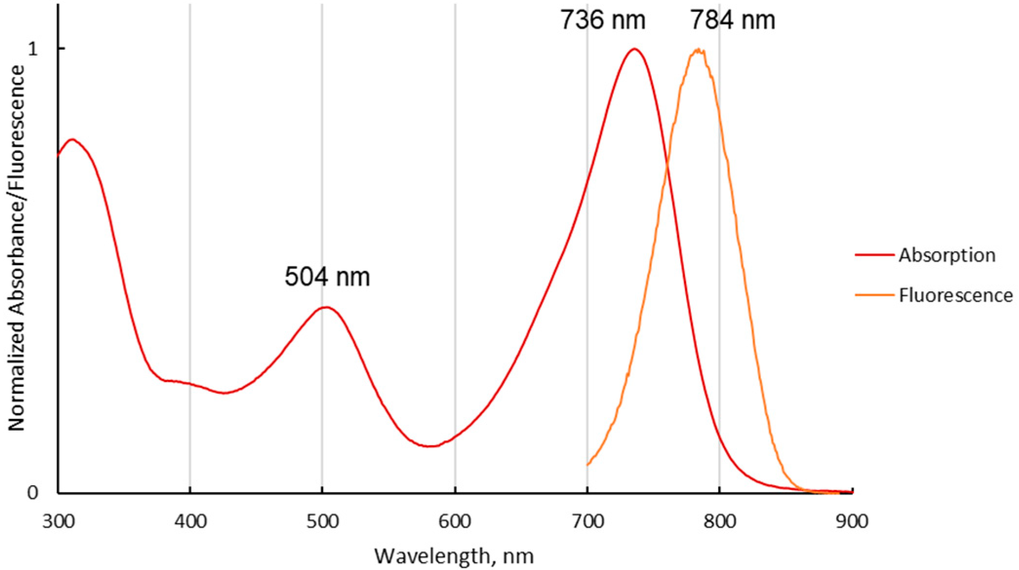
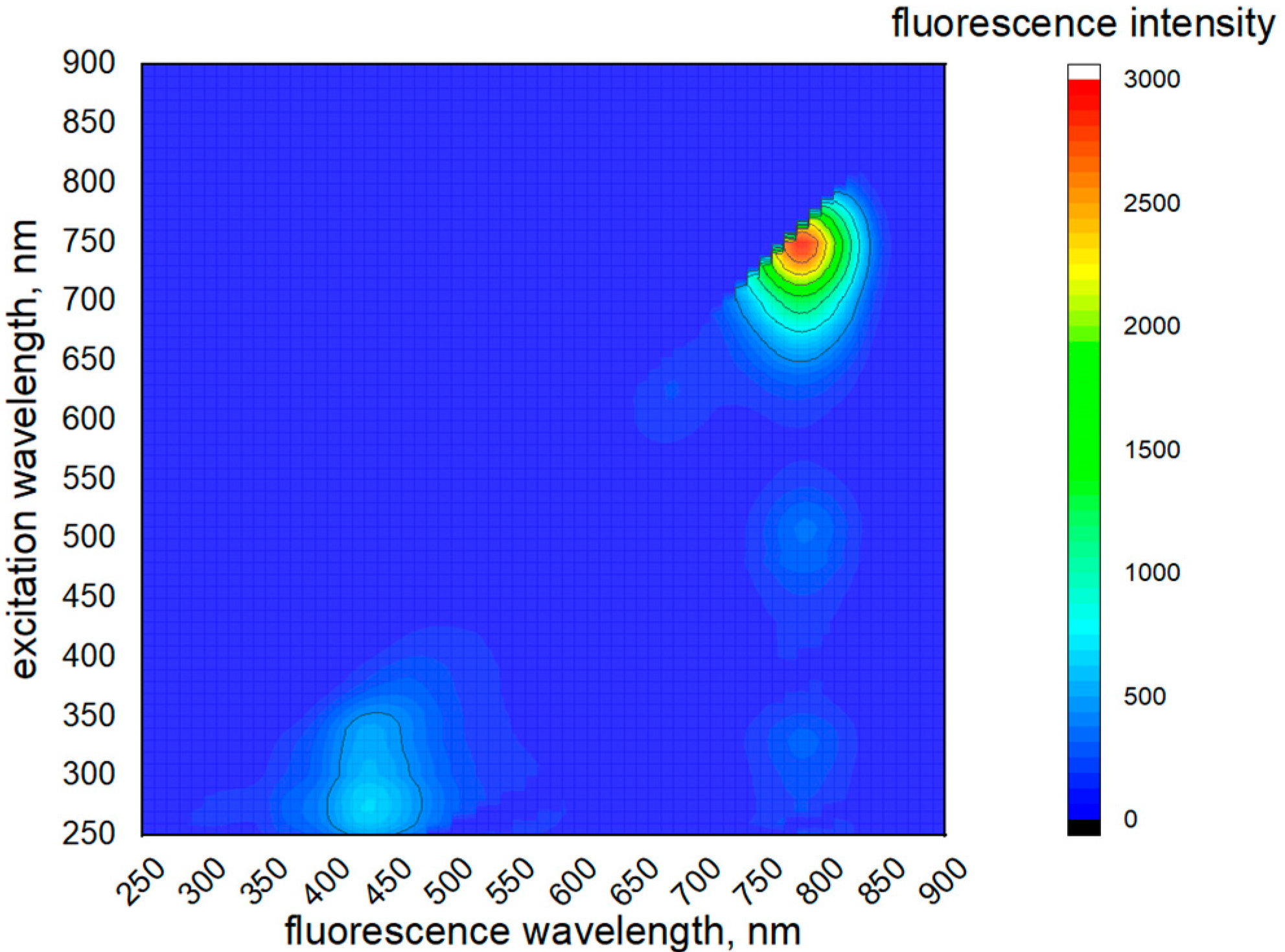
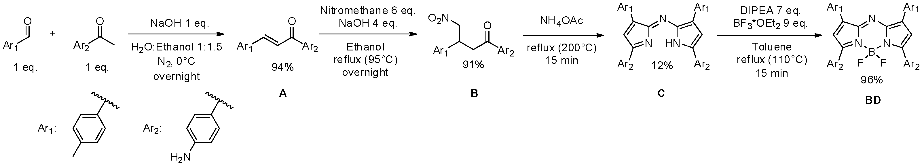
2. Results and Discussion

The common synthesis of aza-BODIPY includes four stages (Scheme 1) [12]. However, the conditions of every step dramatically impact the target substituents in the aza-BODIPY core. In this work, it was found that an inert atmosphere and a low temperature are essential for obtaining pure chalcone A in high yields. The use of NaOH as a base allowed us to simplify the separation of target Michael adduct B as a residue from a water solution. The obtention of aza-DPM C was carried out without solvent, causing the process to last 15 min instead of several hours. The final complexation in toluene allowed us to heat the reaction mixture up to 110 °C and reduce reaction time to 15 min.
The investigation of the spectral properties (Figure 1) showed that the aminophenyl-substituted aza-BODIPY exhibits absorption and fluorescence in the near-IR region of the spectrum, the so-called “first biological window”, making it suitable for application in biological liquids and tissues.
The investigation of the 3D excitation/fluorescence spectrum showed the complex nature of electronic transitions in the molecule BD (Figure 2).
It can be seen that BD exhibits fluorescence in the range of 400 to 460 nm. This region is attributed to the luminescence of individual substituents of the dye. This may mean that there is a low degree of conjugation of the aryl rings with the aza-BODIPY π system. However, this conjugation is still present, which can be seen from the increase in the S1–S0 fluorescence of the complex upon excitation at absorption wavelengths of individual substituents (300–350 nm). This phenomenon can be used for multi-wavelength excitation of the investigated compound to reduce sample damage caused by intense light or by heating.

3. Materials and Methods

All reagents were purchased from Sigma-Aldrich (St. Louis, MO, USA) and were used without further purification. 1H and 13C NMR spectra were acquired using a Bruker Avance 500 spectrometer (Bruker, Ettlingen, Germany) in CDCl3 and DMSO-d6. Mass-spectra were recorded on a Shimadzu MS (AXIMA; Shimadzu Corporation, Kyoto, Japan). Absorption spectra were recorded on Unico SpectroQuest 2802S UV/Vis spectrophotometer (Unico Co., Dayton, NJ, USA). Fluorescence and excitation spectra were recorded using a spectrofluorometer Cary Eclipse (Varian Inc., Palo Alto, CA, USA).
Synthesis of chalcone (A).
An amount of 4-aminoacetophenone (675 mg, 5 mmol, 1 eq.) was dissolved in 7.5 mL ethanol under sonication. The solution was poured into a test tube with a hose connection and mixed in an ice bath. NaOH (200 mg, 5 mmol, 1 eq.) was dissolved in 5 mL of water and poured into the reaction mixture. P-tolualdehyde (589 µL, 5 mmol, 1 eq.) was added to the mixture dropwise to avoid the rapid formation of oily side products. The test tube was immediately closed with a silicone stopper, evacuated and then refilled with nitrogen five times using a Schlenk line. After 3 h of stirring in an ice bath, a white residue of chalcone was fully formed. The mixture was sonicated for 5 min, filtered through a glass Buchner funnel, washed 10 times with water and dried. The product was a pale-yellow powder. The product may be purified by recrystallization from methanol (due to low melting point of the product).
Synthesis of Michael adduct (B).
Chalcone A (474 mg, 2 mmol, 1 eq.) was dissolved in 20 mL of ethanol in a 100 mL round bottom flask upon mixing. NaOH (290 mg, 4 mmol, 2 eq.) was added followed by nitromethane (718 µL, 12 mmol, 6 eq.). The mixture was left stirring under reflux for 12 h. The reaction was quenched by addition of 80 mL of water. The residue was separated by centrifugation and washed with a small amount of water 3 times. The product was purified by column chromatography (SiO2, ethyl acetate, first fraction), and the solvent was removed under vacuum. The product was a brown oil.
Synthesis of aza-DPM (C).
Michael adduct B (298 mg, 1 mmol, 1 eq.) and NH4OAc (4620 mg, 60 mmol, 60 eq.) were mixed in long narrow test tube and heated until boiling (≈200 °C). The mixture was left to boil and was stirred for 15 min. A change of color to dark bluegreen was observed.
The mixture was cooled, dissolved in 50 mL of water, filtered and washed with water 10 times. The product was purified by chromatography (SiO2, ethyl acetate, first fraction). The resulting product was a black-blue coppery powder.
MALDI-TOF, m/z: calc. 507.24, found, 507.6317 (Figure S6).
Synthesis of aza-BODIPY (BD).
Aza-DPM C (507 mg, 1 mmol, 1 eq.) was dissolved in 5 mL of toluene in a long narrow test tube whilst stirring. DIPEA (1217 µL, 7 mmol, 7 eq.) was added. Then, BF3(OEt)2 (1111 µL, 9 mmol, 9 eq.) was added and the mixture was kept under reflux for 30 min. A change in color was not observed.
The reaction mixture was cooled and 50 mL of water was added. The product was extracted with 5 mL of DCM and washed with water 5 times. The compound changed in color to pale-pink. The organic layer was dried over Na2SO4 and purified by chromatography (SiO2, ethyl acetate, first fraction). The solvent was removed under vacuum. The resulting product was a black-pink coppery powder.
1H NMR (500 MHz, chloroform-d) δ 7.99 (dd, J = 10.5, 8.2 Hz, 8H), 7.00 (s, 2H), 6.74 (d, J = 8.4 Hz, 4H), 4.27–3.97 (m, 4H), 3.78–3.47 (m, 4H), 2.43 (s, 6H). 1H NMR (500 MHz, DMSO-d6) δ 8.08 (d, J = 7.8 Hz, 4H), 8.02 (d, J = 8.4 Hz, 4H), 7.49 (s, 2H), 7.33 (d, J = 7.8 Hz, 4H), 6.69 (d, J = 8.5 Hz, 4H), 6.32 (s, 4H), 2.40 (s, 6H). MALDI-TOF, m/z: calc. 555.24, found, 555.0887 (Figures S1–S6).

Supplementary Materials

Figure S1: 1H NMR spectrum for aza-BODIPY BD in CDCl3 (baseline-corrected, solvent peaks removed); Figure S2: 1H NMR spectrum for aza-BODIPY BD in CDCl3 (RAW data); Figure S3: 1H NMR spectrum for aza-BODIPY BD in DMSO-d6 (baseline-corrected, solvent peaks removed); Figure S4: 1H NMR spectrum for aza-BODIPY BD in DMSO-d6 (RAW data); Figure S5: 11B NMR spectrum for aza-BODIPY BD in CDCl3; Figure S6: MALDI-TOF mass spectra for aza-DPM 3 (top) and aza-BODIPY BD (bottom); Figure S7: TLC plates with spots of all investigated compounds. Dashed lines show the initial spots position and the distance moved by solvent.

Author Contributions

Conceptualization, D.M.; methodology, Y.M. and D.M.; investigation, D.M. and T.K.; writing—original draft preparation, D.M.; writing—review and editing, Y.M. funding acquisition, Y.M. All authors have read and agreed to the published version of the manuscript.

Funding

This research was partially funded with the support of the scholarship of the President of the Russian Federation for young scientists and graduate students carrying out promising research and development in priority areas of modernization of the Russian economy for 2021–2023 (SP-5338.2021.4). This work was supported by the Russian Science Foundation (22-73-10167) in part of aza-BODIPY synthesis.

Institutional Review Board Statement

Not applicable.

Informed Consent Statement

Not applicable.

Data Availability Statement

Not applicable.

Acknowledgments

The study was carried out using the resources of the Center for Shared Use of Scientific Equipment of the ISUCT (with the support of the Ministry of Science and Higher Edu-cation of Russia, grant No. 075-15-2021-671).

Conflicts of Interest

The authors declare no conflict of interest.

References

  1. Shi, Z.; Han, X.; Hu, W.; Bai, H.; Peng, B.; Ji, L.; Fan, Q.; Li, L.; Huang, W. Bioapplications of Small Molecule Aza-BODIPY: From Rational Structural Design to In Vivo Investigations. Chem. Soc. Rev. 2020, 49, 7533–7567. [Google Scholar] [CrossRef] [PubMed]
  2. Avellanal-Zaballa, E.; Gartzia-Rivero, L.; Arbeloa, T.; Bañuelos, J. Fundamental Photophysical Concepts and Key Structural Factors for the Design of BODIPY-Based Tunable Lasers. Int. Rev. Phys. Chem. 2022, 41, 177–203. [Google Scholar] [CrossRef]
  3. Wang, L.; Xiong, Z.; Ran, X.; Tang, H.; Cao, D. Recent Advances of NIR Dyes of Pyrrolopyrrole Cyanine and Pyrrolopyrrole Aza-BODIPY: Synthesis and Application. Dye. Pigment. 2022, 198, 110040. [Google Scholar] [CrossRef]
  4. Ivaniuk, K.; Pidluzhna, A.; Stakhira, P.; Baryshnikov, G.V.; Kovtun, Y.P.; Hotra, Z.; Minaev, B.F.; Ågren, H. BODIPY-Core 1,7-Diphenyl-Substituted Derivatives for Photovoltaics and OLED Applications. Dye. Pigment. 2020, 175, 108123. [Google Scholar] [CrossRef]
  5. Jiang, X.D.; Guan, J.; Li, Q.; Sun, C. New Near-Infrared-Fluorescent Aza-BODIPY Dyes with 1-Methyl-1 H-Pyrrolyl Substituents at the 3,5-Positions. Asian J. Org. Chem. 2016, 5, 1063–1067. [Google Scholar] [CrossRef]
  6. Bai, L.; Sun, P.; Liu, Y.; Zhang, H.; Hu, W.; Zhang, W.; Liu, Z.; Fan, Q.; Li, L.; Huang, W. Novel Aza-BODIPY Based Small Molecular NIR-II Fluorophores for: In Vivo Imaging. Chem. Commun. 2019, 55, 10920–10923. [Google Scholar] [CrossRef] [PubMed]
  7. Chen, D.; Zhong, Z.; Ma, Q.; Shao, J.; Huang, W.; Dong, X. Aza-BODIPY-Based Nanomedicines in Cancer Phototheranostics. ACS Appl. Mater. Interfaces 2020, 12, 26914–26925. [Google Scholar] [CrossRef] [PubMed]
  8. Li, C.; Wang, Q. Advanced NIR-II Fluorescence Imaging Technology for In Vivo Precision Tumor Theranostics. Adv. Ther. 2019, 2, 1900053. [Google Scholar] [CrossRef]
  9. Ullah, Z.; Kraimi, A.; Kim, H.J.; Jang, S.; Mary, Y.S.; Kwon, H.W. Selective Detection of F− Ion and SO2 Molecule: An Experimental and DFT Study. J. Mol. Liq. 2022, 359, 119329. [Google Scholar] [CrossRef]
  10. Ullah, Z.; Sonawane, P.M.; Nguyen, T.S.; Garai, M.; Churchill, D.G.; Yavuz, C.T. Bisphenol—Based Cyanide Sensing: Selectivity, Reversibility, Facile Synthesis, Bilateral “OFF-ON” Fluorescence, C2 Structural and Conformational Analysis. Spectrochim. Acta Part A Mol. Biomol. Spectrosc. 2021, 259, 119881. [Google Scholar] [CrossRef] [PubMed]
  11. Bodio, E.; Denat, F.; Goze, C. BODIPYS and Aza-BODIPY Derivatives as Promising Fluorophores for in Vivo Molecular Imaging and Theranostic Applications. In Porphyrin Science by Women; World Scientific: Singapore, 2021; pp. 116–140. [Google Scholar]
  12. Gresser, R.; Hartmann, H.; Wrackmeyer, M.; Leo, K.; Riede, M. Synthesis of Thiophene-Substituted Aza-BODIPYs and Their Optical and Electrochemical Properties. Tetrahedron 2011, 67, 7148–7155. [Google Scholar] [CrossRef]
Scheme 1. Synthetic route to aminophenyl-substituted aza-BODIPY (BD).
Molbank 2022 m1530 sch001
Figure 1. Normalized absorption (red) and emission (orange) spectra of BD in DCM. The excitation wavelength is 680 nm.
Molbank 2022 m1530 g001
Figure 2. 3D excitation/fluorescence spectrum of BD in DCM.
Molbank 2022 m1530 g002
Publisher’s Note: MDPI stays neutral with regard to jurisdictional claims in published maps and institutional affiliations.

