Article

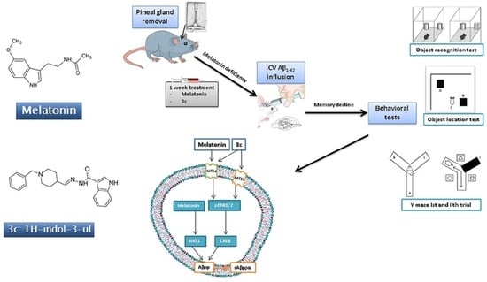
Protective Effect of the Novel Melatonin Analogue Containing Donepezil Fragment on Memory Impairment via MT/ERK/CREB Signaling in the Hippocampus in a Rat Model of Pinealectomy and Subsequent Aβ1-42 Infusion

by
Jana Tchekalarova
1,*,
Petya Ivanova
1,
Desislava Krushovlieva
1,
Lidia Kortenska
1 and
Violina T. Angelova
2
1
Institute of Neurobiology, Bulgarian Academy of Sciences, 1113 Sofia, Bulgaria
2
Department of Chemistry, Faculty of Pharmacy, Medical University of Sofia, 1000 Sofia, Bulgaria
*
Author to whom correspondence should be addressed.
Int. J. Mol. Sci. 2024, 25(3), 1867; https://doi.org/10.3390/ijms25031867
Submission received: 18 January 2024 / Revised: 31 January 2024 / Accepted: 1 February 2024 / Published: 3 February 2024
(This article belongs to the Special Issue Molecular Mechanism in Neurodegeneration)

Abstract

:
A reduction in melatonin function contributes to the acceleration of Alzheimer’s disease (AD), and understanding the molecular processes of melatonin-related signaling is critical for intervention in AD progression. Recently, we synthesized a series of melatonin analogues with donepezil fragments and tested them in silico and in vitro. In this study, one of the most potent compounds, 3c, was evaluated in a rat model of pinealectomy (pin) followed by icvAβ1-42 infusion. Melatonin was used as the reference drug. Treatment with melatonin and 3c (10 mg/kg, i.p. for 14 days) had a beneficial effect on memory decline and the concomitant increase in hippocampal Aβ1-42 and pTAU in the pin+icvAβ1-42 rats. Melatonin supplementation facilitated non-amyloidogenic signaling via non-receptor (histone deacetylase sirtuin 1, SIRT1) and receptor-related signaling (MT/ERK/CREB). The hybrid 3c analogue up-regulated the MT1A and MT2B receptors, pERK and pCREB. Our results strongly support the hypothesis that melatonin-related analogues may become a promising drug candidate for Alzheimer’s disease therapy.

Graphical Abstract

1. Introduction

Alzheimer’s disease (AD) is a progressive brain disorder characterized by severe dementia that affects approximately 10% of the world’s elderly population [1]. The accumulation of extracellular amyloid plaques, composed mainly of amyloid (A)β, and the intracellular formation of hyperphosphorylated TAU protein (pTAU) are considered the unique hallmarks of AD pathogenesis. The imbalance between Aβ production and Aβ clearance with the formation of neurotoxic Aβ1-40 and Aβ1-42 fragments, leading to an increase in oxidative stress in the brain, is one of the accepted hypotheses for the etiology of AD. The accompanying behavioral disturbances include circadian rhythm disruption, sleep disturbance, impaired cognitive function and memory loss. Today, the priority in AD research is to discover the signaling pathways that are closely associated with predisposition to the development of AD pathogenesis. This issue is a crucial step in finding new targets to manipulate and thus control the progression of this neurological disease. Although there is considerable funding for research into practical therapeutic approaches, there is still no approved drug with the three “golden” characteristics needed to reach the market, i.e., (i) to be able to suppress neurodegeneration and disease progression; (ii) to have no side effects in chronic treatment and (iii) to be inexpensive.
Experimental and clinical evidence suggests that the circadian dysregulation of the sleep–wake cycle is part of the early symptoms of the disease predisposing to memory deficits and AD progression [2]. The treatment of sleep–wake cycle disruption at the onset or before the onset of AD symptoms has been suggested to reduce or even prevent pathogenesis development [3]. For example, suvorexant, an orexin receptor antagonist, was recently approved by the Food and Drug Administration in the US for the treatment of sleep–wake circadian disruption in the early stages of AD [4]. The beneficial role of melatonin in AD pathogenesis has also been extensively studied [5] due to its primary function in regulating the circadian rhythms in the body, including synchronizing the sleep–wake cycle and promoting sleep. Melatonin is released from the pineal gland during the dark phase when the inhibitory control from the suprachiasmatic nucleus is weakened by exposure to light.
The main points that point to a close link between the melatoninergic system and AD pathogenesis are as follows: (1) disturbed circadian rhythms (melatonin plays a key role in the resynchronization of circadian rhythms); (2) disturbed pro-oxidant/antioxidant balance (melatonin is a potent antioxidant and free radical scavenger); and (3) neuroinflammation (the hormone has anti-inflammatory activity). Regarding the role of the melatonin system in the three main hallmarks of AD, i.e., Aβ plaques formation, p-TAU, and cholinergic system dysfunction, a growing body of evidence points to a putative neuroprotective effect of the hormone [6,7]. There is ongoing research to explore its therapeutic potential and the development of melatonin-related compounds for treating AD pathogenesis. However, the underlying mechanism associated with the beneficial effect of the melatonin system is still an area of ongoing research.
It is hypothesized that melatonin deficiency and pineal gland dysfunction, in particular, may be critical triggers that predispose to AD pathogenesis [8]. Specifically, patients with AD had diminished function of this gland due to a reduction in the size and calcification [9,10], which decreased melatonin levels in the cerebrospinal fluid (CSF), serum, and urine [8,11,12,13].
The pineal ablation gland in rodents has been accepted as a relevant model to study the detrimental effects of melatonin deficiency, which resemble the signaling dysfunction associated with neurodegenerative diseases, including AD [14]. Recently, we reported that simultaneous removal of the pineal gland and intracerebroventricular (icv) infusion of Aβ1-42 exacerbated behavioral responses and concomitant oxidative stress in the hippocampus and the frontal cortex [15]. Our research team synthesized and tested a series of new melatonin analogues and sulfonyl hydrazone compounds with a melatonin scaffold [16]. Docking analysis revealed a plausible mechanism of action for one of the most potent compounds, the 3c, which is associated with melatonin/MT/receptors and the inhibition of acetylcholinesterase (AchE) and butyrylcholinesterase (BchE). In the present study, the most effective and potent compound, 3c, with low in vitro neurotoxicity, was selected and tested in a rat model of melatonin deficiency induced by pinealectomy (pin) followed by icv1-42 infusion. Melatonin was used as the referent drug. The underlying protective mechanism of the new compound was determined, including the possible signaling pathway closely related to the melatonin system and involved in the suppression of Aβ1-42 accumulation in the hippocampus. We hypothesized that this novel melatonin analogue, 3c, behaves as a potent MT receptor agonist promoting the non-amyloidogenic pathway by facilitating the MT receptor-related ERK/CREB signaling process. The timeline of the experimental steps is shown in Figure 1.

2. Results

2.1. The Circadian Pattern of Motor Activity and Effects of Melatonin and 3c Compound in a Rat Model of Pinealectomy and icvAβ1-42 Infusion

The novel melatonin analogue, 3c, containing a donepezil fragment, was synthesized by a condensation reaction of a 1-benzylpiperidine-4-carbaldehyde and 1H-Indole-5-carbohydrazide, at a molar ratio of 1:1, in abs. ethanol for 1–2 h [16] (Figure 2).
The 24 h registration of the horizontal and vertical activity of the control group (sham-veh-veh group), model group (pin+Aβ1-42-veh), positive control (pin+Aβ1-42-mel) and treatment group (pin+Aβ1-42-3c) was performed in the actimeter. All groups showed a circadian pattern of motor activity, which was also confirmed by the cosinor analysis (Figure 3A,B; Table 1).
The horizontal and vertical activity, total distance and mean velocity, measured in the light and the dark phases, respectively, were also evaluated (Figure 4A–D). The daily pattern of activity was verified for all groups (p < 0.05, p < 0.001, dark vs. light phase). The model group was characterized by an increased horizontal activity compared to the control group (p < 0.05), as well as increased vertical activity compared to the control group (p < 0.001) (Figure 4A,B). Both drugs tended to compensate for the pin+Aβ1-42-induced changes in horizontal activity (Figure 4A). At the same time, only the melatonin treatment attenuated the enhanced effect of pin+Aβ1-42 on vertical activity (p < 0.001 vs. pin+Aβ1-42-veh group) (Figure 4B). There was no difference in the total distance and velocity among groups (p > 0.05) (Figure 4C,D).

2.2. Effects of Melatonin and 3c Compound on Memory Impairment Induced by Pinealectomy and icvAβ1-42 Infusion

To evaluate the efficacy of the new hybrid compound, 3c, and melatonin, used as a reference to compensate for the pinealectomy-induced melatonin deficiency and the Aβ1-42-related memory impairment, we performed a battery of cognitive tests, including the Object recognition test (ORT), the Object location test (OLT) and the Y-maze test.

2.2.1. Object Recognition Test

The ORT was used to assess the cognitive abilities related to the natural curiosity of rodents to explore novel objects. The effects of melatonin and 3c in rats with pinealectomy and icvAβ1-42 infusion on short-term novelty recognition memory were evaluated. The vehicle-treated pin+Aβ1-42 group significantly reduced the Discrimination index (DI) for both time (p < 0.05 vs. sham-veh-veh group) and counts (p < 0.05 vs. sham-veh-veh group) (Figure 5A,B). While the reference drug significantly alleviated DI (count) vs. pin+Aβ1-42-veh group (p < 0.05), the sub-chronic treatment with melatonin or 3c showed a tendency to reverse the impaired memory recognition ((p > 0.05 vs. sham-veh-veh group) and (Figure 5B)).

2.2.2. Object Location Test

The OLT was used to assess the effect of melatonin and 3c on pinealectomy-induced and icvAβ1-42 infusion-induced deficits in short-term spatial memory. For this purpose, the spontaneous response of rats to preferentially explore the novel object placed in an open field, instead of a familiar object placed in the same position, as in the sample phase, (see Section 4) was measured. As in the ORT, the pin+Aβ1-42-veh group had a lower DI than the control group (p < 0.001), indicating impaired short-term spatial memory (Figure 6). Although treatment with the hybrid compound 3c showed a significantly lower DI than the sham-veh-veh group (p < 0.05), both melatonin and 3c reversed the pin+Aβ1-42-induced memory impairment (p < 0.001 and p < 0.05 vs. pin-Aβ1-42-veh group, respectively).

2.2.3. Y-Maze Test

Spontaneous alternation behavior (SAB) was assessed in the first trial of the Y-maze test used to evaluate spatial memory. Lower SAB was found in the pin-Aβ1-42-veh group compared to the sham-veh-veh group (p < 0.001) (Figure 7A). Both the reference group melatonin and the hybrid compound 3c alleviated the impaired SAB (p < 0.05 and p < 0.001 vs. pin-Aβ1-42-veh group, respectively).
Short-term spatial memory was also assessed in the Y maze second trial. As in the OLT, the melatonin deficit model and subsequent icv1-42 infusion reduced DI (time) and DI (count) (p < 0.001 vs. control) (Figure 7B,C), respectively. Treatment with both the positive control melatonin and compound 3c reduced the pin+icv1-42-induced impairment of spatial memory (DI (time): p < 0.001, pin-Aβ1-42-mel group vs. pin-Aβ1-42-veh group; p < 0.05, pin-Aβ1-42-3c group vs. pin-Aβ1-42-veh group (Figure 7B); and DI (count): p < 0.01, pin-Aβ1-42-veh group vs. pin-Aβ1-42-mel and pin-Aβ1-42-3c group, respectively, (Figure 7C). However, the treatment with compound 3c was unable to correct the pin-Aβ1-42-induced reduction in DI (time) (p < 0.001 vs. sham-veh-veh group) (Figure 7B).

2.3. The Expression of Markers of ADs and Effects of Melatonin and Compound 3c in a Rat Model of Pinealectomy and icvAβ1-42 Infusion

The effects of sub-chronic treatment with the positive control melatonin and the hybrid compound 3c on pathological markers closely associated with ADs, including the expression of Aβ1-42, pTAU protein and the hippocampal AchE level, were evaluated following the induction of melatonin deficiency and concomitant icvAβ1-42 infusion one week after pinealectomy. The hippocampus procedure isolation was performed 24 h after the last memory test (see Figure 1).
The expression of Aβ1-42 and pTAU in the hippocampus was significantly increased in the model group treated with a vehicle compared to the control group (p < 0.05 and p < 0.05, respectively) (Figure 8A,B). At the same time, there was no change in the level of AchE among the four groups (Figure 8C). Treatment with melatonin and compound 3c failed to reverse the model-induced elevation of AD markers (p > 0.05 vs. control group).

2.4. Neuroprotective Effect of Melatonin and the Hybrid Compound 3c on Aβ1-42 Neurotoxicity via MT Receptors/ERK/CREB Signaling

Previous studies have shown that melatonin exerts neuroprotection against Aβ neurotoxicity by redirecting the APP metabolism to the non-amyloidogenic pathways. In the present study, we investigated the possible involvement of this molecular mechanism in the beneficial effects of melatonin and melatonin-related compound 3c on memory decline in the pin+Aβ1-42 rat model. We measured the levels of several signaling molecules associated with the non-amyloidogenic pathway, including the expression of the two receptors MT1A and MT2B, respectively, sirtuin 1 (SIRT1), pERK1/2 and pCREB.
The effects of sub-chronic treatment with the positive control melatonin and the hybrid compound 3c on the signaling molecules associated with the non-amyloidogenic pathway activated via MT1A/MT2B receptor subtypes in the hippocampus were assessed. The pinealectomy model, followed by icvAβ1-42 infusion, down-regulated the two MT receptor subtypes in the hippocampus compared to the sham+veh-veh group (p < 0.05 and p < 0.01, respectively) (Figure 9A,B). Treatment with the positive control melatonin and compound 3c reversed the pin+icvAβ1-42-related decreased expression in the MT1A receptor subtype (p < 0.05 vs. control group), whereas only the novel melatonin-related hybrid up-regulated the pin-Aβ decrease in the MT2B receptor subtype (p < 0.01 vs. pin-Aβ1-42-veh group) in the hippocampus. Interestingly, melatonin failed to correct the pin-Aβ1-42-related down-regulation of the MT2B receptor subtype in the hippocampus (p < 0.05, vs. pin-Aβ1-42-veh group) (Figure 9B).
Furthermore, the compound 3c reversed the pin-Aβ1-42-induced decreased expression of both the pERK1/2 (p = 0.01 vs. pin-Aβ1-42-veh group) and pCREB in the hippocampus (p < 0.05 vs. pin-Aβ1-42-veh group) (Figure 10A,B). At the same time, the positive effect of melatonin on these signaling molecules was even higher (p < 0.001 vs. pin-Aβ1-42-veh group, Figure 10A and p < 0.05 vs. sham-veh-veh and pin-Aβ1-42-veh group, Figure 10B, respectively). Similarly, melatonin treatment significantly increased the SIRT1 in the hippocampus (p < 0.01 vs. sham-veh-veh group, p < 0.001 vs. pin-Aβ1-42-veh group), while the hybrid compound 3c was ineffective (p > 0.05 vs. pin-Aβ1-42-veh group; p = 0.01 vs. pin-Aβ1-42-mel group) (Figure 10C).

3. Discussion

Recently, our team designed, synthesized and characterized a series of melatonin-based hybrid compounds with hydrazine scaffolds to determine whether they have the potential for further evaluation in AD therapy [16]. Based on in silico and in vitro data, one of the two lead compounds, 3c, was selected for further in vivo study in a rat model of pinealectomy followed by icv1-42 infusion. Melatonin was used as a positive control. The potential protective effect of the hybrid compound 3c against the Aβ protein-related neurotoxicity and a plausible underlying mechanism were explored. The hypothesis that melatonin and the compound 3c could stimulate non-amyloidogenic signaling in the hippocampus via activity on MT1A/MT2B receptors in the pin+icv1-42 model was based on the recently reported potency of the novel hybrid compound to bind to the two MT receptor subtypes by molecular docking analysis [16]. Our results showed that, while the beneficial effect of the positive control melatonin on AD-related pathogenesis involves the stimulation of the non-amyloidogenic pathway via both non-receptor (SIRT1) and MT1A-receptor-related ERK1/2/CREB signaling, the novel analogue 3c only attenuated memory decline via the MT1A/MT2B receptor-related ERK1/2/CREB signaling in a rat model of pin+icvAβ1-42. The schematic presentation of the proposed mechanism underlying the effects of melatonin and the compound 3c is shown in Scheme 1.
We previously reported that the simultaneous induction of melatonin deficiency by pinealectomy and AD-related pathogenesis by icv infusion of Aβ1-42 in rats induces typical AD behavioral symptom changes such as increased anxiety and cognitive impairment [15]. Demir et al. (2017) and Zhu et al. (2004) reported that melatonin deficiency exacerbated AD-like changes and memory impairment [17,18].
The idea that CSF melatonin levels decrease in the preclinical stages of AD when patients do not show cognitive impairment [8] suggests that melatonin deficiency may be a critical factor predisposing to the development AD. In the present study, we modified the model previously reported by our team [15] in order to simulate the preclinical stage of AD with a drop in melatonin blood levels induced by pinealectomy to assess the efficacy of advanced prophylactic treatment with the hormone and the novel melatonin hybrid compound 3c on AD pathogenesis induced by a subsequent icv1-42-infusion. Long-term supplementation with a high dose of melatonin (50 mg/kg for 40 days) reversed behavioral impairments and the associated increase in oxidative stress in the frontal cortex and the hippocampus [15]. Melatonin may play a role in the modulating of spatial learning and memory, and its potential effects on these cognitive functions in AD models have been investigated in several studies [19,20,21,22]. The beneficial effects of the melatonin system on spatial memory may be related to its ability to regulate disrupted circadian rhythms, neuroprotection, antioxidant and anti-inflammatory properties. The hippocampus is known to be a brain region critical for spatial learning and memory. Melatonin receptors are present in the hippocampus [23] and, so, the hormone could affect synaptic plasticity and the processes closely linked to memory formation in this region.
In addition, the modulatory role of melatonin on acetylcholine [7], the neurotransmitter that plays a crucial role in cognition [24], is also an essential factor suggesting the beneficial effects of melatonin on spatial memory. Our results showed that, while the novel hybrid compound 3c is ineffective against impaired non-spatial cognitive performance, such as the ORT in a rat model of pin+icv1-42, this melatonin analogue has a comparable potency to the positive control melatonin in correcting the impaired short-term and spatial hippocampus-dependent memory. The beneficial role of melatonin supplementation on spatial memory decline has been demonstrated in several models of AD, including the hereditary form of AD, such as mouse transgenic and knockout mice [19,25,26], and the sporadic form of AD, such as senescence-accelerated OXYS rats [21], streptozotosin- [27,28] and icv1-42-induced models [29].
There is sufficient evidence in the literature to support the idea that the soluble Aβ oligomers and the total Aβ levels, in particular, are responsible for progressive memory decline in AD [30,31], suggesting that these factors may be more important than the formation of plaques in understanding the pathology of AD. Both experimental reports using transgenic APP mice [32,33] and human brain tissue [30,31] have shown that cognitive changes occur before the formation of plaques, supporting the notion that soluble forms of Aβ may cause cognitive impairment. In addition, soluble Aβ oligomers, containing Aβ1–40 or Aβ1-42, detected in AD patients are thought to contribute more to the neurotoxicity associated with AD than Aβ deposition. Thereby, interventions aimed at reducing or preventing the neurotoxicity associated with the soluble Aβ oligomers may be crucial for the sequential treatment of AD pathogenesis. To verify the model of melatonin deficiency induced by pinealectomy followed by icv1-42 infusion, we first examined the expression of Aβ1-42, pTAU and AchE levels in the hippocampus. The observed increase in Aβ1-42 is consistent with our previous report, in which the pineal gland was removed at the same time as the toxic oligomer infusion [15].
The present study also confirmed that pTAU was significantly elevated in the hippocampus. The lack of difference between the control group and the model group for this standard marker associated with the pathogenesis of AD with regard to AChE may be related to the fact that we measured the levels but not the enzyme activity, which is a limitation of the study and could be taken into account in further studies. We can also speculate that, in our model, the detected enzyme levels in the hippocampus were obtained before the formation of β-amyloid plaques, where the increased activity of AChE has been reported [34]. In addition, higher AChE activity in the vicinity of amyloid plaques has been shown to enhance Aβ aggregation, making it more toxic than Aβ fibrils [35]. Pretreatment with both the positive control melatonin and the compound 3c reduced the model-induced increase in the Aβ and pTAU expression in the hippocampus of pin+Aβ1-42 rats to control levels. Interestingly, both the Aβ1-40 and Aβ1-42 impaired melatonin production and receptor signaling in cultured cells [36]. Therefore, melatonin deficiency in the pin+icv1-42 rat model may be associated not only with pineal ablation but also with decreased hormone production in other tissues.
The underlying molecular mechanism of melatonin and the compound 3c was also reviewed and discussed. The accumulated evidence supports the hypothesis that melatonin may induce the non-amyloidogenic processing of APP while suppressing amyloidogenic processing, thus preventing the formation of neurotoxic Aβ oligomers and the further accumulation of Aβ in plaques [25]. The endogenous hormone stimulates the activity of alpha-secretases (ADAM10 and ADAM17) at the transcriptional level [6,37]. In contrast, melatonin suppresses amyloidogenic processing by downregulating beta-secretase (BACE1) (transcription, translation and enzyme activity). Furthermore, reduced BACE1 activity keeps the cholinergic system intact and suppresses neuronal damage and memory decline in parallel with the suppression of Aβ40/42 production [38,39].
The neuroprotective effects of melatonin may be mediated by both receptor-dependent and receptor-independent mechanisms [25]. The reduction in SIRT1 levels is negatively correlated with the duration of AD symptoms [40,41,42]. Melatonin promotes the expression of SIRT1 in primary neurons shortly after exposure for up to 24 h via a receptor-independent mode [43], suggesting that the upregulation of SIRT1 may promote the expression of ADAM10, which is involved in non-amyloidogenic processing, thereby leading to a reduced production of Aβ.
Melatonin can also exert its effects through MT1 and MT2 receptors, which are expressed jointly and individually in brain structures [23,44]. While the MT1 receptor is widely distributed in various tissues, the MT2 receptor has a more restricted distribution and is mainly found in the brain, including the hippocampus [23]. Melatonin, through its plasma receptors, induces ERK1/2 phosphorylation via various signaling pathways and activates transcription factors such as CREB [7]. Our results showed that, while melatonin up-regulated SIRT1 expression, the melatonin-related compound 3c was ineffective, suggesting that the hybrid compound could mimic the hormone’s effects mainly through MT receptor activation. Furthermore, our results showed that, while melatonin and the compound 3c reversed the model-related down-regulation of MT1A receptors in the hippocampus, only the novel melatonin-like hybrid corrected the Aβ-induced reduced MT1B receptors. The positive effect of the two pretreatments (melatonin and 3c) on the pCREB expression suggests that the ERK1/2/CREB signaling is the underlying mechanism involved in the protective effects of the two drugs against Aβ formation via the activation of MT1A receptors (melatonin and 3c) and MT2B (3c). However, the role of SIRT1 in the effects of melatonin, which may be be realized via a non-receptor mode, cannot be excluded.

4. Materials and Methods

The compound 3c was synthesized according to procedure described previously in the literature [16] and the structure was proved by 1H, 13C NMR spectra and HRMS.

4.1. Animals

Adult male Wistar rats (250–300 g bw), purchased by the vivarium of the Institute of Neurobiology, BAS, were acclimatized in standard conditions: Plexiglas cages in groups of four; 12/12 cycled light/dark regime; average temperature at 22–23 °C; and food and water ad libitum. The experimental design was prepared in full accordance with the European Communities Council Directive 2010/63/E.U. and approved by the Bulgarian Food Safety Agency (research project: #347).

4.2. Experimental Design

Rats were randomly assigned to four groups (n = 7–10 rats/group) as follows: Group 1 (sham-veh-veh; a control sham-operated rats, i.p. injected after surgery with saline once a day for 14 days and icv infused with saline after a week), group 2 (pin-Aβ-veh; a model group with pinealectomy, i.p. injected after surgery with saline as controls and icv infused with Aβ1-42), group 3 (pin-Aβ-mel; a group with pinealectomy, i.p. injected after surgery with melatonin (10 mg/kg, i.p. for 14 days) and icv infused with Aβ1-42) and group 4 (pin-Aβ-3c; a group with pinealectomy, i.p. injected after surgery with 3c compound (10 mg/kg, i.p. for 14 days) and icv infused with Aβ1-42.

4.2.1. Surgery and icv Injection of Aβ1-42

The surgical procedure was performed as described in our previous study [15], except the fact that pineal removal was performed a week prior to icv Ab1-42 infusion. The sham-operated group was treated in the same way but without pinealectomy and with icv infusion of vehicle instead of Aβ1-42. The neurotoxic fibrils were prepared according to the procedure described in a previous study [15]. Briefly, the stock solution (1 µg/1 µL) was incubated for seven days at room temperature prior to infusion.

4.2.2. Behavioral Test

Actimeter

Locomotion and diurnal rhythm changes were monitored for 26 h using an infrared actimeter (Bioseb, Paris, France). The first two hours were considered an adaptation period and were not included in the results. Each rat was tested in a sensor box (20 cm high, 45 cm wide and 45 cm long), measuring horizontal and vertical activity, distance and mean velocity. For the 24 h activity, cosinor analysis was performed to determine the mean amplitude, acrophase and mensor of the groups in relation to their rhythmic variations, together with the zero amplitude test.

Object Recognition Test (ORT)

The ORT was performed according to the procedures described in our previous study [45]. Briefly, after 24 h of habituation to an empty open field apparatus (50 × 50 × 50 cm), the rats were placed in it next to two identical plastic objects (referred to as “F”) for a 5 min (Training phase). After a sixty-minute interval, the rats were returned to the same box for the Testing phase. In this phase, one of the objects was replaced with a novel object (designated “N”) for 5 min. Throughout both the Training and Testing phases, careful observations were made, and the time spent and the number of sniffs of each object were recorded. Exploration time was quantified in seconds, and each object count (number of sniffs) was documented. The DI was then calculated using the formula: (N)/(N + F).

Object Location Test (OLT)

The OLT consists of several steps. During the adaptation phase, each animal was placed in a box (50 × 50 cm). The adaptation phase lasted 10 min, allowing the animals to become accustomed to their environment without being scored. After 24 h, the animals were transferred to a dimly lit test room 30 min before the start of the test. Two identical objects were placed individually in the same box, positioned in opposite corners. The animals were given 5 min to freely explore and interact with the objects. After 60 min, the animals were subjected to the test phase. One of the objects was moved to the opposite corner, creating both a familiar and a novel location. The animals’ behavior during this 5 min period was recorded. The duration (in seconds) of sniffing at the familiar and novel locations was recorded. The discrimination index was calculated using the formula: DI = (NO × 100)/(NO + FO). This index provided insight into the animals’ ability to distinguish between familiar and novel object locations. To maintain consistency and avoid any unwanted odors, the arms of the maze were wiped with vinegar or alcohol between each test.

Y-Maze Test

The Y-maze test consisted of a setup with three steel arms positioned at 120° angles to each other. Working memory was assessed in the first trial. Each rat was placed in the center of the apparatus and given 8 min to freely explore the three arms. Access to these arms was manually recorded by two persons blind to the schedule. SAB was calculated on the basis of visits to triads consisting of three arms. SAB was determined using a formula that took into account the number of arm entries and triads; alternation % = (number of correct entries × 100)/(total entries (N) − 2). In the second trial, conducted at least 5 days later, a pretest was performed by closing off one arm and allowing the rat to explore only two arms for 10 min. After a 30 min interval, the rat was placed in the arm opposite to the two arms initially explored. During this phase, the time spent in the two new arms and the number of entries into each arm were measured.

4.2.3. Detection of Aβ, pTAU, AchE, pERK, pCREB and SIRT1 in the Homogenates from the Hippocampus

After the last memory test, the animals were euthanized by guillotine, and the left and right hippocampi were carefully isolated, snap-frozen in liquid nitrogen and stored in a refrigerator until further analysis (8 samples/group). Hippocampal samples were weighed and homogenized with HEPES buffer (20 mM HEPES; 1 mM EGTA; 210 mM Mannithol; 70 mM sucrose; pH 7.2) and protease inhibitor cocktail (100 mM PMSF, 100 mM NaF, 35 mM EDTA). The homogenates were centrifuged at 10,000× g for 5 min at 4 °C. The supernatants were used for the determination of total protein by the Bradford method followed by ELISA tests to measure Aβ, pTAU and AchE. The ELISA measurements were performed according to the manufacturer’s guidance for the specific kits (Elabscience Rat Aβ1-42, E-EL-R1402; Elabscience Rat pMAPT/pTAU (phosphorylated microtubule-associated protein tau), E-EL-R1090; Elabscience Rat AChE (Acetylcholinesterase), E-EL-R0355).

Statistical Analysis

The 24 circadian rhythms of motor activity in the actimeter were assessed by two-way repeated ANOVA. Two-way ANOVA was applied to determine the diurnal variations in horizontal and vertical activity, distance and mean velocity. Memory and biochemical data were evaluated by one-way ANOVA. The Shapiro–Wilk post hoc test was used for homogenously distributed data, and the Kruskal–Wallis test on ranks followed by the Mann–Whitney U test were applied for non-parametric data. The significant level was set at p < 0.05.

5. Conclusions

Melatonin supplementation facilitated the non-amyloidogenic signaling via non-receptor (histone deacetylase sirtuin 1, SIRT1) and receptor MT1A-related signaling (ERK1/2/CREB) in a rat model of pin+icvAβ1-42. The novel hybrid analogue 3c positively affected the pin+icvAβ1-42-induced hippocampus-dependent spatial memory impairment by stimulating MT1A/MT2B receptors, which trigger ERK1/2/CREB non-amyloidogenic signaling in rats. Melatonin supplementation or other interventions targeting the melatoninergic system may hold promise as potential strategies for treating aspects of AD. Given that pineal gland dysfunction may be only one of many causes of AD pathogenesis, further research is needed to fully understand the underlying mechanisms and develop practical therapeutic approaches.

Author Contributions

Conceptualization, J.T.; methodology, J.T., V.T.A., L.K. P.I. and D.K.; validation, J.T.; formal analysis, D.K. and P.I.; investigation, P.I. and J.T.; resources, J.T.; data curation, P.I. and J.T.; writing—original draft preparation, J.T.; writing—review and editing, J.T.; project administration, J.T. and V.T.A.; funding acquisition, J.T. and V.T.A. All authors have read and agreed to the published version of the manuscript.

Funding

This research was funded by the Bulgarian National Science Fund, grant number KP-06-N63/11; 14.12.2022.

Institutional Review Board Statement

The study was conducted in accordance with the Declaration of Helsinki, and the experimental design was prepared in full accordance with the European Communities Council Directive 2010/63/E.U. and approved by the Bulgarian Food Safety Agency (research project: #347).

Informed Consent Statement

Not applicable.

Data Availability Statement

The data that support the findings of this study are available from the corresponding author upon reasonable request.

Conflicts of Interest

The authors declare no conflicts of interest.

References

  1. Selkoe, D.J.; Hardy, J. The Amyloid Hypothesis of Alzheimer’s Disease at 25 Years. EMBO Mol. Med. 2016, 8, 595. [Google Scholar] [CrossRef] [PubMed]
  2. Benedict, C.; Byberg, L.; Cedernaes, J.; Hogenkamp, P.S.; Giedratis, V.; Kilander, L.; Lind, L.; Lannfelt, L.; Schiöth, H.B. Self-Reported Sleep Disturbance Is Associated with Alzheimer’s Disease Risk in Men. Alzheimers Dement. 2015, 11, 1090–1097. [Google Scholar] [CrossRef] [PubMed]
  3. Bonanni, E.; Maestri, M.; Tognoni, G.; Fabbrini, M.; Nucciarone, B.; Manca, M.L.; Gori, S.; Iudice, A.; Murri, L. Daytime Sleepiness in Mild and Moderate Alzheimer’s Disease and Its Relationship with Cognitive Impairment. J. Sleep Res. 2005, 14, 311–317. [Google Scholar] [CrossRef] [PubMed]
  4. Musiek, E.S.; Xiong, D.D.; Holtzman, D.M. Sleep, Circadian Rhythms, and the Pathogenesis of Alzheimer Disease. Exp. Mol. Med. 2015, 47, e148. [Google Scholar] [CrossRef] [PubMed]
  5. Anderson, G.; Maes, M. Anderson, G.; Maes, M. A Role for the Regulation of the Melatonergic Pathways in Alzheimer’s Disease and Other Neurodegenerative and Psychiatric Conditions. In Serotonin and Melatonin; CRC Press: Boca Raton, FL, USA, 2016; pp. 443–466. [Google Scholar] [CrossRef]
  6. Shukla, M.; Htoo, H.H.; Wintachai, P.; Hernandez, J.F.; Dubois, C.; Postina, R.; Xu, H.; Checler, F.; Smith, D.R.; Govitrapong, P.; et al. Melatonin Stimulates the Nonamyloidogenic Processing of ΒAPP through the Positive Transcriptional Regulation of ADAM10 and ADAM17. J. Pineal Res. 2015, 58, 151–165. [Google Scholar] [CrossRef]
  7. Shukla, M.; Govitrapong, P.; Boontem, P.; Reiter, R.J.; Satayavivad, J. Mechanisms of Melatonin in Alleviating Alzheimer’s Disease. Curr. Neuropharmacol. 2017, 15, 1010–1031. [Google Scholar] [CrossRef]
  8. Claustrat, B.; Brun, J.; Borson-Chazot, F. Melatonin and Circadian Rhythms. Curr. Top. Med. Chem. 2002, 2, 199–209. [Google Scholar] [CrossRef]
  9. Wu, Y.H.; Feenstra, M.G.P.; Zhou, J.N.; Liu, R.Y.; Toranõ, J.S.; Van Kan, H.J.M.; Fischer, D.F.; Ravid, R.; Swaab, D.F. Molecular Changes Underlying Reduced Pineal Melatonin Levels in Alzheimer Disease: Alterations in Preclinical and Clinical Stages. J. Clin. Endocrinol. Metab. 2003, 88, 5898–5906. [Google Scholar] [CrossRef]
  10. Mahlberg, R.; Walther, S.; Kalus, P.; Bohner, G.; Haedel, S.; Reischies, F.M.; Kühl, K.P.; Hellweg, R.; Kunz, D. Pineal Calcification in Alzheimer’s Disease: An in Vivo Study Using Computed Tomography. Neurobiol. Aging 2008, 29, 203–209. [Google Scholar] [CrossRef] [PubMed]
  11. Ohashi, Y.; Okamoto, N.; Uchida, K.; Iyo, M.; Mori, N.; Morita, Y. Daily Rhythm of Serum Melatonin Levels and Effect of Light Exposure in Patients with Dementia of the Alzheimer’s Type. Biol. Psychiatry 1999, 45, 1646–1652. [Google Scholar] [CrossRef] [PubMed]
  12. Zhou, J.N.; Liu, R.Y.; Kamphorst, W.; Hofman, M.A.; Swaab, D.F. Early Neuropathological Alzheimer’s Changes in Aged Individuals Are Accompanied by Decreased Cerebrospinal Fluid Melatonin Levels. J. Pineal Res. 2003, 35, 125–130. [Google Scholar] [CrossRef] [PubMed]
  13. Luboshitzky, R.; Shen-Orr, Z.; Tzischichinsky, O.; Maldonado, M.; Herer, P.; Lavie, P. Actigraphic Sleep-Wake Patterns and Urinary 6-Sulfatoxymelatonin Excretion in Patients with Alzheimer’s Disease. Chronobiol. Int. 2001, 18, 513–524. [Google Scholar] [CrossRef] [PubMed]
  14. Beriwal, N.; Namgyal, T.; Sangay, P.; Al Quraan, A.M. Role of Immune-Pineal Axis in Neurodegenerative Diseases, Unraveling Novel Hybrid Dark Hormone Therapies. Heliyon 2019, 5, E01190. [Google Scholar] [CrossRef] [PubMed]
  15. Tzoneva, R.; Georgieva, I.; Ivanova, N.; Uzunova, V.; Nenchovska, Z.; Apostolova, S.; Stoyanova, T.; Tchekalarova, J. The Role of Melatonin on Behavioral Changes and Concomitant Oxidative Stress in IcvAβ1-42 Rat Model with Pinealectomy. Int. J. Mol. Sci. 2021, 22, 12763. [Google Scholar] [CrossRef] [PubMed]
  16. Angelova, V.T.; Georgiev, B.; Pencheva, T.; Pajeva, I.; Rangelov, M.; Todorova, N.; Zheleva-Dimitrova, D.; Kalcheva-Yovkova, E.; Valkova, I.V.; Vassilev, N.; et al. Design, Synthesis, In Silico Studies and In Vitro Evaluation of New Indole- and/or Donepezil-like Hybrids as Multitarget-Directed Agents for Alzheimer’s Disease. Pharmaceuticals 2023, 16, 1194. [Google Scholar] [CrossRef] [PubMed]
  17. Demir, M.; Yilmaz, U.; Colak, C.; Cigremis, Y.; Ozyalin, F.; Tekedereli, I.; Kilincli, A.; Sandal, S. The Effects of Lack of Melatonin in Experimental Rat Model of Alzheimer’s Disease: Relationship with FEZ1 Gene Expression. Med. Sci. 2016, 6, 217–223. [Google Scholar] [CrossRef]
  18. Ling, Q.Z.; Shao, H.W.; Zhi, Q.L.; Dan, L.W.; Wang, J.Z. Effect of Inhibiting Melatonin Biosynthesis on Spatial Memory Retention and Tau Phosphorylation in Rat. J. Pineal Res. 2004, 37, 71–77. [Google Scholar] [CrossRef]
  19. Liu, Y.C.; Hsu, W.L.; Ma, Y.L.; Lee, E.H.Y. Melatonin Induction of APP Intracellular Domain 50 SUMOylation Alleviates AD through Enhanced Transcriptional Activation and Aβ Degradation. Mol. Ther. 2021, 29, 376–395. [Google Scholar] [CrossRef]
  20. Jürgenson, M.; Zharkovskaja, T.; Noortoots, A.; Morozova, M.; Beniashvili, A.; Zapolski, M.; Zharkovsky, A. Effects of the Drug Combination Memantine and Melatonin on Impaired Memory and Brain Neuronal Deficits in an Amyloid-Predominant Mouse Model of Alzheimer’s Disease. J. Pharm. Pharmacol. 2019, 71, 1695–1705. [Google Scholar] [CrossRef]
  21. Rudnitskaya, E.A.; Muraleva, N.A.; Maksimova, K.Y.; Kiseleva, E.; Kolosova, N.G.; Stefanova, N.A. Melatonin Attenuates Memory Impairment, Amyloid-β Accumulation, and Neurodegeneration in a Rat Model of Sporadic Alzheimer’s Disease. J. Alzheimer’s Dis. 2015, 47, 103–116. [Google Scholar] [CrossRef]
  22. Sun, C.; Qiu, X.; Wang, Y.; Liu, J.; Li, Q.; Jiang, H.; Li, S.; Song, C. Long-Term Oral Melatonin Alleviates Memory Deficits, Reduces Amyloid-β Deposition Associated with Downregulation of BACE1 and Mitophagy in APP/PS1 Transgenic Mice. Neurosci. Lett. 2020, 735, 135192. [Google Scholar] [CrossRef]
  23. Ng, K.Y.; Leong, M.K.; Liang, H.; Paxinos, G. Melatonin Receptors: Distribution in Mammalian Brain and Their Respective Putative Functions. Brain Struct. Funct. 2017, 222, 2921–2939. [Google Scholar] [CrossRef]
  24. Roy, J.; Tsui, K.C.; Ng, J.; Fung, M.L.; Lim, L.W. Regulation of Melatonin and Neurotransmission in Alzheimer’s Disease. Int. J. Mol. Sci. 2021, 22, 6841. [Google Scholar] [CrossRef]
  25. Li, Y.; Zhang, J.; Wan, J.; Liu, A.; Sun, J. Melatonin Regulates Aβ Production/Clearance Balance and Aβ Neurotoxicity: A Potential Therapeutic Molecule for Alzheimer’s Disease. Biomed. Pharmacother. 2020, 132, 110887. [Google Scholar] [CrossRef] [PubMed]
  26. Labban, S.; Alshehri, F.S.; Kurdi, M.; Alatawi, Y.; Alghamdi, B.S. Melatonin Improves Short-Term Spatial Memory in a Mouse Model of Alzheimer’s Disease. Degener. Neurol. Neuromuscul. Dis. 2021, 11, 15. [Google Scholar] [CrossRef] [PubMed]
  27. Kamsrijai, U.; Wongchitrat, P.; Nopparat, C.; Satayavivad, J.; Govitrapong, P. Melatonin Attenuates Streptozotocin-Induced Alzheimer-like Features in Hyperglycemic Rats. Neurochem. Int. 2020, 132, 104601. [Google Scholar] [CrossRef] [PubMed]
  28. Andrade, M.K.; Souza, L.C.; Azevedo, E.M.; Bail, E.L.; Zanata, S.M.; Andreatini, R.; Vital, M.A.B.F. Melatonin Reduces β-Amyloid Accumulation and Improves Short-Term Memory in Streptozotocin-Induced Sporadic Alzheimer’s Disease Model. IBRO Neurosci. Rep. 2023, 14, 264. [Google Scholar] [CrossRef] [PubMed]
  29. Keymoradzadeh, A.; Komaki, A.R.; Bakhshi, A.; Faraji, N.; Golipoor, Z.; Shahshahani, P. The Effect of Different Doses of Melatonin on Learning and Memory Deficit in Alzheimer Model of Rats. Casp. J. Neurol. Sci. 2021, 7, 1–9. [Google Scholar] [CrossRef]
  30. Ha, S.; Redmond, L. ERK Mediates Activity Dependent Neuronal Complexity via Sustained Activity and CREB-Mediated Signaling. Dev. Neurobiol. 2008, 68, 1565–1579. [Google Scholar] [CrossRef] [PubMed]
  31. Watson, D.; Castaño, E.; Kokjohn, T.A.; Kuo, Y.M.; Lyubchenko, Y.; Pinsky, D.; Connolly, E.S.; Esh, C.; Luehrs, D.C.; Blaine Stine, W.; et al. Physicochemical Characteristics of Soluble Oligomeric Abeta and Their Pathologic Role in Alzheimer’s Disease. Neurol. Res. 2005, 27, 869–881. [Google Scholar] [CrossRef] [PubMed]
  32. Holcomb, L.; Gordon, M.N.; Mcgowan, E.; Yu, X.; Benkovic, S.; Jantzen, P.; Wright, K.; Saad, I.; Mueller, R.; Morgan, D.; et al. Accelerated Alzheimer-Type Phenotype in Transgenic Mice Carrying Both Mutant Amyloid Precursor Protein and Presenilin 1 Transgenes. Nat. Med. 1998, 4, 97–100. [Google Scholar] [CrossRef]
  33. Cleary, J.P.; Walsh, D.M.; Hofmeister, J.J.; Shankar, G.M.; Kuskowski, M.A.; Selkoe, D.J.; Ashe, K.H. Natural Oligomers of the Amyloid-Beta Protein Specifically Disrupt Cognitive Function. Nat. Neurosci. 2005, 8, 79–84. [Google Scholar] [CrossRef]
  34. Morán, M.A.; Mufson, E.J.; Gómez-Ramos, P. Colocalization of Cholinesterases with Beta Amyloid Protein in Aged and Alzheimer’s Brains. Acta Neuropathol. 1993, 85, 362–369. [Google Scholar] [CrossRef]
  35. Reyes, A.E.; Chacón, M.A.; Dinamarca, M.C.; Cerpa, W.; Morgan, C.; Inestrosa, N.C. Acetylcholinesterase-Aβ Complexes Are More Toxic than Aβ Fibrils in Rat Hippocampus: Effect on Rat β-Amyloid Aggregation, Laminin Expression, Reactive Astrocytosis, and Neuronal Cell Loss. Am. J. Pathol. 2004, 164, 2163. [Google Scholar] [CrossRef]
  36. Cecon, E.; Chen, M.; Marçola, M.; Fernandes, P.A.C.; Jockers, R.; Markus, R.P. Amyloid β Peptide Directly Impairs Pineal Gland Melatonin Synthesis and Melatonin Receptor Signaling through the ERK Pathway. FASEB J. 2015, 29, 2566–2582. [Google Scholar] [CrossRef]
  37. Panmanee, J.; Nopparat, C.; Chavanich, N.; Shukla, M.; Mukda, S.; Song, W.; Vincent, B.; Govitrapong, P. Melatonin Regulates the Transcription of ΒAPP-Cleaving Secretases Mediated through Melatonin Receptors in Human Neuroblastoma SH-SY5Y Cells. J. Pineal Res. 2015, 59, 308–320. [Google Scholar] [CrossRef] [PubMed]
  38. Ohno, M.; Sametsky, E.A.; Younkin, L.H.; Oakley, H.; Younkin, S.G.; Citron, M.; Vassar, R.; Disterhoft, J.F. BACE1 Deficiency Rescues Memory Deficits and Cholinergic Dysfunction in a Mouse Model of Alzheimer’s Disease. Neuron 2004, 41, 27–33. [Google Scholar] [CrossRef] [PubMed]
  39. Ohno, M.; Cole, S.L.; Yasvoina, M.; Zhao, J.; Citron, M.; Berry, R.; Disterhoft, J.F.; Vassar, R. BACE1 Gene Deletion Prevents Neuron Loss and Memory Deficits in 5XFAD APP/PS1 Transgenic Mice. Neurobiol. Dis. 2007, 26, 134–145. [Google Scholar] [CrossRef] [PubMed]
  40. Wang, J.; Fivecoat, H.; Ho, L.; Pan, Y.; Ling, E.; Pasinetti, G.M. The Role of Sirt1: At the Crossroad between Promotion of Longevity and Protection against Alzheimer’s Disease Neuropathology. Biochim. Biophys. Acta 2010, 1804, 1690–1694. [Google Scholar] [CrossRef] [PubMed]
  41. Albani, D.; Polito, L.; Batelli, S.; De Mauro, S.; Fracasso, C.; Martelli, G.; Colombo, L.; Manzoni, C.; Salmona, M.; Caccia, S.; et al. The SIRT1 Activator Resveratrol Protects SK-N-BE Cells from Oxidative Stress and against Toxicity Caused by Alpha-Synuclein or Amyloid-Beta (1-42) Peptide. J. Neurochem. 2009, 110, 1445–1456. [Google Scholar] [CrossRef] [PubMed]
  42. Qin, W.; Yang, T.; Ho, L.; Zhao, Z.; Wang, J.; Chen, L.; Zhao, W.; Thiyagarajan, M.; MacGrogan, D.; Rodgers, J.T.; et al. Neuronal SIRT1 Activation as a Novel Mechanism Underlying the Prevention of Alzheimer Disease Amyloid Neuropathology by Calorie Restriction. J. Biol. Chem. 2006, 281, 21745–21754. [Google Scholar] [CrossRef] [PubMed]
  43. Cristòfol, R.; Porquet, D.; Corpas, R.; Coto-Montes, A.; Serret, J.; Camins, A.; Pallàs, M.; Sanfeliu, C. Neurons from Senescence-Accelerated SAMP8 Mice Are Protected against Frailty by the Sirtuin 1 Promoting Agents Melatonin and Resveratrol. J. Pineal Res. 2012, 52, 271–281. [Google Scholar] [CrossRef] [PubMed]
  44. Lacoste, B.; Angeloni, D.; Dominguez-Lopez, S.; Calderoni, S.; Mauro, A.; Fraschini, F.; Descarries, L.; Gobbi, G. Anatomical and Cellular Localization of Melatonin MT1 and MT2 Receptors in the Adult Rat Brain. J. Pineal Res. 2015, 58, 397–417. [Google Scholar] [CrossRef] [PubMed]
  45. Shishmanova-Doseva, M.; Peychev, L.; Yoanidu, L.; Uzunova, Y.; Atanasova, M.; Georgieva, K.; Tchekalarova, J.; Shishmanova-Doseva, M.; Peychev, L.; Yoanidu, L.; et al. Anticonvulsant Effects of Topiramate and Lacosamide on Pilocarpine-Induced Status Epilepticus in Rats: A Role of Reactive Oxygen Species and Inflammation. Int. J. Mol. Sci. 2021, 22, 2264. [Google Scholar] [CrossRef]
Figure 1. Timeline of experimental steps. Abbreviations: veh—vehicle; pin—pinealectomy; mel—melatonin; 1—Y maze, 2—ORT, 3—OLT, A—novel object location; AchE—acetylcholinesterase; MT—melatonin receptor; SIRT—sirtuin.
Ijms 25 01867 g001
Figure 2. Chemical structures of melatonin and the compound 3c. In red color—indole ring as fragment of melatonin and in blue color—the donepezil fragment.
Ijms 25 01867 g002
Figure 3. Effect of control (sham-veh-veh) group (n = 8), Aβ1-42-veh group (n = 7), Aβ1-42-mel group (n = 7) and Aβ1-42-3c group (n = 7) on 24 h motor activity measured in the actimeter. Data are presented as mean ± SEM. Repeated two-way ANOVA demonstrated a main Time effect [F23,479 = 34,427, p < 0.001] as well as Group x Time interaction [F69,479 = 1422, p = 0.026] for the horizontal activity. A main Time effect [F23,479 = 15,916, p < 0.001] was detected for the vertical activity. Post hoc test showed in all groups higher horizontal (A) and vertical (B) activity in the zeitgeber (ZT) 12–23 (the dark period) compared to the ZT 0–11 (the light period). Further, a significant difference in activity was detected at ZT8-ZT19 (pin+Aβ1-42-veh vs. sham-veh-veh group (p < 0.05) (B). On the right of the figures are the inserted cosinor data.
Ijms 25 01867 g003
Figure 4. Effect of sham-veh-veh group (n = 8), Aβ1-42-veh group (n = 7), Aβ1-42-mel group (n = 7) and Aβ1-42-3c group (n = 7) on the daily rhythm of 24 h recorded horizontal activity (counts) (A), vertical activity (B), distance (C) and mean velocity (D) measured in the actimeter. Open and black rectangles represent the light and the dark phases above the figures. Data are presented as mean ± SEM, n = 7–8. Two-way ANOVA revealed a main Phase effect for horizontal activity [F3,56 = 133,318, p < 0.001], a main Phase [F1,55 = 64.475, p < 0.001], Treatment [F3,56 = 6.519, p = 0.001] as well as Phase x Treatment interaction [F3,56 = 6.273, p = 0.001] for vertical activity, a main Phase [F1,55 = 21.866, p < 0.001] and Phase x Treatment interaction [F3,56 = 18.262, p < 0.001] for distance and a main Phase effect [F1,56 = 100.719, p < 0.001] for mean velocity. ### p < 0.001, # p < 0.05, dark vs. light phase (AD); * p < 0.05, pin+Aβ1-42-veh group vs. sham-veh-veh (A); *** p < 0.001, pin+Aβ1-42-veh-mel group vs. pin+Aβ1-42-veh group (B).
Ijms 25 01867 g004
Figure 5. Effect of control (sham-veh-veh) group (n = 10), pin-Aβ1-42-veh group (n = 8), pin-Aβ1-42-mel group (n = 8) and pin-Aβ1-42-3c group (n = 8) on short-term recognition memory tested in the object recognition test. Data are presented as the mean ± SEM. The Kruskal–Wallis analysis revealed a main Group effect [H = 9.366, p = 0.025] for DI (sec) and [H = 21.933, p < 0.001] for DI (count); * p < 0.05, pin-Aβ1-42-veh group vs. sham-veh-veh group (A); * p < 0.05, pin-Aβ1-42-veh group vs. sham-veh-veh group; * p < 0.05, pin-Aβ1-42-mel group vs. pin-Aβ1-42-veh group (B).
Ijms 25 01867 g005
Figure 6. Effect of control (sham-veh-veh) group (n = 10), pin-Aβ1-42-veh group (n = 8), pin-Aβ1-42-mel group (n = 8) and pin-Aβ1-42-3c group (n = 8) on short-term spatial memory as assessed in the Object location test (OLT). Data are presented as the mean ± SEM. One-way ANOVA revealed a main Group effect [F3,35 = 11,367, p < 0.001]; *** p < 0.001, pin-Aβ1-42-veh group vs. sham-veh-veh group; *** p < 0.001, pin-Aβ1-42-veh group vs. pin-Aβ1-42-mel; * p < 0.05, pin-Aβ1-42-3c vs. sham-veh-veh group; * p < 0.05, pin-Aβ1-42-3c vs. pin-Aβ1-42-veh group.
Ijms 25 01867 g006
Figure 7. Effect of control (sham-veh-veh) (n = 10), pin-Aβ1-42-veh (n = 8), pin-Aβ1-42-mel (n = 8) and pin-Aβ1-42-3c (n = 8) on Spontaneous alternation behavior (SAB) (in percent) in the first trial (A), DI (sec) (B) and DI (count) (C), respectively, in the second trial in the Y-maze task. Data are presented as the mean ± SEM. One-way ANOVA demonstrated a main Group effect [F3,35 = 10,054, p < 0.001] for SAB; a main Group effect [F3,35 = 25,859, p < 0.001] for DI (time) and [H = 16,521, p < 0.001] for DI (counts) (Kruskal-Wallis test); *** p < 0.001, pin-Aβ1-42-veh group vs. sham-veh-veh group; * p = 0.02, pin-Aβ1-42-veh group vs. pin-Aβ1-42-mel group; *** p < 0.001 pin-Aβ1-42-veh group vs. pin-Aβ1-42-3c group (A); *** p < 0.001, pin-Aβ1-42-veh group vs. sham-veh-veh group; *** p < 0.01, pin-Aβ1-42-veh group vs. pin-Aβ1-42-mel group; *** p < 0.001 pin-Aβ1-42-3c group vs. sham-veh-veh group; * p < 0.05, pin-Aβ1-42-3c group vs. pin-Aβ1-42-veh group (B); *** p < 0.001, pin-Aβ1-42-veh group vs. sham-veh-veh group; ** p < 0.01, pin-Aβ1-42-veh group vs. pin-Aβ1-42-mel and pin-Aβ1-42-3c group, respectively (C).
Ijms 25 01867 g007
Figure 8. Effect of control (sham-veh-veh), pin-Aβ1-42-veh, pin-Aβ1-42-mel, and pin-Aβ1-42-3c on Aβ1-42 protein expression (A), p-TAU protein expression (B) and AchE levels (C) in the hippocampus measured by the ELISA. Data are presented as the mean ± SEM. One-way ANOVA demonstrated a main Group effect [F3,31 = 3,125, p = 0.042] for Aβ1-42 protein, and [H = 8936, p = 0.03] (Kruskal–Wallis test) for pTAU protein; * p < 0.05, pin-Aβ1-42-veh group vs. sham-veh-veh group (A); * p < 0.05, pin-Aβ1-42-veh group vs. sham-veh-veh group (B) (n = 8).
Ijms 25 01867 g008
Figure 9. Effect of control (sham-veh-veh), pin-Aβ1-42-veh, pin-Aβ1-42-mel and pin-Aβ1-42-3c on MT1A (A) and MT2B (B) receptor subtypes in the hippocampus, measured by the ELISA. Data are presented as the mean ± SEM. One-way ANOVA demonstrated a main Group effect [H = 21,361, p < 0.001] (Kruskal–Wallis test) for MT1A and [F3,31 = 7,771, p < 0.001] (Shapiro–Wilk test) for MT2B receptor subtype; * p < 0.05, pin-Aβ1-42-veh group vs. sham-veh-veh group, pin-Aβ1-42-mel and pin-Aβ1-42-3c, respectively (A); ** p < 0.01, pin-Aβ1-42-veh group vs. sham-veh-veh group; * p < 0.05, pin-Aβ1-42-mel group vs. sham-veh-veh group; ** p < 0.01, pin-Aβ1-42-3c group vs. pin-Aβ1-42-veh group (B) (n = 8).
Ijms 25 01867 g009
Figure 10. Effect of control (sham-veh-veh), pin-Aβ1-42-veh, pin-Aβ1-42-mel and pin-Aβ1-42-3c on pERK 1/2 (A), pCREB (B) and SIRT1 (C) expression in the hippocampus, measured by the ELISA. Data are presented as the mean ± SEM. One-way ANOVA demonstrated a main Group effect [F3,31 = 10,069, p < 0.001] (Shapiro–Wilk) for pERK1/2; [H = 17,782, p < 0.001] (Kruskal–Wallis test) for pCREB, [F3,31 = 11,774, p < 0.001] (Shapiro–Wilk) for SIRT1;. *** p < 0.001, pin-Aβ1-42-mel group vs. pin-Aβ1-42-veh group; ** p = 0.01, pin-Aβ1-42-3c group vs. pin-Aβ1-42-veh group (A); * p < 0.05, pin-Aβ1-42-mel group vs. sham-veh-veh and pin-Aβ1-42-veh group; * p < 0.05, pin-Aβ1-42-3c group vs. pin-Aβ1-42-veh group (B); *** p < 0.001, pin-Aβ1-42-mel group vs. pin-Aβ1-42-veh group; ** p < 0.01, pin-Aβ1-42-mel group vs. sham-veh-veh group; ** p < 0.01, pin-Aβ1-42-mel group vs. pin-Aβ1-42-3c group (n = 8).
Ijms 25 01867 g010
Scheme 1. Illustration of the non-receptor and receptor-related signaling molecules involved in the mechanism of action of melatonin and the novel hybrid melatonin analogue 3c in the pin+icvAβ1-42 rat model.
Ijms 25 01867 sch001
Table 1. Cosinor-based rhythm parameters for 24 h monitored motor activity of sham-veh-veh, pin+Aβ1-42-veh, pin+Aβ1-42-veh-mel and pin+Aβ1-42-veh-3c.
MesorAmplitudeAcrophase (Hours, Decimal)F-Valuep-Value
(Zero Amplitude Test)
sham-veh-veh1887.411093.9417.4533.89<0.00001
pin+Aβ1-42-veh2431.161489.9115.6384.68<0.00001
pin+Aβ1-42-veh-mel2309.211320.8917.5734.93<0.00001
pin+Aβ1-42-veh-3c2179.111216.8817.6426.26<0.00001
Disclaimer/Publisher’s Note: The statements, opinions and data contained in all publications are solely those of the individual author(s) and contributor(s) and not of MDPI and/or the editor(s). MDPI and/or the editor(s) disclaim responsibility for any injury to people or property resulting from any ideas, methods, instructions or products referred to in the content.

