Article

The Specificities of Lysophosphatidic Acid Acyltransferase and Fatty Acid Desaturase Determine the High Content of Myristic and Myristoleic Acids in Cyanobacterium sp. IPPAS B-1200

K.A. Timiryazev Institute of Plant Physiology, Russian Academy of Sciences, Botanicheskaya Street 25, 127276 Moscow, Russia
*
Author to whom correspondence should be addressed.
Int. J. Mol. Sci. 2024, 25(2), 774; https://doi.org/10.3390/ijms25020774
Submission received: 6 December 2023 / Revised: 31 December 2023 / Accepted: 5 January 2024 / Published: 7 January 2024

Abstract

:
The cyanobacterial strain Cyanobacterium sp. IPPAS B-1200 isolated from Lake Balkhash is characterized by high relative amounts of myristic (30%) and myristoleic (10%) acids. The remaining fatty acids (FAs) are represented mainly by palmitic (20%) and palmitoleic (40%) acids. We expressed the genes for lysophosphatidic acid acyltransferase (LPAAT; EC 2.3.1.51) and Δ9 fatty acid desaturase (FAD; EC 1.14.19.1) from Cyanobacterium sp. IPPAS B-1200 in Synechococcus elongatus PCC 7942, which synthesizes myristic and myristoleic acids at the level of 0.5–1% and produces mainly palmitic (~60%) and palmitoleic (35%) acids. S. elongatus cells that expressed foreign LPAAT synthesized myristic acid at 26%, but did not produce myristoleic acid, suggesting that Δ9-FAD of S. elongatus cannot desaturate FAs with chain lengths less than C16. Synechococcus cells that co-expressed LPAAT and Δ9-FAD of Cyanobacterium synthesized up to 45% palmitoleic and 9% myristoleic acid, suggesting that Δ9-FAD of Cyanobacterium is capable of desaturating saturated acyl chains of any length.

1. Introduction

The biosynthesis of glycerolipids in cyanobacteria has been well studied [1,2]. The first reaction in it is the acylation of glycerol-3-phosphate at the sn-1 position with the formation of a molecule of lysophosphatidic acid. As a result of the subsequent acylation of the sn-2 position, phosphatidic acid is formed, which is a precursor of diacylglycerides. Thus, the substrate specificity of the acyltransferase may play an important role in the formation of the final FA composition of cyanobacteria.
Analysis of FAs from the cyanobacterium Cyanothece sp. PCC 8801 (more recent species name—Rippkaea orientalis PCC 8801) revealed that this species contained high levels of myristic acid (14:0; nearly 50% of total FAs) and linoleic acid in its glycerolipids, with minor contributions from palmitic acid (16:0), stearic acid, and oleic acid. Myristic acid was esterified primarily to the sn-2 position of the glycerol moiety of glycerolipids [3]. This characteristic is unique because, in cyanobacterial strains, the sn-2 position is usually occupied by C16 FAs, e.g., 16:0. Transformation of Synechocystis sp. PCC 6803 with the PCC8801_1274 gene for lysophosphatidic acid acyltransferase (LPAAT; 1-acyl-sn-glycerol-3-phosphate acyltransferase) from Cyanothece sp. PCC 8801 shifted the level of 14:0 from 1–2% to 17% in all lipid classes. These findings suggest that the high content of 14:0 in Cyanothece sp. PCC 8801 might be a result of the high specificity of this acyltransferase toward the 14:0-acyl-carrier protein [3].
Another representative of cyanobacteria with high C14 content is the prochlorophyte, Prochlorothrix hollandica, which contains 14:0 (10–14%), 14:1Δ9 (17–33%), 16:0 (23–30%), 16:1Δ9 (18–23%), 18:0 (1–10%), and 18:1 (3–6%) in membrane lipids depending on storage and growth conditions [4].
The representatives of Cyanobacterium spp. are characterized by the presence of only one gene for a fatty acid desaturase (FAD), namely, Δ9-FAD of type 1 [5,6,7,8], and display a simple FA profile: 14:0 (20%), 14:1Δ9 (10%), 16:0 (20%), 16:1Δ9 (45%), 18:0, and 18:1Δ9 (1–2% each) [5,8]. Cyanobacterium sp. IPPAS B-1200, a cyanobacterium isolated from samples obtained from the saline lake Balkhash, contains 30–40% C14 saturated myristic and monounsaturated myristoleic acids [5]. An even more simple FA profile is characteristic for the cyanobacterium Synechococcus elongatus PCC 7942—14:0 (<1%), 16:0 (45%), 16:1 (50%), 18:0 (1%), and 18:1Δ9 (3%) [9]. S. elongatus PCC 7942 has been widely used as a model to study the process of FA desaturation [9,10] and the properties of FADs [11,12].
Here we aimed to expressed LPAAT and Δ9-FAD from Cyanobacterium sp. IPPAS B-1200 (separately and jointly) in Synechococcus elongatus PCC 7942 in order to assess the suggestion that chain-length-specific LPAAT is involved in the synthesis of 14:0. With expression of Δ9-FAD from Cyanobacterium sp. in Synechococcus elongatus, we aimed to clarify the FA length specificity of FADs from different cyanobacterial species.

2. Results

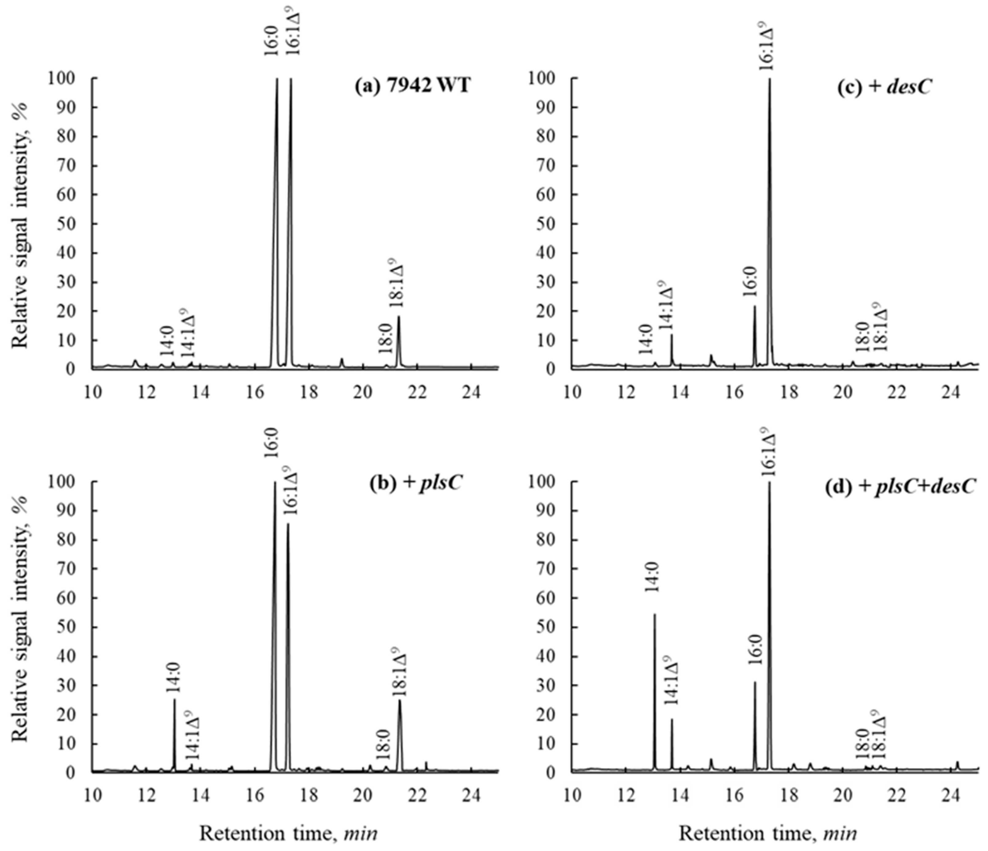
2.1. Changes in FA Composition of Total Lipids in S. elongatus Transformants

The plsC and desC genes were cloned from the genome of the Cyanobacterium sp. strain IPPAS B-1200 enriched with myristic and myristoleic acids. These genes were successfully expressed in cells of the model cyanobacterium S. elongatus containing minor amounts of 14:0 and 14:1∆9 acids. A significant amount of myristic acid appeared when the gene for acyltransferase was used, and the balance shifted towards an increase in the proportion of monounsaturated acids in each of the transformant lines (Figure 1, Table 1).
S. elongatus transformed with the plsC gene for lysophosphotidyl-acyl-CoA:acyltransferase (LPPAT) displayed a nearly 20× increase in the amount of 14:0 compared with wild-type cells (from 0.5 to 10%). However, the absolute and relative amounts of 14:1 did not change much (from 0.5 to 0.8%), suggesting that Δ9-FAD of S. elongatus poorly uses 14:0 as a substrate.
S. elongatus transformed with the desC gene of Cyanobacterium for Δ9-FAD had its amount of 14:1 increased from 0.5 to 9%, indicating that this FAD successfully desaturated minor amounts of 14:0 synthesized by the wild-type cells of Synechococcus. Moreover, the amount of 16:1 increased from 32 to 73% (Table 1), suggesting that, in addition to myristic acid, Δ9-FAD of Cyanobacterium efficiently utilized palmitic acid as a substrate.
Co-expression of LPAAT and Δ9-FAD of Cyanobacterium in S. elongatus cells resulted in a significant increase in 14:0 (more than 26%) and 14:1 (9%). The level of 16:0 dropped from 50 to 15%, while the level of 16:1 increased from 32 (WT) to 49% (Figure 1, Table 1).
Apparently, these shifts in the C16/C14 proportions in S. elongatus were due to the appearance of significant amounts of C14 FAs, caused by the activities of LPAAT and Δ9-FAD of Cyanobacterium sp.

2.2. Stereospecific Positioning of C14 FAs

LPAAT is an enzyme that catalyzes the acylation of lysophosphatidic acid to the sn-2 position. In cyanobacteria, usually, this position is occupied by unsaturated 16:0 [13,14]. However, the PlsC1200 enzyme is presumably characterized by specificity for myristic acid. To confirm the specificity of the enzyme toward the sn-position, we isolated lysophosphatidic acid (LPA) and phosphatidic acid (PA) from S. elongatus wild-type cells and the transformant cell line expressing LPAAT.
Four distinct areas were detected after the separation of phospholipids, which differed in Rfs with the standards PA and LPA (Figure 2). These mismatches may be due to the difference in acyl composition or to the fact that the sodium salts of PA and LPA had been applied as the standards. Based on Rfs, FA composition, and amounts of FAs, we assume that PA and LPA are represented in areas 2 and 1, respectively. The Rf values of lyso-forms of acids are usually lower than that of PA. LPA is characterized by the predominance of saturated FAs and a small amount of phospholipids in a pool. Due to a high content of 16:1Δ9, area 2 probably corresponds to PA [15].
The main FAs in the LPAs of wild-type and transformant cells are represented by 16:0 and 18:0 and then by 16:1Δ9. In the PlsC transformant, the amount of myristic acid reached 12%. The transition from LPA to PA was carried out by LPAAT, which operates at the sn-2 position, thus the accumulation of 14:0 occurs at the sn-2 position. This suggestion is confirmed by the appearance of 12% of 14:0 in the analyzed area 2 (Table 2).

2.3. Analysis of FA Composition in Individual Lipid Classes

Fatty acids analysis of individual glycerolipids from the control showed that all classes contained 16:0 and 16:1 as major FAs. Relatively high levels of myristic acid have been found in all lipid classes. All lipid classes of the transformant cells expressing PlsC revealed a decrease in the amount of 16:0 compared with wild-type cells (Table 3). These changes correlated with the changes in total FA composition (Table 1).

3. Discussion

The transition from LPA to PA is carried out by LPAAT, which introduces a particular set of fatty acids at the sn-2 position of phospholipids in pro- and eukaryotes. Many bacteria have multiple LPAAT paralogs; these enzymes generate a variety of phospholipids with unique fatty acid compositions and have different FA specificities [15]. The cyanobacterium Synechococcus elongatus PCC 7942 has one plsC gene for LPAAT, which, according to the FA composition, is specific to 16:0 [8]. Transformation of Synechococcus with the gene for LPAAT of the Cyanobacterium sp. strain IPPAS B-1200 resulted in the accumulation of 12% of 14:0 at the sn-2 position (Table 2). These results correlate with data obtained for the LPAAT of Cyanothece sp. PCC 8801, where stereospecific positioning was analyzed with the sn-1-specific lipase from Rhizopus delemar [3].
Previously, LPAAT from Cyanothece sp. PCC 8801 was expressed in Synechocystis sp. PCC 6803, resulting in a significant increase in the proportion of myristic acid at the sn-2 position in glycerolipids. This result was explained by the high specificity of this enzyme for the 14:0 acyl-carrying protein [3]. In addition, LPAAT from the thermophilic bacterium Thermus thermophilus HB8 showed increased specificity for 14:0-CoA and 16:1∆9-CoA substrates [16].
Synechocystis sp. PCC 6803 belongs to Group 4 of the cyanobacteria classified according to their FA composition [8]. This strain carries four genes for FADs (namely, desC for Δ9-FAD; desA for Δ12-FAD; desD for Δ6-FAD; and desB for ω3-FAD). As a result, it is characterized by a complicated FA composition, consisting of 16:0, 16:1; 18:0, 18:1, 18:2, 18:3, and 18:4 acids, the proportions of which vary depending on growth temperature [17]. Such a model system complicates analysis of the FA composition of the transformants. On the other hand, the primary Δ9-FAD of Synechocystis sp. PCC 6803 belongs to type 1 of the Δ9-FADs [8], which desaturate FAs only at the sn-1 position [17]. Because 14:0 was assigned to position sn-2, its desaturation by the Δ9-FAD of Synechocystis was not anticipated [3].
Here, we applied the simple model strain Synechococcus elongatus PCC 7942, which belongs to Group 1 and has only one FAD, Δ9-FAD, capable of desaturating the FAs of lipids at the sn-1 and sn-2 positions [18]. Expression of the plsC gene for LPAAT from Cyanobacterium sp. IPPAS B-1200 in S. elongatus PCC 7942 led to an increase in the proportion of saturated myristic acid up to 26%. However, the latter was not converted into monounsaturated myristoleic acid as expected. This implies that the only Δ9-FAD of S. elongatus PCC 7942 is unable to efficiently desaturate 14:0. Expression of the ∆9-FAD gene from Cyanobacterium in S. elongatus led to an increase in the proportion of monounsaturated acids, particularly in palmitoleic acid, 16:1∆9. Actually, in the absence of C14 FAs, the foreign desaturase duplicated the activity of the native ∆9-FAD of S. elongatus. Co-expression of LPAAT and ∆9-FAD of Cyanobacterium in S. elongatus increased the proportion of 14:1 and 16:1 at the expense of 14:0 and 16:0, suggesting that Δ9-FAD of Cyanobacterium is capable of desaturating both saturated FAs, C14 and C16.
The Δ9-FADs of cyanobacteria belong to a class of integral membrane acyl-lipid desaturases that operate on acyl chains attached to the glycerol moiety. The crystal structure of these acyl-lipid desaturases has yet to be determined. However, the structures of functionally comparable stearoyl-CoA desaturases are accessible [19,20]. These enzymes are membrane-integrated homodimers that form a hydrophobic tunnel to accommodate substrates [21]. The tunnel’s length forces carbons 9 and 10 of the acyl chain to align proximal to the relatively buried catalytic metal (Fe or Zn) atoms coupled to His-clusters inside the enzyme’s catalytic site [19]. The shape of the hydrophobic cavity is hypothesized to determine a FAD’s regioselectivity for cis dehydrogenation of the substrate [19,20]. The location of a catalytic site in the tunnel may also influence the length of the acyl chain that can be processed by a particular FAD. The latter may explain the observed differences in substrate preference between the ∆9-FADs of S. elongatus and Cyanobacterium.
Cyanobacteria are distinguished by a wide range of component fatty acids in their lipids, which corresponds to a wide range of phenotypes and environmental conditions, many of which are quite extreme. S. elongatus and Cyanobacterium sp. both belong to Group I of the cyanobacteria, which possess only ∆9-FAD activity and are capable of synthesizing unsaturated and monounsaturated FAs [8]. This FA composition is characteristic for thermophilic strains that are usually not exposed to low temperatures and cannot synthesize polyunsaturated FAs in response to cold. C14-rich Cyanobacterium sp. IPPAS B-1200 was isolated from Lake Balkhash (Kazakhstan) with a salinity of 6.0 g L−1. Lake Balkhash is a semi-freshwater lake: the western part of the lake is almost fresh (mineralization is 0.74 g L−1), while the eastern part has higher salinity (3.5–6.0 g L−1). Water temperature at the surface of the lake varies from 0 °C in December to 28 °C in July. Therefore, Cyanobacterium sp. must acclimate to multiple changing parameters, such as temperature, salinity, and maybe pH. It is known that an increase in C14 FAs and 16:0 is a feature of the acclimation to high temperatures [22]. High amounts of C14 FAs may rigidify the membranes, which is important for survival under heat stress.
Myristic acid is a component of cell membranes and seed oils and a source for protein myristoylation [23]. It is applied in various industries and manufacturing processes, e.g., as flow agent and emulsifier in foods and beverages; as a surfactant in soaps, detergents, and textiles; and as a lubricant in plastics. Myristoleic acid is also located in cell membranes and plant oils. Esterified to cetyl alcohol, myristoleic acid is modified to cetyl myristoleate, a drug for osteoarthritis treatment [24].
Myristic and myristoleic acids are usually produced from seeds or Myristicaceae plants, such as nutmeg (Myristica frangans). Nutmeg butter is rich in trimyristin (up to 75%)—a triglyceride with three saturated myristic acid residues. Another source is the seeds of African nutmeg (Pycnanthus angolensis or P. kombo), which contain up to 60% so-called combo oil, rich in myristic (58–64%) and myristoleic (19–26%) acids [25].
Cyanobacteria are fast-growing cells that are cultured in photobioreactors that produce substantial amounts of biomass for various purposes [26]. In particular, Synechococcus elongatus PCC 7942 is often used for “light-driven autotrophic cell factories” to produce biofuels and various fine chemicals directly from CO2 [27]. The recombinant C14-producing Synechococcus strain described here may be used for the biotechnological production of myristic and myristoleic acids.

4. Materials and Methods

4.1. Cyanobacterial Strains and Growth Conditions

In the course of the experiment, strains of the model cyanobacterium Synechococcus elongatus PCC 7942 and the Cyanobacterium sp. strain IPPAS B-1200, rich in short FAs, were used. Cultivation was carried out under conditions selected during the isolation of this strain [5]. Synechococcus cells were maintained on a solid BG-11 medium [28] with an agar content of 1.2%. The experimental growth conditions were as follows: liquid medium BG-11 with the addition of HEPES-NaOH pH 7.5 in a volume of 250 mL under a constant illumination of 50 µE m−2 s−1 at 33 °C. The cultures were doused with sterile air that contained 1.5% CO2. The samples were fixed in the middle of the exponential growth stage (OD750~1); planting and cultivation were done under aseptic conditions.

4.2. Cloning and Expression of the desC and plsC Genes

Genomic DNA from Cyanobacterium IPPAS B-1200 was isolated according to Williams [29]. The desC and plsC genes were amplified using primers containing the sequences of certain restriction sites: desC1200F ATAACCATGGCAGTTTCAAC; desC1200R TTTGAAGCTTTTATTATGCC; plsC1200F GAATTAGACCATGGCTAAGG; and plsC1200R CTAACACTTGCCTAAGCTTAAAC. Phusion High-Fidelity DNA Polymerase (New England Biolabs, Ipswich, MA, USA) was used for PCR. The amplified DNA fragments were digested with the appropriate restriction enzymes (Nco I and Hind III), purified using the Cleanup Standard kit (Evrogen, Ltd., Moscow, Russia), and ligated with the linearized pTrc99A vector (Pharmacea, Uppsala, Sweden; https://www.addgene.org/vector-database/4402 (accessed on 31 December 2023)). The vector was generated in E. coli strain XL-1 (Stratagene, La Jolla, CA, USA) and used as a template for further cloning. The genes of interest under the control of the constitutive Trc promoter were amplified into the pAM1303 ([30] https://www.addgene.org/40243 (accessed on 31 December 2023) and pNS2 vectors (the latter vector is similar to pAM1303, but contains other neutral recombination sites (NS2) of the S. elongatus genome and a kanamycin resistance cassette). Amplification primers contained the Xma I restriction site sequence. The vectors were linearized with this restriction enzyme and treated with FastAP alkaline phosphatase (Thermo Scientific, Waltham, MA, USA). The resulting vectors were used to transform S. elongatus PCC 7942 by homologous recombination. The selection of transformants was carried out on a solid BG-11 medium containing spectinomycin and kanamycin at final concentrations of 30 and 25 µg ml−1, respectively.

4.3. FA Analysis of Total Lipids

Wild-type S. elongatus PCC 7942 cells and three transformants (pAM-desC, pNS2-plsC, and the double transformant pAM-desC + pNS2-plsC) were used to analyze the FA composition [31]. To 200 µg of harvested cells (wet weight) 1 mL of 80% aqueous ethanol solution of 1 M KOH was added; the samples were vortexed and incubated at 70 °C for 1 h. The samples were then washed twice with 500 µL n-hexane to remove unsaponifiable compounds, e.g., free sterols, pigments, etc. The excess KOH was neutralized by adding 50 µL of 20% sulfuric acid to a slightly acidic level (pH of 4.5–4.7 according to the paper test) to the obtained samples. Free FAs were extracted from the samples with n-hexane (300 µL) and evaporated to dryness, and 200 µL 1% sulfuric acid in methanol was added. FA methylation was carried out for 30 min at 55 °C. FA methyl esters (FAMEs) were extracted into n-hexane in a volume of 200 µL.

4.4. GC-MS

FAMEs were analyzed using an Agilent 7890A gas–liquid chromatograph with an Agilent 5975C mass spectrometric detector (Agilent Technology Systems, Santa Clara, CA, USA). A capillary 60 m HP-88 column (inner diameter ∅ 0.25 mm, film thickness 0.2 µm, Agilent J&W, Santa Clara, CA, USA) was used. Other analysis conditions: carrier gas helium, flow rate 1 mL/min, sample volume 1 µL, flow divider 1:20, evaporation temperature 260 °C. Program for gradient analysis: from 130 °C to 170 °C in steps of 6.5 °C/min; 170 to 215 °C in 2.75 °C/min increments; 215 °C for 25 min; 215 to 240 °C in 5 °C/min increments; and a final stage of 50 min at 240 °C. The operating temperature of the mass spectrometric detector was 240 °C; the ionization energy was 70 eV.

4.5. Analysis of Lysophosphatidic and Phosphatidic Acids

The total lipid extract was obtained by the classical Bligh-Dyer method [31] and purified through PTFE filters (Agilent, 5190–5265). To isolate lysophosphatidic (LPA) and phosphatidic (PA) acids, the total extract was separated by TLC (Fluka Silica gel TLC Al foils 10 × 10 cm) in hexane:acetone:acetic acid (40:50:2). Phospholipids remained in situ and were detected by staining with primulin (0.05% in acetone:water 80:20); a mixture of 1-oleoyl LPA (http://www.gzsopo.com/product/PNOALD-O353308.html. CAS: 655528-98-5; accessed on 31 December 2023), PA sodium salt (http://www.gzsopo.com/product/PNOMACKLIN-L864045.html. CAS 383907-53-7; accessed on 31 December 2023), and phosphatidylcholine (PC) was used as a standard. The appropriate silica gel sections were taken from the plate and transferred to Eppendorf tubes, where they were washed with a chloroform–methanol (1:1) mixture followed by an equivalent volume of water. The bottom fraction containing phospholipids was chosen after centrifugation. To separate phospholipids, the chloroform:methanol:acetone:water:acetic acid (6:2:8:1:1:1) system was utilized. The LPA and PA standards had Rfs of 0.4 and 0.7, respectively. To detect lipid groups, primulin staining was applied. Lipids from all four detected groups were isolated, extracted similarly to phospholipids, and treated in methanol with 1% H2SO4 for 30 min at 55 °C [32]. The FAMEs were then extracted with n-hexane and analyzed using GC-MS as described above.

4.6. Analysis of Fatty Compositions of Individual Classes of Glycerolipids

The extracts of total lipids were separated by two-dimensional TLC [33] (the solvent for the first dimension was chloroform:methanol:water (75:25:2.5) and for the second was chloroform:methanol:acetic acid:water (80:9:12:2)). The results of separation were visualized using 0.01% primuline in 80% acetone. Each lipid group was extracted and treated in methanol with 1% H2SO4 for 30 min at 55 °C. The FAMEs were then extracted with hexane and analyzed using GC-MS as described above.

5. Conclusions

Our findings reveal that LPAAT from Cyanobacterium sp. assures 14:0 synthesis in S. elongatus (up to 25%). However, this did not result in any significant 14:1 rise, indicating that S. elongatus Δ9-FAD is unable to desaturate FAs shorter than 16:0. Co-expression of LPAAT and Δ9-FAD (desC) of Cyanobacterium sp. in S. elongatus resulted in the accumulation of 14:0, 14:1, and 16:1 in Synechococcus cells, indicating that Δ9-FAD of Cyanobacterium has a broad range of length specificity and can accommodate and desaturate both C14- and C16-saturated FAs. Thus, the acyltransferase LPAAT, which drives 14:0 synthesis, and Δ9-FAD, which can accommodate and dehydrogenate myristic acid as a substrate, ensure unusual C14-rich (up to 40%) FA composition in Cyanobacterium sp. IPPAS B-1200. Recombinant C14-producing Synechococcus cells may be used for biotechnological production of myristic and myristoleic acids.

Author Contributions

Conceptualization, A.Y.S. and D.A.L.; methodology, A.Y.S., R.A.S. and D.A.L.; investigation and validation, A.Y.S., R.A.S., K.S.M. and D.A.L.; resources, D.A.L.; writing—original draft preparation, A.Y.S. and D.A.L.; review and editing, D.A.L.; funding acquisition, D.A.L. All authors have read and agreed to the published version of the manuscript.

Funding

This research was funded by the Russian Science Foundation (RSF grant no. 21-74-30003 to D.A.L.) and partially supported by the Ministry of Science and Higher Education of the Russian Federation (theme no. 122042700043-9).

Institutional Review Board Statement

Not applicable.

Informed Consent Statement

Not applicable.

Data Availability Statement

Data are contained within the article.

Acknowledgments

In this work, the large-scale research facilities of the Collection of Microalgae and Cyanobacteria IPPAS (K.A. Timiryazev Institute of Plant Physiology RAS, Moscow, Russia) were used.

Conflicts of Interest

The authors declare no conflicts of interest.

References

  1. Boudière, L.; Michaud, M.; Petroutsos, D.; Rébeillé, F.; Falconet, D.; Bastien, O.; Roy, S.; Finazzi, G.; Rolland, N.; Jouhet, J.; et al. Glycerolipids in photosynthesis: Composition, synthesis and trafficking. Biochim. Biophys. Acta Bioenerg. 2014, 1837, 470–480. [Google Scholar] [CrossRef]
  2. Sato, N.; Wada, H. Lipid biosynthesis and its regulation in cyanobacteria. In Lipids in Photosynthesis; Advances in Photosynthesis and Respiration; Wada, H., Murata, N., Eds.; Springer: Dordrecht, The Netherlands, 2009; Volume 30, pp. 157–177. [Google Scholar] [CrossRef]
  3. Saito, M.; Endo, K.; Kobayashi, K.; Watanabe, M.; Ikeuchi, M.; Murakami, A.; Murata, N.; Wada, H. High myristic acid content in the cyanobacterium Cyanothece sp. PCC 8801 results from substrate specificity of lysophosphatidic acid acyltransferase. Biochim. Biophys. Acta Mol. Cell Biol. Lipids 2018, 1863, 939–947. [Google Scholar] [CrossRef]
  4. Gombos, Z.; Murata, N. Lipids and fatty acids of Prochlorothrix hollandica. Plant Cell Physiol. 1991, 32, 73–77. [Google Scholar] [CrossRef]
  5. Sarsekeyeva, F.K.; Usserbaeva, A.A.; Zayadan, B.K.; Mironov, K.S.; Sidorov, R.A.; Kozlova, A.Y.; Kupriyanova, E.V.; Sinetova, M.A.; Los, D.A. Isolation and characterization of a new cyanobacterial strain with a unique fatty acid composition. Adv. Microbiol. 2014, 4, 1033–1043. [Google Scholar] [CrossRef]
  6. Shih, P.M.; Wu, D.; Latifi, A.; Axen, S.D.; Fewer, D.P.; Talla, E.; Calteau, A.; Cai, F.; Tandeau de Marsac, N.; Rippka, R.; et al. Improving the coverage of the cyanobacterial phylum using diversity-driven genome sequencing. Proc. Natl. Acad. Sci. USA 2013, 110, 1053–1058. [Google Scholar] [CrossRef]
  7. Starikov, A.Y.; Usserbaeva, A.A.; Sinetova, M.A.; Sarsekeyeva, F.K.; Zayadan, B.K.; Ustinova, V.V.; Kupriyanova, E.V.; Los, D.A.; Mironov, K.S. Draft genome sequence of Cyanobacterium sp. strain IPPAS B-1200 with a unique fatty acid composition. Genome Announc. 2016, 4, e01306-16. [Google Scholar] [CrossRef]
  8. Murata, N.; Wada, H.; Gombos, Z. Modes of fatty-acid desaturation in cyanobacteria. Plant Cell Physiol. 1992, 33, 933–941. [Google Scholar] [CrossRef]
  9. Santos-Merino, M.; Gargantilla-Becerra, Á.; de la Cruz, F.; Nogales, J. Highlighting the potential of Synechococcus elongatus PCC 7942 as platform to produce α-linolenic acid through an updated genome-scale metabolic modeling. Front. Microbiol. 2023, 14, 1126030. [Google Scholar] [CrossRef]
  10. Wada, H.; Gombos, Z.; Murata, N. Enhancement of chilling tolerance of a cyanobacterium by genetic manipulation of fatty acid desaturation. Nature 1990, 347, 200–203. [Google Scholar] [CrossRef]
  11. Starikov, A.Y.; Sidorov, R.A.; Mironov, K.S.; Goriainov, S.V.; Los, D.A. Delta or Omega? Δ12 (ω6) fatty acid desaturases count 3C after the pre-existing double bond. Biochimie 2020, 179, 46–53. [Google Scholar] [CrossRef]
  12. Starikov, A.Y.; Sidorov, R.A.; Goriainov, S.V.; Los, D.A. Acyl-lipid Δ6-desaturase may act as a first FAD in cyanobacteria. Biomolecules 2022, 12, 1795. [Google Scholar] [CrossRef]
  13. Brown, A.P.; Slabas, A.R.; Denton, H. Substrate selectivity of plant and microbial lysophosphatidic acid acyltransferases. Phytochemistry 2002, 61, 493–501. [Google Scholar] [CrossRef]
  14. Okazaki, K.; Sato, N.; Tsuji, N.; Tsuzuki, M.; Nishida, I. The significance of C16 fatty acids in the sn-2 positions of glycerolipids in the photosynthetic growth of Synechocystis sp. PCC 6803. Plant Physiol. 2006, 141, 546–556. [Google Scholar] [CrossRef]
  15. Ogawa, T.; Kuboshima, M.; Suwanawat, N.; Kawamoto, J.; Kurihara, T. Division of the role and physiological impact of multiple lysophosphatidic acid acyltransferase paralogs. BMC Microbiol. 2022, 22, 241. [Google Scholar] [CrossRef]
  16. Ogawa, T.; Suwanawat, N.; Toyotake, Y.; Watanabe, B.; Kawamoto, J.; Kurihara, T. Lysophosphatidic acid acyltransferase from the thermophilic bacterium Thermus thermophilus HB8 displays substrate promiscuity. Biosci. Biotechnol. Biochem. 2020, 84, 1831–1838. [Google Scholar] [CrossRef]
  17. Wada, H.; Murata, N. Temperature-induced changes in the fatty acid composition of the cyanobacterium, Synechocystis PCC6803. Plant Physiol. 1990, 92, 1062–1069. [Google Scholar] [CrossRef]
  18. Khan, M.U.; Mackenzie, S.L.; Williams, J.P. Modulation of fatty acids in the membranes of Anacystis nidulans (cyanobacteria): Incorporation of odd-numbered carbon fatty acids. J. Phycol. 1996, 32, 970–973. [Google Scholar] [CrossRef]
  19. Wang, H.; Klein, M.G.; Zou, H.; Lane, W.; Snell, G.; Levin, I.; Li, K.; Sang, B.C. Crystal structure of human stearoyl-coenzyme A desaturase in complex with substrate. Nat. Struct. Mol. Biol. 2015, 22, 581–585. [Google Scholar] [CrossRef]
  20. Bai, Y.; McCoy, J.G.; Levin, E.J.; Sobrado, P.; Rajashankar, K.R.; Fox, B.G.; Zhou, M. X-ray structure of a mammalian stearoyl-CoA desaturase. Nature 2015, 524, 252–256. [Google Scholar] [CrossRef]
  21. Li, D.; Moorman, R.; Vanhercke, T.; Petrie, J.; Singh, S.; Jackson, C.J. Classification and substrate head-group specificity of membrane fatty acid desaturases. Comput. Struct. Biotechnol. J. 2016, 14, 341–349. [Google Scholar] [CrossRef]
  22. Maslova, I.P.; Mouradyan, E.A.; Lapina, S.S.; Klyachko-Gurvich, G.L.; Los, D.A. Lipid fatty acid composition and thermophilicity of cyanobacteria. Russ. J. Plant Physiol. 2004, 51, 353–360. [Google Scholar] [CrossRef]
  23. Meinnel, T.; Dian, C.; Giglione, C. Myristoylation, an ancient protein modification mirroring eukaryogenesis and evolution. Trends Biochem. Sci. 2020, 45, 619–632. [Google Scholar] [CrossRef] [PubMed]
  24. Ameye, L.G.; Chee, W.S. Osteoarthritis and nutrition. From nutraceuticals to functional foods: A systematic review of the scientific evidence. Arthritis Res. Ther. 2006, 8, R127. [Google Scholar] [CrossRef] [PubMed]
  25. Obranović, M.; Bryś, J.; Repajić, M.; Balbino, S.; Škevin, D.; Bryś, A.; Tonković, P.; Medved, A.M.; Uzelac, V.D.; Kraljić, K. Fatty acid and sterol profile of nutmeg (Myristica fragrans) and star anise (Illicium verum) extracted using three different methods. Proceedings 2021, 70, 33. [Google Scholar] [CrossRef]
  26. Melis, A.; Hidalgo Martinez, D.A.; Betterle, N. Perspectives of cyanobacterial cell factories. Photosynth. Res. 2023. [Google Scholar] [CrossRef]
  27. Li, Z.; Li, S.; Chen, L.; Sun, T.; Zhang, W. Fast-growing cyanobacterial chassis for synthetic biology application. Crit. Rev. Biotechnol. 2023. [Google Scholar] [CrossRef]
  28. Rippka, R. Isolation and purification of cyanobacteria. Methods Enzymol. 1988, 167, 3–27. [Google Scholar] [CrossRef]
  29. Williams, J.G.K. Construction of specific mutations in photosystem II photosynthetic reaction center by genetic engineering methods in Synechocystis 6803. Methods Enzymol. 1988, 167, 766–778. [Google Scholar] [CrossRef]
  30. Andersson, C.R.; Tsinoremas, N.F.; Shelton, J.; Lebedeva, N.V.; Yarrow, J.; Min, H.; Golden, S.S. Application of bioluminescence to the study of circadian rhythms in cyanobacteria. Methods Enzymol. 2000, 305, 527–542. [Google Scholar] [CrossRef]
  31. Bligh, E.G.; Dyer, W.J. A rapid method of total lipid extraction and purification. J. Biochem. Physiol. 1959, 37, 911–917. [Google Scholar] [CrossRef]
  32. Pinault, M.; Guimaraes, C.; Dumas, J.; Servais, S.; Chevalier, S.; Besson, P.; Goupille, C. A 1D high performance thin layer chromatography method validated to quantify phospholipids including cardiolipin and monolysocardiolipin from biological samples. Eur. J. Lipid Sci. Technol. 2020, 122, 1900240. [Google Scholar] [CrossRef]
  33. Sallal, A.K.; Nimer, N.A.; Radwan, S.S. Lipid and fatty acid composition of freshwater cyanobacteria. J. Gen. Microbiol. 1990, 136, 2043–2048. [Google Scholar] [CrossRef]
Figure 1. Separation of FA methyl esters obtained from the total lipids of S. elongatus wild-type (a) and transformant cells expressing plsC (b), desC (c), plsC, and desC (d) genes (total ion current).
Ijms 25 00774 g001
Figure 2. TLC separation of phospholipid fractions from cells of S. elongatus transformed with plsC1200 (LPAAT) from Cyanobacterium sp. IPPAS 1200. The mobile phase is chloroform:methanol:acetone:water:acetic acid (6:2:8:1:1:1). Primulin-stained samples were illuminated at 366 nm. PA—phostphatidic acid standard, LPA—lysophosphatidic acid sodium salt standard. 1–4—isolated and analyzed phospholipid fractions. Marked areas have been withdrawn and analyzed.
Ijms 25 00774 g002
Table 1. FA composition (mass %) of total lipids of Synechococcus elongatus PCC 7942 wild-type (7942) and its transformants expressing LPAAT plsC1200 (plsC) and Δ9-FAD desC1200 (desC) and co-expressing plsC1200 together with desC1200 (plsC + desC).
FAs7942plsCdesCplsC + desC
14:00.510.20.626.5
14:1∆90.50.89.09.0
16:050.340.416.015.2
16:1∆931.734.673.148.5
18:01.22.60.60.2
18:1∆913.09.40.50.5
18:1∆112.82.00.20.1
FAs: 14:0—myristic acid; 14:1∆9—myristoleic acid; 16:0—palmitic acid; 16:1∆9—palmitoleic acid; 18:0—stearic acid; 18:1∆9—oleic acid; 18:1∆11—cis-vaccenic acid. Individual peaks were identified with Agilent MSD ChemStation 4.0.3 software and the NIST library. The experiments have been repeated three times. Standard deviations are in the range of 0.1–0.3%.
Table 2. FA compositions of LPA and PA (mass %) obtained by separation of phospholipid fractions (Figure 2). WT, phospholipids from S. elongatus PCC 7942 wild-type cells; PlsC, phospholipids from S. elongatus PCC 7942 cells expressing PlsC (LPAAT) from Cyanobacterium sp. IPPAS 1200.
WTPlsC
FAsLPAPALPAPA
14:00.80.6312.3
14:1Δ9*10.51.2
16:057.449.453.638
16:1Δ9432.34.835.2
18:037.1937.58
18:1Δ90.77.70.65.3
* At standard parameters (width: 2–5 s, threshold: 5, base in fractions of width: 0.5 Min, mV: 0) the peak was not integrated, but the spectrum of the corresponding methyl esters could be detected. Standard deviations are in the range of 0.1–0.3%.
Table 3. The content of FAs in individual areas (mass %) obtained by separation of glycerolipid fractions. WT, glycerolipids from S. elongatus PCC 7942 wild-type cells; PlsC, glycerolipids from S. elongatus PCC 7942 cells expressing PlsC (LPAAT) from Cyanobacterium sp. IPPAS 1200. MGDG—monogalactosyl diacylglycerol; DGDG—digalactosyl diacylglycerol; SQDG—sulfoquinovosyl diacylglycerol; PG—phosphatidylglycerol.
WTPlsC
FAsMGDGDGDGSQDGPGMGDGDGDGSQDGPG
14:01.22.31.10.914.619.77.67.9
14:1Δ90.11.10.81.01.20.80.61.7
16:048.140.356.249.531.230.948.741.9
16:1Δ946.242.934.332.348.143.435.533.0
18:00.810.76.05.21.12.13.24.7
18:1Δ93.62.71.611.13.83.14.410.8
Disclaimer/Publisher’s Note: The statements, opinions and data contained in all publications are solely those of the individual author(s) and contributor(s) and not of MDPI and/or the editor(s). MDPI and/or the editor(s) disclaim responsibility for any injury to people or property resulting from any ideas, methods, instructions or products referred to in the content.

