Article

Ganoderma adspersum (Ganodermataceae): Investigation of Its Secondary Metabolites and the Antioxidant, Antimicrobial, and Cytotoxic Potential of Its Extracts

1
Laboratory of Pharmacognosy, Faculty of Health Sciences, School of Pharmacy, Aristotle University of Thessaloniki, 54124 Thessaloniki, Greece
2
Department of Chemistry and Chemical Engineering, Faculty of Agronomy, University of Kragujevac, Cara Dušana 34, 32000 Čačak, Serbia
*
Author to whom correspondence should be addressed.
Int. J. Mol. Sci. 2024, 25(1), 516; https://doi.org/10.3390/ijms25010516
Submission received: 13 October 2023 / Revised: 23 December 2023 / Accepted: 24 December 2023 / Published: 30 December 2023

Abstract

:
Ganoderma is a genus of wood-degrading mushrooms with medicinal importance. Most Ganoderma species have been studied extensively for their secondary metabolites, biological activities, and ecological value. In this study, the biological activities of the extracts of G. adspersum growing wild on Morus alba trees in the region of Western Thrace (Greece) were evaluated, and the petroleum ether, dichloromethanolic, and methanolic extracts were studied further for their secondary metabolites. Six substances were isolated by chromatographic (Clumn Chromatography (C.C.), High Performance Liquid Chromatography (HPLC)) and spectroscopic methods (Nuclear Magnetic Resonance (NMR)), which were classified in the following categories: (a) unsaturated fatty acids: cis-oleic acid (1); (b) sterols: ergosta-7,22-dien-3-one (2), ergosta-7,22-dien-3-ol (3), and ergosta-5,7,22-trien-3-ol (4); and (c) lanostane-type triterpenoids: applanoxidic acid G (5) and applanoxidic acid A (6). Finally, the biological activities of the extracts were estimated for their antioxidant, antimicrobial, and cytotoxic potential. The methanolic extract of G. adspersum showed the highest total antioxidant activity. The results of the antimicrobial activities indicated that all of the extracts had a minimum inhibitory concentration (MIC) ranging between 39.1 and 312.5 μg/mL. The evaluation of the cytotoxic activity of the samples showed once again that the methanolic extract was the most potent among the examined extracts, with half-maximal inhibitory concentration (IC50) values of 19.22 μg/mL (Hep2c cells), 32.9 μg/mL (RD cells), and 8.94 μg/mL (L2OB cells). Moreover, the bioactivity scores of the isolated secondary metabolites were calculated using the online computer software program Molinspiration. The compounds showed promising bioactivity scores for drug targets.

1. Introduction

Ganoderma constitutes a large and diverse genus of wood-decaying basidiomycetes belonging to the Ganodermataceae family [1]. The fungi of this genus grow on the trunks of different deciduous trees, such as oak, willow, elm, acacia, etc. [2]. The genus was first reported and named by Finnish mycologist Petter Adolf Karsten in 1881 [3]. The name arises from the Greek words ”ganos” and ”derma” that signify the shiny skin caused by the morphological characteristics of the fungus [4]. The classification of the genus has been characterized as chaotic due to the difficulty in precisely identifying the miscellaneous features of the species, while some botanists focus on the basidiocarp color for the classification of the mushroom [5,6,7].
Ganoderma is widespread worldwide, mainly in temperate and tropical climates, specifically in Asia, Africa, and Europe, counting more than 250 species [8,9]. The most popular ones include G. lucidum, G. sinensis, G. applanatum, G. tsugae, and G. atrum [10]. Nevertheless, only the species G. lucidum and G. sinense have been accepted from Traditional Chinese Medicine and recognized as “Ling-zhi” by the Chinese or “Reishi” by the Japanese [11]. Despite the therapeutic use of certain Ganoderma spp., some species exhibit phytopathogenicity. Only a few species are widely recognized for their phytopathogenicity on both landscape and commercially significant trees. The oil palm tree is harmed by G. boninense and G. zonatum. [12,13,14].
Ganoderma has been thoroughly studied for its medicinal properties, as it has been used traditionally in China since antiquity for the treatment and prevention of various diseases and the promotion of health and longevity [15]. It has also been considered as an “elixir” that represents immortality [16]. The Pharmacopoeia of China highlights the beneficial effects of G. lucidum in regulating heart function, relieving the symptoms of asthma and dyspnea, boosting the memory, activating the immune system, and handling irregular sleeping. Today, many products derived from Ganoderma are consumed in the form of powders, food supplements, and teas [17]. Several reports elucidate the antioxidant, anti-inflammatory, anti-hyperlipidemic, anticancer, anti-aging, antimicrobial, antiviral, hepatoprotective, neuroprotective, and antidepressant properties of various Ganoderma species extracts from different parts of the mushroom, both in vitro and in vivo [7,18,19,20,21,22,23,24,25,26,27].
There is a diversity of bioactive constituents and secondary metabolites isolated from Ganoderma species, contributing to their therapeutic properties. The major substances identified belong to the classes of polysaccharides, triterpenes, meroterpenoids, sterols, alkaloids, and phenolic compounds. Furthermore, proteins, peptides, vitamins, amino acids, fatty acids, nucleosides, and inorganic elements has also been found [28,29,30]. Ganoderma lucidum polysaccharides have exhibited significant pharmacological activity. α- and β-glucans, heteroglucans, peptidoglucans, and polysaccharide–protein conjugates have exerted biological action. They are considered to be potent immunomodulatory agents, as they affect the differentiation and enhance the activity of immune cells (e.g., natural killers, B cells, T cells, dendritic cells, etc.) and act on the release of cytokines (tumor necrosis factor (TNF)-α), interleukins (IL-1β, IL-6), and prostaglandin E, while also displaying antitumor and chemopreventive effects through these mechanisms.
Ganoderma adspersum (Schulz., 1969) Donk (=G. europaeum (Stayert, 1961)) is a parasitic species, frequently confused with the species G. applanatum, distributed mainly in southern regions of Europe, such as the Balkan Peninsula and Mediterranean Basin, while it is absent from Scandinavian countries [31,32,33]. To date, there are limited reports referring to the phytochemical profile of the species. Tel-Cayan et al. isolated three lanostane-type triterpenes and one steroid from G. adspersum extracts [34]. Mayaka and his team isolated three sterol-type triterpenoids from Kenyan mushrooms and studied their antimicrobial activities [35]. The triterpenes and sterol characterized belonged to applanoxidic acid and stigmastane-type derivatives, respectively. In addition, they identified and quantified certain phenolic acids (caffeic acid, rosmarinic acid, ferulic acid, 2,4-dihydrobenzoic acid, and ellagic acid) present in the species. The extracts and the pure compounds were evaluated in vitro for their biological activities. Significant antioxidant and anticholinesterase activities were observed. Specifically, the sterol exhibited remarkable inhibitory effects on lipid peroxidation and butyrylcholinesterase action. Two novel heteropolysaccharides detected in the mushroom, belonging to the class of galactomannans, also indicated potent inhibitory activity [36]. Being a promising therapeutic agent, G. adspersum should undergo further study for the protection provided against oxidative stress and Alzheimer’s disease. In addition, the ethanolic extract from a Turkish mushroom was examined for its phenol content and its antioxidant and antimicrobial activities [37].
The aim of this study was the isolation and characterization of mycochemical constituents of wild Ganoderma adspersum (Figure 1). Furthermore, the biological activities of extracts produced from the fungus were evaluated as part of this investigation. The fungus was collected from the tree Morus alba, grown in the region of Western Thrace, Greece, where it is consumed as a remedy for the treatment of asthma and bronchitis. As the use of Ganoderma spp. is rising in Greece, there is a compelling need to investigate the medicinal potential of this mushroom. In this report, we elucidate the extraction and isolation process of six pure compounds derived from G. adspersum, and we examine their potential relationships with target receptors (e.g., kinase inhibitors, nuclear receptors, ion-channel modulators) according to the Molinspiration cheminformatics program, assessing their physicochemical properties. Different extracts of the product were also estimated for their total phenolic, flavonoid, and tannin contents and their antioxidant activity in vitro. This study is one of a few to report on the in vitro antimicrobial and cytotoxic effects exerted by G. adspersum. Moreover, the results of the current study provide the opportunity for further comparison among the species, as well as the introduction of novel drug candidates derived from this fungus.

2. Results

2.1. Mycochemical Profile and Total Antioxidant Activity of G. adspersum Extracts

Ganoderma is a valuable mushroom genus due to its nutritional and pharmaceutical properties. The mycochemical analysis of species belonging to the genus Ganoderma has been demonstrated in recent decades. The mycochemical analysis and antioxidant profile of the extracts from G. adspersum are listed in Table 1.

Inhibitory Effect against Lipid Peroxidation and ROS Scavenging Activities

As we mentioned above, the methanolic extract of G. adspersum was the one with the highest total antioxidant activity. In this study, the inhibitory effect against lipid peroxidation and the scavenging activities against reactive oxygen species (ROS) were measured (Table 2).
Among the tested extracts, the methanolic extract had the highest phenolic and polyphenolic contents; therefore, it showed the highest antioxidant activity (Table 1). The explanation of these results is related to the polarity of the phenolic compounds (phenolic acids and flavonoids). Higher quantities of phenolic compounds accumulate in polar extracts compered to non-polar extracts. Moreover, it was demonstrated that the methanol extract presented the greatest antioxidant activity and inhibitory effect against lipid peroxidation, followed by the dichloromethanolic extract and, lastly, by the petroleum ether extract. On the other hand, in the case of metal-chelating ability, the non-polar extracts were the most potent (Table 2).

2.2. Antimicrobial Activity of G. adspersum Extracts

The inhibitory effects of the G. adspersum extracts were evaluated against the bacteria Staphylococcus aureus, Klebsiella pneumoniae, Escherichia coli, Proteus vulgaris, P. mirabilis, and Bacillus subtilis and against the fungi Candida albicans and Aspergillus niger. As standard antibiotics, we used Amracin as an antibacterial and nystatin as an antifungal (Table 3).
Concerning the antimicrobial activity (Table 3), the results indicate that all of the examined extracts expressed weak antimicrobial activity, with a range of minimum inhibitory concentrations (MICs) between 39.1 and 312.5 μg/mL. The best recorded MICs of the petroleum ether extract were those against Escherichia coli, with an MIC of 39.1 μg/mL, and Aspergillus niger, with an MIC of 78.1 μg/mL. On the other hand, the dichloromethanolic extract showed remarkable effects against Staphylococcus aureus and Candida albicans, with an MIC of 39.1 μg/mL, and also against Escherichia coli and Proteus vulgaris, with an MIC of 78.1 μg/mL. Finally, the methanolic extract was most potent against Staphylococcus aureus, with an MIC of 39.1 μg/mL, and against Klebsiella pneumoniae, Escherichia coli, Proteus mirabilis, and Bacillus subtilis with an MIC of 78.1 μg/mL. These results are consistent with previous reports [35,36].

2.3. Cytotoxic Activities of G. adspersum Extracts

The cytotoxic effects of the G. adspersum extracts were evaluated in vitro against the cell lines Hep2c (human cervix carcinoma), RD (human rhabdomyosarcoma), and L2OB (murine fibroblasts) (Table 4).

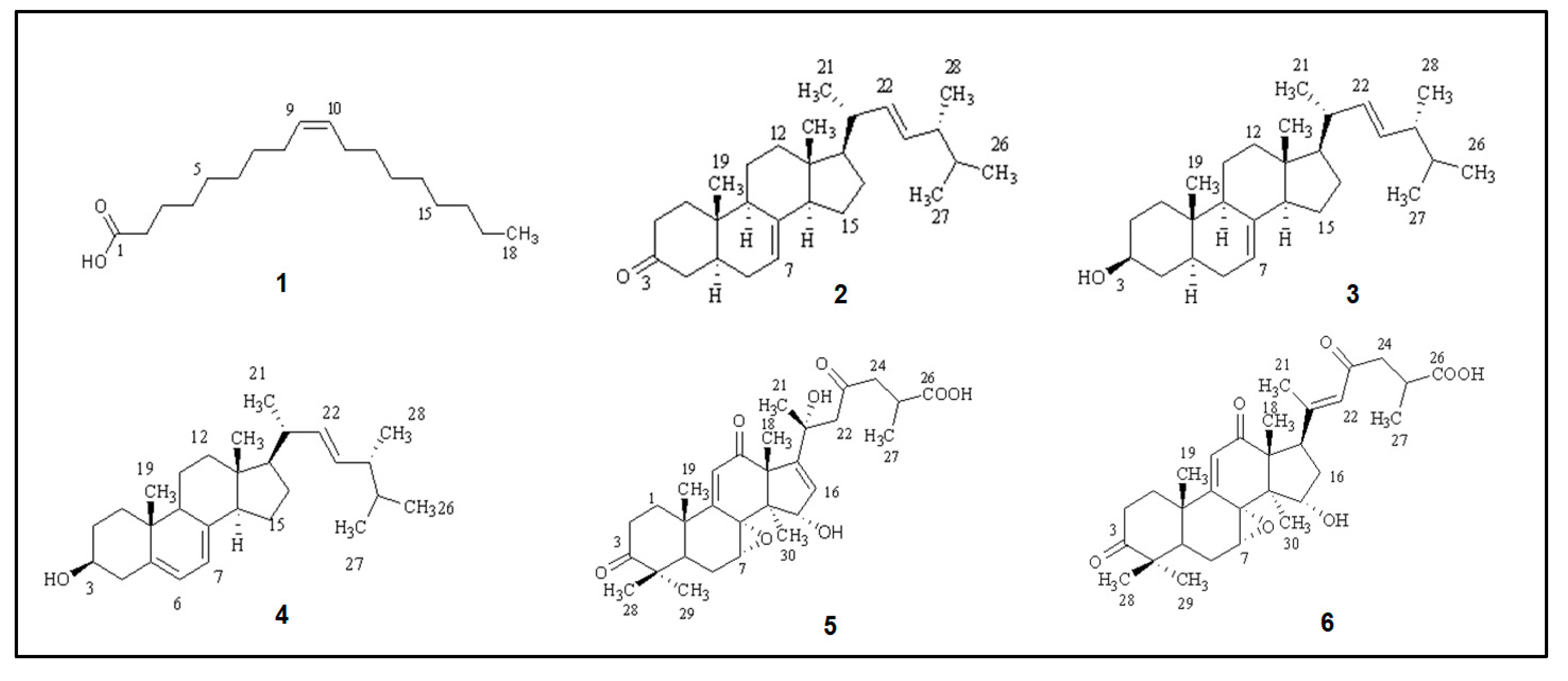
2.4. Characterization of Isolated Compounds

On the basis of 1H, 13C, and 2D NMR (gDQCOSY, gHSQCAD, and gHMBCAD, respectively) spectroscopic analysis, and through comparison with the literature data, the chemical structures of six isolated compounds were established (Figure 2): cis-oleic acid (1) [38], ergosta-7,22-dien-3-one (2) [39], ergosta-7,22-dien-3-ol (3) [40], ergosta-5,7,22-trien-3-ol (4) [35], applanoxidic acid G (5) [41], and applanoxidic acid A (6) [42]. The structures of the isolates are given in Figure 2 and were elucidated based on 1D and 2D NMR spectral analyses (see the Supplementary Materials: Tables S1–S5 and Figures S1–S25).

2.5. In Silico Analysis of the Secondary Metabolites Isolated from G. adspersum

Utilizing computational chemistry methods to examine sizable chemical databases in order to find potential novel drug candidates is known as virtual screening or in silico screening. From basic virtual screening methods that check for the presence or absence of specified substructures or matches in calculated chemical characteristics, to complex virtual docking procedures designed to fit potential ligand molecules into the target receptor site, there are many different virtual screening techniques available [43]. The results of the secondary metabolites isolated from G. adspersum are listed in Table 5.

3. Discussion

Many studies have been conducted to promote Ganoderma extracts as an adjuvant remedy for various diseases. The pharmacological potential of this mushroom has led to its nomination as the greatest adaptogen in nature and has triggered the interest for constant investigation, aiming at the isolation and characterization of its secondary metabolites [44]. Moreover, they have been tested for their antioxidant and antimetastatic properties [45,46]. Triterpenoids constitute the main bioactive compounds of this fungus [47].
To the best of our knowledge, there are not many studies concerning the polyphenol composition of G. adspersum and its biological activities. According to Shomali et al., ethanolic extracts show high contents of both flavonoids and phenolics, which are related to their antioxidant activity [37]. In another study, Raks et al. performed various extraction methods in six different Ganoderma spp. collected from trees in Turkey and investigated their antioxidant activities [48]. The same study reported the results of the total phenolic and flavonoid contents. Apart from the extraction method, another parameter that effects the amounts of total phenolics and flavonoids (and, thus, the bioactivity of the Ganoderma extracts) seems to be the origin of the fungus (i.e., wild or cultivated). The difference in that case was that the mushroom material was not wild but from mycelial cultures. The results of recent studies confirm the aforementioned conclusion [49,50]. Furthermore, it has also been reported that the freshness (i.e., fresh or dry) of the mushroom material plays a key role in the amounts of bioactive components recovered [51]. As was expected, the mycochemical analysis of our wild mushroom material showed that the presence of total phenolics, flavonoids, condensed tannins, and gallotannins increased as the solvent polarity increased.
Researchers’ interest in Ganoderma extracts’ antitumor properties has grown over time. The antitumor action is associated with increasing the host cell’s immunological regulatory function and encouraging tumor cell death [4]. In the present study, we examined the cytotoxic effects of G. adspersum extracts. The results proved once more that the methanolic extract was the most potent (among the tested extracts), with half-maximal inhibitory concentration (IC50) values of 19.22 μg/mL, 32.9 μg/mL, and 8.94 μg/mL against Hep2c, RD, and L2OB cells, respectively, followed by the dichloromethanolic extract and, finally, by the petroleum ether extract (Table 4). In terms of the basic criterion for the cytotoxic activity of a plant extract according to the American National Cancer Institute (i.e., activity < 30 µg/mL), the methanolic extract proved to be active against the cancer cell lines Hep2c (19.22 ± 0.93 μg/mL) and L2OB (8.94 μg/mL) [52].
More than 300 triterpenes have been isolated from Ganoderma species, belonging to the class of lanostanes. Lanostane-type triterpenes have been examined for their cytotoxicity against different cancer cell lines, including lung, colon, pancreas, and breast cancers. These compounds, including the characteristic ganoderic acids, downregulated the proliferation and growth cycle of the tumor cells [53]. Even in silico studies have demonstrated their antiproliferative effects through the interaction with nuclear receptors such as the vitamin D receptor, diminishing the cell growth and activating cell differentiation [54]. The androgenic outcomes of lanostanoids have been indicated by another in silico analysis explaining the affinity between the C3- ketonic group of the triterpenes and the androgen receptor belonging to the nuclear receptor superfamily [55]. All of the abovementioned reports explain the high bioactivity scores that all of the isolated secondary metabolites expressed in the in silico study of the present research. It seems that the structures of the lanostanoids and sterols play a key role in the case of nuclear receptor ligands. Secondary metabolites isolated from G. adspersum showed promising bioactivity scores for drug targets according to the Molinspiration software, e.g., for nuclear receptor ligands (compounds 2, 3, 4, 5, and 6). It must be noted that all of the reported compounds seem to be less effective as kinase inhibitors (Table 5).
All of the secondary metabolites isolated from the extracts of G. adspersum were triterpenoids. The mycochemical analysis led to the isolation of three sterols (compounds 24) and two lanostanoids (compounds 5 and 6). This also underlines the lanostanoids’ antimycobacterial and anti-complement activity [56,57]. Reports also suggest their inhibitory action against angiotensin-converting enzyme, α-glucosidase, and cholinesterase [58,59]. In addition, ergostane-type sterols have been isolated and identified from Ganoderma species. These metabolites are known for their anti-inflammatory activity in suppressing lipopolysaccharide-induced inflammation and reducing the expression of cyclooxygenase-2 and tumor necrosis factor (TNF-α). Therefore, they may be investigated for the treatment of chronic ailments associated with the above pathophysiological conditions [60].
Cis-oleic acid (1) is a well-known unsaturated free fatty acid that is widely distributed in plants and animals [61]. It is a primary metabolite, and its presence was expected. There are various bibliographic data reporting the presence of fatty acids in Ganoderma spp. [62,63].
Compounds 2, 3, and 4 are triterpenoids with a steroidal structure. Ergosterols form the basic membrane lipids of fungi [36,37]. Ergosta-5,7,22-trien-3-ol (4) is commonly known as ergosterol and has been isolated from many Ganoderma spp., such as G. lucidum [64], G. colossum [65], G. lipsiense, G. applanatum, G. australe, and G. fornicatum [66]. According to a recent study, ergosterol showed possible inhibitory effects against MDA-MB-231 (epithelial human breast cancer cells), HepG2 (human hepatoma cells), and HUVECs (human umbilical vein endothelial cells), indicating that it has antitumor and anti-angiogenesis properties. It must be mentioned that there was no evident cytotoxicity against normal cells according to the literature [64].
Ergosta-7,22-dien-3β-ol (3) is also isolated from many Ganoderma spp., such as G. lucidum, G. amboinense, G. carnosum, G. tsugae, G. applanatum, G. neo-japonicum, G. lipsiense, G. australe, G. annulare, G. pfeifferi, and G. lucidum [66]. Ergosta-7,22-dien-3-one (2) is also a very abundant compound in the genus Ganoderma and has been reported in G. australe, G. lucidum, G. applanatum, G. neo-japonicum, G. lipsiense, G. concinna [16], G. oerstedii [66], and G. concinnum [67].
Compounds 5 and 6 are lanostane-type triterpenes, one of the main classes of bioactive components of Ganoderma spp. [68]. The name of compound 6 (applanoxidic acid A) was given after Ganoderma applanatum, which was the fungus from which applanoxidic acids were isolated for the very first time in 1991 by Tokuyama and his colleagues [42]. Some years later, the members of the same team isolated applanoxidic acid G (5), among other compounds [41]. To date, these two secondary metabolites have also been isolated from G. annulare, G. pfeifferi, and G. australe [16]. According to the literature, both applanoxidic acids A and G exert inhibitory effects on EBV-EA activation and cytotoxicity against HL-60 cells. Also, applanoxidic acid A has antifungal activity against the growth of Micronosporum cannis and Trichophyton mentagrophyte [47].

4. Materials and Methods

4.1. General Experimental Procedures

Column chromatography (CC) was carried out on silica gel 60 (Merck Art. 9385, Darmstadt, Germany) with gradient elution, with the solvent mixtures indicated in each case.
Vacuum liquid chromatography (VLC) was carried out on silica gel 60 H (Merck Art. 7736) with gradient elution, with the solvent mixtures indicated in each case. Thin-layer chromatography (TLC) was carried out on silica gel plates (Kieselgel F254, Merck, Art. 5554), and detection was carried out on TLC plates under UV light (absorbance: 254 and 366 nm). For the visualization of the chromatograms on silica gel, vanillin–H2SO4 spray reagent was used. For high-performance liquid chromatography (HPLC), a Lab Alliance Series III pump (LabAlliance, Scientific Systems, Inc., 349 N Science Park Rd., State College PA 16803) equipped with Clarity software (version 9.0.) and a Shodex RI-101 Detector (Kawasaki, Japan) was used, using a C18 ODS1 Spherisorb with a 10 µm column that measured 250 mm × 10 mm (Waters). Spectroscopic NMR data: The 1H-NMR and 13C-NMR spectra were recorded in CD3OD and CDCl3 using an AGILENT DD2 500 (500.1 MHz for 1H-NMR and 125.5 MHz for 13C-NMR) spectrometer. The chemical shifts are provided in δ (ppm) values relative to TMS (CD3OD: 3.31 ppm for 1H-NMR and 49.05 ppm for 13C-NMR; CDCl3: 7.26 ppm for 1H-NMR and 77.6 ppm 13C-NMR).
Mushroom material: G. adspersum (Schulz.) Donk. was collected from Heliopetra in the municipality of Topiros, Xanthi, Greece and was identified by Dr. Zacharoula Gkonou at the Department of Biology of the National and Kapodistrian University of Athens (Athens, Greece).

4.2. Extraction and Isolation

4.2.1. Extraction

The naturally air-dried (in a shady place) fruiting parts of G. adspersum (562.0 g) were finely ground and exhaustively extracted at room temperature with petroleum ether (40 °C–60 °C) (PE), dichloromethane (DM), methanol (M), and a mixture of methanol:water (MW) (70:30), respectively. In each case, the extract was filtered and evaporated in vacuum. In this study, the biological activities and total phenolic compounds of the first three extracts were evaluated.

4.2.2. Compound Isolation

The petroleum ether extract (GPE) (1.46 g) was subjected to column chromatography (CC) on silica gel (17 × 3.5 cm), using increasingly polar eluent systems of hexane (He), dichloromethane (DM), and methanol (M). Eventually, 39 fractions (GPE-A to GPE-ZO) were obtained in total. Fraction GPE-V (196.6 mg, eluted with DM:M 99.5:0.5) consisted of a mixture of compounds 3 (ergosta-7,22-dien-3-ol) and 4 (ergasta-5,7,22-trien-3-ol). Fraction GPE-ZA (32.1 mg, eluted with DM:M 98.5:1.5) was identified as compound 1 (cis-oleic acid). Fraction GPE-Q (49.9 mg), eluted with the less polar solvent systems He:DM 20:80 and DM 100%, was characterized as compound 2 (ergosta-7,22-dien-3-one). In addition, the dichloromethane (GDM) extract (1.19 g) was subjected to CC on silica gel (15 × 3.5 cm). Augmenting the polarity of the solvent systems composed of He, DM, and M, this chromatographic separation led to 24 fractions (GDM-A to GDM-Y). Fraction GDM-R (42.6 mg, eluted with DM:M 92:8) was further fractionated by semi-preparative HPLC, applying isocratic elution (acetonitrile:water, 3:2, 1.00 mL/min) and resulting in the isolation of compound 6 (applanoxidic acid A, tR = 33.8 min, 8.3 mg). Fraction GDM-S (32.4 mg, eluted with DM:M 90:10) was purified by semi-preparative HPLC (isocratic elution, acetonitrile:H2O, 2:3, 1.00 mL/min), allowing for the isolation of compound 6 (applanoxidic acid A, tR = 33.5 min, 5.9 mg). Fraction GDM-C (373.6 mg) was subjected to CC on silica gel (45 × 2.5 cm). Mixtures of DM and M of increasing polarity were considered suitable for the elution of 31 fractions (GDM-CA to GDM-CZF). GDM-CP (21.3 mg) was eluted with both DM:M 98:2 and DM:M 95:5 and then identified as compound 3 (ergostan-7,22-dien-3-ol). The methanolic GM extract (11.74 g) was separated by VLC (vacuum liquid chromatography) on silica gel (10 × 7 cm), utilizing mixtures of DM, M, and H2O as eluents. During the separation, the polarity of the mobile phase was increased, yielding 13 fractions (GM-A to GM-N) of approximately 300 mL each. The GM-A fraction (2.38 g), eluted with DM 100%, was subjected to CC on silica gel (18 × 3.5 cm). The elution strength was increased gradually, using mixtures of the following solvents: DM, M, and H2O. A total of 25 fractions of 30 mL each were collected (GM-AA to GM-AZ). The GM-AK fraction (47.2 mg, eluted with DM:M:H2O 90:10:1) was further purified by semi-preparative HPLC (isocratic elution, acetonitrile:H2O, 2:3, 1.00 mL/min), resulting in the identification of compound 5 (applanoxidic acid G, tR = 46.3 min, 4.8 mg).

4.3. Antimicrobial Activity

The antimicrobial activity of the extracts was tested in vitro against the bacteria Staphylococcus aureus ATCC 25923, Klebsiella pneumoniae ATCC 13883, Escherichia coli ATCC 25922, Proteus vulgaris ATCC 13315, Proteus mirabilis ATCC 14153, and Bacillus subtilis ATCC 6633, as well as the fungi Candida albicans ATCC 10231 and Aspergillus niger ATCC 16404. The experiments were performed according to the method of Mirković et al. (2022) [69].

Minimum Inhibitory Concentrations (MICs)

The minimum inhibitory concentration (MICs) of the extracts and cirsimarin against the test bacteria were determined by microdilution in 96-well microliter plates according to the method of Satyajit et al. (2007) [70]. The MIC value was determined to be the lowest concentration at which a color change occurred. The MICs for the investigated chemicals and the reference medicines (Amracin for bacteria and nystatin for fungi) were determined by averaging three results. The results were expressed in μg/mL [71].

4.4. Measurement of Cytotoxic Activity by MTT Assay

The influence of the sample extracts on the growth of malignantly transformed cell lines was evaluated by the MTT assay. The following cell lines were used (cell lines were donated from the collection of cell lines of the Institute of Virology, Vaccines, and Serums “Torlak”, Belgrade, Serbia): RD (substrate: MEM Eagle/10% FCS) (cell line derived from human rhabdomyosarcoma), Hep2c (medium: MEM Eagle/5% FCS) (cell line derived from human cervix carcinoma—HeLa derivative), and L2OB (medium: MEM Eagle/10% FCS) (cell line derived from murine fibroblasts). The identification of the examined tumor cells was confirmed by the Diagnostics Laboratory of the Torlak Institute in Belgrade, Serbia.
The measurements are represented as the percentage of positive control growth, with cis-diamminedichloroplatinum (cis-DDP) determined in positive control wells being taken as 100% growth [72,73,74]. According to the American National Cancer Institute (NCI), the criterion of cytotoxic activity for plant extracts is IC50 < 30 µg/mL [52]. All experiments were conducted in triplicate.

4.5. Determination of the Secondary Metabolite Contents

4.5.1. Determination of Total Phenolic Contents

Total phenols were estimated according to the Folin–Ciocâlteu method of Singleton et al. [75].

4.5.2. Determination of Total Flavonoid Contents

Total flavonoids were determined as described by Brighente et al. [76].

4.5.3. Determination of Condensed Tannins

The method for the determination of condensed tannins relied on the precipitation of proanthocyanidins with formaldehyde according to the method of Verrmeris and Nicholson (2006) [77].

4.5.4. Determination of Condensed Gallotannins

Gallotannins are hydrosoluble tannins containing a gallic acid residue esterified to a polyol. Gallotannins can be detected quantitatively by the potassium iodate assay. The interaction of potassium iodate (KIO3) with galloyl esters produces a red intermediate and, eventually, a yellow product. The method used was the one described by Verrmeris and Nicholson in 2006 [77].

4.6. Antioxidant Activity

4.6.1. Determination of Total Antioxidant Capacity

The total antioxidant activity of the Ganoderma adspersum extracts was evaluated by the phosphomolybdenum method as described by Prieto et al. (1999) [78].

4.6.2. Determination of DPPH Free Radical Scavenging Activity

The method used by Takao et al. [79] was adopted, with suitable modifications from the work of Kumarasamy et al. [80].

4.6.3. Determination of Hydroxyl Radical Scavenging Activity

The ability of the extracts derived from Ganoderma adspersum extracts to inhibit non-site-specific hydroxyl-radical-mediated peroxidation was determined according to the method described by Hinneburg et al. [81].

4.6.4. ABTS Radical Scavenging Assay

The antioxidant capacity was estimated in terms of the ABTS●+ radical scavenging activity, following the procedure described by Delgado-Andrade et al. [82].

4.6.5. Determination of the Inhibitory Activity toward Lipid Peroxidation

The antioxidant activity of the extracts from G. adspersum was determined by the thiocyanate method, as described by Hsu et al. [83].

4.6.6. Measurement of Ferrous-Ion-Chelating Ability

The ferrous-ion-chelating activity of the three extracts was measured by the decrease in absorbance of the iron(II)–ferrozine complex at 562 nm, as described by Carter and Yan et al. [84,85].

4.7. In Silico Study

SMILES notations of all of the isolated secondary metabolites were fed into the online Molinspiration software, version 2011.06 (accessed on 14 January 2023) (www.molinspiration.com), for the prediction of bioactivity scores for drug targets (GPCR ligands, kinase inhibitors, ion-channel modulators, enzymes, and nuclear receptors).

5. Conclusions

The mycochemical investigation of Ganoderma adspersum proved that this species is a rich source of triterpenoids such as sterols and lanostanoids. We extracted and characterized six known compounds (16) from the fruiting parts of wild G. adspersum. Moreover, this study investigated the phenolic contents of extracts from G. adspersum, proving that the highest concentrations of phenolics, flavonoids, condensed tannins, and gallotannins appeared in the methanol extract in comparison to the non-polar extracts (i.e., dichloromethane and petroleum ether), which expressed higher metal-chelating activity. Also, the methanolic extract exhibited the strongest antioxidant activity and inhibitory effect against lipid peroxidation. None of the examined extracts expressed satisfactory antimicrobial activity. It is not surprising that the polar extract of Ganoderma showed the best results for cytotoxicity against the examined cancer cell lines. It is known that the compounds biosynthesized from various Ganoderma species are valuable candidates for anticancer activity. The in silico analyses of the isolated secondary metabolites of G. adspersum were in accordance with the bibliographical data.

Supplementary Materials

The supporting information can be downloaded at: https://www.mdpi.com/article/10.3390/ijms25010516/s1.

Author Contributions

Conceptualization, D.L. and R.C.; methodology, D.L., R.C., S.K., P.M., D.L. and O.S.T.; software, O.S.T., S.K. and D.L; validation, O.S.T., R.C., S.K., P.M. and D.L.; formal analysis, O.S.T., D.L, S.K. and R.C.; investigation, R.C., S.K., O.S.T., P.M. and D.L.; resources, O.S.T., D.L. and R.C.; data curation, O.S.T., D.L., R.C., S.K. and P.M.; writing—original draft preparation, D.L., R.C., O.S.T. and S.K.; writing—review and editing, O.S.T., S.K., D.L. and R.C.; visualization, R.C., S.K., O.S.T. and D.L.; supervision, R.C., O.S.T. and D.L.; project administration, R.C., P.M. and D.L. All authors have read and agreed to the published version of the manuscript.

Funding

This research received no external funding.

Institutional Review Board Statement

Not applicable.

Informed Consent Statement

Not applicable.

Data Availability Statement

Data are contained within the article and Supplementary Materials.

Acknowledgments

All authors are grateful to Sampri Emin Kechagia for the collection of the mushroom material and to Zacharoula Gkonou, Department of Biology, National and Kapodistrian University of Athens for the identification of the fungi.

Conflicts of Interest

The authors declare no conflicts of interest.

References

  1. Wadt, N.S.Y.; Okamoto, M.K.H.; Hi, E.M.B.H.; Bach, E.E. Chemical, toxicological, anti-inflammatory and antimicrobial evaluation of Ganoderma lucidum extracts. Emir. J. Food Agric. 2015, 27, 577–584. [Google Scholar] [CrossRef]
  2. Ekiz, E.; Oz, E.; Abd El-Aty, A.M.; Proestos, C.; Brennan, C.; Zeng, M.; Tomasevic, I.; Elobeid, T.; Çadırcı, K.; Bayrak, M.; et al. Exploring the Potential Medicinal Benefits of Ganoderma lucidum: From Metabolic Disorders to Coronavirus Infections. Foods 2023, 12, 1512. [Google Scholar] [CrossRef] [PubMed]
  3. Karsten, P. Enumeratio Boletinearum et Polyporearum Fennicarum, systemate novo dispositarum. Rev. Mycol. 1881, 3, 16–19. [Google Scholar]
  4. Wang, L.; Li, J.Q.; Zhang, J.; Li, Z.M.; Liu, H.G.; Wang, Y.Z. Traditional uses, chemical components and pharmacological activities of the genus Ganoderma P. Karst.: A review. RSC Adv. 2020, 10, 42084–42097. [Google Scholar] [CrossRef] [PubMed]
  5. Oke, M.A.; Afolabi, F.J.; Oyeleke, O.O.; Kilani, T.A.; Adeosun, A.R.; Olanbiwoninu, A.A.; Adebayo, E.A. Ganoderma lucidum: Unutilized natural medicine and promising future solution to emerging diseases in Africa. Front. Pharmacol. 2022, 13, 952027. [Google Scholar] [CrossRef] [PubMed]
  6. Jargalmaa, S.; Eimes, J.A.; Park, M.S.; Park, J.Y.; Oh, S.Y.; Lim, Y.W. Taxonomic evaluation of selected Ganoderma species and database sequence validation. PeerJ 2017, 5, e3596. [Google Scholar] [CrossRef] [PubMed]
  7. Sułkowska-Ziaja, K.; Balik, M.; Szczepkowski, A.; Trepa, M.; Zengin, G.; Kała, K.; Muszyńska, B. A Review of Chemical Composition and Bioactivity Studies of the Most Promising Species of Ganoderma spp. Diversity 2023, 15, 882. [Google Scholar] [CrossRef]
  8. Luangharn, T.; Karunarathna, S.C.; Dutta, A.K.; Paloi, S.; Promputtha, I.; Hyde, K.D.; Xu, J.; Mortimer, P.E. Ganoderma (Ganodermataceae, Basidiomycota) Species from the Greater Mekong Subregion. J. Fungi 2021, 7, 819. [Google Scholar] [CrossRef]
  9. Richter, C.; Wittstein, K.; Kirk, P.M.; Stadler, M. An assessment of the taxonomy and chemotaxonomy of Ganoderma. Fungal Divers. 2015, 71, 1–15. [Google Scholar] [CrossRef]
  10. Cao, Y.; Xu, X.; Liu, S.; Huang, L.; Gu, J. Ganoderma: A Cancer Immunotherapy Review. Front. Pharmacol. 2018, 9, 1217. [Google Scholar] [CrossRef]
  11. Lawal, T.O.; Wicks, S.M.; Calderon, A.I.; Mahady, G.B. Bioactive Molecules, Pharmacology and Future Research Trends of Ganoderma lucidium as a Cancer Chemotherapeutic Agent. In New Look to Phytomedicine; Khan, M.S., Ahmad, I., Chattopadhyay, D., Eds.; Academic Press (Elsevier): Cambridge, MA, USA, 2019; pp. 159–178. [Google Scholar] [CrossRef]
  12. Ho, C.-L. Comparative Genomics Analysis of Ganoderma Orthologs Involved in Plant-Pathogenesis. Forests 2023, 14, 653. [Google Scholar] [CrossRef]
  13. Elkhateeb, W.A.; Daba, G.M. The Precious Ganoderma Mushroom and Plant Diseases. J. Microbiol. Biotechnol. 2022, 7. [Google Scholar] [CrossRef] [PubMed]
  14. Loyd, A.L.; Linder, E.R.; Anger, N.A.; Richter, B.S.; Blanchette, R.A.; Smith, J.A. Pathogenicity of Ganoderma Species on Landscape Trees in the Southeastern United States. Plant Dis. 2018, 102, 1944–1949. [Google Scholar] [CrossRef] [PubMed]
  15. Ahmad, M.F. Ganoderma lucidum: A Macro Fungus with Phytochemicals and Their Pharmacological Properties. In Plant and Human Health; Ozturk, M., Hakeem, K., Eds.; Springer: New York, NY, USA, 2019; Volume 2, pp. 491–515. [Google Scholar] [CrossRef]
  16. Baby, S.; Johnson, A.J.; Govindan, B. Secondary metabolites from Ganoderma. Phytochemistry 2015, 114, 66–101. [Google Scholar] [CrossRef]
  17. Wachtel-Galor, S.; Yuen, J.; Buswell, J.A.; Benzie, I.F.F. Chapter 9. Ganoderma lucidum (Lingzhi or Reishi). In Herbal Medicine: Biomolecular and Clinical Aspects, 2nd ed.; CRC Press: Boca Raton, FL, USA; Taylor & Francis: Oxfordshire, UK, 2011. [Google Scholar]
  18. Kolniak-Ostek, J.; Oszmiański, J.; Szyjka, A.; Moreira, H.; Barg, E. Anticancer and Antioxidant Activities in Ganoderma lucidum Wild Mushrooms in Poland, as Well as Their Phenolic and Triterpenoid Compounds. Int. J. Mol. Sci. 2022, 23, 9359. [Google Scholar] [CrossRef]
  19. Ahmad, M.F.; Ahmad, F.A.; Khan, M.I.; Alsayegh, A.A.; Wahab, S.; Alam, M.I.; Ahmed, F. Ganoderma lucidum: A potential source to surmount viral infections through β-glucans immunomodulatory and triterpenoids antiviral properties. Int. J. Biol. Macromol. 2021, 187, 769–779. [Google Scholar] [CrossRef]
  20. Xu, J.; Xiao, C.M.; Xu, H.S.; Yang, S.X.; Chen, Z.M.; Wang, H.Z.; Zheng, B.S.; Mao, B.S.; Wu, X.Q. Anti-inflammatory effects of Ganoderma lucidum sterols via attenuation of the p38 MAPK and NF-κB pathways in LPS-induced RAW 264.7 macrophages. Food Chem. Toxicol. 2021, 150, 112073. [Google Scholar] [CrossRef]
  21. Huang, C.H.; Lin, W.K.; Chang, S.H.; Tsai, G.J. Ganoderma lucidum culture supplement ameliorates dyslipidemia and reduces visceral fat accumulation in type 2 diabetic rats. Mycology 2020, 12, 94–104. [Google Scholar] [CrossRef]
  22. Sun, X.Z.; Liao, Y.; Li, W.; Guo, L.M. Neuroprotective effects of ganoderma lucidum polysaccharides against oxidative stress-induced neuronal apoptosis. Neural Regen. Res. 2017, 12, 953–958. [Google Scholar] [CrossRef]
  23. Wang, J.; Cao, B.; Zhao, H.; Feng, J. Emerging Roles of Ganoderma Lucidum in Anti-Aging. Aging Dis. 2017, 8, 691–707. [Google Scholar] [CrossRef]
  24. Wu, H.; Tang, S.; Huang, Z.; Zhou, Q.; Zhang, P.; Chen, Z. Hepatoprotective Effects and Mechanisms of Action of Triterpenoids from Lingzhi or Reishi Medicinal Mushroom Ganoderma lucidum (Agaricomycetes) on α-Amanitin-Induced Liver Injury in Mice. Int. J. Med. Mushrooms 2016, 18, 841–850. [Google Scholar] [CrossRef] [PubMed]
  25. Vazirian, M.; Faramarzi, M.A.; Ebrahimi, S.E.; Esfahani, H.R.; Samadi, N.; Hosseini, S.A.; Asghari, A.; Manayi, A.; Mousazadeh, A.; Asef, M.R.; et al. Antimicrobial effect of the Lingzhi or Reishi medicinal mushroom, Ganoderma lucidum (higher Basidiomycetes) and its main compounds. Int. J. Med. Mushrooms 2014, 16, 77–84. [Google Scholar] [CrossRef] [PubMed]
  26. Matsuzaki, H.; Shimizu, Y.; Iwata, N.; Kamiuchi, S.; Suzuki, F.; Iizuka, H.; Hibino, Y.; Okazaki, M. Antidepressant-like effects of a water-soluble extract from the culture medium of Ganoderma lucidum mycelia in rats. BMC Complement. Altern. Med. 2013, 13, 370. [Google Scholar] [CrossRef] [PubMed]
  27. Pan, D.; Zhang, D.; Wu, J.; Chen, C.; Xu, Z.; Yang, H.; Zhou, P. Antidiabetic, antihyperlipidemic and antioxidant activities of a novel proteoglycan from Ganoderma lucidum fruiting bodies on db/db mice and the possible mechanism. PLoS ONE 2013, 8, e68332. [Google Scholar] [CrossRef] [PubMed]
  28. Blundell, R.; Camilleri, E.; Baral, B.; Karpiński, T.M.; Neza, E.; Atrooz, O.M. The Phytochemistry of Ganoderma Species and their Medicinal Potentials. Am. J. Chin. Med. 2023, 51, 859–882. [Google Scholar] [CrossRef] [PubMed]
  29. Gong, T.; Yan, R.; Kang, J.; Chen, R. Chemical Components of Ganoderma. Adv. Exp. Med. Biol. 2019, 1181, 59–106. [Google Scholar] [CrossRef]
  30. Ahmad, M.F. Ganoderma lucidum: Persuasive biologically active constituents and their health endorsement. Biomed. Pharmacother. 2018, 107, 507–519. [Google Scholar] [CrossRef]
  31. Schwarze, F.W.M.R.; Ferner, D. Ganoderma on trees-Differentiation of species and studies of invasiveness. Arboric. J. 2003, 27, 59–77. [Google Scholar] [CrossRef]
  32. Tortic, M. Distribution of polypores in Yugoslavia. ii. Ganoderma. Acta Bot. Croat. 1985, 44, 59–71. [Google Scholar]
  33. Tortic, M. Ganoderma adspersum (S. Schulz.) Donk (= Ganoderma europaeum Steyaert) and its distribution in Jugoslavia. Acta Bot. Croat. 1971, 30, 113–118. [Google Scholar]
  34. Tel-Cayan, G.; Öztürk, M.; Duru, M.E.; Rehman, M.U.; Adhikari, A.; Türkoglu, A.; Choudhary, M.I. Phytochemical investigation, antioxidant and anticholinesterase activities of Ganoderma adspersum. Ind. Crops Prod. 2015, 76, 749–754. [Google Scholar] [CrossRef]
  35. Mayaka, R.K.; Njue, A.W.; Langat, M.K.; Cheplogoi, P.K.; Omolo, J.O. Antimicrobial compounds from the Kenyan Ganoderma adspersum (Schulz.) Donk species. Int. J. Biol. Chem. Sci. 2019, 13, 3390–3397. [Google Scholar] [CrossRef]
  36. Tel-Çayan, G.; Muhammad, A.; Deveci, E.; Duru, M.E.; Öztürk, M. Isolation, structural characterization, and biological activities of galactomannans from Rhizopogon luteolus and Ganoderma adspersum mushrooms. Int. J. Biol. Macromol. 2020, 165 Pt B, 2395–2403. [Google Scholar] [CrossRef]
  37. Shomali, N.; Onar, O.; Alkan, T.; Demirtas, N.; Akata, I.; Yildirim, Ö. Investigation of Polyphenol Composition, Biological Activities and Detoxification Properties of Some Medicinal Mushrooms from Turkey. Turk. J. Pharm. Sci. 2019, 16, 155–160. [Google Scholar] [CrossRef]
  38. Akita, C.; Kawaguchi, T.; Kaneko, F.; Yamamoto, H. Solid-state 13C-NMR Study on Order f Disorder Phase Transition in Oleic Acid. J. Phys. Chem. B 2004, 108, 4862–4868. [Google Scholar] [CrossRef]
  39. Protiva, J.; Skorkovska, H.; Urban, J.; Vystrcil, A. Triterpenes and steroids from Ganoderma applanatum. Collect. Czechoslov. Chem. Commun. 1980, 45, 2710–2713. [Google Scholar] [CrossRef]
  40. Ripperger, H.; Budzikiewicz, H. Steroide aus Ganoderma applanatum. Phytochemistry 1975, 14, 2297. [Google Scholar] [CrossRef]
  41. Chairul, S.M.; Hayashi, Y. Lanostanoid triterpenes from Ganoderma applanatum. Phytochemistry 1994, 35, 1305–1308. [Google Scholar] [CrossRef]
  42. Chairul, T.; Hayashi, Y.; Nishizawa, M.; Tokuda, H.; Chairul, S.M.; Hayashi, Y. Applanoxidic acids A, B, C and D, biologically active tetracyclic triterpenes from Ganoderma applanatum. Phytochemistry 1991, 30, 4105–4109. [Google Scholar] [CrossRef]
  43. Available online: https://www.molinspiration.com/docu/miscreen/virtualscreening.html (accessed on 14 January 2023).
  44. Abate, M.; Pepe, G.; Randino, R.; Pisanti, S.; Basilicata, M.G.; Covelli, V.; Bifulco, M.; Cabri, W.; D’Ursi, A.M.; Campiglia, P.; et al. Ganoderma lucidum Ethanol Extracts Enhance Re-Epithelialization and Prevent Keratinocytes from Free-Radical Injury. Pharmaceuticals 2020, 13, 224. [Google Scholar] [CrossRef]
  45. Venturella, G.; Ferraro, V.; Cirlincione, F.; Gargano, M.L. Medicinal Mushrooms: Bioactive Compounds, Use, and Clinical Trials. Int. J. Mol. Sci. 2021, 22, 634. [Google Scholar] [CrossRef]
  46. Zeng, P.; Chen, Y.; Zhang, L.; Xing, M. Chapter Ten—Ganoderma lucidum polysaccharide used for treating physical frailty in China. Prog. Mol. Biol. Transl. Sci. 2019, 163, 179–219. [Google Scholar] [CrossRef]
  47. Galappaththi, M.C.A.; Patabendige, N.M.; Premarathne, B.M.; Hapuarachchi, K.K.; Tibpromma, S.; Dai, D.Q.; Suwannarach, N.; Rapior, S.; Karunarathna, S.C. A Review of Ganoderma Triterpenoids and Their Bioactivities. Biomolecules 2022, 13, 24. [Google Scholar] [CrossRef]
  48. Raks, V.; Öztürk, M.; Vasylchenko, O.; Raks, M. Ganoderma species extracts: Antioxidant activity and chromatography. Biotechnol. Acta 2018, 11, 69–77. [Google Scholar] [CrossRef]
  49. Sułkowska-Ziaja, K.; Zengin, G.; Gunia-Krzyżak, A.; Popiół, J.; Szewczyk, A.; Jaszek, M.; Rogalski, J.; Muszyńska, B. Bioactivity and Mycochemical Profile of Extracts from Mycelial Cultures of Ganoderma spp. Molecules 2022, 27, 275. [Google Scholar] [CrossRef]
  50. Hamwenye, K.K.; Ueitele, I.S.; Kadhila, N.P.; Embashu, W.; Nantanga, K.K. Towards medicinal tea from untapped Namibian Ganoderma: Phenolics and in vitro antioxidant activity of wild and cultivated mushrooms. S. Afr. J. Sci. 2022, 118. [Google Scholar] [CrossRef]
  51. Kıvrık, M.; Süfer, Ö.; Bozok, F. A Research on Quality Evaluation of Eight Wild Edible Macrofungi Collected from East Mediterranean Region of Turkey. Chem Biodivers. 2022, 19, e202100967. [Google Scholar] [CrossRef]
  52. Itharat, A.; Houghton, P.; Eno-Amooguaye, E.; Burke, P.; Sampson, J.; Raman, A. In vitro cytotoxic activity of Thai medicinal plant used traditionally to treat cancer. J. Ethopharmacol. 2004, 90, 33–38. [Google Scholar] [CrossRef]
  53. Ma, Q.; Zhang, S.; Yang, L.; Xie, Q.; Dai, H.; Yu, Z.; Zhao, Y. Lanostane Triterpenoids and Ergostane Steroids from Ganoderma luteomarginatum and Their Cytotoxicity. Molecules 2022, 27, 6989. [Google Scholar] [CrossRef]
  54. Suarez-Medellin, J.; Meza-Menchaca, T.; Carranza, J.A.; Trigos, A.; Vidal-Limon, A.M. In Silico Analysis of Lanostanoids Characterized in Ganoderma Mushrooms (Agaricomycetes) as Potential Ligands of the Vitamin D Receptor. Int. J. Med. Mushrooms 2016, 18, 1037–1047. [Google Scholar] [CrossRef]
  55. Vidal-Limon, A.M.; Luna-Martinez, O.D.; Rojas-Durán, F.; Meza-Menchaca, T.; Hernández-Aguilar, M.E.; Trigos, A.; Suárez-Medellín, J. Molecular Dynamics and Virtual Screening Analysis of Lanosterol Derivatives from Ganoderma Medicinal Mushrooms (Agaricomycetes) as Selective Ligands of Human Androgen Receptor. Int. J. Med. Mushrooms 2017, 19, 595–605. [Google Scholar] [CrossRef]
  56. Isaka, M.; Chinthanom, P.; Kongthong, S.; Srichomthong, K.; Choeyklin, R. Lanostane triterpenes from cultures of the Basidiomycete Ganoderma orbiforme BCC 22324. Phytochemistry 2013, 87, 133–139. [Google Scholar] [CrossRef]
  57. Seo, H.W.; Hung, T.M.; Na, M.; Jung, H.J.; Kim, J.C.; Choi, J.S.; Kim, J.H.; Lee, H.K.; Lee, I.; Bae, K.; et al. Steroids and triterpenes from the fruit bodies of Ganoderma lucidum and their anti-complement activity. Arch. Pharm. Res. 2009, 32, 1573–1579. [Google Scholar] [CrossRef]
  58. Yu, J.H.; Yu, S.J.; Liu, K.L.; Wang, C.; Liu, C.; Sun, J.Y.; Zhang, H. Cytotoxic ergostane-type steroids from Ganoderma lingzhi. Steroids 2021, 165, 108767. [Google Scholar] [CrossRef]
  59. Lee, I.; Ahn, B.; Choi, J.; Hattori, M.; Min, B.; Bae, K. Selective cholinesterase inhibition by lanostane triterpenes from fruiting bodies of Ganoderma lucidum. Bioorg. Med. Chem. Lett. 2011, 21, 6603–6607. [Google Scholar] [CrossRef]
  60. Zhabinskii, V.N.; Drasar, P.; Khripach, V.A. Structure and Biological Activity of Ergostane-Type Steroids from Fungi. Molecules 2022, 27, 2103. [Google Scholar] [CrossRef]
  61. Alexandri, E.; Ahmed, R.; Siddiqui, H.; Choudhary, M.I.; Tsiafoulis, C.G.; Gerothanasis, I.P. High Resolution NMR Spectroscopy as a Structural and Analytical Tool for Unsaturated Lipids in Solution. Molecules 2017, 22, 1663. [Google Scholar] [CrossRef]
  62. Tokul Ölmez, Ö.; Kaplaner, E.; Öztürk, M.; Ullah, Z.; Duru, M.E. Fatty acid profile of four Ganoderma species collected from various host trees with chemometric approach. Biochem. Syst. Ecol. 2018, 78, 91–97. [Google Scholar] [CrossRef]
  63. Lv, G.P.; Zhao, J.; Duan, J.A.; Tang, Y.P.; Li, S.P. Comparison of sterols and fatty acids in two species of Ganoderma. Chem. Cent. J. 2012, 6, 10. [Google Scholar] [CrossRef]
  64. Chen, S.; Yong, T.; Zhang, Y.; Su, J.; Jiao, C.; Xie, Y. Anti-tumor and Anti-angiogenic Ergosterols from Ganoderma lucidum. Front Chem. 2017, 5, 85. [Google Scholar] [CrossRef]
  65. El Dine, R.S.; El Halawany, A.M.; Nakamura, N.; Ma, C.M.; Hattori, M. New Lanostane Triterpene Lactones from the Vietnamese Mushroom Ganoderma colossum (FR.) C. F. BAKER. Chem. Pharm. Bull. 2008, 56, 642–646. [Google Scholar] [CrossRef] [PubMed]
  66. Ramos-Ligonio, A.; López-Monteon, A.; Lagunes-Castro, M.S.; Suarez-Medellin, J.; Espinoza, C.; Mendoza, G.; Trigos, A. In Vitro Expression of Toll-Like Receptors and Proinflammatory Molecules Induced by Ergosta-7,22-Dien-3-One Isolated from a Wild Mexican Strain of Ganoderma oerstedii (Agaricomycetes). Int. J. Med. Mushrooms 2017, 19, 203–211. [Google Scholar] [CrossRef] [PubMed]
  67. González, A.G.; León, F.; Rivera, A.; Padrón, J.; González-Plata, J.; Zuluaga, J.; Quintana, J.; Estévez, F.; Bermejo, J. New lanostanoids from the fungus Ganoderma concinna. J. Nat. Prod. 2002, 65, 417–421. [Google Scholar] [CrossRef] [PubMed]
  68. Xia, Q.; Zhang, H.; Sun, X.; Zhao, H.; Wu, L.; Zhu, D.; Yang, G.; Shao, Y.; Zhang, X.; Mao, X.; et al. A Comprehensive Review of the Structure Elucidation and Biological Activity of Triterpenoids from Ganoderma spp. Molecules 2014, 19, 17478–17535. [Google Scholar] [CrossRef] [PubMed]
  69. Mirković, M.; Filipović, S.; Kalijadis, A.; Mašković, P.; Mašković, J.; Vlahović, B.; Pavlović, V. Hydroxyapatite/TiO2 Nanomaterial with Defined Microstructural and Good Antimicrobial Properties. Antibiotics 2022, 11, 592. [Google Scholar] [CrossRef] [PubMed]
  70. Satyajit, D.; Sarker, L.N.; Kumarasamy, Y. Microtitre plate based antibacterialassay incorporating resazurin as indicator of cell growth, and its application in the in vitro antibacterial screening of phytochemicals. Methods 2007, 42, 321–324. [Google Scholar] [CrossRef]
  71. Mašković, P.; Maksimović, J.; Maksimović, V.; Blagojević, J.; Vujošević, M.; Manojlović, N.; Radojković, M.; Cvijović, M.; Solujić, S. Biological activities of phenolic compounds and ethanolic extract of Halacsya sendtneri (Boiss) Dőrfler. Open Life Sci. 2012, 7, 327–333. [Google Scholar] [CrossRef]
  72. Mosmann, T. Rapid colorimetric assay for cellular growth and survival: Application to proliferation and cytotoxicity assays. J. Immunol. Meth. 1983, 65, 55–63. [Google Scholar] [CrossRef]
  73. Dighe, R.D.; Shiradkara, M.R.; Rohomb, S.S.; Dighe, P.D. Synthesis and SAR of methyl linked cyclohexyl thiophenyl triazoles for their Anti-Alzheimer activity. Der Chem. Sin. 2011, 2, 70–87. [Google Scholar]
  74. Baviskar, B.A.; Khadabadia, S.S.; Deore, S.L.; Shiradkar, M.R. Synthesis of clubbed Triazolyl Indeno [1,2-C] Isoquinolines as an Novel Anticancer Agent. Der Chem. Sin. 2012, 3, 24–30. [Google Scholar]
  75. Singleton, V.; Orthofer, R.; Lamuela-Raventos, R.M. Analysis of total phenols and other oxidation substrates and antioxidants by means of Folin-Ciocalteu reagent. Methods Enzymol. 1999, 299, 152–175. [Google Scholar] [CrossRef]
  76. Brighente, I.M.C.; Dias, M.; Verdi, L.G.; Pizzolatti, M.G. Antioxidant activity and total phenolic content of some Brazilian species. Pharm. Biol. 2007, 45, 156–161. [Google Scholar] [CrossRef]
  77. Verrmeris, W.; Nicholson, R. Phenolic Compound Biochemistry; Springer: Dordrecht, The Netherlands, 2006. [Google Scholar]
  78. Prieto, P.; Pineda, M.; Aguilar, M. Spectrophotometric Quantitation of Antioxidant Capacity through the Formation of a Phosphomolybdenum Complex: Specific Application to the Determination of Vitamin E1. Anal. Biochem. 1999, 269, 337–341. [Google Scholar] [CrossRef] [PubMed]
  79. Takao, T.; Watanabe, N.; Yagi, I.; Sakata, K. A simple screening method for antioxidants and isolation of several antioxidants produced by marine bacteria from fish and shellfish. Biosci. Biotechnol. Biochem. 1994, 58, 1780–1783. [Google Scholar] [CrossRef]
  80. Kumarasamy, Y.; Byres, M.; Cox, P.J.; Jaspars, M.; Nahar, L.; Sarker, S.D. Screening seeds of some Scottish plants for free-radical scavenging activity. Phytother. Res. 2007, 21, 615–621. [Google Scholar] [CrossRef]
  81. Hinneburg, I.; Dorman, H.J.D.; Hiltunen, R. Antioxidant activities of extracts from selected culinary herbs and spices. Food Chem. 2006, 97, 122–129. [Google Scholar] [CrossRef]
  82. Delgado-Andrade, C.; Rufián-Henares, J.A.; Morales, F.J. Assessing the Antioxidant Activity of Melanoidins from Coffee Brews by Different Antioxidant Methods. J. Agric. Food Chem. 2005, 53, 7832–7836. [Google Scholar] [CrossRef]
  83. Hsu, C.K.; Chiang, B.H.; Chen, Y.S.; Yang, J.H.; Liu, C.L. Improving the antioxidant activity of buckwheat (Fagopyrum tataricm Gaertn) sprout with trace element water. Food Chem. 2008, 108, 633–641. [Google Scholar] [CrossRef]
  84. Carter, P. Spectrophotometric determination of serum iron at the submicrogram level with a new reagent—Ferrozine. Anal. Biochem. 1971, 40, 450–458. [Google Scholar] [CrossRef]
  85. Lim, Y.Y.; Lim, T.T.; Tee, J.J. Antioxidant properties of Guava fruits: Comparison with some local fruits. Sunway Acad. J. 2006, 3, 9–20. [Google Scholar]
Figure 1. Ganoderma adspersum collected from Morus alba in order perform the investigation (photo by Msc. Raichan Chafouz).
Ijms 25 00516 g001
Figure 2. Isolated compounds from Ganoderma adspersum.
Ijms 25 00516 g002
Table 1. Mycochemical profile and antioxidant activity of G. adspersum extracts.
SampleTotal Phenolics
(mg GA/g)
Flavonoids
(mg RU/g)
Condensed Tannins
(mg GA/g)
Gallotannins
(mg GA/g)
Total Antioxidant Capacity
(μg AA/g)
GM67.87 ± 0.2743.41 ± 1.1345.42 ± 0.6731.12 ± 0.3387.14 ± 0.99
GDM59.25 ± 0.5537.86 ± 0.3733.65 ± 0.9426.45 ± 0.1471.05 ± 1.25
GPE38.06 ± 0.0620.79 ± 0.7025.23 ± 0.5319.06 ± 0.4350.54 ± 1.03
GPE: G. adspersum petroleum ether extract; GDM: G. adspersum dichloromethane extract; GM: G. adspersum methanol extract. Results are the mean values ± SD from three experiments.
Table 2. Inhibitory effect against lipid peroxidation and ROS scavenging activities.
SampleDPPH Scavenging ActivityInhibitory Effect against Lipid PeroxidationMetal-Chelating ActivityHydroxyl Radical Scavenging ActivityABTS Radical Scavenging Assay
IC50 (μg/mL)
GM21.45 ± 0.8728.64 ± 0.3145.26 ± 0.1437.59 ± 0.9525.71 ± 0.69
GDM43.07 ± 0.9941.34 ± 0.8440.20 ± 0.3669.18 ± 0.7938.20 ± 1.04
GPE46.79 ± 0.7347.09 ± 0.5840.14 ± 0.5785.99 ± 0.8651.10 ± 1.02
Gallic acid3.79 ± 0.69255.43 ± 11.68-59.14 ± 1.101.96 ± 0.41
Ascorbic acid6.05 ± 0.34>1000-160.55 ± 2.3110.98 ± 0.95
BHT15.61 ± 1.261.00 ± 0.23-33.92 ± 0.797.23 ± 0.87
α-Tocopherol-0.48 ± 0.05---
GPE: G. adspersum petroleum ether extract; GDM: G. adspersum dichloromethane extract; GM: G. adspersum methanol extract. Results are the mean values ± SD from three experiments.
Table 3. Antimicrobial activities of G. adspersum extracts.
Microbial StrainsGMGDMGPEAN
MIC (μg/mL)
Staphylococcus aureus ATCC 2592339.1 ± 0.0639.1 ± 0.06312.5 ± 0.490.97 ± 0.24-
Klebsiella pneumoniae ATCC 1388378.125 ± 0.49156.25 ± 0.97312.5 ± 0.030.49 ± 0.06-
Escherichia coli ATCC 2592278.125 ± 0.2478.125 ± 0.2439.1 ± 0.060.97 ± 0.03-
Proteus vulgaris ATCC 13315312.5 ± 0.1278.125 ± 0.12312.5 ± 0.970.49 ± 0.015-
Proteus mirabilis ATCC 1415378.125 ± 0.97156.25 ± 0.24312.5 ± 0.490.49 ± 0.24-
Bacillus subtilis ATCC 663378.125 ± 0.49312.5 ± 0.49312.5 ± 0.240.24 ± 0.06-
Candida albicans ATCC 10231156.25 ± 0.0639.1 ± 0.06312.5 ± 1.95-1.95 ± 0.24
Aspergillus niger ATCC 16404312.5 ± 0.12156.25 ± 0.0378.125 ± 0.97-0.97 ± 0.12
GPE: G. adspersum petroleum ether extract; GDM: G. adspersum dichloromethane extract; GM: G. adspersum methanol extract; A: Amracin, N: nystatin. Results are the mean values ± SD from three experiments.
Table 4. Cytotoxic activities of G. adspersum extracts.
SampleHep2c CellsRD CellsL2OB Cells
IC50 (μg/mL)
GM19.22 ± 0.9332.99 ± 4.738.94 ± 0.85
GDM28.69 ± 0.5155.51 ± 1.9921.25 ± 1.06
GPE42.83 ± 0.4679.65 ± 0.3936.25 ± 0.57
cis-platin0.94 ± 0.551.4 ± 0.970.72 ± 0.64
GPE: G. adspersum petroleum ether extract; GDM: G. adspersum dichloromethane extract; GM: G. adspersum methanol extract. Results are the mean values ± SD from three experiments.
Table 5. Bioactivity scores for drug targets according to Molinspiration software.
CompoundGPCR ReceptorIon-Channel ModulatorKinase InhibitorNuclear Receptor LigandProtease Inhibitor
(2) Ergosta-7,22-dien-3-one0.05−0.03−0.560.59−0.08
(3) Ergosta-7,22-dien-3-ol0.200.12−0.270.660.05
(4) Ergosta-5,7,22-trien-3-ol0.14−0.14−0.400.82−0.15
(5) Applanoxidic acid G0.19−0.20−0.530.670.17
(6) Applanoxidic acid A0.11−0.21−0.670.800.05
The best score is around 1.
Disclaimer/Publisher’s Note: The statements, opinions and data contained in all publications are solely those of the individual author(s) and contributor(s) and not of MDPI and/or the editor(s). MDPI and/or the editor(s) disclaim responsibility for any injury to people or property resulting from any ideas, methods, instructions or products referred to in the content.

