Article

Holoprosencephaly with a Special Form of Anophthalmia Result from Experimental Induction of bmp4, Oversaturating BMP Antagonists in Zebrafish

by
Johannes Bulk
,
Valentyn Kyrychenko
,
Philipp M. Rensinghoff
,
Zahra Ghaderi Ardekani
and
Stephan Heermann
*
Department of Molecular Embryology, Institute of Anatomy and Cell Biology, Faculty of Medicine, University Freiburg, 79104 Freiburg, Germany
*
Author to whom correspondence should be addressed.
Int. J. Mol. Sci. 2023, 24(9), 8052; https://doi.org/10.3390/ijms24098052
Submission received: 6 April 2023 / Revised: 18 April 2023 / Accepted: 27 April 2023 / Published: 29 April 2023
(This article belongs to the Special Issue Gene and Cell Therapy for Ophthalmology Disease)

Abstract

:
Vision is likely our most prominent sense and a correct development of the eye is at its basis. Early eye development is tightly connected to the development of the forebrain. A single eye field and the prospective telencephalon are situated within the anterior neural plate (ANP). During normal development, both domains are split and consecutively, two optic vesicles and two telencephalic lobes emerge. If this process is hampered, the domains remain condensed at the midline. The resulting developmental disorder is termed holoprosencephaly (HPE). The typical ocular finding associated with intense forms of HPE is cyclopia. However, also anophthalmia and coloboma can be associated with HPE. Here, we report that a correct balance of Bone morphogenetic proteins (BMPs) and their antagonists are important for forebrain and eye field cleavage. Experimental induction of a BMP ligand results in a severe form of HPE showing anophthalmia. We identified a dysmorphic forebrain containing retinal progenitors, which we termed crypt-oculoid. Optic vesicle evagination is impaired due to a loss of rx3 and, consecutively, of cxcr4a. Our data further suggest that the subduction of prospective hypothalamic cells during neurulation and neural keel formation is affected by the induction of a BMP ligand.

1. Introduction

Vision is likely our most prominent sense. A correct development of the eye is at its basis. Overall, eye development is an intricate yet fascinating process. During gastrulation, a single eye field within the anterior neural plate (ANP) is divided and, subsequently, two optic vesicles emerge at the lateral surface of the anterior neuroectoderm. The progenitors within the eye field behave like single cells [1] and migrate differently compared to the telencephalic progenitors [2]. During neurulation the telencephalic progenitors converge towards the midline while the eye field progenitors are aligned more laterally and move even further away from the midline [1]. This out-pocketing of optic vesicles is dependent on rx3 and rx3-dependent expression of cxcr4 [3]. Importantly, the division of the ANP is also supported by a subduction movement of hypothalamic progenitors occurring during neurulation and neural keel formation. These progenitors are located posterior to the eye field originally but then migrate ventrally and rostrally inducing neural keel formation. This movement indirectly supports the division of the ANP [4]. However, not only must the eye field be divided, but the prospective telencephalic domain within the ANP must also split, resulting in two telencephalic lobes. Failures during these processes cause holoprosencephaly (HPE). The spectrum of HPE phenotypes varies between mild forms and intense forms including the classical ocular phenotype cyclopia, but in other cases also anophthalmia or coloboma [5]. HPE is mostly genetically linked. The most prominent factor affecting ANP division is the Sonic hedgehog protein (Shh), derived from the prechordal plate [6,7]. BMP signaling must be activated at a specific level during the initiation of the ANP to facilitate the expression of Wnt antagonists in the anterior neural border [8] which in combination with Wnt ligand expression in posterior domains ensure a functional Wnt gradient over the ANP [9]. This is important for ANP domain specification. BMP signaling was found to be favoring the telencephalic progenitor fate over the fate of eye field progenitors [3]. BMP signaling can be affected by different means, e.g., by regulation of ligand or receptor expression but also by induction of BMP antagonists. The latter are frequently found expressed redundantly, underlining their pivotal role during embryogenesis [10,11]. BMP antagonists, derived from the mouse node and axial mesendoderm, are essential for head development. A combined loss of chordin and noggin affects the formation of the prosencephalon and can also result in cyclopia [10], thus providing embryos with variable intense phenotypes in the HPE spectrum. A loss of BMP antagonists will result in an overactivation of BMP signaling. Nevertheless, an enlargement of the telencephalon at the expense of the eye field as seen after BMP signaling activation in zebrafish [3] was not described resulting from the loss of BMP antagonists in mice. This may be due to species differences [10].
As mentioned above, HPE can also be associated with a coloboma. We previously described a coloboma resulting from hampered morphogenetic movements during optic cup formation rather than from a fusion defect [12,13]. We found a BMP antagonist expressed at sites of high cellular mobility and showed that an induction of a BMP ligand (bmp4) is capable of halting optic cup morphogenesis [12,13,14]. We reasoned that such a “morphogenetic coloboma” is more likely linked to HPE than a coloboma resulting from a fusion defect of the optic fissure margins, happening significantly later during development, and asked if BMP antagonism is also important for proper forebrain cleavage and if an induction of a BMP ligand is potentially hampering early forebrain development.
In this study we thus addressed the role of BMP antagonism during ANP and eye field development in zebrafish (Danio rerio). We found BMP antagonists, fsta, chrd, nog2 and grem2b, expressed in the ANP at 11 hpf (hours post fertilization) and 12 hpf. To oversaturate the BMP antagonists in this domain, we experimentally induced bmp4 expression at 8.5 hpf. This induction resulted in anophthalmia. Notably, however, we found rx2 and pax6 positive retinal progenitor cells being stuck in the forebrain. Expression analysis of markers within the ANP at 11 hpf showed that the division of the telencephalic field and the division of the eye field were hampered. We also found a loss of rx3 and cxcr4 expression, a reduced expression of shha/b in the prechordal plate and a ceased zic2 expression in the ANP. Further analyses suggest a failure of neural keel formation including an impaired hypothalamic subduction movement during neurulation.

2. Results

2.1. Bmp4 Induction at 8.5 hpf Results in HPE and a “Crypt-Oculoid”

We investigated whether BMP antagonists are important for the division of the ANP including the eye field. To this end, we first addressed the expression of BMP antagonists in the ANP. We found BMP antagonists fsta, chrd, nog2 and grem2b expressed in the region of the ANP at 11 hpf and 12 hpf (Figure 1A–Q, dotted lines and arrows). The expression of nog2 was broader at 11 hpf and condensed to the midline at 12 hpf. Subsequently, it did no longer reached the most anterior domain of the embryo (Figure 1A–D, dotted lines and arrows). The expression of chrd was weak and found along the midline at 11 hpf and was almost absent at 12 hpf (Figure 1E–H, dotted lines and arrows). At 11 hpf the expression of fsta was found weak along the midline of the body axis while the expression at 12 hpf was found bulging in the anterior domain reminiscent of the out-pocketing optic vesicles (Figure 1I–M, dotted lines and arrows). The expression of grem2b was also found along the midline. However, an onset of bulging in the area of the future optic vesicles was detectable already at 11 hpf and was increased at 12 hpf (Figure 1N–Q, dotted lines and arrows).
We next aimed to oversaturate these expression domains in the ANP region using a transgenic line allowing a heat-shock-induced expression of bmp4 (tg(hsp70l:bmp4, cmlc2:GFP)) mated with wildtype zebrafish. We were aware that this approach would not allow a precise spatial induction, yet the timing of induction is well controllable. We have shown previously that an induction of bmp4 is inducing BMP signaling [12,14,15]. Here, we performed bmp4 induction at 8.5 hpf (Figure 1R). First, we showed that embryos of the tg(hsp70l:bmp4, cmlc2:GFP)) are responsive to an induction at 8.5 hpf by using a BMP reporter line (Figure S1 Supplemental Information). At this developmental stage the fluorescent reporter (cmlc2:GFP) indicating the inducible bmp4 was not yet visible. Therefore, many embryos of several clutches were subjected to a heat shock. We next performed a gross morphological analysis at 48 hpf using a stereomicroscope. At this age, the embryos containing the transgene could be identified by the transgenesis marker (cmlc2:GFP). Heat shocked embryos without the transgene served as controls (36 embryos) (Figure 1S,T). In bmp4-induced embryos, we found a dysmorphic forebrain and could not detect any eyes in 97.5% of the analyzed embryos (39 out of 40). In addition, we observed a pericardial edema and a curved tail (Figure 1U,V).
We next asked whether residual “retinal tissue” or retinal precursors could exist inside the dysmorphic forebrain. To test this, we used another transgenic zebrafish line tg(rx2:GFPcaax) [13], in which retinal progenitors are expressing GFP, localized to membranes. We crossed fish from tg(rx2:GFPcaax) and fish from tg(hsp70l:bmp4, cmlc2:GFP), injected RNA coding for LY-tdtomato and subjected the offspring to a heat shock at 8.5 hpf. We used confocal microscopy for analysis of the embryos at 24 hpf. At this age the transgenesis marker of tg(hsp70l:bmp4, cmlc2:GFP) was visible, enabling separation of bmp4-induced from un-induced embryos. The latter served as controls. Embryos showing GFP expression in the optic cups or inside the forebrain were analyzed with a confocal microscope. In controls (eight embryos), GFP expression was found regularly in retinal progenitors inside the optic cups (Figure 1W). In bmp4-induced embryos (20 embryos), we found many GFP expressing cells inside the forebrain. These cells were found close to the midline and no clear separation into left and right domains could be distinguished. Furthermore, no optic cups could be found (Figure 1X).
Besides the GFP expression driven by the rx2 cis regulatory element, we also addressed the endogenous rx2 transcript in controls (five embryos) and bmp4-induced embryos (five) using whole mount in situ hybridization (WMISH). The WMISH showed rx2 transcript expression corresponding nicely to the GFP expression driven by the rx2 cis regulatory element (Figure 1Y–Bb). Together this indicates that induction of bmp4 at 8.5 hpf is hampering eye field splitting and optic vesicle out-pocketing, since no eye was formed, yet retinal precursors were identified inside the forebrain. We termed this phenotype “crypt-oculoid” (Figure 1X,Aa,Bb) as a specific form of anophthalmia.

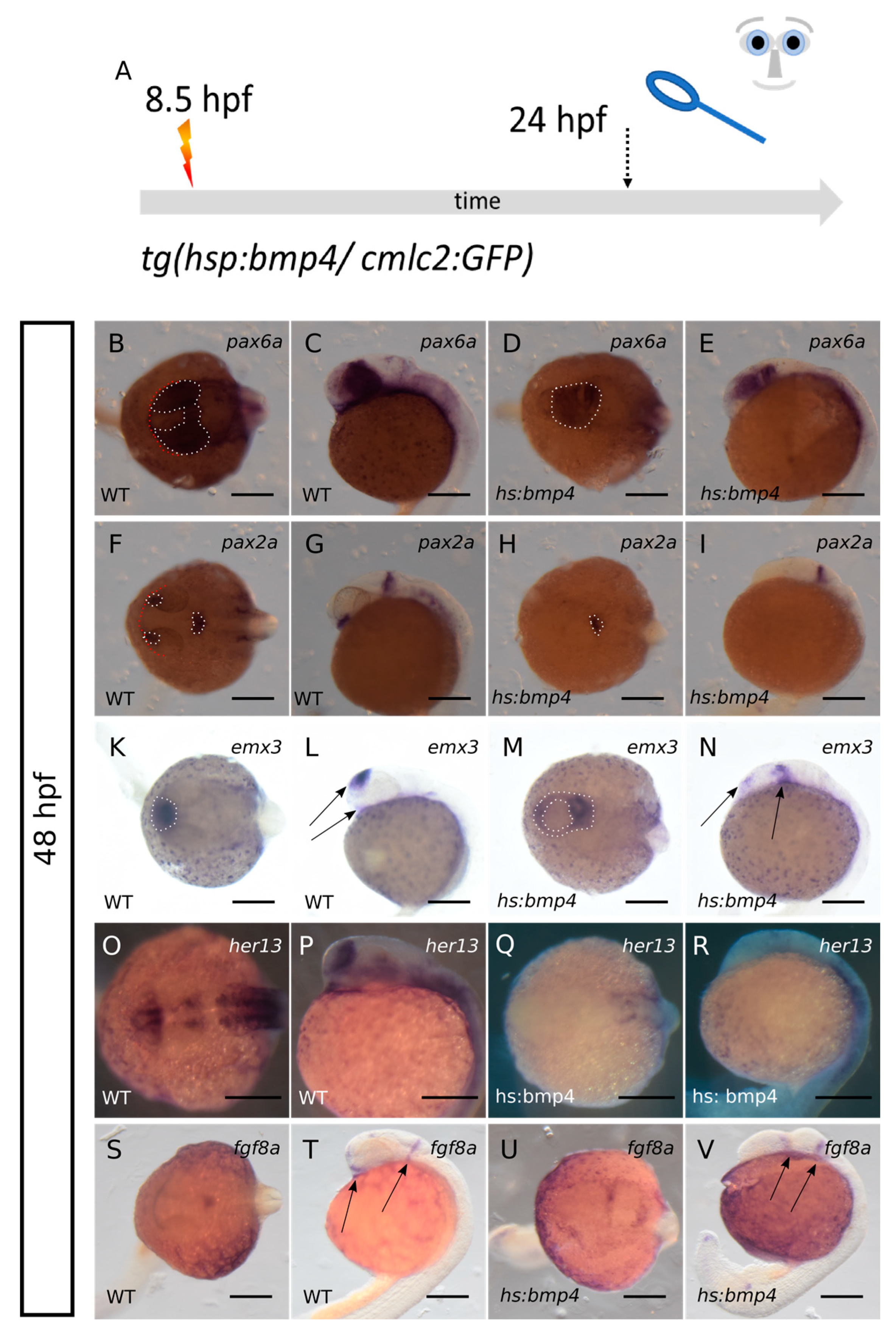
2.2. ANP Development Is Severely Hampered after bmp4 Induction at 8.5 hpf

Having observed that bmp4 induction hampers eye field splitting, we next addressed the effect of bmp4 induction on the development of the ANP. To this end, we induced bmp4 by heat shock at 8.5 hpf using tg(hsp70l:bmp4, cmlc2:GFP) fish mated with wildtype zebrafish and fixed the embryos at 11 hpf. Consecutively, we processed the embryos for WMISH (Figure 2A). We used markers for the prospective forebrain (emx3 and foxg1) domain and the prospective eye field (six3b and rx3). The transgenesis marker (cmlc2:GFP) was not yet active at 11 hpf. Thus, the WMISH was performed blinded and the genotype (wt vs. hsp70l:bmp4, cmlc2:GFP) was determined after the acquisition of the expression patterns. After genotyping the recorded expression patterns were grouped in “control” and “bmp4-induced” samples (Figure 2, wt vs. hs:bmp4). Based on the literature it was expected that an excess of a BMP ligand is potentially resulting in an expansion of the prospective telencephalic domain at the expense of the eye field [3]. Nevertheless, in our paradigm we could not detect a shift in fate between these two domains. We rather found that, resulting from the induction of bmp4, the forebrain domain (emx3) and the eye field (six3b) were both condensed at the midline (Figure 2B–E arrows, Figure 2K–N/4; bmp4-induced embryos and four control embryos were analyzed, respectively). This indicates that the induction of bmp4 was hampering the division of both the eye field and the prospective forebrain domain, being the hallmark of HPE. Interestingly, the expression of rx3 and foxg1a were ceased/lost after bmp4 induction (Figure 2F–I,O–R arrows/4 bmp4-induced embryos and four control embryos were analyzed, respectively). This suggests that either the induction or the maintenance of the expression of these two transcription factors was sensitive to elevated levels of bmp4. Foxg1 is an essential transcription factor involved in many aspects of telencephalic development including also the growth of this brain region [16]. Foxg1 was also shown to be essential for the development of the olfactory system including the olfactory epithelium and also the olfactory bulb, a telencephalic region [17]. A loss of foxg1 expression could thus likely be resulting in reduced size of the telencephalon. A loss of rx3 could be a plausible reason for the anophthalmia phenotype [1,2,3,18].

2.3. Bmp4 Induction Results in Loss of rx3 Expression but Sustained Expression of rx2

We found it surprising, however, that rx3 expression was lost after bmp4 induction while rx2 positive progenitors could still be detected at a later stage of development (Figure 1Y–Bb and Figure 2O–R). In the zebrafish rx3 mutant chokh, it was shown that rx2 expression depends on rx3 expression [18]. In medaka, however, the expression of rx2 is sustained when rx3 is lost [19]. We next targeted the rx3 locus of the zebrafish genome with CRISPR/Cas9 and analyzed F0 Crispants (Figure 3A), using the protocol of Wu and colleagues [20]. The specific sequences chosen for sgRNA preparation are given in the materials and methods section. 118 zygotes were injected. Of these, 59% (70) developed an anophthalmic phenotype. Twenty embryos (17%) showed normal eyes, while the remaining embryos showed differential ocular phenotypes such as bilateral microphthalmia (16), a single lateral eye (six), dysmorphic eyes (three) and unilateral microphthalmia (three). Thus, we state that we reproducibly observed anophthalmia and severe microphthalmia in our F0 Crispants (Figure 3B–E). We next analyzed the rx2 expression in F0 rx3 Crispants by WMISH (Figure 3F–I). Notably, we found severely reduced to absent expression levels of rx2 in our F0 rx3 Crispants (strong phenotype), correlating nicely with the efficacy of the CRISPR-induced anophthalmia phenotype (nine embryos). This supports the finding that rx2 is dependent on rx3 expression in zebrafish. However, this finding does not explain why rx3 is absent after bmp4 induction, while rx2 expression is present afterwards (Figure 1Aa,Bb and Figure 2Q,R). Overall, our findings show two different forms of anophthalmia. We will discuss this alleged contradiction between these below.

2.4. Bmp4 Induction Alters Expression of zic2a, shha/b, alcamb and cxcr4a

Next, we addressed factors influencing forebrain and eye field splitting. We performed this analysis at 11 hpf, subsequent to a heat shock at 8.5 hpf, on embryos derived from mating tg(hsp70l:bmp4, cmlc2:GFP) with wildtype zebrafish and the experimental design as aforementioned. The most prominent cause of HPE is the loss of Shh secreted from the prechordal plate. We thus addressed the expression of shha/b in controls and bmp4-induced embryos (Figure 4A–I). In controls, we found shha (four embryos) and shhb (three embryos) expressed in the axial mesendoderm (Figure 4B,C,F,G). The expression of shha/b was reaching far to the anterior end of the embryo, where the domain was broadened and showing strong expression, (arrows in Figure 4B,C,F,G) corresponding to the prechordal plate. After induction of bmp4, we found the expression of both shha (11 embryos) and shhb (nine embryos with strong repression, two embryos with wildtype-like expression) variably reduced (Figure 4D,E,H,I), yet not absent. Notably, the anterior domain of shha was less broad and less intense, suggesting an affected prechordal plate (Figure 4D,E arrows).
The transcription factor zic2 also is an established HPE-related gene. If mutated it hampers prechordal plate development [21] and thus results in HPE. Later in development, zic2 was, however, also suggested to act downstream of Shh, limiting the expression of six3 in the developing forebrain [22]. We thus next addressed zic2a expression in controls and after bmp4 induction (Figure 4K–N). While in controls (four embryos) zic2a is expressed at the border of the ANP among other domains, we found zic2a almost absent in this region after bmp4 induction (five embryos) (Figure 4M,N).
Next, we addressed the expression of the chemokine receptor cxcr4a, which was shown to be regulated by rx3 [3] and, in combination with sdf1b from the underlying mesendoderm, is supposed to be important for eye field evagination. We found a strongly reduced level of cxcr4a expression in the eye field after induction of bmp4 (four embryos) compared to controls (four embryos) (Figure 4O–R). This is in line with previous findings [3] and supporting the idea that the loss of rx3 and consecutively the loss of cxcr4a is a reason for anophthalmia resulting from induction of bmp4.

2.5. Bmp4 Induction Hampers Hypothalamic Subduction during Neurulation and Neural Keel Formation

As abovementioned, a proper interaction of the prechordal plate and the ANP via Shh is crucial for normal forebrain cleavage [23,24]. Consecutively, during normal neurulation a part of the future hypothalamic domain is subducting and moving in an anterior direction underneath the rest of the ANP [4]. This is an important aspect of neural keel formation which is also increasing the height along the dorso-ventral axis of the head.
We addressed the arrangement of the forebrain at 24 hpf by expression of marker genes for the telencephalic precursors, retinal precursors and hypothalamic precursors, thereby aiming at understanding the effect of an induction of bmp4 on the hypothalamic subduction during early forebrain development. To this end, we subjected embryos of tg(hsp70l:bmp4, cmlc2:GFP) crossed to wildtype zebrafish to a heat shock at 8.5 hpf. At 24 hpf the embryos were sorted in cmlc2:GFP-negative (wildtype/controls) and cmlc2:GFP-positive (bmp4-induced). Consecutively, the embryos were fixed and processed for WMISH.
First, we addressed the expression of pax6a and pax2a, two important transcription factors for eye development. Pax2 is important for proximal fates and optic stalk development and pax6 is important for optic vesicle/cup development [25] (Figure 5B–I). While in controls (six embryos) we found pax6a expressed in the optic cups and diencephalon, we found it expressed in the domain of the crypt-oculoid and a domain posterior to this, likely corresponding to the diencephalic domain, after bmp4 induction (six embryos) (Figure 5B–E, dotted lines). Pax2a expression was detected in the optic stalk and the midbrain-hindbrain boundary in controls (six embryos) (Figure 5F,G, dotted lines). After bmp4 induction, the expression in the midbrain hindbrain boundary was still visible, while optic stalks could not be identified (five embryos) (dotted line Figure 5H,I). Together, our data indicate that the induction of bmp4 results in a failure of eye field separation and optic vesicle out-pocketing. It also showed that even though retinal precursors (positive for rx2 and pax6a) were present, no eye was formed.
Next, we addressed expression of emx3 at 24 hpf. Emx3 is a marker for the prospective telencephalon at early stages (11 hpf), but is also expressed within the diencephalon/hypothalamus at later embryonic stages [26,27]. In controls (five embryos) emx3 mainly shows expression in the developing telencephalon and mild expression in the anterior ventral diencephalon (Figure 5K,L dotted line and arrows). After bmp4 induction (five embryos), emx3 is broadly expressed caudally to the crypt-oculoid. Compared to the control, only a small emx3-positive domain is visible in the residual anterior region, also entangling the crypt-oculoid (Figure 5M,N dotted line and arrows).
The localization of the vast emx3-positive domain posterior to the crypt-oculoid suggests a failure of the hypothalamic subduction movement. Moreover, the height of the dorso-ventral axis is reduced after bmp4 induction resulting in a more linear arrangement of the forebrain domains. The vast emx3-expressing domain posterior to the crypt-oculoid likely does not solely correspond to the ventral anterior hypothalamic domain in controls. It cannot be ruled out that telencephalic precursors are also misplaced to this domain (Figure 5N right arrow).
We next addressed the expression of other genes marking in part diencephalic/hypothalamic identity. We addressed the expression of her13 and fgf8a (Figure 5O–V). In controls (four embryos) her13 is expressed in telencephalic and anterior ventral diencephalic regions (Figure 5O,P). After bmp4 induction (three embryos), however, her13 expression ceased, especially in anterior regions (Figure 5Q,R). Fgf8a is expressed in the midbrain/hindbrain boundary and in anterior regions, including the ventral anterior diencephalon in controls (five embryos) (Figure 5S,T arrows). After induction of bmp4 (three embryos) the anterior ventral expression is ceased, while two expression domains posterior to the crypt-oculoid could be detected (Figure 5U,V arrows), indicative for a subduction defect of the hypothalamic precursors.
Taken together, induced expression of bmp4 resulted in a linear arrangement of forebrain domains and a reduced dorso-ventral height of the forebrain, and hypothalamic precursors which did not subduct to the anterior ventral diencephalic position, both indicative of a hampered neural keel formation.

3. Discussion

Holoprosencephaly (HPE) is the most frequent developmental forebrain disorder in humans. The incidence in live births is approximately 1/10000. However, the estimated incidence per conception is much higher, 0.4% determined in abortions [28]. The intensity of the HPE phenotype can be variably pronounced as well as the ocular phenotypes observed concomitantly [29]. Cyclopia, anophthalmia but also coloboma can be found [5,29].
In this study (Figure 6, scheme, summary of major findings) we set out to address the role of BMP antagonism during early forebrain and eye development. We found different BMP antagonists (grem2b, chrd, nog2 and fsta) expressed in the region of the ANP at 11 hpf and 12 hpf. We have previously shown that an induction of bmp4 can induce BMP signaling in domains of BMP antagonists [12,13] and halt morphogenetic movements during eye development.
Here, we induced bmp4 at 8.5 hpf and found a severe form of HPE associated with anophthalmia in 97.5% of the cases, indicating a robust experimental paradigm. In mice, a compound knock-out of two BMP antagonists, chordin and noggin, resulted in severe forebrain defects presenting cases of HPE with cyclopia but also more severe cases with aprosencephaly [10]. Even though the phenotypes of the compound mouse mutants and ours, induced by experimental bmp4 induction, were both showing severe HPE, there were also marked differences. Importantly, we found anophthalmia with crypt-oculoids instead of cyclopia.
Our analyses of the ANP at 11 hpf revealed a splitting defect of the eye field and the prospective telencephalon. We found markers for the future telencephalon (emx3) and the eye field (six3b) condensed at the midline. The proper interaction of the prechordal plate mesoderm with the developing prosencephalon, the ANP at this stage, is likely the most important step for normal cleavage of the telencephalic field and the eye field [23]. Most HPE-related genes and also non-genetic risk factors can be linked to this step [30]. Shh is the essential factor secreted from the prechordal plate [24] and in turn is directing correct forebrain development. An upstream regulator for prechordal plate development during mid-gastrulation and thus indirectly also of Shh secretion from this domain was found with ZIC2, analyzed in Zic2 mutants in mice [21]. We found that zic2a expression was ceased in the ANP domain after induction of bmp4. In our paradigm we induce bmp4 at 8.5 hpf, a timepoint later than mid-gastrulation [31]. However, we also found a mild and variable decrease in expression of shha/b in the anterior region, the prechordal plate. We can neither be sure nor rule out the possibility that the loss of zic2a is causing the reduced levels of shha/b in the prechordal plate, even if the onset of bmp4 induction is later than mid-gastrulation. Alternatively, the induction of bmp4 could have affected the expression of zic2a and shha/b independently. Our findings indicate, however, that BMP antagonists are important for zic2a and shha/b induction or maintenance in the ANP region. It was shown before that the expression of six3b was depending on Hh signaling [22]. In our analysis, the expression of six3b was not lost after bmp4 induction, but was condensed at the midline. This suggests that the level of shha/b was sufficiently high for six3b induction. Zic2 was also found to act downstream of Shh to affect six3 expression in the forebrain in zebrafish [22]. This effect was, however, considerably later, at mid-somitogenesis, and influenced the development of the prethalamus [22].
We further found that rx3 and foxg1a expression were lost from the eye field and the future telencephalon, respectively. The loss of foxg1a well explains the growth defects of the telencephalon afterwards, which we noticed at 24 hpf. Astonishing, however, was the finding of a ceased rx3 expression, because at 24 hpf we found a condensed domain of rx2 and pax6a-positive tissue in the dysmorphic forebrain, the crypt-oculoid. In anophthalmic zebrafish rx3 mutants (chokh), rx2 expression was shown to be depending on rx3 [18]. Our own analysis of rx3 Crispants from this study is well in line with this. In the anophthalmic medaka rx3 mutants (eyeless, el), however, rx2 expression could be detected [19]. This difference may be due to the difference in species, but also the nature of the mutation could play an important role. In the zebrafish chokh mutant a point mutation (s399) results in a premature stop and thus in a truncated rx3. In medaka eyeless, it is an intronic insertion that results in a transcriptional repression, which is also temperature-sensitive. In our bmp4 induction paradigm we do not mutate the rx3 locus, but rather negatively regulate the expression of rx3. It is conceivable that rx3 is expressed at a level which is not detectable with WMISH but sufficient to induce rx2 expression in our paradigm and maybe also in the medaka eyeless embryos. In both, the level of rx3 was, nevertheless, not sufficient to facilitate eye field splitting and optic vesicle out-pocketing (Rembold et al., 2006, this study). Irrespectively of the exact level of rx3 expression/repression, we detected a dramatic reduction in cxcr4a expression within the eye field after bmp4 induction. Cxcr4 was show to act downstream of rx3 and together with sdf1b from the surface mesoderm to facilitate the evagination of the eye field [3]. The “loss” of rx3 and subsequently of cxcr4a in our bmp4 induction paradigm, thus, very likely explains the lack of optic vesicle out-pocketing. Bearing in mind also our previous findings [13,15], our data indicate that BMP antagonism is continuously important for morphogenetic movements during optic cup development starting with optic vesicle evagination. This also demonstrates nicely the link between “morphogenetic coloboma” that we observed previously and the HPE phenotype from this analysis.
Moreover, investigating the arrangement of the forebrain at 24 hpf in succession of an induction of bmp4, we found that the dysmorphic forebrain is flatter than in controls and the domains were arranged in a more linear way. This could be explained in part by the failure in optic vesicle out-pocketing. However, we also found that the hypothalamic subduction, normally occurring during neurulation, must have been hampered. The anterior ventral hypothalamic domain did not reach its position, but rather remained posterior to the crypt-oculoid. The subduction movement was shown to be important for eye field separation [4] and our data suggest that BMP antagonism is also important to facilitate this process.

4. Materials and Methods

4.1. Zebrafish Care

Zebrafish were kept in accordance with local animal welfare law and with the permit 35-9185.64/1.1 from Regierungspräsidium Freiburg. Fish were maintained in a constant recirculating system at 28 °C on a 12 h light: 12 h dark cycle. The following transgenic lines were used: tg(hsp70l:bmp4, myl7:eGFP) [14] tg(Ola.rx2:bmp4, myl7:eGFP) [13] tg(BRE-AAVmlp:eGFP) [15,32]. Zebrafish embryos were grown at 28 °C in petri dishes in zebrafish medium, consisting of 0.3 g/L sea salt in deionized water. If melanin-based pigmentation needed to be inhibited for downstream applications, embryos were grown in 0.2 mM phenylthiourea.

4.1.1. Heat Shock Procedures

For induction of heat-shock inducible transgenes, embryos were transferred to 1.5 mL reaction tubes and incubated at 37 °C in a heating block (Eppendorf Thermomixer). The onset and the duration of the respective heat shocks varied details are given in the results.

4.1.2. Laser Scanning Confocal Microscopy

Confocal images were recorded with an inverted TCS SP8 microscope (Leica). Embryos were embedded in 1% low-melting agarose (Roth) in glass-bottom dishes (MatTek). Live embryos were anaesthetized with MS-222 (Tricaine, Sigma-Aldrich) for imaging. Image stacks were recorded with a z-spacing of 3 µm, unless specified otherwise.

4.1.3. Image Processing

Images from microscopy were edited for presentation using ImageJ (Fiji) software [33].

4.1.4. In Situ hybridization

Whole-mount ISH was performed according to an established protocol [34].
The following probes were created cloning free, according to [35]: Chrd
The following primers/probes were used:
Emx3 F
5′-GAAGTGCTTCACGATTGAATC-3′; R 5′-TGAAATGACGTCAATGTCCTC-3′
Fgf8a F:
5′-GACTCATACCTTCACGGTTGAG-3′; R: 5′-TGCGTTTAGTCCGTCTGTTG-3′
Foxg1a F:
5′-ATGTTGGATATGGGAGAAAG-3′; R: 5′-AAGAAATAACTGGTCTGACC-3′
Fsta see Knickmeyer et al. 2018
Grem2b see Knickmeyer et al. 2018
Nog2: For:
5′-ATGGGCAGCATCACCCG-3′ Rev: 5′- TCAGCACGAGCACTTGCA-3′
Chrd: For:
5′-TTGTATGGCAGCAGGCGTAT-3′ Rev: 5′-TTGTATGGCAGCAGGCGTAT-3′
Her13 F:
5′-CCACGCTGCTGAACTTAGAAA-3′; R: 5′-TCATCCAGGTCAGAGCAGAGA-3′
Pax2a see Eckert et al. 2019
Pax6a F:
5′-AGATGGTTGCCAACAGTCAG-3′; R: 5′-GGGACATGTCTGGTTCACTG-3′
Rx2 F:
5′-GCCTCTCCACAGAAAGCTAC-3′; R: 5′-CGATACTAGAACTGCGGTCG-3′
Rx3 F:
5′-ATGAGGCTTGTTGGATCTCAG-3′; R: 5′-ATGAGGCTTGTTGGATCTCAG-3′
Shha see Knickmeyer et al. 2021
Shhb see Knickmeyer et al. 2021
Six3b F:
5′-TTTGGTCGTTGCCCGTAGCACC-3′; R: 5′-CATCGAAATCAGAGTCACTGTC-3′
Zic2a F:
5′-ATTAAGCAAGAGCTCATCTG-3′; R: 5′-AACTGTGGACCGCTGAGGAAG-3′

4.1.5. CRISPR/Cas9 F0 Analysis (Crispants)

Embryos in 1-cell stage were microinjected with 1 µM Cas9 protein (Alt-R S.p. Cas9 Nuclease V3, 1081059, Integrated DNA Technologies) and 1µg/µL sgRNA mix as described [20]. sgRNAs were designed using CCTop (http://crispr.cos.uni-heidelberg.de (accessed on 18 April 2023)) [36]. Sequences for sgRNAs:
rx3 T1:
CCCGGCGTTTCCATATGGAT
rx3 T2:
TGAACGTGGTTCGGTTCCGC
rx3 T3:
CTTCGAGAAGTCGCACTATC
rx3 T4:
GAGATGGGGCCGGTCAACCA

5. Conclusions

Here we show that BMP antagonism is important for different aspects of forebrain and early eye development. We found at least four BMP antagonists expressed in the ANP domain during ANP cleavage. Induced bmp4 expression affects various aspects of ANP development, e.g., expression of shha/b in the prechordal plate, expression of zic2a in the ANP, expression of foxg1a, rx3 and cxcr4a in the ANP, respectively. Moreover, the hypothalamic subduction and neural keel formation was hampered. Together these changes result in anophthalmia with a crypt-oculoid in a dysmorphic forebrain. It will be interesting in future analyses to address further the link between the bmp4 induction, the regulation of shha/b and zic2a and the subduction movement of the hypothalamic domain. Further, it will be interesting to address why the splitting defect of the ANP resulted in anophthalmia with a crypt-oculoid and not in cyclopia.

Supplementary Materials

The following supporting information can be downloaded at: https://www.mdpi.com/article/10.3390/ijms24098052/s1.

Author Contributions

Conceptualization, S.H.; methodology, S.H.; validation, J.B.,V.K., P.M.R. and S.H.; formal analysis, J.B., V.K., P.M.R. and Z.G.A.; investigation, J.B.,V.K., P.M.R., Z.G.A. and S.H.; resources, S.H.; data curation, J.B., V.K. and P.M.R.; writing—original draft preparation, S.H.; writing—review and editing J.B.,V.K., P.M.R. and S.H.; visualization, J.B.,V.K., P.M.R. and S.H.; supervision, S.H.; project administration, S.H.; funding acquisition, S.H. All authors have read and agreed to the published version of the manuscript.

Funding

This research received no external funding. In respect to publishing, we acknowledge support by the Open Access Publication Fund of the University of Freiburg.

Institutional Review Board Statement

The study did not require ethical approval.

Informed Consent Statement

The study did not involve humans.

Data Availability Statement

No additional data is published with this article.

Acknowledgments

We thank all members of the Heermann lab for insightful discussions and Ute Baur for great technical assistance. We want to thank Eleni Roussa and Klaus Unsicker for support of the Heermann lab.

Conflicts of Interest

The authors declare no conflict of interest.

References

  1. Rembold, M.; Loosli, F.; Adams, R.J.; Wittbrodt, J. Individual Cell Migration Serves as the Driving Force for Optic Vesicle Evagination. Science 2006, 313, 1130–1134. [Google Scholar] [CrossRef] [PubMed]
  2. Brown, K.E.; Keller, P.J.; Ramialison, M.; Rembold, M.; Stelzer, E.H.K.; Loosli, F.; Wittbrodt, J. Nlcam Modulates Midline Convergence during Anterior Neural Plate Morphogenesis. Dev. Biol. 2010, 339, 14–25. [Google Scholar] [CrossRef]
  3. Bielen, H.; Houart, C. BMP Signaling Protects Telencephalic Fate by Repressing Eye Identity and Its Cxcr4-Dependent Morphogenesis. Dev. Cell 2012, 23, 812–822. [Google Scholar] [CrossRef] [PubMed]
  4. England, S.J.; Blanchard, G.B.; Mahadevan, L.; Adams, R.J. A Dynamic Fate Map of the Forebrain Shows How Vertebrate Eyes Form and Explains Two Causes of Cyclopia. Development 2006, 133, 4613–4617. [Google Scholar] [CrossRef]
  5. Fallet-Bianco, C. Neuropathology of Holoprosencephaly. Am. J. Med. Genet. C Semin. Med. Genet. 2018, 178, 214–228. [Google Scholar] [CrossRef] [PubMed]
  6. Chiang, C.; Litingtung, Y.; Lee, E.; Young, K.E.; Corden, J.L.; Westphal, H.; Beachy, P.A. Cyclopia and Defective Axial Patterning in Mice Lacking Sonic Hedgehog Gene Function. Nature 1996, 383, 407–413. [Google Scholar] [CrossRef]
  7. Roessler, E.; Belloni, E.; Gaudenz, K.; Jay, P.; Berta, P.; Scherer, S.W.; Tsui, L.C.; Muenke, M. Mutations in the Human Sonic Hedgehog Gene Cause Holoprosencephaly. Nat. Genet. 1996, 14, 357–360. [Google Scholar] [CrossRef]
  8. Houart, C.; Caneparo, L.; Heisenberg, C.; Barth, K.; Take-Uchi, M.; Wilson, S. Establishment of the Telencephalon during Gastrulation by Local Antagonism of Wnt Signaling. Neuron 2002, 35, 255–265. [Google Scholar] [CrossRef]
  9. Cavodeassi, F. Integration of Anterior Neural Plate Patterning and Morphogenesis by the Wnt Signaling Pathway. Dev. Neurobiol. 2014, 74, 759–771. [Google Scholar] [CrossRef]
  10. Bachiller, D.; Klingensmith, J.; Kemp, C.; Belo, J.A.; Anderson, R.M.; May, S.R.; McMahon, J.A.; McMahon, A.P.; Harland, R.M.; Rossant, J.; et al. The Organizer Factors Chordin and Noggin Are Required for Mouse Forebrain Development. Nature 2000, 403, 658–661. [Google Scholar] [CrossRef] [PubMed]
  11. Khokha, M.K.; Yeh, J.; Grammer, T.C.; Harland, R.M. Depletion of Three BMP Antagonists from Spemann’s Organizer Leads to a Catastrophic Loss of Dorsal Structures. Dev. Cell 2005, 8, 401–411. [Google Scholar] [CrossRef] [PubMed]
  12. Eckert, P.; Knickmeyer, M.D.; Schütz, L.; Wittbrodt, J.; Heermann, S. Morphogenesis and Axis Specification Occur in Parallel during Optic Cup and Optic Fissure Formation, Differentially Modulated by BMP and Wnt. Open. Biol. 2019, 9, 180179. [Google Scholar] [CrossRef] [PubMed]
  13. Heermann, S.; Schütz, L.; Lemke, S.; Krieglstein, K.; Wittbrodt, J. Eye Morphogenesis Driven by Epithelial Flow into the Optic Cup Facilitated by Modulation of Bone Morphogenetic Protein. eLife 2015, 4, e05216. [Google Scholar] [CrossRef]
  14. Knickmeyer, M.D.; Mateo, J.L.; Eckert, P.; Roussa, E.; Rahhal, B.; Zuniga, A.; Krieglstein, K.; Wittbrodt, J.; Heermann, S. TGFβ-Facilitated Optic Fissure Fusion and the Role of Bone Morphogenetic Protein Antagonism. Open Biol. 2018, 8, 170134. [Google Scholar] [CrossRef]
  15. Eckert, P.; Knickmeyer, M.D.; Heermann, S. In Vivo Analysis of Optic Fissure Fusion in Zebrafish: Pioneer Cells, Basal Lamina, Hyaloid Vessels, and How Fissure Fusion Is Affected by BMP. Int. J. Mol. Sci. 2020, 21, 2760. [Google Scholar] [CrossRef]
  16. Hettige, N.C.; Ernst, C. FOXG1 Dose in Brain Development. Front. Pediatr. 2019, 7, 482. [Google Scholar] [CrossRef] [PubMed]
  17. Duggan, C.D.; DeMaria, S.; Baudhuin, A.; Stafford, D.; Ngai, J. Foxg1 Is Required for Development of the Vertebrate Olfactory System. J. Neurosci. 2008, 28, 5229–5239. [Google Scholar] [CrossRef] [PubMed]
  18. Loosli, F.; Staub, W.; Finger-Baier, K.C.; Ober, E.A.; Verkade, H.; Wittbrodt, J.; Baier, H. Loss of eyes in zebrafish caused by mutation of chokh/rx3. EMBO Rep. 2003, 4, 894–899. [Google Scholar] [CrossRef]
  19. Loosli, F.; Winkler, S.; Burgtorf, C.; Wurmbach, E.; Ansorge, W.; Henrich, T.; Grabher, C.; Arendt, D.; Carl, M.; Krone, A.; et al. Medaka Eyeless Is the Key Factor Linking Retinal Determination and Eye Growth. Development 2001, 128, 4035–4044. [Google Scholar] [CrossRef]
  20. Wu, R.S.; Lam, I.I.; Clay, H.; Duong, D.N.; Deo, R.C.; Coughlin, S.R. A Rapid Method for Directed Gene Knockout for Screening in G0 Zebrafish. Dev. Cell. 2018, 46, 112–125.e4. [Google Scholar] [CrossRef] [PubMed]
  21. Warr, N.; Powles-Glover, N.; Chappell, A.; Robson, J.; Norris, D.; Arkell, R.M. Zic2 -Associated Holoprosencephaly Is Caused by a Transient Defect in the Organizer Region during Gastrulation. Hum. Mol. Genet. 2008, 17, 2986–2996. [Google Scholar] [CrossRef] [PubMed]
  22. Sanek, N.A.; Taylor, A.A.; Nyholm, M.K.; Grinblat, Y. Zebrafish Zic2a Patterns the Forebrain through Modulation of Hedgehog-Activated Gene Expression. Development 2009, 136, 3791–3800. [Google Scholar] [CrossRef]
  23. Li, H.; Tierney, C.; Wen, L.; Wu, J.Y.; Rao, Y. A Single Morphogenetic Field Gives Rise to Two Retina Primordia under the Influence of the Prechordal Plate. Development 1997, 124, 603–615. [Google Scholar] [CrossRef]
  24. Sagai, T.; Amano, T.; Maeno, A.; Ajima, R.; Shiroishi, T. SHH Signaling Mediated by a Prechordal and Brain Enhancer Controls Forebrain Organization. Proc. Natl. Acad. Sci. USA 2019, 116, 23636–23642. [Google Scholar] [CrossRef]
  25. Chow, R.L.; Lang, R.A. Early Eye Development in Vertebrates. Annu. Rev. Cell Dev. Biol. 2001, 17, 255–296. [Google Scholar] [CrossRef]
  26. Jung, J.; Choi, I.; Ro, H.; Huh, T.-L.; Choe, J.; Rhee, M. march5 Governs the Convergence and Extension Movement for Organization of the Telencephalon and Diencephalon in Zebrafish Embryos. Mol. Cells 2020, 43, 76–85. [Google Scholar] [CrossRef] [PubMed]
  27. Paridaen, J.T.M.L.; Danesin, C.; Elas, A.T.; van de Water, S.; Houart, C.; Zivkovic, D. Apc1 Is Required for Maintenance of Local Brain Organizers and Dorsal Midbrain Survival. Dev. Biol. 2009, 331, 101–112. [Google Scholar] [CrossRef] [PubMed]
  28. Matsunaga, E.; Shiota, K. Holoprosencephaly in Human Embryos: Epidemiologic Studies of 150 Cases. Teratology 1977, 16, 261–272. [Google Scholar] [CrossRef]
  29. Pineda-Alvarez, D.E.; Solomon, B.D.; Roessler, E.; Balog, J.Z.; Hadley, D.W.; Zein, W.M.; Hadsall, C.K.; Brooks, B.P.; Muenke, M. A Broad Range of Ophthalmologic Anomalies Is Part of the Holoprosencephaly Spectrum. Am. J. Med. Genet. A 2011, 155A, 2713–2720. [Google Scholar] [CrossRef]
  30. Roessler, E.; Hu, P.; Muenke, M. Holoprosencephaly in the Genomics Era. Am. J. Med. Genet. C Semin. Med. Genet. 2018, 178, 165–174. [Google Scholar] [CrossRef]
  31. Kimmel, C.B.; Ballard, W.W.; Kimmel, S.R.; Ullmann, B.; Schilling, T.F. Stages of Embryonic Development of the Zebrafish. Dev. Dyn. 1995, 203, 253–310. [Google Scholar] [CrossRef] [PubMed]
  32. Collery, R.F.; Link, B.A. Dynamic Smad-Mediated BMP Signaling Revealed through Transgenic Zebrafish. Dev. Dyn. 2011, 240, 712–722. [Google Scholar] [CrossRef]
  33. Schindelin, J.; Arganda-Carreras, I.; Frise, E.; Kaynig, V.; Longair, M.; Pietzsch, T.; Preibisch, S.; Rueden, C.; Saalfeld, S.; Schmid, B.; et al. Fiji: An Open-Source Platform for Biological-Image Analysis. Nat. Methods 2012, 9, 676–682. [Google Scholar] [CrossRef] [PubMed]
  34. Quiring, R.; Wittbrodt, B.; Henrich, T.; Ramialison, M.; Burgtorf, C.; Lehrach, H.; Wittbrodt, J. Large-Scale Expression Screening by Automated Whole-Mount in Situ Hybridization. Mech. Dev. 2004, 121, 971–976. [Google Scholar] [CrossRef] [PubMed]
  35. Hua, R.; Yu, S.; Liu, M.; Li, H. A PCR-Based Method for RNA Probes and Applications in Neuroscience. Front. Neurosci. 2018, 12, 266. [Google Scholar] [CrossRef]
  36. Stemmer, M.; Thumberger, T.; del Sol Keyer, M.; Wittbrodt, J.; Mateo, J.L. CCTop: An Intuitive, Flexible and Reliable CRISPR/Cas9 Target Prediction Tool. PLoS ONE 2015, 10, e0124633. [Google Scholar] [CrossRef]
Figure 1. (AQ): expression of bmp-antagonists nog2, chrd, fsta and grem2b in the ANP at 11 and 12 hpf. (AD): ISH for nog2. (A,B): 11 hpf; (C,D): 12 hpf. (EH): ISH for chrd. (E,F): 11 hpf; (G,H): 12 hpf. (IM): ISH for fsta. (I,K): 11 hpf; (L,M): 12 hpf. (NQ): ISH for grem2b. (N,O): 11 hpf; (P,Q): 12 hpf. (A,C,E,G,I,L,N,P): dorsal view. (B,D,F,H,K,M,O,Q): lateral view. Scalebars indicate 200 µm. (R): summary of experimental procedure. (SBb): Embryos were heat shocked at 8.5 hpf and observed at 24 hpf or 48 hpf, resp. (S,T): bright-field image of wildtype at 48 hpf. (U,V): bmp4-induced larva at 48 hpf. (S,U): dorsal view. (T,V): lateral view. Scalebars indicate 250 µm. (W,X): transversal confocal sections of larvae with rx2:GFP at 24 hpf. injection of LY-tdTomato mRNA in zygote. (W): wildtype. (X): bmp4-induced, scalebars indicate 50 µm. (YBb): ISH for rx2 at 24 hpf. (Y,Z): wildtype. (Aa,Bb): bmp4-induced. (Y,Aa): dorsal view; (Z,Bb): lateral view. Scalebars indicate 250 µm.
Ijms 24 08052 g001
Figure 2. ISH at 11 hpf after bmp4-induction at 8.5 hpf. left columns wildtypes, right columns bmp4-induced. first and third column dorsal view, second and fourth column lateral view. (A): summary of experimental procedure. Embryos were heat-shocked at 8.5 hpf and analyzed at 11 hpf. (BE): Expression of emx3 is condensed at the midline after bmp4-induction (arrows). (FI): the foxg1a domain in the forebrain (arrow) is lost after bmp4-induction. (KN): expression of eye-field marker six3b is also condensed at the midline after bmp4-induction. (OR): expression of eye-field marker rx3 (arrow) is lost after bmp4-induction. Scalebars indicate 200 µm.
Ijms 24 08052 g002
Figure 3. (A): summary of experimental procedure: embryos were injected at 1-cell-stage and analyzed at 24 hpf or 48 hpf. (BE): Brightfield-images at 48 hpf. We observed anophthalmia in F0 rx3 Crispants. (FI): ISH for rx2 at 24 hpf. The expression of rx2 is absent in F0 rx3 Crispants at 24 hpf, correlating with the severity of the phenotype. (B,C,F,G): wildtype. (D,E,H,I): rx3-crispant. (B,D,F,H) dorsal view. (C,E,G,I) lateral view. scalebars indicate 250 µm.
Ijms 24 08052 g003
Figure 4. ISH at 11 hpf after bmp4-induction at 8.5 hpf. (A): summary of experimental procedure: embryos were heat-shocked at 8.5 hpf and analyzed at 11 hpf; left columns wildtypes, right columns bmp4-induced;first and third column dorsal view, second and fourth column lateral view. (BE): Expression of shha is reduced in the area of the prechordal plate (arrows) after bmp4-induction yet still present. (FI): shhb expression is also still present after bmp4-induction but reduced in intensity (arrows). (KN): Expression of zic2a is absent in the ANP domain (arrows) after bmp4-induction. (OR): expression of cxcr4a in the eye field is strongly reduced after bmp4-induction. scalebars indicate 200 µm.
Ijms 24 08052 g004
Figure 5. Analysis of marker genes of telencephalic, retinal and hypothalamic precursors after bmp4-induction. (A): summary of experimental procedure: embryos were heat-shocked at 8.5 hpf and analyzed at 24 hpf. Left columns wildtypes, right columns bmp4-induced embryos. First and third column dorsal view, second and fourth column lateral view. (BE): ISH for pax6a at 24 hpf. Pax6a (dotted lines) is expressed in the crypt-oculoid and in the diencephalic domain after bmp4-induction. (FI): ISH for pax2a (dotted lines) at 24 hpf. The expression of pax2a in the MHB is conserved after bmp4-induction but the optic stalk domain is lost. (KN): ISH for emx3 at 24 hpf. Emx3 is present after bmp4-induction but the expression pattern is changed (arrows). (OR): ISH for her13 at 24 hpf. The expression of her13 is lost in anterior regions after bmp4-induction. (SV): ISH for fgf8a at 24 hpf. Two expression domains posterior to the crypt-oculoid are visible after bmp4-induction (arrows). The ventral expression domain (left arrow in T) is ceased after bmp4-induction. Scalebars indicate 200 µm.
Ijms 24 08052 g005
Figure 6. Scheme, summary of major findings. (A): Experimental induction of bmp4 results in a suppression of rx3 and shha/b with a consecutive suppression of cxcr4a and zic2a. The ANP is not splitting in a proper manner, resulting in a crypt-oculoid. Six3b and emx3 are found condensed at the midline. (B): scheme showing the pathological development of the ANP resulting from bmp4 induction, including a defect in the hypothalamic subduction. (C): brief summary of findings.
Ijms 24 08052 g006
Disclaimer/Publisher’s Note: The statements, opinions and data contained in all publications are solely those of the individual author(s) and contributor(s) and not of MDPI and/or the editor(s). MDPI and/or the editor(s) disclaim responsibility for any injury to people or property resulting from any ideas, methods, instructions or products referred to in the content.

