
Synthesis and Characterisation of Fluorescent Novel Pt(II) Cyclometallated Complexes with Anticancer Activity

 , , , and
Abstract
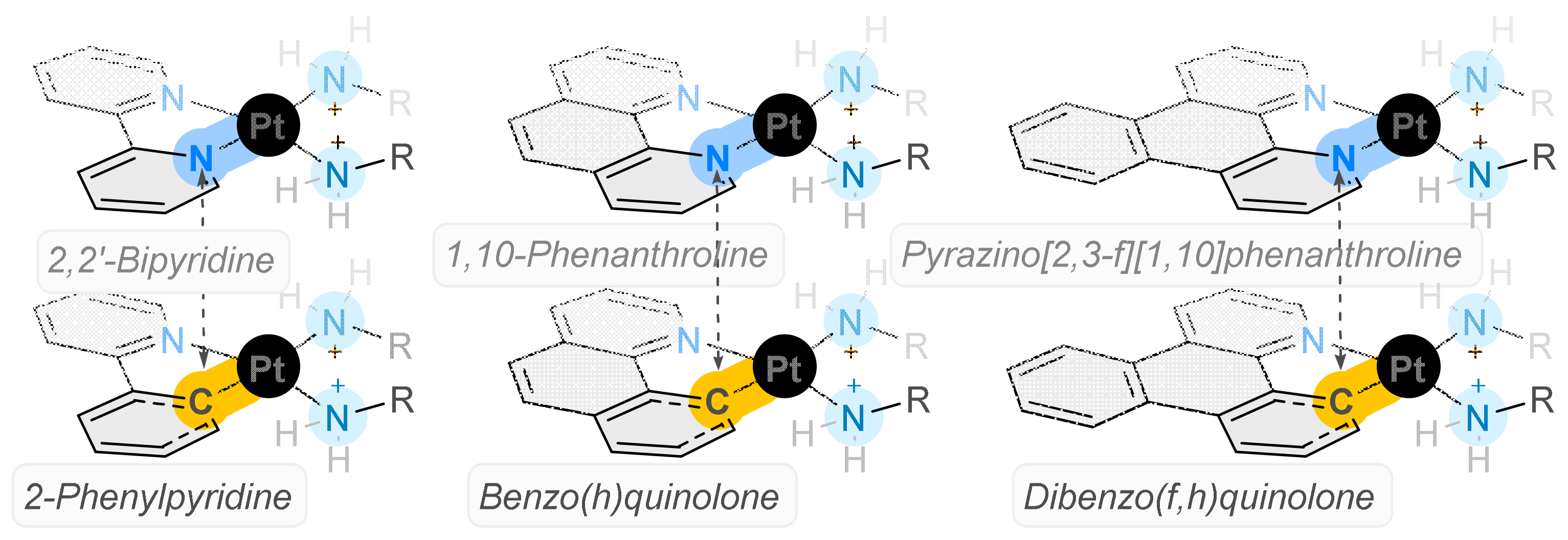
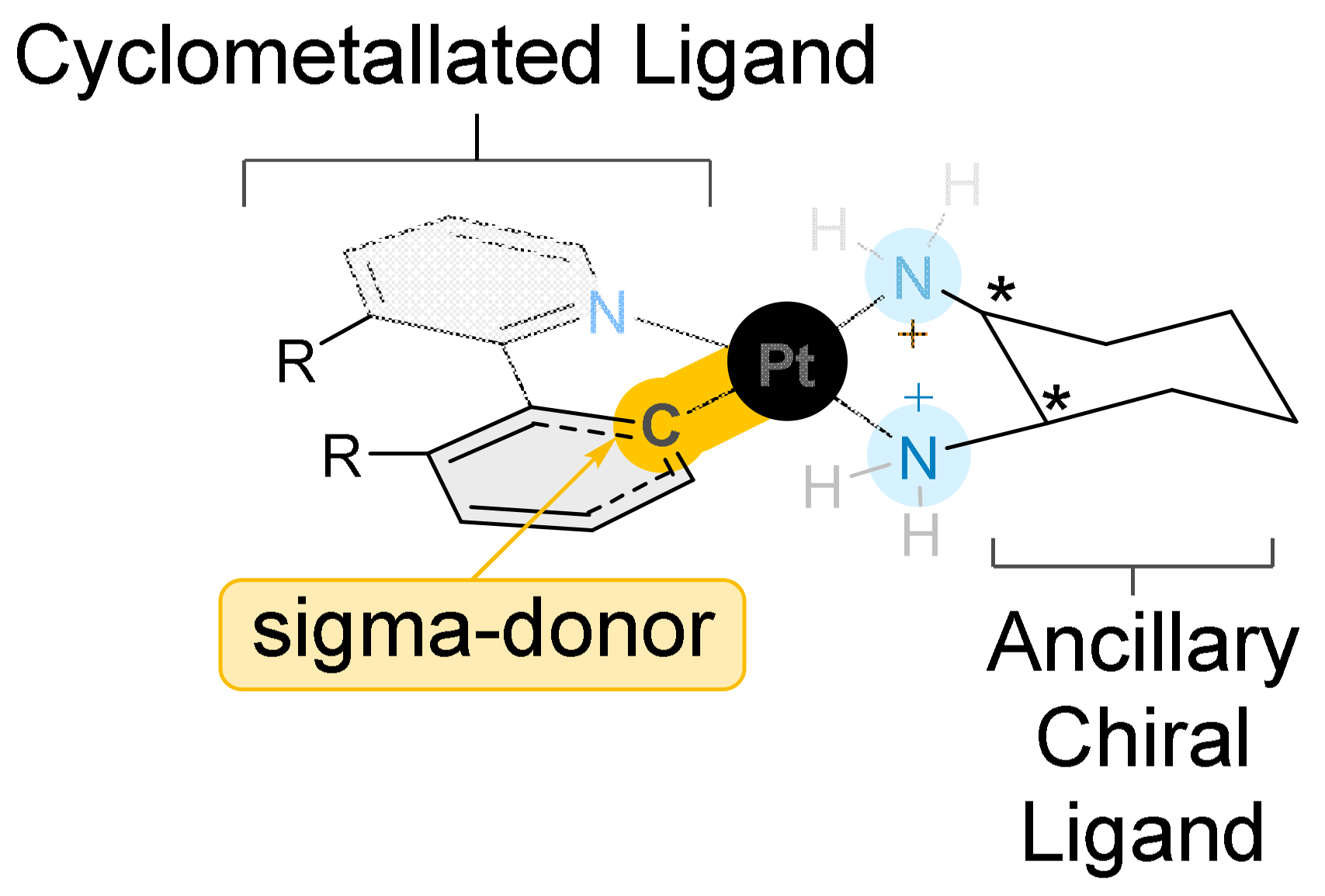
:1. Introduction
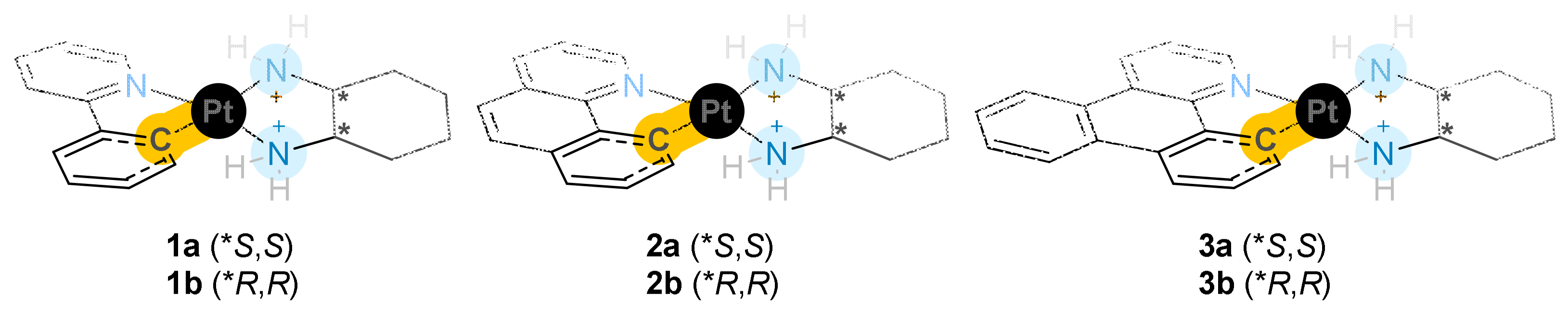
2. Results and Discussion
2.1. Synthesis and Characterisation
2.2. Biophysical Characterisation
2.3. CD ctDNA Experiments
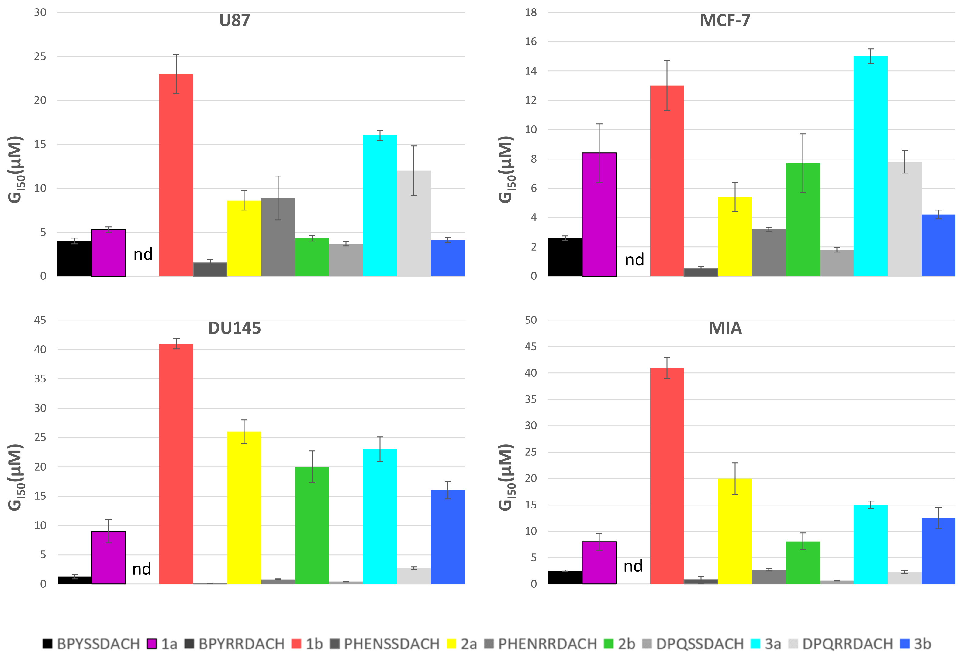
2.4. Cytotoxicity
3. Materials and Methods
3.1. Materials and Preparation
3.2. Synthesis
3.2.1. Synthesis of [Pt(CL)(Cl)2]−
3.2.2. Synthesis of [Pt(DACH)(CL)]+ Complexes
3.3. Cytotoxicity Methodology
3.4. Biophysical Characterisation
4. Conclusions
5. Patents
Supplementary Materials
Author Contributions
Funding
Institutional Review Board Statement
Informed Consent Statement
Data Availability Statement
Acknowledgments
Conflicts of Interest
References
- Sung, H.; Ferlay, J.; Siegel, R.; Laversanne, M.; Soerjomataram, I.; Jemal, A.; Bray, F. Global Cancer Statistics 2020: GLOBOCAN Estimates of Incidence and Mortality Worldwide for 36 Cancers in 185 Countries. CA Cancer J. Clin. 2021, 71, 209–249. [Google Scholar] [CrossRef]
- Bray, F.; Laversanne, M.; Weiderpass, E.; Soerjomataram, I. The Ever-Increasing Importance of Cancer as a Leading Cause of Premature Death Worldwide. Cancer 2021, 127, 3029–3030. [Google Scholar] [CrossRef] [PubMed]
- Cancer (IARC), T.I.A. for R. on Global Cancer Observatory. Available online: https://gco.iarc.fr/ (accessed on 7 February 2022).
- Wheate, N.J.; Walker, S.; Craig, G.E.; Oun, R. The Status of Platinum Anticancer Drugs in the Clinic and in Clinical Trials. Dalton Trans. 2010, 39, 8113–8127. [Google Scholar] [CrossRef] [Green Version]
- Siddik, Z.H. Cisplatin: Mode of Cytotoxic Action and Molecular Basis of Resistance. Oncogene 2003, 22, 7265–7279. [Google Scholar] [CrossRef] [PubMed] [Green Version]
- Kartalou, M.; Essigmann, J.M. Mechanisms of Resistance to Cisplatin. Mutat. Res. Mol. Mech. Mutagen. 2001, 478, 23–43. [Google Scholar] [CrossRef]
- Deo, K.M.; Ang, D.L.; McGhie, B.; Rajamanickam, A.; Dhiman, A.; Khoury, A.; Holland, J.; Bjelosevic, A.; Pages, B.; Gordon, C.; et al. Platinum Coordination Compounds with Potent Anticancer Activity. Coord. Chem. Rev. 2018, 375, 148–163. [Google Scholar] [CrossRef]
- McGhie, B.S.; Sakoff, J.; Gilbert, J.; Aldrich-Wright, J.R. Synthesis and Characterisation of Platinum(IV) Polypyridyl Complexes with Halide Axial Ligands. Inorg. Chim. Acta 2019, 495, 118964. [Google Scholar] [CrossRef]
- Khoury, A.; Deo, K.M.; Aldrich-Wright, J.R. Recent Advances in Platinum-Based Chemotherapeutics That Exhibit Inhibitory and Targeted Mechanisms of Action. J. Inorg. Biochem. 2020, 207, 111070. [Google Scholar] [CrossRef]
- Deo, K.M.; Sakoff, J.; Gilbert, J.; Zhang, Y.; Wright, J.R.A. Synthesis, Characterisation and Influence of Lipophilicity on Cellular Accumulation and Cytotoxicity of Unconventional Platinum(IV) Prodrugs as Potent Anticancer Agents. Dalton Trans. 2019, 48, 17228–17240. [Google Scholar] [CrossRef]
- Fisher, D.M.; Bednarski, P.J.; Grünert, R.; Turner, P.; Fenton, R.R.; Aldrich-Wright, J.R. Chiral Platinum(II) Metallointercalators with Potent in Vitro Cytotoxic Activity. ChemMedChem 2007, 2, 488–495. [Google Scholar] [CrossRef] [PubMed]
- Kemp, S.; Wheate, N.J.; Buck, D.P.; Nikac, M.; Collins, J.G.; Aldrich-Wright, J.R. The Effect of Ancillary Ligand Chirality and Phenanthroline Functional Group Substitution on the Cytotoxicity of Platinum(II)-Based Metallointercalators. J. Inorg. Biochem. 2007, 101, 1049–1058. [Google Scholar] [CrossRef]
- Pages, B.J.; Zhang, Y.; Li, F.; Sakoff, J.; Gilbert, J.; Aldrich-Wright, J.R. Cytotoxicity and Structural Analyses of 2,2′-Bipyridine-, 4,4′-Dimethyl-2,2′-Bipyridine- and 2-(2′-Pyridyl)Quinoxalineplatinum(II) Complexes. Eur. J. Inorg. Chem. 2015, 2015, 4167–4175. [Google Scholar] [CrossRef]
- McGhie, B. N-Halogen Succinimides: Alternative Oxidants of Platinum Anticancer Agents. Aust. J. Chem. 2018, 71, 397. [Google Scholar] [CrossRef]
- Anderson, C.M.; Martínez, M.; Crespo, M. Cyclometallated Platinum Complexes with Heterocyclic Ligands. J. Organomet. Chem. 2004, 689, 1496–1502. [Google Scholar] [CrossRef]
- Brooks, J.; Babayan, Y.; Lamansky, S.; Djurovich, P.I.; Tsyba, I.; Bau, R.; Thompson, M.E. Synthesis and Characterization of Phosphorescent Cyclometalated Platinum Complexes. Inorg. Chem. 2001, 41, 3055–3066. [Google Scholar] [CrossRef] [PubMed]
- Darani, F.A.; Esmaeilbeig, A.R.; Hosseini, F.N.; Nabavizadeh, S.M. Behavior of the Bischelate Platinum(II) Complexes [Pt(S^N)(C^N)] (S^N = Pyridine-2-Thionate, C^N = 2-Phenylpyridinate or Benzo[h]Quinolate) toward C–I Bond Activation; a DFT Study. Struct. Chem. 2015, 26, 961–969. [Google Scholar] [CrossRef]
- Jamshidi, M.; Yousefi, R.; Nabavizadeh, S.M.; Rashidi, M.; Haghighi, M.G.; Niazi, A.; Moosavi-Movahedi, A.-A. Anticancer Activity and DNA-Binding Properties of Novel Cationic Pt(II) Complexes. Int. J. Biol. Macromol. 2014, 66, 86–96. [Google Scholar] [CrossRef]
- Katkova, S.A.; Sokolova, E.V.; Kinzhalov, M.A. Platinum(II) Bisisocyanide Cyclometallated Complexes: Synthesis, Structure, Photophysical Properties, and Mechanochromic Behavior. Russ. J. Gen. Chem. 2023, 93, 43–55. [Google Scholar] [CrossRef]
- Lázaro, A.; Bosque, R.; Ward, J.S.; Rissanen, K.; Crespo, M.; Rodríguez, L. Toward Near-Infrared Emission in Pt(II)-Cyclometallated Compounds: From Excimers’ Formation to Aggregation-Induced Emission. Inorg. Chem. 2023, 62, 2000–2012. [Google Scholar] [CrossRef]
- Thomas, S.W.; Venkatesan, K.; Müller, P.; Swager, T.M. Dark-Field Oxidative Addition-Based Chemosensing: New Bis-Cyclometalated PtII Complexes and Phosphorescent Detection of Cyanogen Halides. J. Am. Chem. Soc. 2006, 128, 16641–16648. [Google Scholar] [CrossRef]
- Osakada, K. 8.08—Platinum–Carbon σ-Bonded Complexes. In Comprehensive Organometallic Chemistry III; Elsevier: Cambridge, MA, USA, 2007; Volume 8; pp. 445–609. [Google Scholar] [CrossRef]
- Ruiz, J.; Vicente, C.; de Haro, C.; Espinosa, A. Synthesis and Antiproliferative Activity of a C,N-Cycloplatinated(II) Complex with a Potentially Intercalative Anthraquinone Pendant. Inorg. Chem. 2011, 50, 2151–2158. [Google Scholar] [CrossRef]
- Shahsavari, H.R.; Hu, J.; Chamyani, S.; Sakamaki, Y.; Aghakhanpour, R.B.; Salmon, C.; Fereidoonnezhad, M.; Mojaddami, A.; Peyvasteh, P.; Beyzavi, H. Fluorinated Cycloplatinated(II) Complexes Bearing Bisphosphine Ligands as Potent Anticancer Agents. Organometallics 2021, 40, 72–82. [Google Scholar] [CrossRef] [PubMed]
- Cutillas, N.; Yellol, G.S.; de Haro, C.; Vicente, C.; Rodríguez, V.; Ruiz, J. Anticancer Cyclometalated Complexes of Platinum Group Metals and Gold. Coord. Chem. Rev. 2013, 257, 2784–2797. [Google Scholar] [CrossRef]
- Tan, C.-P.; Zhong, Y.-M.; Ji, L.-N.; Mao, Z.-W. Phosphorescent Metal Complexes as Theranostic Anticancer Agents: Combining Imaging and Therapy in a Single Molecule. Chem. Sci. 2021, 12, 2357–2367. [Google Scholar] [CrossRef]
- Qiu, K.; Chen, Y.; Rees, T.W.; Ji, L.; Chao, H. Organelle-Targeting Metal Complexes: From Molecular Design to Bio-Applications. Coord. Chem. Rev. 2019, 378, 66–86. [Google Scholar] [CrossRef]
- Ding, S.; Qiao, X.; Suryadi, J.; Marrs, G.S.; Kucera, G.L.; Bierbach, U. Using Fluorescent Post-Labeling To Probe the Subcellular Localization of DNA-Targeted Platinum Anticancer Agents. Angew. Chem. Int. Ed. 2013, 52, 3350–3354. [Google Scholar] [CrossRef] [Green Version]
- Xu, G.-X.; Mak, E.C.-L.; Lo, K.K.-W. Photofunctional Transition Metal Complexes as Cellular Probes, Bioimaging Reagents and Phototherapeutics. Inorg. Chem. Front. 2021, 8, 4553–4579. [Google Scholar] [CrossRef]
- McGhie, B.S.; Aldrich-Wright, J.R. Photoactive and Luminescent Transition Metal Complexes as Anticancer Agents: A Guiding Light in the Search for New and Improved Cancer Treatments. Biomedicines 2022, 10, 578. [Google Scholar] [CrossRef]
- McGhie, B.S.; Sakoff, J.; Gilbert, J.; Gordon, C.P.; Aldrich-Wright, J.R. Novel Planar Pt(II) Cyclometallated Cytotoxic Complexes with G-Quadruplex Stabilisation and Luminescent Properties. Int. J. Mol. Sci. 2022, 23, 10469. [Google Scholar] [CrossRef]
- Garbutcheon-Singh, K.B.; Myers, S.; Harper, B.W.; Ng, N.S.; Dong, Q.; Xie, C.; Aldrich-Wright, J.R. The Effects of 56MESS on Mitochondrial and Cytoskeletal Proteins and the Cell Cycle in MDCK Cells. Metallomics 2013, 5, 1061–1067. [Google Scholar] [CrossRef]
- Loro, C.; Molteni, L.; Papis, M.; Beccalli, E.M.; Nava, D.; Presti, L.L.; Brenna, S.; Colombo, G.; Foschi, F.; Broggini, G. Direct Synthesis of Fluorescent Oxazolo-Phenoxazines by Copper-Catalyzed/Hypervalent Iodine(III)-Mediated Dimerization/Cyclization of 2-Benzylamino-Phenols. J. Org. Chem. 2022, 87, 1032–1042. [Google Scholar] [CrossRef] [PubMed]
- Eustáquio, R.; Ramalho, J.P.P.; Caldeira, A.T.; Pereira, A. New Red-Shifted 4-Styrylcoumarin Derivatives as Potential Fluorescent Labels for Biomolecules. Molecules 2022, 27, 1461. [Google Scholar] [CrossRef] [PubMed]
- Cisse, L.; Djande, A.; Capo-Chichi, M.; Khonté, A.; Bakhoum, J.-P.; Delattre, F.; Yoda, J.; Saba, A.; Tine, A.; Aaron, J.-J. Quantitative study of the substituent effects on the electronic absorption and fluorescence spectra of coumarins. J. Phys. Org. Chem. 2020, 33, e4014. [Google Scholar] [CrossRef]
- Tavadyan, L.A.; Minasyan, S.H.; Kocharyan, G.H.; Antonyan, A.P.; Sahakyan, V.G.; Parsadanyan, M.A.; Vardevanyan, P.O. Exploring the Interaction of Ethidium Bromide and HOECHST 33258 with DNA by Means of Electrochemical Approach. Biophys. Rev. Lett. 2017, 12, 151–161. [Google Scholar] [CrossRef] [Green Version]
- Tarleton, M.; Gilbert, J.; Robertson, M.J.; McCluskey, A.; Sakoff, J.A. Library Synthesis and Cytotoxicity of a Family of 2-Phenylacrylonitriles and Discovery of an Estrogen Dependent Breast Cancer Lead Compound. MedChemComm. 2011, 2, 31–37. [Google Scholar] [CrossRef]
- Brouwer, A.M. Standards for Photoluminescence Quantum Yield Measurements in Solution (IUPAC Technical Report). Pure Appl. Chem. 2011, 83, 2213–2228. [Google Scholar] [CrossRef] [Green Version]
- Klose, M.H.M.; Theiner, S.; Varbanov, H.P.; Hoefer, D.; Pichler, V.; Galanski, M.; Meier-Menches, S.M.; Keppler, B.K. Development and Validation of Liquid Chromatography-Based Methods to Assess the Lipophilicity of Cytotoxic Platinum(IV) Complexes. Inorganics 2018, 6, 130. [Google Scholar] [CrossRef] [Green Version]
- Reithofer, M.R.; Bytzek, A.K.; Valiahdi, S.M.; Kowol, C.R.; Groessl, M.; Hartinger, C.G.; Jakupec, M.A.; Galanski, M.; Keppler, B.K. Tuning of Lipophilicity and Cytotoxic Potency by Structural Variation of Anticancer Platinum(IV) Complexes. J. Inorg. Biochem. 2011, 105, 46–51. [Google Scholar] [CrossRef]
- Valkó, K. Application of High-Performance Liquid Chromatography Based Measurements of Lipophilicity to Model Biological Distribution. J. Chromatogr. A 2004, 1037, 299–310. [Google Scholar] [CrossRef] [PubMed]


| Complex | Yield (%) | ESI-MS (m/z) | Lipophilicity × 10−2 | UV/λmax (nm) (ε/mol−1·dm3.cm−1) × 102 |
|---|---|---|---|---|
| [M]+ Calc. (Found) | ||||
| 1a | 78.9 | 463.15 (463.1557) | 1.41 ± 0.0049 | 241 (213 ± 1.07), 287 (75 ± 0.42) |
| 2a | 78.32 | 487.15 (487.1436) | 2.06 ± 0.097 | 241 (129 ± 1.71), 272 (173 ± 0.80) |
| 3a | 79.36 | 537.16 (538.1541) | 0.77 ± 0.032 | 256 (216 ± 0.69), 235 (27 ± 0.74) |
| 1b | 74.23 | 463.15 (463.1463) | 1.52 ± 0.063 | 241 (358 ± 1.11), 287 (128 ± 0.36) |
| 2b | 77.95 | 487.15 (487.1436) | 3.97 ± 0.053 | 241 (173 ± 0.71), 272 (53 ± 0.21) |
| 3b | 73.26 | 537.16 (538.1475) | 0.4 ± 0.029 | 256 (303 ± 1.93), 235 (155 ± 1.41) |
| Complex | Exmax | Emmax | ΦF |
|---|---|---|---|
| 1a | 287.96 | 486.00 | 0.160 |
| 2a | 262.03 | 483.03 | 0.218 |
| 3a | 262.03 | 361.07 | 0.176 |
| 1b | 338.00 | 484.84 | 0.103 |
| 2b | 378.00 | 524.02 | 0.187 |
| 3b | 300.00 | 361.07 | 0.315 |
| Complex | Point of Experimental Stoichiometry of Binding | ΔFsat | Ka × 104 | n | Kf | Kq | KSV |
|---|---|---|---|---|---|---|---|
| 1a | 2.84 | 83.82 | 2.75 | 0.778 | 4.35 | 0.329 | 1.988 |
| 1b | 2.65 | 86.38 | 1.58 | 0.757 | 4.23 | 0.328 | 1.957 |
| 2a | 2.52 | 89.29 | 1.58 | 0.592 | 3.25 | 0.241 | 1.339 |
| 2b | 2.47 | 89.01 | 1.59 | 0.668 | 3.78 | 0.271 | 1.662 |
| 3a | 2.38 | 91.83 | 1.07 | 0.579 | 3.18 | 0.237 | 1.342 |
| 3b | 2.62 | 82.38 | 3.98 | 0.757 | 4.22 | 0.297 | 1.727 |
| Ka × 104 | R2 | |
|---|---|---|
| 1a | 2.20 | 0.997 |
| 1b | 1.46 | 0.994 |
| 2a | 1.11 | 0.997 |
| 2b | 1.39 | 0.995 |
| 3a | 1.11 | 0.998 |
| 3b | 3.68 | 0.953 |
| GI50 Concentration (µM) That Inhibits Cell Growth by 50% | ||||||||
|---|---|---|---|---|---|---|---|---|
| 1a | 1b | 2a | 2b | 3a | 3b | Cisplatin | 56MESS | |
| HT29 Colon | 13 ± 1.0 | 41 ± 2.7 | 16 ± 1.3 | 30 ± 2.3 | 16 ± 0.00 | 16 ± 0.3 | 11.3 ± 1.9 | nd |
| U87 Glioblastoma | 5.3 ± 0.3 | 23 ± 2.2 | 8.6 ± 1.1 | 4.3 ± 0.3 | 16 ± 0.6 | 4.1 ± 0.3 | 3.8 ± 1.1 | 0.076 ± 0.014 |
| MCF-7 Breast | 8.4 ± 2.0 | 13 ± 1.7 | 5.4 ± 1.0 | 7.7 ± 2.0 | 15 ± 0.5 | 4.2 ± 0.3 | 6.5 ± 0.8 | 0.050 ± 0.020 |
| A2780 Ovarian | 12 ± 0.7 | 31 ± 2.9 | 18 ± 2.3 | 5.0 ± 0.3 | 12 ± 0.9 | 4.5 ± 0.3 | 1 ± 0.1 | 0.030 ± 0.004 |
| H460 Lung | 5.8 ± 0.4 | 25 ± 2.9 | 11 ± 1.5 | 8.1 ± 1.0 | 16 ± 0.9 | 8.2 ± 2.9 | 0.9 ± 0.2 | 0.037 ± 0.009 |
| A431 Skin | 4.2 ± 0.5 | 29 ± 2.6 | 12 ± 2.4 | 3.4 ± 0.3 | 14 ± 0.6 | 3.9 ± 0.4 | 2.4 ± 0.3 | 0.051 ± 0.021 |
| Du145 Prostate | 9 ± 2.0 | 41 ± 0.9 | 26 ± 2.0 | 20 ± 2.7 | 23 ± 2.1 | 16 ± 1.5 | 1.2 ± 0.1 | 0.007 ± 0.002 |
| BE2-C Neuroblastoma | 34 ± 7.5 | 38 ± 4.6 | 23 ± 3.5 | 48 ± 1.7 | 9 ± 1.3 | 33 ± 1.2 | 1.9 ± 0.2 | 0.10 ± 0.016 |
| SJ-G2 Glioblastoma | 11 ± 0.7 | 20 ± 4.8 | 13 ± 2.2 | 10 ± 2.2 | 17 ± 0.6 | 10.1 ± 2.6 | 0.4 ± 0.1 | 0.074 ± 0.018 |
| MIA Pancreas | 8 ± 1.6 | 41 ± 2.0 | 20 ± 3.0 | 8.1 ± 1.6 | 15 ± 0.7 | 12.5 ± 2.0 | 7.5 ± 1.3 | 0.015 ± 0.002 |
| MCF10A Breast (normal) | 6.4 ± 0.5 | 32 ± 2.7 | 20 ± 1.0 | 10.3 ± 1.9 | 16 ± 0.6 | 8.5 ± 1.4 | nd | 0.032 ± 0.007 |
| ADDP Cis Res Ovarian | 5.1 ± 0.3 | 26 ± 0.6 | 14 ± 0.3 | 19 ± 3.0 | 15 ± 0.00 | 6.9 ± 0.4 | nd | nd |
| SI | 0.76 | 2.46 | 3.70 | 1.33 | 1.07 | 2.02 | nd | 0.64 |
Disclaimer/Publisher’s Note: The statements, opinions and data contained in all publications are solely those of the individual author(s) and contributor(s) and not of MDPI and/or the editor(s). MDPI and/or the editor(s) disclaim responsibility for any injury to people or property resulting from any ideas, methods, instructions or products referred to in the content. |
© 2023 by the authors. Licensee MDPI, Basel, Switzerland. This article is an open access article distributed under the terms and conditions of the Creative Commons Attribution (CC BY) license (https://creativecommons.org/licenses/by/4.0/).
Share and Cite
McGhie, B.S.; Sakoff, J.; Gilbert, J.; Gordon, C.P.; Aldrich-Wright, J.R. Synthesis and Characterisation of Fluorescent Novel Pt(II) Cyclometallated Complexes with Anticancer Activity. Int. J. Mol. Sci. 2023, 24, 8049. https://doi.org/10.3390/ijms24098049
McGhie BS, Sakoff J, Gilbert J, Gordon CP, Aldrich-Wright JR. Synthesis and Characterisation of Fluorescent Novel Pt(II) Cyclometallated Complexes with Anticancer Activity. International Journal of Molecular Sciences. 2023; 24(9):8049. https://doi.org/10.3390/ijms24098049
Chicago/Turabian StyleMcGhie, Brondwyn S., Jennette Sakoff, Jayne Gilbert, Christopher P. Gordon, and Janice R. Aldrich-Wright. 2023. "Synthesis and Characterisation of Fluorescent Novel Pt(II) Cyclometallated Complexes with Anticancer Activity" International Journal of Molecular Sciences 24, no. 9: 8049. https://doi.org/10.3390/ijms24098049