Perspective

Can Neutrophils Prevent Nosocomial Pneumonia after Serious Injury?

by
Kristína Macáková
1,2,
Elzbieta Kaczmarek
1 and
Kiyoshi Itagaki
1,*
1
Department of Surgery, Beth Israel Deaconess Medical Center/Harvard Medical School, Boston, MA 02215, USA
2
Institute of Molecular Biomedicine, Faculty of Medicine, Comenius University, Sasinkova 4, 811 08 Bratislava, Slovakia
*
Author to whom correspondence should be addressed.
Int. J. Mol. Sci. 2023, 24(8), 7627; https://doi.org/10.3390/ijms24087627
Submission received: 22 March 2023 / Revised: 11 April 2023 / Accepted: 18 April 2023 / Published: 21 April 2023
(This article belongs to the Special Issue The Role and Characterization of Neutrophils in Human Diseases)

Abstract

:
Nosocomial pneumonia is a leading cause of critical illness and mortality among seriously injured trauma patients. However, the link between injury and the development of nosocomial pneumonia is still not well recognized. Our work strongly suggests that mitochondrial damage-associated molecular patterns (mtDAMPs), especially mitochondrial formyl peptides (mtFPs) released by tissue injury, play a significant role in developing nosocomial pneumonia after a serious injury. Polymorphonuclear leukocytes (neutrophils, PMN) migrate toward the injury site by detecting mtFPs through formyl peptide receptor 1 (FPR1) to fight/contain bacterial infection and clean up debris. Activation of FPR1 by mtFPs enables PMN to reach the injury site; however, at the same time it leads to homo- and heterologous desensitization/internalization of chemokine receptors. Thus, PMN are not responsive to secondary infections, including those from bacteria-infected lungs. This may enable a progression of bacterial growth in the lungs and nosocomial pneumonia. We propose that the intratracheal application of exogenously isolated PMN may prevent pneumonia coupled with a serious injury.

1. Introduction

1.1. A Serious Risk of the Development of Pneumonia after Trauma

Trauma is the leading cause of death in people under age 45 and the third death cause overall, with nosocomial pneumonia being accountable for morbidity in trauma patients [1,2,3,4]. Thus, preventing nosocomial pneumonia would significantly reduce morbidity, mortality, and hospital costs. Additionally, intubated injured patients suffer pneumonia far more often than uninjured intubated patients do [5]. However, the mechanistic links between injury and nosocomial pneumonia are mostly unknown. Our recent data confirm a strong association between tissue injury and nosocomial pneumonia and suggest a causal relationship with limited PMN migration to the lung under extrapulmonary injury conditions.

1.2. Mitochondrial DAMPs Released from Injured Tissues Predispose to Pneumonia Due to Reduced PMN Recruitment to the Lungs

Mitochondria are widely recognized to originate as bacterial endosymbionts [6]. They are hidden in the cells from immune recognition and produce energy as adenosine triphosphate (ATP). Still, when released from injured tissues, they present bacteria-specific molecular motifs, including N-formylated peptides (mtFPs) and mitochondrial DNA (mtDNA) that can act as DAMPs capable of modulating systemic immune responses as if bacterial infection happened in the body [7,8,9,10,11,12]. Mitochondria contain 13 mtFPs [13], which are similar to N-formyl peptides derived from bacterial proteins and hence may act as powerful PMN chemoattractants via FPR1 [13]. We have shown that exposure to mtFPs originating from clinical injuries renders PMN less sensitive to lung-derived chemokines due to chemokine receptor desensitization/internalization, thus decreasing PMN migration and PMN recruitment to the lungs after bacterial inoculation [7,14,15,16]. These events correlate with attenuated bacterial clearance as shown in the lungs of injured mice [12]. Therefore, activation of PMN FPR1 by mtFPs released from injury sites is a critical factor in the potentiation of post-traumatic pneumonia.

1.3. Novel, Non-Antibiotic, Cell-Based Therapies May Prevent Pneumonia after Trauma

Bacteria are increasingly resistant to available antibiotics, with some strains resistant to most or all available treatments. This includes S. aureus, a common pathogen responsible for pulmonary infections after trauma [17,18,19,20]. Antibiotic-resistant infections may cause severe morbidity and mortality and higher healthcare costs [21]. Moreover, new antibiotics are not expected to be readily available in the near future [22,23]. If one of the defense mechanisms against pneumonia after an injury is missing, such as exhaustion of PMN function due to their partial activation, direct application of functionally active PMN to the airway may have a therapeutic value. Therefore, we propose studying such a novel, cell-based therapy and believe it could become a new form of prevention of nosocomial pneumonia in trauma patients.

1.4. Granulocyte Transfusion

We expect that this is feasible as granulocyte transfusion (iv) has been performed at major hospitals worldwide for neutropenic patients, including cancer patients after chemotherapy [24]. Granulocytes must be used within 24 h after isolation while still viable. They are frequently contaminated with red blood cells (RBS), T-cells, and other cells. Thus, ABO-matched granulocytes are irradiated before transfusion to reduce T-cell content, which could lead to transfusion-associated graft-versus-host disease (TA-GVHD). Irradiation prevents T-cells proliferation after transfusion but does not affect PMN that do not proliferate and die within ~24 h [25].
No human leukocyte antigen (HLA) is examined before transfusion. Thus, repeated PMN transfusions from various donors will decrease the number of PMN in circulation after transfusion due to anti-HLA antibody production from previous donors’ PMN. It also strictly suggests that clinicians weigh the danger of transfusion vs. its benefits to the patients [24,26] since it is not a well-established procedure and can be fatal to the recipients. Thus, if the pure PMN can be isolated and stored, these could be used for granulocyte transfusion that requires more frequent and increased PMN.

2. Our Vision: Application of PMN to Human Lungs Will Prevent/Cure Nosocomial Pneumonia after Injury

2.1. What Kind of PMN Can Be Applied Safely to the Lungs of Seriously Injured Trauma Patients to Prevent Nosocomial Pneumonia?

There are no reports on PMN airway instillation as a clinical treatment. Therefore, the safety of this procedure has to be investigated. We plan to examine what kind of PMN can be used safely. Moreover, we will establish the best methods of PMN isolation and storage before application. If the results are promising, we propose to use PMN as a cell therapy for trauma patients.

2.1.1. Any PMN Isolated from Human Donors Will Work?

Many reports suggest that whole blood transfusion (A, B, O-matched) does not cause any issues for recipients [27,28]. It means that blood cells, including PMN, may cause no negative reaction and that HLA match may not be a hampering factor. Unlike other leukocytes, PMN are short-lived cells [29]. Therefore, after instillation they can kill bacteria in the lungs via phagocytosis and neutrophil extracellular traps (NETs) and die shortly after and phagocytosed by the recipients’ macrophages [30]. Again, high purity is the key to avoiding above-mentioned TA-GVHD. How can we prove that any PMN should work without causing any adverse effects on the recipients? Can PMN be purified in a hospital setting at high purity? Maybe not. If there is a simple system to isolate human PMN from peripheral blood at high purity, these PMN can be applied to patients.

2.1.2. Autologous PMN

The trauma patient’s own PMN could be isolated and re-applied. It is well documented that the number of PMN in trauma patients is higher than in healthy people, depending on injury severity [31]. However, it is also well known that the functions of trauma patients’ PMN are reduced compared to those of healthy volunteers [25]. PMN’s migration, phagocytosis, reactive oxygen generation, and NETs formation are significantly decreased by trauma [32,33,34,35,36,37]. Reversing the trauma patients’ PMN to the functional cells quickly would make them safe to apply to the patients even with limited purity. Again, at this time, there is no system to isolate a high purity of PMN at hospitals.

2.1.3. PMN Modified to Be Negative for HLA

As HLAs are the significant components recognized by recipients’ immune cells, PMN from HLA-disrupted iPS cells could be used for instillation. Our collaborator has established the methods of preparation of such cells [38], and these cells are investigated by our group as potential donor cells. We are evaluating ex vivo functions of PMN developed from HLA(-)iPS cells. We will then examine these PMN in our mouse model of injury and lung infection model to compare to freshly isolated human PMN for the ability to kill bacteria in the lungs after injury.

2.2. How to Store PMN before Applying Them to Trauma Patients

Cryopreservation of Human PMN

PMN are well known to be fragile after freeze–thaw in osmotically stressed hypertonic media [39]. It would be best if we could keep PMN frozen and thaw them when needed. No established method has been reported that shows high viability and normal functions of PMN after freeze–thaw. Recently, the freezing media CryoStor CS10 from Stem Cell Technologies showed very promising results. Frozen human PMN are available in CS10 media from Stem Cell Technologies. This is very promising. We are evaluating the viability and functions of PMN after freeze–thawing in CS10 using our volunteers’ PMN, which are essential for successful PMN storage. Once established, the successful freeze–thaw of a large number of pure HLA(-)PMN will benefit patients who might develop nosocomial pneumonia after injury but also who are neutropenia after chemotherapy [40,41], who are severe congenital neutropenia [42], and who do not have less functional PMN such as elderly people [43]. Thus, the application of PMN can be useful for wide variety of people.

3. Supporting Data

3.1. Mouse Injury and Lung Infection Model

To mimic sterile, typical abdominal extrapulmonary injury in humans, we instilled mtDAMPs intraperitoneally (i.p.). This injury decreased the number of PMN migrating toward secondary infection in the lungs, thus bacterial clearance was reduced compared to control mice [7,14,44].

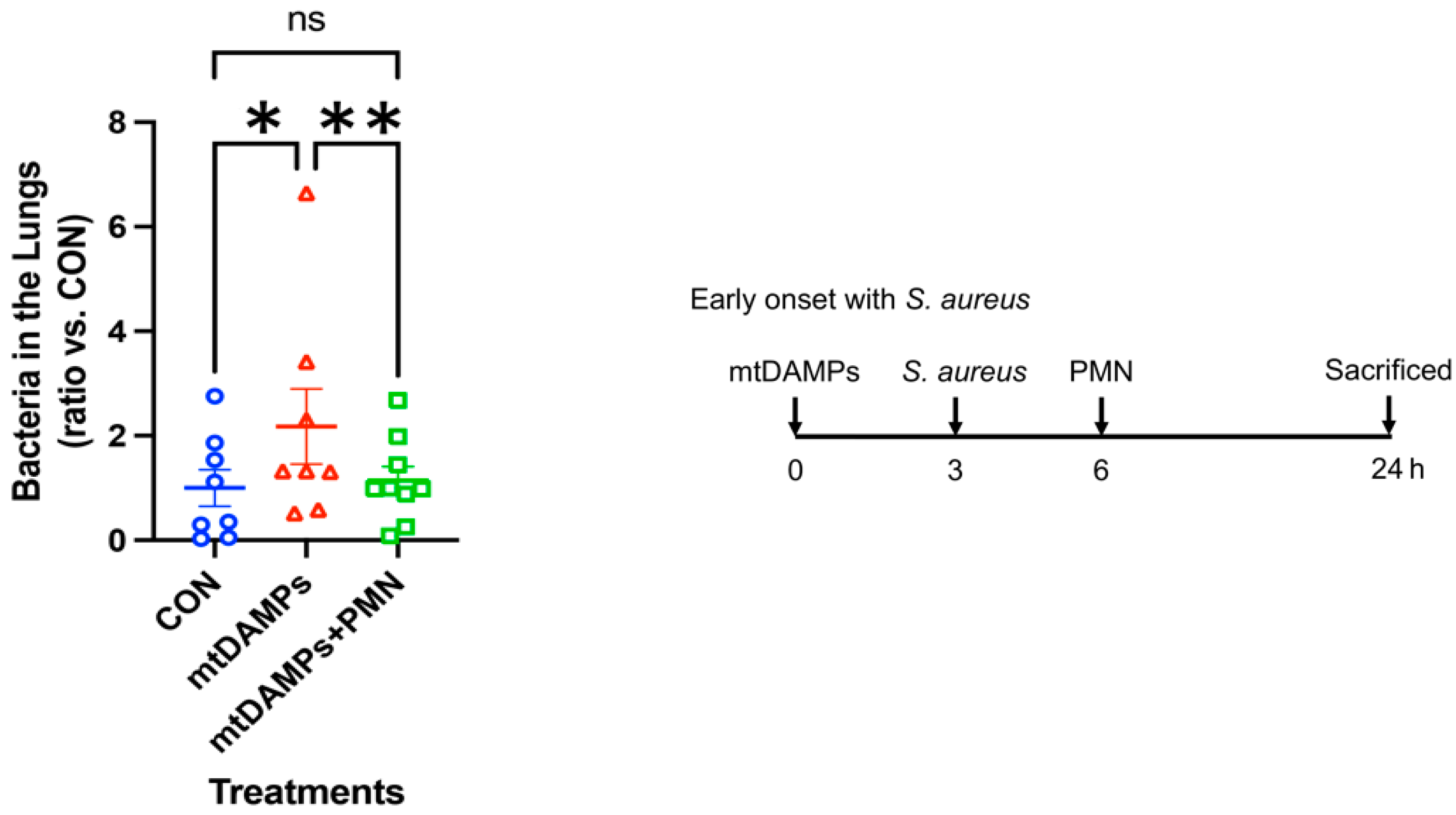
3.2. Prevention of Early Onset of Pneumonia

First, we mimicked the early onset of pneumonia with Staphylococcus aureus (SA). CON (control, non-injury, infected) and two injury groups were injected with mtDAMPs. Then, all mice were infected with SA in the lungs. One injury group received PMN from the same strain of mice to the lungs as a treatment (mtDAMPs + PMN). A day later, lung bacteria were examined compared to CON mice. Injury without treatment mice (mtDAMPs) showed the greatest number of SA in the lungs. CON and injury and PMN-treated mice showed better SA clearance in the lungs. Data suggest that injury reduced SA clearance in the lungs likely due to limited PMN migration toward the lungs. However, exogenous PMN application to the lungs increased bacterial clearance in the lungs (Figure 1) [14].

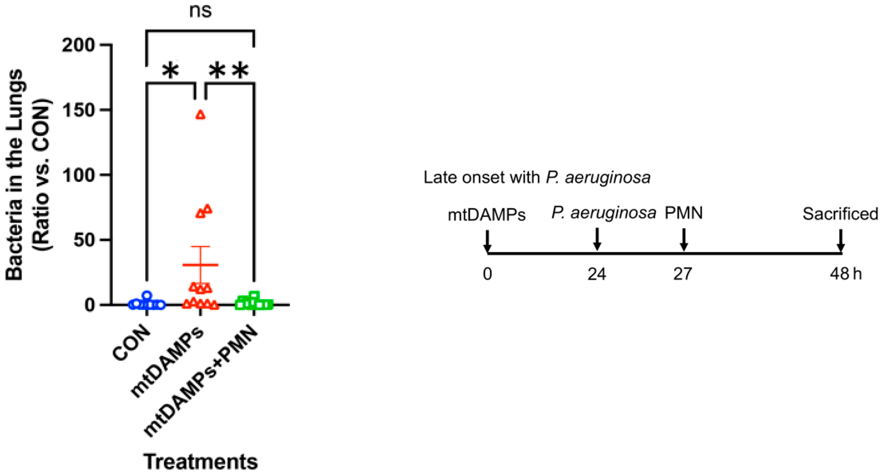
3.3. Prevention of Late-Onset Pneumonia

Next, we mimicked the late onset of pneumonia with Pseudomonas aeruginosa (P. aeruginosa, PA). As shown in Figure 1, animals were divided into three groups, CON, injury, and injury followed by PMN treatment. As a late onset of pneumonia, we infected animals later than above and applied PMN simultaneously after infection. Similar to the early onset of pneumonia, the late onset of pneumonia was significantly reduced by PMN application (Figure 2) [14].

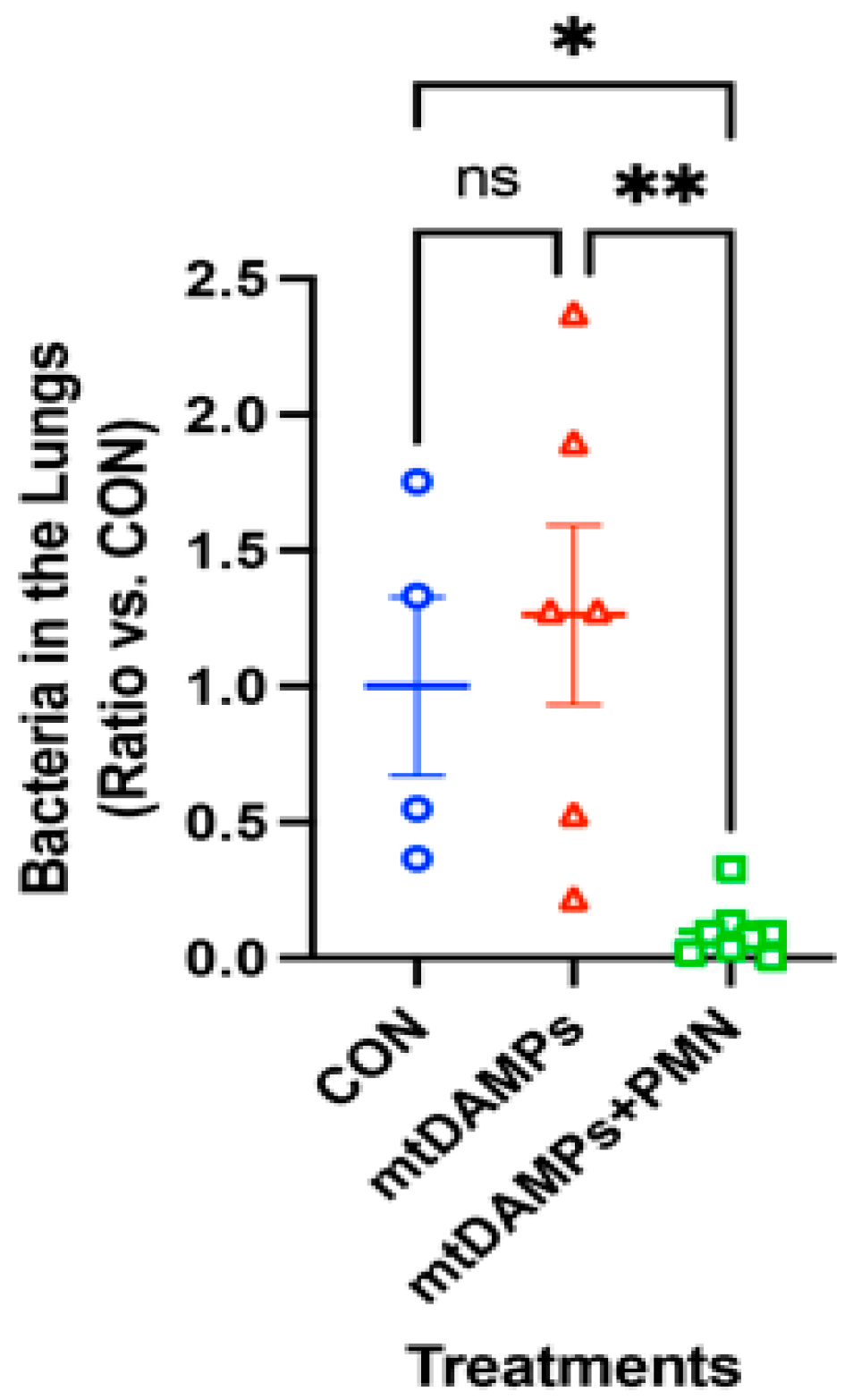
3.4. Prevention of Late Onset of Pneumonia with an Increased Number of P. aeruginosa

We applied an increased number of PA. The rest are the same as in Figure 2. Even the CON group could not clear the bacteria well, so there is no significant difference between CON and injury alone groups. However, the application of PMN (mtDAMPs + PMN) significantly reduced lung bacteria (Figure 3) [14].

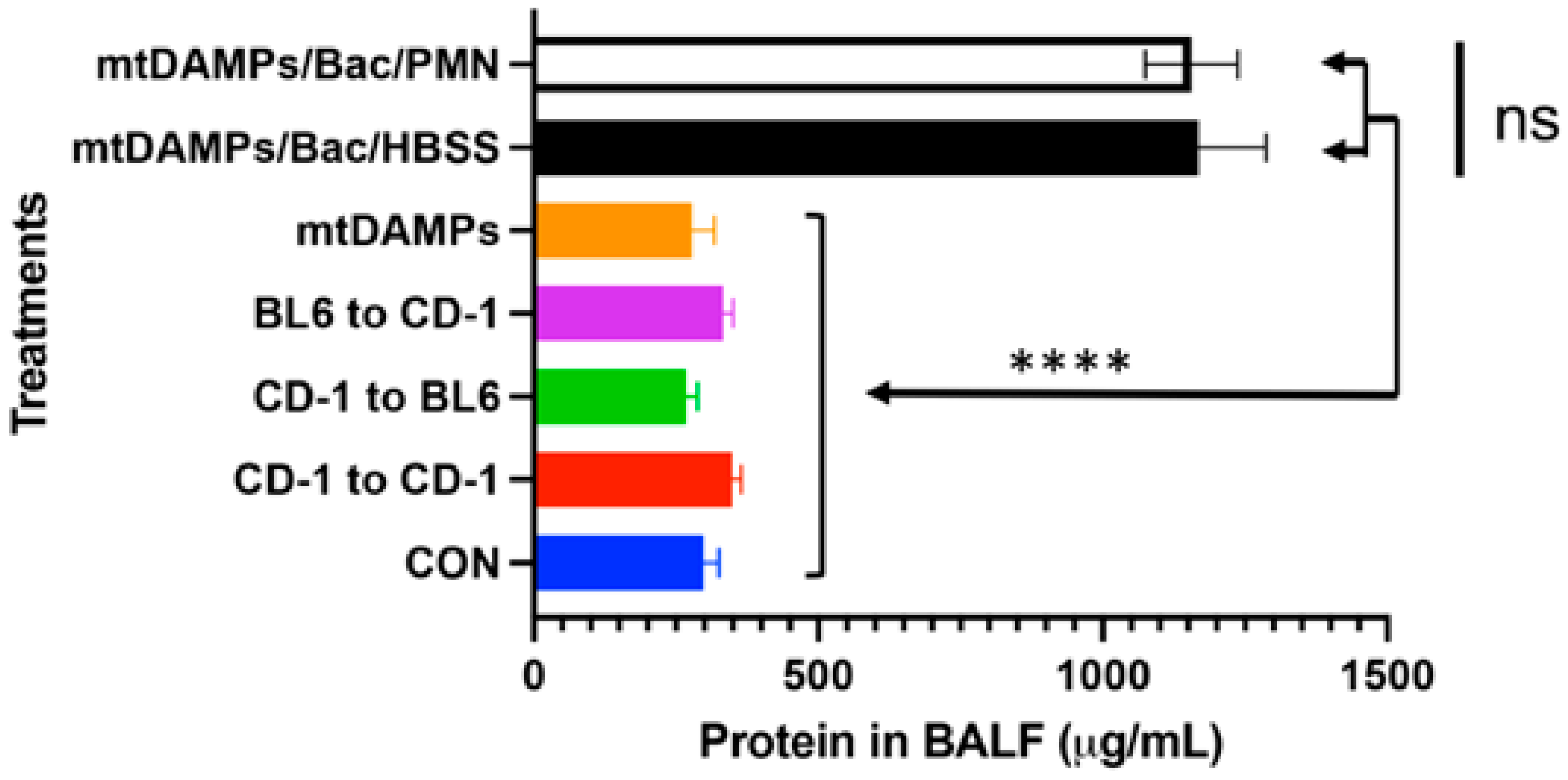
3.5. Evaluation of Potential Lung Injury Due to PMN Instillation

Although PMN application to the lungs effectively cleared secondary bacterial infection to the lungs after injury, we have to confirm the application will not damage the recipients’ lungs. We collected bronchoalveolar lavage fluid (BALF) after various treatments. For example, “CD-1 to BL6” means PMN from CD-1 were applied to the lungs of BL6 mice. Cross-strain application of PMN did not cause lung damage compared to CON mice, and mtDAMPs application did not cause any. mtDAMPs followed by bacteria and PMN or HBSS buffer increased lung damage significantly compared to other groups. However, PMN did not have any effect compared to HBSS. Thus, bacterial infection caused the lung damage detected by protein concentrations in BALF (Figure 4) not PMN application [14].

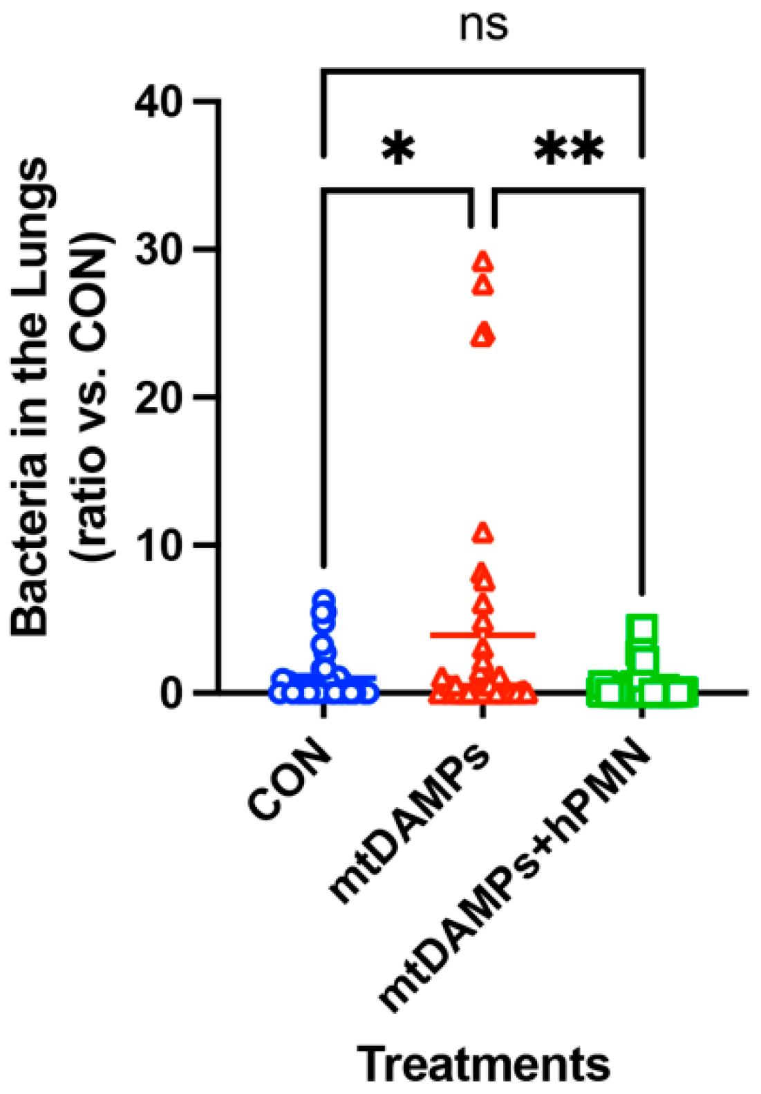
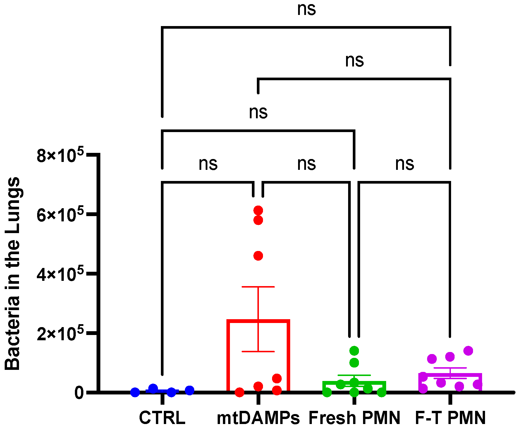
3.6. Human PMN Clears Bacteria in the Lungs without Causing Adverse Effects on the Recipients

Finally, we applied human PMN to our mouse model described in Figure 1 to determine whether human PMN can clear bacteria in the lungs without damaging lungs. CON- and PMN-treated group showed significantly increased bacterial clearance in the lungs (Figure 5) [44]. We also observed mice for up to 28 days after the application of human PMN. We examined the long-term effect of lung damage after applying human PMN via histology with H&E staining, and Trichome and Sirius Red staining to evaluate lung fibrosis. We found no lung damage or lung fibrosis. Additionally, human PMN was detected a day after application but not detected in BALF after 48 h by flow cytometry (CD16(+)/CD49d(-)).

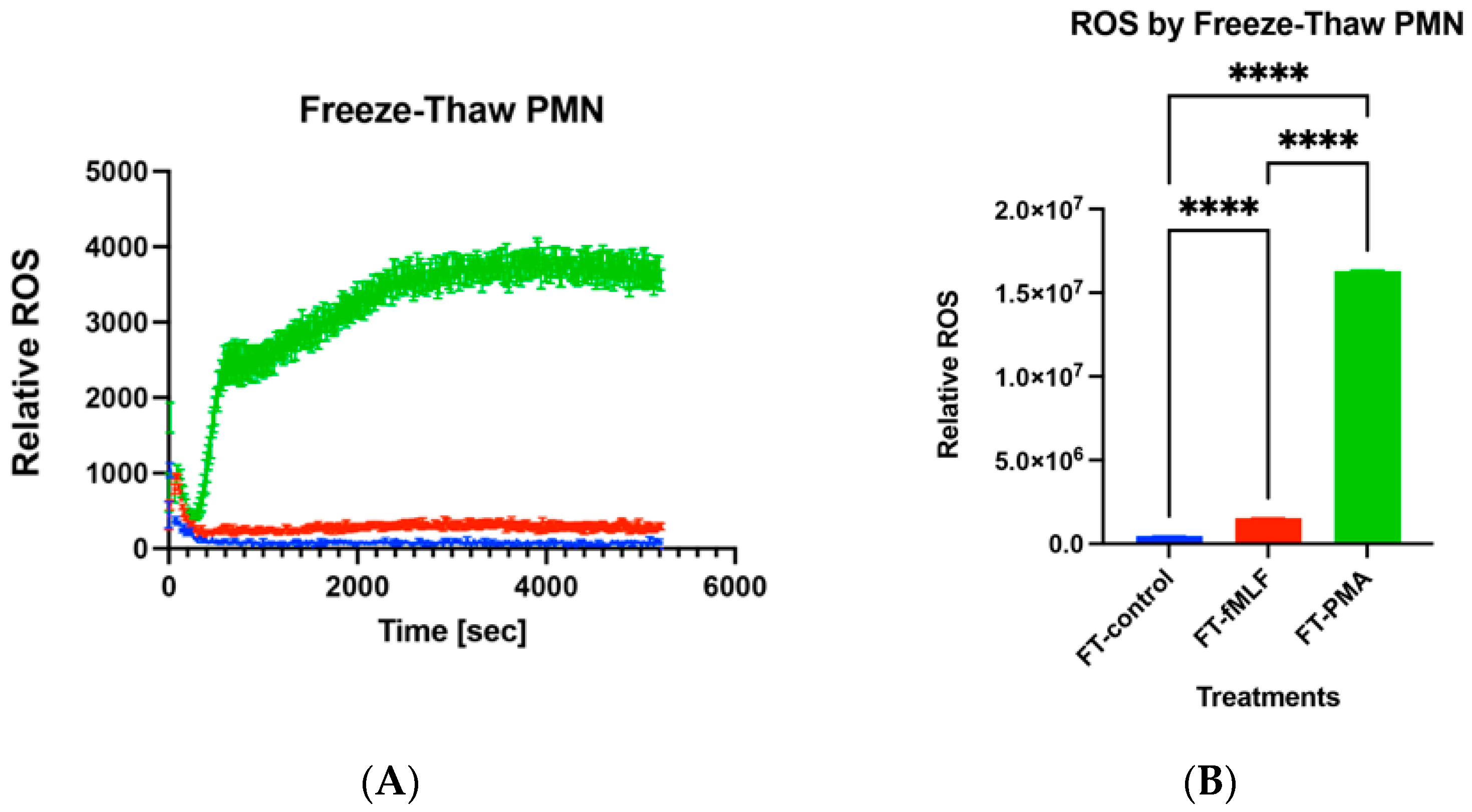
3.7. Functions of Freeze–Thaw PMN

Recently, we found freeze–thaw PMN exhibited reactive oxygen species (ROS) production by stimulation of 100 nM fMLF and 100 nM PMA. Figure 6A shows real-time relative ROS production. Figure 6B shows ROS production by area under curve (AUC) [45]. PMA stimulation induced a lot of ROS production compared to fMLF. Compared to medium, fMLF and PMA induced significantly larger ROS production. ROS production by freeze–thaw PMN was significantly reduced compared to that of freshly isolated PMN. Then, we applied freshly isolated PMN and freeze–thaw PMN to our mouse injury and bacterial infection model described above. Preliminary data suggested that freeze–thaw PMN application enhanced the bacterial clearance in the lung as much as freshly isolated PMN even though there was no significant difference compared to mtDAMPs/bacterial infection group (Figure 7). These are encouraging data that need further examination. So far, the application of human PMN did not cause any damage to the recipient mice. This is not surprising since human PMN are known to have a very short life [29]. They kill bacteria after application and die and then are phagocytosed by residential macrophages. Advancing this method toward possible human use requires study in a large-animal model. Pigs are recognized as an appropriate animal model for studying infectious diseases since their immune system shares many structural and functional similarities with humans [46]. Moreover, pig lungs and airways are anatomically similar to humans [47].

4. Summary

So far, we have shown that the application of mouse PMN of different strains or even human PMN can help clear lung bacteria after injury using our established mouse models without causing any adverse effects on recipients. Up to 28 days after human PMN application, we could not detect any lung injury, including fibrosis, mainly due to the short life of PMN. Though the instillation of exogenous PMN into the lung seems contradictory since the patients with pneumonia already have many PMN in the lungs, these instilled PMN will be activated by bacteria and will damage various tissues, including the lungs. This may not be the case. We believe “the timing is everything”. The lungs of established pneumonia patients have many PMN. The important point is when PMN migrated to the lungs. PMN could not migrate to the lungs when the bacterial infection started due to receptor desensitization as a result of the increased amount of circulating mtDAMPs. PMN could migrate to the lungs only after overwhelming bacterial growth, pneumonia was established, and mtDAMPs in circulation finally decreased. Applying PMN at the early stage of bacterial infection is the key to killing them. No one has ever examined the changes in the number of PMN in the lungs of trauma patients who gradually developed nosocomial pneumonia. We think PMN will not migrate to the lungs after serious injury compared to the uninjured patients. Later, we find many PMN even from trauma patients who developed pneumonia. Recently, Paul Kubes and others suggested that PMN will repair many organs even though they may damage at the beginning when they need to phagocytose cellular debris and bacteria after injury and infections [48]. PMN even contribute to angiogenesis and dying/being phagocytosed by macrophages. Thus, an increasing number of new findings support that additional PMN will do good.

5. More toward Human

It seems that intratracheal application of various types of PMN including human origin did not cause any adverse effects to the recipient mice and effectively cleared bacteria in the lungs. However, the question is, “Is it really so in the case of humans?” PMN live only for a short time, thus the applied PMN will be cleared from the recipients without/before causing any issues. This could be true. Our method is being applied to the pig models under the same concept for the next step. This is an important step toward human use as described before. On the other hand, “Is HLA match an important factor?” PMN without HLA will be the perfect candidates for safe PMN application when they are proven to function similarly to freshly isolated PMN and can maintain functions after freeze–thaw. Once established, these PMN can be used even for neutropenic patients that require many PMN to be infused to prevent infection.

6. Detailed Methods

For readability and nature of this article, we list detailed methods here with references. Here, we list selected papers that will help understand our vision.
Human PMN preparation [49], mouse BM-PMN preparation [50], detailed mouse injury/lung infection models [14,44], mitochondrial DAMPs [7,11,51].

Author Contributions

Conceptualization, K.I.; methodology, K.I., K.M. and E.K.; data curation, K.I., E.K. and K.M.; writing—original draft preparation, K.I.; writing—review and editing, K.I., K.M. and E.K.; visualization, K.I.; supervision. K.I.; project administration, K.I.; funding acquisition, K.I. All authors have read and agreed to the published version of the manuscript.

Funding

National Institute of Allergy and Infectious Diseases (NIAID)/National Institute of Health (NIH). Funding number is 5R03AI135346-02.

Institutional Review Board Statement

Experiments involved in human specimens and animals were approved by our institutional review boards, IRB and IACUC, respectively. The approval numbers are 2019P000243 for human research and 002-2019 for animal research.

Informed Consent Statement

Informed consent was obtained from all subjects involved in the study.

Data Availability Statement

No new data were created. We are happy to share any information to researchers interested in our work.

Conflicts of Interest

The authors declare no conflict of interest.

References

  1. Chevret, S.; Hemmer, M.; Carlet, J.; Langer, M. Incidence and risk factors of pneumonia acquired in intensive care units. Results from a multicenter prospective study on 996 patients. European Cooperative Group on Nosocomial Pneumonia. Intensive Care Med. 1993, 19, 256–264. [Google Scholar]
  2. Papia, G.; Louie, M.; Tralla, A.; Johnson, C.; Collins, V.; Simor, A.E. Screening high-risk patients for methicillin-resistant Staphylococcus aureus on admission to the hospital: Is it cost effective? Infect. Control Hosp. Epidemiol. 1999, 20, 473–477. [Google Scholar] [CrossRef]
  3. Papia, G.; McLellan, B.A.; El-Helou, P.; Louie, M.; Rachlis, A.; Szalai, J.P.; Simor, A.E. Infection in hospitalized trauma patients: Incidence, risk factors, and complications. J. Trauma 1999, 47, 923–927. [Google Scholar] [CrossRef]
  4. Wallace, W.C.; Cinat, M.; Gornick, W.B.; Lekawa, M.E.; Wilson, S.E. Nosocomial infections in the surgical intensive care unit: A difference between trauma and surgical patients. Am. Surg. 1999, 65, 987–990. [Google Scholar] [CrossRef]
  5. Kozka, M.; Sega, A.; Wojnar-Gruszka, K.; Tarnawska, A.; Gniadek, A. Risk Factors of Pneumonia Associated with Mechanical Ventilation. Int. J. Environ. Res. Public Health 2020, 17, 656. [Google Scholar] [CrossRef]
  6. Margulis, L. Genetic and evolutionary consequences of symbiosis. Exp. Parasitol. 1976, 39, 277–349. [Google Scholar]
  7. Itagaki, K.; Kaczmarek, E.; Kwon, W.Y.; Chen, L.; Vlkova, B.; Zhang, Q.; Rica, I.; Yaffe, M.B.; Campbell, Y.; Marusich, M.F.; et al. Formyl Peptide Receptor-1 Blockade Prevents Receptor Regulation by Mitochondrial Danger-Associated Molecular Patterns and Preserves Neutrophil Function After Trauma. Crit. Care Med. 2020, 48, e123–e132. [Google Scholar]
  8. Raoof, M.; Zhang, Q.; Itagaki, K.; Hauser, C.J. Mitochondrial peptides are potent immune activators that activate human neutrophils via FPR-1. J. Trauma 2010, 68, 1328–1332; discussion 1332–1334. [Google Scholar] [CrossRef]
  9. Sun, S.; Sursal, T.; Adibnia, Y.; Zhao, C.; Zheng, Y.; Li, H.; Otterbein, L.E.; Hauser, C.J.; Itagaki, K. Mitochondrial DAMPs increase endothelial permeability through neutrophil dependent and independent pathways. PLoS ONE 2013, 8, e59989. [Google Scholar] [CrossRef]
  10. Sursal, T.; Stearns-Kurosawa, D.J.; Itagaki, K.; Oh, S.-Y.; Sun, S.; Kurosawa, S.; Hauser, C.J. Plasma bacterial and mitochondrial DNA distinguish bacterial sepsis from sterile systemic inflammatory response syndrome and quantify inflammatory tissue injury in nonhuman primates. Shock 2013, 39, 55–62. [Google Scholar] [CrossRef]
  11. Zhang, Q.; Raoof, M.; Chen, Y.; Sumi, Y.; Sursal, T.; Junger, W.; Brohi, K.; Itagaki, K.; Hauser, C.J. Circulating mitochondrial DAMPs cause inflammatory responses to injury. Nature 2010, 464, 104–107. [Google Scholar] [CrossRef]
  12. Zhao, C.; Itagaki, K.; Gupta, A.; Odom, S.; Sandler, N.; Hauser, C.J. Mitochondrial damage-associated molecular patterns released by abdominal trauma suppress pulmonary immune responses. J. Trauma Acute Care Surg. 2014, 76, 1222–1227. [Google Scholar] [CrossRef]
  13. Kaczmarek, E.; Hauser, C.J.; Kwon, W.Y.; Rica, I.; Chen, L.; Sandler, N.; Otterbein, L.E.; Campbell, Y.; Cook, C.H.; Yaffe, M.B.; et al. A subset of five human mitochondrial formyl peptides mimics bacterial peptides and functionally deactivates human neutrophils. J. Trauma Acute Care Surg. 2018, 85, 936–943. [Google Scholar] [CrossRef] [PubMed]
  14. Itagaki, K.; Rica, I.; Zhang, J.; Gallo, D.; DePrato, M.; Otterbein, L.E.; Hauser, C.J. Intratracheal instillation of neutrophils rescues bacterial overgrowth initiated by trauma damage-associated molecular patterns. J. Trauma Acute Care Surg. 2017, 82, 853–860. [Google Scholar] [CrossRef] [PubMed]
  15. Tarlowe, M.H.; Duffy, A.; Kannan, K.B.; Itagaki, K.; Lavery, R.F.; Livingston, D.H.; Bankey, P.; Hauser, C.J. Prospective study of neutrophil chemokine responses in trauma patients at risk for pneumonia. Am. J. Respir. Crit. Care Med. 2005, 171, 753–759. [Google Scholar] [CrossRef] [PubMed]
  16. Tarlowe, M.H.; Kannan, K.B.; Itagaki, K.; Adams, J.M.; Livingston, D.H.; Hauser, C.J. Inflammatory chemoreceptor cross-talk suppresses leukotriene B4 receptor 1-mediated neutrophil calcium mobilization and chemotaxis after trauma. J. Immunol. 2003, 171, 2066–2073. [Google Scholar] [CrossRef] [PubMed]
  17. Bronchard, R.; Albaladejo, P.; Brezac, G.; Geffroy, A.; Seince, P.F.; Morris, W.; Branger, C.; Marty, J. Early onset pneumonia: Risk factors and consequences in head trauma patients. Anesthesiology 2004, 100, 234–239. [Google Scholar] [CrossRef] [PubMed]
  18. Feng, X.; Maze, M.; Koch, L.G.; Britton, S.L.; Hellman, J. Exaggerated Acute Lung Injury and Impaired Antibacterial Defenses During Staphylococcus aureus Infection in Rats with the Metabolic Syndrome. PLoS ONE 2015, 10, e0126906. [Google Scholar] [CrossRef] [PubMed]
  19. Launey, Y.; Asehnoune, K.; Lasocki, S.; Dahyot-Fizelier, C.; Huet, O.; Le Pabic, E.; Malejac, B.; Seguin, P.; AtlanRea, G. Risk factors for ventilator-associated pneumonia due to Staphylococcus aureus in patients with severe brain injury: A multicentre retrospective cohort study. Anaesth. Crit. Care Pain Med. 2021, 40, 100785. [Google Scholar] [CrossRef]
  20. Rello, J.; Ausina, V.; Castella, J.; Net, A.; Prats, G. Nosocomial respiratory tract infections in multiple trauma patients. Influence of level of consciousness with implications for therapy. Chest 1992, 102, 525–529. [Google Scholar] [CrossRef]
  21. Kelly, E.; Bailey, C.P.; Henderson, G. Agonist-selective mechanisms of GPCR desensitization. Br. J. Pharmacol. 2008, 153 (Suppl. S1), S379–S388. [Google Scholar] [CrossRef]
  22. Chambers, H.F.; Deleo, F.R. Waves of resistance: Staphylococcus aureus in the antibiotic era. Nat. Rev. Microbiol. 2009, 7, 629–641. [Google Scholar] [CrossRef]
  23. Lowy, F.D. Staphylococcus aureus infections. N. Engl. J. Med. 1998, 339, 520–532. [Google Scholar] [CrossRef]
  24. Gea-Banacloche, J. Granulocyte transfusions: A concise review for practitioners. Cytotherapy 2017, 19, 1256–1269. [Google Scholar] [CrossRef] [PubMed]
  25. Hazeldine, J.; Hampson, P.; Lord, J.M. The impact of trauma on neutrophil function. Injury 2014, 45, 1824–1833. [Google Scholar] [CrossRef]
  26. Estcourt, L.J.; Stanworth, S.J.; Hopewell, S.; Doree, C.; Trivella, M.; Massey, E. Granulocyte transfusions for treating infections in people with neutropenia or neutrophil dysfunction. Cochrane Database Syst. Rev. 2016, 4, CD005339. [Google Scholar] [CrossRef]
  27. Malkin, M.; Nevo, A.; Brundage, S.I.; Schreiber, M. Effectiveness and safety of whole blood compared to balanced blood components in resuscitation of hemorrhaging trauma patients-A systematic review. Injury 2021, 52, 182–188. [Google Scholar] [CrossRef]
  28. Xiao, W.; Mindrinos, M.N.; Seok, J.; Cuschieri, J.; Cuenca, A.G.; Gao, H.; Hayden, D.L.; Hennessy, L.; Moore, E.E.; Minei, J.P.; et al. A genomic storm in critically injured humans. J. Exp. Med. 2011, 208, 2581–2590. [Google Scholar] [CrossRef]
  29. Kolaczkowska, E.; Kubes, P. Neutrophil recruitment and function in health and inflammation. Nat. Rev. Immunol. 2013, 13, 159–175. [Google Scholar] [CrossRef]
  30. Abraham, E. Neutrophils and acute lung injury. Crit. Care Med. 2003, 31 (Suppl. S4), S195–S199. [Google Scholar] [CrossRef]
  31. Paladino, L.; Subramanian, R.A.; Bonilla, E.; Sinert, R.H. Leukocytosis as prognostic indicator of major injury. West. J. Emerg. Med. 2010, 11, 450–455. [Google Scholar] [PubMed]
  32. Junger, W.G.; Rhind, S.G.; Rizoli, S.B.; Cuschieri, J.; Baker, A.J.; Shek, P.N.; Hoyt, D.B.; Bulger, E.M. Prehospital hypertonic saline resuscitation attenuates the activation and promotes apoptosis of neutrophils in patients with severe traumatic brain injury. Shock 2013, 40, 366–374. [Google Scholar] [PubMed]
  33. Junger, W.G.; Rhind, S.G.; Rizoli, S.B.; Cuschieri, J.; Shiu, M.Y.; Baker, A.J.; Li, L.; Shek, P.N.; Hoyt, D.B.; Bulger, E.M. Resuscitation of traumatic hemorrhagic shock patients with hypertonic saline-without dextran-inhibits neutrophil and endothelial cell activation. Shock 2012, 38, 341–350. [Google Scholar] [PubMed]
  34. Kanyilmaz, S.; Hepguler, S.; Atamaz, F.C.; Gokmen, N.M.; Ardeniz, O.; Sin, A. Phagocytic and oxidative burst activity of neutrophils in patients with spinal cord injury. Arch. Phys. Med. Rehabil. 2013, 94, 369–374. [Google Scholar] [CrossRef]
  35. Kinoshita, M.; Miyazaki, H.; Ono, S.; Inatsu, A.; Nakashima, H.; Tsujimoto, H.; Shinomiya, N.; Saitoh, D.; Seki, S. Enhancement of neutrophil function by interleukin-18 therapy protects burn-injured mice from methicillin-resistant Staphylococcus aureus. Infect. Immun. 2011, 79, 2670–2680. [Google Scholar] [CrossRef]
  36. Kurihara, T.; Jones, C.N.; Yu, Y.M.; Fischman, A.J.; Watada, S.; Tompkins, R.G.; Fagan, S.P.; Irimia, D. Resolvin D2 restores neutrophil directionality and improves survival after burns. FASEB J. 2013, 27, 2270–2281. [Google Scholar]
  37. Liao, Y.; Liu, P.; Guo, F.; Zhang, Z.Y.; Zhang, Z. Oxidative burst of circulating neutrophils following traumatic brain injury in human. PLoS ONE 2013, 8, e68963. [Google Scholar] [CrossRef]
  38. Xu, H.; Wang, B.; Ono, M.; Kagita, A.; Fujii, K.; Sasakawa, N.; Ueda, T.; Gee, P.; Nishikawa, M.; Nomura, M.; et al. Targeted Disruption of HLA Genes via CRISPR-Cas9 Generates iPSCs with Enhanced Immune Compatibility. Cell Stem Cell 2019, 24, 566–578.e7. [Google Scholar] [CrossRef]
  39. Takahashi, T.; Inada, S.; Pommier, C.G.; O’Shea, J.J.; Brown, E.J. Osmotic stress and the freeze-thaw cycle cause shedding of Fc and C3b receptors by human polymorphonuclear leukocytes. J. Immunol. 1985, 134, 4062–4068. [Google Scholar] [CrossRef]
  40. Lima, S.S.; Franca, M.S.; Godoi, C.C.; Martinho, G.H.; de Jesus, L.A.; Romanelli, R.M.; Clemente, W.T. Neutropenic patients and their infectious complications at a University Hospital. Rev. Bras. Hematol. Hemoter. 2013, 35, 18–22. [Google Scholar] [CrossRef]
  41. Lustberg, M.B. Management of neutropenia in cancer patients. Clin. Adv. Hematol. Oncol. 2012, 10, 825–826. [Google Scholar]
  42. Boxer, L.A. Severe congenital neutropenia: Genetics and pathogenesis. Trans. Am. Clin. Climatol. Assoc. 2006, 117, 13–31; discussion 31-2. [Google Scholar] [PubMed]
  43. Sapey, E.; Greenwood, H.; Walton, G.; Mann, E.; Love, A.; Aaronson, N.; Insall, R.H.; Stockley, R.A.; Lord, J.M. Phosphoinositide 3-kinase inhibition restores neutrophil accuracy in the elderly: Toward targeted treatments for immunosenescence. Blood 2014, 123, 239–248. [Google Scholar] [CrossRef] [PubMed]
  44. Zhang, Q.; Kwon, W.Y.; Vlkova, B.; Riça, I.; Kaczmarek, E.; Park, J.; Kim, H.I.; Konecna, B.; Jung, F.; Douglas, G.; et al. Direct airway instillation of neutrophils overcomes chemotactic deficits induced by injury. Shock 2021, 56, 119–124. [Google Scholar] [CrossRef]
  45. Kim, H.I.; Park, J.; Konecna, B.; Huang, W.; Rica, I.; Gallo, D.; Otterbein, L.E.; Itagaki, K.; Hauser, C.J. Plasma and wound fluids from trauma patients suppress neutrophil extracellular respiratory burst. J. Trauma Acute Care Surg. 2022, 92, 330–338. [Google Scholar] [CrossRef] [PubMed]
  46. Meurens, F.; Summerfield, A.; Nauwynck, H.; Saif, L.; Gerdts, V. The pig: A model for human infectious diseases. Trends Microbiol. 2012, 20, 50–57. [Google Scholar] [CrossRef]
  47. Judge, E.P.; Hughes, J.M.; Egan, J.J.; Maguire, M.; Molloy, E.L.; O’Dea, S. Anatomy and bronchoscopy of the porcine lung. A model for translational respiratory medicine. Am. J. Respir. Cell Mol. Biol. 2014, 51, 334–343. [Google Scholar] [CrossRef]
  48. Wang, J.; Hossain, M.; Thanabalasuriar, A.; Gunzer, M.; Meininger, C.; Kubes, P. Visualizing the function and fate of neutrophils in sterile injury and repair. Science 2017, 358, 111–116. [Google Scholar] [CrossRef]
  49. Itagaki, K.; Kannan, K.B.; Livingston, D.H.; Deitch, E.A.; Fekete, Z.; Hauser, C.J. Store-operated calcium entry in human neutrophils reflects multiple contributions from independently regulated pathways. J. Immunol. 2002, 168, 4063–4069. [Google Scholar] [CrossRef]
  50. Swamydas, M.; Lionakis, M.S. Isolation, purification and labeling of mouse bone marrow neutrophils for functional studies and adoptive transfer experiments. J. Vis. Exp. 2013, 77, e50586. [Google Scholar]
  51. Itagaki, K.; Rica, I.; Konecna, B.; Kim, H.I.; Park, J.; Kaczmarek, E.; Hauser, C.J. Role of Mitochondria-Derived Danger Signals Released after Injury in Systemic Inflammation and Sepsis. Antioxid. Redox Signal. 2021, 35, 1273–1290. [Google Scholar] [CrossRef] [PubMed]
Figure 1. Effects of mtDAMPs and external mouse PMN on S. aureus (SA) clearance in lung. Left: CD-1 mice were separated into three groups. 1. CON: Saline i.p. injection (time 0) followed by S. aureus (3 h) i.t. 2. mtDAMPs: mtDAMPs i.p. followed by S. aureus, 3. mtDAMPs + PMN: mtDAMPs i.p. followed by S. aureus and BM-PMN i.t. (6 h). Animals were sacrificed t = 24 h. Lung homogenates were prepared to determine bacterial presence. mtDAMPs prepared from 10% of total liver in saline. 50 µL of OD600 = 0.1 S. aureus was injected intratracheally. BM-PMN were freshly prepared from donor CD-1 mice and ~2 × 10⁶ cells were injected intratracheally. The numbers of animals used for CON, mtDAMPs, and mtDAMPs + PMN were N = 8, N = 8, and N = 9, respectively. Mean and SE values are shown. * denotes a significant difference by one-way ANOVA, Tukey [14]. *: p = 0.027, **: p = 0.015. ns: not significant. Right: The animal protocol as early onset with S. aureus.
Ijms 24 07627 g001
Figure 2. Effects of mtDAMPs and external mouse PMN on P. aeruginosa clearance (PA) in lung. Left: CD-1 mice were separated into three groups. 1. CON: Saline i.p. injection (time 0) followed by P. aeruginosa (24 h) i.t. 2. mtDAMPs: mtDAMPs i.p. (time 0) followed by P. aeruginosa i.t. (24 h), 3. mtDAMPs + PMN: mtDAMPs i.p. (time 0) followed by P. aeruginosa i.t. (24 h) and BM-PMN i.t. (27 h). Animals were sacrificed t = 48 h. Lung homogenates were prepared to determine bacterial presence. mtDAMPs were prepared from 10% of total liver in saline. A total of 50 μL of OD600 = 0.1, P. aeruginosa was injected intratracheally. BM-PMN were freshly prepared from donor CD-1 mice and ~2 × 10⁶ cells were injected intratracheally. The numbers of animals used for CON, mtDAMPs, and mtDAMPs + PMN were N = 10, N = 11, and N = 12, respectively. Mean and SE values are shown. * denotes a significant difference by one-way ANOVA, Tukey [14]. *: p = 0.047, **: p = 0.038. ns: not significant. Right: The animal protocol as late onset with P. aeruginosa.
Ijms 24 07627 g002
Figure 3. Effects of exogenous PMN on clearance of Pseudomonas pneumonia. Protocols are similar to Figure 2; however, an increased number of P. aeruginosa (OD = 0.111) was injected to the lungs. The numbers of animals used for CON, mtDAMPs, and mtDAMPs + PMN were N = 4, N = 6, and N = 8, respectively. Mean and SE values are shown. *: denotes a significant difference by one-way ANOVA, Tukey [14]. *: p = 0.036, **: p = 0.004. ns: not significant.
Ijms 24 07627 g003
Figure 4. Exogenous PMN do not injure the lung. Lung injury was evaluated by assaying protein leak into BAL fluids. CON: saline i.t. (n = 5), CD-1 to CD-1: BM-PMN from CD-1 mice were instilled i.t. into CD-1 mice, 2 × 10⁶ i.t. (n = 5), CD-1 to BL6: BM-PMN from CD-1 mice were instilled i.t. into BL6 mice (n = 2). BL6 to CD-1: BM-PMN from BL6 mice (n = 2) were instilled i.t. into CD-1 mice. mtDAMPs/Bac/HBSS: mtDAMPs from 10% liver is given i.p. at t = 0. S. aureus is given i.t. (8.6 × 10⁶ CFU) at T = 3 h and followed by HBSS (vehicle for PMN) (n = 3). mtDAMPs/Bac/PMN: As in mtDAMPs/Bac but followed by PMN i.t. (1 × 10⁶) at T = 6 h (n = 3). *: denotes a significant difference by one-way ANOVA with Tukey’s test. ns denotes p = 0.090. ****: p < 0.0001. ns: not significant. Sample collection: Control (saline) and CD-1 to CD-1 PMN i.t. 24 h, BL6 to CD-1 or CD-1 to BL6: 72 h, mtDAMPs/Bac/HBSS, and mtDAMPs/Bac/PMN: 23 h. The number in bracket represents the number of animals used [14].
Ijms 24 07627 g004
Figure 5. Effects of mtDAMPs and external human PMN on S. aureus (SA) clearance in lung. C57BL6 mice were separated into three groups. Protocols are similar to Figure 1. 1. CON: Saline i.p. injection (time 0) followed by S. aureus (3 h) i.t. 2. mtDAMPs: mtDAMPs i.p. (time 0) followed by S. aureus i.t. (3 h) and saline i.t. (6 h). 3. mtDAMPs + hPMN: mtDAMPs i.p. (time 0) followed by S. aureus i.t. (3 h) and human PMN i.t. (6 h). Animals were sacrificed t = 20 h. Lung homogenates were prepared to determine bacterial presence. MTD were prepared from 10% of total liver in saline. A total of 50 μL of OD600 = 0.1, S. aureus was applied intratracheally. Human PMN were freshly prepared from healthy donor and ~2 × 10⁶ cells were injected intratracheally. The numbers of animals used for CON, mtDAMPs, and mtDAMPs + PMN were n = 18, n = 21, and n = 21, respectively. Mean and SE values are shown. *: denotes a significant difference by one-way ANOVA, Tukey [44]. *: p = 0.0129, **: p = 0.0020. ns: not significant.
Ijms 24 07627 g005
Figure 6. Reactive oxygen species production by freeze–thaw PMN. Freeze–thaw human PMN were loaded with Luminor and 100 nM fMLF (red), PMA (green), or buffer (blue) were applied to stimulate PMN to produce reactive oxygen species (ROS). Real time ROS production is shown in (A). Area under curve (AUC) for 5200 s was calculated to compare the ROS production (B). Experiments were done in quadruplicates. Mean and SE values are shown. *: denotes a significant difference by one-way ANOVA, Tukey. ****: p < 0.0001.
Ijms 24 07627 g006
Figure 7. Freeze–thaw PMN induce bacterial killing in vivo. Similar to Figure 5, we applied freshly isolated and freeze–thaw human PMN to our mouse injury/lung bacterial infection model. Although there was no significant difference (ns), we could see a tendency that freeze–thaw PMN could be as effective as freshly isolated human PMN. The numbers of animals used for control (CTRL), mtDAMPs, mtDAMPs + fresh PMN (Fresh PMN), and mtDAMPs + freeze–thaw PMN (F-T PMN) were n = 4, n = 7, and n = 8, and n = 8, respectively. Mean and SE values are shown. There was no significant difference by one-way ANOVA, Tukey.
Ijms 24 07627 g007
Disclaimer/Publisher’s Note: The statements, opinions and data contained in all publications are solely those of the individual author(s) and contributor(s) and not of MDPI and/or the editor(s). MDPI and/or the editor(s) disclaim responsibility for any injury to people or property resulting from any ideas, methods, instructions or products referred to in the content.

