
Pharmaco-Omics in Psoriasis: Paving the Way towards Personalized Medicine

Abstract
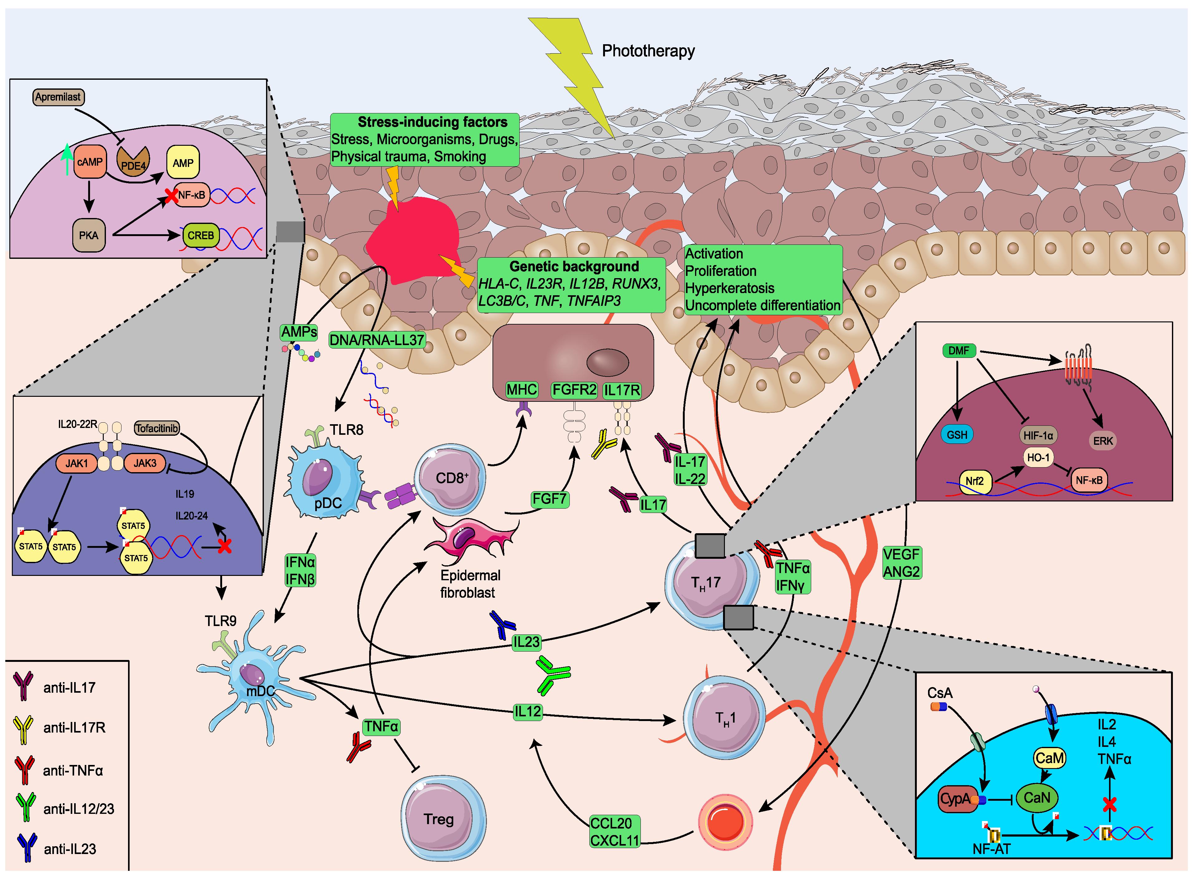
:1. Introduction
2. Pathophysiology of Psoriasis
3. Traditional Therapies
3.1. Acitretin
3.2. Cyclosporine
3.3. Methotrexate
3.4. Phototherapy
4. Small Molecules
4.1. Apremilast
4.2. Fumaric Acid Esters
4.3. JAK and TYK2 Inhibitors

| Study, Year | Drug | Method | Sample | Time Sample | Clinical Outcome | Time Point | Sample Size | Main Results |
|---|---|---|---|---|---|---|---|---|
| Pharmacogenetics | ||||||||
| Gambichler et al., 2013 [94] | DMF | Genotyping | gDNA | n.p. | PASI75 | 3 months | 106 | No evidence for association for GSTT1 alleles. |
| Pharmacogenomics | ||||||||
| Verbenko et al., 2020 [87] | APR | GWAS | gDNA | n.p. | PASI75 | 6 months | 34 | Association of the ARSF rs35084576 SNP. |
| Pharmacotranscriptomics | ||||||||
| Onderdijk et al., 2014 [95] | FAEs | Microarray | Lesional skin | Baseline, 12 weeks | PASI75 | 3 months | 50 | Overexpression of anti-inflammatory pathways after FAE treatment. |
| Tahvili et al., 2015 [96] | DMF | RT-qPCR | Plasma | Baseline | n.p. | n.p. | 35 | DMF suppressed TH1 and TH17 signaling post-treatment. |
| Catlett et al., 2022 [100] | DEUC | RNA-seq | Lesional skin | Baseline, 2 weeks, 12 weeks | n.s. | 3 months | 267 | Alleviation of central pathogenic molecules post-treatment. |
| Additional approaches | ||||||||
| Garcet et al., 2018 [90] | APR | Immunoassay | Plasma | Baseline, 2,4,16,24,32,52 weeks | PASI75 | 4 months | 129 | Suppression of circulating psoriasis pathogenic molecules, including IL-17 cytokine family and TNF. |
| Campanati et al., 2020 [91] | APR | ICC, IHC | Lesional skin | Baseline, 12 weeks | PASI75 | 3 months | 9 | Suppressed expression of VEGF, iNOS and IDO in keratinocytes. |
| Medvedeva et al., 2020 [92] | APR | Protein and RNA profiling | Plasma | Baseline | PASI75 | 4 months | 93 | Suppression of IL-17A and KLK-7 levels post-treatment. |
| Mazzilli et al., 2020 [93] | APR | Metabolic profiling | Plasma | Baseline, 24 weeks, 52 weeks | n.s. | 12 months | 113 | APR reversed metabolic markers. |
| Holzer et al., 2020 [97] | FAEs | Luminex Assay | Plasma | Baseline, 3 months, 6 months | PASI75 | 3 months | 32 | FAEs reduce apolipoprotein B and cholesterol. |
| Gambichler et al., 2012 [98] | DMF, MMF | ELISA | Plasma | Baseline, 3 months | n.s. | 3 months | 32 | AMP levels increased post-treatment. |
5. Biological Agents
5.1. Anti-TNF Agents
5.2. Anti-IL23 Agents
5.3. Anti-IL17 Agents
6. Discussion
Author Contributions
Funding
Institutional Review Board Statement
Informed Consent Statement
Data Availability Statement
Conflicts of Interest
References
- Motulsky, A.G. Drug Reactions, Enzymes, and Biochemical Genetics. JAMA 1957, 165, 835. [Google Scholar] [CrossRef] [PubMed]
- Kalow, W. Human Pharmacogenomics: The Development of a Science. Hum. Genom. 2004, 1, 375. [Google Scholar] [CrossRef] [PubMed] [Green Version]
- Miteva-Marcheva, N.N.; Ivanov, H.Y.; Dimitrov, D.K.; Stoyanova, V.K. Application of Pharmacogenetics in Oncology. Biomark. Res. 2020, 8, 32. [Google Scholar] [CrossRef] [PubMed]
- Yun, C.-H.; Mengwasser, K.E.; Toms, A.V.; Woo, M.S.; Greulich, H.; Wong, K.-K.; Meyerson, M.; Eck, M.J. The T790M Mutation in EGFR Kinase Causes Drug Resistance by Increasing the Affinity for ATP. Proc. Natl. Acad. Sci. USA 2008, 105, 2070–2075. [Google Scholar] [CrossRef] [Green Version]
- DISCO (Deciphering disorders Involving Scoliosis and COmorbidities) Study; Liu, J.; Zhou, Y.; Liu, S.; Song, X.; Yang, X.-Z.; Fan, Y.; Chen, W.; Akdemir, Z.C.; Yan, Z.; et al. The Coexistence of Copy Number Variations (CNVs) and Single Nucleotide Polymorphisms (SNPs) at a Locus Can Result in Distorted Calculations of the Significance in Associating SNPs to Disease. Hum. Genet. 2018, 137, 553–567. [Google Scholar] [CrossRef] [PubMed]
- Eichelbaum, M.; Ingelman-Sundberg, M.; Evans, W.E. Pharmacogenomics and Individualized Drug Therapy. Annu. Rev. Med. 2006, 57, 119–137. [Google Scholar] [CrossRef] [PubMed]
- Evans, W.E.; Relling, M.V. Moving towards Individualized Medicine with Pharmacogenomics. Nature 2004, 429, 464–468. [Google Scholar] [CrossRef]
- Klein, T.E.; Chang, J.T.; Cho, M.K.; Easton, K.L.; Fergerson, R.; Hewett, M.; Lin, Z.; Liu, Y.; Liu, S.; Oliver, D.E.; et al. Integrating Genotype and Phenotype Information: An Overview of the PharmGKB Project. Pharm. J. 2001, 1, 167–170. [Google Scholar] [CrossRef] [PubMed] [Green Version]
- Dinama, O.; Warren, A.M.; Kulkarni, J. The Role of Pharmacogenomic Testing in Psychiatry: Real World Examples. Aust. N. Z. J. Psychiatry 2014, 48, 778. [Google Scholar] [CrossRef]
- Wang, B.; Yang, L.-P.; Zhang, X.-Z.; Huang, S.-Q.; Bartlam, M.; Zhou, S.-F. New Insights into the Structural Characteristics and Functional Relevance of the Human Cytochrome P450 2D6 Enzyme. Drug Metab. Rev. 2009, 41, 573–643. [Google Scholar] [CrossRef]
- Yan, S.-K.; Liu, R.-H.; Jin, H.-Z.; Liu, X.-R.; Ye, J.; Shan, L.; Zhang, W.-D. “Omics” in Pharmaceutical Research: Overview, Applications, Challenges, and Future Perspectives. Chin. J. Nat. Med. 2015, 13, 3–21. [Google Scholar] [CrossRef]
- Pivarcsi, A.; Meisgen, F.; Xu, N.; Ståhle, M.; Sonkoly, E. Changes in the Level of Serum MicroRNAs in Patients with Psoriasis after Antitumour Necrosis Factor-α Therapy. Br. J. Dermatol. 2013, 169, 563–570. [Google Scholar] [CrossRef] [PubMed]
- Xu, M.; Deng, J.; Xu, K.; Zhu, T.; Han, L.; Yan, Y.; Yao, D.; Deng, H.; Wang, D.; Sun, Y.; et al. In-Depth Serum Proteomics Reveals Biomarkers of Psoriasis Severity and Response to Traditional Chinese Medicine. Theranostics 2019, 9, 2475–2488. [Google Scholar] [CrossRef]
- Kamleh, M.A.; Snowden, S.G.; Grapov, D.; Blackburn, G.J.; Watson, D.G.; Xu, N.; Ståhle, M.; Wheelock, C.E. LC–MS Metabolomics of Psoriasis Patients Reveals Disease Severity-Dependent Increases in Circulating Amino Acids That Are Ameliorated by Anti-TNFα Treatment. J. Proteome Res. 2015, 14, 557–566. [Google Scholar] [CrossRef] [Green Version]
- Rosenblum, M.D.; Gratz, I.K.; Paw, J.S.; Abbas, A.K. Treating Human Autoimmunity: Current Practice and Future Prospects. Sci. Transl. Med. 2012, 4, 125sr1. [Google Scholar] [CrossRef] [Green Version]
- Greiner, W.; Patel, K.; Crossman-Barnes, C.-J.; Rye-Andersen, T.V.; Hvid, C.; Vandebrouck, T. High-Expenditure Disease in the EU-28: Does Drug Spend Correspond to Clinical and Economic Burden in Oncology, Autoimmune Disease and Diabetes? Pharm. Econ. Open 2021, 5, 385–396. [Google Scholar] [CrossRef] [PubMed]
- Parisi, R.; Iskandar, I.Y.K.; Kontopantelis, E.; Augustin, M.; Griffiths, C.E.M.; Ashcroft, D.M. National, Regional, and Worldwide Epidemiology of Psoriasis: Systematic Analysis and Modelling Study. BMJ 2020, 369, m1590. [Google Scholar] [CrossRef]
- Lønnberg, A.S.; Skov, L.; Skytthe, A.; Kyvik, K.O.; Pedersen, O.B.; Thomsen, S.F. Heritability of Psoriasis in a Large Twin Sample. Br. J. Dermatol. 2013, 169, 412–416. [Google Scholar] [CrossRef]
- Tsoi, L.C.; Stuart, P.E.; Tian, C.; Gudjonsson, J.E.; Das, S.; Zawistowski, M.; Ellinghaus, E.; Barker, J.N.; Chandran, V.; Dand, N.; et al. Large Scale Meta-Analysis Characterizes Genetic Architecture for Common Psoriasis Associated Variants. Nat. Commun. 2017, 8, 15382. [Google Scholar] [CrossRef] [Green Version]
- Nakamizo, S.; Dutertre, C.-A.; Khalilnezhad, A.; Zhang, X.M.; Lim, S.; Lum, J.; Koh, G.; Foong, C.; Yong, P.J.A.; Tan, K.J.; et al. Single-Cell Analysis of Human Skin Identifies CD14+ Type 3 Dendritic Cells Co-Producing IL1B and IL23A in Psoriasis. J. Exp. Med. 2021, 218, e20202345. [Google Scholar] [CrossRef] [PubMed]
- Ji, Y.-Z.; Liu, S.-R. Koebner Phenomenon Leading to the Formation of New Psoriatic Lesions: Evidences and Mechanisms. Biosci. Rep. 2019, 39, BSR20193266. [Google Scholar] [CrossRef] [PubMed] [Green Version]
- Balak, D.; Hajdarbegovic, E. Drug-Induced Psoriasis: Clinical Perspectives. PTT 2017, 7, 87–94. [Google Scholar] [CrossRef] [PubMed] [Green Version]
- Teng, Y.; Xie, W.; Tao, X.; Liu, N.; Yu, Y.; Huang, Y.; Xu, D.; Fan, Y. Infection-provoked Psoriasis: Induced or Aggravated (Review). Exp. Ther. Med. 2021, 21, 567. [Google Scholar] [CrossRef]
- Nestle, F.O.; Conrad, C.; Tun-Kyi, A.; Homey, B.; Gombert, M.; Boyman, O.; Burg, G.; Liu, Y.-J.; Gilliet, M. Plasmacytoid Predendritic Cells Initiate Psoriasis through Interferon-α Production. J. Exp. Med. 2005, 202, 135–143. [Google Scholar] [CrossRef] [PubMed]
- Lande, R.; Botti, E.; Jandus, C.; Dojcinovic, D.; Fanelli, G.; Conrad, C.; Chamilos, G.; Feldmeyer, L.; Marinari, B.; Chon, S.; et al. The Antimicrobial Peptide LL37 Is a T-Cell Autoantigen in Psoriasis. Nat. Commun. 2014, 5, 5621. [Google Scholar] [CrossRef] [Green Version]
- Gisondi, P.; Del Giglio, M.; Girolomoni, G. Treatment Approaches to Moderate to Severe Psoriasis. Int. J. Mol. Sci. 2017, 18, 2427. [Google Scholar] [CrossRef] [Green Version]
- Gubán, B.; Vas, K.; Balog, Z.; Manczinger, M.; Bebes, A.; Groma, G.; Széll, M.; Kemény, L.; Bata-Csörgő, Z. Abnormal Regulation of Fibronectin Production by Fibroblasts in Psoriasis. Br. J. Dermatol. 2016, 174, 533–541. [Google Scholar] [CrossRef] [PubMed] [Green Version]
- Xiao, Y.; Wang, C.; Zeng, B.; Tang, X.; Zhang, Y.; Xiang, L.; Mi, L.; Pan, Y.; Wang, H.; Yang, Z. MiR124-3p/FGFR2 Axis Inhibits Human Keratinocyte Proliferation and Migration and Improve the Inflammatory Microenvironment in Psoriasis. Mol. Immunol. 2020, 122, 89–98. [Google Scholar] [CrossRef] [PubMed]
- Kanda, N.; Hoashi, T.; Saeki, H. The Defect in Regulatory T Cells in Psoriasis and Therapeutic Approaches. J. Clin. Med. 2021, 10, 3880. [Google Scholar] [CrossRef]
- Yawalkar, N.; Karlen, S.; Hunger, R.; Brand, C.U.; Braathen, L.R. Expression of Interleukin-12 Is Increased in Psoriatic Skin. J. Investig. Dermatol. 1998, 111, 1053–1057. [Google Scholar] [CrossRef]
- Di Cesare, A.; Di Meglio, P.; Nestle, F.O. The IL-23/Th17 Axis in the Immunopathogenesis of Psoriasis. J. Investig. Dermatol. 2009, 129, 1339–1350. [Google Scholar] [CrossRef] [PubMed] [Green Version]
- Wilson, N.J.; Boniface, K.; Chan, J.R.; McKenzie, B.S.; Blumenschein, W.M.; Mattson, J.D.; Basham, B.; Smith, K.; Chen, T.; Morel, F.; et al. Development, Cytokine Profile and Function of Human Interleukin 17–Producing Helper T Cells. Nat. Immunol. 2007, 8, 950–957. [Google Scholar] [CrossRef] [PubMed]
- Ogawa, E.; Sato, Y.; Minagawa, A.; Okuyama, R. Pathogenesis of Psoriasis and Development of Treatment. J. Dermatol. 2018, 45, 264–272. [Google Scholar] [CrossRef] [PubMed] [Green Version]
- Gottlieb, A.B.; Krueger, J.G.; Wittkowski, K.; Dedrick, R.; Walicke, P.A.; Garovoy, M. Psoriasis as a Model for T-Cell–Mediated Disease: Immunobiologic and Clinical Effects of Treatment With Multiple Doses of Efalizumab, an Anti–CD11a Antibody. Arch. Dermatol. 2002, 138, 591–600. [Google Scholar] [CrossRef] [Green Version]
- Clark, R.A. Resident Memory T Cells in Human Health and Disease. Sci. Transl. Med. 2015, 7, 269rv1. [Google Scholar] [CrossRef] [Green Version]
- Liu, J.; Chang, H.-W.; Huang, Z.-M.; Nakamura, M.; Sekhon, S.; Ahn, R.; Munoz-Sandoval, P.; Bhattarai, S.; Beck, K.M.; Sanchez, I.M.; et al. Single-Cell RNA Sequencing of Psoriatic Skin Identifies Pathogenic Tc17 Cell Subsets and Reveals Distinctions between CD8+ T Cells in Autoimmunity and Cancer. J. Allergy Clin. Immunol. 2021, 147, 2370–2380. [Google Scholar] [CrossRef] [PubMed]
- Di Meglio, P.; Villanova, F.; Navarini, A.A.; Mylonas, A.; Tosi, I.; Nestle, F.O.; Conrad, C. Targeting CD8+ T Cells Prevents Psoriasis Development. J. Allergy Clin. Immunol. 2016, 138, 274–276.e6. [Google Scholar] [CrossRef] [PubMed] [Green Version]
- Elnabawi, Y.A.; Garshick, M.S.; Tawil, M.; Barrett, T.J.; Fisher, E.A.; Lo Sicco, K.; Neimann, A.L.; Scher, J.U.; Krueger, J.; Berger, J.S. CCL20 in Psoriasis: A Potential Biomarker of Disease Severity, Inflammation, and Impaired Vascular Health. J. Am. Acad. Dermatol. 2021, 84, 913–920. [Google Scholar] [CrossRef] [PubMed]
- Grossman, R.M.; Krueger, J.; Yourish, D.; Granelli-Piperno, A.; Murphy, D.P.; May, L.T.; Kupper, T.S.; Sehgal, P.B.; Gottlieb, A.B. Interleukin 6 Is Expressed in High Levels in Psoriatic Skin and Stimulates Proliferation of Cultured Human Keratinocytes. Proc. Natl. Acad. Sci. USA 1989, 86, 6367–6371. [Google Scholar] [CrossRef] [PubMed] [Green Version]
- Lemster, B.H.; Carroll, P.B.; Rilo, H.R.; Johnson, N.; Nikaein, A.; Thomson, A.W. IL-8/IL-8 Receptor Expression in Psoriasis and the Response to Systemic Tacrolimus (FK506) Therapy. Clin. Exp. Immunol. 2008, 99, 148–154. [Google Scholar] [CrossRef] [PubMed]
- Edson-Heredia, E.; Sterling, K.L.; Alatorre, C.I.; Cuyun Carter, G.; Paczkowski, R.; Zarotsky, V.; Maeda-Chubachi, T. Heterogeneity of Response to Biologic Treatment: Perspective for Psoriasis. J. Investig. Dermatol. 2014, 134, 18–23. [Google Scholar] [CrossRef] [PubMed] [Green Version]
- Carretero, G. Risk of Serious Adverse Events Associated With Biologic and Nonbiologic Psoriasis Systemic Therapy: Patients Ineligible vs Eligible for Randomized Controlled Trials. Arch. Dermatol. 2012, 148, 463. [Google Scholar] [CrossRef] [PubMed] [Green Version]
- Spuls, P.I.; Lecluse, L.L.A.; Poulsen, M.-L.N.F.; Bos, J.D.; Stern, R.S.; Nijsten, T. How Good Are Clinical Severity and Outcome Measures for Psoriasis?: Quantitative Evaluation in a Systematic Review. J. Investig. Dermatol. 2010, 130, 933–943. [Google Scholar] [CrossRef] [PubMed] [Green Version]
- Lee, C.S.; Li, K. A Review of Acitretin for the Treatment of Psoriasis. Expert Opin. Drug Saf. 2009, 8, 769–779. [Google Scholar] [CrossRef]
- Nast, A.; Smith, C.; Spuls, P.I.; Avila Valle, G.; Bata-Csörgö, Z.; Boonen, H.; De Jong, E.; Garcia-Doval, I.; Gisondi, P.; Kaur-Knudsen, D.; et al. EuroGuiDerm Guideline on the Systemic Treatment of Psoriasis Vulgaris—Part 2: Specific Clinical and Comorbid Situations. Acad. Dermatol. Venereol. 2021, 35, 281–317. [Google Scholar] [CrossRef] [PubMed]
- Young, H.S.; Summers, A.M.; Read, I.R.; Fairhurst, D.A.; Plant, D.J.; Campalani, E.; Smith, C.H.; Brenchley, P.E.C.; Griffiths, C.E.M. Interaction between Genetic Control of Vascular Endothelial Growth Factor Production and Retinoid Responsiveness in Psoriasis. J. Investig. Dermatol. 2006, 126, 453–459. [Google Scholar] [CrossRef]
- Chen, W.; Wu, L.; Zhu, W.; Chen, X. The Polymorphisms of Growth Factor Genes (VEGFA & EGF) Were Associated with Response to Acitretin in Psoriasis. Pers. Med. 2018, 15, 181–188. [Google Scholar] [CrossRef]
- Campalani, E.; Allen, M.H.; Fairhurst, D.; Young, H.S.; Mendonca, C.O.; Burden, A.D.; Griffiths, C.E.M.; Crook, M.A.; Barker, J.N.W.N.; Smith, C.H. Apolipoprotein E Gene Polymorphisms Are Associated with Psoriasis but Do Not Determine Disease Response to Acitretin. Br. J. Dermatol. 2006, 154, 345–352. [Google Scholar] [CrossRef]
- Zhu, T.; Jin, H.; Shu, D.; Li, F.; Wu, C. Association of IL36RN Mutations with Clinical Features, Therapeutic Response to Acitretin, and Frequency of Recurrence in Patients with Generalized Pustular Psoriasis. Eur. J. Dermatol. 2018, 28, 217–224. [Google Scholar] [CrossRef] [PubMed]
- Zhou, X.; He, Y.; Kuang, Y.; Chen, W.; Zhu, W. HLA-DQA1 and DQB1 Alleles Are Associated with Acitretin Response in Patients with Psoriasis. Front. Biosci. 2022, 27, 266. [Google Scholar] [CrossRef]
- Chen, W.; Zhang, X.; Zhang, W.; Peng, C.; Zhu, W.; Chen, X. Polymorphisms of SLCO1B1 Rs4149056 and SLC22A1 Rs2282143 Are Associated with Responsiveness to Acitretin in Psoriasis Patients. Sci. Rep. 2018, 8, 13182. [Google Scholar] [CrossRef] [PubMed]
- Zhou, X.; Zhu, W.; Shen, M.; He, Y.; Peng, C.; Kuang, Y.; Su, J.; Zhao, S.; Chen, X.; Chen, W. Frizzled-Related Proteins 4 (SFRP4) Rs1802073G Allele Predicts the Elevated Serum Lipid Levels during Acitretin Treatment in Psoriatic Patients from Hunan, China. PeerJ 2018, 6, e4637. [Google Scholar] [CrossRef] [Green Version]
- Baran, A.; Kiluk, P.; Świderska, M.; Maciaszek, M.; Myśliwiec, H.; Flisiak, I. Adipocyte Fatty Acid-Binding Protein as a Novel Marker of Psoriasis and Clinical Response to Acitretin. Lipids 2019, 54, 445–452. [Google Scholar] [CrossRef] [PubMed]
- Antonatos, C.; Patsatsi, A.; Zafiriou, E.; Stavrou, E.F.; Liaropoulos, A.; Kyriakoy, A.; Evangelou, E.; Digka, D.; Roussaki-Schulze, A.; Sotiriadis, D.; et al. Protein Network and Pathway Analysis in a Pharmacogenetic Study of Cyclosporine Treatment Response in Greek Patients with Psoriasis. Pharm. J. 2023, 23, 8–13. [Google Scholar] [CrossRef] [PubMed]
- O’Rielly, D.D.; Rahman, P. Pharmacogenetics of Psoriasis. Pharmacogenomics 2011, 12, 87–101. [Google Scholar] [CrossRef] [PubMed]
- Vasilopoulos, Y.; Sarri, C.; Zafiriou, E.; Patsatsi, A.; Stamatis, C.; Ntoumou, E.; Fassos, I.; Tsalta, A.; Karra, A.; Roussaki-Schulze, A.; et al. A Pharmacogenetic Study of ABCB1 Polymorphisms and Cyclosporine Treatment Response in Patients with Psoriasis in the Greek Population. Pharm. J. 2014, 14, 523–525. [Google Scholar] [CrossRef] [PubMed]
- Chernov, A.; Kilina, D.; Smirnova, T.; Galimova, E. Pharmacogenetic Study of the Impact of ABCB1 Single Nucleotide Polymorphisms on the Response to Cyclosporine in Psoriasis Patients. Pharmaceutics 2022, 14, 2441. [Google Scholar] [CrossRef] [PubMed]
- Haider, A.S.; Lowes, M.A.; Suárez-Fariñas, M.; Zaba, L.C.; Cardinale, I.; Khatcherian, A.; Novitskaya, I.; Wittkowski, K.M.; Krueger, J.G. Identification of Cellular Pathways of “Type 1,” Th17 T Cells, and TNF- and Inducible Nitric Oxide Synthase-Producing Dendritic Cells in Autoimmune Inflammation through Pharmacogenomic Study of Cyclosporine A in Psoriasis. J. Immunol. 2008, 180, 1913–1920. [Google Scholar] [CrossRef] [Green Version]
- Grabarek, B.O.; Wcisło-Dziadecka, D.; Michalska-Bańkowska, A.; Gola, J. Evaluation of Expression Pattern of Selected Genes Associated with IL12/23 Signaling Paths in Psoriatic Patients during Cyclosporine A Therapy. Dermatol. Ther. 2019, 32, e13129. [Google Scholar] [CrossRef] [PubMed]
- Michalska-Bańkowska, A.; Wcisło-Dziadecka, D.; Grabarek, B.; Brzezińska-Wcisło, L.; Mazurek, U.; Salwowska, N.; Bańkowski, M. Variances in the MRNA Expression Profile of TGF-Β1–3 Isoforms and Its TGF-ΒRI–III Receptors during Cyclosporin a Treatment of Psoriatic Patients. Pdia 2018, 35, 502–509. [Google Scholar] [CrossRef]
- Eşrefoğlu, M.; Gül, M.; Seyhan, M. Ultrastructural Findings and Tumor Necrosis Factor-Alpha and Intercellular Adhesion Molecule-1 Expression in Psoriasis Patients Before and After Oral Cyclosporin A Therapy. Ultrastruct. Pathol. 2006, 30, 95–102. [Google Scholar] [CrossRef] [PubMed]
- Yan, K.; Zhang, Y.; Han, L.; Huang, Q.; Zhang, Z.; Fang, X.; Zheng, Z.; Yawalkar, N.; Chang, Y.; Zhang, Q.; et al. Safety and Efficacy of Methotrexate for Chinese Adults With Psoriasis With and Without Psoriatic Arthritis. JAMA Dermatol. 2019, 155, 327. [Google Scholar] [CrossRef] [PubMed]
- Fan, Z.; Zhang, Z.; Huang, Q.; Han, L.; Fang, X.; Yang, K.; Wu, S.; Zheng, Z.; Yawalkar, N.; Wang, Z.; et al. The Impact of ANxA6 Gene Polymorphism on the Efficacy of Methotrexate Treatment in Psoriasis Patients. Dermatology 2021, 237, 579–587. [Google Scholar] [CrossRef] [PubMed]
- West, J.; Ogston, S.; Berg, J.; Palmer, C.; Fleming, C.; Kumar, V.; Foerster, J. HLA-Cw6-Positive Patients with Psoriasis Show Improved Response to Methotrexate Treatment. Clin. Exp. Dermatol. 2017, 42, 651–655. [Google Scholar] [CrossRef] [PubMed] [Green Version]
- Mao, M.; Kuang, Y.; Chen, M.; Yan, K.; Lv, C.; Liu, P.; Lu, Y.; Chen, X.; Zhu, W.; Chen, W. The HLA-Cw*06 Allele May Predict the Response to Methotrexate (MTX) Treatment in Chinese Arthritis-Free Psoriasis Patients. Arch. Dermatol. Res. 2022, 2022, 1–7. [Google Scholar] [CrossRef]
- Yan, K.X.; Zhang, Y.J.; Han, L.; Huang, Q.; Zhang, Z.H.; Fang, X.; Zheng, Z.Z.; Yawalkar, N.; Chang, Y.L.; Zhang, Q.; et al. TT Genotype of Rs10036748 in TNIP 1 Shows Better Response to Methotrexate in a Chinese Population: A Prospective Cohort Study. Br. J. Dermatol. 2019, 181, 778–785. [Google Scholar] [CrossRef]
- Warren, R.B.; Smith, R.L.; Campalani, E.; Eyre, S.; Smith, C.H.; Barker, J.N.W.N.; Worthington, J.; Griffiths, C.E.M. Genetic Variation in Efflux Transporters Influences Outcome to Methotrexate Therapy in Patients with Psoriasis. J. Investig. Dermatol. 2008, 128, 1925–1929. [Google Scholar] [CrossRef]
- Grželj, J.; Marovt, M.; Marko, P.B.; Mlinarič-Raščan, I.; Gmeiner, T.; Šmid, A. Polymorphism in Gene for ABCC2 Transporter Predicts Methotrexate Drug Survival in Patients with Psoriasis. Medicina 2021, 57, 1050. [Google Scholar] [CrossRef] [PubMed]
- Voron’ko, O.E.; Baskaev, K.K.; Sobolev, V.V.; Denisova, E.V.; Korsunskaya, I.M. Genetic Markers of Therapeutic Efficacy of Methotrexate in Patients with Psoriasis. Bull. Exp. Biol. Med. 2022, 172, 460–463. [Google Scholar] [CrossRef] [PubMed]
- Grželj, J.; Mlinarič-Raščan, I.; Marko, P.B.; Marovt, M.; Gmeiner, T.; Šmid, A. Polymorphisms in GNMT and DNMT3b Are Associated with Methotrexate Treatment Outcome in Plaque Psoriasis. Biomed. Pharmacother. 2021, 138, 111456. [Google Scholar] [CrossRef]
- Campalani, E.; Arenas, M.; Marinaki, A.M.; Lewis, C.M.; Barker, J.N.W.N.; Smith, C.H. Polymorphisms in Folate, Pyrimidine, and Purine Metabolism Are Associated with Efficacy and Toxicity of Methotrexate in Psoriasis. J. Investig. Dermatol. 2007, 127, 1860–1867. [Google Scholar] [CrossRef] [Green Version]
- Zhang, Y.; Ding, X.; Meng, Z.; Chen, M.; Zheng, X.; Cai, M.; Wu, J.; Chang, Y.; Zhang, Q.; Jin, L.; et al. A Genome-wide Association Study Identified HLA-C Associated with the Effectiveness of Methotrexate for Psoriasis Treatment. Acad. Dermatol. Venereol. 2021, 35, e898–e900. [Google Scholar] [CrossRef] [PubMed]
- Goldminz, A.M.; Suárez-Fariñas, M.; Wang, A.C.; Dumont, N.; Krueger, J.G.; Gottlieb, A.B. CCL20 and IL22 Messenger RNA Expression After Adalimumab vs Methotrexate Treatment of Psoriasis: A Randomized Clinical Trial. JAMA Dermatol. 2015, 151, 837. [Google Scholar] [CrossRef] [PubMed] [Green Version]
- Esawy, F.M.E.; Ahmed, I.A.; Fallah, A.A.E.; Salem, R.M. Methotrexate Mechanism of Action in Plaque Psoriasis: Something New in the Old View. J. Clin. Aesthet. Dermatol. 2022, 15, 42–46. [Google Scholar]
- Correa da Rosa, J.; Kim, J.; Tian, S.; Tomalin, L.E.; Krueger, J.G.; Suárez-Fariñas, M. Shrinking the Psoriasis Assessment Gap: Early Gene-Expression Profiling Accurately Predicts Response to Long-Term Treatment. J. Investig. Dermatol. 2017, 137, 305–312. [Google Scholar] [CrossRef] [Green Version]
- Indhumathi, S.; Rajappa, M.; Chandrashekar, L.; Ananthanarayanan, P.H.; Thappa, D.M.; Negi, V.S. Pharmacogenetic Markers to Predict the Clinical Response to Methotrexate in South Indian Tamil Patients with Psoriasis. Eur. J. Clin. Pharmacol. 2017, 73, 965–971. [Google Scholar] [CrossRef] [PubMed]
- Abdelaal, N.H.; Elhefnawy, N.G.; Abdulmonem, S.R.; Sayed, S.; Saleh, N.A.; Saleh, M.A. Evaluation of the Expression of the Stromal Cell-derived Factor-1 Alpha (CXCL 12) in Psoriatic Patients after Treatment with Methotrexate. J. Cosmet. Dermatol. 2020, 19, 253–258. [Google Scholar] [CrossRef]
- Yan, K.X.; Meng, Q.; He, H.; Zhu, H.W.; Wang, Z.C.; Han, L.; Huang, Q.; Zhang, Z.H.; Yawalkar, N.; Zhou, H.; et al. iTRAQ-based Quantitative Proteomics Reveals Biomarkers/Pathways in Psoriasis That Can Predict the Efficacy of Methotrexate. Acad. Dermatol. Venereol. 2022, 36, 1784–1795. [Google Scholar] [CrossRef] [PubMed]
- Qiu, Q.; Deng, J.; Deng, H.; Yao, D.; Yan, Y.; Ye, S.; Shang, X.; Deng, Y.; Han, L.; Zheng, G.; et al. Association of the Characteristics of the Blood Metabolome and Gut Microbiome with the Outcome of Methotrexate Therapy in Psoriasis. Front. Immunol. 2022, 13, 937539. [Google Scholar] [CrossRef] [PubMed]
- Wong, T.; Hsu, L.; Liao, W. Phototherapy in Psoriasis: A Review of Mechanisms of Action. J. Cutan. Med. Surg. 2013, 17, 6–12. [Google Scholar] [CrossRef]
- Hairutdinov, V.; Moshkalov, A.; Samtsov, A.; Buslov, K.; Kuligina, E.; Mitiushkina, N.; Suspitsin, E.; Togo, A.; Hanson, K.; Imyanitov, E. Apoptosis-Deficient Pro Allele of Gene Is Associated with the Resistance of Psoriasis to the UV-Based Therapy. J. Dermatol. Sci. 2005, 37, 185–187. [Google Scholar] [CrossRef] [PubMed]
- Ryan, C.; Renfro, L.; Collins, P.; Kirby, B.; Rogers, S. Clinical and Genetic Predictors of Response to Narrowband Ultraviolet B for the Treatment of Chronic Plaque Psoriasis: Predictors of Response to NB-UVB for Psoriasis. Br. J. Dermatol. 2010, 163, 1056–1063. [Google Scholar] [CrossRef] [PubMed]
- Rácz, E.; Prens, E.P.; Kurek, D.; Kant, M.; de Ridder, D.; Mourits, S.; Baerveldt, E.M.; Ozgur, Z.; van IJcken, W.F.J.; Laman, J.D.; et al. Effective Treatment of Psoriasis with Narrow-Band UVB Phototherapy Is Linked to Suppression of the IFN and Th17 Pathways. J. Investig. Dermatol. 2011, 131, 1547–1558. [Google Scholar] [CrossRef] [PubMed] [Green Version]
- Hochberg, M.; Zeligson, S.; Amariglio, N.; Rechavi, G.; Ingber, A.; Enk, C.D. Genomic-Scale Analysis of Psoriatic Skin Reveals Differentially Expressed Insulin-like Growth Factor-Binding Protein-7 after Phototherapy. Br. J. Dermatol. 2007, 156, 289–300. [Google Scholar] [CrossRef] [PubMed]
- Ele-Refaei, A.M.; El-Esawy, F.M. Effect of Narrow-Band Ultraviolet B Phototherapy and Methotrexate on MicroRNA (146a) Levels in Blood of Psoriatic Patients. Dermatol. Res. Pract. 2015, 2015, 145769. [Google Scholar] [CrossRef] [Green Version]
- Lo, Y.-H.; Torii, K.; Saito, C.; Furuhashi, T.; Maeda, A.; Morita, A. Serum IL-22 Correlates with Psoriatic Severity and Serum IL-6 Correlates with Susceptibility to Phototherapy. J. Dermatol. Sci. 2010, 58, 225–227. [Google Scholar] [CrossRef]
- Verbenko, D.A.; Karamova, A.E.; Artamonova, O.G.; Deryabin, D.G.; Rakitko, A.; Chernitsov, A.; Krasnenko, A.; Elmuratov, A.; Solomka, V.S.; Kubanov, A.A. Apremilast Pharmacogenomics in Russian Patients with Moderate-to-Severe and Severe Psoriasis. J. Pers. Med. 2020, 11, 20. [Google Scholar] [CrossRef] [PubMed]
- Oehrl, S.; Prakash, H.; Ebling, A.; Trenkler, N.; Wölbing, P.; Kunze, A.; Döbel, T.; Schmitz, M.; Enk, A.; Schäkel, K. The Phosphodiesterase 4 Inhibitor Apremilast Inhibits Th1 but Promotes Th17 Responses Induced by 6-Sulfo LacNAc (Slan) Dendritic Cells. J. Dermatol. Sci. 2017, 87, 110–115. [Google Scholar] [CrossRef] [Green Version]
- Schafer, P.H.; Truzzi, F.; Parton, A.; Wu, L.; Kosek, J.; Zhang, L.-H.; Horan, G.; Saltari, A.; Quadri, M.; Lotti, R.; et al. Phosphodiesterase 4 in Inflammatory Diseases: Effects of Apremilast in Psoriatic Blood and in Dermal Myofibroblasts through the PDE4/CD271 Complex. Cell. Signal. 2016, 28, 753–763. [Google Scholar] [CrossRef] [Green Version]
- Garcet, S.; Nograles, K.; Correa da Rosa, J.; Schafer, P.H.; Krueger, J.G. Synergistic Cytokine Effects as Apremilast Response Predictors in Patients with Psoriasis. J. Allergy Clin. Immunol. 2018, 142, 1010–1013.e6. [Google Scholar] [CrossRef] [PubMed]
- Campanati, A.; Caffarini, M.; Diotallevi, F.; Radi, G.; Lucarini, G.; Di Vincenzo, M.; Orciani, M.; Offidani, A. The Efficacy of in Vivo Administration of Apremilast on Mesenchymal Stem Cells Derived from Psoriatic Patients. Inflamm. Res. 2021, 70, 79–87. [Google Scholar] [CrossRef] [PubMed]
- Medvedeva, I.V.; Stokes, M.E.; Eisinger, D.; LaBrie, S.T.; Ai, J.; Trotter, M.W.B.; Schafer, P.; Yang, R. Large-Scale Analyses of Disease Biomarkers and Apremilast Pharmacodynamic Effects. Sci. Rep. 2020, 10, 605. [Google Scholar] [CrossRef] [PubMed] [Green Version]
- Mazzilli, S.; Lanna, C.; Chiaramonte, C.; Cesaroni, G.M.; Zangrilli, A.; Palumbo, V.; Cosio, T.; Dattola, A.; Gaziano, R.; Galluzzo, M.; et al. Real Life Experience of Apremilast in Psoriasis and Arthritis Psoriatic Patients: Preliminary Results on Metabolic Biomarkers. J. Dermatol. 2020, 47, 578–582. [Google Scholar] [CrossRef] [PubMed]
- Gambichler, T.; Kreuter, A.; Susok, L.; Skrygan, M.; Rotterdam, S.; Höxtermann, S.; Müller, M.; Tigges, C.; Altmeyer, P.; Lahner, N. Glutathione-S-Transferase T1 Genotyping and Phenotyping in Psoriasis Patients Receiving Treatment with Oral Fumaric Acid Esters. J. Eur. Acad. Dermatol. Venereol. 2014, 28, 574–580. [Google Scholar] [CrossRef] [PubMed]
- Onderdijk, A.J.; Balak, D.M.W.; Baerveldt, E.M.; Florencia, E.F.; Kant, M.; Laman, J.D.; IJcken, W.F.J.; Racz, E.; Ridder, D.; Thio, H.B.; et al. Regulated Genes in Psoriatic Skin during Treatment with Fumaric Acid Esters. Br. J. Dermatol. 2014, 171, 732–741. [Google Scholar] [CrossRef] [PubMed]
- Tahvili, S.; Zandieh, B.; Amirghofran, Z. The Effect of Dimethyl Fumarate on Gene Expression and the Level of Cytokines Related to Different T Helper Cell Subsets in Peripheral Blood Mononuclear Cells of Patients with Psoriasis. Int. J. Dermatol. 2015, 54, e254–e260. [Google Scholar] [CrossRef]
- Holzer, G.; Hoke, M.; Sabeti-Sandor, S.; Perkmann, T.; Rauscher, A.; Strassegger, B.; Radakovic, S.; Tanew, A. Disparate Effects of Adalimumab and Fumaric Acid Esters on Cardiovascular Risk Factors in Psoriasis Patients: Results from a Prospective, Randomized, Observer-blinded Head-to-head Trial. Acad. Dermatol. Venereol. 2021, 35, 441–449. [Google Scholar] [CrossRef]
- Gambichler, T.; Bechara, F.G.; Scola, N.; Rotterdam, S.; Altmeyer, P.; Skrygan, M. Serum Levels of Antimicrobial Peptides and Proteins Do Not Correlate with Psoriasis Severity and Are Increased after Treatment with Fumaric Acid Esters. Arch. Dermatol. Res. 2012, 304, 471–474. [Google Scholar] [CrossRef]
- Shang, L.; Cao, J.; Zhao, S.; Zhang, J.; He, Y. TYK2 in Immune Responses and Treatment of Psoriasis. JIR 2022, 15, 5373–5385. [Google Scholar] [CrossRef]
- Catlett, I.M.; Hu, Y.; Gao, L.; Banerjee, S.; Gordon, K.; Krueger, J.G. Molecular and Clinical Effects of Selective Tyrosine Kinase 2 Inhibition with Deucravacitinib in Psoriasis. J. Allergy Clin. Immunol. 2022, 149, 2010–2020.e8. [Google Scholar] [CrossRef]
- Tian, F.; Chen, Z.; Xu, T. Efficacy and Safety of Tofacitinib for the Treatment of Chronic Plaque Psoriasis: A Systematic Review and Meta-Analysis. J. Int. Med. Res. 2019, 47, 2342–2350. [Google Scholar] [CrossRef] [PubMed]
- Bing, N.; Zhou, H.; Chen, X.; Hirose, T.; Kochi, Y.; Tsuchida, Y.; Ishigaki, K.; Sumitomo, S.; Fujio, K.; Zhang, B.; et al. Contribution of a European-Prevalent Variant near CD83 and an East Asian–Prevalent Variant near IL17RB to Herpes Zoster Risk in Tofacitinib Treatment: Results of Genome-Wide Association Study Meta-Analyses. Arthritis Rheumatol. 2021, 73, 1155–1166. [Google Scholar] [CrossRef]
- Krueger, J.; Clark, J.D.; Suárez-Fariñas, M.; Fuentes-Duculan, J.; Cueto, I.; Wang, C.Q.; Tan, H.; Wolk, R.; Rottinghaus, S.T.; Whitley, M.Z.; et al. Tofacitinib Attenuates Pathologic Immune Pathways in Patients with Psoriasis: A Randomized Phase 2 Study. J. Allergy Clin. Immunol. 2016, 137, 1079–1090. [Google Scholar] [CrossRef] [PubMed] [Green Version]
- Ludbrook, V.J.; Hicks, K.J.; Hanrott, K.E.; Patel, J.S.; Binks, M.H.; Wyres, M.R.; Watson, J.; Wilson, P.; Simeoni, M.; Schifano, L.A.; et al. Investigation of Selective JAK1 Inhibitor GSK2586184 for the Treatment of Psoriasis in a Randomized Placebo-Controlled Phase IIa Study. Br. J. Dermatol. 2016, 174, 985–995. [Google Scholar] [CrossRef]
- Kim, J.; Tomalin, L.; Lee, J.; Fitz, L.J.; Berstein, G.; Correa-da Rosa, J.; Garcet, S.; Lowes, M.A.; Valdez, H.; Wolk, R.; et al. Reduction of Inflammatory and Cardiovascular Proteins in the Blood of Patients with Psoriasis: Differential Responses between Tofacitinib and Etanercept after 4 Weeks of Treatment. J. Investig. Dermatol. 2018, 138, 273–281. [Google Scholar] [CrossRef] [PubMed] [Green Version]
- Wu, J.J.; Strober, B.E.; Hansen, P.R.; Ahlehoff, O.; Egeberg, A.; Qureshi, A.A.; Robertson, D.; Valdez, H.; Tan, H.; Wolk, R. Effects of Tofacitinib on Cardiovascular Risk Factors and Cardiovascular Outcomes Based on Phase III and Long-Term Extension Data in Patients with Plaque Psoriasis. J. Am. Acad. Dermatol. 2016, 75, 897–905. [Google Scholar] [CrossRef] [PubMed]
- Brownstone, N.; Hong, J.; Mosca, M.; Hadeler, E.; Liao, W.; Bhutani, T.; Koo, J. Biologic Treatments of Psoriasis: An Update for the Clinician. BTT 2021, 15, 39–51. [Google Scholar] [CrossRef]
- Vasilopoulos, Y.; Manolika, M.; Zafiriou, E.; Sarafidou, T.; Bagiatis, V.; Krüger-Krasagaki, S.; Tosca, A.; Patsatsi, A.; Sotiriadis, D.; Mamuris, Z.; et al. Pharmacogenetic Analysis of TNF, TNFRSF1A, and TNFRSF1B Gene Polymorphisms and Prediction of Response to Anti-TNF Therapy in Psoriasis Patients in the Greek Population. Mol. Diagn. Ther. 2012, 16, 29–34. [Google Scholar] [CrossRef] [PubMed]
- Wang, Z.; Kong, L.; Zhang, H.; Sun, F.; Guo, Z.; Zhang, R.; Dou, Y. Tumor Necrosis Factor Alpha -308G/A Gene Polymorphisms Combined with Neutrophil-to-Lymphocyte and Platelet-to-Lymphocyte Ratio Predicts the Efficacy and Safety of Anti-TNF-α Therapy in Patients with Ankylosing Spondylitis, Rheumatoid Arthritis, and Psoriasis Arthritis. Front. Pharmacol. 2022, 12, 811719. [Google Scholar] [CrossRef]
- Ito, M.; Hirota, T.; Momose, M.; Ito, T.; Umezawa, Y.; Fukuchi, O.; Asahina, A.; Nakagawa, H.; Tamari, M.; Saeki, H. Lack of Association of TNFA, TNFRSF1B and TNFAIP3 Gene Polymorphisms with Response to Anti-tumor Necrosis Factor Therapy in Japanese Patients with Psoriasis. J. Dermatol. 2020, 47, e110–e111. [Google Scholar] [CrossRef] [PubMed]
- Antonatos, C.; Stavrou, E.F.; Evangelou, E.; Vasilopoulos, Y. Exploring Pharmacogenetic Variants for Predicting Response to Anti-TNF Therapy in Autoimmune Diseases: A Meta-Analysis. Pharmacogenomics 2021, 22, 435–445. [Google Scholar] [CrossRef] [PubMed]
- Higgins, J.P.T. Measuring Inconsistency in Meta-Analyses. BMJ 2003, 327, 557–560. [Google Scholar] [CrossRef] [PubMed] [Green Version]
- Coto-Segura, P.; González-Lara, L.; Batalla, A.; Eiris, N.; Queiro, R.; Coto, E. NFKBIZ and CW6 in Adalimumab Response Among Psoriasis Patients: Genetic Association and Alternative Transcript Analysis. Mol. Diagn. Ther. 2019, 23, 627–633. [Google Scholar] [CrossRef]
- Ovejero-Benito, M.C.; Prieto-Pérez, R.; Llamas-Velasco, M.; Muñoz-Aceituno, E.; Reolid, A.; Saiz-Rodríguez, M.; Belmonte, C.; Román, M.; Ochoa, D.; Talegón, M.; et al. Polymorphisms Associated with Adalimumab and Infliximab Response in Moderate-to-Severe Plaque Psoriasis. Pharmacogenomics 2018, 19, 7–16. [Google Scholar] [CrossRef] [PubMed] [Green Version]
- Talamonti, M.; Galluzzo, M.; Zangrilli, A.; Papoutsaki, M.; Egan, C.G.; Bavetta, M.; Tambone, S.; Fargnoli, M.C.; Bianchi, L. HLA-C*06:02 Does Not Predispose to Clinical Response Following Long-Term Adalimumab Treatment in Psoriatic Patients: A Retrospective Cohort Study. Mol. Diagn. Ther. 2017, 21, 295–301. [Google Scholar] [CrossRef] [PubMed]
- Murdaca, G.; Negrini, S.; Magnani, O.; Penza, E.; Pellecchio, M.; Puppo, F. Impact of Pharmacogenomics upon the Therapeutic Response to Etanercept in Psoriasis and Psoriatic Arthritis. Expert Opin. Drug Saf. 2017, 16, 1173–1179. [Google Scholar] [CrossRef] [PubMed]
- Prieto-Pérez, R.; Solano-López, G.; Cabaleiro, T.; Román, M.; Ochoa, D.; Talegón, M.; Baniandrés, O.; López-Estebaranz, J.L.; de la Cueva, P.; Daudén, E.; et al. New Polymorphisms Associated with Response to Anti-TNF Drugs in Patients with Moderate-to-Severe Plaque Psoriasis. Pharm. J. 2018, 18, 70–75. [Google Scholar] [CrossRef] [PubMed] [Green Version]
- Ovejero-Benito, M.C.; Muñoz-Aceituno, E.; Reolid, A.; Fisas, L.H.; Llamas-Velasco, M.; Prieto-Pérez, R.; Abad-Santos, F.; Daudén, E. Polymorphisms Associated with Anti-TNF Drugs Response in Patients with Psoriasis and Psoriatic Arthritis. J. Eur. Acad. Dermatol. Venereol. 2019, 33, e175–e177. [Google Scholar] [CrossRef] [PubMed]
- Tejasvi, T.; Stuart, P.E.; Chandran, V.; Voorhees, J.J.; Gladman, D.D.; Rahman, P.; Elder, J.T.; Nair, R.P. TNFAIP3 Gene Polymorphisms Are Associated with Response to TNF Blockade in Psoriasis. J. Investig. Dermatol. 2012, 132, 593–600. [Google Scholar] [CrossRef] [Green Version]
- Torii, K.; Morita, A. Specific Single Nucleotide Polymorphism Genotypes and Association of an IL-12B Polymorphism with Secondary Failure of Infliximab Therapy in Japanese Psoriasis Patients. J. Dermatol. Sci. 2020, 99, 135–136. [Google Scholar] [CrossRef] [PubMed]
- Nani, P.; Ladopoulou, M.; Papaioannou, E.H.; Papagianni, E.D.; Antonatos, C.; Xiropotamos, P.; Kapsoritakis, A.; Potamianos, P.S.; Karmiris, K.; Tzathas, C.; et al. Pharmacogenetic Analysis of the MIR146A Rs2910164 and MIR155 Rs767649 Polymorphisms and Response to Anti-TNF Treatment in Patients with Crohn’s Disease and Psoriasis. Genes 2023, 14, 445. [Google Scholar] [CrossRef] [PubMed]
- Loft, N.D.; Skov, L.; Iversen, L.; Gniadecki, R.; Dam, T.N.; Brandslund, I.; Hoffmann, H.J.; Andersen, M.R.; Dessau, R.B.; Bergmann, A.C.; et al. Associations between Functional Polymorphisms and Response to Biological Treatment in Danish Patients with Psoriasis. Pharm. J. 2018, 18, 494–500. [Google Scholar] [CrossRef] [Green Version]
- Nishikawa, R.; Nagai, H.; Bito, T.; Ikeda, T.; Horikawa, T.; Adachi, A.; Matsubara, T.; Nishigori, C. Genetic Prediction of the Effectiveness of Biologics for Psoriasis Treatment. J. Dermatol. 2016, 43, 1273–1277. [Google Scholar] [CrossRef] [PubMed] [Green Version]
- Ovejero-Benito, M.C.; Muñoz-Aceituno, E.; Sabador, D.; Almoguera, B.; Prieto-Pérez, R.; Hakonarson, H.; Coto-Segura, P.; Carretero, G.; Reolid, A.; Llamas-Velasco, M.; et al. Genome-wide Association Analysis of Psoriasis Patients Treated with Anti-TNF Drugs. Exp. Dermatol. 2020, 29, 1225–1232. [Google Scholar] [CrossRef] [PubMed]
- Ren, Y.; Wang, L.; Dai, H.; Qiu, G.; Liu, J.; Yu, D.; Liu, J.; Lyu, C.-Z.; Liu, L.; Zheng, M. Genome-Wide Association Analysis of Anti-TNF-α Treatment Response in Chinese Patients with Psoriasis. Front. Pharmacol. 2022, 13, 968935. [Google Scholar] [CrossRef]
- Antonatos, C.; Panoutsopoulou, M.; Georgakilas, G.K.; Evangelou, E.; Vasilopoulos, Y. Gene Expression Meta-Analysis of Potential Shared and Unique Pathways between Autoimmune Diseases under Anti-TNFα Therapy. Genes 2022, 13, 776. [Google Scholar] [CrossRef] [PubMed]
- Shen, H.; Wang, D.; Zhan, M.; Ding, H.; Zhao, H. MicroRNA-146a and MicroRNA-146b Deficiency Correlates with Exacerbated Disease Activity, and Their Longitude Increment Relates to Etanercept Response in Psoriasis Patients. Clin. Lab. Anal. 2022, 36, e24198. [Google Scholar] [CrossRef] [PubMed]
- Pei, D.; Cao, J.; Qin, G.; Wang, X. Measurement of Circulating MiRNA-125a Exhibits Good Value in the Management of Etanercept-treated Psoriatic Patients. J. Dermatol. 2020, 47, 140–146. [Google Scholar] [CrossRef]
- Skarmoutsou, E.; Trovato, C.; Granata, M.; Rossi, G.A.; Mosca, A.; Longo, V.; Gangemi, P.; Pettinato, M.; D’Amico, F.; Mazzarino, M.C. Biological Therapy Induces Expression Changes in Notch Pathway in Psoriasis. Arch. Dermatol. Res. 2015, 307, 863–873. [Google Scholar] [CrossRef] [PubMed]
- Raaby, L.; Langkilde, A.; Kjellerup, R.B.; Vinter, H.; Khatib, S.H.; Hjuler, K.F.; Johansen, C.; Iversen, L. Changes in mRNA Expression Precede Changes in Micro RNA Expression in Lesional Psoriatic Skin during Treatment with Adalimumab. Br. J. Dermatol. 2015, 173, 436–447. [Google Scholar] [CrossRef] [PubMed]
- Balato, A.; Schiattarella, M.; Di Caprio, R.; Lembo, S.; Mattii, M.; Balato, N.; Ayala, F. Effects of Adalimumab Therapy in Adult Subjects with Moderate-to-Severe Psoriasis on Th17 Pathway. J. Eur. Acad. Dermatol. Venereol. 2014, 28, 1016–1024. [Google Scholar] [CrossRef]
- Luan, L.; Han, S.; Wang, H.; Liu, X. Down-Regulation of the Th1, Th17, and Th22 Pathways Due to Anti-TNF-α Treatment in Psoriasis. Int. Immunopharmacol. 2015, 29, 278–284. [Google Scholar] [CrossRef]
- Sato, Y.; Kajihara, I.; Yamada-Kanazawa, S.; Jinnin, M.; Ihn, H. S100A7 Expression Levels in Coordination with Interleukin-8 Indicate the Clinical Response to Infliximab for Psoriasis Patients. J. Dermatol. 2017, 44, 838–839. [Google Scholar] [CrossRef] [PubMed]
- Vageli, D.P.; Exarchou, A.; Zafiriou, E.; Doukas, P.G.; Doukas, S.; Roussaki-Schulze, A. Effect of TNF-α Inhibitors on Transcriptional Levels of pro-Inflammatory Interleukin-33 and Toll-like Receptors-2 and -9 in Psoriatic Plaques. Exp. Ther. Med. 2015, 10, 1573–1577. [Google Scholar] [CrossRef] [PubMed] [Green Version]
- Kusumoto, S.; Kajihara, I.; Nagamoto, E.; Makino, K.; Ichihara, A.; Aoi, J.; Johno, T.; Makino, T.; Fukushima, S.; Jinnin, M.; et al. Increased CCL 22 Expression in Psoriatic Skin Predicts a Good Response to Infliximab Therapy. Br. J. Dermatol. 2014, 171, 1259–1261. [Google Scholar] [CrossRef]
- Suárez-Fariñas, M.; Fuentes-Duculan, J.; Lowes, M.A.; Krueger, J.G. Resolved Psoriasis Lesions Retain Expression of a Subset of Disease-Related Genes. J. Investig. Dermatol. 2011, 131, 391–400. [Google Scholar] [CrossRef] [PubMed] [Green Version]
- Ahn, R.; Gupta, R.; Lai, K.; Chopra, N.; Arron, S.T.; Liao, W. Network Analysis of Psoriasis Reveals Biological Pathways and Roles for Coding and Long Non-Coding RNAs. BMC Genom. 2016, 17, 841. [Google Scholar] [CrossRef] [PubMed] [Green Version]
- Foulkes, A.C.; Watson, D.S.; Carr, D.F.; Kenny, J.G.; Slidel, T.; Parslew, R.; Pirmohamed, M.; Anders, S.; Reynolds, N.J.; Griffiths, C.E.M.; et al. A Framework for Multi-Omic Prediction of Treatment Response to Biologic Therapy for Psoriasis. J. Investig. Dermatol. 2019, 139, 100–107. [Google Scholar] [CrossRef] [PubMed] [Green Version]
- Tsoi, L.C.; Patrick, M.T.; Shuai, S.; Sarkar, M.K.; Chi, S.; Ruffino, B.; Billi, A.C.; Xing, X.; Uppala, R.; Zang, C.; et al. Cytokine Responses in Nonlesional Psoriatic Skin as Clinical Predictor to Anti-TNF Agents. J. Allergy Clin. Immunol. 2022, 149, 640–649.e5. [Google Scholar] [CrossRef]
- Tomalin, L.E.; Kim, J.; Correa da Rosa, J.; Lee, J.; Fitz, L.J.; Berstein, G.; Valdez, H.; Wolk, R.; Krueger, J.G.; Suárez-Fariñas, M. Early Quantification of Systemic Inflammatory Proteins Predicts Long-Term Treatment Response to Tofacitinib and Etanercept. J. Investig. Dermatol. 2020, 140, 1026–1034. [Google Scholar] [CrossRef] [PubMed]
- Zhao, L.; Zhang, X.; Zhu, L.; Geng, S.; Guo, K. Effectiveness and Safety of Adalimumab in Psoriasis and Its Influence on Gut Microbiome. Microb. Pathog. 2022, 162, 105308. [Google Scholar] [CrossRef]
- Nwanaji-Enwerem, J.C.; Nwanaji-Enwerem, U.; Baccarelli, A.A.; Williams, R.F.; Colicino, E. Anti-tumor Necrosis Factor Drug Responses and Skin-blood DNA Methylation Age: Relationships in Moderate-to-severe Psoriasis. Exp. Dermatol. 2021, 30, 1197–1203. [Google Scholar] [CrossRef] [PubMed]
- Ovejero-Benito, M.C.; Cabaleiro, T.; Sanz-García, A.; Llamas-Velasco, M.; Saiz-Rodríguez, M.; Prieto-Pérez, R.; Talegón, M.; Román, M.; Ochoa, D.; Reolid, A.; et al. Epigenetic Biomarkers Associated with Antitumour Necrosis Factor Drug Response in Moderate-to-Severe Psoriasis. Br. J. Dermatol. 2018, 178, 798–800. [Google Scholar] [CrossRef] [PubMed]
- Roberson, E.D.O.; Liu, Y.; Ryan, C.; Joyce, C.E.; Duan, S.; Cao, L.; Martin, A.; Liao, W.; Menter, A.; Bowcock, A.M. A Subset of Methylated CpG Sites Differentiate Psoriatic from Normal Skin. J. Investig. Dermatol. 2012, 132, 583–592. [Google Scholar] [CrossRef] [PubMed] [Green Version]
- Ciric, B.; El-behi, M.; Cabrera, R.; Zhang, G.-X.; Rostami, A. IL-23 Drives Pathogenic IL-17-Producing CD8+ T Cells. J. Immunol. 2009, 182, 5296–5305. [Google Scholar] [CrossRef] [PubMed] [Green Version]
- van Vugt, L.J.; van den Reek, J.M.P.A.; Hannink, G.; Coenen, M.J.H.; de Jong, E.M.G.J. Association of HLA-C*06:02 Status With Differential Response to Ustekinumab in Patients With Psoriasis: A Systematic Review and Meta-Analysis. JAMA Dermatol. 2019, 155, 708. [Google Scholar] [CrossRef] [PubMed] [Green Version]
- Morelli, M.; Galluzzo, M.; Scarponi, C.; Madonna, S.; Scaglione, G.L.; Girolomoni, G.; Talamonti, M.; Bianchi, L.; Albanesi, C. Allelic Variants of HLA-C Upstream Region, PSORS1C3, MICA, TNFA and Genes Involved in Epidermal Homeostasis and Barrier Function Influence the Clinical Response to Anti-IL-12/IL-23 Treatment of Patients with Psoriasis. Vaccines 2022, 10, 1977. [Google Scholar] [CrossRef] [PubMed]
- Galluzzo, M.; Boca, A.N.; Botti, E.; Potenza, C.; Malara, G.; Malagoli, P.; Vesa, S.; Chimenti, S.; Buzoianu, A.D.; Talamonti, M.; et al. IL12B (P40) Gene Polymorphisms Contribute to Ustekinumab Response Prediction in Psoriasis. Dermatology 2016, 232, 230–236. [Google Scholar] [CrossRef]
- van den Reek, J.M.P.A.; Coenen, M.J.H.; van de L’Isle Arias, M.; Zweegers, J.; Rodijk-Olthuis, D.; Schalkwijk, J.; Vermeulen, S.H.; Joosten, I.; van de Kerkhof, P.C.M.; Seyger, M.M.B.; et al. Polymorphisms in CD84, IL12B and TNFAIP3 Are Associated with Response to Biologics in Patients with Psoriasis. Br. J. Dermatol. 2017, 176, 1288–1296. [Google Scholar] [CrossRef] [PubMed]
- Masouri, S.; Stefanaki, I.; Ntritsos, G.; Kypreou, K.P.; Drakaki, E.; Evangelou, E.; Nicolaidou, E.; Stratigos, A.J.; Antoniou, C. A Pharmacogenetic Study of Psoriasis Risk Variants in a Greek Population and Prediction of Responses to Anti-TNF-α and Anti-IL-12/23 Agents. Mol. Diagn. Ther. 2016, 20, 221–225. [Google Scholar] [CrossRef]
- Connell, W.T.; Hong, J.; Liao, W. Genome-Wide Association Study of Ustekinumab Response in Psoriasis. Front. Immunol. 2022, 12, 815121. [Google Scholar] [CrossRef]
- Gedebjerg, A.; Johansen, C.; Kragballe, K.; Iversen, L. IL-20, IL-21 and P40: Potential Biomarkers of Treatment Response for Ustekinumab. Acta Derm. Venerol. 2013, 93, 150–155. [Google Scholar] [CrossRef]
- Zhou, J.; Shen, J.-Y.; Liu, L.-F.; Chen, J.-S.; Dou, T.-T.; Zheng, M.; Cai, S.-Q. Indirect Regulation and Equilibrium of P35 and P40 Subunits of Interleukin (IL)-12/23 by Ustekinumab in Psoriasis Treatment. Med. Sci. Monit. 2020, 26, e920371-1–e920371-8. [Google Scholar] [CrossRef] [PubMed]
- Baerveldt, E.M.; Onderdijk, A.J.; Kurek, D.; Kant, M.; Florencia, E.F.; Ijpma, A.S.; van der Spek, P.J.; Bastiaans, J.; Jansen, P.A.; van Kilsdonk, J.W.J.; et al. Ustekinumab Improves Psoriasis-Related Gene Expression in Noninvolved Psoriatic Skin without Inhibition of the Antimicrobial Response: Ustekinumab Improves Noninvolved Psoriatic Skin with Normal Antimicrobial Response. Br. J. Dermatol. 2013, 168, 990–998. [Google Scholar] [CrossRef]
- Brodmerkel, C.; Li, K.; Garcet, S.; Hayden, K.; Chiricozzi, A.; Novitskaya, I.; Fuentes-Duculan, J.; Suarez-Farinas, M.; Campbell, K.; Krueger, J.G. Modulation of Inflammatory Gene Transcripts in Psoriasis Vulgaris: Differences between Ustekinumab and Etanercept. J. Allergy Clin. Immunol. 2019, 143, 1965–1969. [Google Scholar] [CrossRef] [Green Version]
- Visvanathan, S.; Baum, P.; Vinisko, R.; Schmid, R.; Flack, M.; Lalovic, B.; Kleiner, O.; Fuentes-Duculan, J.; Garcet, S.; Davis, J.W.; et al. Psoriatic Skin Molecular and Histopathologic Profiles after Treatment with Risankizumab versus Ustekinumab. J. Allergy Clin. Immunol. 2019, 143, 2158–2169. [Google Scholar] [CrossRef] [PubMed] [Green Version]
- Sofen, H.; Smith, S.; Matheson, R.T.; Leonardi, C.L.; Calderon, C.; Brodmerkel, C.; Li, K.; Campbell, K.; Marciniak, S.J.; Wasfi, Y.; et al. Guselkumab (an IL-23–Specific MAb) Demonstrates Clinical and Molecular Response in Patients with Moderate-to-Severe Psoriasis. J. Allergy Clin. Immunol. 2014, 133, 1032–1040. [Google Scholar] [CrossRef] [PubMed] [Green Version]
- Lu, J.; Wang, Y.; Li, Y.; Zhong, X.; Gong, Y.; Ding, Y.; Yu, N.; Shi, Y. Based on Gene Expression Analysis: Low-Density Neutrophil Expression Is a Characteristic of the Fast Responders Treated With Guselkumab for Psoriasis. Front. Immunol. 2022, 13, 865875. [Google Scholar] [CrossRef] [PubMed]
- Zhu, Y.; Xu, Y.; Zhuang, Y.; Piantone, A.; Shu, C.; Chen, D.; Zhou, H.; Xu, Z.; Sharma, A. Evaluating Potential Disease-Mediated Protein-Drug Interactions in Patients With Moderate-to-Severe Plaque Psoriasis Receiving Subcutaneous Guselkumab. Clin. Transl. Sci. 2020, 13, 1217–1226. [Google Scholar] [CrossRef]
- Loesche, M.A.; Farahi, K.; Capone, K.; Fakharzadeh, S.; Blauvelt, A.; Duffin, K.C.; DePrimo, S.E.; Muñoz-Elías, E.J.; Brodmerkel, C.; Dasgupta, B.; et al. Longitudinal Study of the Psoriasis-Associated Skin Microbiome during Therapy with Ustekinumab in a Randomized Phase 3b Clinical Trial. J. Investig. Dermatol. 2018, 138, 1973–1981. [Google Scholar] [CrossRef] [PubMed] [Green Version]
- Paolino, G.; Buratta, S.; Mercuri, S.R.; Pellegrino, R.M.; Urbanelli, L.; Emiliani, C.; Bertuccini, L.; Iosi, F.; Huber, V.; Brianti, P.; et al. Lipidic Profile Changes in Exosomes and Microvesicles Derived From Plasma of Monoclonal Antibody-Treated Psoriatic Patients. Front. Cell Dev. Biol. 2022, 10, 923769. [Google Scholar] [CrossRef]
- Anzengruber, F.; Drach, M.; Maul, J.-T.; Kolios, A.G.; Meier, B.; Navarini, A.A. Therapy Response Was Not Altered by HLA-Cw6 Status in Psoriasis Patients Treated with Secukinumab: A Retrospective Case Series. J. Eur. Acad. Dermatol. Venereol. 2018, 32, e274–e276. [Google Scholar] [CrossRef] [Green Version]
- Costanzo, A.; Bianchi, L.; Flori, M.L.; Malara, G.; Stingeni, L.; Bartezaghi, M.; Carraro, L.; Castellino, G.; the SUPREME Study Group. Secukinumab Shows High Efficacy Irrespective of HLA-Cw6 Status in Patients with Moderate-to-Severe Plaque-Type Psoriasis: SUPREME Study. Br. J. Dermatol. 2018, 179, 1072–1080. [Google Scholar] [CrossRef] [Green Version]
- Papini, M.; Cusano, F.; Romanelli, M.; Burlando, M.; Stinco, G.; Girolomoni, G.; Peris, K.; Potenza, C.; Offidani, A.; Bartezaghi, M.; et al. Secukinumab Shows High Efficacy Irrespective of HLA-Cw6 Status in Patients with Moderate-to-severe Plaque-type Psoriasis: Results from Extension Phase of the SUPREME Study. Br. J. Dermatol. 2019, 181, 413–414. [Google Scholar] [CrossRef] [PubMed]
- Vugt, L.J.; Reek, J.M.P.A.; Meulewaeter, E.; Hakobjan, M.; Heddes, N.; Traks, T.; Kingo, K.; Galluzzo, M.; Talamonti, M.; Lambert, J.; et al. Response to IL-17A Inhibitors Secukinumab and Ixekizumab Cannot Be Explained by Genetic Variation in the Protein-coding and Untranslated Regions of the IL-17A Gene: Results from a Multicentre Study of Four European Psoriasis Cohorts. J. Eur. Acad. Dermatol. Venereol. 2020, 34, 112–118. [Google Scholar] [CrossRef] [PubMed]
- Morelli, M.; Galluzzo, M.; Madonna, S.; Scarponi, C.; Scaglione, G.L.; Galluccio, T.; Andreani, M.; Pallotta, S.; Girolomoni, G.; Bianchi, L.; et al. HLA-Cw6 and Other HLA-C Alleles, as Well as MICB-DT, DDX58, and TYK2 Genetic Variants Associate with Optimal Response to Anti-IL-17A Treatment in Patients with Psoriasis. Expert Opin. Biol. Ther. 2021, 21, 259–270. [Google Scholar] [CrossRef]
- Krueger, J.G.; Fretzin, S.; Suárez-Fariñas, M.; Haslett, P.A.; Phipps, K.M.; Cameron, G.S.; McColm, J.; Katcherian, A.; Cueto, I.; White, T.; et al. IL-17A Is Essential for Cell Activation and Inflammatory Gene Circuits in Subjects with Psoriasis. J. Allergy Clin. Immunol. 2012, 130, 145–154.e9. [Google Scholar] [CrossRef] [PubMed] [Green Version]
- Wang, C.Q.F.; Suárez-Fariñas, M.; Nograles, K.E.; Mimoso, C.A.; Shrom, D.; Dow, E.R.; Heffernan, M.P.; Hoffman, R.W.; Krueger, J.G. IL-17 Induces Inflammation-Associated Gene Products in Blood Monocytes, and Treatment with Ixekizumab Reduces Their Expression in Psoriasis Patient Blood. J. Investig. Dermatol. 2014, 134, 2990–2993. [Google Scholar] [CrossRef] [PubMed] [Green Version]
- Bertelsen, T.; Ljungberg, C.; Litman, T.; Huppertz, C.; Hennze, R.; Rønholt, K.; Iversen, L.; Johansen, C. IκBζ Is a Key Player in the Antipsoriatic Effects of Secukinumab. J. Allergy Clin. Immunol. 2020, 145, 379–390. [Google Scholar] [CrossRef] [Green Version]
- Liu, J.; Chang, H.-W.; Grewal, R.; Cummins, D.D.; Bui, A.; Beck, K.M.; Sekhon, S.; Yan, D.; Huang, Z.-M.; Schmidt, T.H.; et al. Transcriptomic Profiling of Plaque Psoriasis and Cutaneous T-Cell Subsets during Treatment with Secukinumab. JID Innov. 2022, 2, 100094. [Google Scholar] [CrossRef]
- Seeler, S.; Moldovan, L.-I.; Bertelsen, T.; Hager, H.; Iversen, L.; Johansen, C.; Kjems, J.; Sommer Kristensen, L. Global CircRNA Expression Changes Predate Clinical and Histological Improvements of Psoriasis Patients upon Secukinumab Treatment. PLoS ONE 2022, 17, e0275219. [Google Scholar] [CrossRef]
- Russell, C.B.; Rand, H.; Bigler, J.; Kerkof, K.; Timour, M.; Bautista, E.; Krueger, J.G.; Salinger, D.H.; Welcher, A.A.; Martin, D.A. Gene Expression Profiles Normalized in Psoriatic Skin by Treatment with Brodalumab, a Human Anti–IL-17 Receptor Monoclonal Antibody. J. Immunol. 2014, 192, 3828–3836. [Google Scholar] [CrossRef] [PubMed] [Green Version]
- Tomalin, L.E.; Russell, C.B.; Garcet, S.; Ewald, D.A.; Klekotka, P.; Nirula, A.; Norsgaard, H.; Suàrez-Fariñas, M.; Krueger, J.G. Short-Term Transcriptional Response to IL-17 Receptor-A Antagonism in the Treatment of Psoriasis. J. Allergy Clin. Immunol. 2020, 145, 922–932. [Google Scholar] [CrossRef] [Green Version]
- Piros, É.A.; Szabó, Á.; Rencz, F.; Brodszky, V.; Wikonkál, N.; Miheller, P.; Horváth, M.; Holló, P. Anti-Interleukin-17 Therapy of Severe Psoriatic Patients Results in an Improvement of Serum Lipid and Inflammatory Parameters’ Levels, but Has No Effect on Body Composition Parameters. Life 2021, 11, 535. [Google Scholar] [CrossRef]
- Cao, H.; Su, S.; Yang, Q.; Le, Y.; Chen, L.; Hu, M.; Guo, X.; Zheng, J.; Li, X.; Yu, Y. Metabolic Profiling Reveals Interleukin-17A Monoclonal Antibody Treatment Ameliorate Lipids Metabolism with the Potentiality to Reduce Cardiovascular Risk in Psoriasis Patients. Lipids Health Dis. 2021, 20, 16. [Google Scholar] [CrossRef] [PubMed]
- Zhao, Z.; Cai, L.; Zhang, S.; Zhang, H.; Liu, X.; Li, C.; Zhao, Y.; Zhang, J. Effects of Secukinumab and Adalimumab on Serum Uric Acid Level in Patients with Plaque Psoriasis. Chin. Med. J. 2022, 135, 1438–1443. [Google Scholar] [CrossRef] [PubMed]
- Yeh, N.-L.; Hsu, C.-Y.; Tsai, T.-F.; Chiu, H.-Y. Gut Microbiome in Psoriasis Is Perturbed Differently During Secukinumab and Ustekinumab Therapy and Associated with Response to Treatment. Clin. Drug Investig. 2019, 39, 1195–1203. [Google Scholar] [CrossRef] [PubMed]
- Wang, X.; Li, Y.; Wu, L.; Xiao, S.; Ji, Y.; Tan, Y.; Jiang, C.; Zhang, G. Dysregulation of the Gut-Brain-Skin Axis and Key Overlapping Inflammatory and Immune Mechanisms of Psoriasis and Depression. Biomed. Pharmacother. 2021, 137, 111065. [Google Scholar] [CrossRef]
- Marek-Jozefowicz, L.; Czajkowski, R.; Borkowska, A.; Nedoszytko, B.; Żmijewski, M.A.; Cubała, W.J.; Slominski, A.T. The Brain–Skin Axis in Psoriasis—Psychological, Psychiatric, Hormonal, and Dermatological Aspects. Int. J. Mol. Sci. 2022, 23, 669. [Google Scholar] [CrossRef]

| Study, Year | Drug | Method | Sample | Time Sample | Clinical Outcome | Time Point | Sample Size | Main Results |
|---|---|---|---|---|---|---|---|---|
| Pharmacogenetics | ||||||||
| Young et al., 2006 [46] | Acitretin | Genotyping | gDNA | n.p. | PASI75 | 3 months | 106 | Association of VEGF rs833061 SNP. |
| Chen et al., 2018 [47] | Acitretin | Genotyping | gDNA | n.p. | PASI75 | 2 months | 131 | No evidence for associatiation for VEGF and EGF SNPs. |
| Campalani et al., 2006 [48] | Acitretin | Genotyping | gDNA | n.p. | PASI75 | 3 months | 190 | No evidence for association for APOE SNPs. |
| Zhu et al., 2018 [49] | Acitretin | Genotyping | gDNA | n.p. | PASI75 | 24 months | 109 | No evidence for association of IL36RN SNPs. |
| Zhou et al., 2022 [50] | Acitretin | Genotyping | gDNA | n.p. | PASI75 | 2 months | 100 | Association of HLA-DQA1 and DQB1 alleles |
| Chen et al., 2018 [51] | Acitretin | Genotyping | gDNA | n.p. | PASI75 | 2 months | 151 | Association of SLCO1B1 rs4149056 and SLC22A1 rs2282143 SNPs. |
| Zhou et al., 2018 [52] | Acitretin | Genotyping | gDNA | n.p. | PASI75 | 2 months | 84 | Association of SFRP4 rs1802073 with elevated serum lipid levels. |
| Antonatos et al., 2022 [54] | CsA | Genotyping | gDNA | n.p. | PASI75 | 3 months | 176 | Association between CALM1 rs12885713 and MALT1 rs2874116 SNPs. |
| Vasilopoulos et al., 2014 [55] | CsA | Genotyping | gDNA | n.p. | PASI75 | 3 months | 84 | Association of ABCB1 rs1045642 SNP. |
| Chernov et al., 2022 [56] | CsA | Genotyping | gDNA | n.p. | PASI75 | 3 months | 168 | Association between ABCB1 rs1045642, rs2032582 and rs1128503 SNPs. |
| Fan et al., 2021 [63] | MTX | Genotyping | gDNA | n.p. | PASI75 | 12 months | 310 | Association of ANxA6 rs11960458 SNP. |
| West et al., 2017 [64] | MTX | Genotyping | gDNA | n.p. | PASI75 | 12 months | 70 | Association of the HLA-Cw6 allele. |
| Mao et al., 2022 [65] | MTX | Genotyping | gDNA | n.p. | PASI75 | 2 months | 204 | Association of the HLA-Cw6 allele. |
| Yan et al., 2019 [66] | MTX | Genotyping | gDNA | n.p. | PASI75 | 3 months | 221 | Association of TNIP1 rs10036748 SNP. |
| Warren et al., 2008 [67] | MTX | Genotyping | gDNA | n.p. | PASI75 | 3 months | 374 | Association of ABCC1 and ABCG2 SNPs. |
| Grželj et al., 2021 [68] | MTX | Genotyping | gDNA | n.p. | PASI75 | 6 months | 117 | Association of ABCC2 rs717620 SNP. |
| Voron’ko et al., 2022 [69] | MTX | Genotyping | gDNA | n.p. | PASI75 | 1 month | 139 | Association of MTHFR and TYMS SNPs. |
| Grželj et al., 2021 [70] | MTX | Genotyping | gDNA | n.p. | PASI75 | 6 months | 199 | Association of GNMT rs10948059 SNP. |
| Campalani et al., 2007 [71] | MTX | Genotyping | gDNA | n.p. | PASI75 | 3 months | 203 | Association of folate, pyrimidine and pourine gene SNPs. |
| Hairutdinov et al., 2005 [81] | UVB | Genotyping | gDNA | n.p. | PASI75 | 1 month | 110 | Association of the apoptosis-deficient P53 allele. |
| Ryan et al., 2010 [82] | UVB | Genotyping | gDNA | n.p. | PASI75 | 4 months | 119 | Association of VDR rs731236 SNP. |
| Pharmacogenomics | ||||||||
| Zhang et al., 2021 [72] | MTX | GWAS | gDNA | n.p. | PASI75 | 3 months | 441 | Association of rs4713429 SNP. |
| Pharmacotranscriptomics | ||||||||
| Haider et al., 2008 [58] | CsA | Microarray | Lesional Skin | Baseline, 14 days, 1 month, 2 months | PASI75 | 1 month | 11 | Down-regulation of pro-inflammatory cytokines during CsA treatment. |
| Grabarek et al., 2019 [59] | CsA | RT-qPCR | Plasma | 3 months | n.s. | 3 months | 32 | IL12/23 signaling pathway is reversed during CsA treatment. |
| Michalska-Bańkowska et al., 2018 [60] | CsA | RT-qPCR | Plasma | Baseline, 1 month, 3 months | n.s. | 3 months | 32 | Reversion of TGF-β isoforms. |
| Goldminz et al., 2015 [73] | MTX | Microarray | Lesional Skin | Baseline, 1, 2, 4, 16 weeks | PASI75 | 4 months | 30 | MTX administration shows similar reversion profile to ADA. |
| El-Esawy et al., 2022 [74] | MTX | RT-qPCR | Plasma | Baseline and 3 months | PASI75 | 3 months | 148 | Elevated circulating TNFSF12 mRNA and protein after MTX administration. |
| Correa da Rosa et al., 2016 [75] | MTX | Microarray | Lesional Skin | Baseline, 1 week, 2 weeks, 1 month | PASI75 | 1 month | 141 | Expression profile after 2–4 weeks of treatment accurately predicts the clinical outcome at 3 months. |
| Rácz et al., 2011 [83] | UVB | Microarrays | Lesional Skin | Baseline, after therapy | PASI75 | 3 months | 11 | UVB suppresses inflammatory and skin-related pathways. |
| Hochberg et al., 2007 [84] | UVB | Microarrays | Lesional Skin | Baseline, 1 month | PASI75 | 1 month | 12 | UVB induces the IGFBP7 expression. |
| Ele-Refaei et al., 2015 [85] | UVB | RT-qPCR | Plasma | 3 months | PASI75 | 3 months | 40 | UVB reduced miR-146a levels. |
| Additional approaches | ||||||||
| Baran et al., 2019 [53] | Acitretin | ELISA | Plasma | Baseline and 12 weeks | n.s. | 3 months | 33 | Circulating FABP4 levels were not altered after acitretin treatment. |
| Esrefoglu et al., 2006 [61] | CsA | Immunochemistry | Lesional Skin | Baseline, 6 months | n.s. | 6 months | 10 | TNF and ICAM-1 are reduced after CsA treatment. |
| Indhumathi et al., 2017 [76] | MTX | ELISA | Plasma | Baseline, 3 months | PASI75 | 3 months | 189 | Reduced circulating IFN-γ, IL-2, IL-12, IL-23 and increased IL-4 post MTX treatment. |
| Abdelaal et al., 2019 [77] | MTX | Immunochemistry | Skin | Baseline, 1 month | PASI75 | 1 month | 20 | MTX administration supressess lesional CXCL12 expression. |
| Yan et al., 2022 [78] | MTX | iTRAQ | Plasma | 2 months | PASI75 | 2 months | 12 | MTX administration normalizes inflammatory expression levels. |
| Qiu et al., 2022 [79] | MTX | Metagenomics sequencing | Plasma, Stool | Baseline and 4 months | PASI75 | 4 months | 15 | Metabolic and metagenomic profiling can predict MTX response. |
| Lo et al., 2010 [86] | UVB | ELISA | Plasma | Baseline and after therapy | PASI75 | n.s. | 32 | Reduced serum IL-17 and IL-22 levels post UVB treatment. |
| Study, Year | Drug | Method | Sample | Time Sample | Clinical Outcome | Time Point | Sample Size | Main Results |
|---|---|---|---|---|---|---|---|---|
| Pharmacogenetics | ||||||||
| Vasilopoulos et al., 2012 [108] | anti-TNF | Genotyping | gDNA | n.p. | PASI75 | 6 months | 80 | Association of TNF rs1799724 and TNFRSF1B rs1061622 SNPs. |
| Ito et al., 2019 [110] | anti-TNF | Genotyping | gDNA | n.p. | PASI75 | 12 months | 49 | No evidence for association between TNF, TNFSRF1B and TNFAIP3 SNPs. |
| Antonatos et al., 2021 [111] | anti-TNF | Meta-analysis | n.p. | n.p. | PASI75 | n.p. | n.p. | Association of TNF rs361525, rs1800629, rs1799724 and TNFRSF1B rs1061622 SNPs. |
| Coto-Segura et al., 2019 [113] | ADA | Genotyping | gDNA | n.p. | PASI75 | 23 months | 169 | Association of NFKBIZ rs3217713 and HLA-Cw6 alleles. |
| Ovejero-Benito et al., 2018 [114] | anti-TNF | Genotyping | gDNA | n.p. | PASI75 | 3 months | 95 | Association of IVL rs6661932, IL-12B rs2546890, NFKBIA rs2146523, ZNF816A rs9304742 and SLC9A8 rs645544 variants. |
| Talamonti et al., 2017 [115] | ADA | Genotyping | gDNA | n.p. | PASI75 | 36 months | 122 | No evidence for association for the HLA-Cw6 allele. |
| Prieto-Pérez et al., 2016 [117] | anti-TNF | Genotyping | gDNA | n.p. | PASI75 | 3 months | 144 | Association of PGLYRP4-24 rs2916205, ZNF816A rs9304742, CTNNA2 rs11126740, IL-12B rs2546890, MAP3K1 rs96844 and HLA-C rs12191877 SNPs. |
| Ovejero-Benito et al., 2019 [118] | anti-TNF | Genotyping | gDNA | n.p. | PASI75 | 3 months | 20 | Association of TNFAIP3 rs610604 and rs6920220 SNPs. |
| Tejasvi et al., 2012 [119] | anti-TNF | Genotyping | gDNA | n.p. | PASI50 | 6 months | 433 | Association of TNFAIP3 rs610604 SNP. |
| Torii et al., 2020 [120] | IFX | Genotyping | gDNA | n.p. | PASI75 | 12 months | 64 | Association of IL-12B rs2546890 SNP. |
| Nani et al., 2023 [121] | anti-TNF | Genotyping | gDNA | n.p. | PASI75 | 6 months | 100 | Association of MIR146A rs2910164 SNP. |
| Loft et al., 2017 [122] | anti-TNF | Genotyping | gDNA | n.p. | PASI75 | 6 months | 478 | Association of IL-1B rs1143623 and rs1143627, LY96 rs11465996, TLR2 rs11938228 and rs4696480 and TLR9 rs352139 SNPs. |
| Pharmacogenomics | ||||||||
| Nishikawa et al., 2016 [123] | anti-TNF | GWAS | gDNA | n.p. | PASI75 | 6 months | 65 | No evidence for association for 731.442 SNPs (P < 5 × 10⁻⁸). |
| Ovejero-Benito et al., 2020 [124] | anti-TNF | GWAS | gDNA | n.p. | PASI75 | 3 months | 243 | No evidence for association for 584.141 SNPs (P < 5 × 10⁻⁸). |
| Ren et al., 2022 [125] | ETA | GWAS | gDNA | n.p. | PASI75 | 6 months | 209 | No evidence for association for >350.000 SNPs (P < 5 × 10⁻⁸). |
| Pharmacotranscriptomics | ||||||||
| Antonatos et al., 2022 [126] | anti-TNF | Meta-analysis | Lesional Skin | n.p. | PASI75 | n.p. | n.p. | Keratinocyte proliferation is repressed after TNF inhibition. |
| Shen et al., 3033 [127] | ETA | RT-qPCR | Plasma | Baseline, 1 month, 3 months, 6 months | PASI75 | 6 months | 80 | Mirs 146a and 146b were gradually over-expressed during ETA treatment. |
| Pei et al., 2019 [128] | ETA | RT-qPCR | Plasma | Baseline, 1 month, 3 months, 6 months | PASI75 | 6 months | 126 | Baseline under-expression of mir-125a in responders. |
| Skarmoutsou et al., 2015 [129] | ETA | RT-qPCR | Lesional Skin | 3 months | PASI50 | 3 months | 16 | Notch signaling is repressed after TNF inhibition. |
| Raaby et al., 2015 [130] | ADA | Microarray | Lesional Skin | 3 months | PASI75 | 3 months | 10 | Mirs 125a, 203, 21 and 31 displayed a transcriptomic reversion after 3 months of ETA treatment. |
| Balato et al., 2013 [131] | ADA | RT-qPCR | Plasma, Lesional Skin | Baseline, 4 months | PASI75 | 4 months | 20 | ADA administration represses TH17-related cytokines. |
| Luan et al., 2015 [132] | ADA | RT-qPCR | Plasma | Baseline, 3 months | PASI75 | 3 months | 21 | ADA administration decreased pathogenic CD4+ cells and associated transcripts. |
| Sato et al., 2017 [133] | IFX | RT-qPCR | Lesional Skin | 3 months | PASI75 | 3 months | 24 | S1007A and IL-8 transcripts were negativelly correlated with response to IFX. |
| Vageli et al., 2015 [134] | anti-TNF | RT-qPCR | Lesional Skin | Baseline, 3 months | PASI75 | 3 months | 17 | TLRs 2 and 9 were significantly reduced post treatment. |
| Kusumoto et al., 2014 [135] | IFX | RT-qPCR | Lesional Skin | 3 months | PASI75 | 3 months | 17 | CCL22 and related chemokines are overexpressed in IFX responders. |
| Suárez-Fariñas et al., 2011 [136] | ETA | Microarray | Lesional Skin | 3 months | PASI75 | 3 months | 20 | Biologic treatment displays an inadequate molecular reversion. |
| Ahn et al., 2016 [137] | ADA | RNA-seq | Lesional Skin | 1 month | PASI75 | 1 month | 18 | Keratinocyte proliferation is repressed after TNF inhibition. |
| Foulkes et al., 2019 [138] | ETA | RNA-seq, Proteomics | Lesional Skin | 3 months | PASI75 | 3 months | 10 | TNF-induced mRNA changes are the most predictive of the TNF inhibitor response. |
| Tsoi et al., 2021 [139] | ETA | RNA-seq | Lesional Skin | 3 months | PASI75 | 3 months | 42 | Non-lesional USP18 and KRT2 mRNA levels were correlated with PASI improvement. |
| Additional approaches | ||||||||
| Tomalin LE, Kim J et al., 2019 [140] | ETA | Proximity Extension Arrays | Plasma | Baseline, 1 month | PASI75 | 3 months | 128 | Plasma proteome displays a significant, nevertheless inferior to skin proteome predictive accuracy. |
| Zhao et al., 2022 [141] | ADA | 16S rRNA-seq | Stool | Baseline, 3 month | PASI75 | 3 months | 13 | Intestinal microbiome was not significantly affected by treatment response. |
| Nwanaji-Enwerem et al., 2021 [142] | anti-TNF | Methylation | Plasma | Baseline | PASI75 | 3 months | 70 | Partial responders displayed a higher Skin-Blood DNA methylation age. |
| Ovejero-Benito et al., 2017 [143] | anti-TNF | Methylation | Plasma | Baseline | PASI75 | 3 months | 70 | No significant methylation changes were observed. |
| Roberson et al., 2012 [144] | anti-TNF | Pyrosequencing | Lesional Skin | 1 month | PASI75 | 1 month | 5 | TNF inhibition partially restores perturbed CpG methylation status. |
| Study, Year | Drug | Method | Sample | Time Sample | Clinical Outcome | Time Point | Sample Size | Main Results |
|---|---|---|---|---|---|---|---|---|
| Pharmacogenetics | ||||||||
| van Vugt et al., 2019 [146] | UST | Meta-analysis | n.p. | n.p. | PASI75 | n.p. | n.p. | HLA-Cw6 positive patients showed a favored response to UST therapy. |
| Morelli et al., 2022 [147] | UST | Genotyping | gDNA | n.p. | PASI75 | 4 months | 152 | Association of HLA-C variants, PSORS1C3 rs1265181, MICA rs2523497, TNF rs1800610, CDSN rs1042127 and rs4713436 and LCE3A-B rs12030223 and rs6701730 SNPs. |
| Loft et al., 2017 [122] | UST | Genotyping | gDNA | n.p. | PASI75 | 3 months | 134 | Association of IL1B rs1143623 and rs1143627, TIRAP rs8177374 and TLR5 rs5744174 SNPs. |
| Galluzo et al., 2016 [148] | UST | Genotyping | gDNA | n.p. | PASI75 | 12 months | 64 | Association of the HLA-Cw6 allele, and IL12B rs6887695 and rs3212227 SNPs. |
| van den Reek et al., 2016 [149] | UST | Genotyping | gDNA | n.p. | PASI75 | 3 months | 66 | Association of IL12B rs3213094 SNP. |
| Masouri et al., 2016 [150] | UST | Genotyping | gDNA | n.p. | PASI75 | 6 months | 22 | Association of ERAP1 rs121823 and rs26653 SNPs. |
| Pharmacogenomics | ||||||||
| Connet et al., 2021 [151] | UST | Genotyping | gDNA | n.p. | PASI75 | 12 months | 439 | Association of WDR1 rs35569429 SNP. |
| Pharmacotranscriptomics | ||||||||
| Gedebjerg et al., 2013 [152] | UST | RT-qPCR | Lesional skin | Baseline | PASI75 | 4 months | 15 | Baseline over-expression of the p40 subunit in responders. |
| Zhou et al., 2020 [153] | UST | RT-qPCR | Plasma | Baseline, 3 months, 6 months | PASI75 | 3 months | 24 | Increased p40 mRNA throughout the administration of UST. |
| Baerveldt et al., 2013 [154] | UST | RT-qPCR | Lesional skin | Baseline, 3 months | PASI75 | 3 months | 11 | Repression of inflammatory signals without alteration of the antimicrobial response. |
| Brodmerkel et al., 2019 [155] | UST | Microarray | Lesional skin | Baseline, 3 months | PASI75 | 3 months | 19 | UST ameliorates the perturbed TNF signaling stronger than ETA. |
| Visvanathan et al., 2019 [156] | RIS, UST | RNA-seq | Lesional skin | 3 months | PASI75 | 3 months | 81 | RIS induces increased molecular reversion in contrast to UST. |
| Sofen et al., 2014 [157] | GUS | Microarray | Lesional skin | 3 months | PASI75 | 3 months | 24 | Repression of T-cell related gene expression. |
| Lu et al., 2022 [158] | GUS | RNA-seq | Lesional skin | 1 month | PASI75 | 11 months | 37 | Fast GUS responce is associated to chemotaxis. |
| Additional approaches | ||||||||
| Zhu et al., 2020 [159] | GUS | Pharmacokinetics | Plasma | 1 month | PASI75 | 1 month | 16 | GUS does not interact with CYP450 probe substrates. |
| Loesche et al., 2018 [160] | UST | 16s rRNA-seq | Lesional skin | Baseline, 3 months | PASI75 | 3 months | 114 | UST displays a body-site-specific microbiome alteration. |
| Paolino et al., 2022 [161] | UST | EVs | Plasma | n.s. | n.s. | n.s. | 10 | UST reverses lipid profiles of circulating exosomes. |
| Study, Year | Drug | Method | Sample | Time Sample | Clinical Outcome | Time Point | Sample Size | Main Results |
|---|---|---|---|---|---|---|---|---|
| Pharmacogenetics | ||||||||
| Anzengruber et al., 2018 [162] | SEC | Genotypic | n.p. | n.p. | PASI75 | 3 months | 18 | No evidence for association with the HLA-Cw6 allele. |
| Costanzo et al., 2018 [163] | SEC | Genotypic | n.p. | n.p. | PASI75 | 6 months | 431 | No evidence for association with the HLA-Cw6 allele. |
| Papini et al., 2019 [164] | SEC | Genotypic | n.p. | n.p. | PASI75 | 18 months | 431 | No evidence for association with the HLA-Cw6 allele. |
| van Vugt et al., 2020 [165] | SEC, IXE | Genotypic | n.p. | n.p. | PASI75 | 3 months | 134 | No evidence for association with IL-17A SNPs. |
| Morelli et al., 2020 [166] | SEC | Genotypic | n.p. | n.p. | PASI75 | 14 months | 62 | Association of 8 HLA-C, 3 MICB-DT, DDX58 rs34085293 and TYK2 rs2304255 SNPs. |
| Pharmacotranscriptomics | ||||||||
| Krueger et al., 2012 [167] | IXE | Microarray | Lesional skin | 1 month | PASI75 | 1 month | 46 | IXE displays a dosage-dependent efficacious profile. |
| Wang et al., 2014 [168] | IXE | Microarray | Plasma | 2 weeks | PASI75 | 2 weeks | n.s. | Repression of genes associatied with artherosclerosis after IXE treatment. |
| Bertelsen et al., 2020 [169] | SEC | Microarray | Lesional skin | Baseline, 2 weeks, 1 month | PASI75 | 1 month | 14 | SEC rapidly reverses the NF-κB expression. |
| Liu et al., 2022 [170] | SEC | RNA-seq | Lesional skin | 3 months | PASI75 | 3 months | 15 | Incomplete molecular reversion despite the high efficacy levels of SEC. |
| Seeler et al., 2022 [171] | SEC | ncRNA analysis | Lesional skin | Baseline, 4, 14, 42, 84 days | PASI75 | 2 months | 14 | Insights to the regulome of SEC therapy. |
| Russel et al., 2014 [172] | BRO | Microarray | Lesional skin | Baseline, 2 weeks, 1 month | PASI75 | 1 month | 25 | BRO displays a complete molecular reversion. |
| Tomalin et al., 2019 [173] | BRO | Microarray | Lesional skin | Baseline, 3 months | PASI75 | 3 months | 116 | BRO treatment reports a rapid molecular reversion of keratinocyte proliferative marker and higher efficacy versus UST. |
| Additional approaches | ||||||||
| Piros et al., 2021 [174] | SEC, IXE | Metabolomic profiling | Plasma | Baseline, 6 months | PASI75 | 6 months | 35 | Amelioration of circulating inflammatory biomarkers, including C-reactive protein and cholesterol levels after anti-IL17 therapy. |
| Cao et al., 2021 [175] | IXE | Metabolomic profiling | Plasma | Baseline, 3 months | PASI75 | 3 months | 117 | IXE treatment restores the perturbed metabolomic profile, significantly reducing the risk for cardiovascular events. |
| Zheng et al., 2022 [176] | SEC, ADA | Blood biochemistry | Plasma | Baseline, 4 months, 12 months | PASI75 | 12 months | 196 | SEC reduces serum uric acid levels in contrast to ADA. |
| Yeh et al., 2019 [177] | SEC, UST | 16S rRNA-seq | Stool | Baseline, 3 months, 6 months | PASI75 | 6 months | 34 | SEC leads to improvement of the microbial profile. |
Disclaimer/Publisher’s Note: The statements, opinions and data contained in all publications are solely those of the individual author(s) and contributor(s) and not of MDPI and/or the editor(s). MDPI and/or the editor(s) disclaim responsibility for any injury to people or property resulting from any ideas, methods, instructions or products referred to in the content. |
© 2023 by the authors. Licensee MDPI, Basel, Switzerland. This article is an open access article distributed under the terms and conditions of the Creative Commons Attribution (CC BY) license (https://creativecommons.org/licenses/by/4.0/).
Share and Cite
Antonatos, C.; Asmenoudi, P.; Panoutsopoulou, M.; Vasilopoulos, Y. Pharmaco-Omics in Psoriasis: Paving the Way towards Personalized Medicine. Int. J. Mol. Sci. 2023, 24, 7090. https://doi.org/10.3390/ijms24087090
Antonatos C, Asmenoudi P, Panoutsopoulou M, Vasilopoulos Y. Pharmaco-Omics in Psoriasis: Paving the Way towards Personalized Medicine. International Journal of Molecular Sciences. 2023; 24(8):7090. https://doi.org/10.3390/ijms24087090
Chicago/Turabian StyleAntonatos, Charalabos, Paschalia Asmenoudi, Mariza Panoutsopoulou, and Yiannis Vasilopoulos. 2023. "Pharmaco-Omics in Psoriasis: Paving the Way towards Personalized Medicine" International Journal of Molecular Sciences 24, no. 8: 7090. https://doi.org/10.3390/ijms24087090