Article

Myokine Musclin Is Critical for Exercise-Induced Cardiac Conditioning

1
Department of Internal Medicine, Fraternal Order of Eagles Diabetes Center, Abboud Cardiovascular Research Center, University of Iowa, Iowa City, IA 52242, USA
2
Veterans Affairs Medical Center, Iowa City, IA 52246, USA
3
Department of Health and Human Physiology, Fraternal Order of Eagles Diabetes Center, Pappajohn Biomedical Institute, University of Iowa, Iowa City, IA 52242, USA
4
NMR Core Facility and Department of Biochemistry, University of Iowa, Iowa City, IA 52242, USA
*
Author to whom correspondence should be addressed.
These authors contributed equally to this work.
Int. J. Mol. Sci. 2023, 24(7), 6525; https://doi.org/10.3390/ijms24076525
Submission received: 3 February 2023 / Revised: 17 March 2023 / Accepted: 23 March 2023 / Published: 30 March 2023
(This article belongs to the Special Issue Cardiovascular Diseases: Molecular Mechanisms and Potential Therapy)

Abstract

:
This study investigates the role and mechanisms by which the myokine musclin promotes exercise-induced cardiac conditioning. Exercise is one of the most powerful triggers of cardiac conditioning with proven benefits for healthy and diseased hearts. There is an emerging understanding that muscles produce and secrete myokines, which mediate local and systemic “crosstalk” to promote exercise tolerance and overall health, including cardiac conditioning. The myokine musclin, highly conserved across animal species, has been shown to be upregulated in response to physical activity. However, musclin effects on exercise-induced cardiac conditioning are not established. Following completion of a treadmill exercise protocol, wild type (WT) mice and mice with disruption of the musclin-encoding gene, Ostn, had their hearts extracted and exposed to an ex vivo ischemia-reperfusion protocol or biochemical studies. Disruption of musclin signaling abolished the ability of exercise to mitigate cardiac ischemic injury. This impaired cardioprotection was associated with reduced mitochondrial content and function linked to blunted cyclic guanosine monophosphate (cGMP) signaling. Genetic deletion of musclin reduced the nuclear abundance of protein kinase G (PKGI) and cyclic adenosine monophosphate (cAMP) response element binding (CREB), resulting in suppression of the master regulator of mitochondrial biogenesis, peroxisome proliferator-activated receptor γ coactivator 1α (PGC1α), and its downstream targets in response to physical activity. Synthetic musclin peptide pharmacokinetic parameters were defined and used to calculate the infusion rate necessary to maintain its plasma level comparable to that observed after exercise. This infusion was found to reproduce the cardioprotective benefits of exercise in sedentary WT and Ostn-KO mice. Musclin is essential for exercise-induced cardiac protection. Boosting musclin signaling might serve as a novel therapeutic strategy for cardioprotection.

1. Introduction

Significant advances in prevention and mitigation of cardiovascular disease have been made, but related morbidity and mortality remain high [1,2]. These adverse outcomes stem from two main processes: dysrhythmias and derangement of cardiac mechanical function, both related to ongoing cardiac structural and functional deterioration that is independent from the initial insult causing myocardial injury [3,4]. Cardiac conditioning is a therapeutic approach to induce endogenous protective adaptive responses in the heart that counteract myocardial loss or dysfunction [5]. Physical activity is one of the most powerful triggers of cardiac conditioning with proven benefits for both those with healthy and with diseased hearts [6,7,8,9,10,11,12,13,14,15,16,17,18,19]. The full breadth and complexity of cardioprotective mechanisms induced by exercise are still under investigation. A comprehensive exploration of the elements underlying exercise conditioning may reveal mechanisms that could be clinically harnessed to complement traditional cardiac risk prevention and treatment strategies, particularly when exercise activity is not possible or well-tolerated.
Myokines are cytokines or peptides produced and secreted by muscles. There is growing recognition that myokines mediate the local and systemic communication of muscles that is necessary to promote exercise tolerance and overall health, including cardiac conditioning [20,21]. We have previously demonstrated that the myokine musclin, highly conserved across animal species, is upregulated in response to physical activity and augments exercise endurance [22]. Further, musclin has been found to have a myocardial protective effect against injury from ischemia [23] and pressure overload [24]. Yet, despite these important observations and the vital physiological role implied by the evolutionary conservation of musclin expression across animal species [22,25,26,27], a musclin effect on exercise-induced cardiac conditioning has not previously been established.
The musclin peptide was discovered and described by two groups: one as a chondrocyte-derived osteocrin [28] and the other as a skeletal muscle-secreted musclin [29]. In mature mice, musclin production by skeletal muscle predominates [22] and is upregulated by physical activity [22]. Musclin contains a region homologous to members of the natriuretic peptide (NP) family [28,29] and has high affinity for the NPRC clearance receptor, comparable to that of the cardiac NPs but, unlike ANP and BNP, very low affinity for guanylyl cyclase-coupled NPRA and NPRB [28,29,30,31]. Consistent with these biochemical characteristics [28,29,30,31], particularly musclin’s ability to limit clearance of NPs by competing with them for binding to NPRC [30], we’ve shown that musclin, when combined with ANP, boosts cGMP signaling [22,23].
Here, to assess the role of the exercise-induced myokine musclin in promoting exercise-induced cardiac stress resistance, mice genetically engineered to lack musclin expression were subjected to treadmill exercise. This reveals that the absence of musclin signaling significantly attenuates the normal exercise-induced degree of cardiac conditioning and protection from myocardial injury in response to ischemia-reperfusion, as evidenced by larger myocardial infarcts in exercised musclin-deficient mice than exercised control mice. On the molecular level, disruption of normal musclin function results in significantly diminished exercise-triggered stimulation of cGMP/PKGI/CREB/PGC1α and reduced mitochondrial biogenesis in the myocardium. On the other hand, synthetic musclin infusion mimics the cardioprotective effects observed with exercise and can rescue the cardioprotective deficits in mice genetically lacking endogenous musclin production. Thus, this work identifies musclin as a critical mediator of the skeletal muscle-to-heart crosstalk that governs exercise-induced cardioprotection.

2. Results

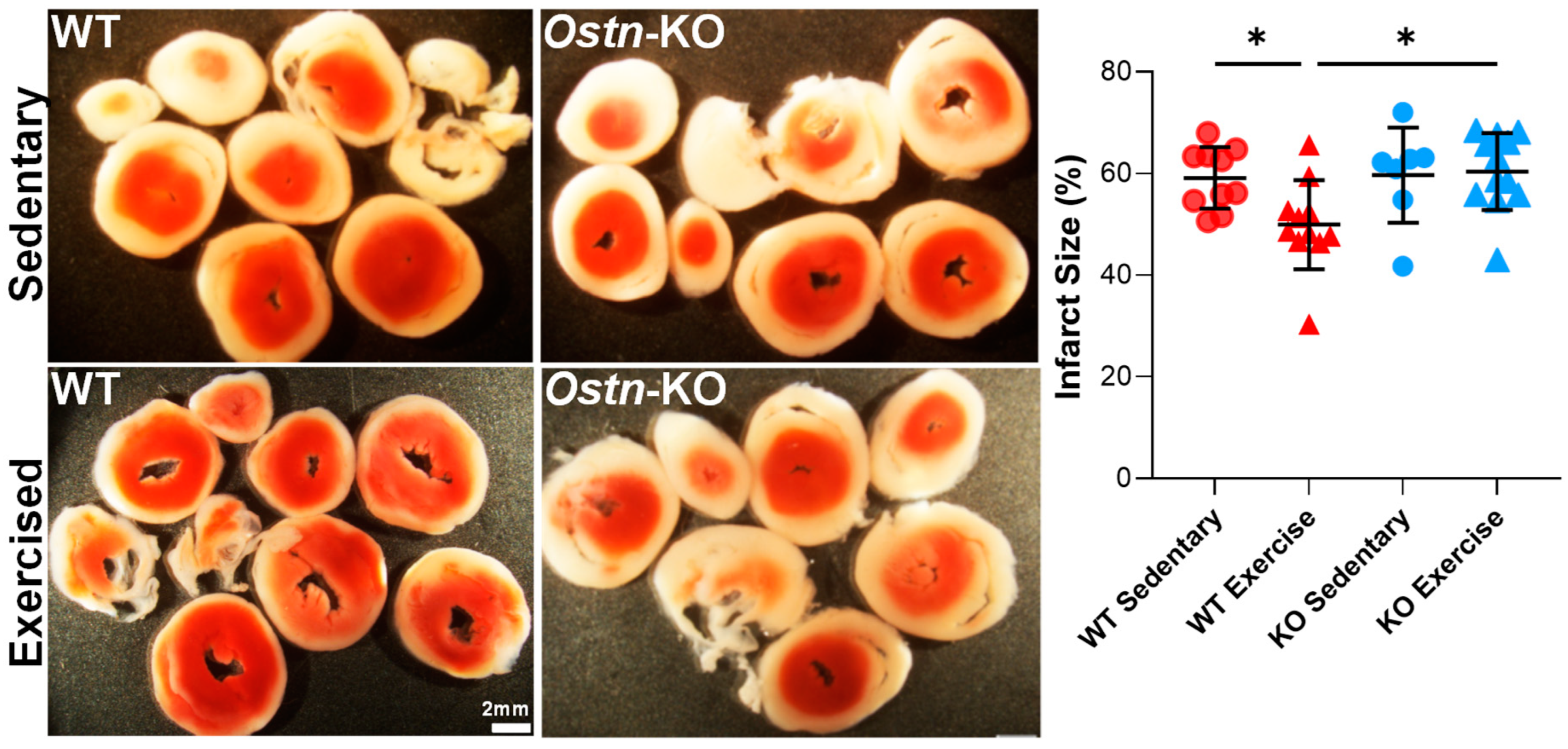
2.1. Musclin Is Required for Exercise-Mediated Protection from Ischemia-Reperfusion Injury

It is well-established that exercise training significantly improves myocardial tolerance to ischemia-reperfusion (IR) injury [32,33,34,35,36,37,38]. Musclin is established as an exercise-induced myokine with cardioprotective properties [22,23], however its role in exercise-induced cardiac protection is unknown. Here, the effect of moderate intensity treadmill exercise training on myocardial resistance to IR injury was compared in WT control vs. a mouse model with ubiquitous disruption of the musclin-encoding gene, Ostn (Ostn-KO, genOway) [22]. Specifically, we used forced treadmill exercise, which combines elements of aerobic and resistance training [22] and was previously linked to enhanced musclin production and secretion [22]. As expected, exposure to exercise training of WT controls resulted in augmented myocardial stress resistance as evidenced by smaller infarct size in response to the IR protocol (Figure 1; 49.94 ± 8.761%, n = 11) compared to that of sedentary WT mice (59.12 ± 6.033%, n = 10, p = 0.031). Importantly, in Ostn-KO mice, exercise training failed to improve resistance to IR injury (60.39 ± 7.58%, n = 12) compared to sedentary Ostn-KO mice (59.67 ± 9.351%, n = 7, p = 0.99). Exercise-trained Ostn-KO exhibited significantly larger infarct sizes than exercise-trained WT mice (p = 0.0169; two-way ANOVA values for all comparisons between groups). Thus, intact musclin production is critical for exercise-related cardioprotection from ischemic injury.

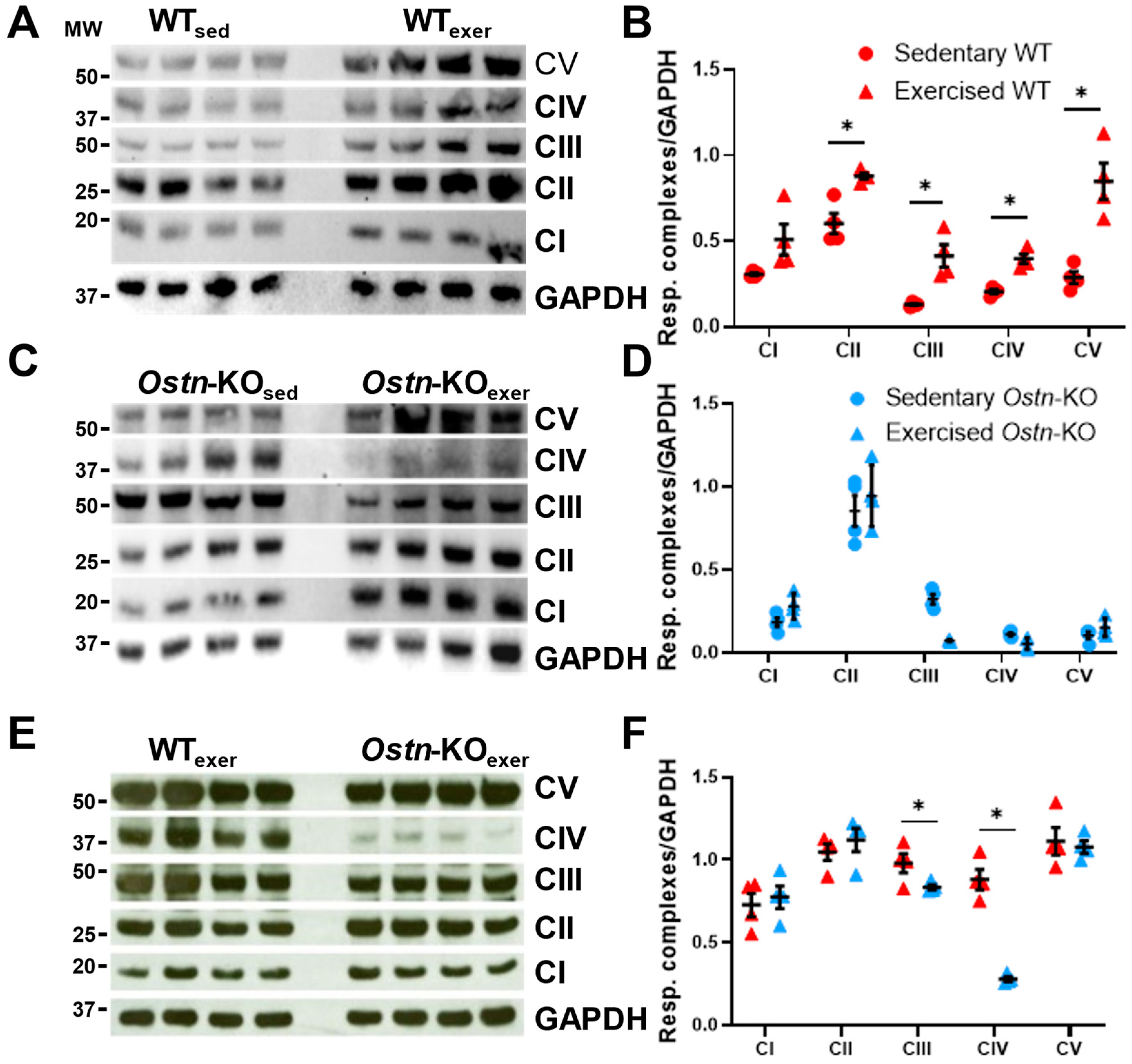
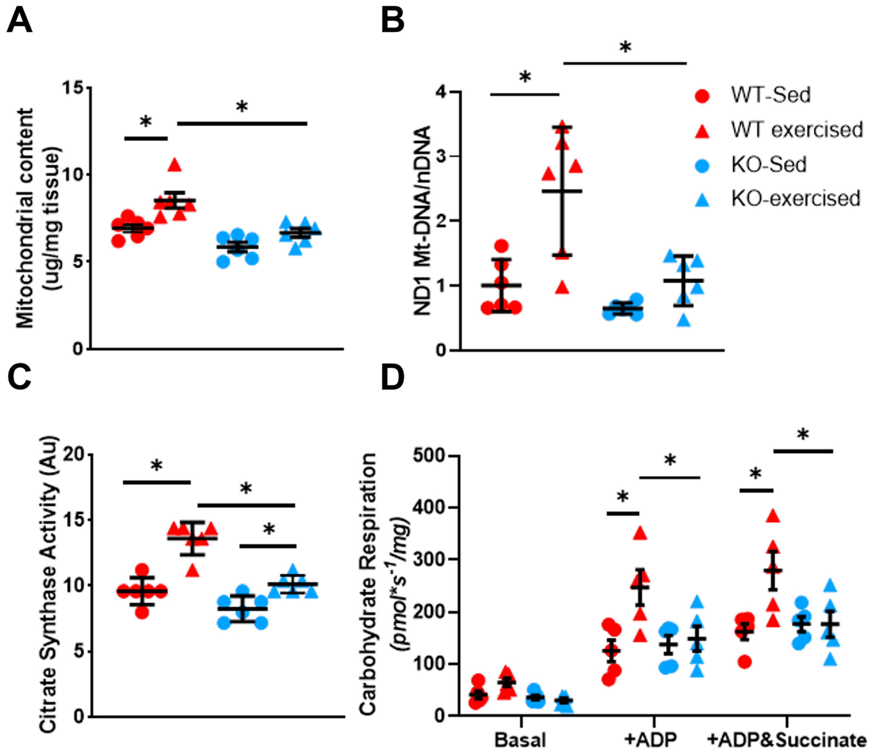
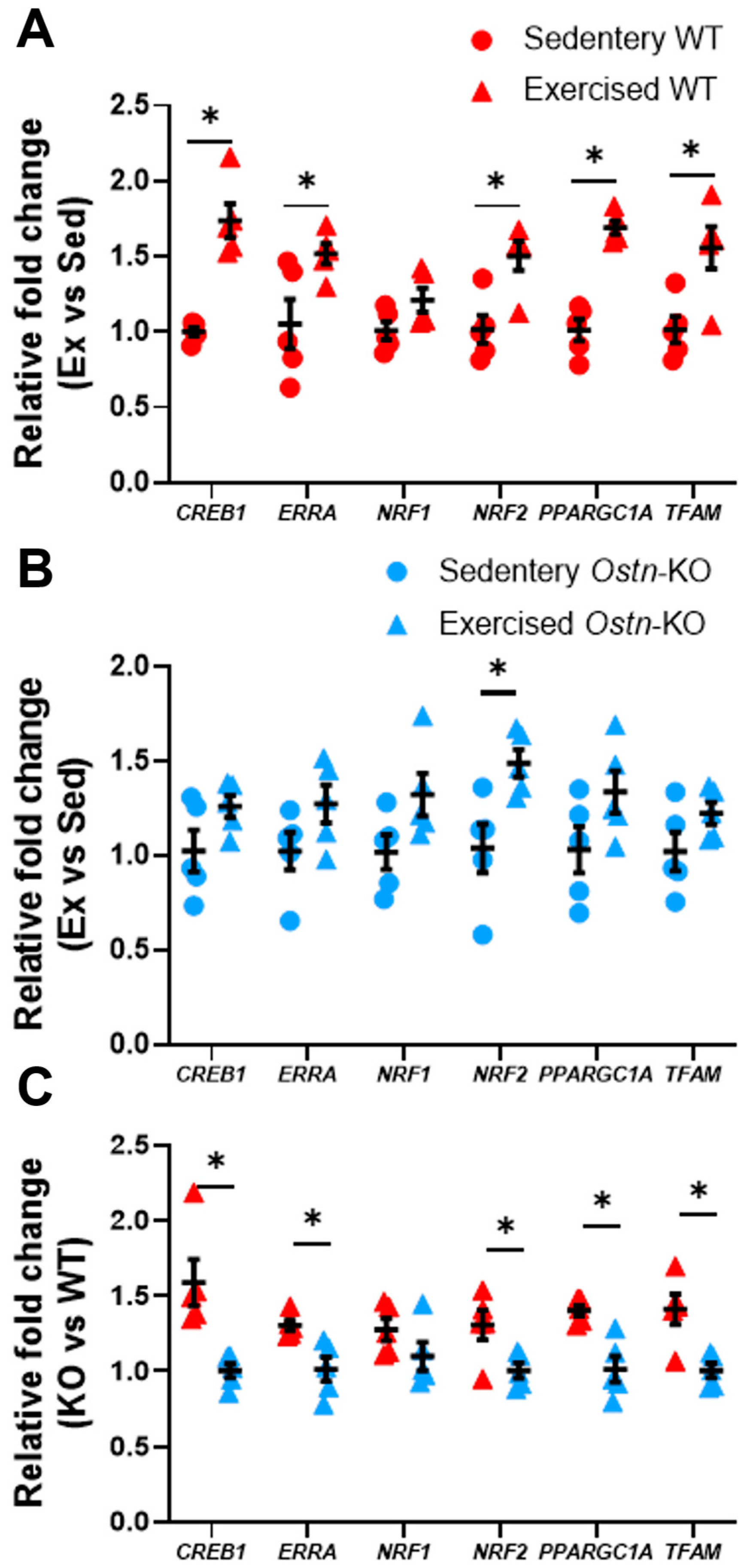
2.2. Musclin Promotes Exercise Training-Induced Enhancements in Mitochondrial Biogenesis and Respiratory Capacity

It has been previously demonstrated that exercise-induced cardiac stress resistance is at least in part mediated through mitochondrial adaptation [39,40,41]. Musclin has been shown to govern exercise-induced mitochondrial biogenesis in skeletal muscles [22]. However, its effect on physical activity-triggered mitochondrial biogenesis in the heart is yet to be established. To this end, the hearts of sedentary or exercise-trained WT and Ostn-KO mice were assessed for mitochondrial content and oxidative phosphorylation potential.
First, protein expression of the subunits of the electron transport chain was measured in both sedentary and exercise-trained Ostn-KO and WT mice. In response to exercise training in WT mice there was a significant increase in complex II expression (0.88 ± 0.04 vs. 0.60 ± 0.12 AU, n = 4, p = 0.005), complex III expression (0.41 ± 0.13 vs. 0.13 ± 0.01 AU, n = 4, p = 0.005), complex IV expression (0.40 ± 0.05 vs. 0.21 ± 0.02 AU, n = 4, p = 0.0006), and complex V expression (0.85 ± 0.21 vs. 0.29 ± 0.07, n = 4, p = 0.002) complex I expression increased, but the change was not statistically significant (0.51 ± 0.18 vs. 0.31 ± 0.02, n = 4, p=0.07; Figure 2A,B). However, in Ostn-KO mice there was no exercise-induced upregulation of OXPHOS protein expression, and significantly lower expression of complex III in comparison to sedentary Ostn-KO mice (0.13 ± 0.01 vs. 0.32 ± 0.06 AU n = 4, p = 0.0002; Figure 2C,D). When comparing WT mice to Ostn-KO mice, hearts from exercise-trained WT mice, vs. exercise-trained Ostn-KO, had significantly higher expression of complex III (0.98 ± 0.12 vs. 0.83 ± 0.03 AU, n = 4, p = 0.0499) and complex IV subunits (0.88 ± 0.12 vs. 0.28 ± 0.03 AU, n = 4, p = 0.0001; Figure 2E,F).
Additionally, there was significantly lower mitochondrial protein content when normalized to wet heart weight in both sedentary and exercise-trained Ostn-KO mice compared to WT mice (5.85 ± 0.66 vs. 6.93 ± 0.52, n = 6 and p = 0.02; 6.67 ± 0.60 vs. 8.52 ± 1.08, n = 6 and p = 0.0003, respectively, in Figure 3A). Importantly, exercise training vs. sedentariness led to a significant increase in mitochondrial protein content only in WT (6.93 ± 0.52 vs. 8.52 ± 1.08, n = 6 and p = 0.0015), but not in Ostn-KO (5.85 ± 0.66 vs. 6.67 ± 0.60, n = 6 and p = 0.122; Figure 3A).
Next, mitochondrial DNA levels were quantified. Specifically, expression was measured for mitochondrial DNA encoded NADH dehydrogenase 1 (ND1) as a marker of mitochondrial biogenesis, normalized to hexokinase 2 as a marker of nuclear DNA. This showed that exercise training promotes increased ND1 expression in WT but not in Ostn-KO mice (2.46 ± 0.99 vs. 1.00 ± 0.40, n = 6 and p = 0.002; 1.08 ± 0.38 vs. 0.65 ± 0.09 n = 6 and p = 0.56, respectively, in Figure 3B). This physical activity-induced ND1 upregulation resulted in a significantly higher ND1 level in exercised WT mice compared to exercised Ostn-KO mice (1.08 ± 0.38 vs. 2.46 ± 0.99, n = 6 and p = 0.0003 in Figure 3B), while there was no difference in ND1 level between the two groups under sedentary conditions (0.65 ± 0.09 vs. 1.00 ± 0.40, n = 6 and p = 0.70 in Figure 3B).
To further ascertain the effect of musclin on exercise-induced cardiac mitochondrial oxidative capacity, citrate synthase activity was measured in isolated mitochondria and maximal mitochondrial respiration in semi-permeabilized cardiac fibers. While both WT and Ostn-KO mice significantly increased their citrate synthase activity with exercise training in mitochondrial extracts (13.59 ± 1.24 vs. 9.59 ± 1.01 AU, n = 6 and p < 0.0001; 10.12 ± 0.65 vs. 8.26 ± 0.97 AU, n = 6 and p = 0.046, respectively), citrate synthase activity was still significantly lower in exercise-trained Ostn-KO mice in comparison to exercise trained WT mice (10.12 ± 0.65 vs. 13.59 ± 1.24 AU, n = 6 and p = 0.004 in Figure 3C).
Similarly, in semi-permeabilized myofibers isolated from hearts, only those of WT mice but not those of Ostn-KO mice exhibited an increase in mitochondrial respiration in response to exercise (Figure 3D). Under carbohydrate-supported conditions, respiration in fibers from hearts of exercise-trained mice was significantly upregulated when stimulated with ADP alone (247.29 ± 75.27 vs. 125.56 ± 46.32 pmol*s−1/mg, n = 5 and p = 0.0006) or with the addition of succinate (279.68 ± 81.51 vs. 162.63 ± 33.89 pmol*s−1/mg, n = 5 and p = 0.001 in Figure 3D). In contrast, fibers from the hearts of exercise-trained Ostn-KO mice did not exhibit upregulation of mitochondrial respiration compared to sedentary controls with ADP stimulation alone (149.11 ± 53.25 vs. 137.88 ± 39.05 pmol*s−1/mg, n = 5 and p = 0.98) or with the addition of succinate (176.76 ± 55.81 vs. 177.17 ± 31.87 pmol*s−1/mg, n = 5 and p > 0.9999). Moreover, when compared to WT mice, Ostn-KO mice had significantly lower oxygen consumption when stimulated with ADP alone (149.11 ± 53.25 vs. 247.29 ± 75.27 pmol*s−1/mg, n = 5 and p = 0.007) or with the addition of succinate (176.76 ± 55.81 vs. 279.68 ± 81.51 pmol*s−1/mg, n = 5 and p = 0.004 in Figure 3D). Differences between sedentary WT and Ostn-KO mice were less marked (Figures S1 and S2), consistent with the finding that baseline (non-exercise) levels of musclin signaling are low in WT mice [22]. Thus, musclin signaling is essential for exercise-induced cardiac mitochondrial adaptations.

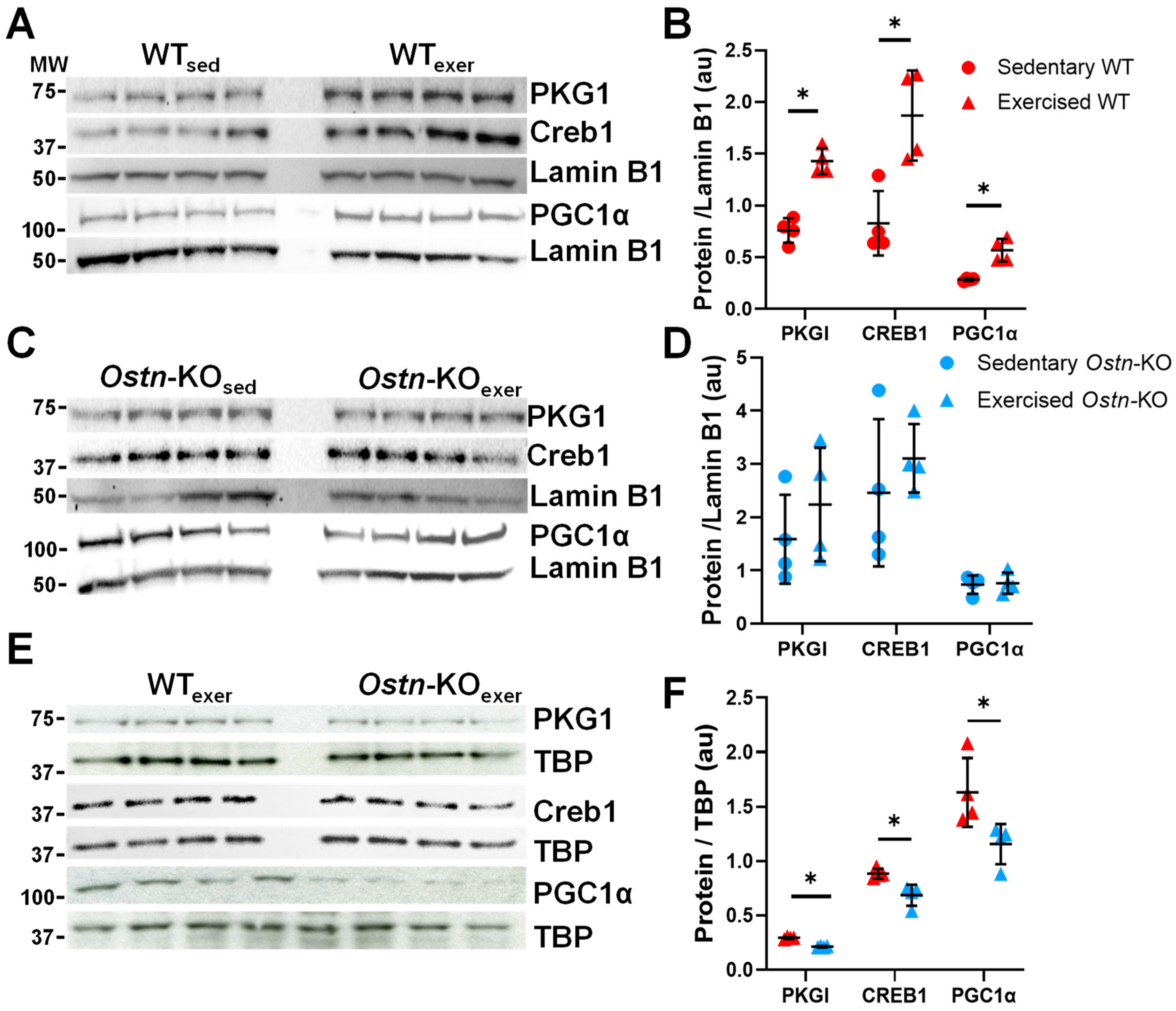
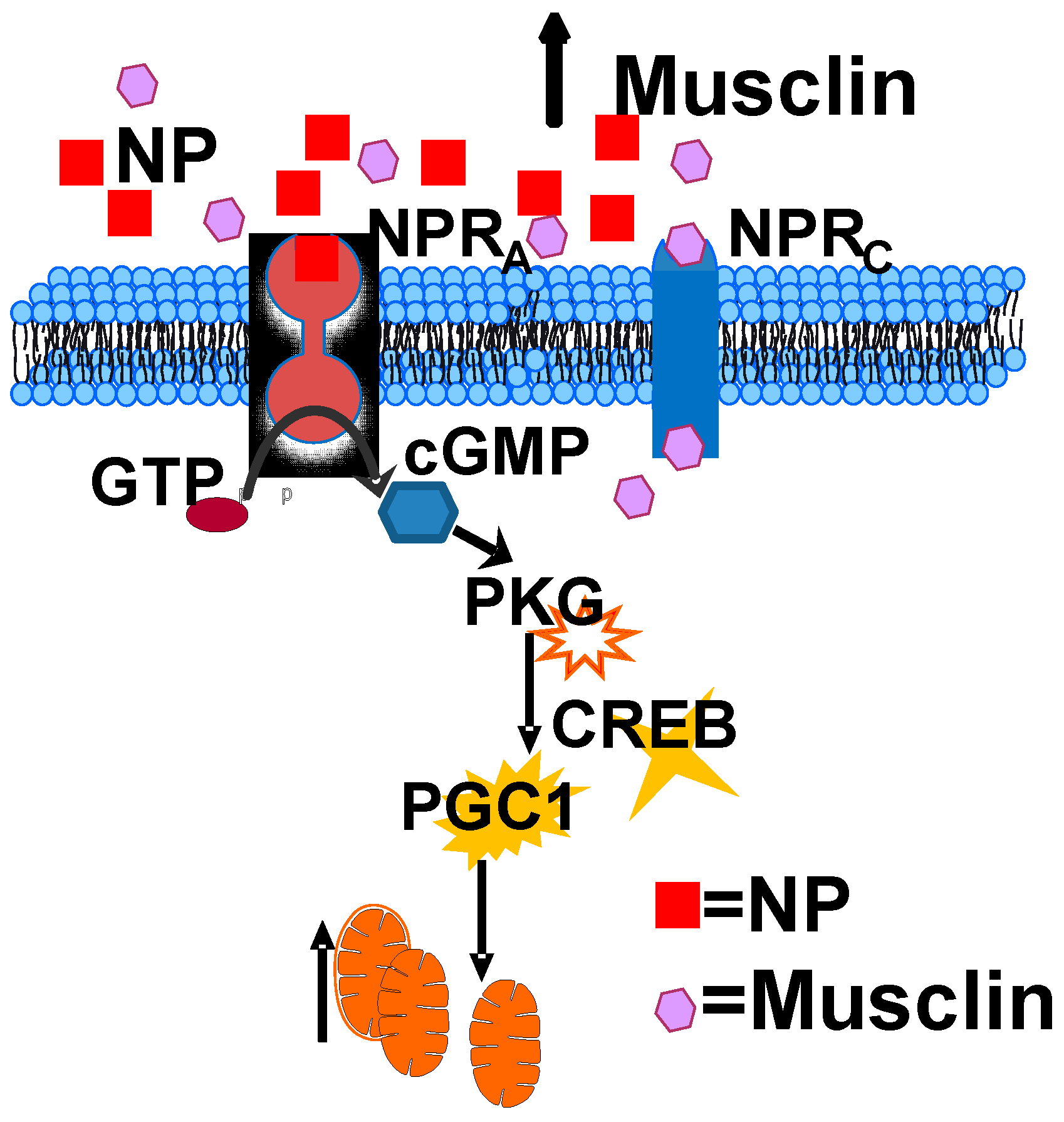
2.3. Musclin Regulates Cardiac cGMP/Protein Kinase G-Dependent Signaling

NP/cGMP signaling is recognized as a key regulator of mitochondrial biogenesis, and, given that we previously demonstrated the role of musclin in augmenting ANP/cGMP signaling in skeletal muscle [22], and others described this in the heart [23,24,42], we hypothesized that musclin would influence exercise-induced cardiac cGMP signaling. To assess this hypothesis, cGMP levels were first measured in the myocardium of exercised WT and Ostn-KO mice. We confirmed that exercise-trained WT mice had significantly higher cardiac cGMP following 8 days of treadmill exercise compared to Ostn-KO mice (98.51 ± 14.16 vs. 61.94 ± 19.66 pMol/mg, n = 5 and p = 0.01). cGMP is known to activate protein kinase G (PKG) [43], which can translocate into the nucleus (and phosphorylates cAMP response element binding (CREB) in baby hamster kidney (BHK) fibroblasts [44,45,46] promoting its nuclear localization and ability to regulate transcription of peroxisome proliferator-activated receptor γ coactivator 1α (PGC1α) [47,48]. To investigate if musclin affects this signaling cascade in the heart, the nuclear expressions of PKG1, CREB1, and PGC1α were quantified. In WT hearts collected immediately after last exercise session compared to those of sedentary WT controls, a significant increase was observed in nuclear localization of PKG1 (1.43 ± 0.13 vs. 0.76 ± 0.12 AU, n = 4 and p = 0.0003), CREB1 (1.87 ± 0.44 vs. 0.83 ± 0.01 AU, n = 4 and p = 0.31), and PGC1α (0.57 ± 0.11 vs. 0.28 ± 0.01 AU, n = 4, p = 0.002, Figure 4A,B). However, in Ostn-KO mice, exercise did not exhibit the same degree of upregulation of PKG1 (2.24 ± 1.07 vs. 1.59 ± 0.84 AU, n = 4 and p = 0.37), CREB1 (3.11 ± 0.64 vs. 2.46 ± 1.38 AU, n = 4 and p = 0.43), or PGC1α (0.76 ± 0.20 vs. 0.73 ± 0.17 AU, n = 4 and p = 0.85 in Figure 4C,D). Additionally, when comparing exercised groups, Ostn-KO had significantly lower nuclear expression than WT of PKG1 (0.21 ± 0.01 vs. 0.29 ± 0.01 AU, n = 4 and p < 0.0001), CREB1 (0.69 ± 0.10 vs. 0.89 ± 0.05 AU, n = 5 and p = 0.01), and PGC1α (1.16 ± 0.19 vs. 1.63 ± 0.32 AU, n = 4 and p = 0.04 in Figure 4E,F).
Similarly, in response to exercise in WT mice, compared to sedentary WT controls, there was upregulation in cardiac mRNA expression of Creb1 (1.73 ± 0.25 vs. 1.00 ± 0.06 fold change, n = 5 and p = 0.0002) and PPARG Coactivator 1 Alpha (Ppargc1a) (1.69 ± 0.09 vs. 1.01 ± 0.16 fold change, n = 5 and p < 0.00004) as well as their downstream targets linked to mitochondrial biogenesis: Estrogen Related Receptor Alpha (Erra) (1.52 ± 0.15 vs. 1.05 ± 0.37 fold change, n = 5 and p = 0.029), Nuclear Respiratory Factor 2 (Nrf2) (1.50 ± 0.22 vs. 1.02 ± 0.21 fold change, n = 5 and p = 0.007), and Transcription Factor A, Mitochondrial (Tfam) (1.56 ± 0.31 vs. 1.01 ± 0.19 fold change, n = 5 and p = 0.011) but not Nuclear Respiratory Factor 1 (Nrf1) (1.21 ± 0.18 vs. 1.01 ± 0.14, n = 5 and p = 0.08 in Figure 5A). This upregulation in gene expression was not observed in response to exercise training in Ostn-KO mice: Creb1 (1.26 ± 0.13 vs. 1.02 ± 0.25 fold change, n = 5 and p = 0.096), Ppargc1a (1.34 ± 0.25 vs. 1.03 ± 0.27 fold change, n = 5, 0.1), Erra (1.27 ± 0.22 vs. 1.02 ± 0.22 fold change, n = 5 and p = 0.11), Nrf1 (1.32 ± 0.25 vs. 1.02 ± 0.21 fold change, n = 5 and p = 0.07), Nrf2 (1.49 ± 0.16 vs. 1.04 ± 0.29 fold change, n = 5 and p = 0.02), and Tfam (1.22 ± 0.13 vs. 1.02 ± 0.23 fold change, n = 5 and p = 0.13 in Figure 5B). Additionally, in exercised WT mice compared to Ostn-KO mice, there was significantly higher expression of Creb1 (1.59 ± 0.34 vs. 1.00 ± 0.10 fold change, n = 5, p = 0.007), Ppargc1a (1.40 ± 0.08 vs. 1.01 ± 0.19 fold change, n = 5 and p = 0.003), Erra (1.30 ± 0.08 vs. 1.01 ± 0.18 fold change, n = 5 and p = 0.009), Nrf2 (1.31 ± 0.22 vs. 1.01 ± 0.11 fold change, n = 5 and p = 0.03), and Tfam (1.41 ± 0.22 vs. 1.01 ± 0.11 fold change, n = 5 and p = 0.006), but not Nrf1 (1.27 ± 0.16 vs. 1.09 ± 0.21 fold change, n = 5 and p = 0.17 in Figure 5C). Hearts of sedentary Ostn-KO and WT mice exhibited similar levels of nuclear PKG1 expression (0.69 ± 0.12 vs. 0.88 ± 0.22 AU, n = 4 and p < 0.18 in Figure S2A,B) and mRNA levels of Creb1 (0.93 ± 0.22 vs. 1.00 ± 0.06 fold change, n = 5 and p = 0.49), Ppargc1a (0.96 ± 0.27 vs. 1.01 ± 0.16 fold change, n = 5 and p = 0.77), Erra (0.97 ± 0.21 vs. 1.05 ± 0.37 fold change, n = 5 and p = 0.68), Nrf1 (0.99 ± 0.19 vs. 0.95 ± 0.19 fold change, n = 5 and p = 0.78), and Tfam (0.94 ± 0.21 vs. 1.01 ± 0.20 fold change, n = 5 and p = 0.60 in Figure S2C), Only expression of Nrf2 was slightly lower in sedentary Ostn-KO mice (0.82 ± 0.17 vs. 1.09 ± 0.15 fold change, n = 5 and p = 0.03 in Figure S2C). Taken together, these results indicate that musclin enhances physical activity-induced cardiac mitochondrial biogenesis through augmentation of cGMP/PKG/CREB-dependent signaling.

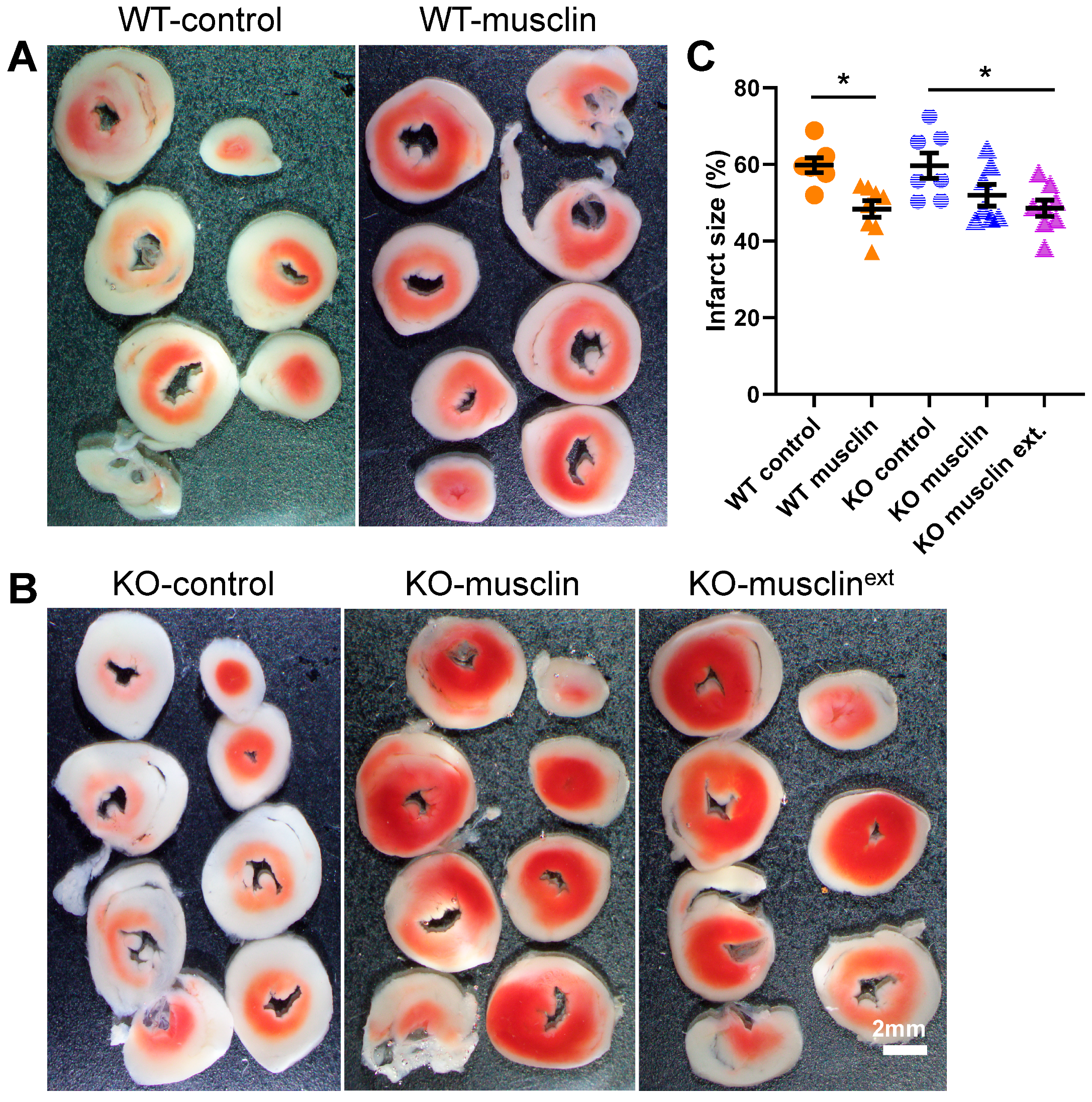
2.4. Synthetic Musclin Infusion Mimics Exercise-Mediated Protection against IR Injury

Exercise is one of the most effective ways to improve cardiac function and stress resistance [6,7,8,9,10,11,12,13,15,16,17,18]. However, adherence among those with heart disease to even short exercise programs, such as cardiac rehabilitation, is suboptimal [49]. The reasons are multifactorial, but a major contributor is baseline exercise intolerance, which is common in those with heart disease [7,50]. Synthetic musclin infusion has been demonstrated to be effective for reduction of myocardial injury and recovery of cardiac function when used after coronary artery ligation [23], but the possibility of using musclin infusion as a therapeutic strategy to induce a profile of benefits overlapping with exercise-induced cardiac conditioning in order to prevent myocardial damage and loss of cardiomyocytes is an intriguing possibility. To test this concept, synthetic peptide, corresponding to aa 80–112 of the murine musclin [22,30], was tested. This fragment has been associated with the most prominent modulation of ANP signaling in previous studies [22,30].
First, this synthetic peptide, containing uniformly labeled with 13C and 15N alanine and valine at positions 26 and 27, was used to define the volume of distribution (Vd) and plasma elimination half-life based on the calculations described in the Methods section (Figure 6A–E). These parameters were used to calculate the infusion rate necessary to maintain a musclin plasma level comparable with endogenously produced levels observed after exercise training [22] (Figure 6A–E).
Musclin treatment for 14 days significantly reduced myocardial infarct size following IR in sedentary WT mice compared to treatment with the control peptide (48.39 ± 2.15 vs. 59.82 ± 1.91%, n = 7–8 and p = 0.002 in Figure 7A,C). While 14 days of musclin infusion did not significantly reduce myocardial infarct size in sedentary Ostn-KO mice (Figure 7B,C), 28 days of treatment reduced infarct size following IR (48.57 ± 2.14 vs. 59.69 ± 3.30%, n = 7–8 and p = 0.013 in Figure 7B,C). Importantly, enhanced cardiac tolerance to IR injury after musclin infusion was associated with markers of increased mitochondrial biogenesis (Figure S3). These results demonstrate that exogenous musclin mimics exercise-induced cardiac conditioning and stress resistance.

3. Discussion

This study establishes that the exercise-responsive myokine musclin [22] is vital for exercise-induced cardiac resistance to IR stress. Disruption of musclin signaling in Ostn-KO mice results in reduced oxidative phosphorylation potential and greater vulnerability to ischemic injury. Musclin replacement therapy restored cardiac resistance to stress in Ostn-KO mice. These findings indicate a previously unrecognized pathway for cardiac adaptation to exercise.
Several studies indicate that exercise training improves myocardial tolerance to IR injury (e.g., myocardial infarction in both young and old, male and female, animals) [32,33,34,36,37,51,52,53,54,55,56,57,58,59,60,61,62,63,64,65,66,67,68,69]. Specifically, studies reveal that exercise training protects the heart against arrhythmias, oxidative injury, mitochondrial damage, and cell death [33,55]. Interestingly, several investigators have shown that short-term exercise training (3–5 consecutive days) provides the same cardioprotection as that observed following long-term (10 weeks) training [34,51,52,55,56,57,62,63,70,71]. Here, we use forced treadmill exercise which combines elements of aerobic and resistance training [22]. We previously demonstrated that this exercise protocol is associated with enhanced musclin production and secretion [22], which in turn modulates atrial natriuretic peptide (ANP)/cyclic GMP signaling [22] in skeletal muscles. The current study confirms that intact musclin function promotes myocardial cGMP signaling triggered by exercise in the heart.
The data in the present study also confirm that intact musclin signaling is necessary for exercise-induced activation of PKGI-CREB/PGC1α, mitochondrial biogenesis, and functional adaptations in the myocardium. Notably, the results indicate significantly greater mitochondrial quantity in WT compared to musclin-deficient hearts by multiple methods and demonstrate the functional importance of the observed changes by revealing a significant deficiency in respiration measured in perforated myofibers isolated from Ostn-KO mice. These findings agree with the previously reported musclin interaction with natriuretic peptide receptors [23,24,30,31,42] and the detection of significantly higher levels of cGMP in the myocardium of WT mice, compared with Ostn-KO mice, after an exercise bout. There are accumulating data suggesting cGMP is a critical driver of mitochondrial biogenesis [72,73,74,75,76,77,78]. In fact, cGMP signaling has been linked to PGC1α–dependent mitochondrial biogenesis in many studies in different tissues and organs, including our own prior study indicating musclin-dependent activation of cGMP/PGC1α-driven mitochondrial biogenesis in skeletal muscles [22], while the current data indicate the significance of this musclin-driven signaling in the heart (Figure 8).
Importantly, different time points after the last exercise training session were used here to reveal specific features of exercise-induced cardiac conditioning. Hearts were collected immediately after exercise for cGMP measurement and 10–15 min after exercise to evaluate the nuclear presence of PKGI and CREB, while nuclear expression of PGC1α and mitochondrial respiratory complexes were evaluated in hearts collected 2 h after the last exercise session to probe the previously reported dynamics of cGMP/PKGI signaling [44,79,80].
Although treadmill exercise training was used here to stimulate musclin signaling, results indicate a phenotype of lower mitochondrial content and reduced mitochondrial respiration even in untrained Ostn-KO mice compared with WT controls. Importantly, these observed trends and changes in sedentary Ostn-KO became much more obvious and significant after exposure to the exercise protocol. These findings can be interpreted to indicate that musclin production and secretion, although more easily demonstrated in response to vigorous exercise, are physiologically relevant, even for routine daily activities.
Cardioprotective properties of musclin were demonstrated previously in different murine models of cardiomyopathies [10,23,81,82]. Our work demonstrates the role of musclin in cardiac conditioning and protection that is specifically exercise-induced. This establishes a previously unrecognized link between muscle activity and cardiac conditioning and supports a therapeutic potential in critically ill, injured, or otherwise high cardiac-risk surgical candidates in whom significant loss of muscle mass due to the catabolic stress of immobility and/or inadequate nutrition associated with any critical illness [83] may undermine optimal intrinsic musclin signaling.
In fact, the data from this study indicate that infusion of a synthetic fragment of musclin peptide mimics exercise benefits with respect to protection of the myocardium from IR injury. This is very encouraging as pharmacological options available to mimic exercise-induced cardioprotection have not previously been clinically available. In contrast to ANP or BNP infusion, which can directly stimulate cGMP synthesis, musclin promotes cGMP signaling through inhibition of other NPs via competitive inhibition of NPRC-driven clearance [22,23]. In this way, musclin infusion would enhance the natural dynamics of NP signaling as well as any potential beneficial effects related to local signaling by multiple NPs that are cleared through the NPRC [84], in contrast to systemic infusion of ANP and BNP that would presumably result in non-physiologically high levels in off-targets and possibly insufficient levels in target tissues such as the myocardium. Interestingly, inhibition of another pathway for NP degradation by neutral endopeptidase has been linked to a survival benefit in heart failure (HF) patients [85]. In contrast, ANP or BNP infusion, while leading to improvement in measures of HF, has been linked to an increased risk of side-effects related to hypotension or nephrotoxicity [86,87,88], supporting the concept that off-target effects of direct infusion of natriuretic peptides are less desirable than methods to boost their intrinsic local concentrations and effects.
The present study also includes pharmacokinetic studies that allow for the calculation of the infusion rate necessary to secure a plasma musclin concentration close to that previously linked to exercise training and enhanced cGMP signaling [22]. As such, the findings of this study may help standardize future experiments with synthetic musclin.
Surprisingly, disruption of musclin signaling in Ostn-KO mice practically abolished exercise-induced cardioprotection. Considering the complexity of exercise-induced adaptations, this is somewhat unexpected. However, this finding may occur since musclin is also important for the skeletal muscle response to physical activity and, as such, Ostn-KO mice may not be getting the full benefits of exercise training [22]. Furthermore, this study investigates the role of musclin in augmenting NP/cGMP signaling. However, there is emerging research that indicates the existence of NPRc—specific signaling [89,90,91]. This possible musclin/NPRc interaction should be the subject of future studies.
Finally, this study further verifies the unique interaction of skeletal muscle and heart through the previously reported co-regulation of endocrine signals: cardiac NPs and the myokine musclin [22,42]. Cardiac NPs, particularly ANP, have been increasingly recognized as powerful exercise-responsive regulators of important physiological functions [92,93,94,95]. Uncovering this musclin-dependent effect on exercise-induced cardiac conditioning broadens appreciation of the complexity and physiologic roles of cardiac and skeletal muscles as endocrine organs. In the future it will be interesting to clarify the effect of musclin signaling on other NP-regulated physiologic phenomenon such as blood pressure homeostasis as well as glucose and fat metabolism [84,96,97]. Additional investigation, including direct comparison of different exercise modes (resistance vs. aerobic) as well as forced vs. voluntary exercise, along with different time exposures, are needed to fully understand the range of musclin-induced adaptations that underlie its beneficial cardioprotective effects. Beyond advancing the understanding of musclin’s effects and musclin’s role in mechanisms of exercise-induced cardiac conditioning, this study has translational implications by extending the concept of musclin-dependent cardioprotection to the preconditioning of hearts to improve their tolerance of future ischemic insults using synthetic musclin infusion. This could represent a new therapeutic strategy to improve outcomes of stressful cardiac events or procedures, especially in exercise-intolerant patients.

4. Methods

4.1. Animal Experiments

All animal protocols conform to the Guide for the Care and Use of Laboratory Animals generated by the Institute for Laboratory Animal Research, National Research Council of the National Academies. All animal protocols were approved by the University of Iowa Institutional Animal Care and Use Committee. For all experiments, mice were anesthetized with inhaled isoflurane (5% induction, 1–1.5% maintenance; Piramal Healthcare) to maintain a respiratory rate of ~50–60 breaths per minute.

4.2. Ostn-KO Mouse Model

Ostn-KO mice were generated as described previously [22]. Vector construction and targeted knockout strategy were designed together with genOway: Mice were generated based on deletion of a 2.1-kb sequence flanking Ostn exon 2, resulting in inactivation of the ATG and signal peptide. All mice used in this study were male, 6–8 weeks of age, and on a C57BL6/N background. Control WT C57BL6/N were originally purchased from Charles River (Wilmington, MA, USA). Heterozygous Ostn-KO mice were bred to obtain homozygous Ostn-KO mice and WT controls.

4.3. Myocardial Ischemia–Reperfusion Injury

Hearts were extracted from anesthetized mice and retrogradely perfused at 90 mm Hg with Krebs–Henseleit buffer bubbled with 95% O2/5% CO2, at 37 °C and pH 7.4. Myocardial ischemia-reperfusion (IR) injury was created as follows: after allowing 20 min for stabilization, the hearts were subjected to 20 min of global ischemia and 45 min of reperfusion. At the end of this treatment, the hearts were removed from the Langendorff perfusion apparatus and immediately frozen at −20 °C. The frozen hearts were then cut into transverse slices of approximately equal thickness (∼0.8 mm) from apex to base. The slices were placed into a small cell culture dish and then incubated in 1% triphenyltetrazolium chloride (TTC) in phosphate buffer (Na2HPO4 88 mmol/L, NaH2PO4 1.8 mmol/L, pH 7.8) at 37 °C for 20 min while gently shaking the dish. The development of the red formazan pigment in living tissues relies on the presence of lactate dehydrogenase, or NADH, while failure to stain red indicates a loss of these constituents from necrotic tissue. After staining, the TTC buffer was replaced by 10% formaldehyde. The slices were fixed for the next 4–6 h, and then the areas of infarct tissue were quantified using ImageJ software. The risk area is taken as the total ventricular cross-sectional area of all slices. The infarct size is presented as infarcted percentage of at-risk area.

4.4. Treadmill Exercise Training

Three days before exercise, mice were acclimated for 45 min daily on a non-moving treadmill (Columbus Instruments, Columbus, OH, USA) followed by 15 min at a velocity of 3.5 m/min at 15° incline. For IR studies and assessment of nuclear PKG and PGC1α, mice were then exercised daily for total of 10 days: five consecutive days (Monday to Friday), followed by a 2-day rest, and then exercised daily for three more days (Monday, Tuesday, Wednesday) at a speed of 15 m/min and inclination of 15° for 45 min. This protocol has been previously shown to increase the production and secretion of musclin [22]. Immediately after the last exercise session, mice were sacrificed, and their hearts were collected for cGMP measurements. To evaluate the nuclear presence of PKGI and CREB, hearts were collected 10–15 min after the last exercise. To test nuclear expression of PGC1α and mitochondrial respiratory complexes, hearts were collected 2 h after the last exercise. Mitochondrial DNA, mRNA, protein expression, and mitochondrial respiration changes were assessed following 15 days of exercise training as described above (5 d/week daily exercise for 3 wks) with hearts collected 2 h after the last exercise.

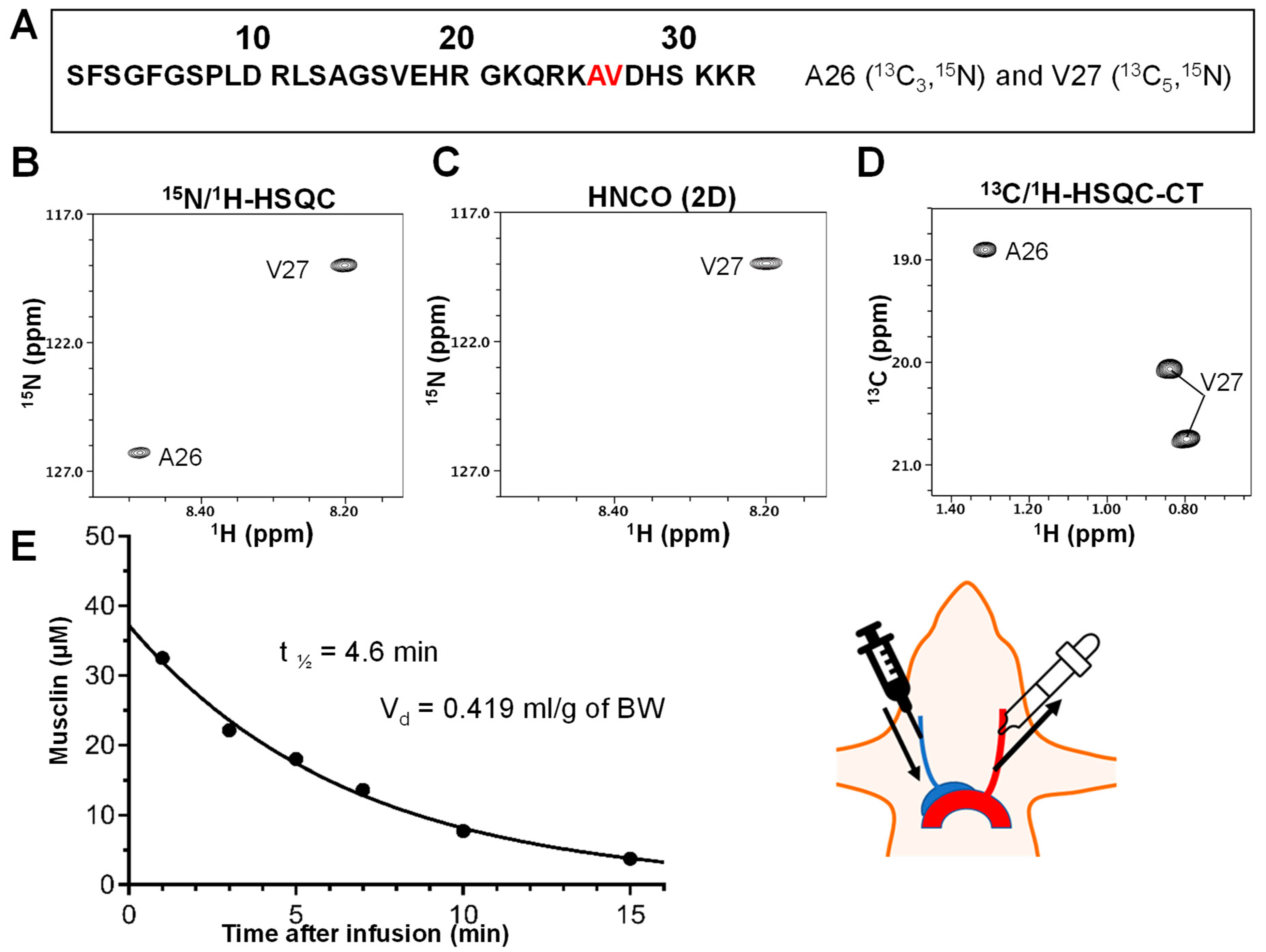
4.5. Determination of Synthetic Musclin Peptide Plasma Elimination Half-Life and Volume of Distribution

The musclin [13C,15N-A26-V27]-labeled peptide sequence used was as follows: SFSGFGSPLDRLSAGSVEHRGKQRKAVDHSKKR, corresponding to amino acids 80–112; GenBank accession no. AAS87598.1 (Bio-synthesis, Inc. Lewisville, TX, USA). A peptide was synthesized with 98% purity, counter-ion exchange (TFA to Cl), and pH adjustment (Bio-synthesis, Inc. Lewisville, TX, USA).
For infusion, 1.7 mg of the chemically synthesized [13C,15N-A26-V27]-labeled musclin peptide was dissolved in 300 µL of PBS. This musclin solution was injected into the jugular vein. A 100 µL blood sample was then drawn from the carotid artery at 1, 3, 5, 7, 10, and 15 min after the peptide infusion. Blood was mixed with 5 µL of 10% NaEDTA and 1 µL of peptidase inhibitor cocktail (Sigma, St. Louis, MI, USA) and kept on ice, then centrifuged at 4 °C for 45 min at 14,000 rpm. Plasma was removed and stored at −80 °C for NMR studies.

4.6. NMR Spectroscopy

All NMR spectra were acquired at 5 °C on a Bruker Avance II 800 MHz NMR spectrometer equipped with a TCI cryoprobe. 100 µL of plasma (see above) was added 150 µL NMR buffer containing 50 mM sodium phosphate, pH 7.1 with a final volume of 250 µL in 8.5% D2O/91.5% H2O. This sample was then transferred to a Shigemi NMR tube for NMR studies. One-dimensional (1D) 1H, two-dimensional (2D) 15N/1H-heteronuclear single-quantum correlation (HSQC), 2D 13C/1H-HSQC with constant time experiment (HSQC-CT), and 2D 15N/1H-slice of the three-dimensional (3D) HNCO experiment [98,99] were acquired for each blood sample drawn at different times after peptide infusion. The collected NMR data were processed using NMRPipe [100] and analyzed using NMRView [101]. The musclin peptide concentration present in these blood samples was calibrated by using a known amount of chemically synthesized [13C,15N-A26-V27]-labeled musclin peptide. The peak intensity of these acquired NMR spectra was measured and fitted to the equation for half-life in exponential decay using GraphPad Prism (GraphPad Software version 9.4.1).

4.7. Pharmacokinetic Calculations

  • Peptide MW: 3649.675
  • Initial concentration: 36 µM = 0.131 mg/mL
  • Q—infusion rate
  • Css—steady state concentration
  • Cl—clearance
  • T1/2—half-life
  • Ke—elimination rate = 0.693/T1/2 = 0.151 min−1
  • Vd—volume of distribution = administered dose/initial concentration
  • 1.7 mg/0.131 mg/mL = 12.997 mL for mice of 31 g → 0.419 mL/g of BW
  • Desired steady state concentration: 200 pg/mL
  • Cl = Ke × Vd = 0.151 min−1 × 0.419 mL/g × 27 g = 1.71 mL/min
  • Q = Css × Cl = 200 pg/mL × 1.71 mL/min = 341.652 pg/min → 20.28 ng/h
  • For mice of 27 g:
  • Q = 174.636 pg/min → 10.478 ng/h → 3520.608 ng for 2 weeks
  • For osmotic pumps with 0.25 µL/h → 20.28 × 4 = 81.966 ng/µL → 8.197 µg per pump

4.8. Osmotic Pumps

Osmotic pumps (model 1002 or 1004, 100 µL volume, 0.25 or 0.11 µL/h release rate, 14–28 d duration; (Alzet Durect) were loaded with saline vs. mouse musclin vs. control peptide. Musclin peptide, corresponding to amino acids 80–112 of full length musclin; GenBank accession no. AAS87598.1. Control peptide has point mutations at amino acid residues that have homology with atrial natriuretic peptide (ANP) (Asp89Gly and Arg90Gly) [23]. Each amino acid sequence is as follows: musclin SFSGFGSPLDRLSAGSVEHRGKQRKAVDHSKKR, and control SFSGFGSPLGGLSAGSVEHRGKQRKAVDHSKKR.
Peptides dissolved in PBS were loaded into osmotic pumps, based on the pharmacokinetic data obtained with labeled peptide (see above). Pumps were surgically implanted within the peritoneal cavity of mice under sterile conditions and general anesthesia.

4.9. Mitochondrial Protein Isolation

Mitochondrial protein fraction was isolated from hearts by using the Mitochondria Isolation Kit (Thermo Scientific, Waltham, MA, USA) according to the manufacturer’s protocol. Mitochondrial protein content was determined as the amount of mitochondrial protein per mg of wet tissue.

4.10. RNA Isolation and qRT-PCR

Total RNA from cardiac tissues was isolated by using the RNeasy RNA Isolation Kit (Qiagen). A 1 µg primary myocyte or tissue RNA sample was used to synthesize cDNA in 50 µL reactions using Oligo(dT) as primer and SuperScript III Reverse Transcriptase (Invitrogen). Quantitative real-time PCR was performed on a Mastercycler EP Gradient S (Eppendorf) by using SYBR green-based PCRs. For qRT-PCR, 1 µL of reverse transcription reaction was mixed with 10 pmol of each specific primer and 12.5 µL of SYBR PCR Master Mix (Bio-Rad, Hercules, CA, USA). The reaction was incubated for 40 cycles consisting of denaturation at 95 °C for 10 s and annealing/extension at 59.9 °C for HPRT and Nrf2, and 56 °C for Creb1, ERRA, PGC1α, Tfam, and Nrf1 for 1 min. The quality of the PCR product was routinely checked by a thermal denaturation curve following the qPCR reactions. The threshold cycle (CT) was determined by Realplex2 software (Eppendorf), and quantification of relative mRNA levels was performed by ∆∆CT method or by calculation of the absolute number of copies based on the standard curve. The primers used in this study are as follows: HPRT forward (F), GGA CCT CTC GAA GTG TTG GAT AC, and HPRT reverse (R), GCT CAT CTT AGG CTT TGT ATT TGG CT; Creb1: Creb1F, CAG TAC ATT GCC ATT ACC CAG G, and Creb1R, GCA CTA GAA TCT GCT GTC CA; ERRA: ERRAF, GGA AGA CAG CCC CAG TGA, and ERRAR, AGT GAC AGT GAG GAG AAG CC; PGC1α: PGC1-αF, TGA TGT GAA TGA CTT GGA TAC AGA CA, and PGC1αR, GCT CAT TGT TGT ACT GGT TGG ATA TG; TfamF, GGA ATG TGG AGC GTG CTA AAA, and TfamR, TGC TGG AAA AAC ACT TCG GAA TA; Nrf1F, CGC AGC ACC TTT GGA GAA, and Nrf1R, CCC GAC CTG TGG AAT ACT TG; and Nrf2F, CAG CTC AAG GGC ACA GTG C, and Nrf2R,– GTG GCC CAA GTC TTG CTC C.

4.11. Western Blotting

Whole-cell protein extracts were prepared by homogenizing cardiac tissue in RIPA Buffer (Sigma), supplemented with protease and phosphatase inhibitors (Roche Inc. Basel, Switzerland). Nuclear extracts were obtained by using the Subcellular Protein Fractionation Kit for Tissues (Thermo Scientific). Electrophoresis was performed on 3–8% gradient Nu-Page Tris-Acetate or 10% Nu-Page Bis-Tris gels and transferred to 0.2 μm Sequi-Blot PVDF membranes (Bio-Rad). The membranes were blotted with total Creb1 (Cell Signaling, Danvers, MA, USA), PKG1 (Cell Signaling), total OXPHOS (Abcam, Cambridge, UK), PGC1α (Abcam), TBP, Lamin B1, and GAPDH (Cell Signaling) antibodies. Densitometric analysis of Western blots was performed using Adobe Photoshop (Adobe Systems, San Jose, CA, USA). For representative individual gel/blots, the reference proteins (laminB1 and TBP) are shown immediately below the proteins of interest.

4.12. CGMP Measurement

cGMP levels were measured using the cyclic GMP enzyme immunoassay (EIA) kit (Cayman Chemical, Ann Arbor, MI). On the day of the experiment, mice were exercised at 12 m/min for 20 min while sedated with isoflurane, and heart tissue was collected immediately at the end of exercise. Cyclic nucleotides were extracted by using 5% (wt/vol) trichloroacetic acid. Samples were acetylated and used for cGMP level measurements according to the manufacturer’s protocol.

4.13. Mitochondrial Respiration in Isolated Myofibers

Mitochondria respiratory function was assessed using an Oroboros Instruments Oxygraph 2K (Innsbruck, Austria) as described previously [102]. Cardiac fibers from a small piece of the endocardium (~15 mg) were dissected and separated into bundles in ice-cold BIOPS buffer (7.23 mM K2EGTA, 2.77 mM CaK2EGTA, 20 mM imidazole, Sigma-Aldrich, I2399), 0.5 mM DTT, 20 mM taurine, 5.7 mM ATP (Sigma-Aldrich, St. Louis, MI, USA, A7699), 14.3 mM phosphocreatine (Sigma-Aldrich, P7936), 6.56 mM MgCl2·6H2O, and 50 mM MES hydrate, pH 7.1). For permeabilization, fibers were then transferred to 1 mL of BIOPS buffer containing 40 µg/mL of saponin (Sigma-Aldrich, S7900) and gently mixed for 30 min at 4 °C. After this point, fibers were then transferred to 1 mL of respiration buffer (105 mM K-MES, 30 mM KCl, 10 mM K2HPO4, 5 mM MgCl2·6H2O, 1 mM EGTA, 20 µm blebbistatin (Selleck Chem, Houston, TX, USA; S7099), 2.5 mg/mL fatty acid free BSA (Sigma-Aldrich, A8806, pH 7.4), and gently mixed for an additional 10 min at 4 °C. Following incubation, 1–1.5 mg of tissue was added into the O2K chamber, which was filled with 2 mL of respiration buffer. Carbohydrate (CHO)-supported respiration was assessed with pyruvate (5 mM) and malate (2 mM) with subsequent addition of ADP (5 mM) and glutamate (5 mM). Maximal CHO-supported respiratory capacity was determined in the presence of these substrates with the subsequent addition of succinate (5 mM).

4.14. Mitochondrial DNA

DNA was isolated by using QIAamp DNA Mini Kit (no. 51034, Qiagen). Mitochondrial DNA (mtDNA) contents were determined by quantitating expression of a mitochondria-encoded gene, ND1 (forward: 5′-CTAGCAGAAACAAACCGGGC-3′; reverse: 5′-CCGGCTGCGTATTCTACGTT-3′) and a nuclear-encoded gene, hexokinase 2 (forward: 5′-GCCAGCCTCTCCTGATTTTAGTGT-3′; reverse: 5′-GGGAACACAAAAGACCTCTTCTGG-3′), as described previously [103]. The expression of ND1 was normalized to the expression of hexokinase 2.

4.15. Citrate Synthase Activity

Citrate synthase activity was determined using a colorimetric assay based on the reaction between 5,5′-dithiobis-(2-nitrobenzoic acid) (DTNB) and CoA-SH to form 5-thio-2-nitrobenzoic acid (TNB), which exhibits maximum absorbance at 412 nm, according to the manufacturer’s instructions (Sigma). A mitochondrial pellet fraction was isolated from the heart by using the Mitochondria Isolation Kit (Thermo Scientific) and resuspended in CS Assay Buffer.

4.16. Statistics

Data are presented as individual data points with means ± the standard deviation. Statistical significance was evaluated using two-way ANOVA with Tukey’s testing for multiple comparisons or a two-sided Student’s t-test with tests for normality/lognormality as appropriate (GraphPad Prism version 9.4).

5. Conclusions

  • The exercise-responsive myokine musclin, which has high homology to natriuretic peptides (NPs), is critical for exercise-induced myocardial protection.
  • Normal musclin signaling is critical for exercise-induced cardiac mitochondrial biogenesis.
  • Musclin-dependent cardioprotective remodeling is driven by activation of cGMP/PKGI/CREB/PGC1α signaling.
  • Uncovering a musclin-dependent effect on exercise-induced cardiac conditioning broadens the appreciation of the complexity and physiologic roles of cardiac and skeletal muscles as endocrine organs.
  • Synthetic musclin infusion reproduces the exercise-induced cardioprotective response to ischemia-reperfusion.

Supplementary Materials

The following supporting information can be downloaded at: https://www.mdpi.com/article/10.3390/ijms24076525/s1.

Author Contributions

Conception and design: D.M.H.-Z. and L.V.Z.; Performance of experiments: M.P.H., S.Z., Z.Z., L.Y. (M.P.H. and S.Z. contributed equally); Methodology V.A.L.; Analysis and/or interpretation of data: all authors; Drafting of the manuscript or revising it critically for important intellectual content: M.P.H., D.M.H.-Z. and L.V.Z. All authors have read and agreed to the published version of the manuscript.

Funding

There are no relationships with industry involved in this work. This work was supported by funding from the Veterans Affairs Merit Review Program (1I0BX004840 to L.V.Z.), the Carver Trust (Medical Research Initiative Grant to D.M.H.-Z.), and the National Institutes of Health (T32 HL07734 to M.P.H.).

Institutional Review Board Statement

Not applicable.

Informed Consent Statement

Not applicable.

Data Availability Statement

The data presented in this study are available in the article.

Conflicts of Interest

The authors declare no conflict of interest.

Abbreviations

ADPadenosine diphosphate
ANOVAanalysis of variance
ANPatrial natriuretic peptide
BNPbrain natriuretic peptide
cGMPcyclic guanosine monophosphate
CREBcyclic adenosine monophosphate response element binding protein
CScitrate synthase
DNAdeoxyribonucleic acid
ERRAestrogen related receptor alpha
GAPDHglyceraldehyde 3-phosphate dehydrogenase
HPRThypoxanthine-guanine phosphoribosyltransferase
IPCischemic pre-conditioning
IRischemia/reperfusion
MImyocardial infarction
ND1NADH dehydrogenase 1
NMRnuclear magnetic resonance
NPnatriuretic peptide
NPRnatriuretic peptide receptor
NRF1nuclear respiratory factor 1
NRF2nuclear respiratory factor 2
OSTNosteocrin
OXPHOSoxidative phosphorylation
PGC1αperoxisome proliferator-activated receptor γ coactivator 1 alpha
PKGcyclic GMP-dependent protein kinase
PPARperoxisome proliferator activated receptor
RNAribonucleic acid
SDstandard deviation
TBPTATA-binding protein
TFAMtranscription factor a, mitochondrial
TTCtriphenyltetrazolium chloride

References

  1. Masaebi, F.; Salehi, M.; Kazemi, M.; Vahabi, N.; Azizmohammad Looha, M.; Zayeri, F. Trend analysis of disability adjusted life years due to cardiovascular diseases: Results from the global burden of disease study 2019. BMC Public Health 2021, 21, 1268. [Google Scholar] [CrossRef]
  2. Roth, G.A.; Mensah, G.A.; Johnson, C.O.; Addolorato, G.; Ammirati, E.; Baddour, L.M.; Barengo, N.C.; Beaton, A.Z.; Benjamin, E.J.; Benziger, C.P.; et al. Global Burden of Cardiovascular Diseases and Risk Factors, 1990–2019: Update from the GBD 2019 Study. J. Am. Coll. Cardiol. 2020, 76, 2982–3021. [Google Scholar] [CrossRef]
  3. McMurray, J.J. Clinical practice. Systolic heart failure. N. Engl. J. Med. 2010, 362, 228–238. [Google Scholar] [CrossRef]
  4. Cokkinos, D.V.; Pantos, C. Myocardial protection in man—From research concept to clinical practice. Heart Fail. Rev. 2007, 12, 345–362. [Google Scholar] [CrossRef]
  5. Juhaszova, M.; Rabuel, C.; Zorov, D.B.; Lakatta, E.G.; Sollott, S.J. Protection in the aged heart: Preventing the heart-break of old age? Cardiovasc. Res. 2005, 66, 233–244. [Google Scholar] [CrossRef]
  6. Eijsvogels, T.M.; Molossi, S.; Lee, D.C.; Emery, M.S.; Thompson, P.D. Exercise at the Extremes: The Amount of Exercise to Reduce Cardiovascular Events. J. Am. Coll. Cardiol. 2016, 67, 316–329. [Google Scholar] [CrossRef] [Green Version]
  7. Pina, I.L.; Apstein, C.S.; Balady, G.J.; Belardinelli, R.; Chaitman, B.R.; Duscha, B.D.; Fletcher, B.J.; Fleg, J.L.; Myers, J.N.; Sullivan, M.J.; et al. Exercise and heart failure: A statement from the American Heart Association Committee on exercise, rehabilitation, and prevention. Circulation 2003, 107, 1210–1225. [Google Scholar] [CrossRef]
  8. Thompson, P.D.; Buchner, D.; Pina, I.L.; Balady, G.J.; Williams, M.A.; Marcus, B.H.; Berra, K.; Blair, S.N.; Costa, F.; Franklin, B.; et al. Exercise and physical activity in the prevention and treatment of atherosclerotic cardiovascular disease: A statement from the Council on Clinical Cardiology (Subcommittee on Exercise, Rehabilitation, and Prevention) and the Council on Nutrition, Physical Activity, and Metabolism (Subcommittee on Physical Activity). Circulation 2003, 107, 3109–3116. [Google Scholar] [CrossRef] [Green Version]
  9. Vujic, A.; Lerchenmuller, C.; Wu, T.D.; Guillermier, C.; Rabolli, C.P.; Gonzalez, E.; Senyo, S.E.; Liu, X.; Guerquin-Kern, J.L.; Steinhauser, M.L.; et al. Exercise induces new cardiomyocyte generation in the adult mammalian heart. Nat. Commun. 2018, 9, 1659. [Google Scholar] [CrossRef] [Green Version]
  10. Anderson, L.; Oldridge, N.; Thompson, D.R.; Zwisler, A.D.; Rees, K.; Martin, N.; Taylor, R.S. Exercise-Based Cardiac Rehabilitation for Coronary Heart Disease: Cochrane Systematic Review and Meta-Analysis. J. Am. Coll. Cardiol. 2016, 67, 1–12. [Google Scholar] [CrossRef] [PubMed] [Green Version]
  11. Puetz, T.W.; Beasman, K.M.; O’Connor, P.J. The effect of cardiac rehabilitation exercise programs on feelings of energy and fatigue: A meta-analysis of research from 1945 to 2005. Eur. J. Cardiovasc. Prev. Rehabil. 2006, 13, 886–893. [Google Scholar] [CrossRef] [PubMed]
  12. Bjarnason-Wehrens, B.; Nebel, R.; Jensen, K.; Hackbusch, M.; Grilli, M.; Gielen, S.; Schwaab, B.; Rauch, B. Exercise-based cardiac rehabilitation in patients with reduced left ventricular ejection fraction: The Cardiac Rehabilitation Outcome Study in Heart Failure (CROS-HF): A systematic review and meta-analysis. Eur. J. Prev. Cardiol. 2019, 27, 2047487319854140. [Google Scholar] [CrossRef] [PubMed]
  13. Mezzani, A.; Hamm, L.F.; Jones, A.M.; McBride, P.E.; Moholdt, T.; Stone, J.A.; Urhausen, A.; Williams, M.A. Aerobic exercise intensity assessment and prescription in cardiac rehabilitation: A joint position statement of the European Association for Cardiovascular Prevention and Rehabilitation, the American Association of Cardiovascular and Pulmonary Rehabilitation and the Canadian Association of Cardiac Rehabilitation. Eur. J. Prev. Cardiol. 2013, 20, 442–467. [Google Scholar] [CrossRef]
  14. Powers, S.K. Exercise: Teaching myocytes new tricks. J. Appl. Physiol. 2017, 123, 460–472. [Google Scholar] [CrossRef] [Green Version]
  15. Hulzebos, E.H.; Smit, Y.; Helders, P.P.; van Meeteren, N.L. Preoperative physical therapy for elective cardiac surgery patients. Cochrane Database Syst. Rev. 2012, 11, CD010118. [Google Scholar] [CrossRef]
  16. Lemanu, D.P.; Singh, P.P.; MacCormick, A.D.; Arroll, B.; Hill, A.G. Effect of preoperative exercise on cardiorespiratory function and recovery after surgery: A systematic review. World J. Surg. 2013, 37, 711–720. [Google Scholar] [CrossRef]
  17. Hulzebos, E.H.; Helders, P.J.; Favie, N.J.; De Bie, R.A.; Brutel de la Riviere, A.; Van Meeteren, N.L. Preoperative intensive inspiratory muscle training to prevent postoperative pulmonary complications in high-risk patients undergoing CABG surgery: A randomized clinical trial. JAMA 2006, 296, 1851–1857. [Google Scholar] [CrossRef] [Green Version]
  18. Hoogeboom, T.J.; Dronkers, J.J.; Hulzebos, E.H.; van Meeteren, N.L. Merits of exercise therapy before and after major surgery. Curr. Opin. Anaesthesiol. 2014, 27, 161–166. [Google Scholar] [CrossRef]
  19. Zhu, Z.; Gao, Z.; Chen, B.; Hall, D.D.; Minerath, R.; Koval, O.; Sierra, A.; Subbotina, E.; Zhu, X.; Kim, Y.R.; et al. Atrial-paced, exercise-similar heart rate envelope induces myocardial protection from ischaemic injury. EP Eur. 2021, 24, 1025–1035. [Google Scholar] [CrossRef]
  20. Chow, L.S.; Gerszten, R.E.; Taylor, J.M.; Pedersen, B.K.; van Praag, H.; Trappe, S.; Febbraio, M.A.; Galis, Z.S.; Gao, Y.; Haus, J.M.; et al. Exerkines in health, resilience and disease. Nat. Rev. Endocrinol. 2022, 18, 273–289. [Google Scholar] [CrossRef]
  21. Sabaratnam, R.; Wojtaszewski, J.F.P.; Hojlund, K. Factors mediating exercise-induced organ crosstalk. Acta Physiol. 2022, 234, e13766. [Google Scholar] [CrossRef] [PubMed]
  22. Subbotina, E.; Sierra, A.; Zhu, Z.; Gao, Z.; Koganti, S.R.; Reyes, S.; Stepniak, E.; Walsh, S.A.; Acevedo, M.R.; Perez-Terzic, C.M.; et al. Musclin is an activity-stimulated myokine that enhances physical endurance. Proc. Natl. Acad. Sci. USA 2015, 112, 16042–16047. [Google Scholar] [CrossRef] [Green Version]
  23. Miyazaki, T.; Otani, K.; Chiba, A.; Nishimura, H.; Tokudome, T.; Takano-Watanabe, H.; Matsuo, A.; Ishikawa, H.; Shimamoto, K.; Fukui, H.; et al. A New Secretory Peptide of Natriuretic Peptide Family, Osteocrin, Suppresses the Progression of Congestive Heart Failure After Myocardial Infarction. Circ. Res. 2018, 122, 742–751. [Google Scholar] [CrossRef]
  24. Szaroszyk, M.; Kattih, B.; Martin-Garrido, A.; Trogisch, F.A.; Dittrich, G.M.; Grund, A.; Abouissa, A.; Derlin, K.; Meier, M.; Holler, T.; et al. Skeletal muscle derived Musclin protects the heart during pathological overload. Nat. Commun. 2022, 13, 149. [Google Scholar] [CrossRef]
  25. Pollen, A.A.; Kriegstein, A.R. Primate Neurons Flex Their Musclin. Neuron 2016, 92, 681–683. [Google Scholar] [CrossRef] [PubMed] [Green Version]
  26. Ataman, B.; Boulting, G.L.; Harmin, D.A.; Yang, M.G.; Baker-Salisbury, M.; Yap, E.L.; Malik, A.N.; Mei, K.; Rubin, A.A.; Spiegel, I.; et al. Evolution of Osteocrin as an activity-regulated factor in the primate brain. Nature 2016, 539, 242–247. [Google Scholar] [CrossRef] [PubMed] [Green Version]
  27. Chiba, A.; Watanabe-Takano, H.; Terai, K.; Fukui, H.; Miyazaki, T.; Uemura, M.; Hashimoto, H.; Hibi, M.; Fukuhara, S.; Mochizuki, N. Osteocrin, a peptide secreted from the heart and other tissues, contributes to cranial osteogenesis and chondrogenesis in zebrafish. Development 2017, 144, 334–344. [Google Scholar] [CrossRef] [PubMed] [Green Version]
  28. Thomas, G.; Moffatt, P.; Salois, P.; Gaumond, M.H.; Gingras, R.; Godin, E.; Miao, D.; Goltzman, D.; Lanctot, C. Osteocrin, a novel bone-specific secreted protein that modulates the osteoblast phenotype. J. Biol. Chem. 2003, 278, 50563–50571. [Google Scholar] [CrossRef] [Green Version]
  29. Nishizawa, H.; Matsuda, M.; Yamada, Y.; Kawai, K.; Suzuki, E.; Makishima, M.; Kitamura, T.; Shimomura, I. Musclin, a novel skeletal muscle-derived secretory factor. J. Biol. Chem. 2004, 279, 19391–19395. [Google Scholar] [CrossRef] [Green Version]
  30. Kita, S.; Nishizawa, H.; Okuno, Y.; Tanaka, M.; Yasui, A.; Matsuda, M.; Yamada, Y.; Shimomura, I. Competitive binding of musclin to natriuretic peptide receptor 3 with atrial natriuretic peptide. J. Endocrinol. 2009, 201, 287–295. [Google Scholar] [CrossRef] [Green Version]
  31. Moffatt, P.; Thomas, G.; Sellin, K.; Bessette, M.C.; Lafreniere, F.; Akhouayri, O.; St-Arnaud, R.; Lanctot, C. Osteocrin is a specific ligand of the natriuretic Peptide clearance receptor that modulates bone growth. J. Biol. Chem. 2007, 282, 36454–36462. [Google Scholar] [CrossRef] [Green Version]
  32. Bowles, D.K.; Starnes, J.W. Exercise training improves metabolic response after ischemia in isolated working rat heart. J. Appl. Physiol. 1994, 76, 1608–1614. [Google Scholar] [CrossRef]
  33. Demirel, H.A.; Powers, S.K.; Caillaud, C.; Coombes, J.S.; Naito, H.; Fletcher, L.A.; Vrabas, I.; Jessup, J.V.; Ji, L.L. Exercise training reduces myocardial lipid peroxidation following short-term ischemia-reperfusion. Med. Sci. Sport. Exerc. 1998, 30, 1211–1216. [Google Scholar] [CrossRef]
  34. Lennon, S.L.; Quindry, J.C.; French, J.P.; Kim, S.; Mehta, J.L.; Powers, S.K. Exercise and myocardial tolerance to ischaemia-reperfusion. Acta Physiol. Scand. 2004, 182, 161–169. [Google Scholar] [CrossRef]
  35. Theodorou, N.A.; Howell, S.L. An assessment of diffusion chambers for use in pancreatic islet cell transplantation. Transplantation 1979, 27, 350–352. [Google Scholar]
  36. Powers, S.K.; Lennon, S.L.; Quindry, J.; Mehta, J.L. Exercise and cardioprotection. Curr. Opin. Cardiol. 2002, 17, 495–502. [Google Scholar] [CrossRef]
  37. Zhang, L.Q.; Zhang, X.Q.; Ng, Y.C.; Rothblum, L.I.; Musch, T.I.; Moore, R.L.; Cheung, J.Y. Sprint training normalizes Ca(2+) transients and SR function in postinfarction rat myocytes. J. Appl. Physiol. 2000, 89, 38–46. [Google Scholar] [CrossRef] [Green Version]
  38. Quindry, J.C.; Franklin, B.A. Exercise Preconditioning as a Cardioprotective Phenotype. Am. J. Cardiol. 2021, 148, 8–15. [Google Scholar] [CrossRef]
  39. Burelle, Y.; Wambolt, R.B.; Grist, M.; Parsons, H.L.; Chow, J.C.; Antler, C.; Bonen, A.; Keller, A.; Dunaway, G.A.; Popov, K.M.; et al. Regular exercise is associated with a protective metabolic phenotype in the rat heart. Am. J. Physiology. Heart Circ. Physiol. 2004, 287, H1055–H1063. [Google Scholar] [CrossRef]
  40. Oi, Y.; Nagoshi, T.; Kimura, H.; Tanaka, Y.; Yoshii, A.; Yasutake, R.; Takahashi, H.; Kashiwagi, Y.; Tanaka, T.D.; Tachibana, T.; et al. Exogenous ANP Treatment Ameliorates Myocardial Insulin Resistance and Protects against Ischemia-Reperfusion Injury in Diet-Induced Obesity. Int. J. Mol. Sci. 2022, 23, 8373. [Google Scholar] [CrossRef]
  41. Lee, Y.; Min, K.; Talbert, E.E.; Kavazis, A.N.; Smuder, A.J.; Willis, W.T.; Powers, S.K. Exercise protects cardiac mitochondria against ischemia-reperfusion injury. Med. Sci. Sport. Exerc. 2012, 44, 397–405. [Google Scholar] [CrossRef]
  42. Sarzani, R.; Allevi, M.; Di Pentima, C.; Schiavi, P.; Spannella, F.; Giulietti, F. Role of Cardiac Natriuretic Peptides in Heart Structure and Function. Int. J. Mol. Sci. 2022, 23, 14415. [Google Scholar] [CrossRef]
  43. Numata, G.; Takimoto, E. Cyclic GMP and PKG Signaling in Heart Failure. Front. Pharm. 2022, 13, 792798. [Google Scholar] [CrossRef]
  44. Gudi, T.; Casteel, D.E.; Vinson, C.; Boss, G.R.; Pilz, R.B. NO activation of fos promoter elements requires nuclear translocation of G-kinase I and CREB phosphorylation but is independent of MAP kinase activation. Oncogene 2000, 19, 6324–6333. [Google Scholar] [CrossRef] [Green Version]
  45. Pilz, R.B.; Broderick, K.E. Role of cyclic GMP in gene regulation. Front. Biosci. 2005, 10, 1239–1268. [Google Scholar] [CrossRef] [Green Version]
  46. Domondon, M.; Nikiforova, A.B.; DeLeon-Pennell, K.Y.; Ilatovskaya, D.V. Regulation of mitochondria function by natriuretic peptides. Am. J. Physiol. Ren. Physiol. 2019, 317, F1164–F1168. [Google Scholar] [CrossRef]
  47. Herzig, S.; Long, F.; Jhala, U.S.; Hedrick, S.; Quinn, R.; Bauer, A.; Rudolph, D.; Schutz, G.; Yoon, C.; Puigserver, P.; et al. CREB regulates hepatic gluconeogenesis through the coactivator PGC-1. Nature 2001, 413, 179–183. [Google Scholar] [CrossRef]
  48. Colbran, J.L.; Roach, P.J.; Fiol, C.J.; Dixon, J.E.; Andrisani, O.M.; Corbin, J.D. cAMP-dependent protein kinase, but not the cGMP-dependent enzyme, rapidly phosphorylates delta-CREB, and a synthetic delta-CREB peptide. Biochem. Cell Biol. 1992, 70, 1277–1282. [Google Scholar] [CrossRef]
  49. Servey, J.T.; Stephens, M. Cardiac Rehabilitation: Improving Function and Reducing Risk. Am. Fam. Physician 2016, 94, 37–43. [Google Scholar]
  50. Working Group on Cardiac Rehabilitation & Exercise Physiology; Working Group on Heart Failure of the European Society of Cardiology. Recommendations for exercise training in chronic heart failure patients. Eur. Heart J. 2001, 22, 125–135. [Google Scholar] [CrossRef] [Green Version]
  51. Brown, D.A.; Chicco, A.J.; Jew, K.N.; Johnson, M.S.; Lynch, J.M.; Watson, P.A.; Moore, R.L. Cardioprotection afforded by chronic exercise is mediated by the sarcolemmal, and not the mitochondrial, isoform of the KATP channel in the rat. J. Physiol. 2005, 569, 913. [Google Scholar] [CrossRef]
  52. Demirel, H.A.; Powers, S.K.; Zergeroglu, M.A.; Shanely, R.A.; Hamilton, K.; Coombes, J.; Naito, H. Short-term exercise improves myocardial tolerance to in vivo ischemia-reperfusion in the rat. J. Appl. Physiol. 2001, 91, 2205–2212. [Google Scholar] [CrossRef]
  53. French, J.P.; Quindry, J.C.; Falk, D.J.; Staib, J.L.; Lee, Y.; Wang, K.K.; Powers, S.K. Ischemia-reperfusion-induced calpain activation and SERCA2a degradation are attenuated by exercise training and calpain inhibition. Am. J. Physiology. Heart Circ. Physiol. 2006, 290, H128–H136. [Google Scholar] [CrossRef] [Green Version]
  54. Hamilton, K.L.; Quindry, J.C.; French, J.P.; Staib, J.; Hughes, J.; Mehta, J.L.; Powers, S.K. MnSOD antisense treatment and exercise-induced protection against arrhythmias. Free. Radic. Biol. Med. 2004, 37, 1360–1368. [Google Scholar] [CrossRef]
  55. Hamilton, K.L.; Staib, J.L.; Phillips, T.; Hess, A.; Lennon, S.L.; Powers, S.K. Exercise, antioxidants, and HSP72: Protection against myocardial ischemia/reperfusion. Free. Radic. Biol. Med. 2003, 34, 800–809. [Google Scholar] [CrossRef]
  56. Hamilton, K.L.; Powers, S.K.; Sugiura, T.; Kim, S.; Lennon, S.; Tumer, N.; Mehta, J.L. Short-term exercise training can improve myocardial tolerance to I/R without elevation in heat shock proteins. Am. J. Physiol. Heart Circ. Physiol. 2001, 281, H1346–H1352. [Google Scholar] [CrossRef]
  57. Lennon, S.L.; Quindry, J.; Hamilton, K.L.; French, J.; Staib, J.; Mehta, J.L.; Powers, S.K. Loss of exercise-induced cardioprotection after cessation of exercise. J. Appl. Physiol. 2004, 96, 1299–1305. [Google Scholar] [CrossRef] [Green Version]
  58. Lennon, S.L.; Quindry, J.C.; Hamilton, K.L.; French, J.P.; Hughes, J.; Mehta, J.L.; Powers, S.K. Elevated MnSOD is not required for exercise-induced cardioprotection against myocardial stunning. Am. J. Physiol. Heart Circ. Physiol. 2004, 287, H975–H980. [Google Scholar] [CrossRef] [Green Version]
  59. Libonati, J.R.; Kendrick, Z.V.; Houser, S.R. Sprint training improves postischemic, left ventricular diastolic performance. J. Appl. Physiol. 2005, 99, 2121–2127. [Google Scholar] [CrossRef] [Green Version]
  60. Libonati, J.R. Exercise training improves left ventricular isovolumic relaxation. Med. Sci. Sport. Exerc. 2000, 32, 1399–1405. [Google Scholar] [CrossRef]
  61. Libonati, J.R.; Gaughan, J.P.; Hefner, C.A.; Gow, A.; Paolone, A.M.; Houser, S.R. Reduced ischemia and reperfusion injury following exercise training. Med. Sci. Sport. Exerc. 1997, 29, 509–516. [Google Scholar] [CrossRef]
  62. Powers, S.K.; Demirel, H.A.; Vincent, H.K.; Coombes, J.S.; Naito, H.; Hamilton, K.L.; Shanely, R.A.; Jessup, J. Exercise training improves myocardial tolerance to in vivo ischemia-reperfusion in the rat. Am. J. Physiol. 1998, 275, R1468–R1477. [Google Scholar] [CrossRef]
  63. Quindry, J.; French, J.; Hamilton, K.; Lee, Y.; Mehta, J.L.; Powers, S. Exercise training provides cardioprotection against ischemia-reperfusion induced apoptosis in young and old animals. Exp. Gerontol. 2005, 40, 416–425. [Google Scholar] [CrossRef]
  64. Starnes, J.W.; Taylor, R.P.; Park, Y. Exercise improves postischemic function in aging hearts. Am. J. Physiol. Heart Circ. Physiol. 2003, 285, H347–H351. [Google Scholar] [CrossRef] [Green Version]
  65. Taylor, R.P.; Harris, M.B.; Starnes, J.W. Acute exercise can improve cardioprotection without increasing heat shock protein content. Am. J. Physiol. 1999, 276, H1098–H1102. [Google Scholar] [CrossRef]
  66. Yamashita, N.; Hoshida, S.; Otsu, K.; Asahi, M.; Kuzuya, T.; Hori, M. Exercise provides direct biphasic cardioprotection via manganese superoxide dismutase activation. J. Exp. Med. 1999, 189, 1699–1706. [Google Scholar] [CrossRef] [Green Version]
  67. Hoshida, S.; Yamashita, N.; Otsu, K.; Hori, M. Repeated physiologic stresses provide persistent cardioprotection against ischemia-reperfusion injury in rats. J. Am. Coll. Cardiol. 2002, 40, 826–831. [Google Scholar] [CrossRef] [Green Version]
  68. Powers, S.K.; Quindry, J.; Hamilton, K. Aging, exercise, and cardioprotection. Ann. N. Y. Acad. Sci. 2004, 1019, 462–470. [Google Scholar] [CrossRef]
  69. Powers, S.K.; Locke; Demirel, H.A. Exercise, heat shock proteins, and myocardial protection from I-R injury. Med. Sci. Sport. Exerc. 2001, 33, 386–392. [Google Scholar] [CrossRef]
  70. Ascensao, A.; Ferreira, R.; Magalhaes, J. Exercise-induced cardioprotection—Biochemical, morphological and functional evidence in whole tissue and isolated mitochondria. Int. J. Cardiol. 2007, 117, 16–30. [Google Scholar] [CrossRef]
  71. Powers, S.K.; Quindry, J.C.; Kavazis, A.N. Exercise-induced cardioprotection against myocardial ischemia-reperfusion injury. Free Radic. Biol. Med. 2008, 44, 193–201. [Google Scholar] [CrossRef]
  72. Brown, G.C. Nitric oxide and mitochondria. Front. Biosci. 2007, 12, 1024–1033. [Google Scholar] [CrossRef] [PubMed]
  73. Nisoli, E.; Clementi, E.; Paolucci, C.; Cozzi, V.; Tonello, C.; Sciorati, C.; Bracale, R.; Valerio, A.; Francolini, M.; Moncada, S.; et al. Mitochondrial biogenesis in mammals: The role of endogenous nitric oxide. Science 2003, 299, 896–899. [Google Scholar] [CrossRef] [PubMed]
  74. Nisoli, E.; Falcone, S.; Tonello, C.; Cozzi, V.; Palomba, L.; Fiorani, M.; Pisconti, A.; Brunelli, S.; Cardile, A.; Francolini, M.; et al. Mitochondrial biogenesis by NO yields functionally active mitochondria in mammals. Proc. Natl. Acad. Sci. USA 2004, 101, 16507–16512. [Google Scholar] [CrossRef] [Green Version]
  75. Seo, D.Y.; Kwak, H.B.; Kim, A.H.; Park, S.H.; Heo, J.W.; Kim, H.K.; Ko, J.R.; Lee, S.J.; Bang, H.S.; Sim, J.W.; et al. Cardiac adaptation to exercise training in health and disease. Pflug. Arch. 2020, 472, 155–168. [Google Scholar] [CrossRef]
  76. Tao, L.; Bei, Y.; Lin, S.; Zhang, H.; Zhou, Y.; Jiang, J.; Chen, P.; Shen, S.; Xiao, J.; Li, X. Exercise Training Protects Against Acute Myocardial Infarction via Improving Myocardial Energy Metabolism and Mitochondrial Biogenesis. Cell. Physiol. Biochem. Int. J. Exp. Cell. Physiol. Biochem. Pharmacol. 2015, 37, 162–175. [Google Scholar] [CrossRef]
  77. Wang, H.; Bei, Y.; Lu, Y.; Sun, W.; Liu, Q.; Wang, Y.; Cao, Y.; Chen, P.; Xiao, J.; Kong, X. Exercise Prevents Cardiac Injury and Improves Mitochondrial Biogenesis in Advanced Diabetic Cardiomyopathy with PGC-1alpha and Akt Activation. Cell. Physiol. Biochem. Int. J. Exp. Cell. Physiol. Biochem. Pharmacol. 2015, 35, 2159–2168. [Google Scholar] [CrossRef]
  78. Vettor, R.; Valerio, A.; Ragni, M.; Trevellin, E.; Granzotto, M.; Olivieri, M.; Tedesco, L.; Ruocco, C.; Fossati, A.; Fabris, R.; et al. Exercise training boosts eNOS-dependent mitochondrial biogenesis in mouse heart: Role in adaptation of glucose metabolism. Am. J. Physiol. Endocrinol. Metab. 2014, 306, E519–E528. [Google Scholar] [CrossRef] [Green Version]
  79. Ciani, E.; Guidi, S.; Bartesaghi, R.; Contestabile, A. Nitric oxide regulates cGMP-dependent cAMP-responsive element binding protein phosphorylation and Bcl-2 expression in cerebellar neurons: Implication for a survival role of nitric oxide. J. Neurochem. 2002, 82, 1282–1289. [Google Scholar] [CrossRef]
  80. Wu, G.Y.; Deisseroth, K.; Tsien, R.W. Activity-dependent CREB phosphorylation: Convergence of a fast, sensitive calmodulin kinase pathway and a slow, less sensitive mitogen-activated protein kinase pathway. Proc. Natl. Acad. Sci. USA 2001, 98, 2808–2813. [Google Scholar] [CrossRef] [Green Version]
  81. Zhang, X.; Hu, C.; Yuan, X.P.; Yuan, Y.P.; Song, P.; Kong, C.Y.; Teng, T.; Hu, M.; Xu, S.C.; Ma, Z.G.; et al. Osteocrin, a novel myokine, prevents diabetic cardiomyopathy via restoring proteasomal activity. Cell. Death Dis. 2021, 12, 624. [Google Scholar] [CrossRef] [PubMed]
  82. Hu, C.; Zhang, X.; Zhang, N.; Wei, W.Y.; Li, L.L.; Ma, Z.G.; Tang, Q.Z. Osteocrin attenuates inflammation, oxidative stress, apoptosis, and cardiac dysfunction in doxorubicin-induced cardiotoxicity. Clin. Transl. Med. 2020, 10, e124. [Google Scholar] [CrossRef]
  83. Drummond, M.J.; Dreyer, H.C.; Fry, C.S.; Glynn, E.L.; Rasmussen, B.B. Nutritional and contractile regulation of human skeletal muscle protein synthesis and mTORC1 signaling. J. Appl. Physiol. 2009, 106, 1374–1384. [Google Scholar] [CrossRef] [PubMed] [Green Version]
  84. Potter, L.R. Natriuretic peptide metabolism, clearance and degradation. FEBS J. 2011, 278, 1808–1817. [Google Scholar] [CrossRef] [PubMed] [Green Version]
  85. McMurray, J.J.; Packer, M.; Desai, A.S.; Gong, J.; Lefkowitz, M.P.; Rizkala, A.R.; Rouleau, J.L.; Shi, V.C.; Solomon, S.D.; Swedberg, K.; et al. Angiotensin-neprilysin inhibition versus enalapril in heart failure. N. Engl. J. Med. 2014, 371, 993–1004. [Google Scholar] [CrossRef] [PubMed] [Green Version]
  86. Gong, B.; Wu, Z.; Li, Z. Efficacy and safety of nesiritide in patients with decompensated heart failure: A meta-analysis of randomised trials. BMJ Open 2016, 6, e008545. [Google Scholar] [CrossRef] [PubMed] [Green Version]
  87. de Sa, D.D.; Chen, H.H. The role of natriuretic peptides in heart failure. Curr. Cardiol. Rep. 2008, 10, 182–189. [Google Scholar] [CrossRef]
  88. Takaya, Y.; Yoshihara, F.; Yokoyama, H.; Kanzaki, H.; Kitakaze, M.; Goto, Y.; Anzai, T.; Yasuda, S.; Ogawa, H.; Kawano, Y.; et al. Impact of decreased serum albumin levels on acute kidney injury in patients with acute decompensated heart failure: A potential association of atrial natriuretic peptide. Heart Vessel. 2017, 32, 932–943. [Google Scholar] [CrossRef]
  89. Moyes, A.J.; Khambata, R.S.; Villar, I.; Bubb, K.J.; Baliga, R.S.; Lumsden, N.G.; Xiao, F.; Gane, P.J.; Rebstock, A.S.; Worthington, R.J.; et al. Endothelial C-type natriuretic peptide maintains vascular homeostasis. J. Clin. Investig. 2014, 124, 4039–4051. [Google Scholar] [CrossRef]
  90. Moyes, A.J.; Chu, S.M.; Aubdool, A.A.; Dukinfield, M.S.; Margulies, K.B.; Bedi, K.C.; Hodivala-Dilke, K.; Baliga, R.S.; Hobbs, A.J. C-type natriuretic peptide co-ordinates cardiac structure and function. Eur. Heart J. 2020, 41, 1006–1020. [Google Scholar] [CrossRef] [Green Version]
  91. Bubb, K.J.; Aubdool, A.A.; Moyes, A.J.; Lewis, S.; Drayton, J.P.; Tang, O.; Mehta, V.; Zachary, I.C.; Abraham, D.J.; Tsui, J.; et al. Endothelial C-Type Natriuretic Peptide Is a Critical Regulator of Angiogenesis and Vascular Remodeling. Circulation 2019, 139, 1612–1628. [Google Scholar] [CrossRef] [PubMed]
  92. Miyashita, K.; Itoh, H.; Tsujimoto, H.; Tamura, N.; Fukunaga, Y.; Sone, M.; Yamahara, K.; Taura, D.; Inuzuka, M.; Sonoyama, T.; et al. Natriuretic peptides/cGMP/cGMP-dependent protein kinase cascades promote muscle mitochondrial biogenesis and prevent obesity. Diabetes 2009, 58, 2880–2892. [Google Scholar] [CrossRef] [PubMed] [Green Version]
  93. Engeli, S.; Birkenfeld, A.L.; Badin, P.M.; Bourlier, V.; Louche, K.; Viguerie, N.; Thalamas, C.; Montastier, E.; Larrouy, D.; Harant, I.; et al. Natriuretic peptides enhance the oxidative capacity of human skeletal muscle. J. Clin. Investig. 2012, 122, 4675–4679. [Google Scholar] [CrossRef] [Green Version]
  94. Zois, N.E.; Bartels, E.D.; Hunter, I.; Kousholt, B.S.; Olsen, L.H.; Goetze, J.P. Natriuretic peptides in cardiometabolic regulation and disease. Nat. Reviews. Cardiol. 2014, 11, 403–412. [Google Scholar] [CrossRef]
  95. Hamasaki, H. The Effects of Exercise on Natriuretic Peptides in Individuals without Heart Failure. Sports 2016, 4, 32. [Google Scholar] [CrossRef] [Green Version]
  96. Kerkela, R.; Ulvila, J.; Magga, J. Natriuretic Peptides in the Regulation of Cardiovascular Physiology and Metabolic Events. J. Am. Heart Assoc. 2015, 4, e002423. [Google Scholar] [CrossRef] [PubMed] [Green Version]
  97. Gruden, G.; Landi, A.; Bruno, G. Natriuretic peptides, heart, and adipose tissue: New findings and future developments for diabetes research. Diabetes Care 2014, 37, 2899–2908. [Google Scholar] [CrossRef] [Green Version]
  98. Yamazaki, T.; Lee, W.; Arrowsmith, C.H.; Muhandiram, D.R.; Kay, L.E. A suite of triple-resonance NMR experiments for the backbone assignment of 15N, 13C, 2H-labeled proteins with high sensitivity. J. Am. Chem. Soc. 1994, 116, 11655–11666. [Google Scholar] [CrossRef]
  99. Clore, G.M.; Gronenborn, A.M. Multidimensional heteronuclear magnetic resonance of proteins. Meths. Enzymol. 1994, 239, 349–363. [Google Scholar]
  100. Delaglio, F.; Grzesiek, S.; Vuister, G.W.; Zhu, G.; Pfeifer, J.; Bax, A. NMRPipe: A multidimensional spectral processing system based on UNIX pipes. J. Biomol. NMR 1995, 6, 277–293. [Google Scholar] [CrossRef]
  101. Johnson, B.A.; Blevins, R.A. NMR View: A computer program for the visualization and analysis of NMR data. J. Biomol. NMR 1994, 4, 603–614. [Google Scholar] [CrossRef] [PubMed]
  102. Harris, M.P.; Zhang, Q.J.; Cochran, C.T.; Ponce, J.; Alexander, S.; Kronemberger, A.; Fuqua, J.D.; Zhang, Y.; Fattal, R.; Harper, T.; et al. Perinatal versus adult loss of ULK1 and ULK2 distinctly influences cardiac autophagy and function. Autophagy 2022, 18, 2161–2177. [Google Scholar] [CrossRef]
  103. Quiros, P.M.; Goyal, A.; Jha, P.; Auwerx, J. Analysis of mtDNA/nDNA Ratio in Mice. Curr. Protoc. Mouse Biol. 2017, 7, 47–54. [Google Scholar] [CrossRef] [Green Version]
Figure 1. Musclin is required for exercise-mediated protection from ischemia-reperfusion injury. Representative transverse sections of individual TTC-stained hearts (Left) (scale bar 2 mm) and summary statistics of infarct size (Right) from sedentary and exercised WT and Ostn-KO mice (n = 7–12). Data are means ± SD; * p < 0.05.
Ijms 24 06525 g001
Figure 2. Musclin promotes exercise training-induced enhancements in respiratory chain enzyme expression. (A) Representative western blots of respiratory chain enzymes and GAPDH, and (B) summary statistics for respiratory complex expression normalized to GAPDH in the hearts of sedentary and exercised WT mice (n = 4). (C) Representative western blots of respiratory chain enzymes and GAPDH, and (D) summary statistics for respiratory complex expression normalized to GAPDH in the hearts of sedentary and exercised Ostn-KO mice (n = 4). (E) Representative western blots of respiratory chain enzymes and GAPDH, and (F) summary statistics for respiratory complex expression normalized to GAPDH in the hearts of exercised WT and Ostn-KO mice (n = 4). Data are means ± SD; * p < 0.05.
Ijms 24 06525 g002
Figure 3. Musclin promotes exercise training-induced enhancements in mitochondrial biogenesis and respiratory capacity. (A) Summary statistics for mitochondrial content by tissue weight in hearts from sedentary and exercised WT and Ostn-KO mice (n = 6). (B) Summary statistics for expression of ND-1 normalized to hexokinase 2 in hearts from sedentary and exercise WT and Ostn-KO mice (n = 6). (C) Summary statistics for citrate synthase activity in hearts from sedentary and exercised WT and Ostn-KO mice (n = 6). (D) Summary statistics for carbohydrate-driven mitochondrial respiration in semi-permeabilized fibers normalized to tissue weight in hearts from sedentary and exercised WT and Ostn-KO mice (n = 5). Data are means ± SD; * p < 0.05.
Ijms 24 06525 g003
Figure 4. Musclin regulates cardiac cGMP/Protein Kinase G-dependent signaling. (A) Representative western blots of PKG1, CREB1, PGC1α and Lamin B1 and (B) summary statistics for PKG1, CREB1, and PGC1α expression normalized to Lamin B1 in nuclear extracts from hearts of sedentary and exercised WT mice (n = 4). (C) Representative western blots of PKG1, CREB1, PGC1α and Lamin B1 and (D) summary statistics for PKG1, CREB1, and PGC1α expression normalized to Lamin B1 in nuclear extracts from hearts of sedentary and exercised Ostn-KO mice (n = 4). (E) Representative western blots of PKG1, CREB1, PGC1α and TATA binding protein (TBP) and (F) summary statistics for PKG1, CREB1, and PGC1α expression normalized to TBP in nuclear extracts from hearts of exercised WT and Ostn-KO mice (n = 4). Data are means ± SD; * p < 0.05.
Ijms 24 06525 g004
Figure 5. Musclin upregulates cardiac transcription of genes involved in mitochondrial biogenesis. Summary statistics for mRNA expression of Creb1, Erra, Nrf1, Nrf2, Ppargc1a, and Tfam in hearts from (A) sedentary and exercised WT mice (n = 6); (B) sedentary and exercised Ostn-KO mice (n = 6); and (C) exercised WT and Ostn-KO mice (n = 6). Data are means ± SD; * p < 0.05.
Ijms 24 06525 g005
Figure 6. Synthetic musclin infusion pharmacokinetics. (A) Chemically synthesized [13C,15N-A26-V27]-labeled musclin peptide. (B) 15N/1H-HSQC spectrum. (C) 2D slice of the HNCO spectrum. (D) 13C/1H-HSQC-CT spectrum. (E) Half-life is determined from the data fit of the peak intensities measured in the HNCO (2D) spectra. Similar half-life values were also obtained from data in the 15N/1H-HSQC or 13C/1H-HSQC-CT spectra.
Ijms 24 06525 g006
Figure 7. Synthetic musclin infusion mimics exercise-mediated protection against IR injury. (A,B)—Representative transverse sections of TTC stained hearts (scale bar = 2 mm) and (C) summary statistics of infarct size in WT and Ostn-KO mice treated with osmotic pumps containing control or musclin peptide for 14 days or 28 (musclin ext) days (n = 7–8). Data are means ± SD; * p < 0.05.
Ijms 24 06525 g007
Figure 8. Molecular mechanisms by which exercise-induced musclin promotes cardiac conditioning. In the heart, musclin competitively interferes with binding to NPRc, allowing increased NP/NPRa binding, which in turn increases cGMP production in the heart. cGMP in turn activates PKG1 which leads to a signaling cascade that results in increased CREB1 nuclear localization and transcription of PGC1α which promotes enhancement of mitochondrial biogenesis and respiratory capacity that support cardiac stress resistance.
Ijms 24 06525 g008
Disclaimer/Publisher’s Note: The statements, opinions and data contained in all publications are solely those of the individual author(s) and contributor(s) and not of MDPI and/or the editor(s). MDPI and/or the editor(s) disclaim responsibility for any injury to people or property resulting from any ideas, methods, instructions or products referred to in the content.

