Article

The Cytosolic Acetoacetyl-CoA Thiolase TaAACT1 Is Required for Defense against Fusarium pseudograminearum in Wheat

1
Hunan Provincial Key Laboratory of Forestry Biotechnology, College of Life Science and Technology, Central South University of Forestry and Technology, Changsha 410004, China
2
The National Key Facility for Crop Gene Resources and Genetic Improvement, Institute of Crop Sciences, Chinese Academy of Agricultural Sciences, Beijing 100081, China
*
Authors to whom correspondence should be addressed.
These authors contributed equally to this work.
Int. J. Mol. Sci. 2023, 24(7), 6165; https://doi.org/10.3390/ijms24076165
Submission received: 3 March 2023 / Revised: 21 March 2023 / Accepted: 22 March 2023 / Published: 24 March 2023
(This article belongs to the Special Issue Molecular Insights into Plant-Biotic Interactions and Crop Yield)

Abstract

:
Fusarium pseudograminearum is a major pathogen for the destructive disease Fusarium crown rot (FCR) of wheat (Triticum aestivum). The cytosolic Acetoacetyl-CoA thiolase II (AACT) is the first catalytic enzyme in the mevalonate pathway that biosynthesizes isoprenoids in plants. However, there has been no investigation of wheat cytosolic AACT genes in defense against pathogens including Fusarium pseudograminearum. Herein, we identified a cytosolic AACT-encoding gene from wheat, named TaAACT1, and demonstrated its positively regulatory role in the wheat defense response to F. pseudograminearum. One haplotype of TaAACT1 in analyzed wheat genotypes was associated with wheat resistance to FCR. The TaAACT1 transcript level was elevated after F. pseudograminearum infection, and was higher in FCR-resistant wheat genotypes than in susceptible wheat genotypes. Functional analysis indicated that knock down of TaAACT1 impaired resistance against F. pseudograminearum and reduced the expression of downstream defense genes in wheat. TaAACT1 protein was verified to localize in the cytosol of wheat cells. TaAACT1 and its modulated defense genes were rapidly responsive to exogenous jasmonate treatment. Collectively, TaAACT1 contributes to resistance to F. pseudograminearum through upregulating the expression of defense genes in wheat. This study sheds new light on the molecular mechanisms underlying wheat defense against FCR.

1. Introduction

The acetoacetyl-CoA thiolase (EC 2.3.1.9), also called thiolase II or Acetyl-CoA acetyltransferase, can catalyze the production of acetoacetyl-CoA from two molecules of acetyl-CoA [1,2]. It is highly conserved in eukaryotes and prokaryotes [3]. In eukarya, including plant species, the cytosol Acetoacetyl-CoA thiolase (AACT)/thiolase II is the first catalytic enzyme in the cytosolic mevalonate pathway required for the biosynthesis of isoprenoids [1,2,4,5,6,7]. Isoprenoids are a large, diverse class of hydrocarbons [5,8,9], and play roles in diverse cellular processes in eukaryotes [1,2,10,11]. For example, some isoprenoids function as defense compounds, are involved in phytohormone biosynthesis, and have an antioxidant function under abiotic and biotic stresses in planta [5,12,13,14,15,16]. However, the genetic study of cytosolic thiolases II is rare, since Arabidopsis thaliana AACT homozygous mutant (T-DNA mutant) is unviable [17] and the stable transgenic plants overexpressing thiolases II are very difficult to obtain [18]. A recent paper reported that the transgenic alfalfa (Medicago sativa L.) plants overexpressing MsAACT1 (thiolase II) increased production of squalene and displayed enhanced tolerance to salt/cold stresses, suggesting that MsAACT1 positively participates in salt/cold stress adaptation by regulating the mevalonate pathway and isoprenoid biosynthesis [1]. However, no genetic study on the function of plant cytosolic AACTs, including wheat cytosolic AACT1, in defense against phytopathogens, has been reported yet.
Wheat (Triticum aestivum L.) is one of the important food sources all over the world. The soil-borne fungus Fusarium pseudograminearum is a main pathogen for Fusarium crown rot (FCR) disease of cereal crops, including wheat. FCR has been become one of the most destructive diseases, seriously threatening the production of wheat in many regions of the world [19,20,21,22]. For example, in Australia and the Pacific Northwest region of the United States, FCR can reduce wheat yield by 35% in severely affected fields [19,20]. Recently, the severity and incidence of FCR has rapidly increased in major wheat-producing regions of China [22]. The planting of disease-resistant varieties is an efficient and environmentally friendly way to control FCR. No wheat germplasm with complete resistance to FCR has been identified, and only some partially resistant genotypes are available [23]. Many quantitative trait loci affecting FCR resistance have been identified [22,23,24]. A recent paper reported that the dirigent gene TaDIR-B1 was a negative contributor to FCR resistance in wheat [25]. The wall-associated kinase TaWAK-6D and DUF26 domain-containing receptor-like kinase TaCRK-7A have been shown to be required for defense responses to Fusarium pseudograminearum infection in wheat [26,27]. However, little is known about involvement of cytosolic AACT/thiolase II in wheat in innate immune responses to F. pseudograminearum infection.
In this study, in order to explore whether and how cytosolic AACT/thiolase II plays a role in wheat defense, we identified an Acetoacetyl-CoA thiolase-encoding gene in wheat, TaAACT1, through haplotype and transcriptional analyses. Subsequently, we analyzed the transcript profile of TaAACT1 in wheat, and investigated the function in the innate immune response of wheat to Fusarium graminearum infection by a combination of VIGS technology and disease assessment. In addition, the cytosolic localization of TaAACT1 protein was validated. This is the first genetic investigation about the function of a cytosolic AACT/thiolase II-encoding gene in plant immunity.

2. Results

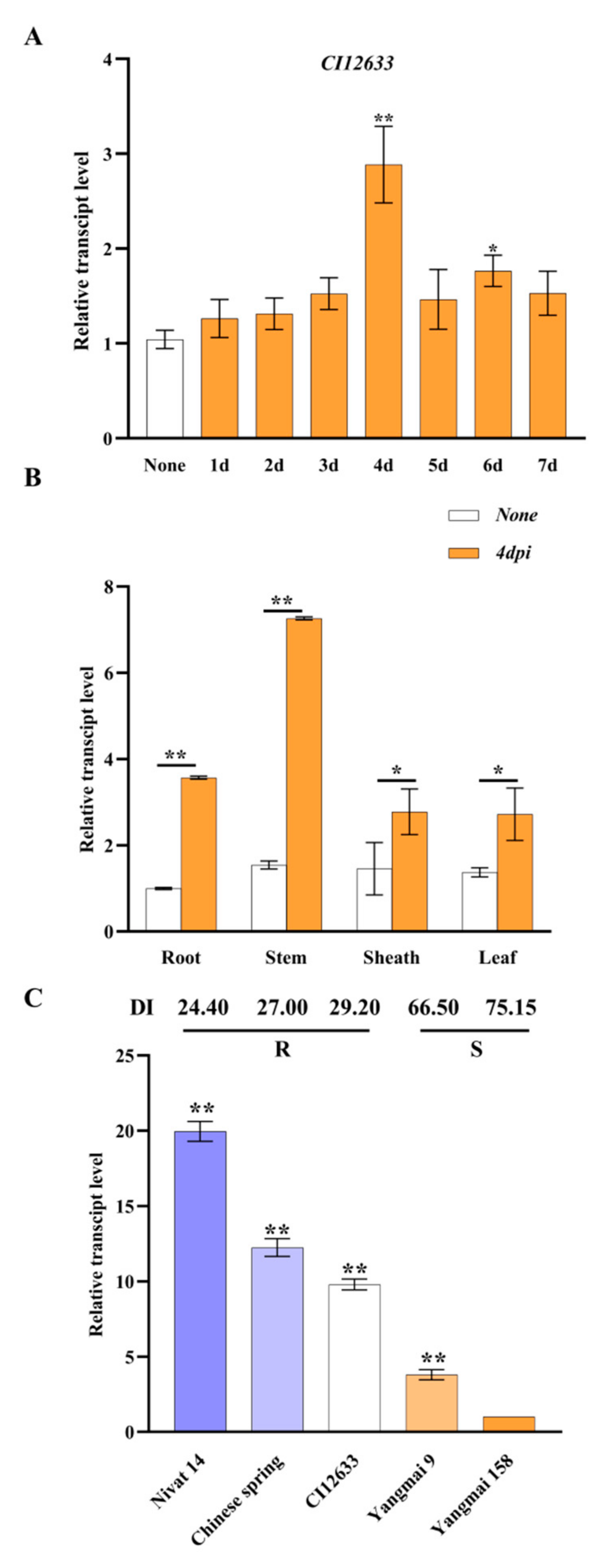
2.1. Identification of TaAACT1 Gene Involved in Wheat Resistance to FCR

By means of bulked segregant RNA sequencing (BSR-Seq) [28] toward the recombinant inbred lines (RILs derived from CI12633/Yangmai 9, F16) combined with wheat variant omics (WheatUnion, http://wheat.cau.edu.cn/WheatUnion/), we analyzed single-nucleotide polymorphisms (SNPs) among the TaAACT1 gene (TraesCS3D02G005500.1) in FCR-resistant wheat cultivars [CI12633, Chinese spring (CS), Jinan 13, Nonglin 10, and Mianyang 26] and its allelic sequences in susceptible wheat cultivars (Handan 6127, Jinan 17, Yangmai 9, and Yangmai158). As a result, 50 SNP sites were found among TaAACT1 gene sequences of the different wheat cultivars, and the majority (47 SNPs) distributed among the last 1300 bp sequence of TaAACT1 (nucleotide no. 2028th to 3327th). Interestingly, 13 nucleotide allelic variants seemed to be associated with FCR resistance/susceptibility of wheat and formed at least 2 haplotypes (Table 1, Figure 1), of which haplotype I was specific to the tested FCR-resistant wheat cultivars (CI12633, CS, Jinan 13, Nonglin 10, and Mianyang 26) but haplotype II was present in the tested susceptible wheat cultivars (Handan 6127, Jinan 17, Yangmai 9, and Yangmai158). As shown in Table 1 and Figure 1, of the 13 SNPs related to FCR resistance, 10 are located in the intron regions of this gene, and the remaining 3 (3156th, 3304th, and 3315th) are located in the gene-coding region. Among the three SNP sites in the coding region, two single-nucleotide mutations are synonymous mutations, which do not cause changes in the amino acid sequence, and one single-nucleotide mutation is a nonsynonymous mutation, which changes the encoded amino acid from serine (S) in the resistant wheat cultivars to proline (P) in the susceptible wheat cultivars.
Reverse-transcription quantitative PCR (RT-qPCR) analysis showed that toward F. pseudograminearum infection, the transcript level of TaAACT1 in stems of FCR-resistant wheat cultivar CI12633 was raised, reaching its peak at 4 days post inoculation (dpi) (about 3-fold over non treatment) (Figure 2A). Subsequently, we examined the gene transcript induction in the tissues of the resistant wheat cultivar CI12633, and the result showed that compared with the nontreatment, the expression level of TaAACT1 in all the tested tissues was elevated after 4 dpi with F. pseudograminearum, and the highest induction occurred in the wheat stems (about 4.8-fold over noninoculation), then in the wheat roots (about 3.9-fold over noninoculation) of the wheat cultivar CI12633 (Figure 2B). In fact, the FCR disease symptom majorly appears in stems and roots of wheat plants [19,20,22]. Furthermore, we tested the transcript level of TaAACT1 in the stems of five different wheat cultivars with different FCR disease indexes [26,27] at 4 dpi with F. pseudograminearum. Expectedly, the transcript level of TaAACT1 was higher in FCR-resistant wheat cultivars than susceptible wheat cultivars (Figure 2C). These results suggest that change in expression of TaAACT1 was associated with wheat resistance to F. pseudograminearum infection, and TaAACT1 might be involved in wheat defense against F. pseudograminearum infection.

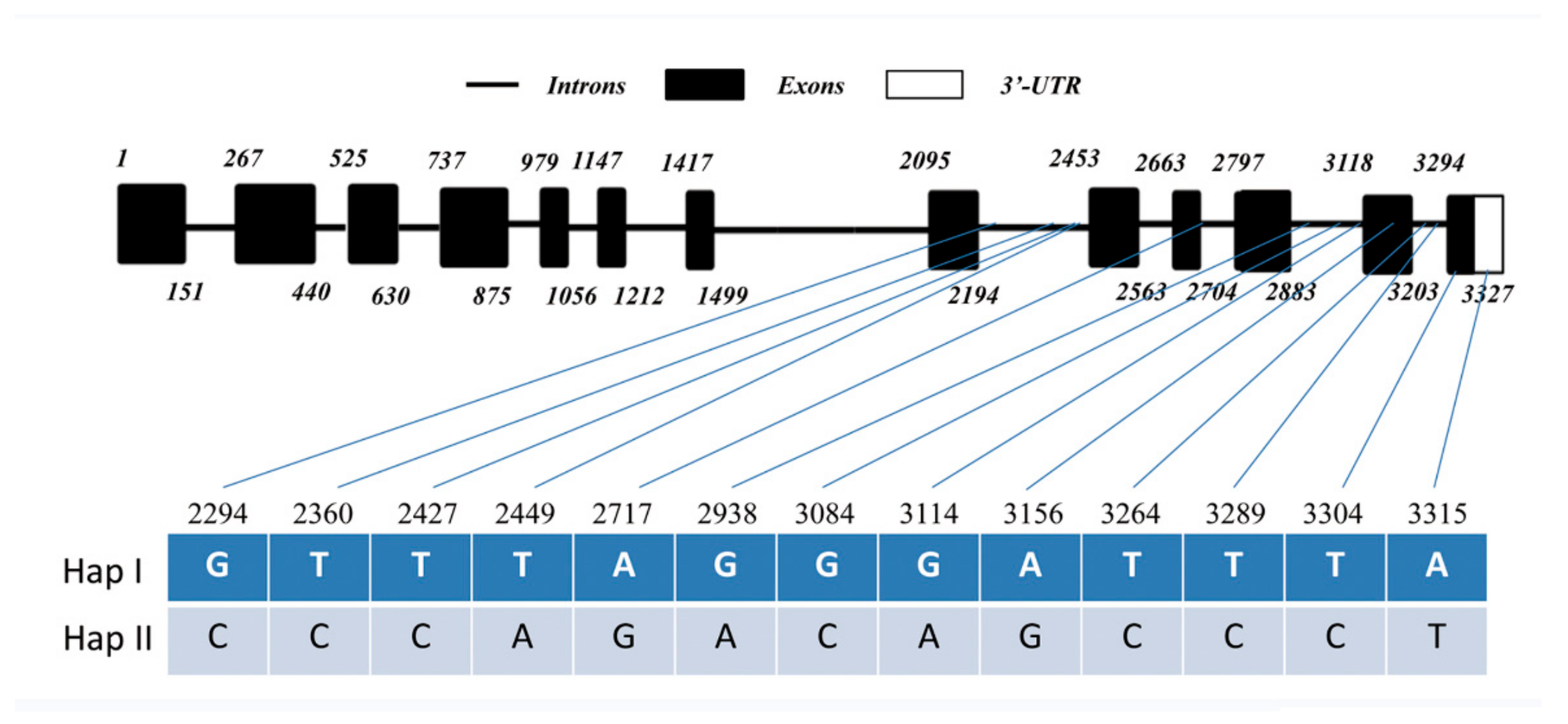
2.2. Sequence and Phylogenetic Characteristics of TaAACT1

The full-length coding sequence and genomic sequence of TaAACT1 were cloned from the partially resistant wheat cultivar CI12633 and had 100% identity with those of CS (TraesCS3D02G005500.1). The TaAACT1 gene contains an open reading frame (ORF) with 1257 bp length. Comparison of the genomic and cDNA sequences showed that the genomic sequence of TaAACT1 was comprised of twelve introns and thirteen exons (Figure 1A). The predicted protein TaAACT1 consists of 418 amino acid (aa) residues with a predicted Mw of 43.02 kDa and theoretical pI of 8.41. TaAACT1 protein has a Thiolase-N domain and a Thiolase-C domain (Figure 3A,B).
Thiolases in plant cells are divided into biosynthetic thiolases and degradative thiolases according to their functions. Degradative thiolases, also known as 3-ketoacyl-CoA thiolase (KAT), belong to peroxisomal thiolases I and can catalyze β-oxidation of fatty acid. Biosynthetic thiolases, also known as Acetoacetyl-CoA thiolases (AACT), belong to cytosolic thiolase II enzymes and catalyze the condensation of two acetyl-CoA to form acetoacetyl-CoA [1,4,11]. To determine the phylogenetic relationship of TaAACT1, we constructed a phylogenetic tree containing TaAACT1 and 15 other thiolase II (AACT/ACAT) proteins as well as 3 thiolase I proteins of other species (Figure 2D). The analysis of phylogenetic tree showed that thiolase I and thiolase II proteins were categorized into two groups (Figure 3C). In the thiolase II group, TaAACT1 is clustered with Aegilops tauschii AACT (XP_0201631811), Triticum dicoccoides AACT (XP_0374108261), Lolium rigidum AACT (XP_0470549061), rice (Oryza sativa) AACT (BAB398721), and corn (Zea mays) AACT (ACG347351) into one branch, while the cytosolic thiolase II (ACAT/AACT) proteins of dicots, namely Arabidopsis thaliana ACAT2 (BAH19918), Medicago sativa AACT1(ACX474701), radish (Raphanus sativus) AACT (CAA550061), and tobacco (Nicotiana tabacum) AACT (AAU956181), were clustered into another branch. AtKAT1 (AEE277361) and AtKAT2 (BAA252481) of Arabidopsis thaliana and rice thiolase I (AAO725881) were categorized to the peroxisomal thiolase I group. Additionally, TaAACT1 is closely related to the corresponding protein of Aegilops tauschii with 100.00% identity at the full-length protein level. These results indicate that TaAACT1 belongs to the cytosolic thiolase II group and that TaAACT1 in wheat might originate from that of Aegilops tauschii.

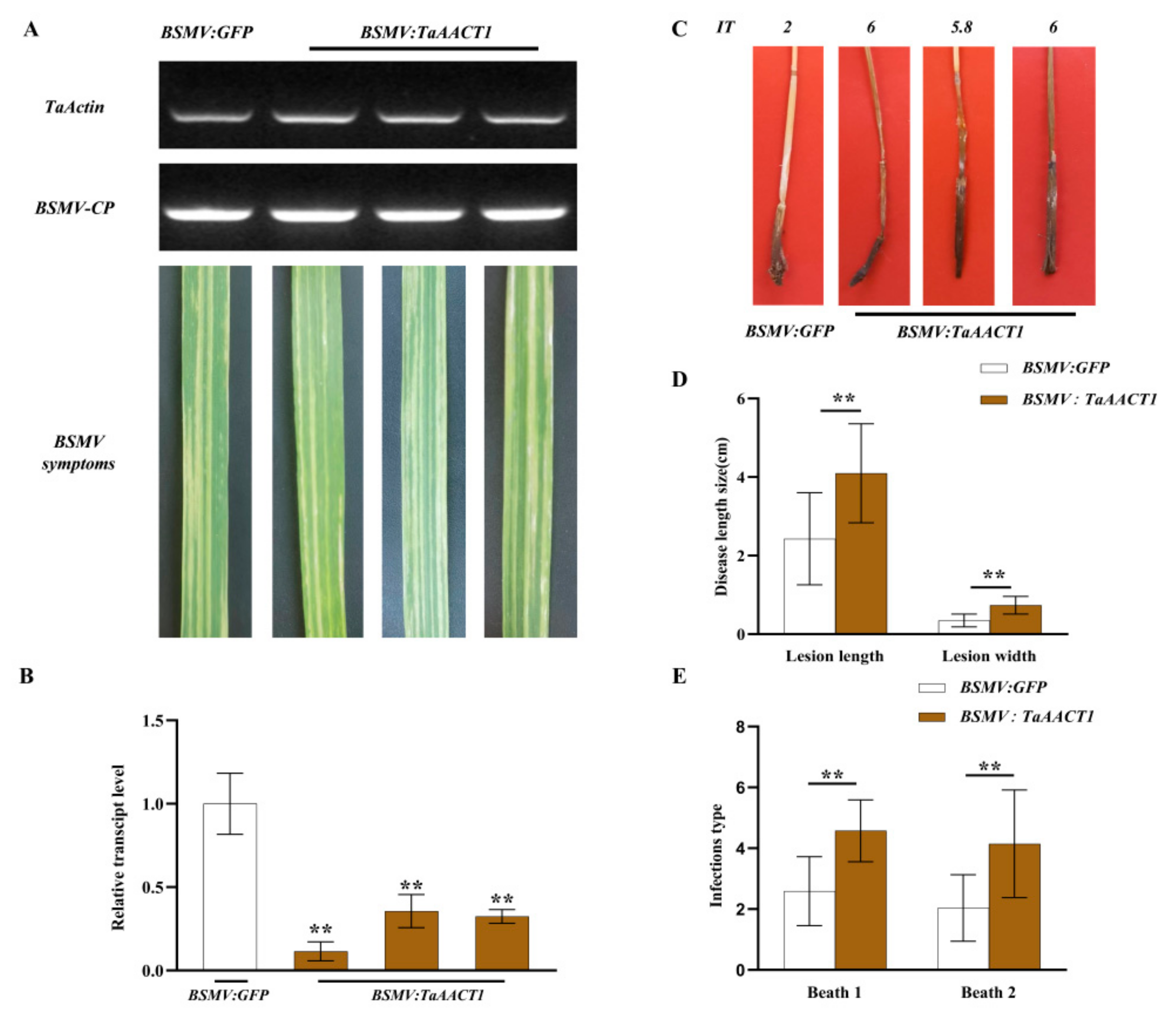
2.3. TaAACT1 Is Required for Wheat Resistance to F. pseudograminearum Infection

The virus-induced gene silencing (VIGS) mediated by barley stripe mosaic virus (BSMV) was used to investigate the defense function of TaAACT1 against F. pseudograminearum infection. The gene-silenced fragment is predicted through Si-Fi software (Figure 4A). The optimum fragment with 194 bp length (covering 166 bp ORF and 28 bp 3’-UTR) of TaAACT1 was subcloned in an antisense orientation into the multicone site of BSMV RNAγ, resulting in the BSMV:TaAACT1 construct (Figure 4B). When the BSMV:GFP/BSMV:TaAACT1 RNAs were transfected to the newly emerging leaves of the mildly-resistant wheat cultivar CI12633 plants at the 3-leaf-stage for 14 days, the symptoms of BSMV infection appeared on the newly emerging 4th leaves and the transcript of the BSMV coat protein gene could be detected in the transfection wheat leaves (Figure 5A). These indicated that the BSMV successfully infected these wheat plants. RT-qPCR analysis showed that compared with the BSMV:GFP-infected CI12633 wheat plants (control), the transcript level of TaAACT1 in BSMV:TaAACT1-infected CI12633 was significantly decreased (Figure 5B), suggesting that the TaAACT1 gene was successfully silenced in these wheat plants mediated by BSMV.
To assess the defensive role of TaAACT1 against F. pseudograminearum infection, TaAACT1-silenced and BSMV:GFP-infected wheat plants were inoculated with F. pseudograminearum WHF220 at the same time. During a period of 28 dpi with F. pseudograminearum, compared with the BSMV:GFP-infected wheat plants, the stems of TaAACT1-silenced CI12633 wheat plants displayed larger lesion sizes of FCR (Figure 5C). In the two experimental batches, the average lesion length on the stems of TaAACT1-silenced CI12633 plants was 4.10 cm, whereas that of BSMV:GFP-infected CI12633 plants was 2.43 cm; the average lesion width on the stems of TaAACT1-silenced CI12633 plants was 0.74 cm, whereas that of BSMV:GFP-infected CI12633 plants was 0.35 cm (Figure 5D). The disease tests showed that the average values of infection types (ITs) on the stems of TaAACT1-silenced CI12633 plants were 4.57 and 4.15, respectively, while the average values of ITs on the stems of BSMV:GFP-infected CI12633 plants were 2.59 and 2.03, respectively (Figure 5E). The above data suggest that the expression of TaAACT1 is required for wheat resistance to F. pseudograminearum infection.

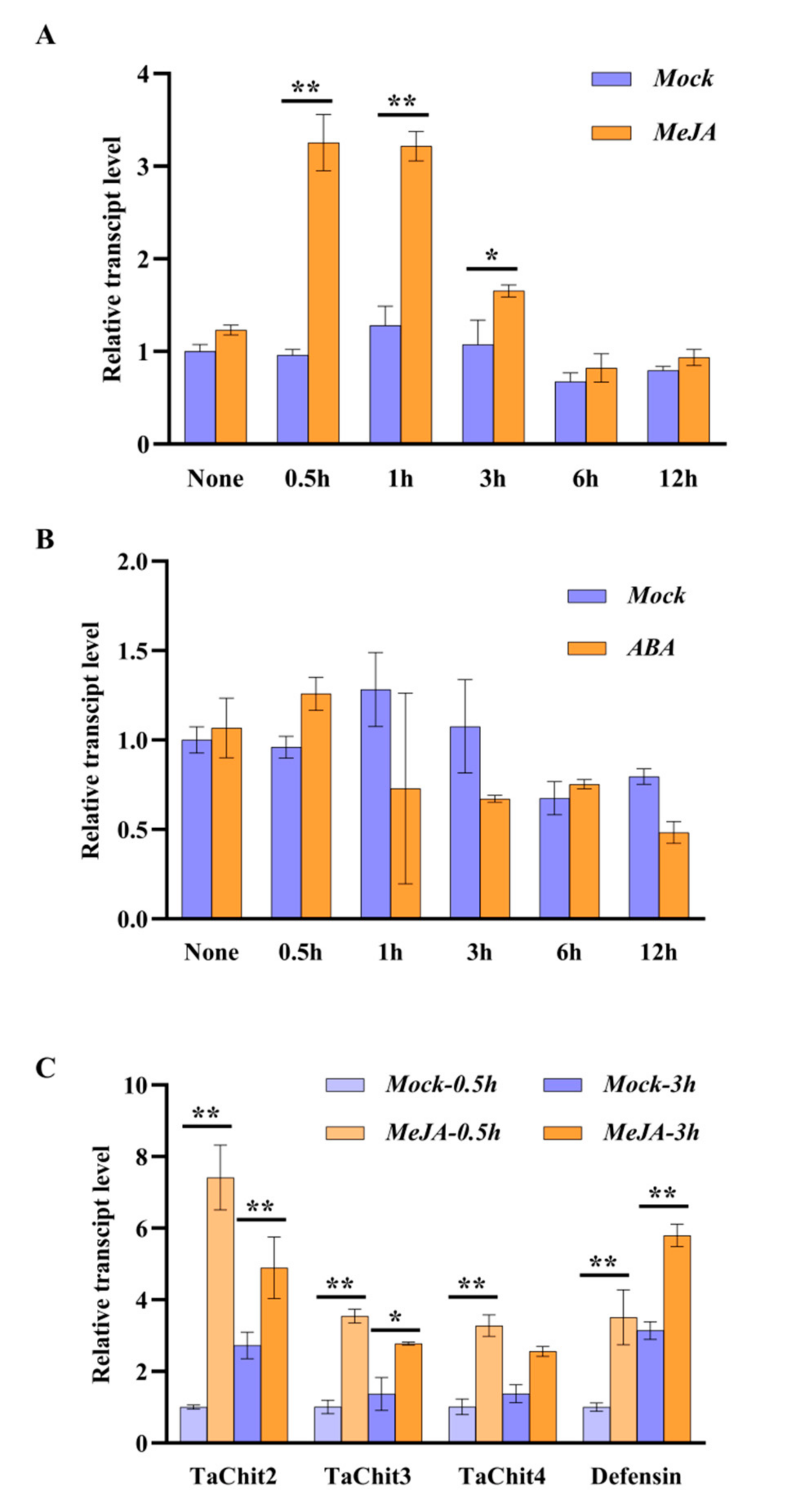
2.4. TaAACT1 Positively Regulates the Expression of Defense Genes

Plant chitinases and defensin have shown to function in directly restricting the growth of pathogenic fungi [29,30,31,32,33,34,35]. In order to explore if TaAACT1 regulates expression of these defense genes during the wheat immune response against F. pseudograminearum infection, we tested the transcript levels of wheat TaChitinase 2 (TaChit2), TaChitinase 3 (TaChit3), TaChitinase 4 (TaChit4), and TaDefensin through RT-qPCR in the TaAACT1-silenced wheat plants and the BSMV:GFP-infected wheat plants inoculated with F. pseudograminearum for 4 d. The results show that the transcript levels of TaChit2, TaChit3, TaChit4, and TaDefensin were significantly decreased in the more susceptibly TaAACT1-silenced wheat plants compared to the BSMV:GFP-infected wheat plants (Figure 6). These results suggest that TaAACT1, acting as an upstream regulator, positively modulates the expression of TaChit2, TaChit3, TaChit4, and TaDefensin during wheat defense responses to F. pseudograminearum infection.

2.5. TaAACT1 and Its Modulated Defense Genes Are Responsive to an Exogenous Jasmonic Acid Stimulus

The promoter sequence of TaAACT1 contains two jasmonic acid (JA)-responsive cis-acting elements (CGTCA motif/TGACG motif) and two abscisic acid (ABA)-responsive cis-acting elements (ABRE, ACGTG motif/GCCGCGTGGC motif). To explore whether TaAACT1 is responsive to JA and ABA, we tested the transcript level of TaAACT1 in the wheat cultivar CI12633 seedlings sprayed with exogenous MeJA (0.05 mM) and ABA (0.1 mM). As results, upon exogenous MeJA treatment, the expression level of TaAACT1 was significantly increased from 0.5 to 3 h, and reached its peak (~3.40-fold compared with the mock (H2O treatment)) at 0.5 h (Figure 7A). However, exogenous ABA treatment did not significantly affect the expression of TaAACT1 (Figure 7B). Moreover, we also examined the transcript profiles of the TaAACT1-regulated defense genes TaChit2, TaChit3, TaChit4, and Defensin in the wheat cultivar CI12633 leaves treated with exogenous MeJA for 0.5 h and 3 h. The results showed that compared with the mock (H2O-treatment), the transcript levels of TaChit2, TaChit3, TaChit4, and Defensin were significantly and rapidly increased by exogenous MeJA (Figure 7C). Taken together, the data suggest that the expression of TaAACT1 and its regulated defense genes (TaChit2, TaChit3, and Defensin) were all in response to the stimulation of exogenous MeJA, and that TaAACT1, TaChit2, TaChit3, TaChit4 and TaDefensin should be located in the JA signaling pathway.

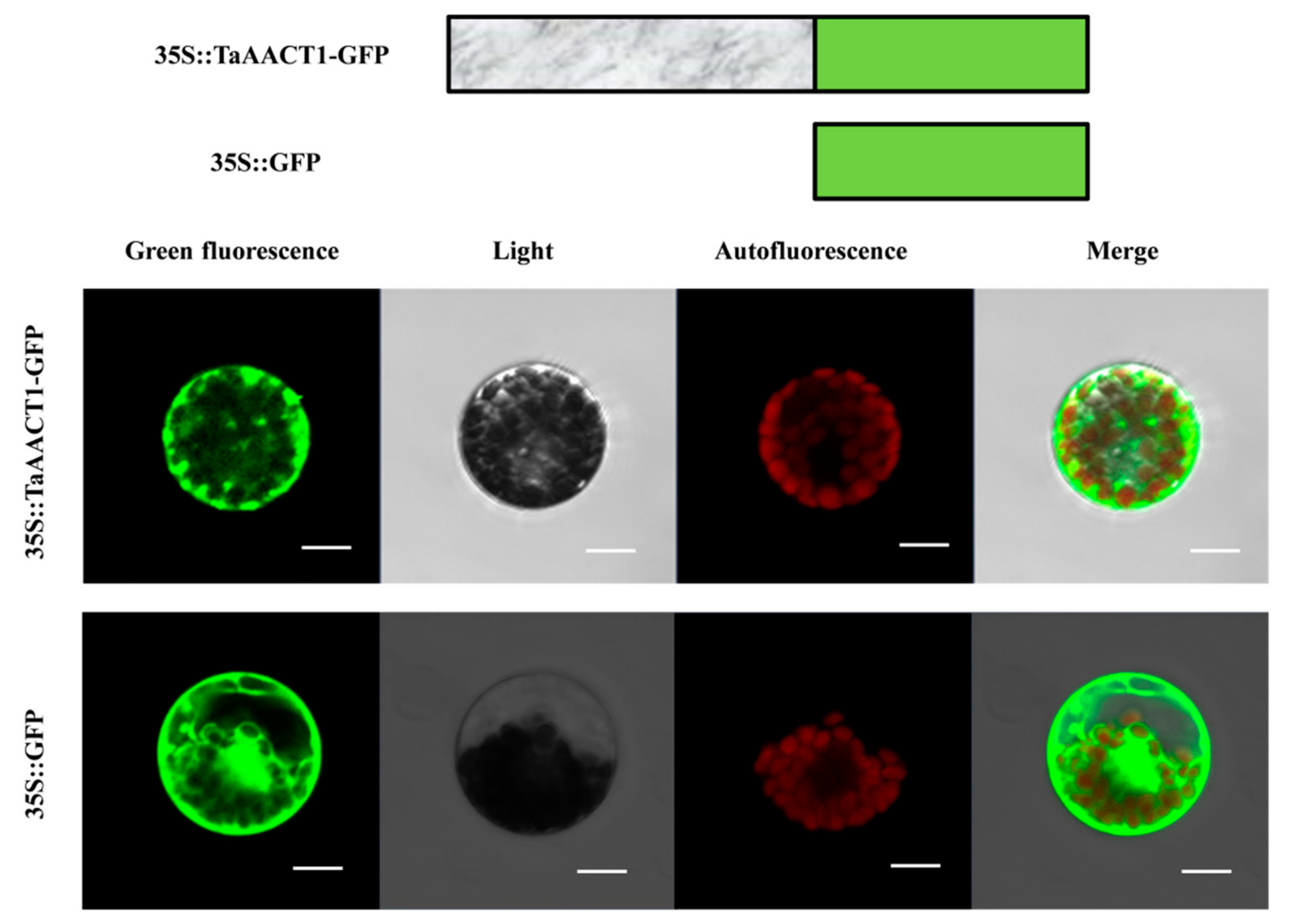
2.6. TaAACT1 Protein Localizes in the Cytosol in Wheat

The subcellular localization of TaAACT1 protein was predicted by PSORT Ⅱ online software. The predicted results are as follows: thiolase II TaAACT1 mainly exists in the cytoplasm, and a small part in the chloroplast. In order to determine the subcellular location of TaAACT1, the full coding sequence of TaAACT1 without the stop codon was subcloned in fusion to the N-terminus of green fluorescent protein (GFP)-coding gene, generating the p35S::TaAACT1-GFP fusion-expressing vector. The resulting p35S::TaAACT1-GFP and the control p35S::GFP were separately introduced into wheat mesophyll protoplasts with the PEG method and transiently expressed in the wheat cells. These fluorescent proteins expressed were observed via a confocal microscope. The results showed that fluorescence images of 35S::TaAACT1-GFP were distributed in the cytosol, while fluorescence images of the GFP control protein were distributed in both the cytoplasm and nucleus (Figure 8). The experimental results suggest that TaAACT1 indeed localizes in the cytosol of wheat, verifying the above prediction for cytoplasm.

3. Discussion

Wheat is an important staple crop and provides 20% of the food calories consumed by human beings worldwide [36]. Its efficient production is often threatened by abiotic and biotic stresses, such as FCR disease. The planting of wheat with resistance is one optimal strategy to control disease. Recently, the dirigent gene TaDIR-B1 (a negative contributor) and two positive regulators, namely TaWAK-6D and TaCRK-7A, were identified to be involved in resistance responses of FCR in wheat [25,26,27]. However, there have been limited papers about FCR-resistant genes and molecular mechanisms underlying wheat defense against Fusarium pseudograminearum infection. Additionally, a recent study showed that the overexpression of alfalfa thiolase II MsAACT1 in transgenic plants enhanced salinity tolerance and the production of squalene in salt-stress conditions [1]. However, little is known about role of cytosolic thiolase II /AACT1 in the plant’s innate immunity to the infection of pathogens.
In the current research, we identified the wheat cytosol thiolase II-encoding gene TaAACT1 associated with FCR resistance of wheat, and provided evidence that TaAACT1, acting as a positive contributor, was required for host defense against F. pseudograminearum infection. By means of analyses in BSR-seq and wheat variant omics, 13 SNPs in TaAACT1, related to FCR resistance, were identified. It is very interesting to develop functional markers based on these SNPs for molecularly breeding wheat with resistance to FCR in future. Transcript analysis results showed that the transcript induction of TaAACT1 by infection of F. pseudograminearum was higher in FCR-resistant wheat cultivars than in susceptible wheat cultivars, and the increase occurred in stems/roots at the greatest extent. Similarly, the expression levels of AtACAT2 and MsAACT1 were higher in roots than in leaves in Arabidopsis thaliana and alfalfa under optimal growth conditions [1,6]. Functional studies on plant thiolases by the genetic method have been rare, since the overexpression of these enzymes during the transformation/regeneration procedure is sufficient to avoid the generation of stable transgenic plants [18], and thiolases II from dicots have an extremely high nucleotide identity, making the RNAi strategy difficult to design with the certainty of ensuring its specificity for plant species [1], and Arabidopsis mutant of AACT 2 with only one copy is nonviable [17]. Fortunately, the VIGS technique can be used as an alternative approach to rapidly analyze gene function [37,38]. For example, due to a lack of generation of transgenic wheat overexpressing a wheat cytochrome P450 gene TaCYP72A, VIGS was used to conduct functional analyses, and the results showed that TaCYP72A enhances resistance to deoxynivalenol and grain number [38]. Here, the VIGS technique was used to study the defensive function of TaAACT1, and the results show that the silencing of TaAACT1 decreased resistance to FCR caused by infection of F. pseudograminearum. To our knowledge, this is the first investigation about the role of plant thiolases in plants’ innate immunity. This study deepens the understanding of plant defense mechanisms against pathogens.
In plant species, many chitinases and defensin peptides show antifungal activity [29,30,35,39]. Chitinases can hydrolyze the chitin (the major structural component) of fungal cell walls, in turn directly inhibiting the growth of pathogenic fungi [29,32]. Antifungal activity of plant defensin requires specific binding to membrane targets to inhibit the growth of fungi [30,31,39]. Additionally, chitinases also have the secondary effect of releasing chitin monomers, which acts as a pathogen/damage-associated molecular pattern for the activation of the plant immune system [40]. Many chitinases and defensin-encoding genes are induced in response to infection by pathogens [41,42], and are also upregulated by upstream immune genes in fungal-infected plants [26,27,43,44,45,46,47,48,49,50]. The current research indicates that expression of TaAACT1 is required for the expression of TaChitinase 2, TaChitinase 3, TaChitinase 4, and TaDefensin in wheat inoculated by F. pseudograminearum, suggesting that TaChitinase 2, TaChitinase 3, TaChitinase 4 and TaDefensin act downstream of TaAACT1 and are positively modulated by upstream TaAACT1. JA is a primary phytohormone associated with plant resistance responses to necrotrophic pathogens [51]. Some kinases and transcription factors, as well as upstream proteins, can respond and transduce JA signaling to the downstream genes [46,47,52]. For instance, the expression of TaSTT3b-2B was significantly induced from 1 to 8 h after MeJA treatment; overexpression of TaSTT3b-2B in transgenic wheat increased endogenous JA content and the expression of JA synthesis-related genes and enhanced resistance to sharp eyespot and grain weight [46]. However, little is known about the effect of JA on thiolase II in wheat plants. Our analyses showed that TaAACT1 rapidly responded to exogenous MeJA stimulus, and MeJA treatment also significantly induced the expression of TaAACT1-modulated defense genes (Chitinases 2/3/4 and defensin). Thus, JA signaling might play an important role in TaAATC1-mediated immune responses to F. pseudograminearum infection. Taken together, these results suggest that TaAACT1 positively contributes to the resistance of wheat against F. pseudograminearum infection by regulating the expression of TaChitinase 2, TaChitinase 3, TaChitinase 4, and TaDefensin in JA signaling.
Protein sequence analysis showed that the TaAACT1-predicted protein contains two domains: a Thiolase-N domain and a Thiolase-C domain, similar to those of Arabidopsis thaliana cytosolic Thiolase II (AtACAT2, BAH19918), and alfalfa cytosolic Thiolase II/AACT1 (MsAACT1, ACX474701), involved in the mevalonate pathway [1,6,7]. Our phylogenetic analysis indicated that the TaAACT1 protein, together with Arabidopsis thaliana cytosolic Thiolase II/AtACAT2 and MsAACT1, as well as other cytosolic Thiolase II proteins, are classified into the same group, and that TaAACT1 in wheat might originate from that of Aegilops tauschii. The subcellular localization experimental assay indicated that TaAACT1 protein indeed localizes in the cytosol of wheat, like the AtACAT2 subcellular localization result [6,7]. In a recent paper, the alfalfa cytosolic Thiolase II MsAACT1 was determined to possess thiolase activity, not only using bacteria as a heterologous system but also in planta, and that the expression of MsAACT1 increased abiotic stress resistance in bacteria and in planta, strongly suggesting that MsAACT1 positively regulates abiotic stress tolerance by increasing squalene biosynthesis [1]. Additionally, infection with avirulent pathogens, tobacco mosaic virus, or Pseudomonas syringae pv. tabaci induced accumulation of polyisoprenoid alcohols in the leaves of resistant tobacco plants but did not cause an increase in polyisoprenoid content in susceptible tobacco plants [15]. In the furture, it is very interesting to investigate if TaAACT1 is required for isoprenoid biosynthesis during wheat resistance responses to F. pseudograminearum.

4. Materials and Methods

4.1. Plant and Fungal Materials, Primers, and Treatments

Five wheat cultivars, including FCR-resistant cultivars (Nivat14, CI12633, Chinese Spring), and susceptible cultivars (Yangmai 6 and Yangmai 158), were used to examine transcript profiles of TaAACT1 with the F. pseudograminearum inoculation.
All of the wheat plants were grown in a 16 h light/8 h dark (22 °C/12 °C) regimen. At the tillering stage, small toothpick fragments harboring the well-developed mycelia of F. pseudograminearum were inoculated between the second base sheaths and stems of wheat plants [26]. The inoculated sheaths of the resistant wheat cultivar CI12633 were collected at noninoculation or 1, 2, 3, 4, 5, 6, or 7 dpi with F. pseudograminearum. The inoculated sheaths of Nivat 14, Chinese spring, CI12633, Yangmai 9, and Yangmai 158 were separately sampled at 4 dpi with F. pseudograminearum for RNA extraction. These samples were quickly frozen in liquid nitrogen and stored at −80 °C prior to extraction of total RNA. The seedlings of the wheat cultivar CI12633 at the two-leaf stage were separately treated with 0.05 mM MeJA and 0.1 mM ABA dissolved in ultrapure water, and ultrapure water solution (mock, as control) for noninoculation and 0.5, 1, 3, 6, and 12 h, as described by Zhang et al. [43]. The treated leaves were collected for RNA extraction.
The pathogenic fungus F. pseudograminearum strain WHF220 was isolated from the FCR-symptomatic wheat sheaths in Shandong by Prof. Jinfeng Yu and Dr. Li Zhang (Shandong Agricultural University, Tai’an, China).
All primers and their sequences in the study are shown in Supplementary Materials: Table S1.

4.2. RNA Extraction, cDNA Synthesis, and RT-qPCR Analysis

Total RNA was extracted from wheat samples tested using Trizol reagent (Invitrogen, Carlsbad, CA, USA) according to the manufacturer’s instruction. RNA was purified and then checked for integrity. Total RNA was reverse-transcribed to cDNA using the FastQuant R T Kit (Tiangen, Beijing, China); RT-qPCR and specific primers were employed to measure expression levels of TaAACT1, TaChit2, TaChit3, TaChit4, and TaDefensin in wheat. RT-qPCR was performed using a SYBR Premix Ex Taq kit (TaKaRa, Tokyo, Japan) in a volume of 20 µL on an ABI 7500 instrument (Applied Biosystems, Foster City, MA, USA). Reactions were set up with the following thermal cycling profile: 95 °C for 3 min followed by 40 cycles of 5 s at 95 °C, 15 s at 55 °C, and 32 s at 72 °C, and completed with a melting curve analysis program. All RT-qPCRs were repeated three times. The relative expression of the tested gene was calculated with the 2−ΔΔCT method [53], where the wheat Actin gene was used to normalize amounts of cDNA among the samples.

4.3. SNP Analysis

The purified RNAs were extracted from the resistant and susceptible RILs (CI12633/Yangmai 9, F16) at 20 dpi and used to BSR-seq using the Illumina HiSeq 2500 sequencing technology platform at Beijing Biomarker Technologies. For each pool, three biological replicates were performed. After discarding low-quality raw reads, the generated 6 Gb clean reads from each library were mapped to wheat genomes. RNA-seq data were analyzed as described previously by Wang et al. [28] The SNP results of RNA-Seq analysis were correlated with the wheat variant omics analysis toward different FCR-resistant/susceptible wheat cultivars. Taken together, the SNPs in TaAACT1 related to FCR resistance of wheat were identified.

4.4. Sequence and Phylogenetic Analyses of TaAACT1

One pair of primers (TaAACT1-F1/TaAACT1-R1) for TaAACT1 were designed and used to amplify the full ORF sequence of TaAACT1 from the cDNA of the wheat cultivar CI12633.The predicted protein sequence was analyzed with the Compute pI/Mw tool (http://web.expasy.org/compute_pi/ (accessed on 25 July 2022)) to determine the theoretical pI (isoelectric point) and Mw (molecular weight), The conserved domains and motifs in the deduced protein sequence were predicted by SMART (http://smart.embl-heidelberg.de/, accessed on 25 July 2022). Sequences were aligned via DNAMAN software. Subcellular localization of TaAACT1 protein was predicted using PSORT Ⅱ online software (https://psort.hgc.jp/form2.html, accessed on 25 July 2022). A phylogenetic tree was constructed by MEGA 11.0 (https://www.megasoftware.net/, accessed on 25 July 2022) software. Cis-elements in the TaAACT1 promoter (2000 bp) were analyzed using the PlantCARE (http://bioinformatics.psb.ugent.be/webtools/plantcare/html/, accessed on 25 July 2022). The JA- and ABA-responsive cis-acting elements in promoter of TaAACT1 are shown in Supplementary Materials: Table S2.

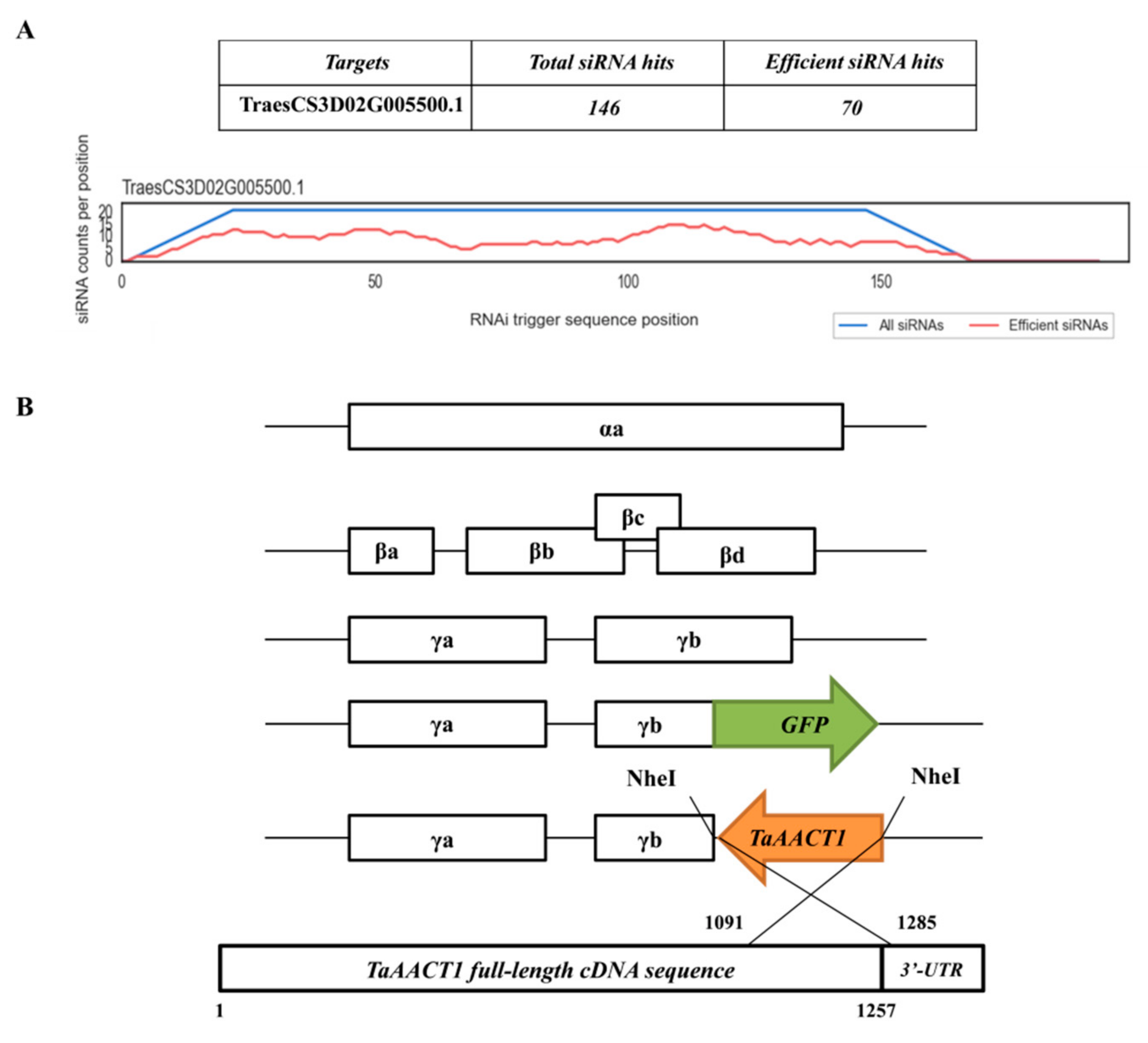
4.5. VIGS of TaAACT1 in Wheat CI12633

A short fragment (194 bp) derived from the coding sequence of TaAACT1 was subcloned in antisense orientation into the multicloning site of the γ chain of the BSMV vector. According to the protocols described previously [44,45,46,48], BSMV:TaAACT1 and BSMV:GFP viruses were transfected into the leaves of the wheat cultivar CI12633 in order to knock down endogenous TaAACT1 transcript in the wheat plants. After 14 d, the fourth leaves of inoculated seedlings were harvested. These samples were quickly frozen in liquid nitrogen and stored at −80 °C prior to extraction of total RNA.

4.6. Assessment and Scoring of FCR Disease in BSMV-VIGS Wheat Plants

About 21 d post-transfection with BSMV, the TaAACT1-silenced and BSMV:GFP-infected (control) wheat plants were inoculated with well-developed F. pseudograminearum mycelia toothpicks and kept at high humidity for 4 d in two batches. After 28 days of fungal inoculation, the average lesion length and width of FCR on the basal sheath of the above wheat plants were measured and recorded, and the FCR disease index was evaluated. The assessment methods of FCR disease severity were referred to the existing researches [22].

4.7. Subcellular Localization of TaAACT1 Protein

The coding region of TaAACT1 lacking the stop codon was amplified and added the restriction enzyme BamH I site using the specific primers (TaAACT1-GFP-F/TaAACT1-GFP-R). The amplified fragment was digested with restriction enzyme BamH I and subcloned in-frame into the N-terminus of the GFP coding region in the p35S::GFP vector, resulting in the TaAACT1-GFP fusion construct p35S::TaAACT1-GFP. The p35S::TaAACT1-GFP fusion construct and p35S::GFP control construct were separately individually introduced into wheat mesophyll protoplasts [54]. After incubation at 25 ℃ for 16 h, GFP signals were observed and photographed using a confocal laser scanning microscope (Zeiss LSM700, Jena, Germany) with a Fluor × 10/0.50 M27 objective lens and SP640 filter.

5. Conclusions

The wheat cytosol thiolase II-encoding gene TaAACT1 was identified to be associated with FCR resistance of wheat. One haplotype ofTaAACT1 gene sequences in analyzed wheat genotypes was related to wheat resistance against FCR, and the gene transcript induction by Fusarium pseudograminearum infection was higher in FCR-resistant wheat cultivars than in susceptible wheat cultivars. Moreover, the transcript induction of this gene was the highest in stems then roots, where FCR disease primarily occurs. VIGS-based functional analyses indicated that the expression of TaAACT1 was required for host defense responses against F. pseudograminearum infection, and TaAACT1 positively modulated the expression of the defense genes TaChitinase 2, TaChitinase 3 TaChitinase 4, and TaDefensin in wheat. This study extends knowledge about wheat-F. pseudograminearum interactions and deepens our understanding of the molecular mechanisms underlying wheat plant immunity to fungal pathogens, especially F. pseudograminearum. The TaAACT1 is a candidate gene for highly efficiently improving the resistance of wheat to FCR disease.

Supplementary Materials

The following supporting information can be downloaded at: https://www.mdpi.com/article/10.3390/ijms24076165/s1.

Author Contributions

Z.Z. designed the experiment, was the program funding winner, and wrote and revised the manuscript. F.X., X.Z., C.L. and Z.L. performed the experiments, analyzed sequences, analyzed the data, and made figures. F.X. and Z.L. wrote the partial draft manuscript except for the Abstract, Discussion, and Conclusions. All authors have read and agreed to the published version of the manuscript.

Funding

This work was supported by the National Key Project for Research on Transgenic Biology, China (2016ZX08002-001-004 to Z.Z.), Natural e Natural Science Foundation of China (Grant No. 31771789 to Z.Z.), and the Natural Science Foundation of Hunan Province of China (Grant No. 2022JJ31002 to L.Z.).

Data Availability Statement

All data supporting the findings of this study are available within the paper and within its Supplementary Materials published online.

Acknowledgments

The authors are grateful to Derong Gao and Xujiang Wu (Institute of Agricultural Science of Lixiahe District in Jiangsu Province) and Jizhong Wu (Jiangsu Academy of Agricultural Sciences) for providing the RILs’ leaves and wheat cultivars; Jinfeng Yu and Li Zhang (College of Plant Protection, Shandong Agricultural University, China) for providing F. pseudograminearum strain WHF220; and Zhiyong Liu (Institute of Genetics and Developmental Biology, Chinese Academy of Sciences, China) for help with the SNP analysis by BSR-seq.

Conflicts of Interest

The authors declare no conflict of interest.

References

  1. Soto, G.; Stritzler, M.; Lisi, C.; Alleva, K.; Pagano, M.E.; Ardila, F.; Mozzicafreddo, M.; Cuccioloni, M.; Angeletti, M.; Ayub, N.D. Acetoacetyl-CoA thiolase regulates the mevalonate pathway during abiotic stress adaptation. J. Exp. Bot. 2011, 62, 5699–5711. [Google Scholar] [CrossRef] [PubMed]
  2. Zhong, Z.H.; Norvienyeku, J.; Yu, J.; Chen, M.L.; Cai, R.L.; Hong, Y.H.; Chen, L.M.; Zhang, D.M.; Wang, B.H.; Zhou, J.; et al. Two different subcellular-localized Acetoacetyl-CoA acetyltransferases differentiate diverse functions in Magnaporthe oryzae. Fungal Genet. Biol. 2015, 83, 58–67. [Google Scholar] [CrossRef] [PubMed]
  3. Wilding, E.I.; Brown, J.R.; Bryant, A.P.; Chalker, A.F.; Holmes, D.J.; Ingraham, K.A.; Iordanescu, S.; So, C.Y.; Rosenberg, M.; Gwynn, M.N. Identification, evolution, and essentiality of the mevalonate pathway for isopentenyl diphosphate biosynthesis in gram-positive cocci. J. Bacteriol. 2000, 182, 4319–4327. [Google Scholar] [CrossRef] [PubMed] [Green Version]
  4. Disch, A.; Hemmerlin, A.; Bach, T.J.; Rohmer, M. Mevalonate-derived isopentenyl diphosphate is the biosynthetic precursor of ubiquinone prenyl side chain in tobacco BY-2 cells. Biochem. J. 1998, 331 Pt 2, 615–621. [Google Scholar] [CrossRef] [Green Version]
  5. Laule, O.; Furholz, A.; Chang, H.-S.; Zhu, T.; Wang, X.; Heifetz, P.B.; Gruissem, W.; Lange, M. Crosstalk between cytosolic and plastidial pathways of isoprenoid biosynthesis in Arabidopsis thaliana. Proc. Natl. Acad. Sci. USA 2003, 100, 6866–6871. [Google Scholar] [CrossRef] [Green Version]
  6. Carrie, C.; Murcha, M.W.; Millar, A.H.; Smith, S.M.; Whelan, J. Nine 3-ketoacyl-CoA thiolases (KATs) and acetoacetyl-CoA thiolases (ACATs) encoded by five genes in Arabidopsis thaliana are targeted either to peroxisomes or cytosol but not to mitochondria. Plant Mol. Biol. 2007, 63, 97–108. [Google Scholar] [CrossRef]
  7. Ahumada, I.; Cairo, A.; Hemmerlin, A.; Gonzalez, V.; Pateraki, I.; Bach, T.J.; Rodriguez-Concepcion, M.; Campos, N.; Boronat, A. Characterisation of the gene family encoding acetoacetyl-CoA thiolase in Arabidopsis. Funct. Plant Biol. FPB 2008, 35, 1100–1111. [Google Scholar] [CrossRef]
  8. Yasmin, S.; Alcazar-Fuoli, L.; Grundlinger, M.; Puempel, T.; Cairns, T.; Blatzer, M.; Lopez, J.F.; Grimalt, J.O.; Bignell, E.; Haas, H. Mevalonate governs interdependency of ergosterol and siderophore biosyntheses in the fungal pathogen Aspergillus fumigatus. Proc. Natl. Acad. Sci. USA 2012, 109, E497–E504. [Google Scholar] [CrossRef] [Green Version]
  9. Moses, T.; Pollier, J.; Thevelein, J.M.; Goossens, A. Bioengineering of plant (tri)terpenoids: From metabolic engineering of plants to synthetic biology invivo and invitro. New Phytol. 2013, 200, 27–43. [Google Scholar] [CrossRef]
  10. Dyer, J.H.; Maina, A.; Gomez, I.D.; Cadet, M.; Oeljeklaus, S.; Schiedel, A.C. Cloning, expression and purification of an acetoacetyl CoA thiolase from sunflower cotyledon. Int. J. Biol. Sci. 2009, 5, 736–744. [Google Scholar] [CrossRef] [Green Version]
  11. Wang, M.; Zheng, Z.; Tian, Z.N.; Zhang, H.; Zhu, C.Y.; Yao, X.Y.; Yang, Y.X.; Cai, X. Molecular cloning and analysis of an Acetyl-CoA C-acetyltransferase gene (EkAACT) from Euphorbia kansui Liou. Plants 2022, 11, 1539. [Google Scholar] [CrossRef]
  12. Maeda, H.; Song, W.; Sage, T.L.; Dellapenna, D. Tocopherols play a crucial role in low-temperature adaptation and phloem loading in Arabidopsis. Plant Cell 2006, 18, 2710–2732. [Google Scholar] [CrossRef] [Green Version]
  13. Abbasi, A.-R.; Hajirezaei, M.; Hofius, D.; Sonnewald, U.; Voll, L.M. Specific roles of alpha- and gamma-tocopherol in abiotic stress responses of transgenic tobacco. Plant Physiol. 2007, 143, 1720–1738. [Google Scholar] [CrossRef] [Green Version]
  14. Matringe, M.; Ksas, B.; Rey, P.; Havaux, M. Tocotrienols, the unsaturated forms of vitamin E, can function as antioxidants and lipid protectors in tobacco leaves. Plant Physiol. 2008, 147, 764–778. [Google Scholar] [CrossRef] [Green Version]
  15. Bajda, A.; Konopka-Postupolska, D.; Krzymowska, M.; Henning, J.; Skorupinska-Tudek, K.; Surmacz, L.; Wojcik, J.; Matysiak, Z.; Chojnacki, T.; Skorzynska-Polit, E.; et al. Role of polyisoprenoids in tobacco resistance against biotic stresses. Physiol. Plant. 2009, 135, 351–364. [Google Scholar] [CrossRef]
  16. Vickers, C.E.; Gershenzon, J.; Lerdau, M.T.; Loreto, F. A unified mechanism of action for volatile isoprenoids in plant abiotic stress. Nat. Chem. Biol. 2009, 5, 283–291. [Google Scholar] [CrossRef]
  17. Suzuki, M.; Nakagawa, S.; Kamide, Y.; Kobayashi, K.; Ohyama, K.; Hashinokuchi, H.; Kiuchi, R.; Saito, K.; Muranaka, T.; Nagata, N. Complete blockage of the mevalonate pathway results in male gametophyte lethality. J. Exp. Bot. 2009, 60, 2055–2064. [Google Scholar] [CrossRef] [Green Version]
  18. Bohmert, K.; Balbo, I.; Steinbuchel, A.; Tischendorf, G.; Willmitzer, L. Constitutive expression of the beta-ketothiolase gene in transgenic plants. A major obstacle for obtaining polyhydroxybutyrate-producing plants. Plant Physiol. 2002, 128, 1282–1290. [Google Scholar] [CrossRef] [Green Version]
  19. Poole, G.J.; Smiley, R.W.; Paulitz, T.C.; Walker, C.A.; Carter, A.H.; See, D.R.; Garland-Campbell, K. Identification of quantitative trait loci (QTL) for resistance to Fusarium crown rot (Fusarium pseudograminearum) in multiple assay environments in the Pacific Northwestern US. Theor. Appl. Genet. 2012, 125, 91–107. [Google Scholar] [CrossRef] [Green Version]
  20. Smiley, R.W.; Gourlie, J.A.; Easley, S.A.; Patterson, L.-M.; Whittaker, R.G. Crop damage estimates for crown rot of wheat and barley in the Pacific Northwest. Plant Dis. 2005, 89, 595–604. [Google Scholar] [CrossRef] [Green Version]
  21. Kazan, K.; Gardiner, D.M. Fusarium crown rot caused by Fusarium pseudograminearum in cereal crops: Recent progress and future prospects. Mol. Plant Pathol. 2018, 19, 1547–1562. [Google Scholar] [CrossRef] [PubMed] [Green Version]
  22. Yang, X.; Pan, Y.B.; Singh, P.K.; He, X.Y.; Ren, Y.; Zhao, L.; Zhang, N.; Cheng, S.H.; Chen, F. Investigation and genome-wide association study for Fusarium crown rot resistance in Chinese common wheat. BMC Plant Biol. 2019, 19, 153. [Google Scholar] [CrossRef] [PubMed] [Green Version]
  23. Liu, C.J.; Ogbonnaya, F.C. Resistance to Fusarium crown rot in wheat and barley: A review. Plant Breed. 2015, 134, 365–372. [Google Scholar] [CrossRef]
  24. Jin, J.J.; Duan, S.N.; Qi, Y.Z.; Yan, S.H.; Li, W.; Li, B.Y.; Xie, C.J.; Zhen, W.C.; Ma, J. Identification of a novel genomic region associated with resistance to Fusarium crown rot in wheat. Theor. Appl. Genet. 2020, 133, 2063–2073. [Google Scholar] [CrossRef]
  25. Yang, X.; Zhong, S.B.; Zhang, Q.J.; Ren, Y.; Sun, C.W.; Chen, F. A loss-of-function of the dirigent gene TaDIR-B1 improves resistance to Fusarium crown rot in wheat. Plant Biotechnol. J. 2021, 19, 866–868. [Google Scholar] [CrossRef]
  26. Qi, H.J.; Guo, F.L.; Lv, L.J.; Zhu, X.L.; Zhang, L.; Yu, J.F.; Wei, X.N.; Zhang, Z.Y. The wheat wall-associated receptor-like kinase TaWAK-6D mediates broad resistance to two fungal pathogens Fusarium pseudograminearum and Rhizoctonia cerealis. Front. Plant Sci. 2021, 12, 2322. [Google Scholar] [CrossRef]
  27. Wu, T.C.; Guo, F.L.; Xu, G.B.; Yu, J.F.; Zhang, L.; Wei, X.N.; Zhu, X.L.; Zhang, Z.Y. The receptor-like kinase TaCRK-7A inhibits Fusarium pseudograminearum growth and mediates resistance to Fusarium crown rot in wheat. Biology 2021, 10, 1122. [Google Scholar] [CrossRef]
  28. Wang, Y.; Xie, J.Z.; Zhang, H.Z.; Guo, B.M.; Ning, S.Z.; Chen, Y.X.; Lu, P.; Wu, Q.H.; Li, M.M.; Zhang, D.Y.; et al. Mapping stripe rust resistance gene YrZH22 in Chinese wheat cultivar Zhoumai 22 by bulked segregant RNA-Seq (BSR-Seq) and comparative genomics analyses. Theor. Appl. Genet. 2017, 130, 2191–2201. [Google Scholar] [CrossRef]
  29. Benhamou, N.; Broglie, K.; Broglie, R.; Chet, I. Antifungal effect of bean endochitinase on Rhizoctonia solani: Ultrastructural changes and cytochemical aspects of chitin breakdown. Can. J. Microbiol. 1993, 39, 318–328. [Google Scholar] [CrossRef]
  30. Thevissen, K.; Terras, F.R.; Broekaert, W.F. Permeabilization of fungal membranes by plant defensins inhibits fungal growth. Appl. Environ. Microbiol. 1999, 65, 5451–5458. [Google Scholar] [CrossRef] [Green Version]
  31. Thomma, B.P.H.J.; Cammue, B.P.A.; Thevissen, K. Plant defensins. Planta 2002, 216, 193–202. [Google Scholar] [CrossRef] [PubMed]
  32. Singh, A.; Kirubakaran, S.I.; Sakthivel, N. Heterologous expression of new antifungal chitinase from wheat. Protein Expr. Purif. 2007, 56, 100–109. [Google Scholar] [CrossRef]
  33. Lacerda, A.F.; Vasconcelos, E.a.R.; Pelegrini, P.B.; De Sa, M.F.G. Antifungal defensins and their role inplant defense. Front. Microbiol. 2014, 5, 116. [Google Scholar] [CrossRef] [PubMed] [Green Version]
  34. Xu, J.; Xu, X.Y.; Tian, L.L.; Wang, G.L.; Zhang, X.Y.; Wang, X.Y.; Guo, W.Z. Discovery and identification of candidate genes from the chitinase gene family for Verticillium dahliae resistance in cotton. Sci. Rep. 2016, 6, 29022. [Google Scholar] [CrossRef] [PubMed]
  35. Su, Q.; Wang, K.; Zhang, Z.Y. Ecotopic expression of the antimicrobial peptide DmAMP1W improves resistance of transgenic wheat to two diseases: Sharp eyespot and common root rot. Int. J. Mol. Sci. 2020, 21, 647. [Google Scholar] [CrossRef] [Green Version]
  36. FAO. World Food and Agriculture—Statistical Yearbook 2020; FAO: Rome, Italy, 2020. [Google Scholar]
  37. Shen, Q.-H.; Saijo, Y.; Mauch, S.; Biskup, C.; Bieri, S.; Keller, B.; Seki, H.; Ulker, B.; Somssich, I.E.; Schulze-Lefert, P. Nuclear activity of MLA immune receptors links isolate-specific and basal disease-resistance responses. Science 2007, 315, 1098–1103. [Google Scholar] [CrossRef] [Green Version]
  38. Gunupuru, L.R.; Arunachalam, C.; Malla, K.B.; Kahla, A.; Perochon, A.; Jia, J.G.; Thapa, G.; Doohan, F.M. A wheat cytochrome P450 enhances both resistance to deoxynivalenol and grain yield. PLoS ONE 2018, 13, e0204992. [Google Scholar] [CrossRef] [Green Version]
  39. Thevissen, K.; Osborn, R.W.; Acland, D.P.; Broekaert, W.F. Specific binding sites for an antifungal plant defensin from Dahlia (Dahlia merckii) on fungal cells are required for antifungal activity. Mol. Plant-Microbe Interact. 2000, 13, 54–61. [Google Scholar] [CrossRef] [Green Version]
  40. Sanz-Martin, J.M.; Pacheco-Arjona, J.R.; Bello-Rico, V.; Vargas, W.A.; Monod, M.; Diaz-Minguez, J.M.; Thon, M.R.; Sukno, S.A. A highly conserved metalloprotease effector enhances virulence in the maize anthracnose fungus Colletotrichum graminicola. Mol. Plant Pathol. 2016, 17, 1048–1062. [Google Scholar] [CrossRef] [Green Version]
  41. Kasprzewska, A. Plant chitinases--regulation and function. Cell. Mol. Biol. Lett. 2003, 8, 809–824. [Google Scholar]
  42. Van Loon, L.C.; Rep, M.; Pieterse, C.M.J. Significance of inducible defense-related proteins in infected plants. Annu. Rev. Phytopathol. 2006, 44, 135–162. [Google Scholar] [CrossRef] [Green Version]
  43. Zhang, Z.Y.; Liu, X.; Wang, X.D.; Zhou, M.P.; Zhou, X.Y.; Ye, X.G.; Wei, X.N. An R2R3 MYB transcription factor in wheat, TaPIMP1, mediates host resistance to Bipolaris sorokiniana and drought stresses through regulation of defense- and stress-related genes. New Phytol. 2012, 196, 1155–1170. [Google Scholar] [CrossRef]
  44. Zhu, X.L.; Qi, L.; Liu, X.; Cai, S.B.; Xu, H.J.; Huang, R.F.; Li, J.R.; Wei, X.N.; Zhang, Z.Y. The wheat ethylene response factor transcription factor PATHOGEN-INDUCED ERF1 mediates host responses to both the necrotrophic pathogen Rhizoctonia cerealis and freezing stresses. Plant Physiol. 2014, 164, 1499–1514. [Google Scholar] [CrossRef] [Green Version]
  45. Zhu, X.L.; Lu, C.G.; Du, L.P.; Ye, X.G.; Liu, X.; Coules, A.; Zhang, Z.Y. The wheat NB-LRR gene TaRCR1 is required for host defence response to the necrotrophic fungal pathogen Rhizoctonia cerealis. Plant Biotechnol. J. 2017, 15, 674–687. [Google Scholar] [CrossRef] [Green Version]
  46. Zhu, X.L.; Rong, W.; Wang, K.; Guo, W.; Zhou, M.P.; Wu, J.Z.; Ye, X.G.; Wei, X.N.; Zhang, Z.Y. Overexpression of TaSTT3b-2B improves resistance to sharp eyespot and increases grain weight in wheat. Plant Biotechnol. J. 2022, 20, 777–793. [Google Scholar] [CrossRef]
  47. Liu, X.; Zhu, X.L.; Wei, X.N.; Lu, C.G.; Shen, F.D.; Zhang, X.W.; Zhang, Z.Y. The wheat LLM-domain-containing transcription factor TaGATA1 positively modulates host immune response to Rhizoctonia cerealis. J. Exp. Bot. 2020, 71, 344–355. [Google Scholar] [CrossRef]
  48. Zheng, H.Y.; Dong, L.L.; Han, X.Y.; Jin, H.B.; Yin, C.C.; Han, Y.L.; Li, B.; Qin, H.J.; Zhang, J.S.; Shen, Q.H.; et al. The TuMYB46L-TuACO3 module regulates ethylene biosynthesis in einkorn wheat defense to powdery mildew. New Phytol. 2020, 225, 2526–2541. [Google Scholar] [CrossRef] [Green Version]
  49. Guo, F.L.; Wu, T.C.; Shen, F.D.; Xu, G.B.A.; Qi, H.J.; Zhang, Z.Y. The cysteine-rich receptor-like kinase TaCRK3 contributes to defense against Rhizoctonia cerealis in wheat. J. Exp. Bot. 2021, 72, 6904–6919. [Google Scholar] [CrossRef]
  50. Wang, K.; Shao, Z.Y.; Guo, F.L.; Wang, K.; Zhang, Z.Y. The mitogen-activated protein kinase kinase TaMKK5 mediates immunity via the TaMKK5-TaMPK3-TaERF3 module. Plant Physiol. 2021, 187, 2323–2337. [Google Scholar] [CrossRef]
  51. Pieterse, C.M.J.; Leon-Reyes, A.; Van Der Ent, S.; Van Wees, S.C.M. Networking by small-molecule hormones in plant immunity. Nat. Chem. Biol. 2009, 5, 308–316. [Google Scholar] [CrossRef] [Green Version]
  52. Peng, X.X.; Wang, H.H.; Jang, J.C.; Xiao, T.; He, H.H.; Jiang, D.; Tang, X.K. OsWRKY80-OsWRKY4 module as a positive regulatory circuit in rice resistance against Rhizoctonia solani. Rice 2016, 9, 63. [Google Scholar] [CrossRef] [PubMed] [Green Version]
  53. Livak, K.J.; Schmittgen, T.D. Analysis of relative gene expression data using real-time quantitative PCR and the 2(-Delta Delta C(T)) Method. Methods 2001, 25, 402–408. [Google Scholar] [CrossRef] [PubMed]
  54. Liu, X.W.X.; Zhang, X.; Zhang, Z. Establishment of a highly-efficient transformation system of wheat protoplasts. J. Plant Genet. Resour. 2017, 18, 117–124. [Google Scholar] [CrossRef]
Figure 1. Genomic structure of the TaAACT1 gene. Black squares indicate exons. Variations in the genomic sequence of TaAACT1 in the test wheat cultivars could be divided into two haplotypes (Hap I and Hap II). The number indicates the nucleotide site.
Ijms 24 06165 g001
Figure 2. Spatiotemporal transcript patterns of TaAACT1 in wheat toward infection of F. pseudograminearum strain WHF220. (A) Transcript abundance of TaAACT1 in the middle-resistant wheat cultivar CI12633 stems inoculated with F. pseudograminearum for 1, 2, 3, 4, 5, 6, and 7 d. The gene transcript level at noninoculation (none) is set to 1. (B) Transcript abundance of TaAACT1 in roots, stems, leaves, and sheaths of wheat cultivar CI12633 plants. The transcript level of TaAACT1 in uninoculated roots was set to 1. (C) Transcript abundance of TaAACT1 in five wheat cultivars at 4 d postinoculation with F. pseudograminearum. The TaAACT1 transcript level in highly susceptible wheat cultivar Yangmai 158 was set to 1. DI represents the FCR disease index. R represents resistance and mild resistance to FCR. S represents susceptibility to FCR. TaActin was used as an internal control gene. Statistically significant differences were derived from the results of three independent replications (t-test: ** p < 0.01; * p < 0.05).
Ijms 24 06165 g002
Figure 3. Sequence and phylogenetic analyses of TaAACT1 protein and 18 other thiolase proteins from other plant species and fungi. (A) Schematic diagram of two domains of TaAACT1 protein. (B) Sequence of TaAACT1 protein. The orange part indicates Thiolase-N domain. The red part indicates Thiolase-C domain. (C) The bootstrapped phylogenetic tree is constructed using the neighbor-joining phylogeny of MEGA 11.0. The scale represents the branch length, and each node reresents bootstrap values from 1000 replicates. † indicates the target protein; The bar (0.1) indicates 10% dissimilarity and distance scale can display the degree of difference.
Ijms 24 06165 g003
Figure 4. (A) Si-Fi software off-target prediction results. (B) Scheme of genomic RNAs of the BSMV and the recombinant virus construct BSMV: TaAACT1. The antisense orientation of the TaAACT1 insert is indicated by orange box.
Ijms 24 06165 g004
Figure 5. TaAACT1 silencing compromises resistance of wheat CI12633 to F. pseudograminearum. (A) RT–PCR analysis of the transcript of BSMV coat protein (CP) in the wheat plants infected by BSMV:GFP or BSMV:TaAACT1 for 14 d. BSMV-infected symptoms were observed on the wheat leaves at 14 d post-transfection with BSMV:GFP or BSMV:TaAACT1. (B) RT-qPCR analysis of the TaAACT1 gene in the wheat plants infected by BSMV:GFP or BSMV:TaAACT1 for 14 d. The relative transcript level of TaAACT1 in BSMV:GFP-infected CI12633 plants was set to 1. TaActin was used as an internal control. (C) FCR symptoms on the stems of BSMV:GFP-infected and TaAACT1-silenced CI12633 plants at 28 d postinoculation (dpi) with F. pseudograminearum strain WHF220. (D) The average lesion sizes of the BSMV:GFP-infected and TaAACT1-silenced plants. (E) The infection types of the BSMV:GFP-infected and TaAACT1-silenced plants in two batches of disease tests ~28 d postinoculation (dpi) with F. pseudograminearum. ** (Student’s t-test, p < 0.01) represents statistically significant differences derived from at least three biological replications. Bars represent standard error of the mean.
Ijms 24 06165 g005
Figure 6. Transcript levels of TaChitinase 2 (TaChit2), TaChitinase 3 (TaChit3), TaChitinase 4 (TaChit4), and TaDefensin in BSMV:GFP-infected and TaAACT1-silenced wheat CI12633 plants at 4 dpi with F. pseudograminearum WHF220. Relative transcript abundances of the tested genes (TaChit2, TaChit3, TaChit4, and TaDefensin) in TaAACT1-silenced CI12633 plants were quantified relative to thosein BSMV:GFP-infected CI12633 plants (set to 1). Statistically significant differences between TaAACT1-silenced and BSMV:GFP-infected CI12633 plants were determined based on three replications using Student’s t-test (t-test: ** p < 0.01). Bars represent standard error of the mean. TaActin was used as internal control.
Ijms 24 06165 g006
Figure 7. The transcript levels of TaAACT1 and its regulated defense genes in wheat treated with exogenous MeJA and ABA. (A) Transcript level of TaAACT1 in leaves of wheat cultivar CI12633 after exogenous application of 0.05 mM MeJA. The transcript level of TaAACT1 in wheat plants at mock (H2O) treatment for 0.5 h is set to 1. (B) Transcript level of TaAACT1 in leaves of wheat cultivar CI12633 after exogenous application of 0.1 mM ABA. The transcript level of TaAACT1 in mock (H2O)-treated (0.5 h) wheat plants is set to 1. (C) Transcript levels of defense genes including TaChit2, TaChit3, TaChit4, and TaDefensin in wheat cultivar CI12633 leaves after exogenous application of 0.05 mM MeJA. The transcript levels of the tested genes in wheat plants at mock (H2O) treatment for 0.5 h is set to 1. Statistically significant differences (* p < 0.05, ** p < 0.01) are analyzed based on three replications using Student’ s t-test. Bars indicated the SD of the mean. TaActin was used as internal control.
Ijms 24 06165 g007
Figure 8. Subcellular localization of TaAACT1 in wheat mesophyll protoplasts. The fused TaAACT1-GFP protein and control GFP protein are transiently expressed in wheat mesophyll protoplasts. Scale bars = 20 μm. Green color represents the green fluorescence. Red color represents chloroplast.
Ijms 24 06165 g008
Table 1. Nucleotide allelic variants of TaAACT1.
SNP Site (no.)Nucleotide
in Haplotype I
Nucleotide
in Haplotype II
Amino Acid
Change
2294GC-
2360TC-
2427TC-
2449TA-
2717AG-
2938GA-
3084GC-
3114GA-
3156AG-
3264TC-
3289TC-
3304TCS→P
3315AT-
Disclaimer/Publisher’s Note: The statements, opinions and data contained in all publications are solely those of the individual author(s) and contributor(s) and not of MDPI and/or the editor(s). MDPI and/or the editor(s) disclaim responsibility for any injury to people or property resulting from any ideas, methods, instructions or products referred to in the content.

