Article

Comparison of Phenotype and Genotype Virulence and Antimicrobial Factors of Salmonella Typhimurium Isolated from Human Milk

by
Joanna Pławińska-Czarnak
1,*,
Karolina Wódz
2,
Magdalena Guzowska
3,
Elżbieta Rosiak
4,
Tomasz Nowak
2,
Zuzanna Strzałkowska
1,
Adam Kwieciński
2,
Piotr Kwieciński
2 and
Krzysztof Anusz
1
1
Department of Food Hygiene and Public Health Protection, Institute of Veterinary Medicine, Warsaw University of Life Sciences, ul. Nowoursynowska 159, 02-776 Warsaw, Poland
2
Laboratory of Molecular Biology, Vet-Lab Brudzew, ul. Turkowska 58c, 62-720 Brudzew, Poland
3
Department of Physiological Sciences, Faculty of Veterinary Medicine, Warsaw University of Life Sciences, ul. Nowoursynowska 159, 02-776 Warsaw, Poland
4
Department of Food Gastronomy and Food Hygiene, Institute of Human Nutrition Sciences, Warsaw University of Life Sciences-SGGW, ul. Nowoursynowska 166, 02-787 Warsaw, Poland
*
Author to whom correspondence should be addressed.
Int. J. Mol. Sci. 2023, 24(6), 5135; https://doi.org/10.3390/ijms24065135
Submission received: 9 February 2023 / Revised: 3 March 2023 / Accepted: 4 March 2023 / Published: 7 March 2023
(This article belongs to the Special Issue Genomics: Infectious Disease and Host-Pathogen Interaction 2.0)

Abstract

:
Salmonella is a common foodborne infection. Many serovars belonging to Salmonella enterica subsp. enterica are present in the gut of various animal species. They can cause infection in human infants via breast milk or cross-contamination with powdered milk. In the present study, Salmonella BO was isolated from human milk in accordance with ISO 6579-1:2017 standards and sequenced using whole-genome sequencing (WGS), followed by serosequencing and genotyping. The results also allowed its pathogenicity to be predicted. The WGS results were compared with the bacterial phenotype. The isolated strain was found to be Salmonella enterica subsp. enterica serovar Typhimurium 4:i:1,2_69M (S. Typhimurium 69M); it showed a very close similarity to S. enterica subsp. enterica serovar Typhimurium LT2. Bioinformatics sequence analysis detected eleven SPIs (SPI-1, SPI-2, SPI-3, SPI-4, SPI-5, SPI-9, SPI-12, SPI-13, SPI-14, C63PI, CS54_island). Significant changes in gene sequences were noted, causing frameshift mutations in yeiG, rfbP, fumA, yeaL, ybeU (insertion) and lpfD, avrA, ratB, yacH (deletion). The sequences of several proteins were significantly different from those coded in the reference genome; their three-dimensional structure was predicted and compared with reference proteins. Our findings indicate the presence of a number of antimicrobial resistance genes that do not directly imply an antibiotic resistance phenotype.

1. Introduction

Salmonella enterica subsp. enterica serovar Typhimurium is a major cause of gastroenteritis and bacteraemia in humans [1,2] and has been included in the group of invasive non-typhoidal Salmonella (iNTS), frequently associated with human and animal diseases [3]. Infection with iNTS can be acquired through contaminated food and water and contact with animals, especially reptiles and amphibians. After infection, the incubation period is short, and gastrointestinal disease often develops within hours or days, during which S. Typhimurium develops alongside intestinal inflammation and has been found to survive and multiply in macrophages.
The invasiveness, virulence, and pathogenicity of Salmonella spp. have been attributed to genes located on Salmonella pathogenicity islands (SPI) [4]. In Salmonella, pathogenic islands such as SPI1 and SPI 2 include genes that are responsible for host-cell invasion, phagocytic cell inactivation, apoptosis, and alteration of intracellular transport pathways [5]. Following successful infection, some animals may remain asymptomatic for many months and may shed bacteria in their stools [6,7].
Salmonella Typhimurium is frequently connected with outbreaks [8]. One such outbreak associated with chocolate products occurred in 2022, during which 453 people, mainly children, from 14 European countries, Canada, and the United States fell ill [9].
Cases of Salmonella spp. infection have been reported in infants following transmission from breastmilk [10,11,12,13,14]. However, a greater risk of Salmonella spp. infection, like Cronobacter sakazakii, is associated with contact between breast milk and PIF (powdered infant formula) via surfaces, bottles, and utensils [15]. Recent publications highlight the clinical importance of the presence of Salmonella enterica subsp. enterica in human milk [16,17,18]. This is an area of particular concern considering the growing use of human milk banks: specialized laboratories operating in hospitals that process milk from donors for infants who cannot be fed with their own mothers’ milk [15].
There have been numerous reports related to the emergence of antibiotic resistance in Salmonella Typhimurium [19,20], and the presence of genes enabling it to multiply in macrophages and use, for example, iron [7]. Following reports of Salmonella occurring in newborns as a zoonosis [21], the aim of the present study was to provide a detailed phenotypic and genotypic analysis of Salmonella Typhimurium isolated from human milk and determine its potential for invasiveness, pathogenicity, and antibiotic resistance using whole-genome sequencing.

2. Results

2.1. Identification of the Isolated Strain

A Salmonella strain was isolated from human milk in accordance with PN-EN ISO 6579-1:2017-04 [22]. The strain was grown on differential solid media for Salmonella spp. (i.e., on XLD and BGA medium). The tested strain of Salmonella indicated characteristic growth on the differential media: convex colonies with a black center and a pink-red halo on a pink-red medium on the XLD agar and pinkish-white colonies on a pinkish-red medium on the BGA agar (Figure S1A,B).
Serological and biochemical analysis indicated that the isolated strain was Salmonella enterica subsp. enterica serovar O:4 (BO) with no characteristic phenotypical antigenic phase. Positive results were obtained for 14 biochemical reactions: H2S production, D-glucose, D-mannitol, D-maltose, D-mannose, gamma-glutamyl-transferase, ornithine decarboxylase, lysine decarboxylase, fermentation/glucose, alpha-galactosidase, phosphatase, coumarate, O/129 resistance, L-malate assimilation, lLATa—L-lactate assimilation. The results of all biochemical reactions are presented in Supplemental Materials, Figure S2.
An automatic analysis based on these findings classified the strains in the following order: Salmonella group; Salmonella spp.; S. Paratyphi B; S. Typhimurium; S. enterica subsp. enterica; S. Enteritidis; S. Pataryphi C.
Subsequent PCR analysis narrowed this classification to Salmonella enterica subsp. enterica serovar Typhimurium.

2.1.1. Whole-Genome Sequencing (WGS) and Bioinformatic Analysis

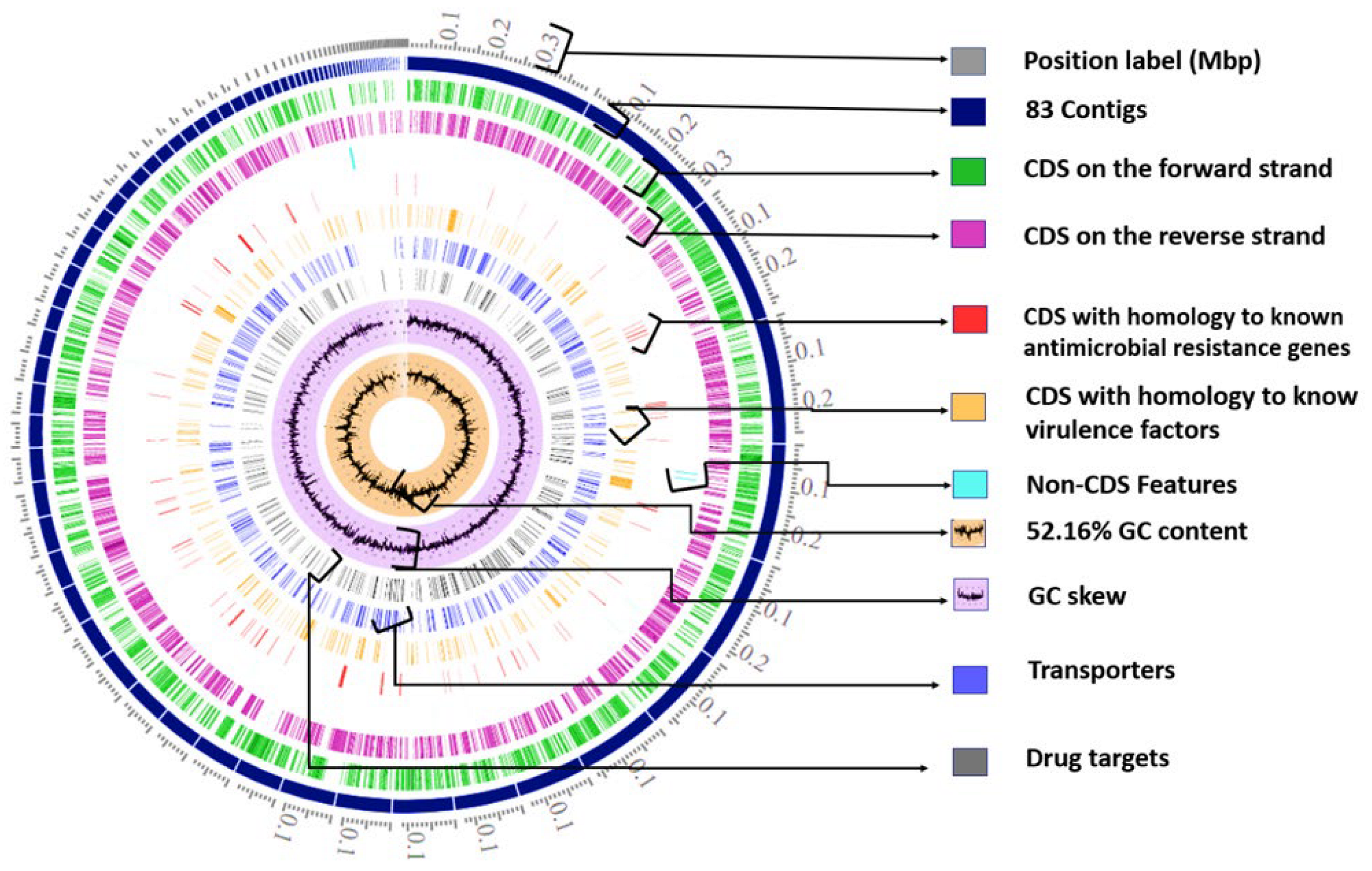
The Genome Assembly and Annotation

Reference-based genome assembly (reference genome ASM694v2) was performed with Bowtie 2 v. 2.4.5 [23]. In total, 83 contigs were obtained for Salmonella enterica subsp. enterica Typhimurium str. 69M (S. Typhimurium 69M), with the result demonstrating 99.7% similarity to S. enterica subsp. enterica serovar Typhimurium strain LT2. The assembled genome was submitted for comprehensive analysis at the on-line PATRIC service [24,25]; based on a comparison with other genomes within this same species, this genome appears to be of good quality (Coarse Consistency 99.7%, Fine Consistency 99.3%, CheckM Completeness 100%).
The genome encompassed 83 contigs, with a total length of 4,784,350 bp, and one plasmid IncFIB(S). The mean G+C content was 52.16%. The arrangement of the individual elements of the S. Typhimurium 69M genome is given in Figure 1; this includes the assembled contigs, coding DNA sequences (CDS), CDS on the forward strand, CDS on the reverse strand, RNA genes, CDS with homology to known antimicrobial resistance genes, CDS with homology to known virulence factors, GC content, and GC skew.
The annotation included 529 hypothetical proteins and 4235 proteins with functional assignments. Those with functional assignments included 1283 with Enzyme Commission (EC) numbers [26], 1048 with Gene Ontology (GO) assignments [27], and 900 proteins that were mapped to KEGG pathways [28]. PATRIC analysis indicated that the genome has 4647 proteins belonging to genus-specific protein families (PLFams) and 4673 belonging to cross-genus protein families (PGFams) [29].
Plasmid IncFIB(S) were observed. These carried critical virulence genes (spvB and spvC) and typical determinants for Salmonella pathogenicity island 1 (SPI-1) and SPI-2. They also harbored genes associated with fimbriae (pefA, pefC, pefD, pefI) and vapB (type II toxin-antitoxin system VapB family antitoxin) (Figure 2).

2.1.2. Serotype Determination Using Whole-Genome Sequencing Data

Genotypic serotyping based on multidirectional WGS analysis showed that O antigen prediction is 4, H1 antigen prediction (fliC)—i, H2 antigen prediction (fljB)—1,2. The strain was tentatively classified as Salmonella enterica subsp. enterica (subspecies I) serotype Typhimurium 4:i:1,2 and named as Salmonella enterica subsp. enterica Typhimurium 4:i:1,2_69M) [30,31].

2.2. Analysis for Virulence and Antibiotic Resistance Genes

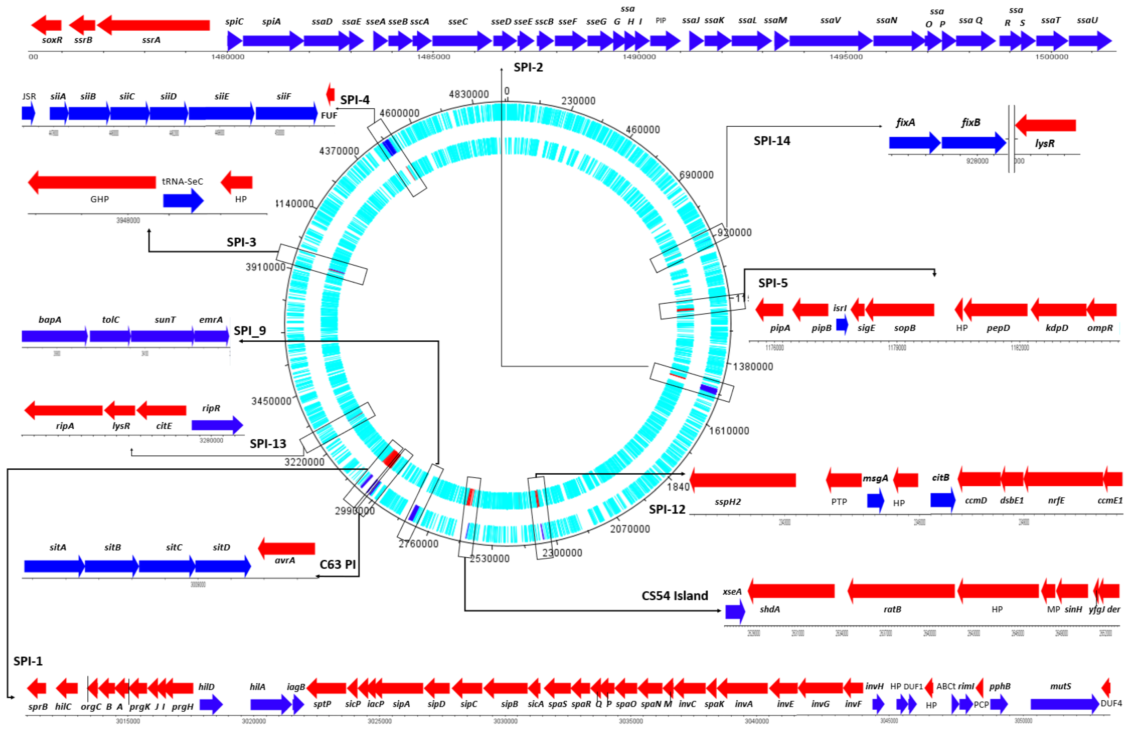
2.2.1. Salmonella Pathogenic Island (SPI)

The sequence of S. Typhimurium 69M was determined using SPIFinder 2.0, the online bioinformatics tool of the Center for Genomic Epidemiology. The results identified eleven SPIs (SPI-1, SPI-2, SPI-3, SPI-4, SPI-5, SPI-9, SPI-12, SPI-13, SPI-14, C63PI, CS54_island) with very high similarity (i.e., 98.58% to 100%) to the reference genome AE006468.2; it also identified other close matches regarding genes and regions of Salmonella spp. given in the NCBI Nucleotide Database, including JN673272, Z95891, AJ000509, Y13864, and AJ576316 (Table S1) [32]. All identified parts of the SPIs and their genes, together with their position in the contig and their accession number, are presented in Table 1.
The S. Typhimurium 69M sequences were compared to the reference strain Salmonella Typhimurium LT2 genome. The results of fragment of SPI 1 with the mutS gene are given in Figure 3, with 100% matches marked in green, and differences such as deletions, insertions, and SNPs in red. Ten single nucleotide insertions and single nucleotide polymorphisms (SNPs) were detected in the mutS gene (Figure 3). Despite these differences, the protein sequences generated by the mutS gene of the test strain demonstrated 98% and 100% similarity with the reference strains U16303 and S. Typhimurium LT2 (ProgramBlast 2 sequences [33]). The entire SPIfinder2.0 analysis is presented in the Supplementary Materials as Table S1.
The gene sequences obtained from S. Typhimurium 69M, particularly those within the identified SPIs, were analyzed using DNAPlotter [34]. It was found that the strain contains 221 genes responsible for virulence factors and antimicrobial resistance. The genes are located within 11 Salmonella pathogenicity islands (Figure 4). SPI-1 includes 42 genes; SPI-2, 33 genes; SPI-3, three genes; SPI-4, nine genes; SPI-5, 10 genes; SPI-9, five genes; SPI-12, eight genes; SPI-13, four genes; and SPI-14, two genes (fixB and fixA). In addition, the CS54 island includes seven genes, and C63-PI includes five genes. The location of individual pathogenicity islands and their genes, along with their arrangement on the forward and the reverse strands, are given in Figure 4.
The WGS analysis showed differences in the arrangement of 14 genes. Most of these, including pipB (SPI-2 type III secretion system effector PipB) and sspH2 (SPI-2 type III secretion system effector E3 ubiquitin transferase SspH2), were transferred from SPI-1 to SPI-5 and SPI-2 to SPI-12 (Figure 4). The relevant SPIs are presented in Table S2, together with their component genes, gene length, strand direction, locus, and a brief description of the product and function.

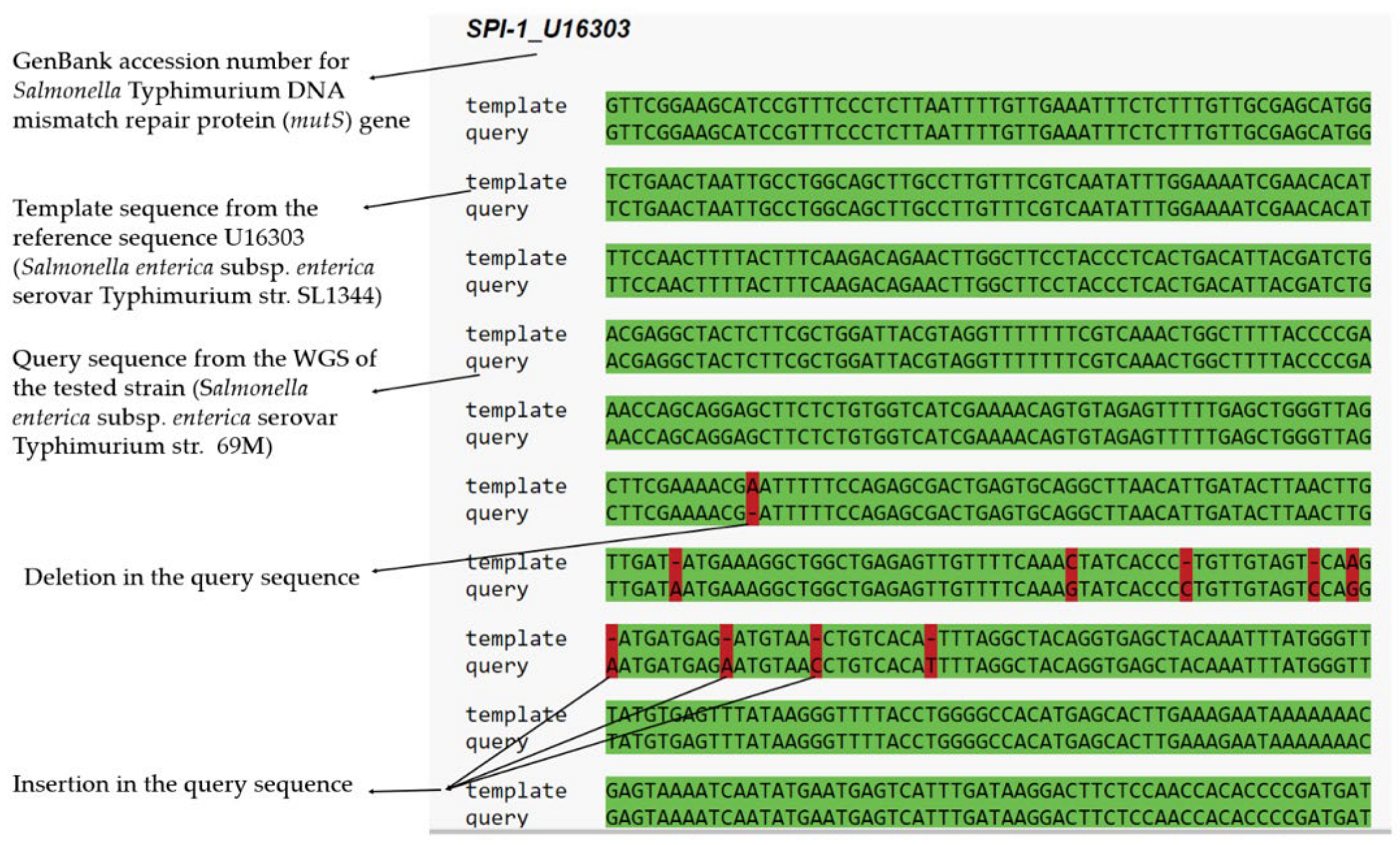
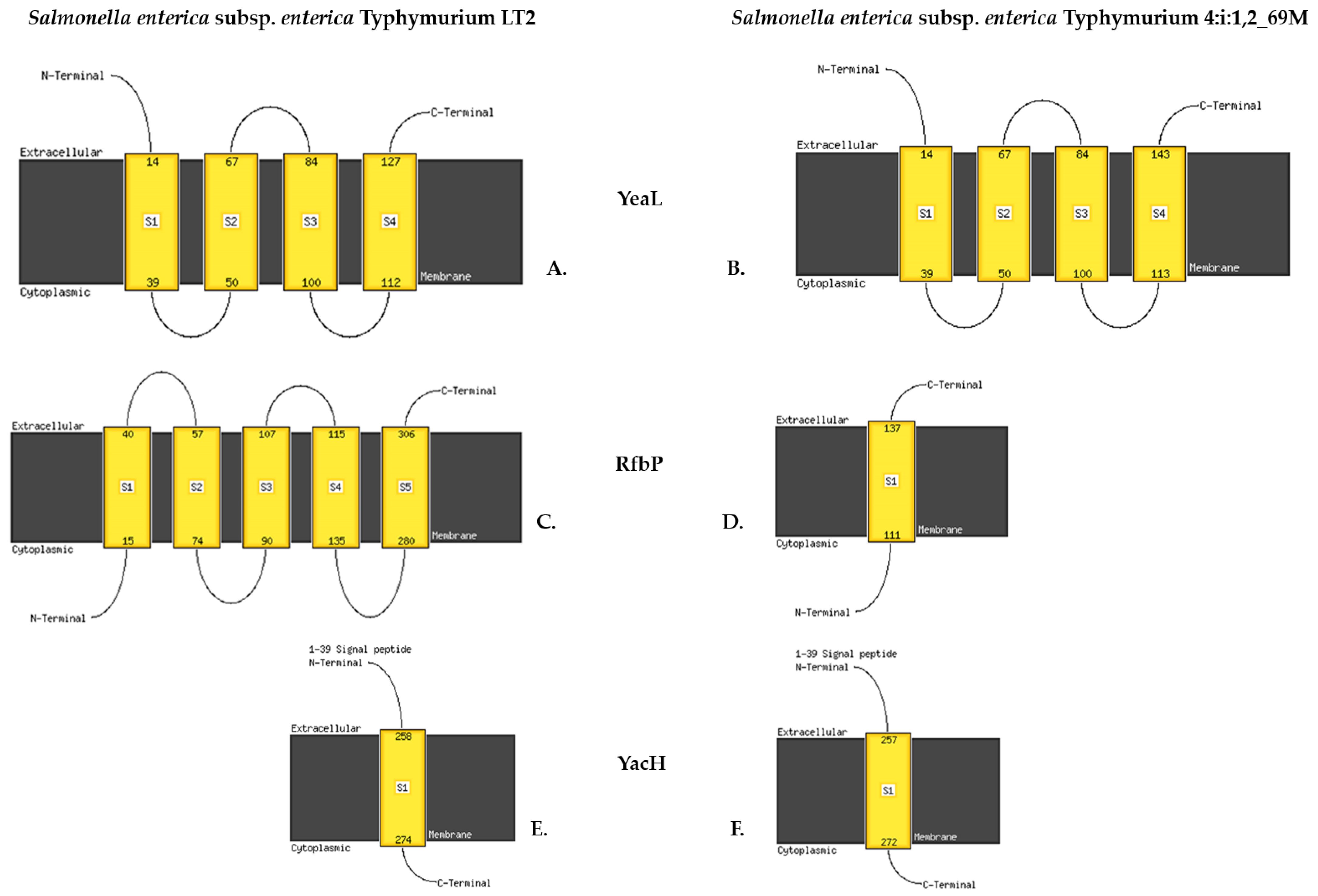
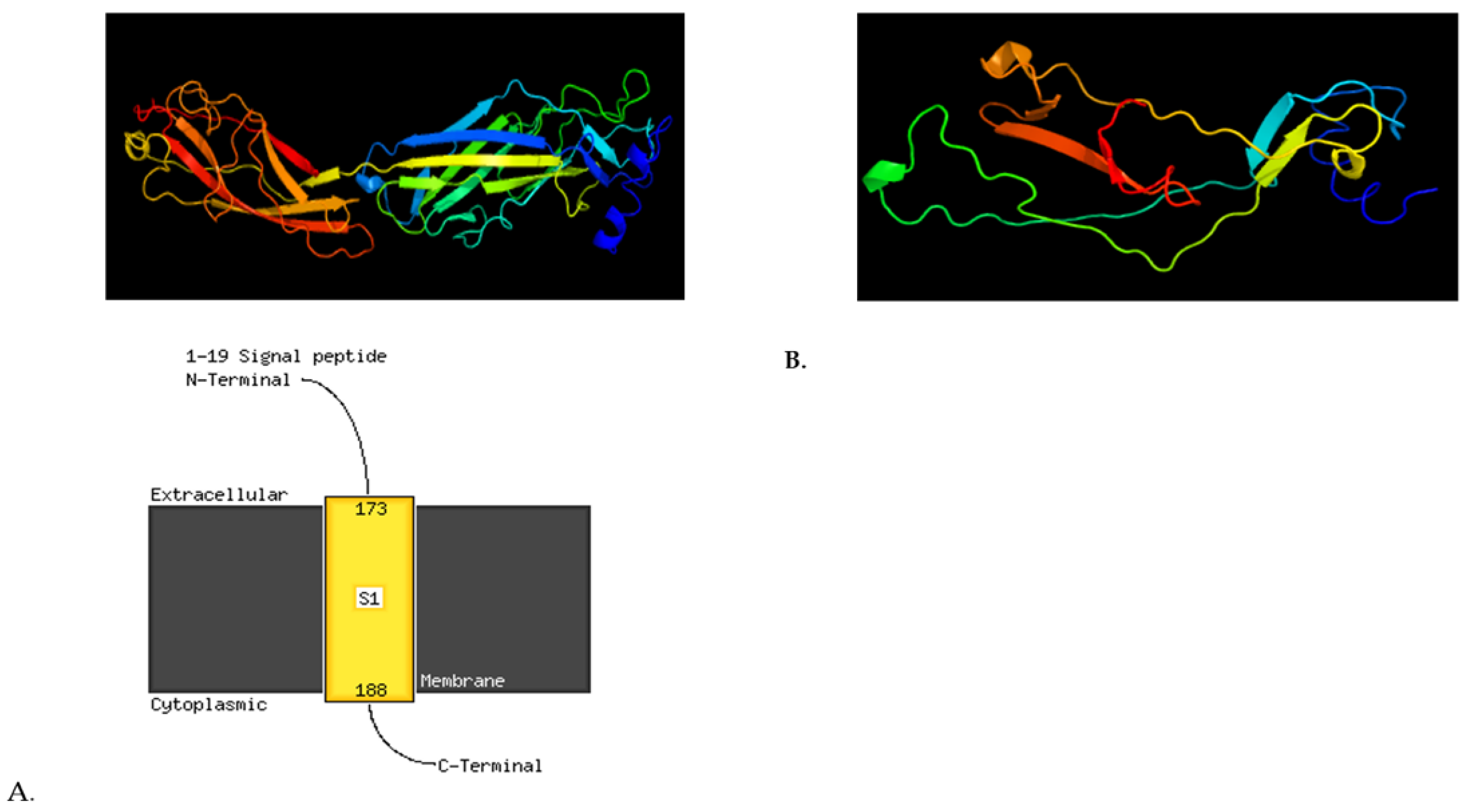
2.2.2. Multiple Sequence Alignment (MSA) and Single-Nucleotide Polymorphism (SNP)

Changes occurring in individual genes were identified using the multiple sequence alignment (MSA) and single-nucleotide polymorphism (SNP) service of the BV-BRC. In total, 700 changes were detected. These included 53 insertions and 24 deletions, of which 36 were classified as having a high impact. These are given in Table 2, with all raw results in Table S3. SNP effects were marked based on the degree of their effect on the encoded protein. Only those genes marked as High SNP Impact Effect, i.e., a destructive effect on the protein, were selected for further analysis (Figure 5). Significant changes were observed in a number of genes due to frameshift variants; these included insertions, such as in yeiG, rfbP, fumA, yeaL, and ybeU, and deletions, as in lpfD, avrA, ratB, and yacH. A stop-gain mutation was observed in shdA and ybfE and a stop loss-mutation in yciW. The most important changes observed in the selected genes are given in Table 2, together with a description of their effect and the possible differences to the S. Typhimurium LT2 referential genes.
Insertions were noted in the yeaL and yeiG genes of S. Typhimurium 69M. These changes cause a frame shift, creating a premature STOP codon and resulting in the loss of a large part of the protein (UPF0756 membrane protein YeaL UniProtKB/Swiss-Prot: Q8ZPW7 [36]). More specifically, the YeaL protein contains 148 aa in the reference strain (NP_460246.2), but only 134 aa in S. Typhimurium 69M: gtg cgg Val, Arg (VR), Val130, Arg131 (Val130_Arg131) Val Arg Ser Ala His STOP (TGA) (Figure 5).
The rfbP gene of Salmonella enterica serovar Typhimurium has two functions: it is involved in the first step of O-antigen synthesis, i.e., the galactosyltransferase [GT] function, and in a later step (the T function as O-antigen transfer protein).
This was first thought to be the flipping of the O-antigen subunit on undecaprenyl-phosphate galactosephosphotransferase (EC 2.7.8.6) from the cytoplasm to the periplasmic space of the cytoplasmic membrane [37].
The detected insertion within the first half of the rfbP gene would result in a frameshift mutation leading to the creation of a STOP codon 25 codons below the frameshift. This would shorten the open reading frame to only 307 codons, compared to 476 codons in the reference-type rfbP gene.
Insertion 390_391insC, Val130_Arg131fs, creates a new STOP codon in the yeaL gene, resulting in the formation of a truncated Yeal multi-pass membrane protein and the disruption of its S4 and C-terminal parts. Similarly, insertion 67_68insT Ile23_Phe24fs causes a premature STOP codon in undecaprenyl-phosphate galactosephosphotransferase/O-antigen transfer protein gene rfbP; this results in the formation of a truncated RfbP protein and loss of transmembrane helix structure (Figure 5).
Insertion 720_721insG Arg240_Lys241fs in yeiG (S-formylglutathione hydrolase gene) also creates a new STOP codon and the formation of a truncated protein. In addition, insertion 139_140insG Ile47_Asp48fs adds a new STOP codon, resulting in the formation of truncated YbeU protein (Figure S3). In Figure S3 protein model with premature STOP codon in yeaL, rfbP, yacH, avrA, and ratB were present.
Insertion 1631_1632insC, Gly544_Ala545fs, deletes the STOP codon in the fumA gene, leading to the formation of a longer protein than in Salmonella Typhimurium LT2 (Figure S4). Table S4. Secondary structure of protein with insertions and deletions in gene of Salmonella Typhimurium LT2 and Salmonella Typhimurium 69 were described in Table S4.
A deletion was found in the gene encoding the long polar fimbrial operon protein (ipfD), i.e., 400_409delTTTGAGAATG, Phe134fs, which adds a new STOP codon. This results in the production of a truncated IpfD protein and loss of its transmembrane structure. Deletion 5807delT Met1936fs in ratB, encoding an outer membrane protein/colonization factor, creates a new STOP codon and potentially leads to the synthesis of a truncated protein (Figure 6). Deletion 850_857delGATCATCA Asp284fs creates a new STOP codon in the outer membrane gene (yacH) resulting in a truncated YacH protein sequence (Figure 5).
Deletion 755delA Glu252fs in avrA again results in the creation of a premature STOP codon. This leads to the formation of a truncated Type III secretion injected virulence protein (YopP, YopJ), thought to be an inner-membrane protein (Table 2).

2.2.3. Antimicrobal Resistance Genes

The isolated strain turned out to be sensitive to many of the tested antibiotics: ampicillin ≤ 2, amoxicillin with clavulanic acid ≤ 2, cephalexin ≤ 4, cephalotin ≤ 2, cefoperazone ≤ 4, ceftiofur ≤ 1, cefquinone ≤ 0.5 imipenem ≤ 0.25, neomycin ≤ 2, flumequin ≤ 1, enrofloxacin ≤ 0.12, marbofloxacin ≤ 0.5 tetracycline ≤ 1 florfenicol 4, trimethoprim/sulfamethoxazole ≤ 20. Salmonella enterica subsp. enterica Typhimurium 4:i:1,2_69M showed phenotypic resistance only to gentamicin.
The phenotype of S. Typhimurium was determined using VITEK2 software v 8.02, Biomerieux, Marcy-l’Étoile, France: β-lactams, wild; aminoglycosides, resistant to Ami mesh (AAC (6′)) wild; quinolones, wild resistant/partially resistant; tetracyclines, wild; phoenixols, wild–resistant; trimethoprim, wild–resistant.
In the Enterobacteriaceae, resistance to aminoglycoside (gentamycin) is associated with AAC(6′), an aminoglycoside acetyltransferase encoded in the plasmid. The aac(6′)-I-cr variant gene can induce resistance against aminoglycoside and fluoroquinolone simultaneously. Our results also indicate the presence of the AcrAD-TolC efflux pump system, associated with aminoglycoside efflux. Although aminoglycoside resistance can also be caused by mutations within the gidB gene, causing changes in the structure of 16s rRNA, no such genetic changes were present in the investigated strain.
Bioinformatic analysis identified genes involved in, broadly understood, antimicrobial resistance involving various mechanisms (Table 3) [24].
Salmonella Typhimurium 69M has both marA, inducing MDR efflux pump AcrAB, and marB, coding the repressor of the mar operon marRAB, which regulates marA expression. Although S. Typhimurium 69M strain carries the gyrB gene, associated with fluoroquinolone resistance, it appears to be sensitive to fluoroquinolone. In addition, the investigated genome contains inhA and kasA, associated with resistance to isoniazid.
Other AMR mechanisms were also detected. These include AcrAB-TolC, a tripartite efflux system that confers resistance to tetracycline, chloramphenicol, ampicillin, nalidixic acid, and rifampin. This was accompanied by the AcrEF-TolC efflux pump system, involved in resistance to fluoroquinolones, and the MdtABC-TolC multidrug efflux system. The latter includes the macA gene, encoding the MacA membrane fusion protein, which forms an antibiotic efflux complex with MacB and TolC. Detailed functions of individual genes are provided in Tables S3 and S4.

3. Discussion

Human milk from the Women’s Milk Bank is not available to private individuals: it is illegal to purchase breast milk or give it to a child outside the hospital premises. The milk collected by milk banks is intended primarily for premature babies or for sick infants as part of nutritional therapy. Therefore, it is very important that potential donors are screened for pathogens, including Salmonella enterica subsp. enterica serotype Typhimurium [15].
The investigated Salmonella strain from human milk could not be serotyped with classical methods according to the White–Kauffmann–Le Minor scheme; this approach is time-consuming and complicated, as it requires above one hundred and fifty specific antisera and well-trained personnel to interpret the results.
For the genus Salmonella, 47 O serogroups and 114 H antigens have been described according to the Kauffmann–White–Le Minor scheme [38,39]. The O antigen (polysaccharide O) is part of the lipopolysaccharide (LPS) component of the outer membrane. It is necessary for the survival of the bacteria and plays a role in the virulence of Salmonella spp. [39].
Often, precise identification is not possible due to the lack of well-expressed flagellar antigens, as was the case in our study. This problem is becoming increasingly common [1,22,40,41], and as such, a number of European countries have included WGS in their screening protocols, such as the UK Standards for Microbiology Investigations [42].
In the present study, the biochemical tests and serotyping of isolated pure colonies allowed only the strain to be identified to a subspecies, i.e., Salmonella enterica subsp. enterica. Subsequent PCR analysis indicated that the strain belonged to the Typhimurium serovar. However, the WGS data confirmed the presence of genes encoding the lipopolysaccharide (O antigen; encoded by rfb genes) and flagellar antigens (phases 1 and 2 of H antigen, encoded by fliC and fljB). The detected antigenic profile was characteristic for the strain of S. Typhimurium (4:i:1,2): O antigen 4, H1 antigen I, and H2 antigen 1,2, despite Salmonella Typhimurium 69M not revealing any flagellar antigens when tested with H sera.
RfbP belongs to a large family of bacterial membrane proteins required for initiation of O antigen synthesis, and which catalyze the transfer of galactose-1-phosphate to undecaprenyl phosphate (Und-P) [43]. The rfpB gene is involved in lipopolysaccharide biosynthesis; it encodes galactosyl transferase, which catalyzes the transfer of galactose to undecaprenol phosphate, which is involved in the initial step in O-polysaccharide synthesis. As noted by Kong et al. (2011), Salmonella with mutated wbaP (rfbP) were significantly attenuated compared to wild-type strains when administered orally to BALB/c mice and were less invasive in host tissues; the mutants also demonstrated substantially reduced bacterial motility [44]. Wand et al. reported the presence of a secondary translation starting within the rfbP gene, resulting in the synthesis of a polypeptide with GT activity [37]. These results indicate that the N- and C-terminal parts of RfbP are the T and GT functional domains, respectively.
The genes affected by the changes within the tested Salmonella Typhimurium 69M strain are believed to be responsible for the chemotaxis and motility of the bacterial cell, as well as its adhesion and colonization factors. One good example is the bapA gene; this encodes the BapA protein, which forms a biofilm together with cellulose, fimbriae, and the lpfD colonization factor, fimbrial gene lpfD, encoding the adhesin at the tip of the Lpf fimbriae [45].
The fumA gene (fumarase A) is responsible for switching flagellar rotation from one direction to another and is hence an essential part of bacterial chemotaxis. In cytoplasm-free bacterial envelopes containing CheY, fumarate has been shown to restore the ability of flagella to switch directions; it also increases the probability of reversal in intact cells. Fumarate acts as a switching factor, presumably by lowering the activation energy of switching; thus, fumarate modulates bacterial flagellar rotation during chemotaxis and plays a role in bacteria metabolism [46]. The encoded protein, FumA (fumarase A), plays an essential role in bacterial chemotaxis through switching the direction of flagellar rotation. Fumarate acts as a switching factor, presumably by lowering the activation energy of switching. Thus fumarate and some of its metabolites may serve as a connection point between the bacterial metabolic state and chemotactic behavior [46].
The outer membrane protein yacH—CpxR is a conserved sensing system, i.e., regulated by genes associated with virulence; they are known to contribute to the resistance of E. coli to cationic antimicrobial peptide stress. In CpxR, extracellular protein transcription is reduced upon exposure to a sublethal dose of the cationic antimicrobial insect peptide cecropin A. Single-deletion strains (ΔyacH) demonstrated better survival than wild-type strains after protamine challenge, suggesting that these target genes contribute to resistance to protamine in E. coli [47,48]. CpxRA is a two-component system that monitors envelope perturbations and responds by altering the gene expression profile to allow Salmonella to survive under harmful conditions. Therefore, CpxRA activation is likely to contribute to Salmonella gut infection. However, the role of the CpxRA-mediated envelope stress response in Salmonella-induced diarrhea is unclear. In S. enterica subsp. enterica serovar Typhimurium, it has been found that CpxRA is not needed for the induction of colitis, but is required for gut colonization [49].
In BALB/c mice, a strain of Salmonella Typhimurium with a deletion in the ratB gene was not able to colonize the cecum, but was still noted in Peyer’s patches, the mesenteric lymph nodes, and spleen. In addition, mutations in shdA, ratB, and sivH resulted in a reduced ability to colonize intestinal tissues. The genes were encoded on the CS54 island and appear to be required for optimal colonization in the mouse cecum [50]. In contrast, despite large deletions and premature codon arrest, shdA from S. Typhi (shdASTy) remained fully functional and was found to allow adherence and invasion in a fibronectin-producing epithelial cell line [51].
A significant change was found in yeiG, encoding S-formylglutathione hydrolase (esterase), which plays a role in L-glutamine production. The change resulted in alanine replacement in the protein YeiG, demonstrating that Ser145, Asp233, and His256 are essential for protein activity: the residues represent a serine hydrolase catalytic triad in the protein. The enzyme also appears to contribute to the detoxification of formaldehyde, and may be involved in the degradation of methylglyoxal and/or other aldehydes [52,53].
The yeaL gene is responsible for the synthesis of the transmembrane protein in inter alia E. coli, Salmonella enterica subsp. Enterica, Klebsiella pneumoniae, and Yersinia enterocolitica [54]. The gene is also believed to involved in cell-wall biogenesis, which is required by S. Typhimurium to survive after desiccation [55].
Our findings indicate that 80.44% of Salmonella enterica subsp. Enterica showed aminoglycoside resistance facilitated by AAC(6′)-Ib-cr. The gene encodes an aminoglycoside acetyltransferase, which acetylates an amino group at position 6′ in aminoglycoside. Despite the presence of a number of potential antibiotic resistant mechanisms, S. Typhimurium 69M was still sensitive to most antibiotics.
Moreover, one plasmid, IncFIB(S), carrying virulence genes spvB and spvC, associated with fimbriae (pefA, pefC, pefD, pefI) and vapB (type II toxin-antitoxin system VapB family antitoxin), was observed. The SpvB protein exhibits a cytotoxic effect on host cells and is required for delayed cell death by apoptosis following intracellular infection. The SpvC protein demonstrates phosphothreonine lyase activity and has been shown to inhibit MAP kinase signaling [56]. Interestingly, strains isolated from HIV positive patients, usually carry spv genes, strongly suggesting that CD4+ T lymphocytes are required to control disease caused by spv-positive Salmonella.
The IncFIB(S) plasmid belongs to the IncF family, which is widely distributed throughout the Enterobacteriaceae, in particular, Salmonella enterica subsp. Enterica. These plasmids carry a variety of virulence factors, such as AMR genes and adhesion factors [57]. In S. Typhimurium, resistance genes are carried predominantly on IncFII(S)/IncFIB(S)/IncQ1-type plasmids. The detected IncFIB(S) plasmid affects the virulence of this serovar but is not involved in antibiotic resistance because of the absence of the AMR gene (blaTEM, tet(A), dfrA15, sul1, catA1, strA/strB, addA1).
In the presence of antibiotic stress, Salmonella overexpresses the global activator protein MarA, which induces MDR efflux pump AcrAB, and downregulates the synthesis of the porin OmpF. In addition, S. Typhimurium 69M showed the presence of the marR gene encoding the MarR protein; this regulates the expression of marA, the activator of multidrug efflux pump AcrAB.
In the stress-response pathways, Salmonella Typhimurium alternates sensitivity to triclosan due to point mutations in the gyrA gene; similarly, point mutations in fabI used in lipid metabolism and fatty acid biosynthesis are disturbed by this biocide.
In addition to direct AMR mechanisms, the Salmonella genome expresses AcrAB-TolC, a tripartite efflux system that spans the cell membrane (AcrB) and the outer-membrane (TolC) and is linked together in the periplasm by AcrA. This efflux pump confers resistance to tetracycline, chloramphenicol, ampicillin, nalidixic acid, and rifampin. The cells also expressed AcrAD-TolC, associated with efflux of aminoglycosides, and the AcrEF-TolC efflux pump system, associated with resistance to fluoroquinolones.
The replacement of classical Salmonella spp. serotyping methods with molecular biology methods, especially WGS, has been extensively discussed in previous studies [58]. Our present analysis of the Salmonella enterica subsp. enterica serotype Typhimurium 4:i:1.2_69M sequence revealed the presence of numerous antimicrobial resistance genes. However, the strain turned out to be sensitive to all antibiotics, except for gentamicin, to which the Enterobacteriacea demonstrate natural resistance. This probably indicates that the strain was not subject to environmental pressure in terms of the presence of antibiotics. However, there is the genetic potential for activation of these genes. In the environment, such a strain may serve as an effective donor of antibiotic resistance genes for other bacteria.

4. Materials and Methods

Salmonella spp. was isolated in accordance with PN-EN ISO 6579-1:2017-04 [59]. A human milk sample was preincubated in buffered peptone water (BPW GRASO, Gdansk, Poland) diluted 1:9 and then transferred to the following media: MSRV agar (modified semi-solid Rappaport–Vassiliadis (MSRV) agar, GRASO, Gdansk, Poland) and the Muller–Kauffmann tetrathionate–novobiocin (MKTTn) broth (GRASO, Gdansk, Poland). The potential Salmonella colonies were transferred from the selective enrichment media, viz. XLD agar (xylose lysine deoxycholate agar, GRASO, Gdansk, Poland) and BGA agar (Brilliant Green agar, OXOID, Hampshire, United Kingdom), to non-selective nutrient agar (GRASO, Gdansk, Poland). The temperature range and incubation time used for the above-mentioned media was described previously [60].

4.1. Serological Testing

Serotyping was performed by slide agglutination according to the White–Kauffmann–Le Minor scheme [61]. Commercial H poly antisera were used to verify the genus of Salmonella enterica (IBSS Biomed, Lublin, Poland), O group antisera to determine the O group, (IBSS Biomed, Lublin, Poland), and H phase and H factor antisera to determine the H phase and H factor (IBSS Biomed, Lublin, Poland, Bio-Rad, Chercules, CA, USA) [62].

4.2. Biochemical Strain Identification

The colonies demonstrating morphology typical of Salmonella spp. on selective agars were subjected to biochemical identification using VITEK2 COMPACT automated system for bacterial identification and VITEK® 2 GN cards (Biomerieux, Marcy-l’Étoile, France). E. coli ATCC 25922, Salmonella Typhimurium ATCC 14028, Salmonella Enteritidis ATCC 13076, and P. aeruginosa ATCC 27853 were used as reference strains. Tests were performed according to the manufacturer’s instructions.

4.3. Confirmation of Salmonella Identification with Molecular Biology Methods

DNA for Real-Time PCR was extracted from bacterial cells using a Kylt DNA Extraction-Mix II kit (Anicon, Emstek, Germany). A Kylt Salmonella spp. kit (Anicon, Emstek, Germany) was used to detect Salmonella spp., and a Spp-Se-St PCR kit (BioChek, Reeuwijk, The Netherlands) to detect Salmonella Enteritidis and Salmonella Typhimurium. Both Real-Time PCR tests were performed according to the manufacturer’s instructions using an Applied Biosystems 7500 Fast Real-Time PCR System (Thermo, Waltham, MA, USA).

4.4. Whole-Genome Sequencing

The whole genome and library gene preparation were sequenced using Illumina DNA Prep, (M) Tagmentation (Illumina, San Diego, CA, USA, Nexter DNA CD Indexes (Illumina, San Diego, CA, USA), PhiX Control v3 (Illumina), and MiSeq Reagent Kit v2 (300-cycles, Illumina) on Illumina MiSeq (Illumina, San Diego, CA, USA). All procedures were performed according to the manufacturer’s instructions.
Sequencing data from the Illumina MiSeq platform were extracted. The quality of the raw sequence reads was assessed with FastQC (v0.11.5) [63]. Low-quality sequences and adapters were removed with Trimmomatic (v0.36) with the following parameters: ILLUMINACLIP:adapters.fasta:2:30:10 LEADING:3 TRAILING:3 SLIDINGWINDOW:4:15 MINLEN:36; adapterst.fasta contained a sequence for Nextera_XT adapters (CTGTCTCTTATACACATCT) [64]. The sequence reads were assembled into the reference genome of Salmonella enterica subsp. enterica serovar Typhimurium str. LT2 (ASM694v2) with Bowtie 2 v. 2.4.5 [23]. SAM files were converted to BAM files with SAMtools [65] and annotated with Bakta v. 1.5.0. [66].
The general information about the assembly quality and gene content of Salmonella enterica subsp. enterica serovar Typhimurium 69M isolates and genomic components was obtained using the genomics tools of the Bacterial and Viral Bioinformatics Resource Center (BV-BRC, https://www.bv-brc.org accessed on 9 July 2022).
The serotypes of the isolated S. Typhimurium 69M strain were identified using a web-based tool: SeqSero 1.2 (https://cge.food.dtu.dk/services/SeqSero/ accessed on 9 July 2022) [30]. Bakta was used for rapid and standardized annotation of the bacterial genome and plasmids; this tool provides dbxref-rich, sORF-including, and taxon-independent annotations in machine-readable JSON and bioinformatics standard file formats for automated analysis of whole-genome sequences (Bakta version 1.4.2 was installed via BioConda with its native database publicly hosted at Zenodo [66]).
The Salmonella Pathogenicity Islands were predicted using the SPIFinder online search tool (https://cge.cbs.dtu.dk/services/SPIFinder accessed on 9 July 2022). Known or potential virulence factors were predicted in silico using the Virulence Factor Database (VFDB) (http://www.mgc.ac.cn/cgi-bin/VFs/v5/main.cgi accessed on 19 November 2022). The result was visualized by DNAPlotter [34], BV-BRC, Phyre2 web portal for protein modeling, prediction and analysis (http://www.sbg. bio.ic.ac.uk/phyre2 accessed on 19 November 2022), and plasmid graphics (SnapGene version 6.2) [67].

4.5. Antimicrobial Sensitivity Testing: Phenotypic Antibiotic Resistance

The antimicrobial susceptibility studies were performed as described previously [60] using a 96-well MICRONAUT plate in a VITEK2 Compact reader-incubator module, and AST-GN96 cards for gram-negative bacteria (BioMérieux, Marcy-l’Étoile, France). The AST card is a miniaturized and abbreviated version of the doubling dilution technique used to determine MICs by microdilution. The MICs were interpreted according to Clinical and Laboratory Standards Institute (CLSI) and FDA breakpoints (CLSI M100-ED28, 2018).

5. Conclusions

Salmonella serovars are typically classified based on serotyping according to the Kauffman and White classification scheme. However, some isolates require several passages through semi-solid media to enhance motility and flagellar antigenic expression. Some strains, like the S. Typhimurium 69M described herein, do not express serotype H antigens; S. Typhimurium 69M possesses a nine-nucleotide deletion (TTTGAGAATG, Phe134fs) in the ipfD gene (long polar fimbrial protein gene, part of the fimbrial operon), which creates a premature STOP codon. Pseudogene formation was also evident in a number of host adhesion factors. Two fimbrial genes including lpfD (encoding the tip adhesin of the Lpf fimbriae) and fimH (encoding the adhesin of the mannose-specific type 1 fimbriae) are inactive in the Sparrow MpSTM strain [45]. It was predicted that the mutated IpfD was truncated, i.e., with only 134 aa; this would result in the loss of its transmembrane structure, and thus potentially limit the value of traditional serotyping. In such cases, WGS Salmonella subtyping may ultimately prove to be more reliable and efficient. Although WGS serotyping requires higher technical and informational capacities, and remains expensive, it is clearly a very useful technique for both identifying Salmonella spp. strains and predicting the potential development of antibiotic resistance.

Supplementary Materials

The following supporting information can be downloaded at: https://www.mdpi.com/article/10.3390/ijms24065135/s1. References [68,69,70,71,72,73,74,75,76,77,78,79,80,81,82,83,84,85,86,87,88,89,90,91,92,93,94,95,96,97,98,99,100,101,102,103,104,105,106,107,108,109,110,111,112] are cited in the supplementary materials.

Author Contributions

Conceptualization, J.P.-C. and K.W.; methodology, J.P.-C., K.W., M.G., T.N.; software, J.P.-C., K.W., M.G., Z.S., A.K.; validation, J.P.-C., K.W., M.G., T.N. and K.A.; formal analysis, P.K., J.P.-C., K.W., M.G., Z.S., T.N.; investigation, E.R., Z.S.; resources, E.R., J.P.-C., K.W., M.G.; data curation, J.P.-C., P.K., K.A.; writing—original draft preparation, J.P.-C., K.W., M.G., E.R., T.N., Z.S., A.K.; writing—review and editing, J.P.-C., K.W., K.A., Z.S., P.K.; visualization, J.P.-C., K.W., M.G.; supervision, K.A., P.K.; project administration, J.P.-C.; funding acquisition, K.A. and P.K. All authors have read and agreed to the published version of the manuscript.

Funding

This research received no external funding.

Institutional Review Board Statement

Not applicable.

Informed Consent Statement

Not applicable.

Data Availability Statement

The data presented in this study are available in Supplementary Materials.

Acknowledgments

Special thanks to Jolanta Przybylska for help with the laboratory work.

Conflicts of Interest

The authors declare no conflict of interest.

References

  1. EFSA (European Food Safety Authority); ECDC (European Centre for Disease Prevention and Control). The European Union Summary Report on Antimicrobial Resistance in zoonotic and indicator bacteria from humans, animals and food in 2019–2020. EFSA J. 2022, 20, 1–197. [Google Scholar]
  2. Havelaar, A.H.; Kirk, M.D.; Torgerson, P.R.; Gibb, H.J.; Hald, T.; Lake, R.J.; Praet, N.; Bellinger, D.C.; de Silva, N.R.; Gargouri, N.; et al. World Health Organization Global Estimates and Regional Comparisons of the Burden of Foodborne Disease in 2010. PLoS Med. 2015, 12, e1001923. [Google Scholar] [CrossRef] [Green Version]
  3. Ao, T.T.; Feasey, N.A.; Gordon, M.A.; Keddy, K.H.; Angulo, F.J.; Crump, J.A. Global burden of invasive nontyphoidal Salmonella disease, 2010. Emerg. Infect. Dis. 2015, 21, 941–949. [Google Scholar] [CrossRef] [PubMed] [Green Version]
  4. Gal-Mor, O.; Finlay, B.B. Pathogenicity islands: A molecular toolbox for bacterial virulence. Cell. Microbiol. 2006, 8, 1707–1719. [Google Scholar] [CrossRef]
  5. Schmidt, H.; Hensel, M. Pathogenicity Islands in Bacterial Pathogenesis. Clin. Microbiol. Rev. 2004, 17, 14–56. [Google Scholar] [CrossRef] [Green Version]
  6. Deriu, E.; Liu, J.Z.; Pezeshki, M.; Edwards, R.A.; Ochoa, R.J.; Contreras, H.; Libby, S.J.; Fang, F.C.; Raffatellu, M. Probiotic bacteria reduce Salmonella Typhimurium intestinal colonization by competing for iron. Cell Host Microbe 2013, 14, 26–37. [Google Scholar] [CrossRef] [Green Version]
  7. Jiang, L.; Wang, P.; Song, X.; Zhang, H.; Ma, S.; Wang, J.; Li, W.; Lv, R.; Liu, X.; Ma, S.; et al. Salmonella Typhimurium reprograms macrophage metabolism via T3SS effector SopE2 to promote intracellular replication and virulence. Nat. Commun. 2021, 12, 879. [Google Scholar] [CrossRef] [PubMed]
  8. Kuhn, G.K.; Ethelberg, S. Investigating Outbreaks of Salmonella Typhimurium Using Case-Control Studies, with a Reference to the One Health Approach, 3rd ed.; Schatten, H., Ed.; Springer: Hatfield, UK, 2021; Volume 2182, ISBN 9781071607909. [Google Scholar]
  9. Lund, S.; Tahir, M.; Vohra, L.I.; Hamdana, A.H.; Ahmad, S. Outbreak of monophasic Salmonella Typhimurium Sequence Type 34 linked to chocolate products. Ann. Med. Surg. 2022, 82, 104597. [Google Scholar] [CrossRef]
  10. Ryder, R.W.; Crosby Ritchie, A.; Mcdonough, B.; Hall, W.J. Human Milk Contaminated with Salmonella Kottbus: A Cause of Nosocomial Illness in Infants. JAMA J. Am. Med. Assoc. 1977, 238, 1533–1534. [Google Scholar] [CrossRef]
  11. Revathi, G.; Mahajan, R.; Faridi, M.M.A.; Kumar, A.; Talwar, V. Transmission of lethal Salmonella Senftenberg from mother’s breast-milk to her baby. Ann. Trop. Paediatr. 1995, 15, 159–161. [Google Scholar] [CrossRef]
  12. Chen, T.L.; Thien, P.F.; Liaw, S.C.; Fung, C.P.; Siu, L.K. First report of Salmonella enterica serotype Panama meningitis associated with consumption of contaminated breast milk by a neonate. J. Clin. Microbiol. 2005, 43, 5400–5402. [Google Scholar] [CrossRef] [PubMed] [Green Version]
  13. Drhová, A.; Dobiásová, V.; Stefkovicová, M. Mother’s milk—Unusual factor of infection transmission in a salmonellosis epidemic on a newborn ward. J. Hyg. Epidemiol. Microbiol. Immunol. 1990, 34, 353–355. [Google Scholar] [PubMed]
  14. Blackshaw, K.; Valtchev, P.; Koolaji, N.; Berry, N.; Schindeler, A.; Dehghani, F.; Banati, R.B. The risk of infectious pathogens in breast-feeding, donated human milk and breast milk substitutes. Public Health Nutr. 2021, 24, 1725–1740. [Google Scholar] [CrossRef]
  15. Wesołowska, A. Banki mleka w Polsce : Funkcjonowanie w Podmiotach Leczniczych: Idea i Praktyka; Wesołowska, A., Ed.; Fundacja Bank Mleka Kobiecego: Warsaw, Poland, 2017; ISBN 9788393793310. [Google Scholar]
  16. Qutaishat, S.S.; Stemper, M.E.; Spencer, S.K.; Opitz, J.C.; Monson, T.A.; Anderson, J.L. Transmission of Salmonella enterica Serotype Typhimurium DT104 to Infants Through Mother’s Breast Milk. Pediatrics 2003, 111, 1442–1446. [Google Scholar] [CrossRef]
  17. Mitchell, H.; Ebbeson, R.; Maclean, M.; Hajek, J. A mother with Salmonella mastitis and a baby with Salmonella bacteremia. Can. Fam. Physician 2021, 67, 747–749. [Google Scholar] [CrossRef]
  18. Seah, X.F.V.; Ngeow, J.H.A.; Thoon, K.C.; Tee, W.S.N.; Maiwald, M.; Chong, C.Y.; Tan, N.W.H. Infant with Invasive Nontyphoidal Salmonellosis and Mastitis. Glob. Pediatr. Health 2015, 2, 2333794X1559156. [Google Scholar] [CrossRef]
  19. Wang, X.; Biswas, S.; Paudyal, N.; Pan, H.; Li, X.; Fang, W.; Yue, M. Antibiotic resistance in Salmonella Typhimurium isolates recovered from the food chain through national antimicrobial resistance monitoring system between 1996 and 2016. Front. Microbiol. 2019, 10, 985. [Google Scholar] [CrossRef] [PubMed] [Green Version]
  20. Karabay, M.; Şehmusoğlu, Z.; Terzi, H.A.; Caner, İ.; Köroğlu, M. Neonatal Sepsis due to Salmonella enteritidis from Colonized Breast Milk; A Case Report and Literature Review of Breast Milk-induced Neonatal Sepsis. Flora J. Infect. Dis. Clin. Microbiol. 2021, 26, 323–327. [Google Scholar] [CrossRef]
  21. Maroszyńska, I.; Fortecka-Piestrzeniewicz, K.; Niedźwiecka, M. Salmonella Typhimurium—A threat for a newborn. Open J. Obstet. Gynecol. 2013, 3, 628–630. [Google Scholar] [CrossRef] [Green Version]
  22. Pławińska-Czarnak, J.; Wódz, K.; Kizerwetter-świda, M.; Nowak, T.; Bogdan, J.; Kwieciński, P.; Kwieciński, A.; Anusz, K. Citrobacter braakii yield false-positive identification as salmonella, a note of caution. Foods 2021, 10, 2177. [Google Scholar] [CrossRef]
  23. Langmead, B.; Salzberg, S.L. Fast gapped-read alignment with Bowtie 2. Nat. Methods 2012, 9, 357–359. [Google Scholar] [CrossRef] [PubMed] [Green Version]
  24. Wattam, A.R.; Davis, J.J.; Assaf, R.; Boisvert, S.; Brettin, T.; Bun, C.; Conrad, N.; Dietrich, E.M.; Disz, T.; Gabbard, J.L.; et al. Improvements to PATRIC, the all-bacterial bioinformatics database and analysis resource center. Nucleic Acids Res. 2017, 45, D535–D542. [Google Scholar] [CrossRef]
  25. Olson, R.D.; Assaf, R.; Brettin, T.; Conrad, N.; Cucinell, C.; Davis, J.J.; Dempsey, D.M.; Dickerman, A.; Dietrich, E.M.; Kenyon, R.W.; et al. Introducing the Bacterial and Viral Bioinformatics Resource Center (BV-BRC): A resource combining PATRIC, IRD and ViPR. Nucleic Acids Res. 2022, 51, 678–689. [Google Scholar] [CrossRef] [PubMed]
  26. Schomburg, I.; Chang, A.; Ebeling, C.; Gremse, M.; Heldt, C.; Huhn, G.; Schomburg, D. BRENDA, the enzyme database: Updates and major new developments. Nucleic Acids Res. 2004, 32, D431–D433. [Google Scholar] [CrossRef] [Green Version]
  27. Finke, W.; Rachimow, C.; Pfützner, B. Untersuchungen zu Wasserdargebot und Wasserverfügbarkeit im Ballungsraum Berlin im Rahmen des Verbundprojektes GLOWA Elbe. Hydrol. Wasserbewirtsch. 2004, 48, 2–11. [Google Scholar]
  28. Kanehisa, M.; Sato, Y.; Kawashima, M.; Furumichi, M.; Tanabe, M. KEGG as a reference resource for gene and protein annotation. Nucleic Acids Res. 2016, 44, D457–D462. [Google Scholar] [CrossRef] [PubMed] [Green Version]
  29. Davis, J.J.; Gerdes, S.; Olsen, G.J.; Olson, R.; Pusch, G.D.; Shukla, M.; Vonstein, V.; Wattam, A.R.; Yoo, H. PATtyFams: Protein families for the microbial genomes in the PATRIC database. Front. Microbiol. 2016, 7, 118. [Google Scholar] [CrossRef] [Green Version]
  30. Zhang, S.; den Bakker, H.C.; Li, S.; Chen, J.; Dinsmore, B.A.; Lane, C.; Lauer, A.C.; Fields, P.I.; Deng, X. SeqSero2: Rapid and improved Salmonella serotype determination using whole-genome sequencing data. Appl. Environ. Microbiol. 2019, 85, e01746-19. [Google Scholar] [CrossRef] [PubMed]
  31. Zhang, S.; Yin, Y.; Jones, M.B.; Zhang, Z.; Kaiser, B.L.D.; Dinsmore, B.A.; Fitzgerald, C.; Fields, P.I.; Deng, X. Salmonella serotype determination utilizing high-throughput genome sequencing data. J. Clin. Microbiol. 2015, 53, 1685–1692. [Google Scholar] [CrossRef] [PubMed] [Green Version]
  32. Roer, L.; Hendriksen, R.S.; Leekitcharoenphon, P.; Lukjancenko, O.; Kaas, S.; Hasman, H. Is the Evolution of Salmonella enterica subsp. enterica Linked to Restriction-Modification Systems? mSystems 2016, 1, e00009-16. [Google Scholar]
  33. Altschul, S.F.; Madden, T.L.; Schäffer, A.A.; Zhang, J.; Zhang, Z.; Miller, W.; Lipman, D.J. Gapped BLAST and PSI-BLAST: A new generation of protein database search programs. Genetics 2000, 156, 1997–2005. [Google Scholar] [CrossRef] [PubMed] [Green Version]
  34. Carver, T.; Thomson, N.; Bleasby, A.; Berriman, M.; Parkhill, J. DNAPlotter: Circular and linear interactive genome visualization. Bioinformatics 2009, 25, 119–120. [Google Scholar] [CrossRef] [Green Version]
  35. Vihinen, M. When a Synonymous Variant Is Nonsynonymous. Genes 2022, 13, 1485. [Google Scholar] [CrossRef] [PubMed]
  36. Boutet, E.; Lieberherr, D.; Tognolli, M.; Schneider, M.; Bansal, P.; Bridge, A.J.; Poux, S.; Bougueleret, L.; Xenarios, I. UniProtKB/Swiss-Prot, the Manually Annotated Section of the UniProt KnowledgeBase: How to Use the Entry View. In Plant Bioinformatics, Methods and Protocols, Methods in Molecular Biology; Edwards, D., Ed.; Springer: Berlin/Heidelberg, Germany, 2007; Volume 1374, pp. 23–54. ISBN 9781493931675. [Google Scholar]
  37. Wang, L.; Liu, D.; Reeves, P.R. C-terminal half of Salmonella enterica WbaP (RfbP) is the galactosyl-1-phosphate transferase domain catalyzing the first step of O-antigen synthesis. J. Bacteriol. 1996, 178, 2598–2604. [Google Scholar] [CrossRef] [Green Version]
  38. Mcquiston, J.R.; Parrenas, R.; Gheesling, L.; Brenner, F.; Fields, P.I. Sequencing and Comparative Analysis of Flagellin Genes fliC, fljB, and flpA from Salmonella. J. Clin. Microbiol. 2004, 42, 1923–1932. [Google Scholar] [CrossRef] [Green Version]
  39. Liu, B.; Knirel, Y.A.; Feng, L.; Perepelov, A.V.; Senchenkova, S.N.; Reeves, P.R.; Wang, L. Structural diversity in Salmonella O antigens and its genetic basis. Fed. Eur. Microbiol. Soc. Microbiol Rev. 2014, 38, 56–89. [Google Scholar]
  40. European, T.; One, U.; Report, Z. The European Union One Health 2020 Zoonoses Report. EFSA J. 2021, 19, e06971. [Google Scholar]
  41. Campos, J.; Mourão, J.; Peixe, L.; Antunes, P. Non-typhoidal salmonella in the pig production chain: A comprehensive analysis of its impact on human health. Pathogens 2019, 8, 19. [Google Scholar] [CrossRef] [Green Version]
  42. Public Health England. UK Standards for Microbiology Investigations. Bacteriology 2015, B55, 1–21. [Google Scholar]
  43. Patel, K.B.; Furlong, S.E.; Valvano, M.A. Functional analysis of the C-terminal domain of the WbaP protein that mediates initiation of O antigen synthesis in Salmonella enterica. Glycobiology 2010, 20, 1389–1401. [Google Scholar] [CrossRef] [PubMed] [Green Version]
  44. Kong, Q.; Yang, J.; Liu, Q.; Alamuri, P.; Roland, K.L.; Curtiss, R. Effect of deletion of genes involved in lipopolysaccharide core and O-antigen synthesis on virulence and immunogenicity of Salmonella enterica serovar Typhimurium. Infect. Immun. 2011, 79, 4227–4239. [Google Scholar] [CrossRef] [PubMed] [Green Version]
  45. Cohen, E.; Azriel, S.; Austeri, O.; Gal, A.; Zitronblat, C.; Mikhlin, S.; Scharte, F.; Hensei, M.; Rahav, G.; Mor, O.G. Pathoadaptation of the passerine-associated Salmonella enterica serovar Typhimurium lineage to the avian host. PLoS Pathog. 2021, 17, e1009451. [Google Scholar] [CrossRef] [PubMed]
  46. Barak, R.; Eisenbach, M. Fumarate or a fumarate metabolite restores switching ability to rotating flagella of bacterial envelopes. J. Bacteriol. 1992, 174, 643–645. [Google Scholar] [CrossRef] [PubMed] [Green Version]
  47. Lu, Y.; Zhao, H.; Sun, J.; Liu, Y.; Zhou, X.; Beier, R.C.; Wu, G.; Hou, X. Characterization of Multidrug-Resistant Salmonella enterica Serovars Indiana and Enteritidis from Chickens in Eastern China. PLoS ONE 2014, 9, e96050. [Google Scholar] [CrossRef] [PubMed]
  48. Hong, R.W.; Shchepetov, M.; Weiser, J.N.; Axelsen, P.H. Transcriptional profile of the Escherichia coli response to the antimicrobial insect peptide cecropin A. Antimicrob. Agents Chemother. 2003, 47, 1–6. [Google Scholar] [CrossRef] [Green Version]
  49. Fujimoto, M.; Goto, R.; Haneda, T.; Okada, N.; Miki, T. Salmonella enterica Serovar Typhimurium CpxRA Two-Component System Contributes to Gut Colonization in Salmonella-Induced Colitis. Infect. Immun. 2018, 86, e00280-18. [Google Scholar] [CrossRef] [Green Version]
  50. Kingsley, R.A.; Humphries, A.D.; Weening, E.H.; De Zoete, M.R.; Winter, S.; Papaconstantinopoulou, A.; Dougan, G.; Ba, A.J. Molecular and Phenotypic Analysis of the CS54 Island of. Infect. Immun. 2003, 71, 629–640. [Google Scholar] [CrossRef] [Green Version]
  51. Urrutia, I.M.; Fuentes, J.A.; Valenzuela, L.M.; Ortega, A.P.; Hidalgo, A.A.; Mora, G.C. Salmonella Typhi shdA: Pseudogene or allelic variant? Infect. Genet. Evol. 2014, 26, 146–152. [Google Scholar] [CrossRef]
  52. Gonzalez, C.F.; Proudfoot, M.; Brown, G.; Korniyenko, Y.; Mori, H.; Savchenko, A.V.; Yakunin, A.F. Molecular basis of formaldehyde detoxification: Characterization of two S-formylglutathione hydrolases from Escherichia coli, FrmB and YeiG. J. Biol. Chem. 2006, 281, 14514–14522. [Google Scholar] [CrossRef] [PubMed] [Green Version]
  53. Chen, N.H.; Djoko, K.Y.; Veyrier, F.J.; McEwan, A.G. Formaldehyde stress responses in bacterial pathogens. Front. Microbiol. 2016, 7, 257. [Google Scholar] [CrossRef] [Green Version]
  54. Kanehara, K.; Ito, K.; Akiyama, Y. YaeL (EcfE) activates the σE pathway of stress response through a site-2 cleavage of anti-σE, RseA. Genes Dev. 2002, 16, 2147–2155. [Google Scholar] [CrossRef] [Green Version]
  55. Mandal, R.K.; Kwon, Y.M. Global screening of Salmonella enterica serovar Typhimurium genes for desiccation survival. Front. Microbiol. 2017, 8, 1723. [Google Scholar] [CrossRef] [PubMed]
  56. Guiney, D.G.; Fierer, J. The role of the spv genes in Salmonella pathogenesis. Front. Microbiol. 2011, 2, 129. [Google Scholar] [CrossRef] [Green Version]
  57. Mansour, M.N.; Yaghi, J.; El Khoury, A.; Felten, A.; Mistou, M.Y.; Atoui, A.; Radomski, N. Prediction of Salmonella serovars isolated from clinical and food matrices in Lebanon and genomic-based investigation focusing on Enteritidis serovar. Int. J. Food Microbiol. 2020, 333, 108831. [Google Scholar] [CrossRef] [PubMed]
  58. Shi, C.; Singh, P.; Ranieri, M.L.; Wiedmann, M.; Moreno Switt, A.I. Molecular methods for serovar determination of Salmonella. Crit. Rev. Microbiol. 2015, 41, 309–325. [Google Scholar] [CrossRef] [PubMed]
  59. EN ISO 6579-1:2017; Microbiology of the food chain—Horizontal method for the detection, enumeration and serotyping of Salmonella—Part 1: Detection of Salmonella spp. International Organization for Standardization: Geneva, Switzerland, 2017.
  60. Pławińska-Czarnak, J.; Wódz, K.; Kizerwetter-Świda, M.; Bogdan, J.; Kwieciński, P.; Nowak, T.; Strzałkowska, Z.; Anusz, K. Multi-Drug Resistance to Salmonella spp. When Isolated from Raw Meat Products. Antibiotics 2022, 11, 876. [Google Scholar] [CrossRef] [PubMed]
  61. Grimont, P.A.D.; Weill, F.-X. Antigenic formulae of the Salmonella serovars. WHO Collab. Cent. Ref. Res. Salmonella 2007, 9, 1–167. [Google Scholar]
  62. Issenhuth-Jeanjean, S.; Roggentin, P.; Mikoleit, M.; Guibourdenche, M.; De Pinna, E.; Nair, S.; Fields, P.I.; Weill, F.X. Supplement 2008–2010 (no. 48) to the White-Kauffmann-Le Minor scheme. Res. Microbiol. 2014, 165, 526–530. [Google Scholar] [CrossRef] [PubMed] [Green Version]
  63. Wingett, S.W.; Andrews, S. Fastq screen: A tool for multi-genome mapping and quality control. [version 2; peer review: 4 approved]. F1000Research 2018, 7, 1338. [Google Scholar] [CrossRef] [PubMed]
  64. Bolger, A.M.; Lohse, M.; Usadel, B. Trimmomatic: A flexible trimmer for Illumina sequence data. Bioinformatics 2014, 30, 2114–2120. [Google Scholar] [CrossRef] [PubMed] [Green Version]
  65. Li, H.; Handsaker, B.; Wysoker, A.; Fennell, T.; Ruan, J.; Homer, N.; Marth, G.; Abecasis, G.; Durbin, R. The Sequence Alignment/Map format and SAMtools. Bioinformatics 2009, 25, 2078–2079. [Google Scholar] [CrossRef] [Green Version]
  66. Schwengers, O.; Jelonek, L.; Dieckmann, M.A.; Beyvers, S.; Blom, J.; Goesmann, A. Bakta: Rapid and standardized annotation of bacterial genomes via alignment-free sequence identification. Microb. Genom. 2021, 7, 000685. [Google Scholar] [CrossRef]
  67. Kelley, L.A.; Mezulis, S.; Yates, C.M.; Wass, M.N.; Sternberg, M.J. The Phyre2 web portal for protein modeling, prediction and analysis. Nat. Protoc. 2015, 10, 845–858. [Google Scholar] [CrossRef] [Green Version]
  68. Ando, H.; Kondo, Y.; Suetake, T.; Toyota, E.; Kato, S.; Mori, T.; Kirikae, T. Identification of katG mutations associated with high-level isoniazid resistance in Mycobacterium tuberculosis. Antimicrob. Agents Chemother. 2010, 54, 1793–1799. [Google Scholar] [CrossRef] [Green Version]
  69. Loewen, P.C.; De Silva, P.M.; Donald, L.J.; Switala, J.; Villanueva, J.; Fita, I.; Kumar, A. KatG-Mediated Oxidation Leading to Reduced Susceptibility of Bacteria to Kanamycin. ACS Omega 2018, 3, 4213–4219. [Google Scholar] [CrossRef] [PubMed] [Green Version]
  70. Holden, E.R.; Webber, M.A. MarA, RamA, and SoxS as Mediators of the Stress Response: Survival at a Cost. Front. Microbiol. 2020, 11, 1–10. [Google Scholar] [CrossRef] [PubMed]
  71. Sharma, P.; Haycocks, J.R.J.; Middlemiss, A.D.; Kettles, R.A.; Sellars, L.E.; Ricci, V.; Piddock, L.J.V.; Grainger, D.C. The multiple antibiotic resistance operon of enteric bacteria controls DNA repair and outer membrane integrity. Nat. Commun. 2017, 8, 1444. [Google Scholar] [CrossRef] [PubMed] [Green Version]
  72. Eaves, D.J.; Ricci, V.; Piddock, L.J. V soxS in Salmonella enterica Serovar Typhimurium: Role in Multiple Antibiotic Resistance Serovar Typhimurium: Role in Multiple Antibiotic Resistance. Antimicrob. Agent Chemother. 2004, 48, 1145–1150. [Google Scholar] [CrossRef] [PubMed] [Green Version]
  73. Galakatos, N.G.; Daub, E.; Botstein, D.; Walsh, C.T. Biosynthetic alr alanine racemase from Salmonella typhimurium: DNA and protein sequence determination. Biochemistry. 1986, 25, 3255–3260. [Google Scholar] [CrossRef] [PubMed]
  74. Ray, S.; Das, S.; Panda, P.K.; Suar, M. Identification of a new alanine racemase in Salmonella Enteritidis and its contribution to pathogenesis. Gut Pathog. 2018, 10, 1–17. [Google Scholar] [CrossRef] [PubMed]
  75. Zawadzke, L.E.; Bugg, T.D.H.; Walsh, C.T. Existence of Two D-Alanine:D-Alanine Ligases in Escherichia coli: Cloning and Sequencing of the ddlA Gene and Purification and Characterization of the DdlA and DdlB Enzymes. Biochemistry 1991, 30, 1673–1682. [Google Scholar] [CrossRef] [PubMed]
  76. Brown, A.C.; Parish, T. Dxr is essential in Mycobacterium tuberculosis and fosmidomycin resistance is due to a lack of uptake. BMC Microbiol. 2008, 8, 78. [Google Scholar] [CrossRef] [PubMed] [Green Version]
  77. Macvanin, M.; Björkman, J.; Eriksson, S.; Rhen, M.; Andersson, D.I.; Hughes, D. Fusidic Acid-Resistant Mutants of Salmonella enterica Serovar Typhimurium with Low Fitness In Vivo are Defective in RpoS Induction. Antimicrob. Agents Chemother. 2003, 47, 3743–3749. [Google Scholar] [CrossRef] [Green Version]
  78. Harvey, K.L.; Jarocki, V.M.; Charles, I.G.; Djordjevic, S.P. The diverse functional roles of elongation factor tu (Eftu) in microbial pathogenesis. Front. Microbiol. 2019, 10, 2351. [Google Scholar] [CrossRef] [PubMed]
  79. Manna, M.S.; Tamer, Y.T.; Gaszek, I.; Poulides, N.; Ahmed, A.; Wang, X.; Toprak, F.C.R.; Woodard, D.N.R.; Koh, Y.; Williams, N.S.; et al. A trimethoprim derivative impedes antibiotic resistance evolution. Nat. Commun. 2021, 12, 2949. [Google Scholar] [CrossRef] [PubMed]
  80. Vedantam, G.; Guay, G.G.; Austria, N.E.; Doktor, S.Z.; Nichols, B.P. Characterization of mutations contributing to sulfathiazole resistance in Escherichia coli. Antimicrob. Agents Chemother. 1998, 42, 88–93. [Google Scholar] [CrossRef] [Green Version]
  81. Weigel, L.M.; Steward, C.D.; Tenover, F.C. gyrA mutations associated with fluoroquinolone resistance in eight species of Enterobacteriaceae. Antimicrob. Agents Chemother. 1998, 42, 2661–2667. [Google Scholar] [CrossRef] [PubMed] [Green Version]
  82. Feng, X.; Zhang, Z.; Li, X.; Song, Y.; Kang, J.; Yin, D.; Gao, Y.; Shi, N.; Duan, J. Mutations in gyrB play an important role in ciprofloxacin-resistant pseudomonas aeruginosa. Infect. Drug Resist. 2019, 12, 261–272. [Google Scholar] [CrossRef] [Green Version]
  83. Webber, M.A.; Randall, L.P.; Cooles, S.; Woodward, M.J.; Piddock, L.J.V. Triclosan resistance in Salmonella enterica serovar Typhimurium. J. Antimicrob. Chemother. 2008, 62, 83–91. [Google Scholar] [CrossRef] [Green Version]
  84. Takahata, S.; Ida, T.; Hiraishi, T.; Sakakibara, S.; Maebashi, K.; Terada, S.; Muratani, T.; Matsumoto, T.; Nakahama, C.; Tomono, K. Molecular mechanisms of fosfomycin resistance in clinical isolates of Escherichia coli. Int. J. Antimicrob. Agents 2010, 35, 333–337. [Google Scholar] [CrossRef]
  85. Hafeezunnisa, M.; Sen, R. The Rho-Dependent Transcription Termination Is Involved in Broad-Spectrum Antibiotic Susceptibility in Escherichia coli. Front. Microbiol. 2020, 11, 605305. [Google Scholar] [CrossRef]
  86. Siu, G.K.H.; Zhang, Y.; Lau, T.C.K.; Lau, R.W.T.; Ho, P.L.; Yew, W.W.; Tsui, S.K.W.; Cheng, V.C.C.; Yuen, K.Y.; Yam, W.C. Mutations outside the rifampicin resistance-determining region associated with rifampicin resistance in Mycobacterium tuberculosis. J. Antimicrob. Chemother. 2011, 66, 730–733. [Google Scholar] [CrossRef]
  87. De Vos, M.; Müller, B.; Borrell, S.; Black, P.A.; Van Helden, P.D.; Warren, R.M.; Gagneux, S.; Victor, T.C. Putative compensatory mutations in the rpoc gene of rifampin-resistant mycobacterium tuberculosis are associated with ongoing transmission. Antimicrob. Agents Chemother. 2013, 57, 827–832. [Google Scholar] [CrossRef] [PubMed] [Green Version]
  88. Radeck, J.; Gebhard, S.; Orchard, P.S.; Kirchner, M.; Bauer, S.; Mascher, T.; Fritz, G. Anatomy of the bacitracin resistance network in Bacillus subtilis. Mol. Microbiol. 2016, 100, 607–620. [Google Scholar] [CrossRef] [Green Version]
  89. Du, D.; Wang, Z.; James, N.R.; Voss, J.E.; Klimont, E.; Ohene-agyei, T.; Venter, H.; Chiu, W.; Luisi, B.F. Structure of the AcrAB—TolC multidrug efflux pump. Nature 2014, 509, 512–515. [Google Scholar] [CrossRef] [Green Version]
  90. Atac, N.; Kurt-Azap, O.; Dolapci, I.; Yesilkaya, A.; Ergonul, O.; Gonen, M.; Can, F. The Role of AcrAB–TolC Efflux Pumps on Quinolone Resistance of E. coli ST131. Curr. Microbiol. 2018, 75, 1661–1666. [Google Scholar] [CrossRef] [PubMed] [Green Version]
  91. Lu, Y.; Wen, Y.; Hu, G.; Liu, Y.; Beier, R.C.; Hou, X. Genomic sequence analysis of the multidrug-resistance region of avian Salmonella enterica serovar Indiana strain MHYL. Microorganisms 2019, 7, 248. [Google Scholar] [CrossRef] [PubMed] [Green Version]
  92. Alav, I.; Bavro, V.N.; Blair, J.M.A. Interchangeability of periplasmic adaptor proteins AcrA and AcrE in forming functional efflux pumps with AcrD in Salmonella enterica serovar Typhimurium. J. Antimicrob. Chemother. 2021, 76, 2558–2564. [Google Scholar] [CrossRef] [PubMed]
  93. Alenazy, R. Antibiotic resistance in Salmonella: Targeting multidrug resistance by understanding efflux pumps, regulators and the inhibitors. J. King Saud Univ.-Sci. 2022, 34, 102275. [Google Scholar] [CrossRef]
  94. Yardeni, E.H.; Zomot, E.; Bibi, E. The fascinating but mysterious mechanistic aspects of multidrug transport by MdfA from Escherichia coli. Res. Microbiol. 2018, 169, 455–460. [Google Scholar] [CrossRef] [PubMed]
  95. Nilsen, I.W.; Bakke, I.; Vader, A.; Olsvik, Ø.; El-Gewely, M.R. Isolation of cmr, a novel Escherichia coli chloramphenicol resistance gene encoding a putative efflux pump. J. Bacteriol. 1996, 178, 3188–3193. [Google Scholar] [CrossRef] [PubMed] [Green Version]
  96. Ayhan, D.H.; Tamer, Y.T.; Akbar, M.; Bailey, S.M.; Wong, M.; Daly, S.M.; Greenberg, D.E.; Toprak, E. Sequence-Specific Targeting of Bacterial Resistance Genes Increases Antibiotic Efficacy. PLoS Biol. 2016, 14, e1002552. [Google Scholar] [CrossRef] [Green Version]
  97. Nishino, K.; Nikaido, E.; Yamaguchi, A. Regulation of multidrug efflux systems involved in multidrug and metal resistance of Salmonella enterica serovar typhimurium. J. Bacteriol. 2007, 189, 9066–9075. [Google Scholar] [CrossRef] [Green Version]
  98. Jibril, A.H.; Okeke, I.N.; Dalsgaard, A.; Menéndez, V.G.; Olsen, J.E. Genomic analysis of antimicrobial resistance and resistance plasmids in salmonella serovars from poultry in Nigeria. Antibiotics 2021, 10, 99. [Google Scholar] [CrossRef]
  99. Holdsworth, S.R.; Law, C.J. Functional and biochemical characterisation of the Escherichia coli major facilitator superfamily multidrug transporter MdtM. Biochimie 2012, 94, 1334–1346. [Google Scholar] [CrossRef] [PubMed]
  100. Spierings, G.; Elders, R.; van Lith, B.; Hofstra, H.; Tommassen, J. Characterization of the Salmonella typhimurium phoE gene and development of Salmonella-specific DNA probes. Gene 1992, 122, 45–52. [Google Scholar] [CrossRef] [PubMed]
  101. Nishino, K.; Latifi, T.; Groisman, E.A. Virulence and drug resistance roles of multidrug efflux systems of Salmonella enterica serovar Typhimurium. Mol. Microbiol. 2006, 59, 126–141. [Google Scholar] [CrossRef]
  102. Kapach, G.; Nuri, R.; Schmidt, C.; Danin, A.; Ferrera, S.; Savidor, A.; Gerlach, R.G.; Shai, Y. Loss of the Periplasmic Chaperone Skp and Mutations in the Efflux Pump AcrAB-TolC Play a Role in Acquired Resistance to Antimicrobial Peptides in Salmonella typhimurium. Front. Microbiol. 2020, 11, 189. [Google Scholar] [CrossRef] [PubMed]
  103. Rodríguez-García, Á.; Mares-Alejandre, R.E.; Muñoz-Muñoz, P.L.A.; Ruvalcaba-Ruiz, S.; González-Sánchez, R.A.; Bernáldez-Sarabia, J.; Meléndez-López, S.G.; Licea-Navarro, A.F.; Ramos-Ibarra, M.A. Molecular analysis of streptomycin resistance genes in clinical strains of mycobacterium tuberculosis and biocomputational analysis of the mtgidb l101f variant. Antibiotics 2021, 10, 807. [Google Scholar] [CrossRef]
  104. Sulaiman, J.E.; Long, L.; Wu, L.; Qian, P.Y.; Lam, H. Comparative proteomic investigation of multiple methicillin-resistant Staphylococcus aureus strains generated through adaptive laboratory evolution. iScience 2021, 24, 102950. [Google Scholar] [CrossRef]
  105. Yang, B.; Yao, H.; Li, D.; Liu, Z. The phosphatidylglycerol phosphate synthase PgsA utilizes a trifurcated amphipathic cavity for catalysis at the membrane-cytosol interface. Curr. Res. Struct. Biol. 2021, 3, 312–323. [Google Scholar] [CrossRef] [PubMed]
  106. Tran, T.T.; Mishra, N.N.; Seepersaud, R.; Diaz, L.; Rios, R.; Dinh, A.Q.; Garcia-de-la-Maria, C.; Rybak, M.J.; Miro, J.M.; Shelburne, S.A.; et al. Mutations in cdsA and pgsA Correlate with Daptomycin Resistance in Streptococcus mitis and S. oralis. Antimicrob. Agents Chemother. 2019, 63, e01531-18. [Google Scholar] [CrossRef] [PubMed] [Green Version]
  107. Kalafatis, M.; Slauch, J.M. Long-distance effects of H-NS binding in the control of hilD expression in the salmonella SPI1 locus. J. Bacteriol. 2021, 203, e0030821. [Google Scholar] [CrossRef]
  108. Johnson, J.R.; Clabots, C.; Rosen, H. Effect of inactivation of the global oxidative stress regulator oxyR on the colonization ability of Escherichia coli O1:K1:H7 in a mouse model of ascending urinary tract infection. Infect. Immun. 2006, 74, 461–468. [Google Scholar] [CrossRef] [PubMed] [Green Version]
  109. Anand, A.; Chen, K.; Catoiu, E.; Sastry, A.V.; Olson, C.A.; Sandberg, T.E.; Seif, Y.; Xu, S.; Szubin, R.; Yang, L.; et al. OxyR Is a Convergent Target for Mutations Acquired during Adaptation to Oxidative Stress-Prone Metabolic States. Mol. Biol. Evol. 2020, 37, 660–666. [Google Scholar] [CrossRef] [PubMed] [Green Version]
  110. Robicsek, A.; Strahilevitz, J.; Jacoby, G.A.; Macielag, M.; Abbanat, D.; Chi, H.P.; Bush, K.; Hooper, D.C. Fluoroquinolone-modifying enzyme: A new adaptation of a common aminoglycoside acetyltransferase. Nat. Med. 2006, 12, 83–88. [Google Scholar] [CrossRef] [PubMed]
  111. Wong, M.H.Y.; Chan, E.W.C.; Liu, L.Z.; Chen, S. PMQR genes oqxAB and aac(6′)Ib-cr accelerate the development of fluoroquinolone resistance in Salmonella Typhimurium. Front. Microbiol. 2014, 5, 521. [Google Scholar] [CrossRef]
  112. Nishino, K.; Yamaguchi, A. Role of Histone-Like Protein H-NS in Multidrug Resistance of Escherichia coli. J. Bacteriol. 2004, 186, 1423–1429. [Google Scholar] [CrossRef] [PubMed] [Green Version]
Figure 1. A circular graphical display of the distribution of the genome annotations of Salmonella enterica subsp. enterica Typhimurium 69M. The colors of the descriptive of the CDS elements of the Salmonella enterica subsp. enterica Typhimurium 69M were changed for the transparency of the view: grey for position label (MBP), navy for the contigs, green for the CDS on the forward strand, violet for the CDS on the reverse strand, turquoise RNA genes, red for the CDS with homology to known antimicrobial resistance genes, orange for the CDS with homology to know virulence factors, blue for the transporters genes, black for the drugs target genes, black line on orange background GC content, and black on purple GC skew (done with PATRIC-BV-BRC, https://www.bv-brc.org, accessed on 17 November 2022).
Ijms 24 05135 g001
Figure 2. A circular graphic of the IncFIB(S) plasmid found in the Salmonella Typhimurium 69M sequence. The length of the plasmid is 93,926 bp, and the virulence genes carried by the IncFIB(S) are marked with maroon arrows (created in SnapGene 6.0.3 (www.snapgene.com, accessed on 2 February 2023)).
Ijms 24 05135 g002
Figure 3. Fragment of SPI 1 with the mutS gene. Analysis performed by SPIFinder 2.0. alignment of the mutS gene; query (Salmonella Typhimurium 69M from human milk) and template (U16303—Salmonella enterica subsp. enterica serovar Typhimurium str. SL1344). The query sequence was found to demonstrate the greatest similarity (99.43%) with the Salmonella Typhimurium DNA mismatch repair protein (mutS) gene sequence (U16303).
Ijms 24 05135 g003
Figure 4. The black line indicates the position on the genome (Kbp); the Salmonella Pathogenity Islands are presented in color: the red arrows indicate genes on the reverse strand and the blue arrows indicate those on the forward strand: SPI-1 includes 38 genes and 5 putative protein genes (sprB, hilC, orgC, orgB, orgA, prgK, prgJ, prgI, prgH, hilD, hilA, iagB, sptP, sicP, iacP, sipA, sipD, sipC, sipB, sicA, spaS, spaR, spaQ, spaP, spaO, spaN, spaM, invC, spaK, invA, invE, invG, invF, invH, InvR, rimI, pphB, mutS, ABCt—ABC transporter, PCP—putative cytoplasmic protein, DUF1—DUF1493 domain-containing protein, DUF4—DUF4440 domain-containing protein); SPI-2 includes 32 genes and 1 putative protein gene (soxR, ssrB, ssrA, spiC, spiA, ssaD, ssaE, sseA, sseB, sscA, sseC, sseD, sseE, sscB, sseF, sseG, ssaG, ssaH, ssaI, ssaJ, PIP, ssaK, ssaL, ssaM, ssaV, ssaN, ssaO, ssaP, ssaQ, ssaR, ssas, ssaT, ssaU) and (on the position from 2424353 to 2425810) nuoN; SPI-3 includes 3 putative protein genes (GHP, tRNA-SeC, HP as Ybl27 hypothetical protein); SPI-4 includes 7 genes and 2 putative protein genes (ssb1, JSR, siiA, siiB, siiC, siiD, siiE, siiF, FUF), JSR-JUMPstart RNA, and FUF—family of unknown function; SPI-5 includes 8 genes and 2 putative protein genes (tRNA-Ser, pipA, pipB, isrI, sigE, sopB, HP, pepD, kdpD, ompR); SPI-9 includes 5 genes (bapA, isrL, tolC, sunT, emrA); SPI-12 includes 7 genes and 2 putative protein genes (sspH2, PTP, isrG, msgA, AH, citB, ccmD, dsbE1, nrfE); SPI-13 includes 4 genes (ripA, lysR, citE, ripR); SPI-14 includes 2 genes, traX and fixA; the CS54 island includes 5 genes and 2 putative protein genes (shdA, ratB, HP, MP, sinH, yfgJ, der); C63 PI includes 5 genes (sitA, sitB, sitC, sitD, avrA); HP-hypothetical protein, PIP—pathogenicity island protein, AH—acyloxyacyl hydrolase, GPH—glycoside–pentoside–hexuronide family transporter.
Ijms 24 05135 g004
Figure 5. Predicted transmembrane helices. Functional (Salmonella enterica subsp. Enterica Typhimurium LT2, left panel) and truncated (Salmonella enterica subsp. Enterica Typhimurium 4:i:1,2_69M, right panel) proteins. YeaL UPF0756 membrane protein (A). YeaL LT2 (148 aa), (B). YeaL 69M (134 aa); RfbP O-antigen transfer protein (C). RfbP LT2 (476 aa), (D). RfbP 69M (307 aa); YacH membrane protein (E). YacH LT2 (540 aa), (F). YacH 69M (285 aa). Created by Phyre2, Protein Homology/analogY Recognition Engine V 2.0, London, UK.
Ijms 24 05135 g005
Figure 6. 3D viewing of the protein model with premature STOP codon. (A). IpfD LT2 (359 aa); (B). IpfD 69M (134 aa), the truncated IpfD protein with loss of transmembrane structure. Created by Phyre2, Protein Homology/analogY Recognition Engine V 2.0, London, UK.
Ijms 24 05135 g006
Table 1. Salmonella Pathogenicity Islands (SPIs) detected in the genome of Salmonella enterica subsp. enterica Typhimurium isolated from human milk (S. Typhimurium 69M) and their references in GeneBank.
SPIIdentity %Query/Template LengthPosition in ContigGenesOrganismAccession Number
C63PI1004000/40003,006,066..3,010,065sitA, sitB, sitC, sitDSalmonella-enterica-Typhimurium-SL1344AF128999
CS54_island99.9225255/252522,627,012..2,652,263xse, shdA, ratC, ratB, ratA, sinI, sinH, yfgKSalmonella-enterica-Typhimurium-ATCC_14028AF140550
SPI-199.962705/27053,010,641..3,013,345sprA, sprBSalmonella-enterica-Typhimurium-SL1344AF148689
SPI-199.77440/4403,015,995..3,016,434prgH, prgI, prgJSalmonella-enterica-Gallinarum-SGB_1AY956822
SPI-1100430/4303,018,105..3,018,534hilDSalmonella-enterica-Gallinarum-SGE_2 AY956823
SPI-1100470/4703,026,813..3,027,282sipDSalmonella-enterica-Gallinarum-SGB_4AY956824
SPI-199.28415/4153,038,600..3,039,014invASalmonella-enterica-Gallinarum-SGB_8AY956825
SPI-198.84259/2573,039,894..3,040,152 invASalmonella-enterica-Typhimurium-J4STEHOJN982040
SPI-199.433155/31413,049,734..3,052,886mutSSalmonella-enterica-Typhimurium-SL1344U16303
SPI-1297.64 9345/110752,342,056..2,351,393sspH2, isrG, citB, ccmD, dsbE1, nrfE, ccmE1Salmonella-enterica-Choleraesuis-SC_B67 NC_006905
SPI-13 99.41 338/3383,279,563..3,279,900gacDSalmonella-enterica-Gallinarum-SGD_3AY956832
SPI-13100404/4043,278,852..3,279,255gtrASalmonella-enterica-Gallinarum-SGG_1AY956833
SPI-13100341/3413,277,144..3,277,484gtrBSalmonella-enterica-Gallinarum-SGA_10AY956834
SPI-14100501/501926,728..927,228gpiASalmonella-enterica-Gallinarum-SGA_8AY956835
SPI-1499.55 441/44132,304..932,744gpiBSalmonella-enterica-Gallinarum-SGC_8AY956836
SPI-2 98.90637/6371,497,019..1,497,655ssaO, ssaPSalmonella-enterica-Gallinarum-SGB_10 AY956826
SPI-2100642/6421,489,709…1,490,350ssaG, ssaH, ssaI, ssaJSalmonella-enterica-Gallinarum-SGC_2AY956827
SPI-2100395/3961,494342..1,494,736ssaVSalmonella-enterica-Gallinarum-SGC_9 AY956828
SPI-2 99.76 425/4251,487,684..1,488,108sscB, sseFSalmonella-enterica-Gallinarum-SGH_1 AY956829
SPI-2 99.82547/5471,477,547..1,478,093ssrASalmonella-enterica-Gallinarum-SGD_8AY956830
SPI-2100384/384 1,480,008..1,480,391spiCSalmonella-enterica-Typhimurium-St11JN673272
SPI-21001252/12521,475,967..1,477,218ssrA, ssrBSalmonella-enterica-TyphimuriumZ95891
SPI-3100738/7383,965,704..3,966,441mgtCSalmonella-enterica-Typhimurium-14028sAJ000509
SPI-31001514/15143,948,169..3,949,682selCSalmonella-enterica-Typhimurium-14028sY13864
SPI-410024660/24660 4,476,929..4,501,588siiABCDEFSalmonella-enterica-Typhimurium-ST4/74 AJ576316
SPI-51009069/90691,175,309..1,184,377Ser_trna, pipA, pipB, isrI, sigE, sopB, pepD, kdpD, ompRSalmonella-Typhimurium-LT2NC_003197
SPI-998.5812647/156962,831,233..2,843,879bapA, isrL, tolC, sunT, emrASalmonella-Typhi-CT18 NC_003198
Table 2. The selected genes with their mutations and potential effects in the identified Salmonella Typhimurium 4:i:1,2_69M sequence; the table also compares the findings with referential genes from Salmonella enterica subsp. enterica serovar Typhimurium str. LT2.
PosRefVarTypeRef_ntVar_ntRef_nt_pos_changeRef_aa_pos_changeLocus
Tag
Gene
Name
FunctionsnpEff
Type
snpEff
Impact
1357016CGCCGGCInsertiongtgcgggtGCCGg390_391insCVal130_Arg131fsSTM1280yeaLUPF0756 membrane protein YeaLFrameshift
variant
HIGH
1543816GGCGCGCInsertionggcgctGCGCgct1631_1632insCGly544_Ala545fsSTM1468fumAFumarate hydratase class I, aerobic (EC 4.2.1.2)Frameshift
variant
HIGH
1794104AGNonsyntgatgG981A>GTer327Trpext *?STM1701yciWUncharacterized protein YciWstop_lostHIGH
184611GTGATGATCGGGDeletiongacgatcatcacgaCC850_857delGATCATCAAsp284fsSTM0157yacHUncharacterized protein YacHFrameshift
variant
HIGH
2162286TAAAAAAAATCAATAAAAAAAAATCAAInsertionttgattttttttaatTTGATTTTTTTTTAat67_68insTIle23_Phe24fsSTM2082rfbPUndecaprenyl-phosphate galactosephosphotransferase (EC 2.7.8.6)Frameshift
variant
HIGH
2292159CGCCGGCInsertioncgcaaaCGGCaaa720_721insGArg240_Lys241fsSTM2194yeiGS-formylglutathione hydrolase (EC 3.1.2.12)Frameshift
variant
HIGH
2633672GANonsyncgaTga28C>TArg10 *STM2513shdAAIDA autotransporter-like proteinstop_gainedHIGH
2635902CATCTDeletionatgAG5807delTMet1936fsSTM2514ratBPutative outer membrane proteinFrameshift
variant
HIGH
3010007GTTTCAGTTCADeletionaatgaaacgaaTGAACg755delAGlu252fsSTM2865avrAType III secretion injected virulence protein (YopP, YopJ, induces apoptosis, prevents cytokine induction, inhibits NFkb activation)Frameshift
variant
HIGH
3823687TACATTCTCAAATADeletiontttgagaatgtaTA400_409delTTTGAGAATGPhe134fsSTM3637lpfDProtein LpfDFrameshift
variant
HIGH
720479TGGGGGATTGGGGGGATInsertiontgggggattTGGGGGGATt139_140insGIle47_Asp48fsSTM0657ybeUhypothetical proteinFrameshift
variant
HIGH
757823GANonsyncagTag280C>TGln94 *STM0695ybfEUncharacterized protein YbfEstop_gainedHIGH
Ref: Salmonella enterica subsp. enterica serovar Typhimurium str. LT2, Var: Salmonella Typhimurium 4:i:1,2_69M, Ref_nt: reference nucleotide, Var_nt: variant nucleotide, Ref_nt_pos_change: reference nucleotide position change, Ref_aa_pos_change: reference amino acid position change, * Nonsyn: nonsynonymous substitution is a nucleotide mutation that alters the aminoacide sequence of a protein. This variant is defined as an alter in the DNA coding nucleotide that affects gene expression and protein production in different form [35].
Table 3. The multi-drug resistance virulence profile of Salmonella enterica subsp. enterica Typhimurium 4:i:1,2_69M isolated from human milk.
AMR MechanismGenes
Antibiotic activation enzymeKatG
Antibiotic inactivation enzymeAAC(6’)-Ic,f,g,h,j,k,l,r-z
Antibiotic resistance gene cluster, cassette, or operonMarA, MarB, MarR
Antibiotic target in susceptible speciesAlr, Ddl, dxr, EF-G, EF-Tu, folA, Dfr, folP, gyrA, gyrB, inhA, fabI, Iso-tRNA, kasA, MurA, rho, rpoB, rpoC, S10p, S12p
Antibiotic target protection proteinBcrC
Efflux pump conferring antibiotic resistanceAcrAB-TolC, AcrAD-TolC, AcrEF-TolC, AcrZ, EmrAB-TolC, MacA, MacB, MdfA/Cmr, MdtABC-TolC, MdtL, MdtM, MexPQ-OpmE, OprM/OprM family, SugE, TolC/OpmH
Gene conferring resistance via absencegidB
Protein altering cell wall charge conferring antibiotic resistanceGdpD, PgsA
Regulator modulating expression of antibiotic resistance genesAcrAB-TolC, EmrAB-TolC, H-NS, OxyR
Disclaimer/Publisher’s Note: The statements, opinions and data contained in all publications are solely those of the individual author(s) and contributor(s) and not of MDPI and/or the editor(s). MDPI and/or the editor(s) disclaim responsibility for any injury to people or property resulting from any ideas, methods, instructions or products referred to in the content.

