
Sustainable Biodegradable Biopolymer-Based Nanoparticles for Healthcare Applications


Abstract
:1. Introduction
2. Polymeric Nanoparticles
2.1. Biopolymers Used for the Preparation of Biodegradable Nanoparticles
2.1.1. Protein Nanoparticles
2.1.2. Polysaccharide Nanoparticles
3. Animal-Based Biopolymeric Nanoparticles Used for Healthcare Applications
3.1. Protein Nanoparticles of Animal Origin
3.1.1. Albumin
3.1.2. Collagen
3.1.3. Other Animal-Based Protein Nanoparticles
3.2. Polysaccharide Nanoparticles of Animal Origin
Chitosan
4. Plant-Based Biopolymeric Nanoparticles Used for Healthcare Applications
4.1. Protein Nanoparticles of Plant Origin
4.1.1. Gliadin
4.1.2. Zein
4.2. Polysaccharide Nanoparticles of Plant Origin
4.2.1. Pectin
4.2.2. Starch
4.2.3. Cellulose
4.2.4. Other Plant-Based Polysaccharide Nanoparticles
5. Algae-Based Biopolymeric Nanoparticles Used for Healthcare Applications
5.1. Polysaccharide Nanoparticles of Algal Origin
5.1.1. Alginate
5.1.2. Carrageenan
6. Fungal-Based Biopolymeric Nanoparticles Used for Healthcare Applications
Polysaccharide Nanoparticles of Fungal Origin
Pullulan
7. Bacterial-Based Biopolymeric Nanoparticles Used for Healthcare Applications
7.1. Polysaccharide Nanoparticles of Bacterial Origin
7.1.1. Dextran
7.1.2. Other Bacterial-Based Polysaccharide Nanoparticles
8. Incorporation of Different Therapeutic Substances into Biopolymeric Nanoparticles for Healthcare Applications
8.1. Incorporation and Delivery of Bioactive Compounds
8.2. Incorporation and Delivery of Conventional Drugs
8.3. Incorporation and Delivery of Antibiotics and Antimicrobial Agents
8.4. Incorporation and Delivery of Extracts
8.5. Incorporation and Delivery of Essential Oils
8.6. Incorporation and Delivery of Different Therapeutic Substances: A Summary
9. Regulatory Aspects of Biopolymer-Based Nanoparticles
10. Conclusions
Author Contributions
Funding
Institutional Review Board Statement
Informed Consent Statement
Data Availability Statement
Conflicts of Interest
References
- Prasad, M.; Lambe, U.P.; Brar, B.; Shah, I.; Jyoti, M.; Ranjan, K.; Rao, R.; Kumar, S.; Mahant, S.; Khurana, S.K.; et al. Nanotherapeutics: An Insight into Healthcare and Multi-Dimensional Applications in Medical Sector of the Modern World. Biomed. Pharmacother. 2018, 97, 1521–1537. [Google Scholar] [CrossRef] [PubMed]
- Joudeh, N.; Linke, D. Nanoparticle Classification, Physicochemical Properties, Characterization, and Applications: A Comprehensive Review for Biologists. J. Nanobiotechnology 2022, 20, 262. [Google Scholar] [CrossRef] [PubMed]
- Vodyashkin, A.A.; Kezimana, P.; Vetcher, A.A.; Stanishevskiy, Y.M. Biopolymeric Nanoparticles–Multifunctional Materials of the Future. Polymers 2022, 14, 2287. [Google Scholar] [CrossRef] [PubMed]
- Ingle, A.P.; Gupta, I.; Duran, N.; Rai, M. Chapter 29—Nanotherapy: A next Generation Hallmark for Combating Cancer. In Nanostructures for Cancer Therapy; Ficai, A., Grumezescu, A.M., Eds.; Micro and Nano Technologies; Elsevier: Amsterdam, The Netherlands, 2017; pp. 811–830. ISBN 978-0-323-46144-3. [Google Scholar]
- Xiao, X.; Teng, F.; Shi, C.; Chen, J.; Wu, S.; Wang, B.; Meng, X.; Essiet Imeh, A.; Li, W. Polymeric Nanoparticles—Promising Carriers for Cancer Therapy. Front. Bioeng. Biotechnol. 2022, 10, 1024143. [Google Scholar] [CrossRef]
- Kolahalam, L.A.; Kasi Viswanath, I.V.; Diwakar, B.S.; Govindh, B.; Reddy, V.; Murthy, Y.L.N. Review on Nanomaterials: Synthesis and Applications. Mater. Today Proc. 2019, 18, 2182–2190. [Google Scholar] [CrossRef]
- Sajid, M. Nanomaterials: Types, Properties, Recent Advances, and Toxicity Concerns. Curr. Opin. Environ. Sci. Health. 2022, 25, 100319. [Google Scholar] [CrossRef]
- Ijaz, I.; Gilani, E.; Nazir, A.; Bukhari, A. Detail Review on Chemical, Physical and Green Synthesis, Classification, Characterizations and Applications of Nanoparticles. Green Chem. Lett. Rev. 2020, 13, 223–245. [Google Scholar] [CrossRef]
- Teleanu, D.M.; Chircov, C.; Grumezescu, A.M.; Teleanu, R.I. Neurotoxicity of Nanomaterials: An Up-to-Date Overview. Nanomaterials 2019, 9, 96. [Google Scholar] [CrossRef]
- Subhan, M.A.; Choudhury, K.P.; Neogi, N. Advances with Molecular Nanomaterials in Industrial Manufacturing Applications. Nanomanufacturing 2021, 1, 75–97. [Google Scholar] [CrossRef]
- Saha, S.; Bansal, S.; Khanuja, M. Chapter 2—Classification of Nanomaterials and Their Physical and Chemical Nature. In Nano-Enabled Agrochemicals in Agriculture; Ghorbanpour, M., Shahid, M.A., Eds.; Academic Press: Cambridge, MA, USA, 2022; pp. 7–34. ISBN 978-0-323-91009-5. [Google Scholar]
- Xiong, R.; Grant, A.M.; Ma, R.; Zhang, S.; Tsukruk, V.V. Naturally-Derived Biopolymer Nanocomposites: Interfacial Design, Properties and Emerging Applications. Mater. Sci. Eng. R Rep. 2018, 125, 1–41. [Google Scholar] [CrossRef]
- Alkaç, İ.M.; Çerçi, B.; Timuralp, C.; Şen, F. 2—Nanomaterials and Their Classification. In Nanomaterials for Direct Alcohol Fuel Cells; Şen, F., Ed.; Micro and Nano Technologies; Elsevier: Amsterdam, The Netherlands, 2021; pp. 17–33. ISBN 978-0-12-821713-9. [Google Scholar]
- Kang, H.; Rho, S.; Stiles, W.R.; Hu, S.; Baek, Y.; Hwang, D.W.; Kashiwagi, S.; Kim, M.S.; Choi, H.S. Size-Dependent EPR Effect of Polymeric Nanoparticles on Tumor Targeting. Adv. Healthc. Mater. 2020, 9, 1901223. [Google Scholar] [CrossRef]
- Distaso, M. Potential Contribution of Nanotechnology to the Circular Economy of Plastic Materials. Acta Innov. 2020, 37, 57–66. [Google Scholar] [CrossRef]
- Vassal, M.; Rebelo, S.; Pereira, M.D.L. Metal Oxide Nanoparticles: Evidence of Adverse Effects on the Male Reproductive System. Int. J. Mol. Sci. 2021, 22, 8061. [Google Scholar] [CrossRef]
- Patel, K.D.; Singh, R.K.; Kim, H.-W. Carbon-Based Nanomaterials as an Emerging Platform for Theranostics. Mater. Horiz. 2019, 6, 434–469. [Google Scholar] [CrossRef]
- Khan, I.; Saeed, K.; Khan, I. Nanoparticles: Properties, Applications and Toxicities. Arab. J. Chem. 2019, 12, 908–931. [Google Scholar] [CrossRef]
- Khalid, K.; Tan, X.; Mohd Zaid, H.F.; Tao, Y.; Lye Chew, C.; Chu, D.-T.; Lam, M.K.; Ho, Y.-C.; Lim, J.W.; Chin Wei, L. Advanced in Developmental Organic and Inorganic Nanomaterial: A Review. Bioengineered 2020, 11, 328–355. [Google Scholar] [CrossRef] [PubMed]
- Wang, H.; Zhou, Y.; Sun, Q.; Zhou, C.; Hu, S.; Lenahan, C.; Xu, W.; Deng, Y.; Li, G.; Tao, S. Update on Nanoparticle-Based Drug Delivery System for Anti-Inflammatory Treatment. Front. Bioeng. Biotechnol. 2021, 9, 630352. [Google Scholar] [CrossRef]
- Chandrakala, V.; Aruna, V.; Angajala, G. Review on Metal Nanoparticles as Nanocarriers: Current Challenges and Perspectives in Drug Delivery Systems. Emergent Mater. 2022, 5, 1593–1615. [Google Scholar] [CrossRef]
- Jamkhande, P.G.; Ghule, N.W.; Bamer, A.H.; Kalaskar, M.G. Metal Nanoparticles Synthesis: An Overview on Methods of Preparation, Advantages and Disadvantages, and Applications. J. Drug Deliv. Sci. Technol. 2019, 53, 101174. [Google Scholar] [CrossRef]
- Amini, E.; Azadfallah, M. In Situ Synthesis of Silver Nanoparticles on Fiber Matrix for Preparing Antibacterial Paper. Biointerface Res. Appl. Chem. 2018, 8, 3449–3456. [Google Scholar]
- Amini, E.; Valls, C.; Yousefi, H.; Roncero, M.B. Ionic Liquid/ZnO Assisted Preparation of High Barrier Cellulose Nanocomposite Films by In Situ Ring-Opening Polymerization of Lactide Monomers. J. Polym. Environ. 2023, 1–19. [Google Scholar] [CrossRef]
- Adepu, S.; Ramakrishna, S. Controlled Drug Delivery Systems: Current Status and Future Directions. Molecules 2021, 26, 5905. [Google Scholar] [CrossRef]
- Feitosa, R.C.; Geraldes, D.C.; Beraldo-de-Araújo, V.L.; Costa, J.S.R.; Oliveira-Nascimento, L. Pharmacokinetic Aspects of Nanoparticle-in-Matrix Drug Delivery Systems for Oral/Buccal Delivery. Front. Pharmacol. 2019, 10, 1057. [Google Scholar] [CrossRef]
- Leitgeb, M.; Knez, Ž.; Primožič, M. Sustainable Technologies for Liposome Preparation. J. Supercrit. Fluids 2020, 165, 104984. [Google Scholar] [CrossRef]
- Kučuk, N.; Primožič, M.; Knez, Ž.; Leitgeb, M. Exosomes Engineering and Their Roles as Therapy Delivery Tools, Therapeutic Targets, and Biomarkers. Int. J. Mol. Sci. 2021, 22, 9543. [Google Scholar] [CrossRef] [PubMed]
- Yetisgin, A.A.; Cetinel, S.; Zuvin, M.; Kosar, A.; Kutlu, O. Therapeutic Nanoparticles and Their Targeted Delivery Applications. Molecules 2020, 25, 2193. [Google Scholar] [CrossRef]
- Yoon, M.S.; Lee, Y.J.; Shin, H.J.; Park, C.-W.; Han, S.-B.; Jung, J.-K.; Kim, J.-S.; Shin, D.H. Recent Advances and Challenges in Controlling the Spatiotemporal Release of Combinatorial Anticancer Drugs from Nanoparticles. Pharmaceutics 2020, 12, 1156. [Google Scholar] [CrossRef] [PubMed]
- Harish, V.; Tewari, D.; Gaur, M.; Yadav, A.B.; Swaroop, S.; Bechelany, M.; Barhoum, A. Review on Nanoparticles and Nanostructured Materials: Bioimaging, Biosensing, Drug Delivery, Tissue Engineering, Antimicrobial, and Agro-Food Applications. Nanomaterials 2022, 12, 457. [Google Scholar] [CrossRef] [PubMed]
- Lu, X.-Y.; Wu, D.-C.; Li, Z.-J.; Chen, G.-Q. Chpater 7—Polymer Nanoparticles. In Progress in Molecular Biology and Translational Science; Villaverde, A., Ed.; Nanoparticles in Translational Science and Medicine; Academic Press: Cambridge, MA, USA, 2011; Volume 104, pp. 299–323. [Google Scholar]
- Guo, S.; Fu, D.; Utupova, A.; Sun, D.; Zhou, M.; Jin, Z.; Zhao, K. Applications of Polymer-Based Nanoparticles in Vaccine Field. Nanotechnol. Rev. 2019, 8, 143–155. [Google Scholar] [CrossRef]
- Noreen, S.; Ma, J.-X.; Saeed, M.; Pervaiz, F.; Hanif, M.F.; Ahmed, B.; Farooq, M.I.; Akram, F.; Safdar, M.; Madni, A.; et al. Natural Polysaccharide-Based Biodegradable Polymeric Platforms for Transdermal Drug Delivery System: A Critical Analysis. Drug. Deliv. Transl. Res. 2022, 12, 2649–2666. [Google Scholar] [CrossRef]
- Gowthaman, N.S.K.; Lim, H.N.; Sreeraj, T.R.; Amalraj, A.; Gopi, S. Chapter 15—Advantages of Biopolymers over Synthetic Polymers: Social, Economic, and Environmental Aspects. In Biopolymers and Their Industrial Applications; Thomas, S., Gopi, S., Amalraj, A., Eds.; Elsevier: Amsterdam, The Netherlands, 2021; pp. 351–372. ISBN 978-0-12-819240-5. [Google Scholar]
- Maghsoudi, S.; Taghavi Shahraki, B.; Rabiee, N.; Fatahi, Y.; Dinarvand, R.; Tavakolizadeh, M.; Ahmadi, S.; Rabiee, M.; Bagherzadeh, M.; Pourjavadi, A.; et al. Burgeoning Polymer Nano Blends for Improved Controlled Drug Release: A Review. Int. J. Nanomed. 2020, 15, 4363–4392. [Google Scholar] [CrossRef] [PubMed]
- Baranwal, J.; Barse, B.; Fais, A.; Delogu, G.L.; Kumar, A. Biopolymer: A Sustainable Material for Food and Medical Applications. Polymers 2022, 14, 983. [Google Scholar] [CrossRef]
- Lakshminarayanan, R.; Ye, E.; Young, D.J.; Li, Z.; Loh, X.J. Recent Advances in the Development of Antimicrobial Nanoparticles for Combating Resistant Pathogens. Adv. Healthc. Mater. 2018, 7, 1701400. [Google Scholar] [CrossRef]
- Muir, V.G.; Burdick, J.A. Chemically Modified Biopolymers for the Formation of Biomedical Hydrogels. Chem. Rev. 2021, 121, 10908–10949. [Google Scholar] [CrossRef]
- Biswas, M.C.; Jony, B.; Nandy, P.K.; Chowdhury, R.A.; Halder, S.; Kumar, D.; Ramakrishna, S.; Hassan, M.; Ahsan, M.A.; Hoque, M.E.; et al. Recent Advancement of Biopolymers and Their Potential Biomedical Applications. J. Polym. Environ. 2022, 30, 51–74. [Google Scholar] [CrossRef]
- Kaur, G.; Kumar, V.; Mishra, A.K.; Mishra, S. Chapter 12—Toxicological Effect of Biopolymers and Their Applications. In Bio-Based Nanomaterials; Mishra, A.K., Hussain, C.M., Eds.; Micro and Nano Technologies; Elsevier: Amsterdam, The Netherlands, 2022; pp. 265–284. ISBN 978-0-323-85148-0. [Google Scholar]
- Kumar, S.; Basumatary, I.B.; Mukherjee, A.; Dutta, J. An Overview of Natural Biopolymers in Food Packaging. In Biopolymer-Based Food Packaging; John Wiley & Sons, Ltd.: Hoboken, NJ, USA, 2022; pp. 1–28. ISBN 978-1-119-70231-3. [Google Scholar]
- Kleber, M.; Reardon, P. Biopolymers and Macromolecules. In Encyclopedia of Geochemistry: A Comprehensive Reference Source on the Chemistry of the Earth; White, W.M., Ed.; Encyclopedia of Earth Sciences Series; Springer International Publishing: Cham, Switzerland, 2018; pp. 148–153. ISBN 978-3-319-39312-4. [Google Scholar]
- Karabasz, A.; Bzowska, M.; Szczepanowicz, K. Biomedical Applications of Multifunctional Polymeric Nanocarriers: A Review of Current Literature. Int. J. Nanomed. 2020, 15, 8673–8696. [Google Scholar] [CrossRef] [PubMed]
- Sun, X.; Wang, H.; Li, S.; Song, C.; Zhang, S.; Ren, J.; Udenigwe, C.C. Maillard-Type Protein–Polysaccharide Conjugates and Electrostatic Protein–Polysaccharide Complexes as Delivery Vehicles for Food Bioactive Ingredients: Formation, Types, and Applications. Gels 2022, 8, 135. [Google Scholar] [CrossRef] [PubMed]
- Bealer, E.J.; Onissema-Karimu, S.; Rivera-Galletti, A.; Francis, M.; Wilkowski, J.; Salas-de la Cruz, D.; Hu, X. Protein–Polysaccharide Composite Materials: Fabrication and Applications. Polymers 2020, 12, 464. [Google Scholar] [CrossRef]
- Pathak, N.; Singh, P.; Singh, P.K.; Sharma, S.; Singh, R.P.; Gupta, A.; Mishra, R.; Mishra, V.K.; Tripathi, M. Biopolymeric Nanoparticles Based Effective Delivery of Bioactive Compounds toward the Sustainable Development of Anticancerous Therapeutics. Front. Nutr. 2022, 9, 963413. [Google Scholar] [CrossRef] [PubMed]
- Patra, J.K.; Das, G.; Fraceto, L.F.; Campos, E.V.R.; Rodriguez-Torres, M.d.P.; Acosta-Torres, L.S.; Diaz-Torres, L.A.; Grillo, R.; Swamy, M.K.; Sharma, S.; et al. Nano Based Drug Delivery Systems: Recent Developments and Future Prospects. J. Nanobiotechnology 2018, 16, 71. [Google Scholar] [CrossRef]
- Nishimoto-Sauceda, D.; Romero-Robles, L.E.; Antunes-Ricardo, M. Biopolymer Nanoparticles: A Strategy to Enhance Stability, Bioavailability, and Biological Effects of Phenolic Compounds as Functional Ingredients. J. Sci. Food Agric. 2022, 102, 41–52. [Google Scholar] [CrossRef] [PubMed]
- Pulingam, T.; Foroozandeh, P.; Chuah, J.-A.; Sudesh, K. Exploring Various Techniques for the Chemical and Biological Synthesis of Polymeric Nanoparticles. Nanomaterials 2022, 12, 576. [Google Scholar] [CrossRef]
- Meng, R.; Zhu, H.; Wang, Z.; Hao, S.; Wang, B. Preparation of Drug-Loaded Albumin Nanoparticles and Its Application in Cancer Therapy. J. Nanomater. 2022, 2022, e3052175. [Google Scholar] [CrossRef]
- Hong, S.; Choi, D.W.; Kim, H.N.; Park, C.G.; Lee, W.; Park, H.H. Protein-Based Nanoparticles as Drug Delivery Systems. Pharmaceutics 2020, 12, 604. [Google Scholar] [CrossRef]
- Bora, A.; Mishra, P. Casein and Ag Nanoparticles: Synthesis, Characterization, and Their Application in Biopolymer-Based Bilayer Film. J. Food Process. Preserv. 2019, 43, e14062. [Google Scholar] [CrossRef]
- Miao, Y.; Yang, T.; Yang, S.; Yang, M.; Mao, C. Protein Nanoparticles Directed Cancer Imaging and Therapy. Nano Converg. 2022, 9, 2. [Google Scholar] [CrossRef] [PubMed]
- Liang, T.; Zhang, Z.; Jing, P. Black Rice Anthocyanins Embedded in Self-Assembled Chitosan/Chondroitin Sulfate Nanoparticles Enhance Apoptosis in HCT-116 Cells. Food Chem. 2019, 301, 125280. [Google Scholar] [CrossRef]
- Jain, A.; Singh, S.K.; Arya, S.K.; Kundu, S.C.; Kapoor, S. Protein Nanoparticles: Promising Platforms for Drug Delivery Applications. ACS Biomater. Sci. Eng. 2018, 4, 3939–3961. [Google Scholar] [CrossRef] [PubMed]
- Lindemann, H.; Kühne, M.; Grune, C.; Warncke, P.; Hofmann, S.; Koschella, A.; Godmann, M.; Fischer, D.; Heinzel, T.; Heinze, T. Polysaccharide Nanoparticles Bearing HDAC Inhibitor as Nontoxic Nanocarrier for Drug Delivery. Macromol. Biosci. 2020, 20, 2000039. [Google Scholar] [CrossRef]
- Chopde, S.; Datir, R.; Deshmukh, G.; Dhotre, A.; Patil, M. Nanoparticle Formation by Nanospray Drying & Its Application in Nanoencapsulation of Food Bioactive Ingredients. J. Agric. Res. 2020, 2, 100085. [Google Scholar] [CrossRef]
- Asadi, M.; Salami, M.; Hajikhani, M.; Emam-Djomeh, Z.; Aghakhani, A.; Ghasemi, A. Electrospray Production of Curcumin-Walnut Protein Nanoparticles. Food Biophys. 2021, 16, 15–26. [Google Scholar] [CrossRef]
- Khalid, A.; Ahmed, N.; Qindeel, M.; Asad, M.I.; Khan, G.M.; ur.Rehman, A. Development of Novel Biopolymer-Based Nanoparticles Loaded Cream for Potential Treatment of Topical Fungal Infections. Drug Dev. Ind. Pharm 2021, 47, 1090–1099. [Google Scholar] [CrossRef]
- Plucinski, A.; Lyu, Z.; Schmidt, J.B.V.K. Polysaccharide Nanoparticles: From Fabrication to Applications. J. Mater. Chem. B 2021, 9, 7030–7062. [Google Scholar] [CrossRef]
- Pedroso-Santana, S.; Fleitas-Salazar, N. Ionotropic Gelation Method in the Synthesis of Nanoparticles/Microparticles for Biomedical Purposes. Polym. Int. 2020, 69, 443–447. [Google Scholar] [CrossRef]
- Kohli, K.; Mujtaba, A.; Malik, R.; Amin, S.; Alam, M.S.; Ali, A.; Barkat, M.A.; Ansari, M.J. Development of Natural Polysaccharide–Based Nanoparticles of Berberine to Enhance Oral Bioavailability: Formulation, Optimization, Ex Vivo, and In Vivo Assessment. Polymers 2021, 13, 3833. [Google Scholar] [CrossRef]
- Habibi, N.; Mauser, A.; Ko, Y.; Lahann, J. Protein Nanoparticles: Uniting the Power of Proteins with Engineering Design Approaches. Adv. Sci. 2022, 9, 2104012. [Google Scholar] [CrossRef] [PubMed]
- Aljabali, A.A.; Rezigue, M.; Alsharedeh, R.H.; Obeid, M.A.; Mishra, V.; Serrano-Aroca, Á.; El-Tanani, M.; Tambuwala, M.M. Protein-Based Nanomaterials: A New Tool for Targeted Drug Delivery. Ther. Deliv. 2022, 13, 321–338. [Google Scholar] [CrossRef]
- Zhang, Y.; Sun, T.; Jiang, C. Biomacromolecules as Carriers in Drug Delivery and Tissue Engineering. Acta Pharm. Sin. B. 2018, 8, 34–50. [Google Scholar] [CrossRef] [PubMed]
- Rizzuti, B. Nanomedicines Meet Disordered Proteins: A Shift from Traditional Materials and Concepts to Innovative Polymers. J. Pers. Med. 2022, 12, 1662. [Google Scholar] [CrossRef]
- Santofimia-Castaño, P.; Xia, Y.; Peng, L.; Velázquez-Campoy, A.; Abián, O.; Lan, W.; Lomberk, G.; Urrutia, R.; Rizzuti, B.; Soubeyran, P.; et al. Targeting the Stress-Induced Protein NUPR1 to Treat Pancreatic Adenocarcinoma. Cells 2019, 8, 1453. [Google Scholar] [CrossRef]
- Rizzuti, B.; Lan, W.; Santofimia-Castaño, P.; Zhou, Z.; Velázquez-Campoy, A.; Abián, O.; Peng, L.; Neira, J.L.; Xia, Y.; Iovanna, J.L. Design of Inhibitors of the Intrinsically Disordered Protein NUPR1: Balance between Drug Affinity and Target Function. Biomolecules 2021, 11, 1453. [Google Scholar] [CrossRef]
- Bonucci, A.; Palomino-Schätzlein, M.; Malo de Molina, P.; Arbe, A.; Pierattelli, R.; Rizzuti, B.; Iovanna, J.L.; Neira, J.L. Crowding Effects on the Structure and Dynamics of the Intrinsically Disordered Nuclear Chromatin Protein NUPR1. Front. Mol. Biosci. 2021, 8, 684622. [Google Scholar] [CrossRef] [PubMed]
- Karami, E.; Behdani, M.; Kazemi-Lomedasht, F. Albumin Nanoparticles as Nanocarriers for Drug Delivery: Focusing on Antibody and Nanobody Delivery and Albumin-Based Drugs. J. Drug Deliv. Sci. Technol. 2020, 55, 101471. [Google Scholar] [CrossRef]
- Carter, B.G.; Cheng, N.; Kapoor, R.; Meletharayil, G.H.; Drake, M.A. Invited Review: Microfiltration-Derived Casein and Whey Proteins from Milk. J. Dairy Sci. 2021, 104, 2465–2479. [Google Scholar] [CrossRef] [PubMed]
- Davison-Kotler, E.; Marshall, W.S.; García-Gareta, E. Sources of Collagen for Biomaterials in Skin Wound Healing. Bioengineering 2019, 6, 56. [Google Scholar] [CrossRef] [PubMed]
- Alipal, J.; Mohd Pu’ad, N.A.S.; Lee, T.C.; Nayan, N.H.M.; Sahari, N.; Basri, H.; Idris, M.I.; Abdullah, H.Z. A Review of Gelatin: Properties, Sources, Process, Applications, and Commercialisation. Mater. Today Proc. 2021, 42, 240–250. [Google Scholar] [CrossRef]
- Donato, R.K.; Mija, A. Keratin Associations with Synthetic, Biosynthetic and Natural Polymers: An Extensive Review. Polymers 2020, 12, 32. [Google Scholar] [CrossRef]
- Su, D.; Ding, S.; Shi, W.; Huang, X.; Jiang, L. Bombyx Mori Silk-Based Materials with Implication in Skin Repair: Sericin versus Regenerated Silk Fibroin. J. Biomater. Appl. 2019, 34, 36–46. [Google Scholar] [CrossRef]
- Mehanna, M.M.; Mneimneh, A.T. Updated but Not Outdated “Gliadin”: A Plant Protein in Advanced Pharmaceutical Nanotechnologies. Int. J. Pharm. 2020, 587, 119672. [Google Scholar] [CrossRef]
- Jaski, A.C.; Schmitz, F.; Horta, R.P.; Cadorin, L.; da Silva, B.J.G.; Andreaus, J.; Paes, M.C.D.; Riegel-Vidotti, I.C.; Zimmermann, L.M. Zein—A Plant-Based Material of Growing Importance: New Perspectives for Innovative Uses. Ind. Crops Prod. 2022, 186, 115250. [Google Scholar] [CrossRef]
- Shahbaz, A.; Hussain, N.; Basra, M.A.R.; Bilal, M. Polysaccharides-Based Nano-Hybrid Biomaterial Platforms for Tissue Engineering, Drug Delivery, and Food Packaging Applications. Starch Stärke 2022, 74, 2200023. [Google Scholar] [CrossRef]
- Sood, A.; Gupta, A.; Agrawal, G. Recent Advances in Polysaccharides Based Biomaterials for Drug Delivery and Tissue Engineering Applications. Carbohydr. Polym. Technol. Appl. 2021, 2, 100067. [Google Scholar] [CrossRef]
- Li, Z.; Lin, Z. Recent Advances in Polysaccharide-Based Hydrogels for Synthesis and Applications. Aggregate 2021, 2, e21. [Google Scholar] [CrossRef]
- Huh, M.S.; Lee, E.J.; Koo, H.; Yhee, J.Y.; Oh, K.S.; Son, S.; Lee, S.; Kim, S.H.; Kwon, I.C.; Kim, K. Polysaccharide-Based Nanoparticles for Gene Delivery. Top. Curr. Chem. 2017, 375, 31. [Google Scholar] [CrossRef]
- Miao, T.; Wang, J.; Zeng, Y.; Liu, G.; Chen, X. Polysaccharide-Based Controlled Release Systems for Therapeutics Delivery and Tissue Engineering: From Bench to Bedside. Adv. Sci. 2018, 5, 1700513. [Google Scholar] [CrossRef]
- Shokrani, H.; Shokrani, A.; Mohammad Sajadi, S.; Yazdi, M.K.; Seidi, F.; Jouyandeh, M.; Zarrintaj, P.; Kar, S.; Kim, S.-J.; Kuang, T.; et al. Polysaccharide-Based Nanocomposites for Biomedical Applications: A Critical Review. Nanoscale Horiz. 2022, 7, 1136–1160. [Google Scholar] [CrossRef]
- Sun, Y.; Jing, X.; Ma, X.; Feng, Y.; Hu, H. Versatile Types of Polysaccharide-Based Drug Delivery Systems: From Strategic Design to Cancer Therapy. Int. J. Mol. Sci. 2020, 21, 9159. [Google Scholar] [CrossRef]
- Khalid, A.; Ahmed, N.; Chaudhery, I.; Al-Jafary, M.A.; Al-Suhaimi, E.A.; Tarhini, M.; Lebaz, N.; Elaissari, A. Polysaccharide Chemistry in Drug Delivery, Endocrinology, and Vaccines. Chem. Eur. J. 2021, 27, 8437–8451. [Google Scholar] [CrossRef]
- Salave, S.; Rana, D.; Sharma, A.; Bharathi, K.; Gupta, R.; Khode, S.; Benival, D.; Kommineni, N. Polysaccharide Based Implantable Drug Delivery: Development Strategies, Regulatory Requirements, and Future Perspectives. Polysaccharides 2022, 3, 625–654. [Google Scholar] [CrossRef]
- Kou, S.G.; Peters, L.M.; Mucalo, M.R. Chitosan: A Review of Sources and Preparation Methods. Int. J. Biol. Macromol. 2021, 169, 85–94. [Google Scholar] [CrossRef]
- Reyes-Batlle, M.; Rodríguez-Talavera, I.; Sifaoui, I.; Rodríguez-Expósito, R.L.; Rocha-Cabrera, P.; Piñero, J.E.; Lorenzo-Morales, J. In vitro Amoebicidal Effects of Arabinogalactan-Based Ophthalmic Solution. Int. J. Parasitol. Drugs Drug Resist. 2021, 16, 9–16. [Google Scholar] [CrossRef]
- Farooq, A.; Patoary, M.K.; Zhang, M.; Mussana, H.; Li, M.; Naeem, M.A.; Mushtaq, M.; Farooq, A.; Liu, L. Cellulose from Sources to Nanocellulose and an Overview of Synthesis and Properties of Nanocellulose/Zinc Oxide Nanocomposite Materials. Int. J. Biol. Macromol. 2020, 154, 1050–1073. [Google Scholar] [CrossRef] [PubMed]
- Dehghani Soltani, M.; Meftahizadeh, H.; Barani, M.; Rahdar, A.; Hosseinikhah, S.M.; Hatami, M.; Ghorbanpour, M. Guar (Cyamopsis tetragonoloba L.) Plant Gum: From Biological Applications to Advanced Nanomedicine. Int. J. Biol. Macromol. 2021, 193, 1972–1985. [Google Scholar] [CrossRef] [PubMed]
- Sharma, G.; Sharma, S.; Kumar, A.; Al-Muhtaseb, A.H.; Naushad, M.; Ghfar, A.A.; Mola, G.T.; Stadler, F.J. Guar Gum and Its Composites as Potential Materials for Diverse Applications: A Review. Carbohydr. Polym. 2018, 199, 534–545. [Google Scholar] [CrossRef]
- Redondo-Cuenca, A.; Herrera-Vázquez, S.E.; Condezo-Hoyos, L.; Gómez-Ordóñez, E.; Rupérez, P. Inulin Extraction from Common Inulin-Containing Plant Sources. Ind. Crops Prod. 2021, 170, 113726. [Google Scholar] [CrossRef]
- Dranca, F.; Oroian, M. Extraction, Purification and Characterization of Pectin from Alternative Sources with Potential Technological Applications. Int. Food Res. J. 2018, 113, 327–350. [Google Scholar] [CrossRef]
- Seddiqi, H.; Oliaei, E.; Honarkar, H.; Jin, J.; Geonzon, L.C.; Bacabac, R.G.; Klein-Nulend, J. Cellulose and Its Derivatives: Towards Biomedical Applications. Cellulose 2021, 28, 1893–1931. [Google Scholar] [CrossRef]
- Khajouei, R.A.; Keramat, J.; Hamdami, N.; Ursu, A.-V.; Delattre, C.; Laroche, C.; Gardarin, C.; Lecerf, D.; Desbrières, J.; Djelveh, G.; et al. Extraction and Characterization of an Alginate from the Iranian Brown Seaweed Nizimuddinia zanardini. Int. J. Biol. Macromol. 2018, 118, 1073–1081. [Google Scholar] [CrossRef]
- Gomaa, M.; Fawzy, M.A.; Hifney, A.F.; Abdel-Gawad, K.M. Use of the Brown Seaweed Sargassum Latifolium in the Design of Alginate-Fucoidan Based Films with Natural Antioxidant Properties and Kinetic Modeling of Moisture Sorption and Polyphenolic Release. Food Hydrocoll. 2018, 82, 64–72. [Google Scholar] [CrossRef]
- Trica, B.; Delattre, C.; Gros, F.; Ursu, A.V.; Dobre, T.; Djelveh, G.; Michaud, P.; Oancea, F. Extraction and Characterization of Alginate from an Edible Brown Seaweed (Cystoseira barbata) Harvested in the Romanian Black Sea. Mar. Drugs 2019, 17, 405. [Google Scholar] [CrossRef]
- Montes, L.; Gisbert, M.; Hinojosa, I.; Sineiro, J.; Moreira, R. Impact of Drying on the Sodium Alginate Obtained after Polyphenols Ultrasound-Assisted Extraction from Ascophyllum Nodosum Seaweeds. Carbohydr. Polym. 2021, 272, 118455. [Google Scholar] [CrossRef] [PubMed]
- Bouasria, M.; El Mendili, Y.; Benzaama, M.-H.; Pralong, V.; Bardeau, J.-F.; Hennequart, F. Valorisation of Stranded Laminaria Digitata Seaweed as an Insulating Earth Material. Constr. Build. Mater. 2021, 308, 125068. [Google Scholar] [CrossRef]
- Cajnko, M.M.; Novak, U.; Likozar, B. Cascade Valorization Process of Brown Alga Seaweed Laminaria Hyperborea by Isolation of Polyphenols and Alginate. J. Appl. Phycol. 2019, 31, 3915–3924. [Google Scholar] [CrossRef]
- Li, S.-Y.; Wang, Z.-P.; Wang, L.-N.; Peng, J.-X.; Wang, Y.-N.; Han, Y.-T.; Zhao, S.-F. Combined Enzymatic Hydrolysis and Selective Fermentation for Green Production of Alginate Oligosaccharides from Laminaria Japonica. Bioresour. Technol. 2019, 281, 84–89. [Google Scholar] [CrossRef] [PubMed]
- Paul, S.; Salavarría, E.; Gil-Kodaka, P.; Villena, G.K. A de Novo Transcriptomic Approach to Study the Influence of Marine Water Depth in Macrocystis pyrifera Alginate Production. Aquat. Bot. 2020, 163, 103211. [Google Scholar] [CrossRef]
- Janarthanan, M.; Senthil Kumar, M. Extraction of Alginate from Brown Seaweeds and Evolution of Bioactive Alginate Film Coated Textile Fabrics for Wound Healing Application. J. Ind. Text. 2019, 49, 328–351. [Google Scholar] [CrossRef]
- Bahari, A.; Moelants, K.; Wallecan, J.; Mangiante, G.; Mazoyer, J.; Hendrickx, M.; Grauwet, T. Understanding the Effect of Time, Temperature and Salts on Carrageenan Extraction from Chondrus Crispus. Algal Res. 2021, 58, 102371. [Google Scholar] [CrossRef]
- Hughes, M.H.; Prado, H.J.; Rodríguez, M.C.; Michetti, K.; Leonardi, P.I.; Matulewicz, M.C. Carrageenans from Sarcothalia crispata and Gigartina skottsbergii: Structural Analysis and Interpolyelectrolyte Complex Formation for Drug Controlled Release. Mar. Biotechnol. 2018, 20, 706–717. [Google Scholar] [CrossRef]
- Naseri, A.; Jacobsen, C.; Sejberg, J.J.P.; Pedersen, T.E.; Larsen, J.; Hansen, K.M.; Holdt, S.L. Multi-Extraction and Quality of Protein and Carrageenan from Commercial Spinosum (Eucheuma denticulatum). Foods 2020, 9, 1072. [Google Scholar] [CrossRef]
- Das, A.K.; Sequeira, R.A.; Maity, T.K.; Prasad, K. Bio-Ionic Liquid Promoted Selective Coagulation of κ-Carrageenan from Kappaphycus Alvarezii Extract. Food Hydrocoll. 2021, 111, 106382. [Google Scholar] [CrossRef]
- Akrong, M.O.; Anning, A.K.; Addico, G.N.D.; Hogarh, J.N.; Adu-Gyamfi, A.; deGraft-Johnson, K.A.A.; Ale, M.; Meyer, A.S. Biomass and Carrageenan Yields of Hypnea Musciformis in Relation to Selected Environmental Variables in the Coastal Waters of Ghana. J. Appl. Phycol. 2022, 34, 2589–2601. [Google Scholar] [CrossRef]
- Haghighatpanah, N.; Mirzaee, H.; Khodaiyan, F.; Kennedy, J.F.; Aghakhani, A.; Hosseini, S.S.; Jahanbin, K. Optimization and Characterization of Pullulan Produced by a Newly Identified Strain of Aureobasidium Pullulans. Int. J. Biol. Macromol. 2020, 152, 305–313. [Google Scholar] [CrossRef] [PubMed]
- Raychaudhuri, R.; Naik, S.; Shreya, A.B.; Kandpal, N.; Pandey, A.; Kalthur, G.; Mutalik, S. Pullulan Based Stimuli Responsive and Sub Cellular Targeted Nanoplatforms for Biomedical Application: Synthesis, Nanoformulations and Toxicological Perspective. Int. J. Biol. Macromol. 2020, 161, 1189–1205. [Google Scholar] [CrossRef] [PubMed]
- Agrawal, S.; Budhwani, D.; Gurjar, P.; Telange, D.; Lambole, V. Pullulan Based Derivatives: Synthesis, Enhanced Physicochemical Properties, and Applications. Drug Deliv. 2022, 29, 3328–3339. [Google Scholar] [CrossRef]
- Abdulhameed, E.; Abdulsattar, J.; Majeed, H. Majeed Production, Characterization, and Antibacterial Effects of Dextran from Saccharomyces Cerevisiae Strains Obtained from Different Commercial Products Available in the Iraq Market. Int. J. Pharm. Res. 2020, 12, 2836–2844. [Google Scholar] [CrossRef]
- Sebastian, J.; Rouissi, T.; Brar, S.K.; Hegde, K.; Verma, M. Microwave-Assisted Extraction of Chitosan from Rhizopus Oryzae NRRL 1526 Biomass. Carbohydr. Polym. 2019, 219, 431–440. [Google Scholar] [CrossRef]
- Díaz-Montes, E. Dextran: Sources, Structures, and Properties. Polysaccharides 2021, 2, 554–565. [Google Scholar] [CrossRef]
- Rosca, I.; Petrovici, A.R.; Peptanariu, D.; Nicolescu, A.; Dodi, G.; Avadanei, M.; Ivanov, I.C.; Bostanaru, A.C.; Mares, M.; Ciolacu, D. Biosynthesis of Dextran by Weissella Confusa and Its In vitro Functional Characteristics. Int. J. Biol. Macromol. 2018, 107, 1765–1772. [Google Scholar] [CrossRef]
- Hu, Y.; Gänzle, M.G. Effect of Temperature on Production of Oligosaccharides and Dextran by Weissella Cibaria 10 M. Int. J. Food Microbiol. 2018, 280, 27–34. [Google Scholar] [CrossRef]
- Hamsan, M.H.; Shukur, M.F.; Aziz, S.B.; Kadir, M.F.Z. Dextran from Leuconostoc Mesenteroides-Doped Ammonium Salt-Based Green Polymer Electrolyte. Bull. Mater. Sci. 2019, 42, 57. [Google Scholar] [CrossRef]
- Jumma Kareem, A.; Abdul Sattar Salman, J. Production of Dextran from Locally Lactobacillus Spp. Isolates. Rep. Biochem. Mol. Biol. 2019, 8, 287–300. [Google Scholar] [PubMed]
- Dev, M.J.; Warke, R.G.; Warke, G.M.; Mahajan, G.B.; Patil, T.A.; Singhal, R.S. Advances in Fermentative Production, Purification, Characterization and Applications of Gellan Gum. Bioresour. Technol. 2022, 359, 127498. [Google Scholar] [CrossRef] [PubMed]
- Han, J.; Feng, H.; Wang, X.; Liu, Z.; Wu, Z. Levan from Leuconostoc Citreum BD1707: Production Optimization and Changes in Molecular Weight Distribution during Cultivation. BMC Biotechnol. 2021, 21, 14. [Google Scholar] [CrossRef] [PubMed]
- Vieira, A.M.; Zahed, F.; Crispim, A.C.; de Souza Bento, E.; França, R.d.F.O.; Pinheiro, I.O.; Pardo, L.A.; Carvalho, B.M. Production of Levan from Bacillus Subtilis Var. Natto and Apoptotic Effect on SH-SY5Y Neuroblastoma Cells. Carbohydr. Polym. 2021, 273, 118613. [Google Scholar] [CrossRef] [PubMed]
- Pei, F.; Ma, Y.; Chen, X.; Liu, H. Purification and Structural Characterization and Antioxidant Activity of Levan from Bacillus Megaterium PFY-147. Int. J. Biol. Macromol. 2020, 161, 1181–1188. [Google Scholar] [CrossRef]
- Kırtel, O.; Öner, E.T. Chapter 6: Levan Polysaccharide for Biomedical Applications. In Soft Matter for Biomedical Applications; Royal Society of Chemistry: London, UK, 2021; pp. 134–154. [Google Scholar]
- Sampaio, I.C.F.; Crugeira, P.J.L.; Soares, L.G.P.; dos Santos, J.N.; de Almeida, P.F.; Pinheiro, A.L.B.; Silveira, L. Composition of Xanthan Gum Produced by Xanthomonas Campestris Using Produced Water from a Carbonated Oil Field through Raman Spectroscopy. J. Photochem. Photobiol. B Biol. 2020, 213, 112052. [Google Scholar] [CrossRef]
- Kupnik, K.; Primožič, M.; Kokol, V.; Leitgeb, M. Nanocellulose in Drug Delivery and Antimicrobially Active Materials. Polymers 2020, 12, 2825. [Google Scholar] [CrossRef]
- Choukaife, H.; Doolaanea, A.A.; Alfatama, M. Alginate Nanoformulation: Influence of Process and Selected Variables. Pharmaceuticals 2020, 13, 335. [Google Scholar] [CrossRef]
- Hoogenboezem, E.N.; Duvall, C.L. Harnessing Albumin as a Carrier for Cancer Therapies. Adv. Drug Deliv. Rev. 2018, 130, 73–89. [Google Scholar] [CrossRef]
- Spada, A.; Emami, J.; Tuszynski, J.A.; Lavasanifar, A. The Uniqueness of Albumin as a Carrier in Nanodrug Delivery. Mol. Pharm. 2021, 18, 1862–1894. [Google Scholar] [CrossRef]
- Yan, F.; Li, H.; Zhong, Z.; Zhou, M.; Lin, Y.; Tang, C.; Li, C. Co-Delivery of Prednisolone and Curcumin in Human Serum Albumin Nanoparticles for Effective Treatment of Rheumatoid Arthritis. Int. J. Nanomed. 2019, 14, 9113–9125. [Google Scholar] [CrossRef] [PubMed] [Green Version]
- Kuten Pella, O.; Hornyák, I.; Horváthy, D.; Fodor, E.; Nehrer, S.; Lacza, Z. Albumin as a Biomaterial and Therapeutic Agent in Regenerative Medicine. Int. J. Mol. Sci. 2022, 23, 10557. [Google Scholar] [CrossRef] [PubMed]
- Maurya, P.; Singh, S.; Mishra, N.; Pal, R.; Singh, N.; Parashar, P.; Saraf, S.A. Chapter 20—Albumin-Based Nanomaterials in Drug Delivery and Biomedical Applications. In Biopolymer-Based Nanomaterials in Drug Delivery and Biomedical Applications; Bera, H., Hossain, C.M., Saha, S., Eds.; Academic Press: Cambridge, MA, USA, 2021; pp. 465–496. ISBN 978-0-12-820874-8. [Google Scholar]
- Gawde, K.A.; Sau, S.; Tatiparti, K.; Kashaw, S.K.; Mehrmohammadi, M.; Azmi, A.S.; Iyer, A.K. Paclitaxel and Di-Fluorinated Curcumin Loaded in Albumin Nanoparticles for Targeted Synergistic Combination Therapy of Ovarian and Cervical Cancers. Colloids Surf. B Biointerfaces 2018, 167, 8–19. [Google Scholar] [CrossRef]
- Zhao, Y.; Cai, C.; Liu, M.; Zhao, Y.; Wu, Y.; Fan, Z.; Ding, Z.; Zhang, H.; Wang, Z.; Han, J. Drug-Binding Albumins Forming Stabilized Nanoparticles for Co-Delivery of Paclitaxel and Resveratrol: In vitro/in Vivo Evaluation and Binding Properties Investigation. Int. J. Biol. Macromol. 2020, 153, 873–882. [Google Scholar] [CrossRef] [PubMed]
- Ambrósio, J.A.R.; Pinto, B.C.D.S.; da Silva, B.G.M.; Passos, J.C.D.S.; Beltrame Junior, M.; Costa, M.S.; Simioni, A.R. BSA Nanoparticles Loaded-Methylene Blue for Photodynamic Antimicrobial Chemotherapy (PACT): Effect on Both Growth and Biofilm Formation by Candida Albicans. J. Biomater. Sci. Polym. Ed. 2020, 31, 2182–2198. [Google Scholar] [CrossRef] [PubMed]
- Arun, A.; Malrautu, P.; Laha, A.; Luo, H.; Ramakrishna, S. Collagen Nanoparticles in Drug Delivery Systems and Tissue Engineering. Appl. Sci. 2021, 11, 11369. [Google Scholar] [CrossRef]
- Xu, N.; Peng, X.-L.; Li, H.-R.; Liu, J.-X.; Cheng, J.-S.-Y.; Qi, X.-Y.; Ye, S.-J.; Gong, H.-L.; Zhao, X.-H.; Yu, J.; et al. Marine-Derived Collagen as Biomaterials for Human Health. Front. Nutr. 2021, 8, 702108. [Google Scholar] [CrossRef]
- Roată, C.-E.; Iacob, Ș.; Morărașu, Ș.; Livadaru, C.; Tudorancea, I.; Luncă, S.; Dimofte, M.-G. Collagen-Binding Nanoparticles: A Scoping Review of Methods and Outcomes. Crystals 2021, 11, 1396. [Google Scholar] [CrossRef]
- Kianfar, E. Protein Nanoparticles in Drug Delivery: Animal Protein, Plant Proteins and Protein Cages, Albumin Nanoparticles. J. Nanobiotechnology 2021, 19, 159. [Google Scholar] [CrossRef]
- Makkithaya, K.N.; Nadumane, S.; Zhuo, G.-Y.; Chakrabarty, S.; Mazumder, N.; Makkithaya, K.N.; Nadumane, S.; Zhuo, G.-Y.; Chakrabarty, S.; Mazumder, N. Nanoparticle Based Collagen Biomaterials for Wound Healing; IntechOpen: London, UK, 2022; ISBN 978-1-80355-412-9. [Google Scholar]
- Vijayakumar, S.; Vaseeharan, B. Antibiofilm, Anti Cancer and Ecotoxicity Properties of Collagen Based ZnO Nanoparticles. Adv. Powder Technol. 2018, 29, 2331–2345. [Google Scholar] [CrossRef]
- Ziaei Amiri, F.; Pashandi, Z.; Lotfibakhshaiesh, N.; Mirzaei-Parsa, M.J.; Ghanbari, H.; Faridi-Majidi, R. Cell Attachment Effects of Collagen Nanoparticles on Crosslinked Electrospun Nanofibers. Int. J. Artif. Organs 2021, 44, 199–207. [Google Scholar] [CrossRef]
- Lo, S.; Fauzi, M.B. Current Update of Collagen Nanomaterials—Fabrication, Characterisation and Its Applications: A Review. Pharmaceutics 2021, 13, 316. [Google Scholar] [CrossRef] [PubMed]
- Mondal, S.; Hoang, G.; Manivasagan, P.; Moorthy, M.S.; Vy Phan, T.T.; Kim, H.H.; Nguyen, T.P.; Oh, J. Rapid Microwave-Assisted Synthesis of Gold Loaded Hydroxyapatite Collagen Nano-Bio Materials for Drug Delivery and Tissue Engineering Application. Ceram. Int. 2019, 45, 2977–2988. [Google Scholar] [CrossRef]
- Gandhi, S.; Roy, I. Doxorubicin-Loaded Casein Nanoparticles for Drug Delivery: Preparation, Characterization and in vitro Evaluation. Int. J. Biol. Macromol. 2019, 121, 6–12. [Google Scholar] [CrossRef]
- Peñalva, R.; Morales, J.; González-Navarro, C.J.; Larrañeta, E.; Quincoces, G.; Peñuelas, I.; Irache, J.M. Increased Oral Bioavailability of Resveratrol by Its Encapsulation in Casein Nanoparticles. Int. J. Mol. Sci. 2018, 19, 2816. [Google Scholar] [CrossRef]
- Du, X.; Jing, H.; Wang, L.; Huang, X.; Mo, L.; Bai, X.; Wang, H. PH-Shifting Formation of Goat Milk Casein Nanoparticles from Insoluble Peptide Aggregates and Encapsulation of Curcumin for Enhanced Dispersibility and Bioactivity. LWT 2022, 154, 112753. [Google Scholar] [CrossRef]
- Peñalva, R.; Esparza, I.; Morales-Gracia, J.; González-Navarro, C.J.; Larrañeta, E.; Irache, J.M. Casein Nanoparticles in Combination with 2-Hydroxypropyl-β-Cyclodextrin Improves the Oral Bioavailability of Quercetin. Int. J. Pharm. 2019, 570, 118652. [Google Scholar] [CrossRef] [PubMed]
- Chen, L.; Wei, J.; An, M.; Zhang, L.; Lin, S.; Shu, G.; Yuan, Z.; Lin, J.; Peng, G.; Liang, X.; et al. Casein Nanoparticles as Oral Delivery Carriers of Mequindox for the Improved Bioavailability. Colloids Surf. B Biointerfaces 2020, 195, 111221. [Google Scholar] [CrossRef]
- Wang, Y.; Xue, Y.; Bi, Q.; Qin, D.; Du, Q.; Jin, P. Enhanced Antibacterial Activity of Eugenol-Entrapped Casein Nanoparticles Amended with Lysozyme against Gram-Positive Pathogens. Food Chem. 2021, 360, 130036. [Google Scholar] [CrossRef]
- Madkhali, O.; Mekhail, G.; Wettig, S.D. Modified Gelatin Nanoparticles for Gene Delivery. Int. J. Pharm. 2019, 554, 224–234. [Google Scholar] [CrossRef]
- Ferroni, C.; Varchi, G. Keratin-Based Nanoparticles as Drug Delivery Carriers. Appl. Sci. 2021, 11, 9417. [Google Scholar] [CrossRef]
- Gao, F.; Li, W.; Deng, J.; Kan, J.; Guo, T.; Wang, B.; Hao, S. Recombinant Human Hair Keratin Nanoparticles Accelerate Dermal Wound Healing. ACS Appl. Mater. Interfaces 2019, 11, 18681–18690. [Google Scholar] [CrossRef]
- Pham, D.T.; Tiyaboonchai, W. Fibroin Nanoparticles: A Promising Drug Delivery System. Drug Deliv. 2020, 27, 431–448. [Google Scholar] [CrossRef] [PubMed]
- Kumar Dan, A.; Aamna, B.; De, S.; Pereira-Silva, M.; Sahu, R.; Cláudia Paiva-Santos, A.; Parida, S. Sericin Nanoparticles: Future Nanocarrier for Target-Specific Delivery of Chemotherapeutic Drugs. J. Mol. Liq. 2022, 368, 120717. [Google Scholar] [CrossRef]
- Hu, Q.; Luo, Y. Recent Advances of Polysaccharide-Based Nanoparticles for Oral Insulin Delivery. Int. J. Biol. Macromol. 2018, 120, 775–782. [Google Scholar] [CrossRef] [PubMed]
- Chandrasekaran, M.; Kim, K.D.; Chun, S.C. Antibacterial Activity of Chitosan Nanoparticles: A Review. Processes 2020, 8, 1173. [Google Scholar] [CrossRef]
- Yanat, M.; Schroën, K. Preparation Methods and Applications of Chitosan Nanoparticles; with an Outlook toward Reinforcement of Biodegradable Packaging. React. Funct. Polym 2021, 161, 104849. [Google Scholar] [CrossRef]
- Herdiana, Y.; Wathoni, N.; Shamsuddin, S.; Joni, I.M.; Muchtaridi, M. Chitosan-Based Nanoparticles of Targeted Drug Delivery System in Breast Cancer Treatment. Polymers 2021, 13, 1717. [Google Scholar] [CrossRef]
- Aibani, N.; Rai, R.; Patel, P.; Cuddihy, G.; Wasan, E.K. Chitosan Nanoparticles at the Biological Interface: Implications for Drug Delivery. Pharmaceutics 2021, 13, 1686. [Google Scholar] [CrossRef] [PubMed]
- Madni, A.; Kousar, R.; Naeem, N.; Wahid, F. Recent Advancements in Applications of Chitosan-Based Biomaterials for Skin Tissue Engineering. J. Bioresour. Bioprod. 2021, 6, 11–25. [Google Scholar] [CrossRef]
- Križnik, L.; Vasić, K.; Knez, Ž.; Leitgeb, M. Hyper-Activation of ß-Galactosidase from Aspergillus Oryzae via Immobilization onto Amino-Silane and Chitosan Magnetic Maghemite Nanoparticles. J. Clean. Prod. 2018, 179, 225–234. [Google Scholar] [CrossRef]
- Hojnik Podrepšek, G.; Knez, Ž.; Leitgeb, M. Development of Chitosan Functionalized Magnetic Nanoparticles with Bioactive Compounds. Nanomaterials 2020, 10, 1913. [Google Scholar] [CrossRef]
- Saeed, R.M.; Dmour, I.; Taha, M.O. Stable Chitosan-Based Nanoparticles Using Polyphosphoric Acid or Hexametaphosphate for Tandem Ionotropic/Covalent Crosslinking and Subsequent Investigation as Novel Vehicles for Drug Delivery. Front. Bioeng. Biotechnol. 2020, 8, 4. [Google Scholar] [CrossRef] [PubMed]
- Shim, S.; Yoo, H.S. The Application of Mucoadhesive Chitosan Nanoparticles in Nasal Drug Delivery. Mar. Drugs 2020, 18, 605. [Google Scholar] [CrossRef] [PubMed]
- Rostami, E. Progresses in Targeted Drug Delivery Systems Using Chitosan Nanoparticles in Cancer Therapy: A Mini-Review. J. Drug Deliv. Sci. Technol. 2020, 58, 101813. [Google Scholar] [CrossRef]
- Ansari, M.M.; Ahmad, A.; Mishra, R.K.; Raza, S.S.; Khan, R. Zinc Gluconate-Loaded Chitosan Nanoparticles Reduce Severity of Collagen-Induced Arthritis in Wistar Rats. ACS Biomater. Sci. Eng. 2019, 5, 3380–3397. [Google Scholar] [CrossRef]
- Tzeyung, A.S.; Md, S.; Bhattamisra, S.K.; Madheswaran, T.; Alhakamy, N.A.; Aldawsari, H.M.; Radhakrishnan, A.K. Fabrication, Optimization, and Evaluation of Rotigotine-Loaded Chitosan Nanoparticles for Nose-To-Brain Delivery. Pharmaceutics 2019, 11, 26. [Google Scholar] [CrossRef]
- Yu, A.; Shi, H.; Liu, H.; Bao, Z.; Dai, M.; Lin, D.; Lin, D.; Xu, X.; Li, X.; Wang, Y. Mucoadhesive Dexamethasone-Glycol Chitosan Nanoparticles for Ophthalmic Drug Delivery. Int. J. Pharm. 2020, 575, 118943. [Google Scholar] [CrossRef] [PubMed]
- Sharkawy, A.; Barreiro, M.F.; Rodrigues, A.E. New Pickering Emulsions Stabilized with Chitosan/Collagen Peptides Nanoparticles: Synthesis, Characterization and Tracking of the Nanoparticles after Skin Application. Colloids Surf. A Physicochem. Eng. Asp. 2021, 616, 126327. [Google Scholar] [CrossRef]
- Ribeiro, E.F.; de Barros-Alexandrino, T.T.; Assis, O.B.G.; Junior, A.C.; Quiles, A.; Hernando, I.; Nicoletti, V.R. Chitosan and Crosslinked Chitosan Nanoparticles: Synthesis, Characterization and Their Role as Pickering Emulsifiers. Carbohydr. Polym. 2020, 250, 116878. [Google Scholar] [CrossRef] [PubMed]
- Babaee, M.; Garavand, F.; Rehman, A.; Jafarazadeh, S.; Amini, E.; Cacciotti, I. Biodegradability, Physical, Mechanical and Antimicrobial Attributes of Starch Nanocomposites Containing Chitosan Nanoparticles. Int. J. Biol. Macromol. 2022, 195, 49–58. [Google Scholar] [CrossRef]
- Fong, S.S.; Foo, Y.Y.; Saw, W.S.; Leo, B.F.; Teo, Y.Y.; Chung, I.; Goh, B.T.; Misran, M.; Imae, T.; Chang, C.-C.; et al. Chitosan-Coated-PLGA Nanoparticles Enhance the Antitumor and Antimigration Activity of Stattic—A STAT3 Dimerization Blocker. Int. J. Nanomed. 2022, 17, 137–150. [Google Scholar] [CrossRef]
- Qu, N.; Sun, Y.; Li, Y.; Hao, F.; Qiu, P.; Teng, L.; Xie, J.; Gao, Y. Docetaxel-Loaded Human Serum Albumin (HSA) Nanoparticles: Synthesis, Characterization, and Evaluation. Biomed. Eng. Online 2019, 18, 11. [Google Scholar] [CrossRef] [PubMed]
- Abolhassani, H.; Shojaosadati, S.A. A Comparative and Systematic Approach to Desolvation and Self-Assembly Methods for Synthesis of Piperine-Loaded Human Serum Albumin Nanoparticles. Colloids Surf. B Biointerfaces 2019, 184, 110534. [Google Scholar] [CrossRef]
- Shafiei, S.; Hassanshahian, M.; Shakeri, S.; Hamayeli, H. Evaluation the Antibacterial Activity of Nanoantibiotics Imipenem and Ciprofloxacin Loaded in Human Serum Albumin against Some Antibiotic-Resistant Pathogenic Bacteria. J. Exp. Nanosci. 2020, 15, 350–362. [Google Scholar] [CrossRef]
- Demirkurt, B.; Cakan-Akdogan, G.; Akdogan, Y. Preparation of Albumin Nanoparticles in Water-in-Ionic Liquid Microemulsions. J. Mol. Liq. 2019, 295, 111713. [Google Scholar] [CrossRef]
- Prajapati, R.; Garcia-Garrido, E.; Somoza, Á. Albumin-Based Nanoparticles for the Delivery of Doxorubicin in Breast Cancer. Cancers 2021, 13, 3011. [Google Scholar] [CrossRef] [PubMed]
- Tang, L.; Fu, L.; Zhu, Z.; Yang, Y.; Sun, B.; Shan, W.; Zhang, Z. Modified Mixed Nanomicelles with Collagen Peptides Enhanced Oral Absorption of Cucurbitacin B: Preparation and Evaluation. Drug Deliv. 2018, 25, 862–871. [Google Scholar] [CrossRef]
- Hasheminejad, N.; Khodaiyan, F.; Safari, M. Improving the Antifungal Activity of Clove Essential Oil Encapsulated by Chitosan Nanoparticles. Food Chem. 2019, 275, 113–122. [Google Scholar] [CrossRef]
- Cai, M.; Wang, Y.; Wang, R.; Li, M.; Zhang, W.; Yu, J.; Hua, R. Antibacterial and Antibiofilm Activities of Chitosan Nanoparticles Loaded with Ocimum Basilicum L. Essential Oil. Int. J. Biol. Macromol. 2022, 202, 122–129. [Google Scholar] [CrossRef] [PubMed]
- Subhaswaraj, P.; Barik, S.; Macha, C.; Chiranjeevi, P.V.; Siddhardha, B. Anti Quorum Sensing and Anti Biofilm Efficacy of Cinnamaldehyde Encapsulated Chitosan Nanoparticles against Pseudomonas Aeruginosa PAO1. LWT 2018, 97, 752–759. [Google Scholar] [CrossRef]
- Soltanzadeh, M.; Peighambardoust, S.H.; Ghanbarzadeh, B.; Mohammadi, M.; Lorenzo, J.M. Chitosan Nanoparticles as a Promising Nanomaterial for Encapsulation of Pomegranate (Punica Granatum L.) Peel Extract as a Natural Source of Antioxidants. Nanomaterials 2021, 11, 1439. [Google Scholar] [CrossRef] [PubMed]
- Soltanzadeh, M.; Peighambardoust, S.H.; Ghanbarzadeh, B.; Mohammadi, M.; Lorenzo, J.M. Chitosan Nanoparticles Encapsulating Lemongrass (Cymbopogon Commutatus) Essential Oil: Physicochemical, Structural, Antimicrobial and in-Vitro Release Properties. Int. J. Biol. Macromol. 2021, 192, 1084–1097. [Google Scholar] [CrossRef]
- Reddy, N.; Rapisarda, M. Properties and Applications of Nanoparticles from Plant Proteins. Materials 2021, 14, 3607. [Google Scholar] [CrossRef] [PubMed]
- Gomes, A.; Sobral, P.J.D.A. Plant Protein-Based Delivery Systems: An Emerging Approach for Increasing the Efficacy of Lipophilic Bioactive Compounds. Molecules 2022, 27, 60. [Google Scholar] [CrossRef] [PubMed]
- Zhang, X.; Zhang, Z.; Liang, H.; Li, J.; Wen, L.; Geng, F.; Li, B. Influence of Solvent Polarity of Ethonal/Water Binary Solvent on the Structural, Emulsifying, Interfacial Rheology Properties of Gliadin Nanoparticles. J. Mol. Liq. 2021, 344, 117976. [Google Scholar] [CrossRef]
- Dubey, A.; Ramteke, S.; Jain, D. Preparation and Characterization of Gliadin Nanoparticles for Brain Drug Delivery. Asian J. Pharm. Clin. Res. 2018, 11, 345–347. [Google Scholar] [CrossRef]
- Alqahtani, M.S.; Syed, R.; Alshehri, M. Size-Dependent Phagocytic Uptake and Immunogenicity of Gliadin Nanoparticles. Polymers 2020, 12, 2576. [Google Scholar] [CrossRef]
- Wu, W.; Kong, X.; Zhang, C.; Hua, Y.; Chen, Y.; Li, X. Fabrication and Characterization of Resveratrol-Loaded Gliadin Nanoparticles Stabilized by Gum Arabic and Chitosan Hydrochloride. LWT 2020, 129, 109532. [Google Scholar] [CrossRef]
- Sharif, N.; Golmakani, M.-T.; Hajjari, M.M. Integration of Physicochemical, Molecular Dynamics, and in vitro Evaluation of Electrosprayed γ-Oryzanol-Loaded Gliadin Nanoparticles. Food Chem. 2022, 395, 133589. [Google Scholar] [CrossRef]
- Abdelsalam, A.M.; Somaida, A.; Ayoub, A.M.; Alsharif, F.M.; Preis, E.; Wojcik, M.; Bakowsky, U. Surface-Tailored Zein Nanoparticles: Strategies and Applications. Pharmaceutics 2021, 13, 1354. [Google Scholar] [CrossRef]
- Zhang, H.; van Os, W.L.; Tian, X.; Zu, G.; Ribovski, L.; Bron, R.; Bussmann, J.; Kros, A.; Liu, Y.; Zuhorn, I.S. Development of Curcumin-Loaded Zein Nanoparticles for Transport across the Blood–Brain Barrier and Inhibition of Glioblastoma Cell Growth. Biomater. Sci. 2021, 9, 7092–7103. [Google Scholar] [CrossRef] [PubMed]
- Cardenas Turner, J.; Collins, G.; Blaber, E.A.; Almeida, E.A.C.; Arinzeh, T.L. Evaluating the Cytocompatibility and Differentiation of Bone Progenitors on Electrospun Zein Scaffolds. J. Tissue. Eng. Regen. Med. 2020, 14, 173–185. [Google Scholar] [CrossRef] [PubMed]
- Nunes, R.; Baião, A.; Monteiro, D.; das Neves, J.; Sarmento, B. Zein Nanoparticles as Low-Cost, Safe, and Effective Carriers to Improve the Oral Bioavailability of Resveratrol. Drug Deliv. Transl. Res. 2020, 10, 826–837. [Google Scholar] [CrossRef] [PubMed]
- Hadavi, M.; Hasannia, S.; Faghihi, S.; Mashayekhi, F.; Homazadeh, H.; Mostofi, S.B. Zein Nanoparticle as a Novel BMP6 Derived Peptide Carrier for Enhanced Osteogenic Differentiation of C2C12 Cells. Artif. Cells Nanomed. Biotechnol. 2018, 46, 559–567. [Google Scholar] [CrossRef] [PubMed]
- Federico, O.-D.; Lizeth, L.-E.C.; Guadalupe, F.-A.K.; Zuleth, G.-R.M.; Gabriel, H.G.-R.; Alfonso, S.-V.J.; Agustín, R.-C. 7—Pectin Nanoparticles: Fabrication and Uses. In Polysaccharide Nanoparticles; Venkatesan, J., Kim, S.-K., Anil, S.P.d.R., Eds.; Micro and Nano Technologies; Elsevier: Amsterdam, The Netherlands, 2022; pp. 163–201. ISBN 978-0-12-822351-2. [Google Scholar]
- Rajapaksha, D.C.; Edirisinghe, S.L.; Nikapitiya, C.; Dananjaya, S.H.S.; Kwun, H.-J.; Kim, C.-H.; Oh, C.; Kang, D.-H.; De Zoysa, M. Spirulina Maxima Derived Pectin Nanoparticles Enhance the Immunomodulation, Stress Tolerance, and Wound Healing in Zebrafish. Mar. Drugs 2020, 18, 556. [Google Scholar] [CrossRef]
- Khotimchenko, M. Pectin Polymers for Colon-Targeted Antitumor Drug Delivery. Int. J. Biol. Macromol. 2020, 158, 1110–1124. [Google Scholar] [CrossRef]
- Jacob, E.M.; Borah, A.; Jindal, A.; Pillai, S.C.; Yamamoto, Y.; Maekawa, T.; Kumar, D.N.S. Synthesis and Characterization of Citrus-Derived Pectin Nanoparticles Based on Their Degree of Esterification. J. Mater. Res. 2020, 35, 1514–1522. [Google Scholar] [CrossRef]
- Bostanudin, M.F.; Arafat, M.; Sarfraz, M.; Górecki, D.C.; Barbu, E. Butylglyceryl Pectin Nanoparticles: Synthesis, Formulation and Characterization. Polymers 2019, 11, 789. [Google Scholar] [CrossRef] [PubMed]
- Troncoso, O.P.; Torres, F.G. Non-Conventional Starch Nanoparticles for Drug Delivery Applications. Med. Devices Sens. 2020, 3, e10111. [Google Scholar] [CrossRef]
- Campelo, P.H.; Sant’Ana, A.S.; Pedrosa Silva Clerici, M.T. Starch Nanoparticles: Production Methods, Structure, and Properties for Food Applications. Curr. Opin. Food Sci. 2020, 33, 136–140. [Google Scholar] [CrossRef]
- Farrag, Y.; Ide, W.; Montero, B.; Rico, M.; Rodríguez-Llamazares, S.; Barral, L.; Bouza, R. Preparation of Starch Nanoparticles Loaded with Quercetin Using Nanoprecipitation Technique. Int. J. Biol. Macromol. 2018, 114, 426–433. [Google Scholar] [CrossRef]
- Chin, S.F.; Romainor, A.N.B.; Pang, S.C.; Lee, B.K.; Hwang, S.S. PH-Responsive Starch-Citrate Nanoparticles for Controlled Release of Paracetamol. Starch Stärke 2019, 71, 1800336. [Google Scholar] [CrossRef]
- Chen, K.; Zhang, S.; Wang, H.; Wang, X.; Zhang, Y.; Yu, L.; Ke, L.; Gong, R. Fabrication of Doxorubicin-Loaded Glycyrrhetinic Acid-Biotin-Starch Nanoparticles and Drug Delivery into HepG2 Cells In vitro. Starch Stärke 2019, 71, 1800031. [Google Scholar] [CrossRef]
- Khine, Y.Y.; Stenzel, M.H. Surface Modified Cellulose Nanomaterials: A Source of Non-Spherical Nanoparticles for Drug Delivery. Mater. Horiz. 2020, 7, 1727–1758. [Google Scholar] [CrossRef]
- Amini, E.; Valls, C.; Roncero, M.B. Ionic Liquid-Assisted Bioconversion of Lignocellulosic Biomass for the Development of Value-Added Products. J. Clean. Prod. 2021, 326, 129275. [Google Scholar] [CrossRef]
- Abdelhamid, H.N.; Mathew, A.P. Cellulose-Based Nanomaterials Advance Biomedicine: A Review. Int. J. Mol. Sci. 2022, 23, 5405. [Google Scholar] [CrossRef]
- Amini, E.; Azadfallah, M.; Layeghi, M.; Talaei-Hassanloui, R. Silver-Nanoparticle-Impregnated Cellulose Nanofiber Coating for Packaging Paper. Cellulose 2016, 23, 557–570. [Google Scholar] [CrossRef]
- Ching, Y.C.; Gunathilake, T.M.S.U.; Chuah, C.H.; Ching, K.Y.; Singh, R.; Liou, N.-S. Curcumin/Tween 20-Incorporated Cellulose Nanoparticles with Enhanced Curcumin Solubility for Nano-Drug Delivery: Characterization and in vitro Evaluation. Cellulose 2019, 26, 5467–5481. [Google Scholar] [CrossRef]
- Zhu, M.; Huan, S.; Liu, S.; Li, Z.; He, M.; Yang, G.; Liu, S.; McClements, D.J.; Rojas, O.J.; Bai, L. Recent Development in Food Emulsion Stabilized by Plant-Based Cellulose Nanoparticles. Curr. Opin. Colloid Interface Sci. 2021, 56, 101512. [Google Scholar] [CrossRef]
- Jiménez-Sánchez, M.; Pérez-Morales, R.; Goycoolea, F.M.; Mueller, M.; Praznik, W.; Loeppert, R.; Bermúdez-Morales, V.; Zavala-Padilla, G.; Ayala, M.; Olvera, C. Self-Assembled High Molecular Weight Inulin Nanoparticles: Enzymatic Synthesis, Physicochemical and Biological Properties. Carbohydr. Polym. 2019, 215, 160–169. [Google Scholar] [CrossRef] [PubMed]
- Chandel, D.; Uppal, S.; Mehta, S.K.; Shukla, G. Preparation and Characterization of Celecoxib Entrapped Guar Gum Nanoparticles Targeted for Oral Drug Delivery against Colon Cancer: An In-Vitro Study. J. Drug Deliv. Ther. 2020, 10, 14–21. [Google Scholar] [CrossRef]
- Verma, D.; Sharma, S.K. Recent Advances in Guar Gum Based Drug Delivery Systems and Their Administrative Routes. Int. J. Biol. Macromol. 2021, 181, 653–671. [Google Scholar] [CrossRef]
- Afinjuomo, F.; Abdella, S.; Youssef, S.H.; Song, Y.; Garg, S. Inulin and Its Application in Drug Delivery. Pharmaceuticals 2021, 14, 855. [Google Scholar] [CrossRef] [PubMed]
- Ayala-Fuentes, J.C.; Gallegos-Granados, M.Z.; Villarreal-Gómez, L.J.; Antunes-Ricardo, M.; Grande, D.; Chavez-Santoscoy, R.A. Optimization of the Synthesis of Natural Polymeric Nanoparticles of Inulin Loaded with Quercetin: Characterization and Cytotoxicity Effect. Pharmaceutics 2022, 14, 888. [Google Scholar] [CrossRef]
- Veverka, M.; Dubaj, T.; Gallovič, J.; Veverková, E.; Šimon, P.; Lokaj, J.; Jorík, V. Formulations of Staphylococcus Aureus Bacteriophage in Biodegradable Beta-Glucan and Arabinogalactan-Based Matrices. J. Drug Deliv. Sci. Technol. 2020, 59, 101909. [Google Scholar] [CrossRef]
- Yu, X.; Wu, H.; Hu, H.; Dong, Z.; Dang, Y.; Qi, Q.; Wang, Y.; Du, S.; Lu, Y. Zein Nanoparticles as Nontoxic Delivery System for Maytansine in the Treatment of Non-Small Cell Lung Cancer. Drug Deliv. 2020, 27, 100–109. [Google Scholar] [CrossRef]
- Merino, N.; Berdejo, D.; Bento, R.; Salman, H.; Lanz, M.; Maggi, F.; Sánchez-Gómez, S.; García-Gonzalo, D.; Pagán, R. Antimicrobial Efficacy of Thymbra capitata (L.) Cav. Essential Oil Loaded in Self-Assembled Zein Nanoparticles in Combination with Heat. Ind. Crops Prod. 2019, 133, 98–104. [Google Scholar] [CrossRef]
- Chinnaiyan, S.K.; Karthikeyan, D.; Gadela, V.R. Development and Characterization of Metformin Loaded Pectin Nanoparticles for T2 Diabetes Mellitus. Pharm. Nanotechnol. 2018, 6, 253–263. [Google Scholar] [CrossRef]
- Acevedo-Guevara, L.; Nieto-Suaza, L.; Sanchez, L.T.; Pinzon, M.I.; Villa, C.C. Development of Native and Modified Banana Starch Nanoparticles as Vehicles for Curcumin. Int. J. Biol. Macromol. 2018, 111, 498–504. [Google Scholar] [CrossRef]
- Wang, T.; Wu, C.; Fan, G.; Li, T.; Gong, H.; Cao, F. Ginkgo Biloba Extracts-Loaded Starch Nano-Spheres: Preparation, Characterization, and in vitro Release Kinetics. Int. J. Biol. Macromol. 2018, 106, 148–157. [Google Scholar] [CrossRef] [PubMed]
- Ding, Y.; Lin, Q.; Kan, J. Development and Characteristics Nanoscale Retrograded Starch as an Encapsulating Agent for Colon-Specific Drug Delivery. Colloids Surf. B: Biointerfaces 2018, 171, 656–667. [Google Scholar] [CrossRef] [PubMed]
- Putro, J.N.; Ismadji, S.; Gunarto, C.; Soetaredjo, F.E.; Ju, Y.H. A Study of Anionic, Cationic, and Nonionic Surfactants Modified Starch Nanoparticles for Hydrophobic Drug Loading and Release. J. Mol. Liq. 2020, 298, 112034. [Google Scholar] [CrossRef]
- Nallasamy, P.; Ramalingam, T.; Nooruddin, T.; Shanmuganathan, R.; Arivalagan, P.; Natarajan, S. Polyherbal Drug Loaded Starch Nanoparticles as Promising Drug Delivery System: Antimicrobial, Antibiofilm and Neuroprotective Studies. Process Biochem. 2020, 92, 355–364. [Google Scholar] [CrossRef]
- Chin, S.F.; Jimmy, F.B.; Pang, S.C. Size Controlled Fabrication of Cellulose Nanoparticles for Drug Delivery Applications. J. Drug Deliv. Sci. Technol. 2018, 43, 262–266. [Google Scholar] [CrossRef]
- Kim, W.-S.; Lee, J.-Y.; Singh, B.; Maharjan, S.; Hong, L.; Lee, S.-M.; Cui, L.-H.; Lee, K.-J.; Kim, G.; Yun, C.-H.; et al. A New Way of Producing Pediocin in Pediococcus Acidilactici through Intracellular Stimulation by Internalized Inulin Nanoparticles. Sci. Rep. 2018, 8, 5878. [Google Scholar] [CrossRef]
- Aswathi Mohan, A.; Robert Antony, A.; Greeshma, K.; Yun, J.-H.; Ramanan, R.; Kim, H.-S. Algal Biopolymers as Sustainable Resources for a Net-Zero Carbon Bioeconomy. Bioresour. Technol. 2022, 344, 126397. [Google Scholar] [CrossRef] [PubMed]
- Hasnain, M.S.; Nayak, A.K.; Kurakula, M.; Hoda, M.N. Chapter 6—Alginate Nanoparticles in Drug Delivery. In Alginates in Drug Delivery; Nayak, A.K., Hasnain, M.S., Eds.; Academic Press: Cambridge, MA, USA, 2020; pp. 129–152. ISBN 978-0-12-817640-5. [Google Scholar]
- Severino, P.; da Silva, C.F.; Andrade, L.N.; de Lima Oliveira, D.; Campos, J.; Souto, E.B. Alginate Nanoparticles for Drug Delivery and Targeting. Curr. Pharm. Des. 2019, 25, 1312–1334. [Google Scholar] [CrossRef]
- Dodero, A.; Alberti, S.; Gaggero, G.; Ferretti, M.; Botter, R.; Vicini, S.; Castellano, M. An Up-to-Date Review on Alginate Nanoparticles and Nanofibers for Biomedical and Pharmaceutical Applications. Adv. Mater. Interfaces 2021, 8, 2100809. [Google Scholar] [CrossRef]
- Li, S.; Zhang, H.; Chen, K.; Jin, M.; Vu, S.H.; Jung, S.; He, N.; Zheng, Z.; Lee, M.-S. Application of Chitosan/Alginate Nanoparticle in Oral Drug Delivery Systems: Prospects and Challenges. Drug Deliv. 2022, 29, 1142–1149. [Google Scholar] [CrossRef]
- Rostami, E. Recent Achievements in Sodium Alginate-Based Nanoparticles for Targeted Drug Delivery. Polym. Bull. 2022, 79, 6885–6904. [Google Scholar] [CrossRef]
- Al-Hatamleh, M.A.I.; Alshaer, W.; Hatmal, M.M.; Lambuk, L.; Ahmed, N.; Mustafa, M.Z.; Low, S.C.; Jaafar, J.; Ferji, K.; Six, J.-L.; et al. Applications of Alginate-Based Nanomaterials in Enhancing the Therapeutic Effects of Bee Products. Front. Mol. Biosci. 2022, 9, 865833. [Google Scholar] [CrossRef]
- Spadari, C.D.C.; Bastiani, F.W.M.D.S.; Lopes, L.B.; Ishida, K. Alginate Nanoparticles as Non-Toxic Delivery System for Miltefosine in the Treatment of Candidiasis and Cryptococcosis. Int. J. Nanomed. 2019, 14, 5187–5199. [Google Scholar] [CrossRef]
- Belguesmia, Y.; Hazime, N.; Kempf, I.; Boukherroub, R.; Drider, D. New Bacteriocins from Lacticaseibacillus paracasei CNCM I-5369 Adsorbed on Alginate Nanoparticles Are Very Active against Escherichia coli. Int. J. Mol. Sci. 2020, 21, 8654. [Google Scholar] [CrossRef]
- Dong, Y.; Wei, Z.; Xue, C. Recent Advances in Carrageenan-Based Delivery Systems for Bioactive Ingredients: A Review. Trends Food Sci. Technol. 2021, 112, 348–361. [Google Scholar] [CrossRef]
- BeMiller, J.N. 13—Carrageenans. In Carbohydrate Chemistry for Food Scientists, 3rd ed.; BeMiller, J.N., Ed.; Elsevier: Amsterdam, The Netherlands, 2019; pp. 279–291. ISBN 978-0-12-812069-9. [Google Scholar]
- Alba, K.; Kontogiorgos, V. Seaweed Polysaccharides (Agar, Alginate Carrageenan). In Encyclopedia of Food Chemistry; Melton, L., Shahidi, F., Varelis, P., Eds.; Academic Press: Oxford, UK, 2019; pp. 240–250. ISBN 978-0-12-814045-1. [Google Scholar]
- Yegappan, R.; Selvaprithiviraj, V.; Amirthalingam, S.; Jayakumar, R. Carrageenan Based Hydrogels for Drug Delivery, Tissue Engineering and Wound Healing. Carbohydr. Polym. 2018, 198, 385–400. [Google Scholar] [CrossRef]
- Fani, N.; Enayati, M.H.; Rostamabadi, H.; Falsafi, S.R. Encapsulation of Bioactives within Electrosprayed κ-Carrageenan Nanoparticles. Carbohydr. Polym. 2022, 294, 119761. [Google Scholar] [CrossRef]
- Vijayakumar, S.; Saravanakumar, K.; Malaikozhundan, B.; Divya, M.; Vaseeharan, B.; Durán-Lara, E.F.; Wang, M.-H. Biopolymer K-Carrageenan Wrapped ZnO Nanoparticles as Drug Delivery Vehicles for Anti MRSA Therapy. Int. J. Biol. Macromol. 2020, 144, 9–18. [Google Scholar] [CrossRef]
- Thomas, D.; KurienThomas, K.; Latha, M.S. Preparation and Evaluation of Alginate Nanoparticles Prepared by Green Method for Drug Delivery Applications. Int. J. Biol. Macromol. 2020, 154, 888–895. [Google Scholar] [CrossRef]
- Kianersi, S.; Solouk, A.; Saber-Samandari, S.; Keshel, S.H.; Pasbakhsh, P. Alginate Nanoparticles as Ocular Drug Delivery Carriers. J. Drug Deliv. Sci. Technol. 2021, 66, 102889. [Google Scholar] [CrossRef]
- Costa, J.R.; Xavier, M.; Amado, I.R.; Gonçalves, C.; Castro, P.M.; Tonon, R.V.; Cabral, L.M.C.; Pastrana, L.; Pintado, M.E. Polymeric Nanoparticles as Oral Delivery Systems for a Grape Pomace Extract towards the Improvement of Biological Activities. Mater. Sci. Eng. C. 2021, 119, 111551. [Google Scholar] [CrossRef]
- Alallam, B.; Altahhan, S.; Taher, M.; Mohd Nasir, M.H.; Doolaanea, A.A. Electrosprayed Alginate Nanoparticles as CRISPR Plasmid DNA Delivery Carrier: Preparation, Optimization, and Characterization. Pharmaceuticals 2020, 13, 158. [Google Scholar] [CrossRef]
- Huang, W.; Wang, L.; Wei, Y.; Cao, M.; Xie, H.; Wu, D. Fabrication of Lysozyme/κ-Carrageenan Complex Nanoparticles as a Novel Carrier to Enhance the Stability and in vitro Release of Curcumin. Int. J. Biol. Macromol. 2020, 146, 444–452. [Google Scholar] [CrossRef]
- Ganie, S.A.; Rather, L.J.; Li, Q. A Review on Anticancer Applications of Pullulan and Pullulan Derivative Nanoparticles. Carbohydr. Polym. Technol. Appl. 2021, 2, 100115. [Google Scholar] [CrossRef]
- Grigoras, A.G. Drug Delivery Systems Using Pullulan, a Biocompatible Polysaccharide Produced by Fungal Fermentation of Starch. Environ. Chem. Lett. 2019, 17, 1209–1223. [Google Scholar] [CrossRef]
- Singh, R.S.; Kaur, N.; Hassan, M.; Kennedy, J.F. Pullulan in Biomedical Research and Development—A Review. Int. J. Biol. Macromol. 2021, 166, 694–706. [Google Scholar] [CrossRef]
- Rai, M.; Wypij, M.; Ingle, A.P.; Trzcińska-Wencel, J.; Golińska, P. Emerging Trends in Pullulan-Based Antimicrobial Systems for Various Applications. Int. J. Mol. Sci. 2021, 22, 13596. [Google Scholar] [CrossRef]
- Tao, X.; Tao, T.; Wen, Y.; Yi, J.; He, L.; Huang, Z.; Nie, Y.; Yao, X.; Wang, Y.; He, C.; et al. Novel Delivery of Mitoxantrone with Hydrophobically Modified Pullulan Nanoparticles to Inhibit Bladder Cancer Cell and the Effect of Nano-Drug Size on Inhibition Efficiency. Nanoscale Res. Lett. 2018, 13, 345. [Google Scholar] [CrossRef]
- Yuan, H.; Zhong, W.; Wang, R.; Zhou, P.; Nie, Y.; Hu, W.; Tao, X.; Yang, P. Preparation of Cholesteryl-Modified Aminated Pullulan Nanoparticles to Evaluate Nanoparticle of Hydrophobic Degree on Drug Release and Cytotoxicity. J. Nanomater. 2020, 2020, e7171209. [Google Scholar] [CrossRef]
- Laha, B.; Maiti, S. Design of Core-Shell Stearyl Pullulan Nanostructures for Drug Delivery. Mater. Today Proc. 2019, 11, 620–627. [Google Scholar] [CrossRef]
- Li, H.; Yu, C.; Zhang, J.; Li, Q.; Qiao, H.; Wang, Z.; Zeng, D. PH-Sensitive Pullulan-Doxorubicin Nanoparticles Loaded with 1,1,2-Trichlorotrifluoroethane as a Novel Synergist for High Intensity Focused Ultrasound Mediated Tumor Ablation. Int. J. Pharm. 2019, 556, 226–235. [Google Scholar] [CrossRef]
- Yu, K.S.; Oh, J.Y.; Kim, M.C.; Kang, S.H.; Lee, N.S.; Han, S.-Y.; Ryu, K.H.; Jeong, Y.G.; Kim, D.K. Controlled Release of Ursodeoxycholic Acid from Pullulan Acetate Nanoparticles to Modulate Glutamate-Induced Excitotoxicity in PC-12 Cells. J. Nanomater. 2018, 2018, e7130450. [Google Scholar] [CrossRef]
- Pavaloiu, R.-D.; Sha’at, F.; Hlevca, C.; Sha’at, M.; Sevcenco, C.; Petrescu, M.; Eremia, M.; Moscovici, M. Preliminary Evaluation of Pullulan Nanoparticles Loaded with Valsartan. Chem. Proc. 2020, 3, 139. [Google Scholar] [CrossRef]
- Yang, W.; Zhang, Y.; Wang, J.; Li, H.; Yang, H. Glycyrrhetinic Acid-Cyclodextrin Grafted Pullulan Nanoparticles Loaded Doxorubicin as a Liver Targeted Delivery Carrier. Int. J. Biol. Macromol. 2022, 216, 789–798. [Google Scholar] [CrossRef]
- Wu, S.; Yang, X.; Yang, X. Methotrexate and 10-Hydroxycamptothecine Loaded Pullulan Nanoparticles with the Targeting Property for Efficient Cancer Therapy. Mater. Technol. 2022, 37, 2777–2784. [Google Scholar] [CrossRef]
- Bera, H.; Abosheasha, M.A.; Ito, Y.; Ueda, M. Hypoxia-Responsive Pullulan-Based Nanoparticles as Erlotinib Carriers. Int. J. Biol. Macromol. 2021, 191, 764–774. [Google Scholar] [CrossRef]
- Ukkund, S.J.; Alke, B.; Taqui, S.N.; Syed, U.T. 1—Dextran Nanoparticles: Preparation and Applications. In Polysaccharide Nanoparticles; Venkatesan, J., Kim, S.-K., Anil, S., P. d, R., Eds.; Micro and Nano Technologies; Elsevier: Amsterdam, The Netherlands, 2022; pp. 1–31. ISBN 978-0-12-822351-2. [Google Scholar]
- Huang, G.; Huang, H. Application of Dextran as Nanoscale Drug Carriers. Nanomedicine 2018, 13, 3149–3158. [Google Scholar] [CrossRef]
- Chen, F.; Huang, G.; Huang, H. Preparation and Application of Dextran and Its Derivatives as Carriers. Int. J. Biol. Macromol. 2020, 145, 827–834. [Google Scholar] [CrossRef]
- Delrish, E.; Ghassemi, F.; Jabbarvand, M.; Lashay, A.; Atyabi, F.; Soleimani, M.; Dinarvand, R. Biodistribution of Cy5-Labeled Thiolated and Methylated Chitosan-Carboxymethyl Dextran Nanoparticles in an Animal Model of Retinoblastoma. J. Ophthalmic. Vis. Res. 2022, 17, 58–68. [Google Scholar] [CrossRef]
- Han, H.; Zhang, Y.; Jin, S.; Chen, P.; Liu, S.; Xie, Z.; Jing, X.; Wang, Z. Paclitaxel-Loaded Dextran Nanoparticles Decorated with RVG29 Peptide for Targeted Chemotherapy of Glioma: An in Vivo Study. New J. Chem. 2020, 44, 5692–5701. [Google Scholar] [CrossRef]
- Jamwal, S.; Ram, B.; Ranote, S.; Dharela, R.; Chauhan, G.S. New Glucose Oxidase-Immobilized Stimuli-Responsive Dextran Nanoparticles for Insulin Delivery. Int. J. Biol. Macromol. 2019, 123, 968–978. [Google Scholar] [CrossRef]
- Prezotti, F.G.; Boni, F.I.; Ferreira, N.N.; Silva, D.S.; Almeida, A.; Vasconcelos, T.; Sarmento, B.; Gremião, M.P.D.; Cury, B.S.F. Oral Nanoparticles Based on Gellan Gum/Pectin for Colon-Targeted Delivery of Resveratrol. Drug Dev. Ind. Pharm. 2020, 46, 236–245. [Google Scholar] [CrossRef]
- Cinan, E.; Cesur, S.; Erginer Haskoylu, M.; Gunduz, O.; Toksoy Oner, E. Resveratrol-Loaded Levan Nanoparticles Produced by Electrohydrodynamic Atomization Technique. Nanomaterials 2021, 11, 2582. [Google Scholar] [CrossRef]
- Sharma, R.; Sharma, U. Formulation and Characterization of Atenolol-Loaded Gellan Gum Nanoparticles. Indian J. Pharm. Sci. 2021, 83, 60–65. [Google Scholar] [CrossRef]
- Feng, Z.; Xu, J.; Ni, C. Preparation of Redox Responsive Modified Xanthan Gum Nanoparticles and the Drug Controlled Release. Int. J. Polym. Mater. Polym. Biomater. 2021, 70, 994–1001. [Google Scholar] [CrossRef]
- Kadota, K.; Yanagawa, Y.; Tachikawa, T.; Deki, Y.; Uchiyama, H.; Shirakawa, Y.; Tozuka, Y. Development of Porous Particles Using Dextran as an Excipient for Enhanced Deep Lung Delivery of Rifampicin. Int. J. Pharm. 2019, 555, 280–290. [Google Scholar] [CrossRef]
- Butzbach, K.; Konhäuser, M.; Fach, M.; Bamberger, D.N.; Breitenbach, B.; Epe, B.; Wich, P.R. Receptor-Mediated Uptake of Folic Acid-Functionalized Dextran Nanoparticles for Applications in Photodynamic Therapy. Polymers 2019, 11, 896. [Google Scholar] [CrossRef]
- Lee, S.; Stubelius, A.; Hamelmann, N.; Tran, V.; Almutairi, A. Inflammation-Responsive Drug-Conjugated Dextran Nanoparticles Enhance Anti-Inflammatory Drug Efficacy. ACS Appl. Mater. Interfaces 2018, 10, 40378–40387. [Google Scholar] [CrossRef]
- Kashyap, A.; Kaur, R.; Baldi, A.; Jain, U.K.; Chandra, R.; Madan, J. Chloroquine Diphosphate Bearing Dextran Nanoparticles Augmented Drug Delivery and Overwhelmed Drug Resistance in Plasmodium Falciparum Parasites. Int. J. Biol. Macromol. 2018, 114, 161–168. [Google Scholar] [CrossRef]
- Madkhali, O.A.; Sivagurunathan Moni, S.; Sultan, M.H.; Bukhary, H.A.; Ghazwani, M.; Alhakamy, N.A.; Meraya, A.M.; Alshahrani, S.; Alqahtani, S.S.; Bakkari, M.A.; et al. Formulation and Evaluation of Injectable Dextran Sulfate Sodium Nanoparticles as a Potent Antibacterial Agent. Sci. Rep. 2021, 11, 9914. [Google Scholar] [CrossRef]
- Muhammad, D.R.A.; Sedaghat Doost, A.; Gupta, V.; bin Sintang, M.D.; Van de Walle, D.; Van der Meeren, P.; Dewettinck, K. Stability and Functionality of Xanthan Gum–Shellac Nanoparticles for the Encapsulation of Cinnamon Bark Extract. Food Hydrocoll. 2020, 100, 105377. [Google Scholar] [CrossRef]
- Verma, M.L.; Dhanya, B.S.; Rani, V.; Thakur, M.; Jeslin, J.; Kushwaha, R. Carbohydrate and Protein Based Biopolymeric Nanoparticles: Current Status and Biotechnological Applications. Int. J. Biol. Macromol. 2020, 154, 390–412. [Google Scholar] [CrossRef] [PubMed]
- Ramachandraiah, K.; Hong, G.-P. Polymer Based Nanomaterials for Strategic Applications in Animal Food Value Chains. Food Rev. Int. 2022, 38, 1577–1606. [Google Scholar] [CrossRef]
- Jain, K.K. An Overview of Drug Delivery Systems. In Drug Delivery Systems; Jain, K.K., Ed.; Methods in Molecular Biology; Springer: New York, NY, USA, 2020; pp. 1–54. ISBN 978-1-4939-9798-5. [Google Scholar]
- Das, R.P.; Gandhi, V.V.; Singh, B.G.; Kunwar, A.; Kumar, N.N.; Priyadarsini, K.I. Preparation of Albumin Nanoparticles: Optimum Size for Cellular Uptake of Entrapped Drug (Curcumin). Colloids Surf. A Physicochem. Eng. Asp. 2019, 567, 86–95. [Google Scholar] [CrossRef]
- Hassani, A.; Mahmood, S.; Enezei, H.H.; Hussain, S.A.; Hamad, H.A.; Aldoghachi, A.F.; Hagar, A.; Doolaanea, A.A.; Ibrahim, W.N. Formulation, Characterization and Biological Activity Screening of Sodium Alginate-Gum Arabic Nanoparticles Loaded with Curcumin. Molecules 2020, 25, 2244. [Google Scholar] [CrossRef]
- Nogueira, L.F.B.; Cruz, M.A.E.; Tovani, C.B.; Lopes, H.B.; Beloti, M.M.; Ciancaglini, P.; Bottini, M.; Ramos, A.P. Curcumin-Loaded Carrageenan Nanoparticles: Fabrication, Characterization, and Assessment of the Effects on Osteoblasts Mineralization. Colloids Surf. B Biointerfaces 2022, 217, 112622. [Google Scholar] [CrossRef]
- Xie, H.; Xiang, C.; Li, Y.; Wang, L.; Zhang, Y.; Song, Z.; Ma, X.; Lu, X.; Lei, Q.; Fang, W. Fabrication of Ovalbumin/κ-Carrageenan Complex Nanoparticles as a Novel Carrier for Curcumin Delivery. Food Hydrocoll. 2019, 89, 111–121. [Google Scholar] [CrossRef]
- Fan, Y.; Yi, J.; Zhang, Y.; Yokoyama, W. Fabrication of Curcumin-Loaded Bovine Serum Albumin (BSA)-Dextran Nanoparticles and the Cellular Antioxidant Activity. Food Chem. 2018, 239, 1210–1218. [Google Scholar] [CrossRef] [PubMed]
- Liu, Q.; Jing, Y.; Han, C.; Zhang, H.; Tian, Y. Encapsulation of Curcumin in Zein/ Caseinate/Sodium Alginate Nanoparticles with Improved Physicochemical and Controlled Release Properties. Food Hydrocoll. 2019, 93, 432–442. [Google Scholar] [CrossRef]
- Yao, K.; Chen, W.; Song, F.; McClements, D.J.; Hu, K. Tailoring Zein Nanoparticle Functionality Using Biopolymer Coatings: Impact on Curcumin Bioaccessibility and Antioxidant Capacity under Simulated Gastrointestinal Conditions. Food Hydrocoll. 2018, 79, 262–272. [Google Scholar] [CrossRef]
- Sorasitthiyanukarn, F.N.; Muangnoi, C.; Ratnatilaka Na Bhuket, P.; Rojsitthisak, P.; Rojsitthisak, P. Chitosan/Alginate Nanoparticles as a Promising Approach for Oral Delivery of Curcumin Diglutaric Acid for Cancer Treatment. Mater. Sci. Eng. C 2018, 93, 178–190. [Google Scholar] [CrossRef]
- Sorasitthiyanukarn, F.N.; Muangnoi, C.; Rojsitthisak, P.; Rojsitthisak, P. Chitosan-Alginate Nanoparticles as Effective Oral Carriers to Improve the Stability, Bioavailability, and Cytotoxicity of Curcumin Diethyl Disuccinate. Carbohydr. Polym. 2021, 256, 117426. [Google Scholar] [CrossRef]
- Wathoni, N.; Meylina, L.; Rusdin, A.; Mohammed, A.F.A.; Tirtamie, D.; Herdiana, Y.; Motoyama, K.; Panatarani, C.; Joni, I.M.; Lesmana, R.; et al. The Potential Cytotoxic Activity Enhancement of α-Mangostin in Chitosan-Kappa Carrageenan-Loaded Nanoparticle against MCF-7 Cell Line. Polymers 2021, 13, 1681. [Google Scholar] [CrossRef]
- Rathore, P.; Arora, I.; Rastogi, S.; Akhtar, M.; Singh, S.; Samim, M. Collagen Nanoparticle-Mediated Brain Silymarin Delivery: An Approach for Treating Cerebral Ischemia and Reperfusion-Induced Brain Injury. Front. Neurosci. 2020, 14, 538404. [Google Scholar] [CrossRef] [PubMed]
- Roy, S.; Rhim, J.-W. Fabrication of Chitosan-Based Functional Nanocomposite Films: Effect of Quercetin-Loaded Chitosan Nanoparticles. Food Hydrocoll. 2021, 121, 107065. [Google Scholar] [CrossRef]
- Li, H.; Wang, D.; Liu, C.; Zhu, J.; Fan, M.; Sun, X.; Wang, T.; Xu, Y.; Cao, Y. Fabrication of Stable Zein Nanoparticles Coated with Soluble Soybean Polysaccharide for Encapsulation of Quercetin. Food Hydrocoll. 2019, 87, 342–351. [Google Scholar] [CrossRef]
- Liang, X.; Cao, K.; Li, W.; Li, X.; McClements, D.J.; Hu, K. Tannic Acid-Fortified Zein-Pectin Nanoparticles: Stability, Properties, Antioxidant Activity, and in vitro Digestion. Food Res. Int. 2021, 145, 110425. [Google Scholar] [CrossRef]
- de Araujo, J.T.C.; Martin-Pastor, M.; Pérez, L.; Pinazo, A.; Sousa, F.F.O. de Development of Anacardic Acid-Loaded Zein Nanoparticles: Physical Chemical Characterization, Stability and Antimicrobial Improvement. J. Mol. Liq. 2021, 332, 115808. [Google Scholar] [CrossRef]
- Lima, R.A.; de Souza, S.L.X.; Lima, L.A.; Batista, A.L.X.; de Araújo, J.T.C.; Sousa, F.F.O.; Rolim, J.P.M.L.; Bandeira, T.D.J.P.G. Antimicrobial Effect of Anacardic Acid–Loaded Zein Nanoparticles Loaded on Streptococcus Mutans Biofilms. Braz. J. Microbiol. 2020, 51, 1623–1630. [Google Scholar] [CrossRef]
- Bernal-Mercado, A.T.; Juarez, J.; Valdez, M.A.; Ayala-Zavala, J.F.; Del-Toro-Sánchez, C.L.; Encinas-Basurto, D. Hydrophobic Chitosan Nanoparticles Loaded with Carvacrol against Pseudomonas Aeruginosa Biofilms. Molecules 2022, 27, 699. [Google Scholar] [CrossRef] [PubMed]
- Jain, A.; Sharma, G.; Kushwah, V.; Ghoshal, G.; Jain, A.; Singh, B.; Shivhare, U.S.; Jain, S.; Katare, O.P. Beta Carotene-Loaded Zein Nanoparticles to Improve the Biopharmaceutical Attributes and to Abolish the Toxicity of Methotrexate: A Preclinical Study for Breast Cancer. Artif. Cells Nanomed. Biotechnol. 2018, 46, 402–412. [Google Scholar] [CrossRef] [PubMed] [Green Version]
- Abid, M.; Naveed, M.; Azeem, I.; Faisal, A.; Faizan Nazar, M.; Yameen, B. Colon Specific Enzyme Responsive Oligoester Crosslinked Dextran Nanoparticles for Controlled Release of 5-Fluorouracil. Int. J. Pharm. 2020, 586, 119605. [Google Scholar] [CrossRef] [PubMed]
- Adeel, M.; Duzagac, F.; Canzonieri, V.; Rizzolio, F. Self-Therapeutic Nanomaterials for Cancer Therapy: A Review. ACS Appl. Nano Mater. 2020, 3, 4962–4971. [Google Scholar] [CrossRef]
- Abnoos, M.; Mohseni, M.; Mousavi, S.A.J.; Ashtari, K.; Ilka, R.; Mehravi, B. Chitosan-Alginate Nano-Carrier for Transdermal Delivery of Pirfenidone in Idiopathic Pulmonary Fibrosis. Int. J. Biol. Macromol. 2018, 118, 1319–1325. [Google Scholar] [CrossRef] [PubMed]
- Thomas, D.; Mathew, N.; Nath, M.S. Starch Modified Alginate Nanoparticles for Drug Delivery Application. Int. J. Biol. Macromol. 2021, 173, 277–284. [Google Scholar] [CrossRef]
- Constantin, M.; Bucatariu, S.; Sacarescu, L.; Daraba, O.M.; Anghelache, M.; Fundueanu, G. Pullulan Derivative with Cationic and Hydrophobic Moieties as an Appropriate Macromolecule in the Synthesis of Nanoparticles for Drug Delivery. Int. J. Biol. Macromol. 2020, 164, 4487–4498. [Google Scholar] [CrossRef]
- Singh, T.A.; Sharma, A.; Tejwan, N.; Ghosh, N.; Das, J.; Sil, P.C. A State of the Art Review on the Synthesis, Antibacterial, Antioxidant, Antidiabetic and Tissue Regeneration Activities of Zinc Oxide Nanoparticles. Adv. Colloid Interface Sci. 2021, 295, 102495. [Google Scholar] [CrossRef]
- Horie, M.; Tabei, Y. Role of Oxidative Stress in Nanoparticles Toxicity. Free Radic. Res. 2021, 55, 331–342. [Google Scholar] [CrossRef]
- Scutera, S.; Argenziano, M.; Sparti, R.; Bessone, F.; Bianco, G.; Bastiancich, C.; Castagnoli, C.; Stella, M.; Musso, T.; Cavalli, R. Enhanced Antimicrobial and Antibiofilm Effect of New Colistin-Loaded Human Albumin Nanoparticles. Antibiotics 2021, 10, 57. [Google Scholar] [CrossRef]
- Lee, N.-Y.; Ko, W.-C.; Hsueh, P.-R. Nanoparticles in the Treatment of Infections Caused by Multidrug-Resistant Organisms. Front. Pharmacol. 2019, 10. [Google Scholar] [CrossRef]
- Benoit, D.S.W.; Sims, K.R.; Fraser, D. Perspective: Nanoparticles for Oral Biofilm Treatments. ACS Nano 2019, 13, 4869–4875. [Google Scholar] [CrossRef]
- Yeh, Y.-C.; Huang, T.-H.; Yang, S.-C.; Chen, C.-C.; Fang, J.-Y. Nano-Based Drug Delivery or Targeting to Eradicate Bacteria for Infection Mitigation: A Review of Recent Advances. Front. Chem. 2020, 8, 286. [Google Scholar] [CrossRef]
- Hill, M.; Twigg, M.; Sheridan, E.A.; Hardy, J.G.; Elborn, J.S.; Taggart, C.C.; Scott, C.J.; Migaud, M.E. Alginate/Chitosan Particle-Based Drug Delivery Systems for Pulmonary Applications. Pharmaceutics 2019, 11, 379. [Google Scholar] [CrossRef]
- Liu, J.; Xiao, J.; Li, F.; Shi, Y.; Li, D.; Huang, Q. Chitosan-Sodium Alginate Nanoparticle as a Delivery System for ε-Polylysine: Preparation, Characterization and Antimicrobial Activity. Food Control 2018, 91, 302–310. [Google Scholar] [CrossRef]
- Falciani, C.; Zevolini, F.; Brunetti, J.; Riolo, G.; Gracia, R.; Marradi, M.; Loinaz, I.; Ziemann, C.; Cossío, U.; Llop, J.; et al. Antimicrobial Peptide-Loaded Nanoparticles as Inhalation Therapy for Pseudomonas Aeruginosa Infections. Int. J. Nanomed. 2020, 15, 1117–1128. [Google Scholar] [CrossRef] [PubMed]
- Yu, H.; Ma, Z.; Meng, S.; Qiao, S.; Zeng, X.; Tong, Z.; Jeong, K.C. A Novel Nanohybrid Antimicrobial Based on Chitosan Nanoparticles and Antimicrobial Peptide Microcin J25 with Low Toxicity. Carbohydr. Polym. 2021, 253, 117309. [Google Scholar] [CrossRef] [PubMed]
- Rahimivand, M.; Tafvizi, F.; Noorbazargan, H. Synthesis and Characterization of Alginate Nanocarrier Encapsulating Artemisia Ciniformis Extract and Evaluation of the Cytotoxicity and Apoptosis Induction in AGS Cell Line. Int. J. Biol. Macromol. 2020, 158, 338–357. [Google Scholar] [CrossRef]
- Beconcini, D.; Fabiano, A.; Zambito, Y.; Berni, R.; Santoni, T.; Piras, A.M.; Di Stefano, R. Chitosan-Based Nanoparticles Containing Cherry Extract from Prunus Avium L. to Improve the Resistance of Endothelial Cells to Oxidative Stress. Nutrients 2018, 10, 1598. [Google Scholar] [CrossRef]
- Hadidi, M.; Pouramin, S.; Adinepour, F.; Haghani, S.; Jafari, S.M. Chitosan Nanoparticles Loaded with Clove Essential Oil: Characterization, Antioxidant and Antibacterial Activities. Carbohydr. Polym. 2020, 236, 116075. [Google Scholar] [CrossRef]
- Vahedikia, N.; Garavand, F.; Tajeddin, B.; Cacciotti, I.; Jafari, S.M.; Omidi, T.; Zahedi, Z. Biodegradable Zein Film Composites Reinforced with Chitosan Nanoparticles and Cinnamon Essential Oil: Physical, Mechanical, Structural and Antimicrobial Attributes. Colloids Surf. B: Biointerfaces 2019, 177, 25–32. [Google Scholar] [CrossRef]
- Song, X.; Wang, L.; Liu, T.; Liu, Y.; Wu, X.; Liu, L. Mandarin (Citrus reticulata L.) Essential Oil Incorporated into Chitosan Nanoparticles: Characterization, Anti-Biofilm Properties and Application in Pork Preservation. Int. J. Biol. Macromol. 2021, 185, 620–628. [Google Scholar] [CrossRef] [PubMed]
- Bagheri, R.; Ariaii, P.; Motamedzadegan, A. Characterization, Antioxidant and Antibacterial Activities of Chitosan Nanoparticles Loaded with Nettle Essential Oil. J. Food Meas. Charact. 2021, 15, 1395–1402. [Google Scholar] [CrossRef]
- Shetta, A.; Kegere, J.; Mamdouh, W. Comparative Study of Encapsulated Peppermint and Green Tea Essential Oils in Chitosan Nanoparticles: Encapsulation, Thermal Stability, in-Vitro Release, Antioxidant and Antibacterial Activities. Int. J. Biol. Macromol. 2019, 126, 731–742. [Google Scholar] [CrossRef]
- Karimirad, R.; Behnamian, M.; Dezhsetan, S. Application of Chitosan Nanoparticles Containing Cuminum Cyminum Oil as a Delivery System for Shelf Life Extension of Agaricus Bisporus. LWT 2019, 106, 218–228. [Google Scholar] [CrossRef]
- Yoncheva, K.; Benbassat, N.; Zaharieva, M.M.; Dimitrova, L.; Kroumov, A.; Spassova, I.; Kovacheva, D.; Najdenski, H.M. Improvement of the Antimicrobial Activity of Oregano Oil by Encapsulation in Chitosan—Alginate Nanoparticles. Molecules 2021, 26, 7017. [Google Scholar] [CrossRef]
- Cai, M.; Zhang, G.; Wang, J.; Li, C.; Cui, H.; Lin, L. Application of Glycyrrhiza Polysaccharide Nanofibers Loaded with Tea Tree Essential Oil/ Gliadin Nanoparticles in Meat Preservation. Food Biosci. 2021, 43, 101270. [Google Scholar] [CrossRef]
- Brotons-Canto, A.; González-Navarro, C.J.; Gil, A.G.; Asin-Prieto, E.; Saiz, M.J.; Llabrés, J.M. Zein Nanoparticles Improve the Oral Bioavailability of Curcumin in Wistar Rats. Pharmaceutics 2021, 13, 361. [Google Scholar] [CrossRef]
- Li, J.; Yang, Y.; Lu, L.; Ma, Q.; Zhang, J. Preparation, Characterization and Systemic Application of Self-Assembled Hydroxyethyl Starch Nanoparticles-Loaded Flavonoid Morin for Hyperuricemia Therapy. Int. J. Nanomed. 2018, 13, 2129–2141. [Google Scholar] [CrossRef]
- Das, S.; Das, M.K. Synthesis and Characterization of Thiolated Jackfruit Seed Starch as a Colonic Drug Delivery Carrier. Int. J. Appl. Pharm. 2019, 11, 53–62. [Google Scholar] [CrossRef]
- Larsson, S.; Jansson, M.; Boholm, Å. Expert Stakeholders’ Perception of Nanotechnology: Risk, Benefit, Knowledge, and Regulation. J. Nanopart. Res. 2019, 21, 57. [Google Scholar] [CrossRef]
- Yuan, H.; Guo, H.; Luan, X.; He, M.; Li, F.; Burnett, J.; Truchan, N.; Sun, D. Albumin Nanoparticle of Paclitaxel (Abraxane) Decreases While Taxol Increases Breast Cancer Stem Cells in Treatment of Triple Negative Breast Cancer. Mol. Pharm. 2020, 17, 2275–2286. [Google Scholar] [CrossRef] [PubMed]


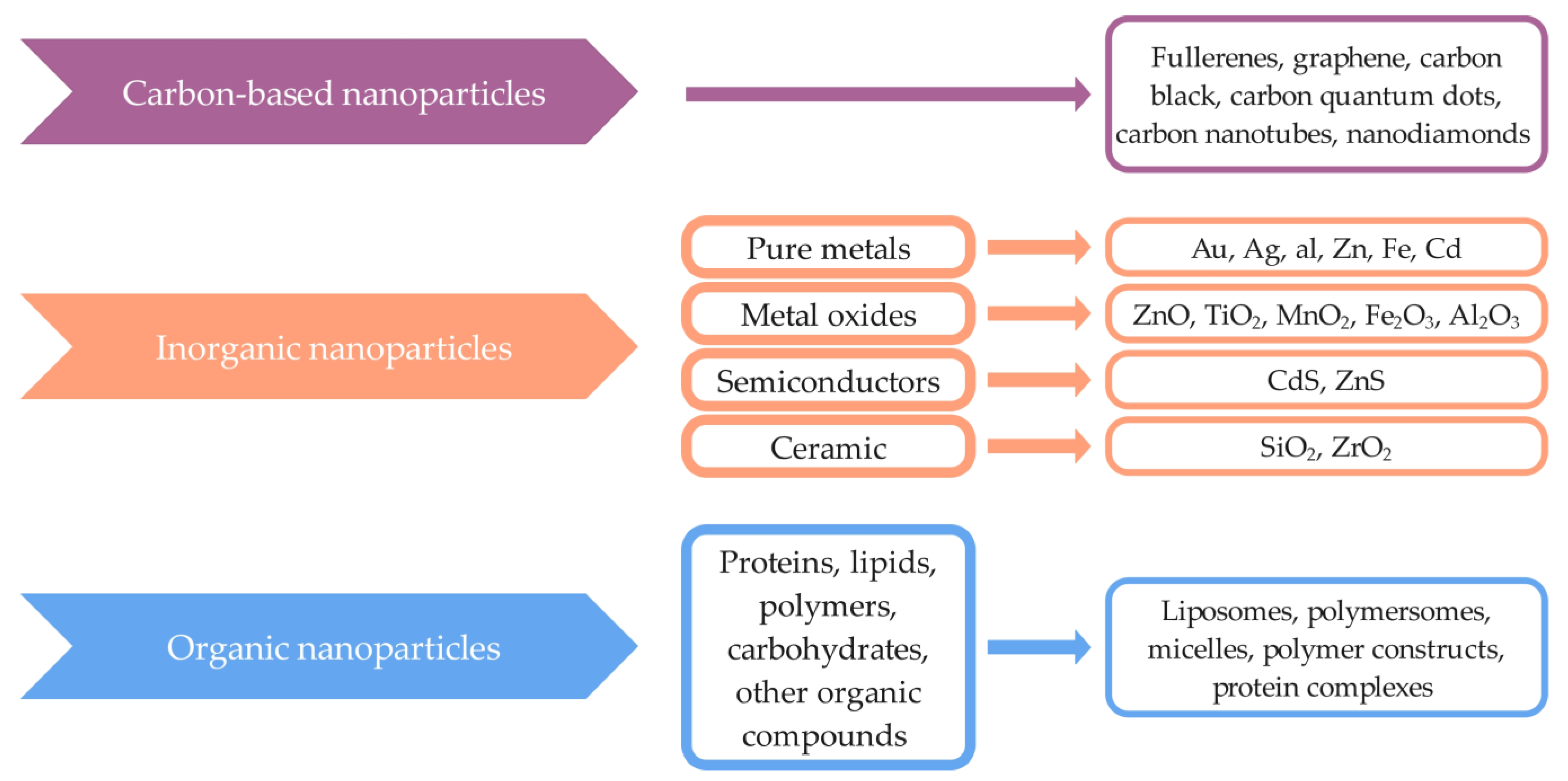
| Origin of Protein | Protein Source | Protein | Ref. |
|---|---|---|---|
| Animals | Ovalbumin and bovine serum albumin | Albumin | [71] |
| Milk | Casein | [72] | |
| Muscles, tendons, skin, bones | Collagen | [73] | |
| Bones, skin, connective tissue | Gelatin | [74] | |
| Hair, nails, skin, feathers, wool, hooves, beaks, horns | Keratin | [75] | |
| Silkworm Bombyx mori | Silk fibroin and sericin | [76] | |
| Plants | Wheat | Gliadin | [77] |
| Corn kernel | Zein | [78] |
| Origin of Polysaccharide | Polysaccharide Source | Polysaccharide | Ref. |
|---|---|---|---|
| Animal | Shells of arthropods such as crabs, prawns, shrimps, and lobsters | Chitosan | [88] |
| Plant | Larix spp. | Arabinogalactan | [89] |
| Agricultural trashes: sugarcane bagasse, rice husks | Cellulose | [37,90] | |
| Plant sources: wood, cotton, jute, hemp, bamboo, coconut, wheat, rice, grass, agave, flax | [37,90] | ||
| Cyamopsis tetragonoloba | Guar gum | [91] | |
| Cyamopsis psoraloides | [92] | ||
| Globe artichoke, topinambur, elecampane, chicory | Inulin | [93] | |
| Citrus peels, apple pomace | Pectin | [94] | |
| Potatoes, corn, tubers, fruits, legumes, roots, cereals, rice, yams, banana wheat, sorghum | Starch | [37] | |
| Plant tissues (trees, cotton) | Cellulose | [95] | |
| Algae | Nizimuddinia zanardini | Alginate | [96] |
| Sargassum latifolium | [97] | ||
| Cystoseira barbata | [98] | ||
| Ascophyllum nodosum | [99] | ||
| Laminaria digitata | [100] | ||
| Laminaria hyperborea | [101] | ||
| Laminaria japonica | [102] | ||
| Macrocystis pyrifera | [103] | ||
| Sargassum wightii | [104] | ||
| Chondrus crispus | Carrageenan | [105] | |
| Sarcothalia crispata | [106] | ||
| Gigartina skottsbergii | [106] | ||
| Eucheuma denticulatum | [107] | ||
| Kappaphycus alvarezii | [108] | ||
| Hypnea musciformis | [109] | ||
| Fungi | Aureobasidium pullulans | Pullulan | [110] |
| Cytaria harioti | [111] | ||
| Cytaria darwinii | [111] | ||
| Cryphonectria parasitica | [111] | ||
| Termella mesenterica | [112] | ||
| Teloschistes falvicans | [112] | ||
| Rhodotorula bacarum | [112] | ||
| Saccharomyces cerevisiae | Dextran | [113] | |
| Rhizopus oryzae | Chitosan | [114] | |
| Bacteria | Lactobacillus plantarum | Dextran | [115] |
| Weissella confuse | [116] | ||
| Weissella cibaria | [117] | ||
| Leuconostoc mesenteroides | [118] | ||
| Leuconostoc reuteri | [117] | ||
| Lactobacillus gasseri | [119] | ||
| Sphingomonas elodea | Gellan gum | [120] | |
| Sphingomonas paucimobilis | [120] | ||
| Leuconostoc citreum | Levan | [121] | |
| Bacillus subtilis | [122] | ||
| Bacillus atrophaeus | [123] | ||
| Acinetobacter nectaris | [124] | ||
| Xanthomonas campestris | Xanthan gum | [125] | |
| Gluconacetobacter xylinus | Cellulose | [126] | |
| Azotobacter spp. | Alginate | [127] | |
| Pseudomonas spp. | [127] |
| Biopolymer | Preparation Method | Particle Size (nm) | Loaded Cargo | Healthcare Application | Ref. |
|---|---|---|---|---|---|
| HSA | Self-assembly method | <200.0 | Docetaxel | Delivery of anticancer drug | [174] |
| HSA | High-pressure homogenization | 150.4 | Prednisolone and curcumin | Drug delivery for the treatment of rheumatoid arthritis | [130] |
| HSA | Desolvation method | <200.0 | Piperine | Drug delivery for the treatment of breast cancer cells (MCF-7) | [175] |
| Self-assembly method | |||||
| HSA | Coacervation method | 85.0–120.0 | Imipenem and ciprofloxacin | Delivery of antibiotics | [176] |
| BSA | Emulsification method | 100.0 | - | Drug delivery studies | [177] |
| BSA | Desolvation method | 156.0–315.0 | Doxorubicin | Delivery of anticancer drug | [178] |
| BSA | Anti-solvent precipitation method | 150.0 | Paclitaxel and resveratrol | Delivery of anticancer drug | [134] |
| BSA | Desolvation method | 100.0–200.0 | Methylene blue | Delivery of antifungal drugs for the treatment of Candida infections | [135] |
| Collagen | Electrospinning/electrospray method | - | - | Appropriate mimicry of the extracellular matrix for biomedical applications | [142] |
| Collagen | Film dispersion method | 35.2–107.0 | Cucurbitacin B | Oral delivery | [179] |
| Chitosan | Ionic gelation method | 106.5 | Zinc gluconate | Drug delivery for the treatment of rheumatoid arthritis | [167] |
| Chitosan | Emulsion-ionic gelation | 40.0–100.0 | Clove essential oil | Superior inhibitory effect against Aspergillus niger | [180] |
| Chitosan | Ionic gelation method | 75.4 | Rotigotine | Nose-to-brain drug delivery | [168] |
| Chitosan | Emulsion-ionic gelation | 198.7–373.4 | Basil essential oil | Delivery of antibacterial substances | [181] |
| Chitosan | Ionic gelation method | 208.1 | Cinnamaldehyde | Delivery of anti-quorum sensing agents | [182] |
| Chitosan | Ionic gelation method | 174.0–898.0 | Pomegranate peel extract | Controlled delivery of natural antioxidants and antimicrobials | [183] |
| Chitosan | Emulsification-ionic gelation method | 174.0–293.0 | Lemongrass essential oil | Delivery of antibacterial substances | [184] |
| Biopolymer | Preparation Method | Particle Size (nm) | Loaded Cargo | Healthcare Application | Ref. |
|---|---|---|---|---|---|
| Gliadin | Desolvation method | 1.0–181.0 | Sumatriptan | Brain-targeted drug delivery | [188] |
| Gliadin | Electrospray method | 481.6 | γ-Oryzanol | Drug delivery to reduce HT-29 cell proliferation | [191] |
| Zein | Nanoprecipitation method | 120.0–180.0 | Resveratrol | Oral drug delivery | [195] |
| Zein | Liquid–liquid phase separation method | 379.4 | BMP6-derived peptide | Bone regenerative medicine | [196] |
| Zein | Phase separation | 37.0–112.3 | Maytansine | Delivery of anticancer drug | [219] |
| Zein | Self-assembly method | 180.0 | Thymbra capitata essential oil | Drug delivery | [220] |
| Pectin | Ionotropic gelation method | 700.0–850.0 | - | Potential oral drug delivery | [200] |
| Pectin | Ionic gelation method | 482.7 | Metformin | Drug delivery for type 2 diabetes mellitus | [221] |
| Starch | Nanoprecipitation method | 500.0 | Quercetin | Estimation of release kinetics | [204] |
| Starch | Nanoprecipitation method | <250.0 | Curcumin | Drug delivery for polyphenols | [222] |
| Starch | Nanoprecipitation method | 255.0–396.0 | Ginkgo biloba extracts | Oral drug delivery | [223] |
| Starch | Emulsification method | 300.0 | 5-Fluorouracil | Colon-specific drug delivery | [224] |
| Starch | Acid hydrolysis and precipitation method | 85.1 | Paclitaxel | Delivery of anticancer drug | [225] |
| Starch | Nanoprecipitation method | 105.0 | Paracetamol | pH-responsive drug delivery system | [205] |
| Starch | Microemulsification method | 282.9 | Triphala Churna | Drug delivery of drugs with antimicrobial, antibiofilm, and neuroprotective effects | [226] |
| Cellulose | Nanoprecipitation method | 70.0–365.0 | Methylene blue | Sustained and controlled release | [227] |
| Guar gum | Emulsification method | 200.0 | Celecoxib | Oral drug delivery | [214] |
| Inulin | Emulsion solvent evaporation method | 224.0–365.0 | - | Intracellular stimulation of probiotics | [228] |
| Inulin | Spray drying method | 289.8 | Quercetin | Colon-targeted drug delivery | [217] |
| Biopolymer | Preparation Method | Particle Size (nm) | Loaded Cargo | Healthcare Application | Ref. |
|---|---|---|---|---|---|
| Alginate | Ionic gelation method | - | Rifampicin | Drug delivery without systemic toxicity | [244] |
| Alginate | Emulsification method | 279.1 | Miltefosine | Antifungal drug delivery | [236] |
| Alginate | Emulsification method | 150.0–270.0 | Betamethasone sodium phosphate | Ocular drug delivery | [245] |
| Alginate | Ionic gelation method | 400.0–1000.0 | Grape pomace extract | Oral delivery | [246] |
| Alginate | Electrospray method | 228.0 | CRISPR plasmids DNA | Gene delivery | [247] |
| Alginate | Emulsification method | 279.1 | Bacteriocins from L. paracasei | Drug delivery for the treatment of fungal diseases caused by Candida spp. and Cryptococcus spp. | [236] |
| κ-Carrageenan | Electrospray method | 309.5–663.8 | D-limonene | Drug delivery of highly sensitive bioactive agents | [242] |
| κ-Carrageenan | Emulsification method | 211.0 | Curcumin | Drug delivery | [248] |
| Biopolymer | Preparation Method | Particle Size (nm) | Loaded Cargo | Healthcare Application | Ref. |
|---|---|---|---|---|---|
| Pullulan | Nanoemulsification method | 98.0 | Ursodeoxycholic acid | Drug delivery for neuroprotective effect | [257] |
| Pullulan | Self-assembly method | 86.4–222.3 | Mitoxantrone | Delivery of anticancer drug | [253] |
| Pullulan | Nanoprecipitation method | <200.0 | Valsartan | Delivery of cardiovascular agents | [258] |
| Pullulan | Emulsion solvent evaporation method | 200.0 | Doxorubicin | Liver-specific drug delivery | [259] |
| Pullulan | Self-assembly method | 185.7 | Methotrexate and 10-hydroxycamptothecine | Tumor-targeted drug delivery | [260] |
| Pullulan | Probe-sonication method | 84.1 | Erlotinib | Drug delivery for cervical cancer therapy | [261] |
| Biopolymer | Preparation Method | Particle Size (nm) | Loaded Cargo | Healthcare Application | Ref. |
|---|---|---|---|---|---|
| Dextran | Spray drying method | - | Rifampicin | Enhanced deep lung delivery | [272] |
| Dextran | Ionic gelation method | 34.0–42.0 | - | Ocular drug delivery | [265] |
| Dextran | Single emulsion evaporation method | 99.8–181.3 | 5, 10, 15, 20-tetraphenyl-21H, 23H-porphyrine | Drug delivery for a tumor cell-targeted photodynamic therapy | [273] |
| Dextran | Self-assembly method | 260.0 | Naproxen | Anti-inflammatory drug delivery | [274] |
| Dextran | Solvent diffusion method | <70.0 | Chloroquine diphosphate | Delivery of antimalarial drug | [275] |
| Dextran | Ionic gelation method | 69.3 | - | Antibacterial agents | [276] |
| Xanthan gum | Anti-solvent precipitation | 149.0–241.0 | Cinnamon bark extract | Controlled delivery of polyphenols | [277] |
| Levan | Electrohydrodynamic atomization method | 82.1 | Resveratrol | Drug delivery for wound healing and tissue engineering | [269] |
| Gellan gum | Nebulization/ionic gelation method | 337.1 | Resveratrol | Oral drug delivery | [268] |
| Gellan gum | Ionic gelation method | 85.6 | Atenol | Delivery of antihypertensive drug | [270] |
| Incorporated Therapeutic Compound | Biopolymer | Administration Route | Therapeutic Application | Ref. |
|---|---|---|---|---|
| Bioactive compounds | ||||
| Sylmarin | Collagen | Intraperitoneal injection | Enhanced neuroprotective effect against acute ischemia/reperfusion injury | [291] |
| Cucurbitacin B | Collagen | Oral delivery | Enhanced absorption of orally administered Cucurbitacin B | [179] |
| Curcumin | BSA/dextran | In vitro | Enhancement of cellular antioxidant activity of curcumin in Caco-2 cells | [285] |
| Curcumin | Zein | Oral administration | Increased oral bioavailability | [324] |
| Curcumin | Zein | Intravenous injection | Potential drug delivery platform for therapy of glioblastoma | [193] |
| Beta-carotene | Zein | Oral administration | Enhanced cellular uptake, cytotoxicity, and improved oral biopharmaceutical performance | [298] |
| Anacardic acid | Zein | In vitro | Enhanced antimicrobial activity | [295] |
| Quercetin | Zein stabilized with soluble soybean polysaccharide | In vitro | Potential all-natural delivery systems for bioactive molecules | [293] |
| Morin | Starch | Intravenous injection | Therapeutic efficacy in the treatment of hyperuricemia | [325] |
| Conventional drugs | ||||
| Prednisolone and curcumin | HSA | Intravenous injection | Improved in vivo therapeutic effect in rats with rheumatoid arthritis | [130] |
| Paclitaxel and resveratrol | BSA | Intravenous injection | Improved suppression of tumor growth without systemic toxicity | [66] |
| Paclitaxel and di-fluorinated curcumin | BSA | Intravenous injection | A promising platform for treating gynecological cancers | [133] |
| Rotigotine | Chitosan | Nose-to-brain administration | Efficient drug delivery as an alternative to conventional routes | [168] |
| Zinc gluconate | Chitosan | Intraperitoneal injection | A potential alternative for the treatment of rheumatoid arthritis | [167] |
| Pirfenidone | Chitosan/alginate | Transdermal administration | Drug delivery for the treatment of idiopathic pulmonary fibrosis | [301] |
| Rifampicin | Alginate | Oral administration | No systemic toxicity and excellent safety after oral administration of nanoparticles | [244] |
| Maytansine | Zein | Intravenous injection | Strong anti-A549 tumor cell activity | [219] |
| Sumatriptan | Gliadin | Nasal administration | Effective crossing of the blood-brain barrier | [188] |
| Metformin | Pectin | In vitro | Oral administration for management of Type 2 Diabetes Mellitus | [221] |
| Ibuprofen | Starch | Oral administration | Colon-targeted drug delivery | [326] |
| Mitoxantrone | Pullulan | In vitro | Inhibition of the growth of bladder cancer cells (MB49) | [253] |
| Ursodeoxycholic Acid | Pullulan | In vitro | Increased neuroprotective effect | [257] |
| Paclitaxel | Dextran | Intravenous injection | Potential for C6 glioma treatment | [266] |
| Chloroquine diphosphate | Dextran | Parenteral administration | Treatment of malaria | [275] |
| Antibiotics and antimicrobial agents | ||||
| Colistin | HSA | In vitro | High antibacterial activity against the growth and inhibition of biofilm formation of multidrug-resistant Gram-negative bacterial species | [306] |
| Tobramycin | Alginate/chitosan | In vitro | Efficient delivery of antimicrobial drug | [310] |
| ε-Polylysine | Alginate/chitosan | In vitro | Delivery of antibacterial agents | [311] |
| SET-M33 peptide | Dextran | Intrapulmonary administration | Efficient treatment of lung infection | [312] |
| Extracts | ||||
| Pomegranate peel extract | Chitosan | In vitro | Enhanced antioxidant and antimicrobial properties | [183] |
| Grape pomace extract | Chitosan/alginate | Oral delivery | Improved biological activity | [246] |
| Artemisia ciniformis extract | Alginate | In vitro | Inhibition of proliferation of AGS cells | [314] |
| Ginkgo biloba extract | Starch | In vitro | Sustained release in artificial gastric and intestinal juices | [223] |
| Essential oils | ||||
| Oregano essential oil | Alginate/chitosan | Dermal administration | Enhanced antimicrobial activity | [322] |
| Clove essential oil | Chitosan | In vitro | Improved antioxidant and antibacterial activity | [316] |
| Basil essential oil | Chitosan | In vitro | Enhanced antimicrobial activity and strong antibiofilm activity | [181] |
| Mandarin essential oil | Chitosan | In vitro | Enhanced inhibition of biofilm formation | [318] |
| Lemongrass and peppermint essential oil | Chitosan | In vitro | Enhanced thermal stability, antioxidant, and antimicrobial activity | [184] |
| Green tea essential oil | Chitosan | In vitro | Enhanced thermal stability, antioxidant, and antimicrobial activity | [320] |
Disclaimer/Publisher’s Note: The statements, opinions and data contained in all publications are solely those of the individual author(s) and contributor(s) and not of MDPI and/or the editor(s). MDPI and/or the editor(s) disclaim responsibility for any injury to people or property resulting from any ideas, methods, instructions or products referred to in the content. |
© 2023 by the authors. Licensee MDPI, Basel, Switzerland. This article is an open access article distributed under the terms and conditions of the Creative Commons Attribution (CC BY) license (https://creativecommons.org/licenses/by/4.0/).
Share and Cite
Kučuk, N.; Primožič, M.; Knez, Ž.; Leitgeb, M. Sustainable Biodegradable Biopolymer-Based Nanoparticles for Healthcare Applications. Int. J. Mol. Sci. 2023, 24, 3188. https://doi.org/10.3390/ijms24043188
Kučuk N, Primožič M, Knez Ž, Leitgeb M. Sustainable Biodegradable Biopolymer-Based Nanoparticles for Healthcare Applications. International Journal of Molecular Sciences. 2023; 24(4):3188. https://doi.org/10.3390/ijms24043188
Chicago/Turabian StyleKučuk, Nika, Mateja Primožič, Željko Knez, and Maja Leitgeb. 2023. "Sustainable Biodegradable Biopolymer-Based Nanoparticles for Healthcare Applications" International Journal of Molecular Sciences 24, no. 4: 3188. https://doi.org/10.3390/ijms24043188