Article

The In Vitro Impact of Isoxazole Derivatives on Pathogenic Biofilm and Cytotoxicity of Fibroblast Cell Line

1
Department of Organic Chemistry and Drug Technology, Faculty of Pharmacy, Wroclaw Medical University, 50-556 Wroclaw, Poland
2
Unique Application Model Laboratory, Department of Pharmaceutical Microbiology and Parasitology, Faculty of Pharmacy, Wroclaw Medical University, 50-556 Wroclaw, Poland
*
Author to whom correspondence should be addressed.
Int. J. Mol. Sci. 2023, 24(3), 2997; https://doi.org/10.3390/ijms24032997
Submission received: 14 December 2022 / Revised: 24 January 2023 / Accepted: 31 January 2023 / Published: 3 February 2023
(This article belongs to the Special Issue Recent Approaches for Wound Treatment)

Abstract

:
The microbial, biofilm-based infections of chronic wounds are one of the major challenges of contemporary medicine. The use of topically administered antiseptic agents is essential to treat wound-infecting microorganisms. Due to observed microbial tolerance/resistance against specific clinically-used antiseptics, the search for new, efficient agents is of pivotal meaning. Therefore, in this work, 15 isoxazole derivatives were scrutinized against leading biofilm wound pathogens Staphylococcus aureus and Pseudomonas aeruginosa, and against Candida albicans fungus. For this purpose, the minimal inhibitory concentration, biofilm reduction in microtitrate plates, modified disk diffusion methods and antibiofilm dressing activity measurement methods were applied. Moreover, the cytotoxicity and cytocompatibility of derivatives was tested toward wound bed-forming cells, referred to as fibroblasts, using normative methods. Obtained results revealed that all isoxazole derivatives displayed antimicrobial activity and low cytotoxic effect, but antimicrobial activity of two derivatives, 2-(cyclohexylamino)-1-(5-nitrothiophen-2-yl)-2-oxoethyl 5-amino-3-methyl-1,2-oxazole-4-carboxylate (PUB9) and 2-(benzylamino)-1-(5-nitrothiophen-2-yl)-2-oxoethyl 5-amino-3-methyl-1,2-oxazole-4-carboxylate (PUB10), was noticeably higher compared to the other compounds analyzed, especially PUB9 with regard to Staphylococcus aureus, with a minimal inhibitory concentration more than x1000 lower compared to the remaining derivatives. The PUB9 and PUB10 derivatives were able to reduce more than 90% of biofilm-forming cells, regardless of the species, displaying at the same time none (PUB9) or moderate (PUB10) cytotoxicity against fibroblasts and high (PUB9) or moderate (PUB10) cytocompatibility against these wound cells. Therefore, taking into consideration the clinical demand for new antiseptic agents for non-healing wound treatment, PUB9 seems to be a promising candidate to be further tested in advanced animal models and later, if satisfactory results are obtained, in the clinical setting.

1. Introduction

The non-healing wound is referred to as the discontinuity of skin and subcutaneous tissue, which does not heal in an orderly set of stages and in a predictable amount of time. It is estimated that procedures related with management and treatment of chronic wounds consume approximately 1–3% of European Union public health budgets [1]. The presence of non-healing wounds significantly increases risk of serious health loss and, in case of wound infection, even death of the patient. The non-healing wounds are frequently colonized by differentiated consortia of bacteria, of whom Gram-positive Staphylococcus aureus and Gram-negative Pseudomonas aeruginosa should be distinguished because of their particular tendency to spread in the wound bed and to cause local, biofilm-based infections, which may develop into the life-threatening systemic forms [2]. In turn, fungi, such as yeast-like Candida albicans, are less frequently isolated from the non-healing wounds. Nevertheless, they are an important component of so called mixed (duo- or multi-species) biofilms in this niche; therefore, their potential impact on the process of wound healing should not be neglected [3]. The present treatment algorithms for critically colonized/infected non-healing wounds consist of wound bed debridement (of surgical, chemical/enzymatical or biological nature), use of modern dressings and application of locally administered antimicrobials referred to as the antiseptics [4]. The last-mentioned measure, used for both prophylactic and treatment purposes, are compounds of rapid antimicrobial action and preferentially of low or none cytotoxicity toward cells forming the wound bed, i.e., fibroblasts and keratinocytes [5]. Nevertheless, in recent years, the phenomenon of increased microbial tolerance and/or resistance toward many of the most widespread antiseptics occurred, just to mention cases of chlorhexidine and octenidine dihydrochloride [6,7]. Because use of antiseptics in the treatment of infected non-healing wounds is a critical factor with regard to clinical success (wound closure), presently the increasing search for new compounds, which may replace or improve activity of existing antiseptics, can be observed. One of the examples of such compounds are bioactive small molecules of a molecular weight <900 Da. These low-mass molecules make up presently 90% of pharmaceutical drugs (including such common drugs as aspirin or insulin). Thanks to their low size, such drugs can be administered orally and are able to pass through cell membranes and reach intracellular targets [8].
Much attention has been paid to the synthesis of heterocycles containing both nitrogen and oxygen due to their broad spectrum of biological and pharmacological activities. Among the wide range of pharmacologically important heterocycles, isoxazoles play a significant role in the field of medicinal chemistry. This heterocycle is considered as an important class of compounds in medicinal chemistry because of its wide spectrum of targets and varied biological application such as antimicrobial [9], antiviral [10], anticancer [11], anti-inflammatory [12], immunomodulatory [13], anticonvulsant [14] or antidiabetic properties [15]. The key feature of these heterocycles is that they exhibit the typical properties of an aromatic system but the same contain a weak nitrogen-oxygen bond that can be easily cleaved under certain reaction conditions. Thus, isoxazoles are very useful substrates since the ring system stability allows the derivatization of substituents to give functionally complex derivatives. Emerging research interest on the isoxazole moiety results from fact that this moiety is a common synthetic building block in searching for new compounds exhibiting antimicrobial and antifungal activities [16,17]. Synthesis of isoxazole derivatives is widely carried out through different methods such as condensation, cyclomerization or cycloaddition. Thus, the synthesis of new isoxazole derivatives is a very attractive aspect in the research and development field for both medicinal and organic chemistry. Due to these facts as well as to its relatively easy synthesis, isoxazole rings have become an object of our interest. Inspired by these facts we planned to synthesize some more derivatives of isoxazoles using different methods of functionalization such as Passerini multicomponent reaction and Michael addition.
Of note, the presence of an isoxazole moiety translates frequently into antimicrobial potential of such low-mass molecules as antibiotics: sulfisoxazole, cloxacilin, dicloxacilin or sulfamethozaole [18]. Isoxazole derivatives obtained earlier by our team also possessed a number of biological activities, including those of immunosuppressive [19,20,21,22], antiviral [23] or anticancer character [24]. It is well-recognized that conjugation of a small molecule to the isoxazole core offers a possibility to obtain new derivatives of biological activity [25]. As an example, maleimides are one of the most widely used functional groups for 1,4-conjugate addition. Their chemistry is widely used in the site-selective modification of thiol- and amine-containing compounds [26]. Although some types of nucleophiles can be involved in such a conjugation, ubiquitous heteronucleophiles (e.g., thiols, amines, and alcohols) are used the most frequently and, therefore, they are the most scrutinized and developed. For a number of years, these conjugate additions were limited to organic chemistry. Nevertheless, in the last few decades they have been also notably used in bioorganic and polymer chemistry for bioconjugation reactions and step-growth polymerizations, respectively, thanks to their excellent efficiency and ambient reactivity [27]. The α,β-unsaturated bond presented in maleimides allows a process of easy functionalization. Moreover, the delocalization of the double bonds (two carbonyl groups and one double bond in the ring) gives maleimides electron acceptor and dienophile characteristics [28].
Thus, click nucleophilic conjugate addition via aza-Michael reaction has become one of the ways to obtain a series of new isoxazole derivatives in this work (MAL1-5 series) (Figure 1). In these reactions, we used 5-amino-3-methyl-isoxazole-4-carbohydrazide as a substrate [29,30]. We have employed this compound because this isoxazole derivative has attracted much attention due to its known biological activity [31]. According to the earlier investigation, this compound exhibits immunomodulatory properties, which makes it useful in further research and chemical modification [32,33]. Because the different approaches of modification of substituents in position 4 of 5-amino-3-methyl-isoxazole-4-carboxylic acid allow to obtain derivatives of different biological activity [34], we decided to employ this unnatural amino acid to a Passerini three-component reaction (PUB1-10 series) (Figure 1). This type of reaction is the oldest isocyanide-based multicomponent reaction employing isocyanide (R2), aldehyde/ketone (R1) and carboxylic acid to give acyclic depsipeptides [35]. This type of peptide contains one or more ester bonds in addition to the amide bonds, and has become important lead structures in the development of new synthetic drugs. One example of this class of compounds is enniatins, which are cyclohexadepsipeptides with a range of biological activities, including those of antibiotic and cytotoxic character [36].
Therefore, the aim of this work was to scrutinize obtained isoxazole derivatives with regard to their antimicrobial (including antibiofilm) potential, as well as their impact on fibroblast cell lines. Moreover, following the recent trends of fortification of wound dressings with antimicrobials other than antibiotics or clinically used antiseptics, we introduced the isoxazole derivatives to the experimental dressing made of polymeric bacterial cellulose and assessed its activity against biofilms [37,38,39]. Such a goal fits in the current trend of research aiming for introduction of new agents which can be considered an alternative for already existing antiseptic agents used in the prophylaxis and treatment of critically colonized non-healing wounds.

2. Results and Discussion

2.1. Chemistry

A series of isoxazole derivatives referred to in this work as the PUB series (PUB1-8) were obtained by our team earlier and their effects on phytohemagglutinin A (PHA)-induced proliferation of human peripheral blood mononuclear cells (PBMC), production of tumor necrosis factor alpha (TNFα) in human whole blood cultures stimulated with lipopolysaccharide (LPS) and two-way mixed lymphocyte reaction (MLR) of PBMC were investigated [25]. These compounds as well as PUB9 and PUB10 were obtained via Passerini three-component reaction by using methods published previously [25]. Briefly, N′-substituted 5-amino-3-methyl-isoxazole-4-carbohydrazide derivatives were synthesized using click nucleophilic conjugate addition via aza-Michael reaction according to Figure 2 presented below.
Maleimide is an unsaturated imide that forms pyrrole-type ring structures (1H-pyrrole-2,5-diones). The double bond presented in maleimides is highly electron-deficient due to the presence of two carbonyl groups, which consequently makes maleimides highly reactive and undergo nucleophilic addition reaction, called Michael addition, and is a type of “click chemistry”. The Michael addition with a primary amine is faster and reaches a higher conversion rate than its reactions with thiols [19]. A new series of N-substituted hydrazide derivatives was obtained by reacting 5-amino-3-methyl-isoxazole-4-carbohydrazide with different commercially available maleimides. This isoxazole derivative exhibits the unique reactivity, which is due to the selectivity of reaction of the amino groups present in this compound. The maleimides react with the amine group presented in hydrazide moiety, whereas we did not observe any product of reaction between the maleimide and NH2 group derived from the isoxazole ring. This unique property of the amino group attached to the isoxazole ring has been examined by our team in different kinds of reaction and as a result it has been shown that it remains unreactive in almost types of reactions [40,41]. The reaction is pH selective and favors primary amines in more alkaline conditions, but it unfortunately also increases the rate of hydrolysis of the maleimide group, resulting in a maleamic acid which is a non-reactive form of maleimide. Therefore, one of the most important tasks in our work was the optimalization of the reaction conditions to avoid this synthetic problem. In the course of the conducted chemical synthesis, it turned out that the most suitable pH for carrying out this reaction was pH = 7.8, which made it possible to use a bicarbonate buffer. By employing a slight excess of 5-amino-3-methyl-isoxazole-4-carbohydrazide, we ensured the full consumption of each of the used maleimides, consequently improving the yields of isolated products of conjugation to obtain a new series of isoxazole derivatives (series MAL1-5) (Figure 2).

2.2. Synthesis and Structural Characterization

The synthesis and characterization of compounds PUB1-8 has been published previously [25]. Compounds PUB9 and PUB10 were obtained according to the method described previously and purified by crystallization from methanol (PUB10) and by column chromatography (PUB9) with silica gel 230–400 mesh (60 Å); mobile phase: ethyl acetate/ chloroform = 3/7 (v/v), sample dissolved in chloroform. Structures of new compounds and their spectral analysis are shown in the Supplementary Material (Figures S16–S21).

2.2.1. 2-(cyclohexylamino)-1-(5-nitrothiophen-2-yl)-2-oxoethyl 5-amino-3-methyl-1,2-oxazole-4-carboxylate (PUB9)

30 % yield; m.p. 199–200 °C, beige solid. 1H NMR (300 MHz, DMSO-d6) δ (ppm): 1.01–1.34 (m, 5H), 1.50–1.80 (m, 6H). 2.25 (s, 3H, CH3 group of isoxazole ring), 4.09 (q, J = 5.3 Hz, 1H), 6.34 (s, 1H), 7.26–7.31 (d, J = 4.22 Hz, 1H), 7.91 (bs, 2H, NH2 group from isoxazole ring), 8.05 (d, J = 4.3 Hz, 1H), 8.42 (d, J = 7.8 Hz, 1H). 13C NMR (75 MHz, DMSO-d6) δ (ppm): 12.43, 24.70, 24.79, 25.52, 31.13, 32.34, 32.47, 39.15, 39.43, 39.71, 39.98, 40.26, 40.54, 40.81, 48.49, 49.05, 70.48, 84.42, 127.18, 129.91, 147.31, 161.31, 165.62, 172.65. ESI-MS: m/z calculated for formula C17H20N4O6S [M-H] 407.103, found 407.103.

2.2.2. 2-(benzylamino)-1-(5-nitrothiophen-2-yl)-2-oxoethyl 5-amino-3-methyl-1,2-oxazole-4-carboxylate (PUB10)

20% yield; m.p. 201–202 °C, beige solid. 1H NMR (300 MHz, DMSO-d6) δ (ppm): 2.25 (s, 3H, CH3 groups of isoxazole ring), 4.29–4.35 (d, J = 5.8 Hz, 2H), 6.44 (s, 1H), 7.16–7.35 (m, 6H), 7.94 (bs, 2H, NH2 group from isoxazole ring), 8.04–8.08 (d, J = 5.5 Hz, 1H), 9.02 (t, J = 5.9 Hz, 1H). 13C NMR (126 MHz, DMSO-d6) δ (ppm): 12.52, 42.80, 70.74, 84.45, 127.38, 127.46, 127.60, 128.82, 129.93, 138.98, 146.86, 151.42, 159.37, 161.40, 166.90, 172.72. ESI-MS: m/z calculated for formula C18H16N4O6S [M-H] 415.072, found 415.076.
The novel target compounds (MAL1-5) were synthesized according to the procedure described below using 5-amino-3-methylisoxazole-4-carbohydrazide, which was obtained by a sequence of very efficient processes described in detail in the following literature entries [29,42,43]) and different commercially available N-substituted maleimides as starting materials.

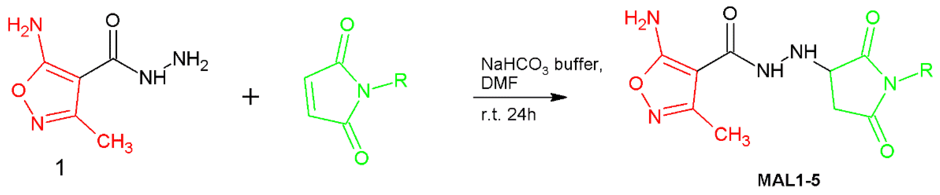
2.2.3. General Procedure for Preparation of a Series of Compounds (MAL1-5)

To a solution of 5-amino-3-methylisoxazole-4-carbohydrazide (1) (2.0 mmol) in DMF (5 mL) was added a solution of each N-maleimide (1.8 mmol) in 5 mL DMF with 2 mL of NaHCO3 buffer (8.4 mg NaHCO3 dissolved in 10 mL of water). The reaction mixture was stirred at room temperature for 24 h, then DMF was removed using a gentle stream of air. The final product was purified by crystallization from methanol. Spectral analysis of new compounds as well as their structures are shown in the Supplementary Material (Figures S1–S15).

2.2.4. 5-amino-N′-(2,5-dioxopyrrolidin-3-yl)-3-methyl-1,2-oxazole-4-carbohydrazide (MAL1)

40% yield; m.p. 220–221 °C, orange solid. 1H NMR (300 MHz, DMSO-d6) δ (ppm): 2.21 (s, 3H, CH3 group of isoxazole ring), 2.56–2.90 (m, 2H), 3.90–4.04 (m, 1H), 5.42–5.76 (m, 1H), 7.25–7.62 (bs, 2H, NH2 group from isoxazole ring), 8.34–8.87 (d, 1H, J = 5.5 Hz), 10.97–11.42 (s, 1H). 13C NMR (75 MHz, DMSO-d6) δ (ppm): 11.54, 35.21, 58.91, 86.23, 157.25, 163.72, 171.02, 177.24, 178.03. ESI-MS: m/z calculated for formula C9H11N5O4 [M+H]+ 254.088, found 254.081.

2.2.5. 5-amino-3-methyl-N′-(1-methyl-2,5-dioxopyrrolidin-3-yl)-1,2-oxazole-4-carbohydrazide (MAL2)

55% yield; m.p. 173–174 °C, orange solid. 1H NMR (300 MHz, DMSO-d6) δ (ppm): 2.19 (s, 3H, CH3 group of isoxazole ring), 2.54–2.64 (m, 1H), 2.82 (s, 3H, CH3 group of maleimide moiety), 2.85–2.92 (m, 1H), 3.95–4.06 (m, 1H), 5.62–5.78 (m, 1H), 7.41–7.48 (bs, 2H, NH2 group from isoxazole ring), 8.56–8.73 (d, 1H, J = 5.39 Hz). 13C NMR (126 MHz, DMSO-d6) δ (ppm): 11.68, 24.43, 34.22, 57.89, 86.62, 157.43, 163.88, 171.19, 176.09, 176.76. ESI-MS: m/z calculated for formula C10H13N5O4 [M+H]+ 268.104, found 268.098.

2.2.6. 6-(3-{2-[(5-amino-3-methyl-1,2-oxazol-4-yl)carbonyl]hydrazinyl}-2,5-dioxopyrrolidin-1-yl)hexanoic acid (MAL3)

37% yield; m.p. 143–144 °C, white solid. 1H NMR (300 MHz, DMSO-d6) δ (ppm): 1.10–1.56 (m, 6H), 2.08–2.18 (m, 2H), 2.19 (s, 3H, CH3 group of isoxazole ring), 2.54–2.67 (m, 2H), 2.80–2.93 (m, 1H), 3.93–4.08 (m, 1H), 5.63–5.82 (m, 1H), 7.37–7.55 (bs, 2H, NH2 group from isoxazole ring), 8.56–8.70 (d, 1H, J = 4.91), 11.81–12.09 (bs, 1H, COOH group from maleimide moiety). 13C NMR (75 MHz, DMSO-d6) δ (ppm): 11.99, 24.45, 26.16, 27.34, 33.86, 34.27, 38.15, 58.00, 86.65, 157.66, 164.32, 171.54, 174.80, 176.31, 177.02. ESI-MS: m/z calculated for formula C15H21N5O6 [M+Na]+ 390.138, found 390.132.

2.2.7. 5-amino-N′-(1-cyclohexyl-2,5-dioxopyrrolidin-3-yl)-3-methyl-1,2-oxazole-4-carbohydrazide (MAL4)

27% yield; m.p. 193–195 °C, white solid. 1H NMR (300 MHz, DMSO-d6) δ (ppm): 0.94–2.08 (m, 10H), 2.20 (s, 3H, CH3 group of isoxazole ring), 2.51–2.61 (m, 1H), 2.74–2.88 (m, 1H), 3.72–3.86 (m, 1H), 3.89–4.00 (m, 1H), 5.62–5.73 (m, 1H), 7.37–7.49 (bs, 2H, NH2 group from isoxazole ring), 8.53–8.73 (d, 1H, J = 5.4 Hz). 13C NMR (75 MHz, DMSO-d6) δ (ppm): 11.53, 24.81, 25.31, 28.36, 33.63, 50.53, 57.09, 86.15, 157.17, 163.85, 171.08, 175.79, 176.55. ESI-MS: m/z calculated for formula C15H21N5O4 [M+H]+ 336.166, found 336.161.

2.2.8. 5-amino-N′-[1-(4-chlorophenyl)-2,5-dioxopyrrolidin-3-yl]-3-methyl-1,2-oxazole-4-carbohydrazide (MAL5)

45% yield; m.p. 217–220 °C, white solid. 1H NMR (300 MHz, DMSO-d6) δ (ppm): 2.22 (s, 3H, CH3 group of isoxazole ring), 2.69–2.87 (m, 1H), 2.95–3.19 (m, 1H), 4.10–4.27 (m, 1H), 5.81–5.94 (m, 1H), 7.25–7.37 (d, 2H, J = 8.11 Hz), 7.44–7.53 (bs, 2H, NH2 group from isoxazole ring), 7.51–7.64 (d, 2H, J = 8.28 Hz). 13C NMR (75 MHz, DMSO-d6) δ (ppm): 11.56, 34.23, 57.98, 86.21, 128.57, 128.96, 131.08, 132.72, 157.25, 163.87, 171.04, 174.77, 175.48. ESI-MS: m/z calculated for formula C15H14ClN5O4 [M+H]+ 364.081, found 364.072.

2.3. Biology

In the first line of biological line of experiments, the minimal inhibitory concentrations of analyzed compounds were evaluated using a microplate model towards Gram-positive and Gram-negative reference pathogens and yeast-like fungus (Table 1). Obtained results indicate that the synthesized compounds acted in a more efficient manner against C. albicans than against S. aureus and P. aeruginosa. Nevertheless, two of the compounds (PUB9 and PUB10) displayed a few hundred times higher activity against S. aureus compared to the remaining compounds, maintaining at the same time comparable (to the remaining compounds) activity against P. aeruginosa and C. albicans.
Compounds PUB9 and PUB10 were thus selected for further analyses. The first of them included assessment of biofilm reduction in the microplate model. Obtained results indicate strong (above 90%) reduction in biofilm, regardless of the strain applied (Figure 3) in concentration range of 0.125–0.25 mg/mL.
Next, the survivability of fibroblast cells (wound bed-forming cells) was analyzed after the exposure to the range of PUB9 and PUB10 compounds (Figure 4). In the case of PUB9, the advantageous results (cytotoxicity close to none) were obtained when 0.2 mg/mL of compounds was applied, while doubling of the compound’s concentration correlated with moderate level of cytotoxicity. In the case of PUB10, higher cytotoxicity level was observed towards fibroblasts (strong and very strong cytotoxicity when concentrations of 0.2 and 0.39 mg/mL, respectively, were applied).
The results presented in Table 1, Figure 3 and Figure 4 allow obtaining data on the investigated compound’s in vitro activity against wound pathogens and also indicate in which concentration the specific compound does not exert (or exert at the acceptable level) cytotoxicity against fibroblast cells. In the next step, another two methods assessing the impact of antimicrobials against different types of microbial consortia were performed. In the first of them, referred to as the modified disk-diffusion model, the only full inhibition of microbial growth (equal 4 mm) was observed when PUB9 against S. aureus was applied (Figure 5).
In the case of PUB10, the impact was also seen only against S. aureus; however, within the zone of growth inhibition (equal 2 mm), distinctive microbial colonies were also observed. Therefore, it was decided to apply another experimental model to get better insight on possible activity of PUB9 and PUB10 against biofilm. Using a modified Antibiofilm Dressing Activity Measurement model, the reduction resulting from activity of PUB9 and PUB10 was assessed. In the case of PUB9, the [%] reduction values were 99 ± 0.1%; 99 ± 0.1%, 90.7 ± 2.4% for S. aureus, C. albicans and P. aeruginosa, respectively, when 0.25 mg/mL of compound was applied. In case of exposure to 0.25 mg/mL of PUB10, the respective values were: 72.8 ± 0.04%; 69.8 ± 0.11% and 77.7 ± 2.4%.
In turn, the cytocompatibility tests of PUB9- and PUB10-containing BC carriers toward the fibroblast cell line corresponded to the obtained data on fibroblasts’ cytotoxicity, i.e., PUB9-BC displayed excellent cytocompatibility while PUB10-BC displayed the moderate cytocompatibility (Figure 6) compared to the biocompatibility of native BC.
The increasing frequency of non-healing wounds occurrence in the developed populations of the Western hemisphere accelerates the search for efficient algorithms in treatment of these disease entities. Infection is considered one of the most dangerous complications of non-healing wounds, and may lead even to the patient’s death. The local application of antiseptic agents is one of the pillars of modern wound treatment [44]. Nevertheless, continuous and prolonged exposure of infected wounds to antiseptics starts to induce processes analogical to those observed during application of antibiotics, namely to the rise of microbial tolerance and resistance [45]. Therefore, new classes of antiseptic agents are needed to overcome the threat of bacterial resistance and to provide wound professionals an effective tool in the fight against microbial biofilms colonizing and infecting wound beds.
The isoxazole derivatives seem to be promising candidates in this regard, thanks to their relatively easy synthesis, chemical stability, low toxicity and good bioactivity at low doses. Although numerous methods for isoxazole-containing drugs have been developed, due to their wide activity, there is still a strong need to synthesize new derivatives containing this valuable moiety with increased chemical and pharmacological properties and test their biological activity. The current research focused on the antimicrobial (including antibiofilm) activity of isoxazole derivatives and, simultaneously, on their impact on wound bed-forming cells, referred to as the fibroblasts. The general aim was thus to investigate potential applicability of isoxazole derivatives in the character of drugs for infected wound care. While the MIC evaluation and modified disk diffusion methods served, to a certain extent, as the predictors of biofilm prevention potential, the MBEC and A.D.A.M. techniques showed the performance of isoxazole derivatives in the character of drugs for biofilm treatment. In turn, tests performed on fibroblasts provided insight on potential interaction of derivatives with wound cells, especially their ability to adhere and proliferate.
The previous research on isoxazoles indicates that the substitution of various groups on the isoxazole ring imparts different activity. It was observed that the introduction of the thiophene moiety to the isoxazole ring increases its antimicrobial activity [46]. Previously, in vitro antibacterial and antifungal activity against S. aureus, B. subtilis, E. coli, P. aeruginosa, A. niger and C.albicans of 4,5-dihydro-5-(substitutedphenyl)-3-(thiophene-2-yl)isoxazole derivatives using the disc diffusion method was investigated by Gautam and Singh [47]. The obtained results clearly demonstrated high impact of the thiophene moiety on the analyzed biological activity. Another useful result was obtained by RamaRao et al. [48], who synthesized heteroarylisoxazoles and evaluated them for antibacterial activity against E. coli, S. aureus and P. aeruginosa. It was found that the isoxazoles substituted with the thiophenyl moiety displayed significant activity. The fact that the thiophene nucleus has been recognized as an important moiety in the synthesis of heterocyclic compounds with promising pharmacological characteristics prompted us to investigate such phenomena in the case of new isoxazole derivatives in the form of Passerini reaction products. In this work, 15 isoxazole derivatives were scrutinized with regard to their antimicrobial effect displayed against S. aureus, P. aeruginosa and C. albicans. The reason standing behind the choice of aforementioned species was not only different structure and composition of their cell wall, but also the fact that both S. aureus and P. aeruginosa belong to the pathogens being frequent etiological factors of non-healing wounds, while C. albicans is a component of mixed wound biofilms. Obtained results (Table 1) indicate that all tested compounds were able to inhibit growth of S. aureus, P. aeruginosa and C. albicans within the tested range of concentrations in the basic microplate model. Of note, in the case of 11/15 tested compounds there were no differences in Minimal Inhibitory Concentrations against S. aureus and P. aeruginosa displayed by a particular compound or these differences were on the level of just one geometric dilution, which may be related with the limitation of the method itself [49]. In turn, in the case of two compounds (PUB9 and PUB10) the MIC value against S. aureus was more than 1000 and 260 times lower, respectively, than against P. aeruginosa. With regard to the chemical composition, PUB9 and PUB10 differed from the other analyzed compounds mainly by the presence of a thiophene ring. It was speculated by another research team that the presence of the positive charge of donor S atoms in the heteroaromatic ring was responsible for damage of the microbial cell wall/membrane, potential leakage of cytoplasmatic content to the environment and eventual cellular damage [50].
In the second line of investigation, the most promising isoxazole derivatives (PUB9 and PUB10) were scrutinized with regard to their antibiofilm properties (Figure 3). The level of biofilm eradication above 90% was achieved when higher concentrations of compounds were applied (compared to the MIC values presented in Table 1). At the same time there were no significant differences in the value of reduction in biofilms formed by applied species (p < 0.05), which suggests the dose-dependent mode of action of tested isoxazole derivatives and the mechanism of biofilm eradication based on killing of microbial cells rather than destruction of biofilm matrix [51].
Nevertheless, such an observation confirms the well-recognized higher tolerance of biofilms against antimicrobials, compared to the tolerance displayed by their planktonic (non-adhered) counterparts [52]. Interestingly, the difference between concentrations necessary to reach MIC value vs. concentrations required to reach >90% of biofilm eradication was noticeably lower in the case of P.aeruginosa or C.albicans (two to four times) than in the case of S.aureus (ca. 2000–4000 times). Such a phenomenon may be related with the hypothetical mechanism of action of PUB9 and PUB10, i.e., the inhibition of GTPase activity and bacterial cell division, which cause bactericidal effects [53]. The appropriate candidate for a wound antiseptic should display not only the sufficient antimicrobial (antibiofilm) activity, but also should not exert the harmful effect on the wound bed cells, such as fibroblasts [54]. Therefore, in the next analysis (Figure 4), the survivability of fibroblast cell line exposed to PUB9 and PUB10 activity was performed for the spectrum of concentrations, including these which proved to be sufficient to eradicate the pathogens in planktonic and biofilm forms (Table 1, Figure 3). The obtained results indicated the concentration-dependent growth of cytotoxic effects displayed by both compounds. Nevertheless, it also occurred that in the concentration range 0.2–0.4 mg/mL, PUB9 displays acceptable (low) level of cytotoxicity, while acceptable level of cytotoxicity was recorded for PUB10 in the concentration range 0.012–0.025 mg/mL. The exact mechanism explaining why certain isoxazole derivatives containing a thiophene ring display a cytotoxic effect toward certain cell lines but not other types still needs to be elucidated [55]. Nevertheless, in the current research two derivatives of low cytotoxicity against fibroblast cells were found, which makes them promising candidates to be investigated more thoroughly. To further analyze the impact of PUB9 and PUB10 on microorganisms, the modified disk diffusion test was performed (Figure 5) [56], which confirmed higher antimicrobial effect exerted by PUB9 than PUB10; the coherent results were obtained in case of the A.D.A.M. test performed, i.e., the higher antibiofilm activity of PUB9 than PUB10. To our knowledge it is the first time the isoxazole derivatives were introduced to the BC carrier, which paves a way to their potential application not only as antiseptic agents in the form of liquid solutions, but also as an antimicrobial component of active dressings designed to treat chronic wounds. To get deeper insight on properties of isoxazole derivatives-containing BC, we performed a cytocompatibility test in vitro (Figure 6). Obtained results revealed that excellent cytocompatibility of BC, indicated by other research teams [57], was reduced moderately (but in a statistically significant manner) by addition of PUB10, but not the PUB9 compound. It may be thus assumed that the PUB9 compound can be considered a candidate designed for treatment purposes (eradication of already formed biofilm), while PUB10 appears to be more suitable for prophylaxis purposes (the lower, non-cytotoxic concentration of PUB10 may be applied against planktonic forms of microorganisms, which did not develop into biofilm structure yet).
Conventional, clinically applied drugs used topically for the treatment of chronic wounds can be divided into antibiotics or antiseptics. Gentamycin is an example of an antibiotic allowed to be used topically in the treatment of chronic wounds. This antibiotic is provided in a collagen scaffold, referred to as a “gentamycin-sponge”, or in a viscous hydrogel carrier. It was previously demonstrated that during the first 60 min of application of the sponge, released gentamycin reached a concentration of 1000 mg/L and during the next 4–5 days after implantation, the antibiotic concentration was at the level of 300–400 mg/L [58,59]. The indicated concentration of gentamycin sufficient to eradicate staphylococcal or pseudomonal biofilm in vitro was between 100–500 mg/L (depending on model applied) [60]. Of note, the concentration of the most efficient isoxazole derivative analyzed in the current work (PUB9) against S. aureus and P. aeruginosa biofilm was 125 mg/L. Such a result shows potential of PUB9 to be applied for treatment of chronic wounds with regard to antibiofilm activity (come pared to antibiotics). The other class of products used to treat/prevent infections of chronic wounds is referred to as the antiseptics. These are mainly liquid solutions containing antimicrobial substances. The modern antiseptics include such antimicrobial compounds as octenidine dihydrochloride, povidone iodine or hypochlorous acids [54]. Due to the various mechanisms of action, the concentrations of various antiseptics necessary to eradicate pathogenic biofilm, differ significantly. The concentration of octenidine dihydrochloride (used in the current study as control of method’s usability) sufficient to eradicate the in vitro staphylococcal and candida biofilm was 62.5 mg/L (Table S1, Supplementary Data). This concentration was two times lower (more favorable with regard to antimicrobial level) than the concentration of the aforementioned PUB9 compound. In turn, with regard to the P. aeruginosa biofilm, PUB9 acted more efficiently (in microtiter plate model) than octenidine dihydrochloride. Such a differentiation in antiseptics’ efficacy may be applied for personalized medicine of treatment of chronic wound infections (under condition of performance of appropriate microbiological diagnostics). On the other hand, the working concentration of another common clinical antiseptic, povidone iodine, is 7.5%, and such a concentration exceeds significantly the microbiologically efficient concentrations of both PUB9 and octenidine dihydrochloride. Therefore, the dissertations on antimicrobial efficacy should be always combined with data on compounds’ cytotoxic effect/cytocompatibility. These parameters were favorable for PUB9 as shown in Figure 4 and Figure 6. The combined data on antimicrobial effect and cytocompatibility indicate that the PUB9 derivative is of potential to be tested further in direction of its application as a compound for chronic wound treatment.
We are aware of the specific disadvantages of this work, derived mainly from its preliminary character, i.e., focusing on such basic parameters of obtained isoxazole derivatives as antimicrobial effect or cytotoxicity and not on, for example, such potentially important aspects as stability of derivatives introduced to the BC carrier, genotoxicity or the analysis of potential to induce antimicrobial resistance. Nevertheless, out of 15 synthesized isoxazole derivatives, two of them (PUB9 and PUB10) displayed satisfactory antimicrobial effects, maintaining at the same time, acceptable level of cytotoxicity (the certain level of this feature is displayed by virtually all antiseptics [41]. These satisfactory (at this stage of research) results justify undertaking other investigation steps, including among others, testing derivatives using a Galleria mellonella infection animal model, performance of above-mentioned stability testing and potential of derivatives to induce antimicrobial resistance using tests as presented recently by Li et al. [61]. Taking into consideration the clinical demand for new antiseptic agents for non-healing wound treatment, the compounds presented in this work seem to be promising candidates to be further tested in more advanced animal models and later, if the satisfactory results are obtained, in the clinical setting.

3. Materials and Methods

3.1. Chemistry

Commercially available reagents, i.e., triethyl orthoacetate, cyanoacetate, hydrazine monohydrate, N-substituted maleimides, aldehydes and isocyanides, were purchased from Sigma-Aldrich (Merck Group, Darmstadt, Germany) or TCI (Tokyo, Japan) and were used without further purification. Thin layer chromatography (TLC) was used to assess the progress and completion of reactions and was carried out using Alugram SIL G/UV 254 nm plates (Macherey-Nagel, Düren, Germany) and the developing system ethyl acetate/chloroform = 3/7 (v/v), and visualized by ultraviolet (UV) light at 254 nm (UV A. KRÜSS Optronic GmbH, Hamburg, Germany). Melting points were determined by uniMELT 2 apparatus (LLG, Meckenheim, Germany) and were uncorrected. 1H NMR and 13C NMR spectra were obtained in DMSO-d6 and recorded using a Bruker ARX 300 MHz spectrometer (using TMS as the internal standard).
All ESI-MS experiments were performed on the LCMS-9030 qTOF Shimadzu (Shimadzu, Kyoto, Japan) device, equipped with a standard ESI source and the Nexera X2 system. Analysis was performed in the positive and negative ion mode between 100–3000 m/z. LCMS-9030 parameters were the following: the nebulizing gas was nitrogen, the nebulizing gas flow was 3.0 L/min, the drying gas flow was 10 L/min, the heating gas flow was 10 L/min, interface temperature was 300 °C, desolvation line temperature was 400 °C, detector voltage was 2.02 kV, interface voltage was 4.0 kV, collision gas was argon, mobile phase (A) was H2O + 0.1% HCOOH, (B) was MeCN + 0.1% HCOOH, and mobile phase total flow was 0.3 mL/min.

3.2. Microorganisms

For research purposes, three following reference strains from the American Type and Culture Collection (ATCC) were used: Staphylococcus aureus 6538, Pseudomonas aeruginosa 15,442 and Candida albicans 103,231.

3.3. Determination of Minimal Inhibitory Concentration Using Microtiter Plate Method

To determine the minimal inhibitory concentration (MIC) values of the tested compounds, the following steps were performed. Firstly, overnight cultures of the strains were prepared in TSB medium (Tryptic Soy Broth, Biomaxima, Lublin, Poland) medium. Next, 100 µL of TBS was added to all wells of 96-well plates (Jet Bio-Filtration Co. Ltd., Guangzhou, China) and 100 µL of the tested substances or DMSO (dimethyl sulfoxide, VWR Chemicals, Radnor, PA, USA) (used as a control of the solvent’s antimicrobial activity) was added to the first columns of the pates, and ten geometric dilutions were performed for each compound. Subsequently, 0.5 MacFarland of bacterial/fungal suspensions were established in 0.9% solution of sodium chloride (NaCl, Stanlab, Lublin, Poland) using a densitometer (Densilameter II Erba Lachema, Brno, the Czech Republic). The suspensions were then diluted 1000 times in TSB and 100 µL was poured to the compounds-containing wells. Control of microorganisms’ growth (suspensions in TSB) and control of medium sterility (medium only) were also prepared. The absorbance of the solution was measured at 580 nm using a spectrophotometer (Multiskan Go, Thermo Fisher Scientific, Vantaa, Finland) and the plates were incubated at 37 °C for 24 h with shaking at 350 rpm (Mini-shaker PSU-2T, Biosan SIA, Riga, Latvia). The absorbance was measured after the incubation at 580 nm. The MIC values against C. albicans were evaluated in the first well, where no visible growth was observed. To determine MIC values against bacteria, 20 µL of 1% (w/v) TTC (2,3,5-triphenyl-tetrazolium chloride, AppliChem Gmbh, Darmstadt, Germany) in TSB was added to all wells and the plates were incubated for 2 h in the same conditions. The MIC value against bacteria was assessed in the first well where no red color was observed. There were three repetitions for each compound. The octenidine dihydrochloride-based antiseptic (Schulke Mayr, Nordstadt, Germany) served as control of method’s usability.

3.4. Determination of Minimal Biofilm Eradication Concentration Using Microtiter Plate Model

The overnight cultures of the strains were prepared in TSB medium. Subsequently, 0.5 MacFarland of bacterial/fungal suspensions were established in 0.9% solution of sodium chloride using a densitometer. The suspensions were then diluted 1000 times in TSB. An amount of 100 µL of such suspension was poured to wells of the 96-well plate and incubated for 18 h/37 °C. After incubation, the medium was removed, leaving biofilm-forming cells only. Next, the 100 µL of sterile TSB was poured to each well of the plate. Subsequently, the geometrical dilutions of tested compounds were performed in the wells of the 96-well plate. The whole experimental setting was then incubated for 18 h/37 °C. Afterwards, 20 µL of 1% (w/v) TTC in TSB was added to all wells and the plates were incubated for 2 h in the same conditions. The MBEC value against bacteria was assessed in the first well where no red color was observed. In the case of C. albicans, 20 µL of 0.1% resazurin in TSB was added to each well and incubated for 3h in the same conditions. The MBEC value against C. albicans was assessed in the first well where blue color was observed. There were three repetitions performed for each compound and each compound’s concentration. The formula used to determine the biofilm eradication was as follows: 100%—(value of absorbance obtained from biofilm treated with analyzed compound/absorbance value obtained from biofilm treated with saline) × 100%. These experiments were done in three repeats. The octenidine dihydrochloride-based antiseptic (Schulke Mayr, Nordstadt, Germany) served as control of method’s usability.

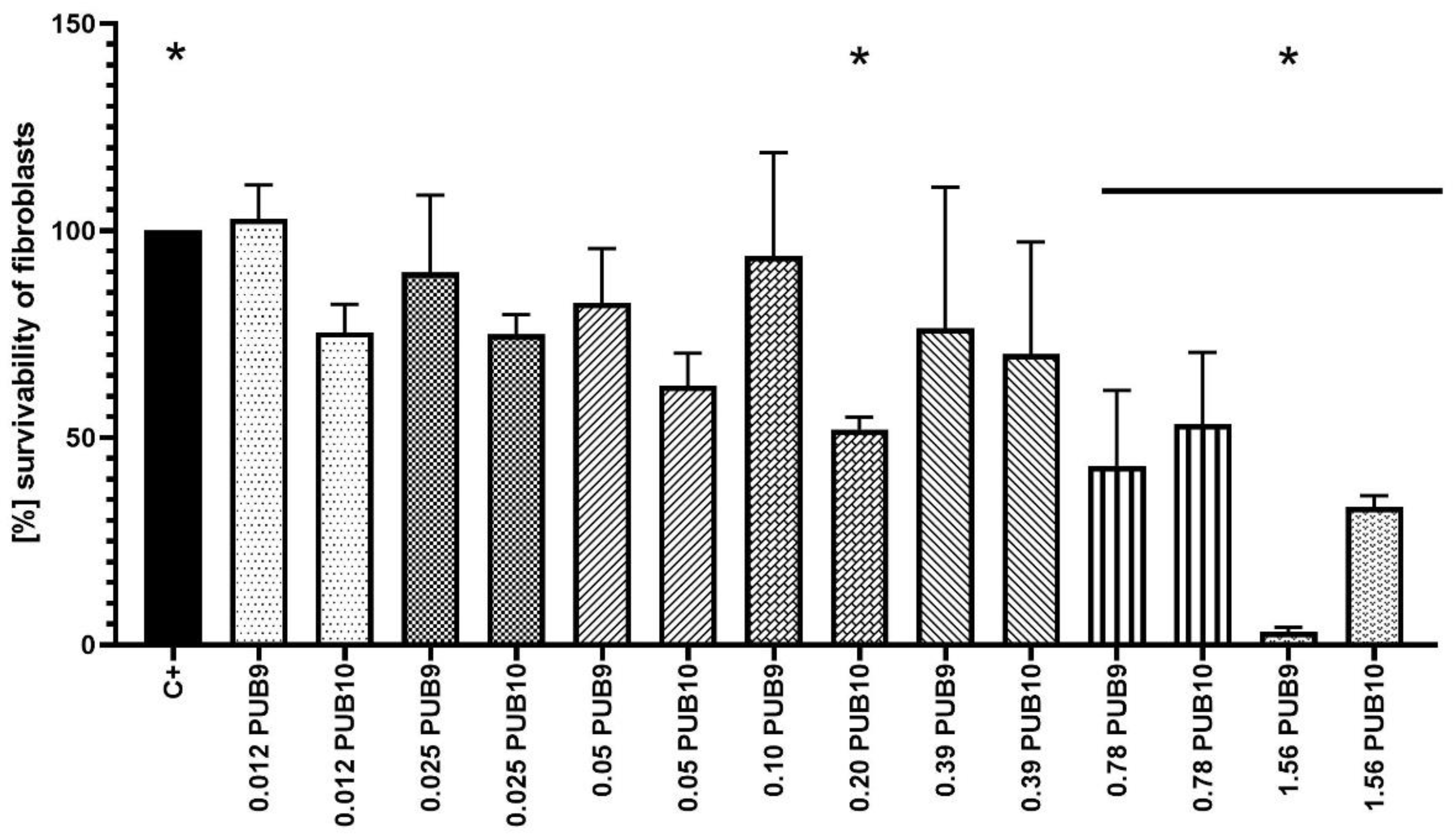
3.5. The Cytotoxicity Assay of Analyzed Compounds towards Fibroblast Cell Line In Vitro

The Neutral Red (NR) cytotoxicity assay was performed toward fibroblast (L929 ATCC, Manassas, VA, USA) in vitro cell cultures treated with the analyzed compounds in concentration equal to their MBEC (or MIC if MBEC was beyond tested range of concentrations) value according to ISO 10993: Biological evaluation of medical devices; Part 5: Tests for in vitro cytotoxicity; Part 12: Biological evaluation of medical devices, sample preparation and reference materials (ISO 10993–5:2009 and ISO/IEC 17025:2005). The analyzed compounds were suspended in the medium for fibroblast culturing (RPMI, Sigma-Aldrich, Darmstad, Germany) and left to dry at room temperature. Next, 150 μL of a de-stain solution (50% ethanol, 96%, 49% deionized water, 1% glacial acetic acid; POCH, Lublin, Poland) was introduced to each well. The plate was shaken vigorously in a shaker (MTS4, IKA-Labortechnik, Berlin, Germany) for 30 min until NR was extracted from the cells and formed a homogenous solution. Finally, the value of NR absorbance was measured spectrometrically using a microplate reader at a wave of 540 nm length. The absorbance value of NR-dyed cells not treated with medium containing analyzed compounds was considered 100% of the potential cellular growth (positive control of growth), while cells treated with 70% EtOH (POCH, Lublin, Polska) for 30 min were considered the control of method’s usability. All analyses were performed in 6 repeats.

3.6. The Synthesis and Purification of Bacterial Cellulose Carrier and Impregnation of Carrier with Tested Compounds

The K. xylinus strain DSM 46604, used to bio-fabricate the BC carrier, was cultivated in stationary conditions for 7 days /28 °C in a 24-well plate (VWR International, Radnor, PA, USA) using a Hestrin-Schramm (H-S) medium. To remove bacterial cells and media components, the BCs were purified in 0.1 M NaOH (POCH, Lublin, Poland) for 90 min/80 °C. Next, BC carriers were immersed in distilled water and incubated with shaking. During this process, the pH value was measured every 3 hrs, until it reached a neutral value. The wet BC carriers were weighted. These of weight equal 1000 ± 100 mg were transferred to the 24-well plate and immersed with 1 mL of solution containing analyzed compounds in a concentration equal to 2 × MIC. The plate was left for 24 h/8 °C. After this time, BC carriers were used for analyses described in the subsequent sections of Material and Methods (Section 3.7, Section 3.8 and Section 3.9).

3.7. The Analysis of Antimicrobial Efficacy of Analyzed Compounds Released from BC Carriers in Modified Disk-Diffusion Method

For control purposes, BC carriers introduced to 1 mL of octenidine dihydrochloride (antiseptic substance of confirmed antimicrobial activity, Schulke-Mayr, Nordstadt, Germany) or 0.9% NaCl were applied. Firstly, the S. aureus, P. aeruginosa or C. albicans of 0.5/0.8 McFarland density were spread over the Muller-Hinton or Sabouraud agar plates, respectively. Next, BC carriers soaked with tested compounds were placed on the plates. The whole setting was subjected to incubation at 37 °C for 24 h. After that, the growth inhibition zone (if occurred) was measured using a ruler. The tests were performed in triplicates.

3.8. The Detection of Antibiofilm Activity of Tested Compounds Using Modified Antibiofilm Dressing Activity Measurement (A.D.A.M.)

The method was described in detail in the earlier work of ours [62]. The exposure time was 24 h. After incubation of preformed biofilm with compounds, biofilm-containing agar disks were transferred to the fresh wells of a 24-well plate. Next, 1 mL of 0.1% saponin solution was subjected to vortex-mixing for 1 min to detach the biofilm cells from the agar surface. Serial dilutions of the obtained suspension in saline solution were performed and then cultured onto Sabouraud or M-H agar plates (for C. albicans and S. aureus/P. aeruginosa, respectively). The plates were incubated at 37 °C for 24 h. After incubation, the cfu number of grown colonies was counted. The formula used to determine the biofilm eradication was as follows: 100%—(value of cfu obtained from biofilm treated with cellulose carrier soaked with analyzed compound)/cfu obtained from biofilm treated with non-impregnated cellulose) × 100%. These experiments were done in three repeats. The octenidine dihydrochloride-based antiseptic (Schulke Mayr, Nordstadt, Germany) served as control of method’s usability.

3.9. The Cytocompatibility of Isoxazole-Fortified BC Carriers to Fibroblast Cell Lines

One mL of culturing medium containing fibroblast cell lines of density equal 1 × 10⁵ were seeded on the BC carriers fortified with the appropriate concentrations of PUB9 and PUB10 (0.25 mg/L) and cultured for 72 h at 37 °C/5% CO2 in an incubator. Next, the procedures utilizing Neutral Red method were performed to assess the survivability of the cells. The medium for fibroblast growth was removed and 100 μL of the NR solution (40 μg/mL; Sigma-Aldrich, Dormstadt, Germany) was introduced to wells of the plate. Cells were incubated with NR for 2 h at 37 °C. After incubation, the dye was removed, wells were rinsed with phosphate buffer saline (PBS, Sigma Aldrich, Dormstadt, Germany) and left to dry at room temperature. Next, 500 μL of a de-stain solution (50% ethanol 96%, 49% deionized water, 1% glacial acetic acid; POCH, Lublin, Poland) was introduced to each well. The plate was shaken vigorously in a microtiter plate shaker (MTS4, IKA-Labortechnik, Berlin, Germany) for 30 min until NR was extracted from the cells and formed a homogenous solution. Finally, the value of NR absorbance was measured spectrometrically using a microplate reader (Multi-scan GO, Thermo Fisher Scientific, Waltham, MA, USA) at 540 nm. The absorbance value of dyed fibroblasts seeded on the BC carriers non-fortified with PUB9 and PUB10 was considered 100% of the potential cellular growth (positive control).

3.10. The Statistical Analysis

Statistical analyses were performed using GraphPad Prism 8.0. (GraphPad Software, San Diego, CA, USA). Normality of distribution was verified using Shapiro–Wilk’s test. To evaluate statistical significance, the ANOVA test with post hoc Dunnett’s multiple comparison (α = 0.05) was performed.

Supplementary Materials

The following supporting information can be downloaded at: https://www.mdpi.com/article/10.3390/ijms24032997/s1.

Author Contributions

Conceptualization, U.B.; methodology, U.B.; formal analysis, U.B.; investigation, A.J., M.B. and U.B.; writing—original draft preparation, U.B.; writing—review and editing, A.J.; supervision, M.M.; funding acquisition, A.J. and M.M. All authors have read and agreed to the published version of the manuscript.

Funding

This research was funded by Wroclaw Medical University, grant number SUBZ.D090.23.027.

Institutional Review Board Statement

Not applicable.

Informed Consent Statement

Not applicable.

Data Availability Statement

All necessary data are presented in the manuscript and the raw data can be provided from the authors upon reasonable request.

Acknowledgments

The authors would like to thank Magdalena Korab for technical support.

Conflicts of Interest

The authors declare no conflict of interest.

References

  1. Frykberg, R.G.; Banks, J. Challenges in the Treatment of Chronic Wounds. Adv. Wound Care 2015, 4, 560–582. [Google Scholar] [CrossRef] [PubMed]
  2. Serra, R.; Grande, R.; Butrico, L.; Rossi, A.; Settimio, U.F.; Caroleo, B.; Amato, B.; Gallelli, L.; de Franciscis, S. Chronic wound infections: The role of Pseudomonas aeruginosa and Staphylococcus aureus. Expert Rev. Anti Infect. Ther. 2015, 13, 605–613. [Google Scholar] [CrossRef] [PubMed]
  3. Durand, B.A.R.N.; Pouget, C.; Magnan, C.; Molle, V.; Lavigne, J.P.; Dunyach-Remy, C. Bacterial Interactions in the Context of Chronic Wound Biofilm: A Review. Microorganisms 2022, 10, 1500. [Google Scholar] [CrossRef] [PubMed]
  4. Barrigah-Benissan, K.; Ory, J.; Sotto, A.; Salipante, F.; Lavigne, J.P.; Loubet, P. Antiseptic Agents for Chronic Wounds: A Systematic Review. Antibiotics 2022, 11, 350. [Google Scholar] [CrossRef]
  5. Müller, G.; Kramer, A. Biocompatibility index of antiseptic agents by parallel assessment of antimicrobial activity and cellular cytotoxicity. J. Antimicrob. Chemother. 2008, 61, 1281–1287. [Google Scholar] [CrossRef]
  6. Kampf, G. Acquired resistance to chlorhexidine—Is it time to establish an ‘antiseptic stewardship’ initiative? J. Hosp. Infect. 2016, 94, 213–227. [Google Scholar] [CrossRef]
  7. Bock, L.J.; Ferguson, P.M.; Clarke, M.; Pumpitakkul, V.; Wand, M.E.; Fady, P.E.; Allison, L.; Fleck, R.A.; Shepherd, M.J.; Mason, A.J.; et al. Pseudomonas aeruginosa adapts to octenidine via a combination of efflux and membrane remodelling. Commun. Biol. 2021, 4, 1058. [Google Scholar] [CrossRef]
  8. Li, Q.; Kang, C. Mechanisms of Action for Small Molecules Revealed by Structural Biology in Drug Discovery. Int. J. Mol. Sci. 2020, 21, 5262. [Google Scholar] [CrossRef]
  9. Zhang, D.; Jia, J.; Meng, L.; Xu, W.; Tang, L.; Wang, J. Synthesis and Preliminary Antibacterial Evaluation of 2-Butyl Succinate-based Hydroxamate Derivatives Containing Isoxazole Rings. Arch. Pharm. Res. 2010, 33, 831–842. [Google Scholar] [CrossRef]
  10. Egorova, A.; Kazakova, E.; Jahn, B.; Ekins, S.; Makarov, V.; Schmidtke, M. Novel pleconaril derivatives: Influence of substituents in the isoxazole and phenyl rings on the antiviral activity against enteroviruses. Eur. J. Med. Chem. 2020, 188, 112007. [Google Scholar] [CrossRef]
  11. Arya, G.C.; Kaur, K.; Jaitak, V. Isoxazole derivatives as anticancer agent: A review on synthetic strategies, mechanism of action and SAR studies. Eur. J. Med. Chem. 2021, 221, 113511. [Google Scholar] [CrossRef]
  12. Razzaq, S.; Minhas, A.M.; Qazi, N.G.; Nadeem, H.; Khan, A.-u.; Ali, F.; Hassan, S.S.u.; Bungau, S. Novel Isoxazole Derivative Attenuates Ethanol-Induced Gastric Mucosal Injury through Inhibition of H+/K+-ATPase Pump, Oxidative Stress and Inflammatory Pathways. Molecules 2022, 27, 5065. [Google Scholar] [CrossRef]
  13. Sysak, A.; Obmińska-Mrukowicz, B. Isoxazole ring as a useful scaffold in a search for new therapeutic agents. Eur. J. Med. Chem. 2017, 137, 292–309. [Google Scholar] [CrossRef]
  14. Huang, X.; Dong, S.; Liu, H.; Wan, P.; Wang, T.; Quan, H.; Wang, Z.; Wang, Z. Design, Synthesis, and Evaluation of Novel Benzo[d]isoxazole Derivatives as Anticonvulsants by Selectively Blocking the Voltage-Gated Sodium Channel NaV1.1. ACS Chem. Neurosci. 2022, 13, 834–845. [Google Scholar] [CrossRef]
  15. Algethami, F.K.; Saidi, I.; Abdelhamid, H.N.; Elamin, M.R.; Abdulkhair, B.Y.; Chrouda, A.; Ben Jannet, H. Trifluoromethylated Flavonoid-Based Isoxazoles as Antidiabetic and Anti-Obesity Agents: Synthesis, In Vitro α-Amylase Inhibitory Activity, Molecular Docking and Structure–Activity Relationship Analysis. Molecules 2021, 26, 5214. [Google Scholar] [CrossRef]
  16. Chikkula, K.V.; Raja, S. Isoxazole-A potent pharmacophore. Int. J. Pharm. Pharm. Sci. 2017, 9, 13–24. [Google Scholar] [CrossRef]
  17. Esfahani, S.N.; Damavandi, M.S.; Sadeghi, P.; Nazifi, Z.; Salari-Jazi, A.; Massah, A.R. Synthesis of some novel coumarin isoxazol sulfonamide hybrid compounds, 3D-QSAR studies, and antibacterial evaluation. Sci. Rep. 2021, 11, 20088. [Google Scholar] [CrossRef]
  18. Shaik, A.; Bhandare, R.R.; Palleapati, K.; Nissankararao, S.; Kancharlapalli, V.; Shaik, S. Antimicrobial, Antioxidant, and Anticancer Activities of Some Novel Isoxazole Ring Containing Chalcone and Dihydropyrazole Derivatives. Molecules 2020, 25, 1047. [Google Scholar] [CrossRef] [PubMed]
  19. Zimecki, M.; Mączyński, M.; Artym, J.; Ryng, S. Closely related isoxazoles may exhibit opposite immunological activities. Acta Pol Pharm.—Drug Res. 2008, 65, 793–794. [Google Scholar]
  20. Mączyński, M.; Ryng, S.; Artym, J.; Kocięba, M.; Zimecki, M.; Brudnik, K.; Jodkowski, J.T. New lead structures in the isoxazole system: Relationship between quantum chemical parameters and immunological activity. Acta Pol Pharm.—Drug Res. 2014, 71, 71–83. [Google Scholar]
  21. Mączyński, M.; Artym, J.; Kocięba, M.; Sochacka-Ćwikła, A.; Drozd-Szczygieł, E.; Ryng, S.; Zimecki, M. Synthesis and immunoregulatory properties of selected 5-amino-3-methyl-4-isoxazolecarboxylic acid benzylamides. Acta Pol. Pharm.—Drug Res. 2016, 73, 1201–1211. [Google Scholar]
  22. Mączyński, M.; Regiec, A.; Sochacka-Ćwikła, A.; Kochanowska, I.; Kocięba, M.; Zaczyńska, E.; Artym, J.; Kałas, W.; Zimecki, M. Synthesis, Physicochemical Characteristics and Plausible Mechanism of Action of an Immunosuppressive Isoxazolo[5,4-e]-1,2,4-Triazepine Derivative (RM33). Pharmaceuticals 2021, 14, 468. [Google Scholar] [CrossRef] [PubMed]
  23. Sochacka-Ćwikła, A.; Regiec, A.; Zimecki, M.; Artym, J.; Zaczyńska, E.; Kocięba, M.; Kochanowska, I.; Bryndal, I.; Pyra, A.; Mączyński, M. Synthesis and Biological Activity of New 7-Amino-oxazolo[5,4-d]Pyrimidine Derivatives. Molecules 2020, 25, 3558. [Google Scholar] [CrossRef]
  24. Sochacka-Ćwikła, A.; Mączyński, M.; Czyżnikowska, Ż.; Wiatrak, B.; Jęśkowiak, I.; Czerski, A.; Regiec, A. New Oxazolo[5,4-d]pyrimidines as Potential Anticancer Agents: Their Design, Synthesis, and In Vitro Biological Activity Research. Int. J. Mol. Sci. 2022, 23, 11694. [Google Scholar] [CrossRef] [PubMed]
  25. Bąchor, U.; Mączyński, M. Selected β2-, β3- and β2,3-Amino Acid Heterocyclic Derivatives and Their Biological Perspective. Molecules 2021, 26, 438. [Google Scholar] [CrossRef]
  26. Uno, B.E.; Deibler, K.K.; Villa, C.; Raghuraman, A.; Karl, A.S. Conjugate Additions of Amines to Maleimides via Cooperative Catalysis. Adv. Synth. Catal. 2018, 360, 1719–1725. [Google Scholar] [CrossRef]
  27. Worch, J.C.; Stubbs, C.J.; Matthew, J.P.; Dove, A.P. Click Nucleophilic Conjugate Additions to Activated Alkynes: Exploring Thiol-yne, Amino-yne, and Hydroxyl-yne Reactions from (Bio)Organic to Polymer Chemistry. Chem. Rev. 2021, 121, 6744–6776. [Google Scholar] [CrossRef]
  28. Dolci, E.; Froidevaux, V.; Joly-Duhamel, C.; Auvergne, R.; Boutevin, B.; Caillol, S. Maleimides as a Building Block for the Synthesis of High Performance Polymers. Polym. Rev. 2016, 56, 512–556. [Google Scholar] [CrossRef]
  29. Regiec, A.; Wojciechowski, P.; Pietraszko, A.; Mączyński, M. Infrared spectra and other properties predictions of 5-amino-3-methyl-4-isoxazolecarbohydrazide with electric field simulation using CPC model. J. Mol. Struct. 2018, 1161, 320–338. [Google Scholar] [CrossRef]
  30. Regiec, A.; Płoszaj, P.; Ryng, S.; Wojciechowski, P. Vibrational spectroscopy of 5-amino-3-methyl-4-isoxazolecarbohydrazide and its N-deuterated isotopologue. Vib. Spectrosc. 2014, 70, 125–136. [Google Scholar] [CrossRef]
  31. Płoszaj, P.; Regiec, A.; Ryng, S.; Piwowar, A.; Kruzel, M.L. Influence of 5-amino-3-methyl-4-isoxazolecarbohydrazide on selective gene expression in Caco-2 cultured cells. Immunopharmacol. Immunotoxicol. 2016, 38, 486–494. [Google Scholar] [CrossRef] [PubMed]
  32. Drynda, A.; Mączyński, M.; Ryng, S.; Obmińska-Mrukowicz, B. In vitro immunomodulatory effects of 5-amino-3-methyl-4-isoxazolecarboxylic acid hydrazide on the cellular immune response. Immunopharmacol. Immunotoxicol. 2014, 36, 150–157. [Google Scholar] [CrossRef]
  33. Drynda, A.; Obmińska-Mrukowicz, B.; Mączyński, M.; Ryng, S. The effect of 5-amino-3-methyl-4-isoxazolecarboxylic acid hydrazide on lymphocyte subsets and humoral immune response in SRBC-immunized mice. Immunopharmacol. Immunotoxicol. 2015, 37, 148–157. [Google Scholar] [CrossRef] [PubMed]
  34. Bąchor, U.; Ryng, S.; Mączyński, M.; Artym, J.; Kocięba, M.; Zaczyńska, E.; Kochanowska, I.; Tykarska, E.; Zimecki, M. Synthesis, immunosuppressive properties, mechanism of action and X-ray analysis of a new class of isoxazole derivatives. Acta Pol. Pharm. Drug Res. 2019, 76, 251–263. [Google Scholar] [CrossRef]
  35. Banfi, L.; Basso, A.; Lambruschini, C.; Moni, L.; Riva, R. The 100 facets of the Passerini reaction. Chem. Sci. 2021, 12, 15445–15472. [Google Scholar] [CrossRef]
  36. Olleik, H.; Nicoletti, C.; Lafond, M.; Courvoisier-Dezord, E.; Xue, P.; Hijazi, A.; Baydoun, E.; Perrier, J.; Maresca, M. Comparative Structure–Activity Analysis of the Antimicrobial Activity, Cytotoxicity, and Mechanism of Action of the Fungal Cyclohexadepsipeptides Enniatins and Beauvericin. Toxins 2019, 11, 514. [Google Scholar] [CrossRef]
  37. Zheng, L.; Li, S.; Luo, J.; Wang, X. Latest Advances on Bacterial Cellulose-Based Antibacterial Materials as Wound Dressings. Front. Bioeng. Biotechnol. 2020, 23, 593768. [Google Scholar] [CrossRef] [PubMed]
  38. de Amorim, J.D.P.; da Silva Junior, C.J.G.; de Medeiros, A.D.M.; do Nascimento, H.A.; Sarubbo, M.; de Medeiros, T.P.M.; Costa, A.F.S.; Sarubbo, L.A. Bacterial Cellulose as a Versatile Biomaterial for Wound Dressing Application. Molecules 2022, 27, 5580. [Google Scholar] [CrossRef]
  39. Lemnaru Popa, G.M.; Truşcă, R.D.; Ilie, C.I.; Țiplea, R.E.; Ficai, D.; Oprea, O.; Stoica-Guzun, A.; Ficai, A.; Dițu, L.M. Antibacterial Activity of Bacterial Cellulose Loaded with Bacitracin and Amoxicillin: In Vitro Studies. Molecules 2020, 25, 4069. [Google Scholar] [CrossRef]
  40. Bąchor, U.; Lizak, A.; Bąchor, R.; Mączyński, M. 5-Amino-3-methyl-Isoxazole-4-carboxylic Acid as a Novel Unnatural Amino Acid in the Solid Phase Synthesis of α/β-Mixed Peptides. Molecules 2022, 27, 5612. [Google Scholar] [CrossRef]
  41. Bąchor, U.; Drozd-Szczygieł, E.; Bąchor, R.; Jerzykiewicz, L.; Wieczorek, R.; Mączyński, M. New water-soluble isoxazole-linked 1,3,4-oxadiazole derivative with delocalized positive charge. RSC Adv. 2021, 11, 29668–29674. [Google Scholar] [CrossRef] [PubMed]
  42. Regiec, A.; Gadziński, P. New Methods for Preparing of Esters of 2-cyano-3- alkoxy-2-butenoate acids. Polish patent- PL 216770 B1, 30 May 2014. [Google Scholar]
  43. Regiec, A.; Gadziński, P.; Płoszaj, P. New methods for preparing of esters of 5-amino-3-methyl-4-isoxazolecarboxylic acid. Polish patent- PL 216764 B1, 30 May 2014. [Google Scholar]
  44. Sopata, M.; Kucharzewski, M.; Tomaszewska, E. Antiseptic with modern wound dressings in the treatment of venous leg ulcers: Clinical and microbiological aspects. J. Wound Care 2016, 25, 419–426. [Google Scholar] [CrossRef]
  45. Alves, P.J.; Barreto, R.T.; Barrois, B.M.; Gryson, L.G.; Meaume, S.; Monstrey, S.J. Update on the role of antiseptics in the management of chronic wounds with critical colonisation and/or biofilm. Int Wound, J. 2021, 18, 342–358. [Google Scholar] [CrossRef] [PubMed]
  46. Agrawal, N.; Pradeep, M. The synthetic and therapeutic expedition of isoxazole and its analogs. Med. Chem. Res. 2018, 27, 1309–1344. [Google Scholar] [CrossRef] [PubMed]
  47. Gautam, K.C.; Singh, D.P. Synthesis and antimicrobial activity of some isoxazole derivatives of thiophene. Chem Sci Trans. 2013, 2, 992–996. [Google Scholar]
  48. RamaRao, R.J.; Rao, A.K.S.B.; Sreenivas, N.; Kumar, B.S.; Murthy, Y.L.N. Synthesis and antibacterial activity of novel 5-(heteroaryl)isoxazole Derivatives. J Korean Chem Soc. 2011, 55, 243–250. [Google Scholar] [CrossRef]
  49. Tasse, J.; Cara, A.; Saglio, M.; Villet, R.; Laurent, F. A Steam-Based Method to Investigate Biofilm. Sci. Rep. 2018, 8, 13040. [Google Scholar] [CrossRef]
  50. Ünver, H.; Cantürk, Z. New thiophene bearing dimethyl-5-hydroxy isophtalate esters and their antimicrobial activities. Appl. Sci. Eng. 2018, 19, 43–49. [Google Scholar] [CrossRef]
  51. Ciecholewska-Juśko, D.; Żywicka, A.; Junka, A.; Woroszyło, M.; Wardach, M.; Chodaczek, G.; Szymczyk-Ziółkowska, P.; Migdał, P.; Fijałkowski, K. The effects of rotating magnetic field and antiseptic on in vitro pathogenic biofilm and its milieu. Sci. Rep. 2022, 12, 8836. [Google Scholar] [CrossRef]
  52. Rabin, N.; Zheng, Y.; Opoku-Temeng, C.; Du, Y.; Bonsu, E.; Sintim, H.O. Biofilm Formation Mechanisms and Targets for Developing Antibiofilm Agents. Future Med. Chem. 2015, 7, 493–512. [Google Scholar] [CrossRef]
  53. Fang, Z.; Li, Y.; Zheng, Y.; Li, X.; Lu, Y.J.; Yan, S.C.; Wong, W.L.; Chan, K.F.; Wong, K.Y.; Sun, N. Antibacterial activity and mechanism of action of a thiophenyl substituted pyrimidine derivative. RSC Adv. 2019, 9, 10739–10744. [Google Scholar] [CrossRef] [PubMed]
  54. Kramer, A.; Dissemond, J.; Kim, S.; Willy, C.; Mayer, D.; Papke, R.; Tuchmann, F.; Assadian, O. Consensus on Wound Antisepsis: Update 2018. Skin Pharmacol. Physiol. 2018, 31, 28–58. [Google Scholar] [CrossRef] [PubMed]
  55. Ghorab, M.M.; Bashandy, M.S.; Alsaid, M.S. Novel thiophene derivatives with sulfonamide, isoxazole, benzothiazole, quinoline and anthracene moieties as potential anticancer agents. Acta Pharm. 2014, 64, 419–431. [Google Scholar] [CrossRef]
  56. Golonka, I.; Greber, K.E.; Oleksy-Wawrzyniak, M.; Paleczny, J.; Dryś, A.; Junka, A.; Sawicki, W.; Musiał, W. Antimicrobial and Antioxidative Activity of Newly Synthesized Peptides Absorbed into Bacterial Cellulose Carrier against Acne vulgaris. Int. J. Mol. Sci. 2021, 22, 7466. [Google Scholar] [CrossRef] [PubMed]
  57. Abdelhamid, H.N.; Mathew, A.P. Cellulose-Based Nanomaterials Advance Biomedicine: A Review. Int. J. Mol. Sci. 2022, 23, 5405. [Google Scholar] [CrossRef] [PubMed]
  58. Ruszczak, Z.; Friess, W. Collagen as a carrier for on-site delivery of antibacterial drugs. Adv. Drug Deliv. Rev. 2003, 55, 1679–1698. [Google Scholar] [CrossRef] [PubMed]
  59. Brehant, O.; Sabbagh, C.; Lehert, P.; Dhahri, A.; Rebibo, L.; Regimbeau, J.M. The gentamicin–collagen sponge for surgical site infection prophylaxis in colorectal surgery: A prospective case-matched study of 606 cases. Int. J. Colorectal Dis. 2013, 28, 119–125. [Google Scholar] [CrossRef]
  60. Mączyńska, B.; Secewicz, A.; Smutnicka, D.; Szymczyk, P.; Dudek-Wicher, R.; Junka, A.; Bartoszewicz, M. In vitro efficacy of gentamicin released from collagen sponge in eradication of bacterial biofilm preformed on hydroxyapatite surface. PLoS ONE 2019, 14, e0217769. [Google Scholar] [CrossRef]
  61. Li, Z.; Shi, L.; Wang, B.; Wei, X.; Zhang, J.; Guo, T.; Kong, J.; Wang, M.; Xu, H. In Vitro Assessment of Antimicrobial Resistance Dissemination Dynamics during Multidrug-Resistant-Bacterium Invasion Events by Using a Continuous-Culture Device. Appl. Environ. Microbiol. 2021, 87, e02659-20. [Google Scholar] [CrossRef]
  62. Krzyżek, P.; Gościniak, G.; Fijałkowski, K.; Migdał, P.; Dziadas, M.; Owczarek, A.; Czajkowska, J.; Aniołek, O.; Junka, A. Potential of Bacterial Cellulose Chemisorbed with Anti-Metabolites, 3-Bromopyruvate or Sertraline, to Fight against Helicobacter pylori Lawn Biofilm. Int. J. Mol. Sci. 2020, 21, 9507. [Google Scholar] [CrossRef]
Figure 1. Designed isoxazole derivatives ((A) MAL1-5 series; (B) PUB1-10 series: R1-aldehyde/ketone residue; R2-isocyanide residue).
Ijms 24 02997 g001
Figure 2. Synthesis of isoxazole linked maleimide conjugates (MAL1-5).
Ijms 24 02997 g002
Figure 3. The [%] biofilm reduction of S. aureus 6538, P. aeruginosa 15,442 and C. albicans 103,231 displayed by PUB9 and PUB10 compounds at specific concentrations tested in microplate model. The red line shows level of 100% reduction while no reduction (0%, positive control of growth) was established for non-treated cells (maximal cellular growth).
Ijms 24 02997 g003
Figure 4. The survivability [%] of L929 fibroblasts exposed to the spectrum of concentrations of PUB9 and PUB10 compounds. The asterisks show significant (p > 0.05, ANOVA test with Tukey’s multiple comparison test) difference in survivability between control setting (non-treated cells, considered 100% growth) and cells treated with 0.78–1.56 mg/mL of PUB9 or PUB10 as well as 0.20 mg/mL of PUB10.
Ijms 24 02997 g004
Figure 5. The inhibition of staphylococcal growth zone resulting from release of PUB9 from BC carrier in modified disk-diffusion model. The diameter of BC carrier is 18 mm.
Ijms 24 02997 g005
Figure 6. Comparison of cytocompatibility of isoxazole derivatives-fortified cellulosic carriers: PUB9-BC and PUB10-BC compared to control setting (cellulose carrier containing no isoxazole derivative) toward fibroblast cell line. Asterisks indicate significant differences (p < 0.05, ANOVA Test with Tukey’s multiple comparison test) between BC vs. PUB10-BC and PUB9-BC vs. PUB10-BC. The red line indicates the maximal potential cellular growth (of non-fortified cellulosic carrier).
Ijms 24 02997 g006
Table 1. The Minimal Inhibitory Concentration [mg/mL] of tested compounds towards S. aureus 6538, P. aeruginosa 15442 and C. albicans 103231 tested in microplate model.
Minimal Inhibitory Concentration [mg/mL]
S. aureusP. aeruginosaC. albicans
PUB10.1250.1250.063
PUB20.1250.1250.063
PUB30.1250.1250.063
PUB40.250.1250.063
PUB50.250.1250.063
PUB60.1250.1250.063
PUB70.1250.1250.063
PUB80.1250.1250.063
PUB90.000120.1250.063
PUB100.000240.0630.02
MAL10.1250.0630.02
MAL20.1250.0630.63
MAL30.1250.1250.063
MAL40.1250.0630.063
MAL50.1250.0630.063
Disclaimer/Publisher’s Note: The statements, opinions and data contained in all publications are solely those of the individual author(s) and contributor(s) and not of MDPI and/or the editor(s). MDPI and/or the editor(s) disclaim responsibility for any injury to people or property resulting from any ideas, methods, instructions or products referred to in the content.

