Article

Genetic Variant Overlap Analysis Identifies Established and Putative Genes Involved in Pulmonary Fibrosis

by
Karlijn Groen
1,
Joanne J. van der Vis
1,2,
Aernoud A. van Batenburg
1,
Karin M. Kazemier
3,4,
Jan C. Grutters
1,4 and
Coline H. M. van Moorsel
1,*
1
Department of Pulmonology, St. Antonius ILD Center of Excellence, St. Antonius Hospital, 3435 CM Nieuwegein, The Netherlands
2
Department of Clinical Chemistry, St. Antonius ILD Center of Excellence, St. Antonius Hospital, 3435 CM Nieuwegein, The Netherlands
3
Center of Translational Immunology, University Medical Center Utrecht, 3508 GA Utrecht, The Netherlands
4
Division of Hearts and Lungs, University Medical Center Utrecht, 3584 CX Utrecht, The Netherlands
*
Author to whom correspondence should be addressed.
Int. J. Mol. Sci. 2023, 24(3), 2790; https://doi.org/10.3390/ijms24032790
Submission received: 15 December 2022 / Revised: 23 January 2023 / Accepted: 28 January 2023 / Published: 1 February 2023

Abstract

:
In only around 40% of families with pulmonary fibrosis (PF) a suspected genetic cause can be found. Genetic overlap analysis of Whole Exome Sequencing (WES) data may be a powerful tool to discover new shared variants in novel genes for PF. As a proof of principle, we first selected unrelated PF patients for whom a genetic variant was detected (n = 125) in established PF genes and searched for overlapping variants. Second, we performed WES (n = 149) and identified novel potentially deleterious variants shared by at least two unrelated PF patients. These variants were genotyped in validation cohorts (n = 2748). In 125 unrelated patients, a potentially deleterious variant was detected in known PF genes of which 15 variants in six genes overlapped, involving 51 patients. Overlap analysis of WES data identified two novel variants of interest: TOM1L2 c.421T > C p.(Y141H) and TDP1c.1373dupG p.(S459fs*5), neither gene had been related to pulmonary fibrosis before. Both proteins were present in the alveolar epithelium. No apparent characteristics of telomere disease were observed. This study underlines the potential of searching for overlapping rare potentially deleterious variants to identify disease-associated variants and genes. A previously unreported variant was found in two putative new PF genes, but further research is needed to determine causality.

Graphical Abstract

1. Introduction

Pulmonary fibrosis (PF) in the context of interstitial lung disease is characterized by progressive scarring of the lung and reduced survival. The most severe form of pulmonary fibrosis is idiopathic pulmonary fibrosis (IPF), a progressive disease of unknown cause with a median survival of 3–5 years after diagnosis [1]. Genetics is known to play an important role in disease development. Approximately 20% of IPF patients have familial pulmonary fibrosis (FPF) with disease usually being transmitted in an autosomal dominant way. A genetic cause can also be suspected in sporadic cases, based on early age of onset or disease phenotype. An increasing number of PF genes is known and the vast majority are related to telomere biology. In approximately 35% of cases, mutations are found in telomere-related genes—such as TERC, RTEL1, and PARN, but most commonly the TERT gene [2,3]. Patients with telomere related gene mutations frequently show short telomere length in blood and lung biopsy, increased DNA-damage in lung tissue, and clinical signs and symptoms of short telomere syndromes [3,4,5,6]. In only 3–8% of suspected genetic PF cases, a mutation can be found in genes of the surfactant system [2,3].
The phenotype of mutation carriers usually involves PF, including IPF, but also other forms of pulmonary fibrosis, such as fibrotic hypersensitivity pneumonitis (fHP) and non-classifiable interstitial lung disease (ILD). However, other lung diseases—such as pediatric ILD, neonatal RDS, and lung cancer in case of surfactant mutations—or pulmonary arteriovenous malformations and emphysema in case of telomere related mutations have also been observed, as reviewed by Hoffman et al. [7] and Van Moorsel et al. [2]. An increasing number of genes, here referred to as PF genes, are now known to be involved in genetic PF. Nonetheless, a causal variant—often called a mutation—can only be found in around 40% of the cases with a suspected monogenetic origin [2,3].
Several strategies for discovering new PF genes and variants involved in PF can be applied. Candidate gene approaches yielded important results such as for SFTPC [8], TERT, and TERC [9]. Linkage analysis yielded SFTPA2 [10] and analysis of genome wide data yielded RTEL1 through segregation analysis [11]. However, this approach requires DNA from multiple affected family members, something that is not often available in this rare disease that mostly presents in the sixth or seventh decade of life and with such short survival time. Alternatively, an genetic overlap based strategy can be applied in unrelated patients with similar disease phenotypes, whereby variants in a single, thus overlapping, gene are sought [12]. As an example, analysis of genome wide data yielded PARN through gene burden analysis [13]. Interestingly, several identical variants have been observed to cause PF in multiple unrelated patients, such as the SFTPC p.I73T and TERT p.R669W mutations [14,15,16,17,18,19]. We thus hypothesized that a modified overlap based strategy, namely genetic variant overlap analysis, may be used to discover new variants in novel genes for PF. Using this strategy, we here search for identical, overlapping variants in unrelated patients with PF.
The St. Antonius ILD Center of Excellence manages the Dutch national ILD-biobank, gathering blood samples and data from patients with ILD in this small-sized country. First, we performed a proof of principle analysis for the genetic variant overlap analysis. To this end, we retrospectively summarized the deleterious variants in known PF genes that had been observed in unrelated patients with PF in the Biobank. Then we identified the number of variants that were shared, thus overlapping, between the patients. Second, we applied the overlap-based strategy as an explorative approach to identify new variants and genes that may be causal to PF. We therefore performed whole exome sequencing (WES) and searched for novel, deleterious variants that are shared amongst multiple unrelated patients. Discovered novel variants were genotyped in ILD-biobank samples, and then analyzed for expression in lung tissue and characteristics of telomere related disease.

2. Results

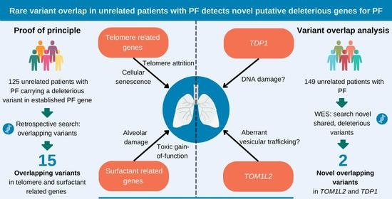
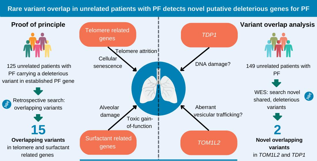
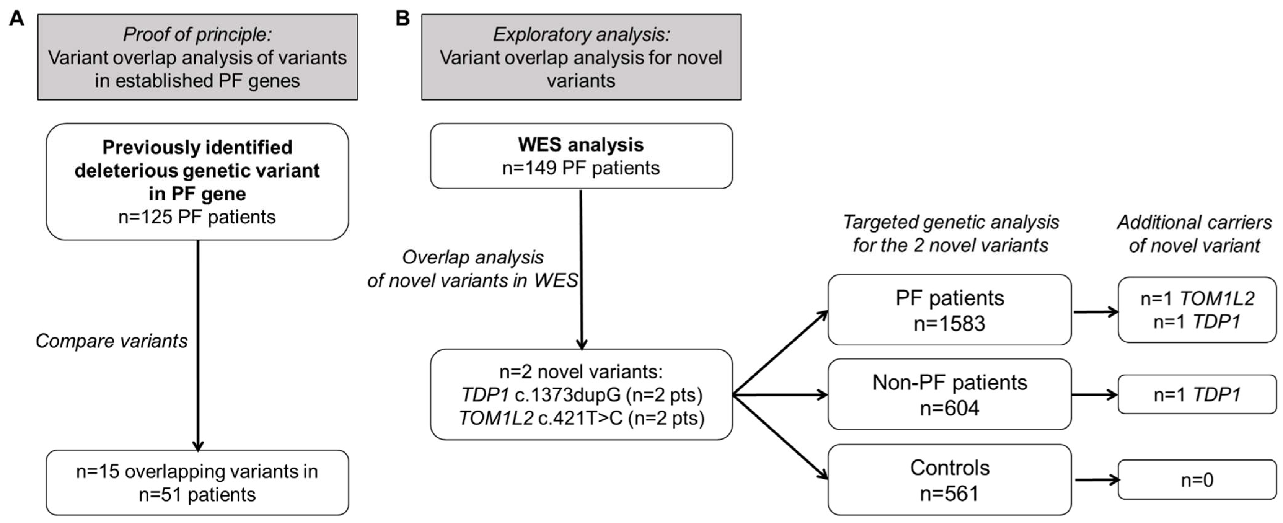
2.1. Overlap of Variants in Established PF Genes

To determine if searching for overlapping variants can be a useful strategy, we checked whether overlapping variants are present amongst our cohort of PF patients with a previously identified deleterious variant. Genetic variants classified by clinical genetic criteria as variants of unknown significance (VUS), likely pathogenic (LP), or pathogenic (P) variant in a PF gene had been previously identified in 125 patients (Figure 1A). These 125 patients jointly carried variants in nine different genes: two surfactant related genes (SFTPA2, SFTPC) and seven telomere related genes, of which the majority were present in the TERT gene. Figure 2A shows the number of different dominant variants per PF gene (variants located in genes associated with recessive disease were omitted here). Fifteen variants were found in more than one presumably unrelated patient (Table 1). The distribution of these shared variants per gene are shown in Figure 2B. Overlap was detected in two surfactant genes and four out of seven telomere related genes and was most common in TERT (7 variants in 28 patients; Figure 2B). No overlap was present in ACD, TERC, or TINF2. Genealogical research to determine possible relatedness, identified distant relatedness for 4 out of 13 patients carrying the TERT p.(Arg669Trp) variant [19], 3 out of 4 patients carrying the TERT p.(Pro771Leu) variant, and 2 out of 2 patients with the POT1 p.(Leu259Ser) [20], RTEL1 p.(Leu658del) or TERT p.(Tyr576His) variant. Thus, there is significant overlap of rare deleterious variants in established PF genes.

2.2. Variant Overlap Analysis to Identify Novel Variants in PF

Genetic variant overlap analysis was then applied to identify novel variants and genes involved in PF. To this end, WES was performed on blood derived DNA in 149 unrelated PF patients. WES data were subjected to filtering to select variants that were: (1) predicted deleterious by in silico prediction models; (2) were absent from population databases; and (3) were shared between at least two patients. After this variant filtering, 25 variants remained, of which 22 were excluded after visual inspection of the raw data. Visual inspection of the TBXT c.457A > C variant was inconclusive, but the variant was absent from subsequent Sanger sequences, and was thus considered a sequencing artefact in the WES. Two variants of interest remained, namely TOM1L2 c.421T > C and TDP1 c.1373dupG (Table 2). Both were present in two patients in the WES cohort. The target of myb1-like 2 membrane trafficking protein (TOM1L2) c.421T > C missense variant is predicted to cause a substitution of the highly conserved hydrophobic tyrosine residue with a positively charged histidine (p.(Y141H)) and is located in the VHS domain (Figure 3A). The tyrosyl-DNA phosphodiesterase 1 (TDP1) c.1373dupG p.(S459fs*5) variant is predicted to cause a frameshift introducing a premature stop codon likely leading to nonsense mediated decay. We examined the WES data for other rare variants (frequency < 1% in population databases) in these two genes. Two more variants were identified in TDP1. One variant was found in two unrelated individuals (TDP1 c.1443G > A). This variant did not pass the original filtering criteria due to its presence in population databases. However, available WES data from a brother with pulmonary fibrosis of one of these patients indicated lack of segregation. The other TDP1 variant (c.1580delA (p.K527fs*6); rs770659676) is a very rare variant which we found in one patient with FPF. However, no material was available for segregation analysis, and this variant was therefore not investigated further. To identify additional carriers of these variants, targeted genotyping was applied in an extended cohort of pulmonary fibrosis patients (n = 1583). We identified one additional heterozygous carrier of the TOM1L2 variant and one heterozygous carrier of the TDP1 c.1373dupG variant in this cohort. Targeted genotyping in non-PF patients (n = 604) identified one non-PF patient carrying the TDP1 c.1373dupG variant. Neither variant was identified in healthy controls (n = 561).

2.3. Novel Variant Patient Characteristics

The TOM1L2 c.421T > C variant was present in three PF patients each with a different diagnosis, namely fHP, IPF, and nonclassifiable interstitial pneumonia (NCIP) (see Table 3). Patient 1 was a 52-year-old male diagnosed with fHP. A causative antigen could not be identified. The patient’s mother had been diagnosed at age 77 with pulmonary fibrosis. On HRCT a pattern of probable usual interstitial pneumonia (UIP) was detected with subpleural reticulation, traction bronchiectasis with basal and peripheral predominance and absence of honeycombing. He received a bilateral lung transplant at age 60. The second patient was a 47-year-old male diagnosed with sporadic IPF and WES was performed as part of an earlier study [4]. Histologic analysis showed a UIP pattern with concomitant DIP-like features. He underwent a unilateral lung transplant at 51 years old. The third patient was a 55-year-old male diagnosed with NCIP with a suspicion of PF. An HRCT pattern of NSIP and a histological pattern of UIP with lymphoid hyperplasia containing B lymphocytes and endogenous lipoid pneumonia were observed.
TDP1 c.1373dupG was initially found in two IPF patients (Patients 4 and 5). Screening in a broader cohort of PF patients, non-PF patients and healthy controls identified two more heterozygous carriers: one patient with lymphangioleiomyomatosis (LAM) (patient 6) and one patient with COPD and PF (patient 7). Patient 4 is a male patient with a familial background of lung fibrosis who was diagnosed with IPF at age 62. The HRCT shows a UIP pattern with suspected diffuse alveolar damage (DAD) and includes emphysematous changes. In a diagnostic biopsy, a UIP pattern with DAD was observed. Patient 5 is a sporadic male IPF patient diagnosed at age 50. Both radiology and histology showed a UIP pattern, which is accompanied by paraseptal emphysema on HRCT. He received a bilateral lung transplant at age 52. Patient 6 is a 46-year-old female diagnosed with sporadic LAM. LAM diagnosis was based on presence of bilateral angiomyolipomas in the kidneys and a strongly increased serum VEGF-D level. Additionally, HRCT displayed mild small thin-walled cysts in the lung. Patient 7 was a 56-year-old male diagnosed with COPD GOLD IV with an HRCT with extensive emphysema and bronchiolitis. He therefore received a unilateral lung transplant (left) at age 57. After lung transplantation, the patient developed progressive fibrosis with radiological UIP pattern features in the native right lung. Analysis of explant lung tissue showed signs of fibrosis i.e., accumulation of interstitial fibroblast in addition to emphysema.

2.4. TOM1L2 Presence in Lung Cells

Double staining in lung tissue of control subjects showed that TOM1L2 protein is located in Type 2 alveolar epithelial cells (AT2 cells) as well as in proSP-C negative cells (indicated by the arrows) in the alveoli of control lung (Figure 4A). Localization is predominantly cytosolic. As a confirmation for the mouse-anti-TOM1L2 antibody, the staining was repeated with a rabbit-anti-TOM1L2 antibody on subsequent slides from the same sample which showed a similar pattern (see Supplemental Results S3). In a sporadic IPF patient and a PF TOM1L2 c.421T > C carrier, TOM1L2 positive staining was present in non-fibrotic (B,D) and fibrotic (C,E) lung tissue in (hyperplasic) AT2 cells, but also in unspecified proSP-C negative cells.
TOM1L2 RNA expression was measured in whole lung tissue from controls, sporadic IPF, and TERT mutation carriers. Low expression of reference genes prevented analysis in TOM1L2 variant carriers. TOM1L2 RNA expression was significantly increased in sporadic IPF (sIPF) compared to control lung (p = 0.040) (Figure 5). When sIPF and TERT mutations carriers were grouped, expression was significantly higher in this group as compared to controls (p = 0.012; see Supplemental Results S4).

2.5. TDP1 Expression in the Lung

TDP1 is present in cells in the alveoli of control lung, as well as in lung tissue of a TERT mutation carrier and Patient 5 carrying the TDP1 c.1373dupG variant (Figure 6). Staining is of limited intensity and most pronounced in the cytosol of proSP-C negative cells, but it can also be observed in the nucleus of both proSP-C positive and negative cells. No differences in TDP1 intensity were optically detected between carriers and non-carriers of the TDP1 c.1373dupG variant.

2.6. Telomere Length

As PF causing mutations are mostly located in telomere related genes and associated with short telomeres, we sought whether variant carriers showed signs of short telomeres. Telomere length in blood of variant carriers is close to the 10th percentile for age except for patient 1 with TOM1L2 c.421T > C variant who had telomere length above the 50th percentile (Figure 7A). Telomere length in lung and other organs based on T/S ratio was determined in autopsy tissue of TDP1 variant carrier Patient 4 (Figure 7B). In IPF and TERT PF tissues, telomere length is shortest in the lungs, which is also observed in the TDP1 variant tissue. Telomere length in lung biopsy is comparable to controls (Figure 7C).

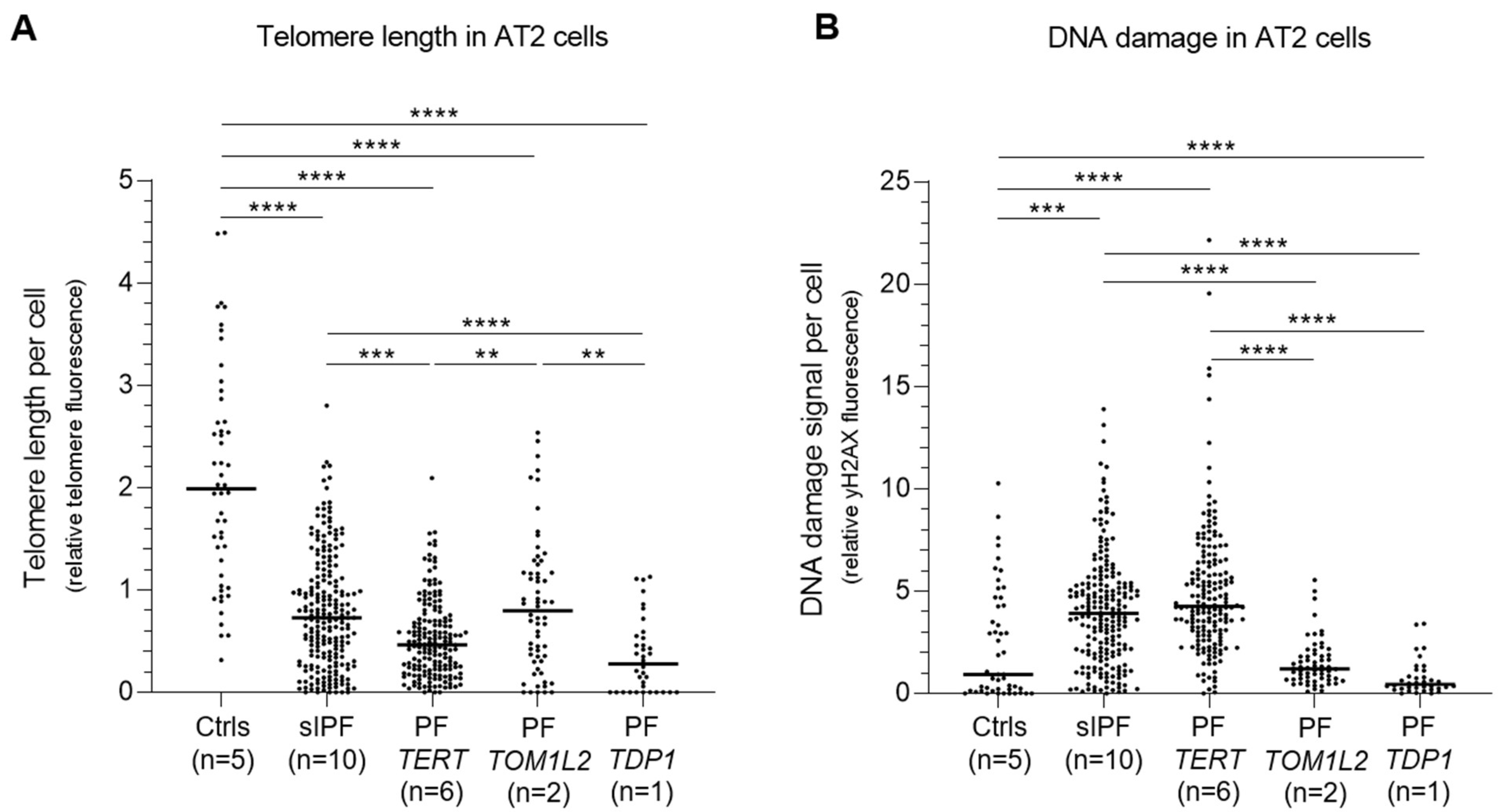
2.7. Telomere Length and DNA Damage in AT2 Cells of TOM1L2 and TDP1 Variant Carriers

AT2 cell telomere length of TOM1L2 and TDP1 variant carriers (Patients 2, 3, and 5) is significantly shorter than in controls. AT2 cell TL in TOM1L2 variant carriers is comparable to that of sporadic IPF patients, whereas that of the TDP1 variant carrier (Patient 5) is significantly shorter (Figure 8). DNA damage in AT2 cells of both variants is considerably lower than that of both sporadic and TERT PF patients.

3. Discussion

In this study, we show that there is considerable overlap of the genetic variants in established PF genes that were present in unrelated patients. Additionally, a strict search for putative damaging variants shared between unrelated patients revealed two variants in two novel genes that may be involved in PF.
Genetic variant overlap may be caused by shared ancestral origin, or by recurrent de novo mutations. Among the well-known mutations in PF, both can be found. The SFTPC mutation I73T has been discovered in patients all over the world, inherited or de novo [15,16,18](. It is the most common of all surfactant mutations, and was previously shown to have originated on different haplotypes in affected children and in adults, proof of recurrent mutation at a mutational hotspot [18,28]. We detected a particularly high number of overlapping variants in telomere related genes, particularly in TERT. Previously, the TERT c.2371G > A and c.2599G > A in cis [35] and the TERT c.2005C > T mutation that was also described here [19] have each been reported to share a common ancestor. For multiple telomere related variants, post hoc genealogical research resulted in the detection of distant relatedness of carriers of identical mutations. The distribution of these variants may be the result of reduced penetrance or genetic anticipation. In families with TRG mutations, telomere length is passed on from generation to generation; hence, in mutation carriers, telomeres shorten in successive generations. Therefore, inheritance through several generations may be required before a telomere related mutation may have caused significant telomere shortening necessary for disease manifestation [19]. Comparison of surrounding haplotypes can confirm distant relatedness among reported carriers, though this was not performed here. Given the considerable overlap in our cohort but also in patients found worldwide, a general database containing detected variants in pulmonary disease, may help establish the pathogenicity of observed variants. Most importantly, the significant overlap of variants in established PF genes showed that such an analysis could also be a powerful tool to discover novel variants in novel genes.
Overlap analysis in our WES cohort revealed two genes that may be involved in PF pathogenesis, one of which is TOM1L2. Interestingly, in the study where Stuart et al. linked PARN and RTEL1 to FPF through gene burden analysis, TOM1L2 was among the top 10 genes of interest (based on p-value), although genome-wide significance was not reached [13]. We demonstrate here that TOM1L2 is expressed in the lung and in the AT2 cells, which are deemed the culprit cell in IPF. Information regarding the function of TOM1L2 is scarce. The variant results in an amino acid substitution in the highly conserved VHS domain. Normal telomere length in blood, and telomere length in AT2 cells that is comparable to sporadic IPF and longer than TERT-IPF, indicate a non-telomere-related pathway. Interestingly, TOM1L2 RNA expression was increased in IPF lung compared to control, which suggests a role for TOM1L2 in disease, though it may be a response to disease processes.
TOM1L2, together with TOM1 and TOM1L1, forms a subfamily of VPS (Vps27-Hrs-STAM) containing proteins. TOM1L2 shows considerable overlap in amino acid sequence with TOM1 (59%) and TOM1L1 (30%) [36] In vitro a fragment of the TOM1L2 GAT domain binds TOLLIP [37] This is of specific interest, as the minor alleles of the common SNPs rs111521887 and rs5743894 in the TOLLIP gene are associated with IPF. These IPF risk alleles are associated with significantly reduced TOLLIP expression, whereas the minor allele of rs5743890 was found to be protective for IPF and associated with disease progression and worse survival [38,39]. In mice expressing low levels of TOM1L2, an abnormal immune response was observed which was characterized by increased incidence of skin and eye infections, splenomegaly, and tumors [40]. Possibly in line with the latter, in vitro, TOM1L2 overexpression exerts an inhibiting effect on mitogenesis [41]. This increased incidence of infections, combined with the well-known role of TOLLIP in the innate immune response with its role in IL1 and Toll-like receptor signaling, suggests a role for impaired host defense. However, our patients with the TOM1L2 variant did not display a specifically inflammatory pulmonary phenotype.
On the other hand, mouse TOM1L2 is located in the trans-Golgi network and a role in vesicular transport and trafficking was suggested [40]. TOM1L2 also interacts with clathrin, and Katoh et al. hypothesize that—due to its similarity to TOM1 and TOM1L1—TOM1L2 is recruited by TOLLIP to endosomes and in turn recruits clathrin to endosomes [37]. TOM1L2 was further shown to interact in vitro with myosin VI by means of co-immunoprecipitation [42] and to bind polyubiquitin chains (both Lys48 and Lys63 linked chains) [43], pointing towards a role in endosomal sorting. Keeping in mind the significance of the AT2 cells in pulmonary fibrosis, it is interesting to speculate on a role for TOM1L2 in these cells. Several studies showed that mutations in SFTPC may result in abnormal trafficking via the plasma membrane followed by internalization to the endosomal compartment [44,45]. It was recently hypothesized that also wild type SFTPC may first travel to the plasma membrane [46]. It would subsequently be internalized via AP2-dependent endocytosis in a clathrin-coated vesicle [46], which was at least in enterocytes shown to depend on myosin VI [47], followed by cleavage in an early and then a later endosomal compartment. Lys63-chain ubiquitination is subsequently necessary for trafficking into the MVBs [46]. Some cautiousness is warranted as the majority of these experiments were performed in HeLa cells, rather than AT2 (resembling) cells [48]. However, it remains interesting that TOM1L2 was shown to associate with multiple players in this process.
The second gene we putatively linked with PF was TDP1. The overlapping variant caused a frameshift, resulting in a stop codon four amino acids downstream. We observed TDP1 protein expression in the lung and AT2 cells, as previously shown by Fam et al. [49]. Fam et al. further found that TDP1 is located mostly in cytoplasm of AT1 cells and more nuclear in AT2 cells. Additionally, they showed that during oxidative stress, TDP1 relocates to the mitochondria in cultured skin fibroblast. TDP1 is involved in different types of DNA repair, of both nuclear and mitochondrial DNA [50]. Here we studied whether presence of γH2AX is increased in AT2 cells of TDP1 c.1373dupG variant carrier with IPF, but found fluorescence to be comparable to controls, in contrast with sporadic and TERT IPF samples where γH2AX was strongly increased. This was unexpected, as TDP1-knockdown human tumor cells that were treated with etoposide, which eventually causes DNA double strand breaks (DSBs), showed increased γH2AX levels [51]. Although we cannot link TDP1 function to pulmonary fibrosis, it is interesting to note that Kosmider et al. showed mitochondrial dysfunction in AT2 cells in emphysema, associated with increased mtDNA damage and reduced TDP1 levels. When tissue from areas with mild and severe emphysema from the same patient was compared, the TDP1 level was lower in the more affected areas [52]. Interestingly, emphysema was present in various degrees in three out of four TDP1 variant carriers. The fourth carrier was diagnosed with LAM at age 46. HRCT showed limited thin-walled cysts, but no signs of emphysema or fibrosis were present on her most recent HRCT at age 45. However, the three other patients were diagnosed with disease at age 62, 50, and 56, thus increased awareness for signs of fibrosis may be warranted during clinical follow-up.
Genetic analysis in PF has shown that all dominant disease-causing variants are rare, and often unique for a specific family. This hampers detection of new genes and variants which has consequences for the diagnosis and management of patients. Clinical genetic analysis is a powerful tool and when a genetic cause can be identified, this aids with disease diagnosis and may provide information regarding prognosis, risk of comorbidities, and response to treatment. Additionally, it can be used to determine disease risk assessment in relatives and aid family planning [53]. This study shows that rare variant overlap analysis using an extensive biobank can be used to detect new genes and variants.
The study has several limitations. The strict variant filtering allowed only inclusion of previously unreported variants, increasing the risk of discarding shared pathogenic variants that are present at low frequency in the general population. Furthermore, only a small number of lung biopsies were available for analysis and as we did not perform functional experiments, the effect of the observed variants on protein function remains unknown. As there is considerable overlap between TOM1L2 and especially TOM1, it may be difficult to discriminate between the two in visualization of the protein. However, the rabbit anti-TOM1L2 antibody used here was also used by Protein Atlas (version 21.1 [54]). No TOM1L2 staining was reported there in alveolar cells, however we used a higher concentration (1:10 vs 1:75) and immunofluorescence staining, a more sensitive method.
The current study shows the potential of searching for overlapping variants as a tool to identify new putative disease-associated variants and with that, genes. In addition, it underlines the value of an extensive well-phenotyped cohort in rare disease. We detected two previously unreported variants, one in TOM1L2 and one in TDP1, in multiple patients with pulmonary disease. To date, neither gene is related to telomere maintenance or the surfactant system, processes that currently cover all known PF genes. This suggests a new mechanism involved in PF disease pathogenesis and further research into the role of these genes and the observed variants is therefore needed.

4. Methods and Materials

4.1. Patients

We retrieved 1731 unrelated patients with PF from the St. Antonius ILD Center of Excellence Biobank (hereafter referred to as ‘ILD Biobank’) and first selected PF patients for whom a genetic variant was detected (n = 125) in established dominant genes associated with PF. These PF genes included SFTPA2, SFTPC, ACD, PARN, POT1, RTEL1, TERC, TERT, and TINF2.
Second, we selected a PF group for WES analysis based on presence of familial disease, age of presentation of ILD below 55 years, suspicion of short telomere syndrome or prior presence of whole exome data. WES was performed for 149 unrelated subjects with pulmonary fibrosis from the ILD Biobank. This includes 102 patients with pulmonary fibrosis in the context of familial disease (68.5%), 9 (6.0%) patients with sporadic pulmonary fibrosis were included because they were younger than 55 years old at diagnosis, 3 (2.0%) because there was suspicion of short telomere syndrome, and 35 (23.5%) IPF patients were previously sequenced as part of a study [4]. Variants of interest were screened using a TaqMan genotyping assay in a larger cohort of other patients with pulmonary fibrosis (n = 1583), non-PF pulmonary patients (n = 604 of which 85.8% had sarcoidosis) and healthy controls (n = 561), all registered in the ILD Biobank. Residual FFPE (lung) tissue from biopsy (Patients 2, 3, and 5) or autopsy (Patient 4) material was collected. The study was approved by the Medical Research Ethics Committees United (MEC-U) of the St. Antonius Hospital (approval number R05-08A) and subjects provided written informed consent.

4.2. Samples

The magnetic beads-based method (chemagic DNA blood 10k kit; Perkin Elmer Inc. Waltham, MA, USA) was used for genomic DNA extraction from peripheral leukocytes. Biopsy material was available from two patients with the TOM1L2 variant and one with the TDP1 variant. Biopsy material of control lung, sporadic IPF and TERT mutation carriers was included as a comparison. Autopsy material from one patient with the TDP1 variant was available from lung, kidney, and liver tissue and compared to previously published data from healthy controls, sporadic IPF patients and an IPF patient carrying a TERT mutation [4].

4.3. Whole Exome Sequencing

Whole Exome Sequencing (WES) was performed in gDNA from peripheral leukocytes by Novogene (Hong Kong, China) using the Agilent Sure-Select Human All Exon V6 kit (Agilent Technologies, Santa Clara, CA, USA) on an Illumina PE150 sequencing platform (Illumina, San Diego, CA, USA) according to standard protocol. On average at least 20-fold read coverage was achieved for 93.7%. Reads were aligned to reference genome assembly hg19/GRCh37.

4.4. Variant Selection

Variant call files were uploaded into Ingenuity Variant Analysis (IVA) software (Qiagen, Hilden, Germany) for filtering. Variants were filtered to: (1) exclude variants present in population databases; (2) include variants that are deleterious as predicted by in silico prediction software; and (3) keep only variants that are present in at least two unrelated patients. For more detail, see Supplemental Methods S1 and Supplemental Results S1. Resulting monoallelic variants were subjected to visual inspection using the Integrative Genomics Viewer (IGV) tool (Supplemental Results S2).

4.5. Targeted Genetic Analysis

Sanger sequencing was used to check the TOM1L2 c.421T > C, TDP1 c.1373dupG and TBXT c.457A > C variants. For primers, see Supplemental Methods S2. TOM1L2 c.421T > C and TDP1 c.1373dupG were subsequently genotyped in an extended cohort using custom Taqman assays and the QuantStudio 5 Real-Time PCR system (both ThermoFisher Scientific, Waltham, MA, USA).

4.6. Immunofluorescent Staining of TOM1L2 and TDP1 in FFPE Diagnostic Lung Biopsies

Staining of TOM1L2 and TDP1 was based on a similar staining procedure as described previously [21]. In short, residual FFPE lung tissue from diagnostic biopsies were cut in 4 μm slices and stored at 4 °C until use. After antigen retrieval, slides were incubated with the following antibodies: mouse anti human TDP1 antibody (C-3, sc-365674, Santa Cruz, Dallas, TX, USA) previously used by Kosmider et al. [52], rabbit anti human TOM1L2 (1:10, HPA023304 R09917, Sigma-Aldrich, Saint Louis, MO, USA), and mouse anti human TOM1L2 (1:10, H00146691-B01 08071 WULZ, Abnova, Taipei, Taiwan). In case of the two mouse antibodies, slides were co-stained with an AT2-specific cell marker (rabbit anti-human pro-Spc antibody, 1:100, AB3786; Merck Millipore, Darmstadt, Germany) and corresponding immunofluorescence secondary antibodies. To reduce auto-fluorescent signals, slides were incubated with Vector TrueView (Vector laboratories, Newark, CA, USA) for 3 min and nuclei were subsequently counterstained with 4′,6-diamidino-2-phenylindole (DAPI; 25 μg/mL). Pictures were taken with a LSM700 laser scanning confocal microscope (Zeiss, Jena, Germany).

4.7. Measurement of Average Telomere Length in Tissue and Blood Using MMqPCR

Slices of FFPE tissue from lung were deparaffinated using paraffin dissolver (Macherey-Nagel, Düren, Germany). DNA was isolated using an AllPrep DNA/RNA FFPE Kit (Qiagen, Hilden, Germany) and quantified using Nanodrop (Thermo Fisher Scientific, Waltham, MA, USA). Measurement of telomere length in peripheral blood leukocytes and FFPE tissue from autopsy section material was performed as previously described (Van Batenburg et al., 2020). To determine T/S ratios as a measure for average relative telomere length, monochrome multiplex qPCR (MMqPCR) was performed on a CFX96™ Single-Color Real-Time PCRDetection System (Bio-Rad, Hercules, CA, USA) using iQ SYBR Green Supermix (Bio-Rad, Hercules, CA, USA).

4.8. Quantification of TOM1L2 Expression with qPCR

The expression of TOM1L2 RNA was measured in lung tissue obtained from biopsy. RNA was isolated from formalin-fixed paraffin-embedded tissue using AllPrep DNA/RNA FFPE Kit (Qiagen, Hilden, Germany). Paraffin was removed first using paraffin dissolver (Machery-Nagel, Düren, Germany). RNA concentration and purity was measured by nanodrop and RNA was reversed transcribed using iScript (Bio-Rad, Hercules, CA, USA). 6 ng cDNA was used in real time RT-PCR which was performed on a CFX96 Single-Color Real-Time PCRDetection System (Bio-Rad, Hercules, CA, USA) using iQ SYBR Green Supermix (Bio-Rad, Hercules, CA, USA). The quantitation was made with the comparative threshold cycles (delta Ct*1000) method where the amount of TOM1L2 target was normalized to the mean of 3 endogenous reference genes: RPL13A, beta-actin and EEF1A1 see Supplemental Methods S2 for primers. Measurements were performed in duplicate.

4.9. Statistical Analysis

Statistical analyses were performed using GraphPad Prism (version 8.3). Differences between groups were analyzed by Mann–Whitney U tests or, in case of multiple groups, Kruskal–Wallis tests as appropriate for non-parametric data. The latter was followed by Dunn’s multiple comparisons test in case of a significant outcome. Values of p < 0.05 were considered as statistically significant.

Supplementary Materials

The supporting information can be downloaded at: https://www.mdpi.com/article/10.3390/ijms24032790/s1.

Author Contributions

Conceptualization, K.G., J.J.v.d.V. and C.H.M.v.M.; Methodology, K.G., J.J.v.d.V., A.A.v.B., K.M.K. and C.H.M.v.M.; Validation, J.C.G.; Formal analysis, K.G., J.J.v.d.V., A.A.v.B. and K.M.K.; Investigation, K.G., J.J.v.d.V., A.A.v.B. and K.M.K.; Data curation, K.G., J.J.v.d.V., A.A.v.B., K.M.K. and J.C.G.; Writing – original draft, K.G., J.J.v.d.V. and C.H.M.v.M.; Writing – review & editing, K.G., J.J.v.d.V., A.A.v.B., K.M.K., J.C.G. and C.H.M.v.M.; Visualization, K.G., J.J.v.d.V., A.A.v.B. and K.M.K.; Supervision, C.H.M.v.M.; Funding acquisition, K.G., J.J.v.d.V., J.C.G. and C.H.M.v.M. All authors have read and agreed to the published version of the manuscript.

Funding

This study received funding from the St. Antonius Onderzoeksfonds. This study was also funded by ZonMW TopZorg St. Antonius Care (grant no. 842002001) and ZonMW Topspecialistische Zorg en Onderzoek (grant no. 10070012010004). The funders had no role in study design, data collection and analysis, decision to publish, or preparation of the manuscript.

Institutional Review Board Statement

The study was approved by the Medical research Ethics Committees United (MEC-U) of the St. Antonius Hospital (R05–08A).

Informed Consent Statement

Informed consent was obtained from all subjects involved in the study.

Data Availability Statement

Data can be requested from the corresponding author.

Conflicts of Interest

The authors declare no conflict of interest.

References

  1. Martinez, F.J.; Collard, H.R.; Pardo, A.; Raghu, G.; Richeldi, L.; Selman, M.; Swigris, J.J.; Taniguchi, H.; Wells, A.U. Idiopathic Pulmonary Fibrosis. Nat. Rev. Dis. Prim. 2017, 3, 17074. [Google Scholar] [CrossRef] [PubMed]
  2. van Moorsel, C.H.M.; van der Vis, J.J.; Grutters, J.C. Genetic Disorders of the Surfactant System: Focus on Adult Disease. Eur. Respir. Rev. 2021, 30, 1–17. [Google Scholar] [CrossRef] [PubMed]
  3. Borie, R.; Kannengiesser, C.; Sicre de Fontbrune, F.; Gouya, L.; Nathan, N.; Crestani, B. Management of Suspected Monogenic Lung Fibrosis in a Specialised Centre. Eur. Respir. Rev. 2017, 26, 160122. [Google Scholar] [CrossRef] [PubMed]
  4. Van Batenburg, A.A.; Kazemier, K.M.; Van Oosterhout, M.F.M.; Van Der Vis, J.J.; Van Es, H.W.; Grutters, J.C.; Goldschmeding, R.; Van Moorsel, C.H.M. From Organ to Cell: Multi-Level Telomere Length Assessment in Patients with Idiopathic Pulmonary Fibrosis. PLoS ONE 2020, 15, e0226785. [Google Scholar] [CrossRef]
  5. Newton, C.A.; Batra, K.; Torrealba, J.; Kozlitina, J.; Glazer, C.S.; Aravena, C.; Meyer, K.; Raghu, G.; Collard, H.R.; Garcia, C.K. Telomere-Related Lung Fibrosis Is Diagnostically Heterogeneous but Uniformly Progressive. Eur. Respir. J. 2016, 48, 1710–1720. [Google Scholar] [CrossRef] [PubMed]
  6. van Batenburg, A.A.; Kazemier, K.M.; van Oosterhout, M.F.M.; van der Vis, J.J.; Grutters, J.C.; Goldschmeding, R.; van Moorsel, C.H.M. Telomere Shortening and DNA Damage in Culprit Cells of Different Types of Progressive Fibrosing Interstitial Lung Disease. ERJ Open Res. 2021, 7, 00691–02020. [Google Scholar] [CrossRef] [PubMed]
  7. Hoffman, T.W.; Van Moorsel, C.H.M.; Borie, R.; Crestani, B. Pulmonary Phenotypes Associated with Genetic Variation in Telomere-Related Genes. Curr. Opin. Pulm. Med. 2018, 24, 269–280. [Google Scholar] [CrossRef]
  8. Nogee, L.M.; Dunbar, A.E.; Wert, S.E.; Askin, F.; Hamvas, A.; Whitsett, J.A. A Mutation in the Surfactant Protein C Gene Associated with Familial Interstitial Lung Disease. N. Engl. J. Med. 2001, 344, 573–579. [Google Scholar] [CrossRef]
  9. Armanios, M.Y.; Chen, J.J.-L.; Cogan, J.D.; Alder, J.K.; Ingersoll, R.G.; Markin, C.; Lawson, W.E.; Xie, M.; Vulto, I.; Phillips, J.A.; et al. Telomerase Mutations in Families with Idiopathic Pulmonary Fibrosis. N. Engl. J. Med. 2007, 356, 1317–1326. [Google Scholar] [CrossRef]
  10. Wang, Y.; Kuan, P.J.; Xing, C.; Cronkhite, J.T.; Torres, F.; Rosenblatt, R.L.; DiMaio, J.M.; Kinch, L.N.; Grishin, N.V.; Garcia, C.K. Genetic Defects in Surfactant Protein A2 Are Associated with Pulmonary Fibrosis and Lung Cancer. Am. J. Hum. Genet. 2009, 84, 52–59. [Google Scholar] [CrossRef] [Green Version]
  11. Cogan, J.D.; Kropski, J.A.; Zhao, M.; Mitchell, D.B.; Rives, L.; Markin, C.; Garnett, E.T.; Montgomery, K.H.; Mason, W.R.; McKean, D.F.; et al. Rare Variants in RTEL1 Are Associated with Familial Interstitial Pneumonia. Am. J. Respir. Crit. Care Med. 2015, 191, 646–655. [Google Scholar] [CrossRef]
  12. Gilissen, C.; Hoischen, A.; Brunner, H.G.; Veltman, J.A. Disease Gene Identification Strategies for Exome Sequencing. Eur. J. Hum. Genet. 2012, 20, 490–497. [Google Scholar] [CrossRef]
  13. Stuart, B.D.; Choi, J.; Zaidi, S.; Xing, C.; Holohan, B.; Chen, R.; Choi, M.; Dharwadkar, P.; Torres, F.; Girod, C.E.; et al. Exome Sequencing Links Mutations in PARN and RTEL1 with Familial Pulmonary Fibrosis and Telomere Shortening. Nat. Genet. 2015, 47, 512–517. [Google Scholar] [CrossRef]
  14. Hong, D.; Dai, D.; Liu, J.; Zhang, C.; Jin, T.; Shi, Y.; Jiang, G.; Mei, M.; Wang, L.; Qian, L. Clinical and Genetic Spectrum of Interstitial Lung Disease in Chinese Children Associated with Surfactant Protein C Mutations. Ital. J. Pediatr. 2019, 45, 117. [Google Scholar] [CrossRef]
  15. Guillot, L.; Epaud, R.; Thouvenin, G.; Jonard, L.; Mohsni, A.; Couderc, R.; Counil, F.; de Blic, J.; Taam, R.A.; Le Bourgeois, M.; et al. New Surfactant Protein C Gene Mutations Associated with Diffuse Lung Disease. J. Med. Genet. 2009, 46, 490–494. [Google Scholar] [CrossRef]
  16. Tredano, M.; Griese, M.; Brasch, F.; Schumacher, S.; de Blic, J.; Marque, S.; Houdayer, C.; Elion, J.; Couderc, R.; Bahuau, M. Mutation of SFTPC in Infantile Pulmonary Alveolar Proteinosis with or without Fibrosing Lung Disease. Am. J. Med. Genet. 2004, 126A, 18–26. [Google Scholar] [CrossRef]
  17. Coghlan, M.A.; Shifren, A.; Huang, H.J.; Russell, T.D.; Mitra, R.D.; Zhang, Q.; Wegner, D.J.; Cole, F.S.; Hamvas, A. Sequencing of Idiopathic Pulmonary Fibrosis-Related Genes Reveals Independent Single Gene Associations. BMJ Open Respir. Res. 2014, 1, e000057. [Google Scholar] [CrossRef]
  18. Cameron, H.S.; Somaschini, M.; Carrera, P.; Hamvas, A.; Whitsett, J.A.; Wert, S.E.; Deutsch, G.; Nogee, L.M. A Common Mutation in the Surfactant Protein C Gene Associated with Lung Disease. J. Pediatr. 2005, 146, 370–375. [Google Scholar] [CrossRef]
  19. van der Vis, J.J.; van der Smagt, J.J.; Hennekam, F.A.M.; Grutters, J.C.; van Moorsel, C.H.M. Pulmonary Fibrosis and a TERT Founder Mutation With a Latency Period of 300 Years. Chest 2020, 158, 612–619. [Google Scholar] [CrossRef]
  20. Kelich, J.; Aramburu, T.; van der Vis, J.J.; Showe, L.; Kossenkov, A.; van der Smagt, J.; Massink, M.; Schoemaker, A.; Hennekam, E.; Veltkamp, M.; et al. Telomere Dysfunction Implicates POT1 in Patients with Idiopathic Pulmonary Fibrosis. J. Exp. Med. 2022, 219, e2021168. [Google Scholar] [CrossRef]
  21. van Batenburg, A.A.; Kazemier, K.M.; Peeters, T.; van Oosterhout, M.F.M.; van der Vis, J.J.; Grutters, J.C.; Goldschmeding, R.; van Moorsel, C.H.M. Cell Type–Specific Quantification of Telomere Length and DNA Double-Strand Breaks in Individual Lung Cells by Fluorescence In Situ Hybridization and Fluorescent Immunohistochemistry. J. Histochem. Cytochem. 2018, 66, 485–495. [Google Scholar] [CrossRef] [PubMed]
  22. Justet, A.; Klay, D.; Porcher, R.; Cottin, V.; Ahmad, K.; Molina, M.M.; Nunes, H.; Reynaud-Gaubert, M.; Naccache, J.M.; Manali, E.; et al. Safety and Efficacy of Pirfenidone and Nintedanib in Patients with Idiopathic Pulmonary Fibrosis and Carrying a Telomere-Related Gene Mutation. Eur. Respir. J. 2021, 57, 4–7. [Google Scholar] [CrossRef] [PubMed]
  23. Dressen, A.; Abbas, A.R.; Cabanski, C.; Reeder, J.; Ramalingam, T.R.; Neighbors, M.; Bhangale, T.R.; Brauer, M.J.; Hunkapiller, J.; Reeder, J.; et al. Analysis of Protein-Altering Variants in Telomerase Genes and Their Association with MUC5B Common Variant Status in Patients with Idiopathic Pulmonary Fibrosis: A Candidate Gene Sequencing Study. Lancet Respir. Med. 2018, 6, 603–614. [Google Scholar] [CrossRef] [PubMed]
  24. Moriya, K.; Niizuma, H.; Rikiishi, T.; Yamaguchi, H.; Sasahara, Y.; Kure, S. Novel Compound Heterozygous RTEL1 Gene Mutations in a Patient With Hoyeraal-Hreidarsson Syndrome. Pediatr. Blood Cancer 2016, 63, 1683–1684. [Google Scholar] [CrossRef]
  25. Borie, R.; Bouvry, D.; Cottin, V.; Gauvain, C.; Cazes, A.; Debray, M.-P.; Cadranel, J.; Dieude, P.; Degot, T.; Dominique, S.; et al. Regulator of Telomere Length 1 (RTEL1) Mutations Are Associated with Heterogeneous Pulmonary and Extra-Pulmonary Phenotypes. Eur. Respir. J. 2019, 53, 1800508. [Google Scholar] [CrossRef]
  26. van Moorsel, C.H.M.; ten Klooster, L.; van Oosterhout, M.F.M.; de Jong, P.A.; Adams, H.; Wouter van Es, H.; Ruven, H.J.T.; van der Vis, J.J.; Grutters, J.C. SFTPA2 Mutations in Familial and Sporadic Idiopathic Interstitial Pneumonia. Am. J. Respir. Crit. Care Med. 2015, 192, 1249–1252. [Google Scholar] [CrossRef]
  27. Nogee, L.M.; Dunbar, A.E.; Wert, S.; Askin, F.; Hamvas, A.; Whitsett, J.A. Mutations in the Surfactant Protein C Gene Associated With Interstitial Lung Disease. Chest 2002, 121, 20S–21S. [Google Scholar] [CrossRef]
  28. Van Moorsel, C.H.M.; Van Oosterhout, M.F.M.; Barlo, N.P.; De Jong, P.A.; Van Der Vis, J.J.; Ruven, H.J.T.; Van Es, H.W.; Van Den Bosch, J.M.M.; Grutters, J.C. Surfactant Protein C Mutations Are the Basis of a Significant Portion of Adult Familial Pulmonary Fibrosis in a Dutch Cohort. Am. J. Respir. Crit. Care Med. 2010, 182, 1419–1425. [Google Scholar] [CrossRef]
  29. Tsakiri, K.D.; Cronkhite, J.T.; Kuan, P.J.; Xing, C.; Raghu, G.; Weissler, J.C.; Rosenblatt, R.L.; Shay, J.W.; Garcia, C.K. Adult-Onset Pulmonary Fibrosis Caused by Mutations in Telomerase. Proc. Natl. Acad. Sci. 2007, 104, 7552–7557. [Google Scholar] [CrossRef]
  30. Diaz de Leon, A.; Cronkhite, J.T.; Katzenstein, A.L.A.; Godwin, J.D.; Raghu, G.; Glazer, C.S.; Rosenblatt, R.L.; Girod, C.E.; Garrity, E.R.; Xing, C.; et al. Telomere Lengths, Pulmonary Fibrosis and Telomerase (TERT) Mutations. PLoS ONE 2010, 5, e10680. [Google Scholar] [CrossRef] [Green Version]
  31. Richards, S.; Aziz, N.; Bale, S.; Bick, D.; Das, S.; Gastier-Foster, J.; Grody, W.W.; Hegde, M.; Lyon, E.; Spector, E.; et al. Standards and Guidelines for the Interpretation of Sequence Variants: A Joint Consensus Recommendation of the American College of Medical Genetics and Genomics and the Association for Molecular Pathology. Genet. Med. 2015, 17, 405–423. [Google Scholar] [CrossRef]
  32. Kumar, P.; Henikoff, S.; Ng, P.C. Predicting the Effects of Coding Non-Synonymous Variants on Protein Function Using the SIFT Algorithm. Nat. Protoc. 2009, 4, 1073–1081. [Google Scholar] [CrossRef]
  33. Adzhubei, I.; Jordan, D.M.; Sunyaev, S.R. Predicting Functional Effect of Human Missense Mutations Using PolyPhen-2. Curr. Protoc. Hum. Genet. 2013, 76, 7–20. [Google Scholar] [CrossRef]
  34. Steinhaus, R.; Proft, S.; Schuelke, M.; Cooper, D.N.; Schwarz, J.M.; Seelow, D. MutationTaster2021. Nucleic Acids Res. 2021, 49, W446–W451. [Google Scholar] [CrossRef]
  35. Alder, J.K.; Cogan, J.D.; Brown, A.F.; Anderson, C.J.; Lawson, W.E.; Lansdorp, P.M.; Phillips, J.A.; Loyd, J.E.; Chen, J.J.-L.; Armanios, M. Ancestral Mutation in Telomerase Causes Defects in Repeat Addition Processivity and Manifests As Familial Pulmonary Fibrosis. PLoS Genet. 2011, 7, e1001352. [Google Scholar] [CrossRef]
  36. Wang, T.; Liu, N.S.; Seet, L.F.; Hong, W. The Emerging Role of VHS Domain-Containing Tom1, Tom1L1 and Tom1L2 in Membrane Trafficking. Traffic 2010, 11, 1119–1128. [Google Scholar] [CrossRef]
  37. Katoh, Y.; Imakagura, H.; Futatsumori, M.; Nakayama, K. Recruitment of Clathrin onto Endosomes by the Tom1-Tollip Complex. Biochem. Biophys. Res. Commun. 2006, 341, 143–149. [Google Scholar] [CrossRef]
  38. Noth, I.; Zhang, Y.; Ma, S.-F.; Flores, C.; Barber, M.; Huang, Y.; Broderick, S.M.; Wade, M.S.; Hysi, P.; Scuirba, J.; et al. Genetic Variants Associated with Idiopathic Pulmonary Fibrosis Susceptibility and Mortality: A Genome-Wide Association Study. Lancet Respir. Med. 2013, 1, 309–317. [Google Scholar] [CrossRef]
  39. Bonella, F.; Campo, I.; Zorzetto, M.; Boerner, E.; Ohshimo, S.; Theegarten, D.; Taube, C.; Costabel, U. Potential Clinical Utility of MUC5B Und TOLLIP Single Nucleotide Polymorphisms (SNPs) in the Management of Patients with IPF. Orphanet J. Rare Dis. 2021, 16, 111. [Google Scholar] [CrossRef]
  40. Girirajan, S.; Hauck, P.M.; Williams, S.; Vlangos, C.N.; Szomju, B.B.; Solaymani-Kohal, S.; Mosier, P.D.; White, K.L.; McCoy, K.; Elsea, S.H. Tom1l2 Hypomorphic Mice Exhibit Increased Incidence of Infections and Tumors and Abnormal Immunologic Response. Mamm. Genome 2008, 19, 246–262. [Google Scholar] [CrossRef]
  41. Franco, M.; Furstoss, O.; Simon, V.; Benistant, C.; Hong, W.J.; Roche, S. The Adaptor Protein Tom1L1 Is a Negative Regulator of Src Mitogenic Signaling Induced by Growth Factors. Mol. Cell. Biol. 2006, 26, 1932–1947. [Google Scholar] [CrossRef] [PubMed]
  42. Tumbarello, D.A.; Waxse, B.J.; Arden, S.D.; Bright, N.A.; Kendrick-Jones, J.; Buss, F. Autophagy Receptors Link Myosin VI to Autophagosomes to Mediate Tom1-Dependent Autophagosome Maturation and Fusion with the Lysosome. Nat. Cell Biol. 2012, 14, 1024–1035. [Google Scholar] [CrossRef] [PubMed]
  43. Nathan, J.A.; Tae Kim, H.; Ting, L.; Gygi, S.P.; Goldberg, A.L. Why Do Cellular Proteins Linked to K63-Polyubiquitin Chains Not Associate with Proteasomes? EMBO J. 2013, 32, 552–565. [Google Scholar] [CrossRef] [PubMed]
  44. Beers, M.F.; Hawkins, A.; Maguire, J.A.; Kotorashvili, A.; Zhao, M.; Newitt, J.L.; Ding, W.; Russo, S.; Guttentag, S.; Gonzales, L.; et al. A Nonaggregating Surfactant Protein C Mutant Is Misdirected to Early Endosomes and Disrupts Phospholipid Recycling. Traffic 2011, 12, 1196–1210. [Google Scholar] [CrossRef]
  45. Hawkins, A.; Guttentag, S.H.; Deterding, R.; Funkhouser, W.K.; Goralski, J.L.; Chatterjee, S.; Mulugeta, S.; Beers, M.F. A Non-BRICHOS SFTPC Mutant (SP-C I73T ) Linked to Interstitial Lung Disease Promotes a Late Block in Macroautophagy Disrupting Cellular Proteostasis and Mitophagy. Am. J. Physiol. Cell. Mol. Physiol. 2014, 308, L33–L47. [Google Scholar] [CrossRef]
  46. Dickens, J.A.; Rutherford, E.N.; Abreu, S.; Chambers, J.E.; Ellis, M.O.; van Schadewijk, A.; Hiemstra, P.S.; Marciniak, S.J. Novel Insights into Surfactant Protein C Trafficking Revealed through the Study of a Pathogenic Mutant. Eur. Respir. J. 2022, 59. [Google Scholar] [CrossRef]
  47. Collaco, A.; Jakab, R.; Hegan, P.; Mooseker, M.; Ameen, N. α-AP-2 Directs Myosin VI-Dependent Endocytosis of Cystic Fibrosis Transmembrane Conductance Regulator Chloride Channels in the Intestine. J. Biol. Chem. 2010, 285, 17177–17187. [Google Scholar] [CrossRef]
  48. Gooptu, B. Surfactant Protein C Mutations and Familial Pulmonary Fibrosis: Stuck in a Loop on the Scenic Route. Eur. Respir. J. 2022, 59, 2102147. [Google Scholar] [CrossRef]
  49. Fam, H.K.; Chowdhury, M.K.; Walton, C.; Choi, K.; Boerkoel, C.F.; Hendson, G. Expression Profile and Mitochondrial Colocalization of Tdp1 in Peripheral Human Tissues. J. Mol. Histol. 2013, 44, 481–494. [Google Scholar] [CrossRef]
  50. Das, B.B.; Antony, S.; Gupta, S.; Dexheimer, T.S.; Redon, C.E.; Garfield, S.; Shiloh, Y.; Pommier, Y. Optimal Function of the DNA Repair Enzyme TDP1 Requires Its Phosphorylation by ATM and/or DNA-PK. EMBO J. 2009, 28, 3667–3680. [Google Scholar] [CrossRef] [Green Version]
  51. Borda, M.A.; Palmitelli, M.; Verón, G.; González-Cid, M.; de Campos Nebel, M. Tyrosyl-DNA-Phosphodiesterase I (TDP1) Participates in the Removal and Repair of Stabilized-Top2α Cleavage Complexes in Human Cells. Mutat. Res.-Fundam. Mol. Mech. Mutagen. 2015, 781, 37–48. [Google Scholar] [CrossRef]
  52. Kosmider, B.; Lin, C.R.; Karim, L.; Tomar, D.; Vlasenko, L.; Marchetti, N.; Bolla, S.; Madesh, M.; Criner, G.J.; Bahmed, K. Mitochondrial Dysfunction in Human Primary Alveolar Type II Cells in Emphysema. EBioMedicine 2019, 46, 305–316. [Google Scholar] [CrossRef]
  53. Borie, R.; Kannengiesser, C.; Antoniou, K.; Bonella, F.; Crestani, B.; Fabre, A.; Froidure, A.; Galvin, L.; Griese, M.; Grutters, J.C.; et al. European Respiratory Society Statement on Familial Pulmonary Fibrosis. Eur. Respir. J. 2022. [Google Scholar] [CrossRef]
  54. Pontén, F.; Jirström, K.; Uhlen, M. The Human Protein Atlas—A Tool for Pathology. J. Pathol. 2008, 216, 387–393. [Google Scholar] [CrossRef]
Figure 1. Variant overlap analysis in unrelated Dutch patients with pulmonary fibrosis (PF). (A) proof of principle for variant overlap analysis showing the number of PF patients from the ILD Biobank with a previously identified deleterious variant in a known PF gene. Deleterious variants had been classified as pathogenic, likely pathogenic, or a variant of uncertain significance (VUS). (B) PF cohort for identification of novel PF genes by variant overlap analysis and screening of a cohort of PF and non-PF patients and controls for these novel variants. PF = pulmonary fibrosis.
Ijms 24 02790 g001
Figure 2. (A) Distribution of number of different variants per established dominant PF gene. A total of 65 out of 76 variants are observed in telomere related genes (blue/green), accounting for 85% of the variants in total. Eleven variants are located in surfactant related genes (red/orange); (B) Distribution of number of overlapping variants per established PF gene. Thirteen out of 15 variants involve telomere related genes, accounting for in total 87% of the observed overlap.
Ijms 24 02790 g002
Figure 3. (A) Schematic representation of the TDP1 protein and amino acid conservation around the c.1373dupG p.(S459fs*5) variant. The variant is predicted to cause a frameshift resulting in a premature stop codon (X); (B) Schematic representation of the TOM1L2 protein with its two functional domains. The predicted p.(Y141H) amino acid alteration caused by the c.421T > C variant is located in the VHS domain. Amino acid conservation around novel variants in TDP1 and TOM1L2 across eight species with the conserved residues in black and the new variants in red.
Ijms 24 02790 g003
Figure 4. TOM1L2 protein expression in alveoli of (A) control tissue; (B,C) non-fibrotic and fibrotic lung area of a sporadic IPF patient, respectively; and (D,E) non-fibrotic and fibrotic lung area of a TOM1L2 c.421T > C variant carrier (Patient 3), respectively. (A1E1): merge of mouse- antiTOM1L2-selective immunofluorescence staining (green), proSP-C specific staining as a marker for alveolar type 2 cells (red) and DAPI DNA staining (blue); (A2E2): only TOM1L2 specific staining; (A3E3): TOM1L2 staining merged with DAPI staining. Arrows indicate proSP-C negative cells in the alveoli that are positive for TOM1L2 staining.
Ijms 24 02790 g004
Figure 5. TOM1L2 RNA expression in whole lung tissue from controls, sporadic IPF patients (sIPF) and IPF patients with a TERT mutation (TERT). TOM1L2 expression is relative compared to the mean of three reference genes multiplied by 1000. * = p < 0.05.
Ijms 24 02790 g005
Figure 6. TDP1 protein expression in control lung (A), fibrotic area of PF TERT lung (B), and non-fibrotic (C) and fibrotic (D) lung area of TDP1 c.1373dupG variant carrier (Patient 5). (A1D1): merge of TDP1-selective immunofluorescence staining (green), proSP-C specific staining as a marker for type 2 alveolar cells (red) and DAPI DNA staining (blue); (A2D2): only TDP1 staining; (A3D3): overlay of TDP1 staining with DAPI staining.
Ijms 24 02790 g006
Figure 7. (A) Telomere length in blood plotted against age as measured by MMqPCR. Circles indicate patients carrying the TOM1L2 c.421T > C variant, triangles indicate patients carrying the TDP1 c.1373dupG variant. The straight line indicates 50th percentile of control subjects; dotted lines the 1st, 10th, 90th, and 99th percentile of control subjects. (B) Telomere length in autopsy FFPE tissue from different organs. PF TERT = PF patient with a mutation in the TERT gene; PF TDP1 = Patient 4; (C) Telomere length in biopsy tissue of whole lung. Data on controls, IPF, and TERT mutation carriers in (B,C) were previously published [4]. No statistical analysis was applied due to small group size.
Ijms 24 02790 g007
Figure 8. Cell specific quantification of telomere length (A) and DNA damage (B) in alveolar type 2 (AT2) cells of control, sporadic IPF (sIPF) and pulmonary fibrosis with a mutation in TERT or variant in TOM1L2 or TDP1. Telomere length is measured by FISH and DNA damage by immunofluorescence for γH2AX. **: p < 0.01; ***: p < 0.001; ****: p < 0.0001, calculated by Kruskal–Wallis multiple comparison tests and Dunn’s multiple comparison test.
Ijms 24 02790 g008
Table 1. Overlapping variants in established pulmonary fibrosis (PF) genes
GeneNumber of Unrelated PatientsTranscriptNucleotide ChangeAmino Acid ChangeACMG ClassificationPublication *
PARN2NM_001134477.2c.1214G > Cp.(Ser405Thr)VUS
PARN4NM_001134477.2c.98C > Tp.(Pro33Leu)VUSDressen et al. (2018); Justet et al. (2021); Van Batenburg et al. (2018) [21,22,23]
POT12NM_015450.2c.776T > Cp.(Leu259Ser)LPKelich et al. (2022) [20]
RTEL13NM_001283009.1c.1972_1974delp.(Leu658del)LPJustet et al. (2021) [22]
RTEL12NM_001283009.1c.3811C > Tp.(Arg1271Trp)VUS
RTEL15NM_001283009.1c.2956C > Tp.(Arg986 *)PBorie et al. (2019); Moriya et al. (2016); Van Batenburg et al. (2020) [4,24,25]
SFTPA22NM_001098668.2c.629A > Cp.(Asn210Thr)LPVan Moorsel et al. (2015) [26]
SFTPC3NM_003018.3c.218T > Cp.(Ile73Thr)PNogee et al. (2002); Van Moorsel et al. (2010) and many others [27,28]
TERT2NM_198253.2c.2701C > Tp.(Arg901Trp)P
TERT2NM_198253.2c.1726C > Tp.(Tyr576His)VUS
TERT3NM_198253.2c.1584T > Gp.(Cys528Trp)LPJustet et al. (2021) [22]
TERT4NM_198253.2c.2312C > Tp.(Pro771Leu)LP
TERT2NM_198253.2c.2594G > Ap.(Arg865His)PDiaz de Leon et al. (2010); Justet et al. (2021); Newton et al. (2016); Tsakiri et al. (2007) [5,22,29,30]
TERT2NM_198253.2c.299G > Ap.(Gly100Asp)VUSJustet et al. (2021) [22]
TERT13NM_198253.2c.2005C > Tp.(Arg669Trp)LPDressen et al. (2018); Justet et al. (2021); Newton et al. (2016); van der Vis et al. (2020) # [5,19,22,23]
VUS = variant of unknown significance; LP = likely pathogenic; P = pathogenic variant as classified according to the ACMG guidelines [31]. # post-hoc genealogical analysis revealed distant relatedness to the fourth degree and up, see references for details), * = Publications in italics are reports including patients from the Dutch PF cohort presented in this paper.
Table 2. Variants of interest. TDP1 and TOM1L2 are located on chromosomes 14 and 17, respectively. Predictions by SIFT [32], PolyPhen-2 [33], and CADD score were obtained from the IVA software. Prediction by Mutation Taster was obtained from Mutation Taster2021 [34].
GeneTranscriptNucleotide ChangeAmino Acid ChangeTranslation ImpactSIFTPolyPhen-2CADDMutation Taster
TDP1NM_018319.4c.1373dupGp.(Ser459fs*5)frameshift--35.0Deleterious
TOM1L2NM_001288786.2c.421T > Cp.(Tyr141His)missenseDamagingProbably Damaging28.1Benign (deleterious for all other transcipts)
Table 3. Clinical characteristics of TOM1L2 c.421T > C or TDP1 c.1373dupG variant carriers. fHP = fibrotic hypersensitivity pneumonitis; IPF = idiopathic pulmonary fibrosis; LAM= lymphangioleiomyomatosis; LTx = lung transplant; b = bilateral and u = unilateral transplant; UIP = usual interstitial pneumonia; NSIP = nonspecific interstitial pneumonia; DAD = diffuse alveolar damage; DIP = desquamative interstitial pneumonia; TL = telomere length in peripheral blood. # = biopsy tissue was used in this study.
IDVariantFamilial
Disease
SexDiagnosisAge at Diagnosis (Death)Age LTxHRCT PatternHistology (Tissue)TL < 10th Percentile for Age
Patient 1TOM1L2 c.421T > C YesMalefHP + secondary UIP characteristics5260 (b)probable UIPUIP characteristics (explant)No
Patient 2 #TOM1L2 c.421T > C NoMaleIPF47 (55)51 (u)UIPUIP + DIP component (explant)Yes
Patient 3 #TOM1L2 c.421T > C NoMaleNCIP55 (59)NANSIPUIP (VATS biopsy)No
Patient 4TDP1 c.1373dupGYesMaleIPF62 (63)NAUIP with DADUIP (VATS biopsy)Yes
Patient 5 #TDP1 c.1373dupGNoMaleIPF50 (61)52 (b)UIP, paraseptal emphysemaUIP (explant)No
Patient 6TDP1 c.1373dupGNo FemaleLAM46NAmultiple thin-walled cystsNANo
Patient 7TDP1 c.1373dupGNoMalepretransplant COPD/emphysema; posttransplant progressive pulmonary fibrosis in right lung with UIP characteristics56 (62)57 (u)emphysema,
UIP
emphysema, presence of fibroblast foci (explant)Yes
Disclaimer/Publisher’s Note: The statements, opinions and data contained in all publications are solely those of the individual author(s) and contributor(s) and not of MDPI and/or the editor(s). MDPI and/or the editor(s) disclaim responsibility for any injury to people or property resulting from any ideas, methods, instructions or products referred to in the content.

