

Synthesis, In Silico Study, Antibacterial and Antifungal Activities of N-phenylbenzamides
 , , and
Abstract
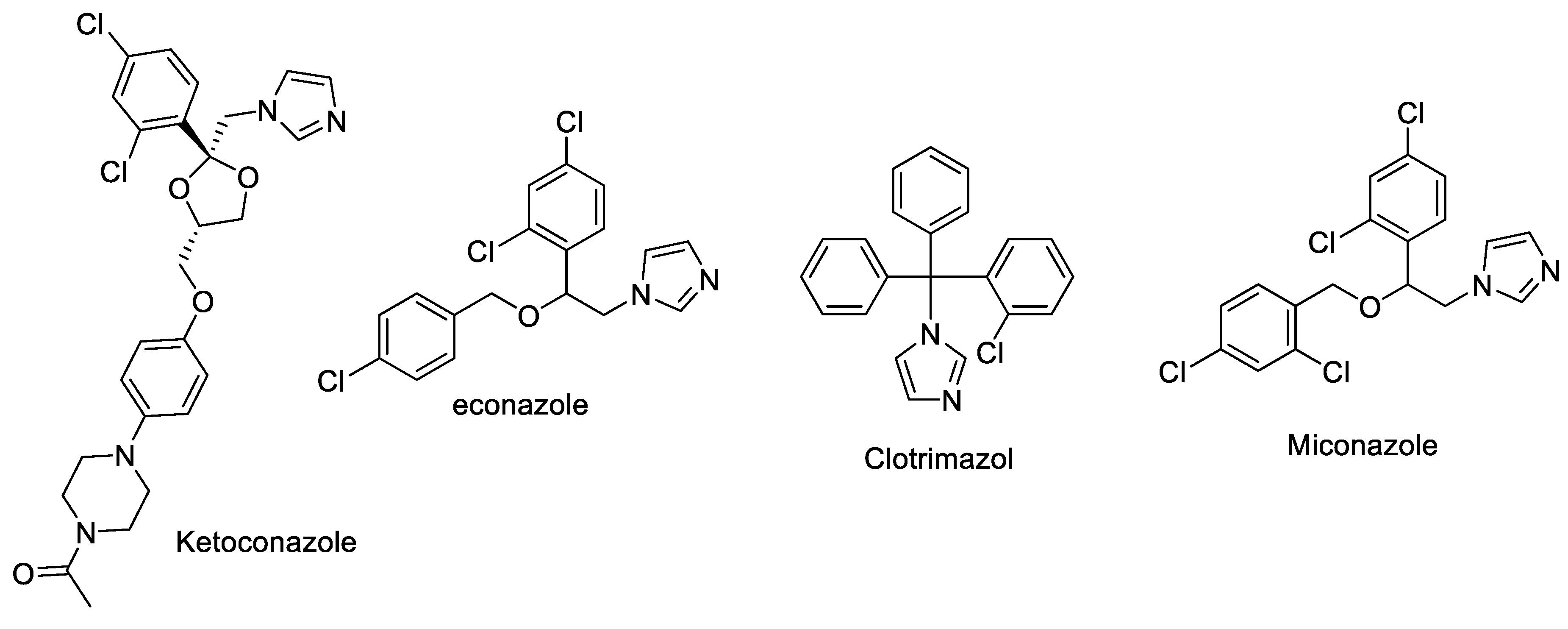
:1. Introduction
2. Results and Discussion
2.1. Synthesis
2.2. In Silico
2.2.1. ADMET Predictions
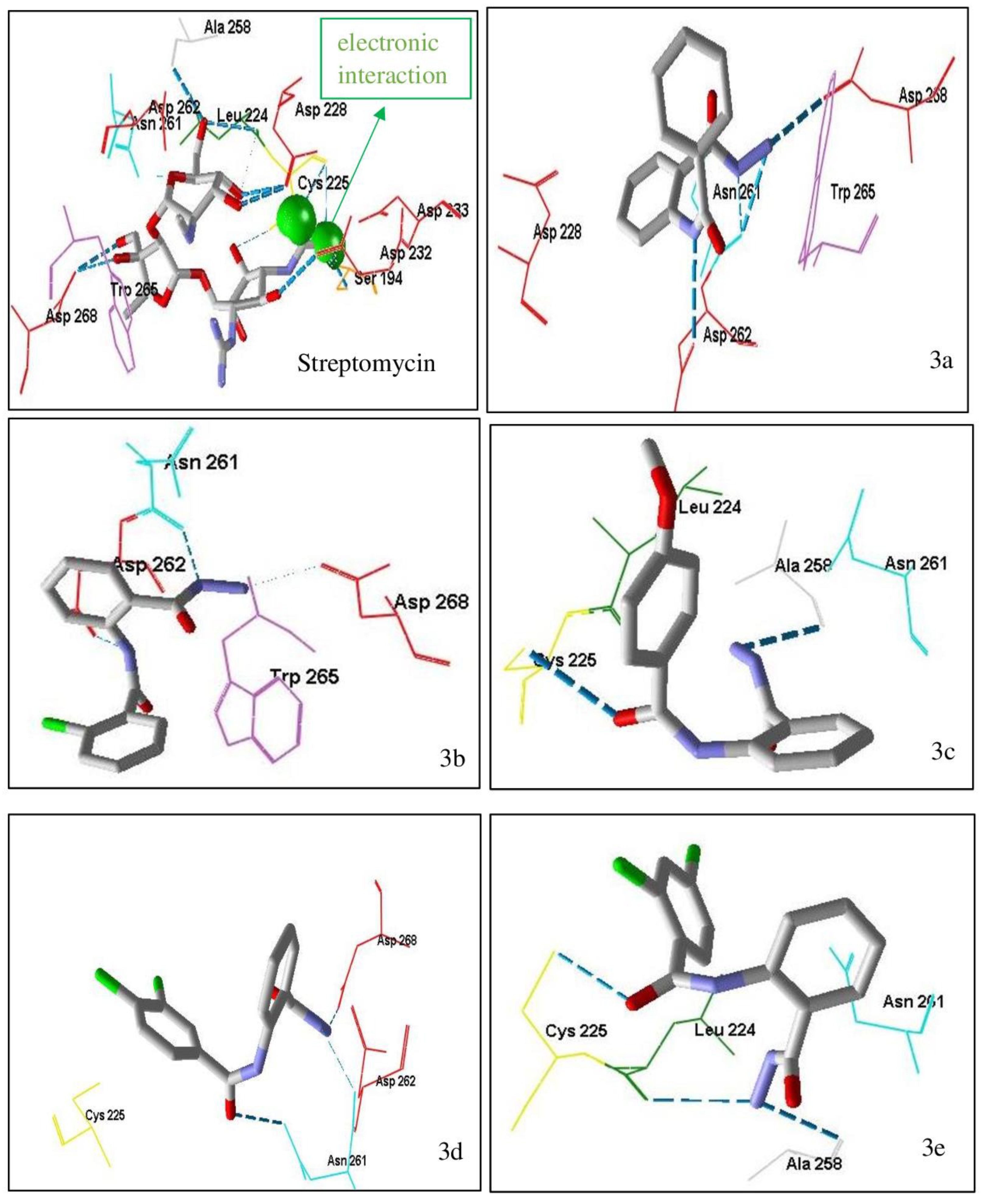
2.2.2. Molecular Docking Studies
Antibacterial Docking Studies
Antifungal Docking Studies
2.2.3. Antibacterial and Antifungal Activities
3. Materials and Methods
3.1. Synthesis
3.1.1. N-(2-(hydrazinecarbonyl)phenyl)benzamide (3a)
3.1.2. 2-chloro-N-(2-(hydrazinecarbonyl)phenyl)benzamide (3b)
3.1.3. N-(2-(hydrazinecarbonyl)phenyl)-4-methoxybenzamide (3c)
3.1.4. 3,4-dichloro-N-(2-(hydrazinecarbonyl)phenyl)benzamide (3d)
3.1.5. 2,4-dichloro-N-(2-(hydrazinecarbonyl)phenyl)benzamide (3e)
3.2. Synthesis
3.2.1. ADMET Prediction Studies
3.2.2. Molecular Docking Studies
3.3. In Vitro Assay
3.3.1. Antibacterial Activity Studies
3.3.2. Antifungal Activity Studies
4. Conclusions
Author Contributions
Funding
Institutional Review Board Statement
Informed Consent Statement
Data Availability Statement
Acknowledgments
Conflicts of Interest
References
- WHO. 2022. Available online: https://www.emro.who.int/health-topics/infectious-diseases/index.html (accessed on 1 November 2022).
- Conteh, L.; Kingori, P. Per diems in Africa: A counter-argument. Trop. Med. Int. Health 2010, 15, 1553–1555. [Google Scholar] [CrossRef] [PubMed]
- Hotez, P.J.; Molyneux, D.H.; Fenwick, A.; Kumaresan, J.; Sachs, S.E.; Sachs, J.D.; Savioli, L. Control of neglected tropical diseases. N. Engl. J. Med. 2007, 357, 1018–1027. [Google Scholar] [CrossRef] [PubMed]
- National Basic Health Research, Ministry of Health of Indonesia. Available online: https://www.litbang.kemkes.go.id/laporan-riset-kesehatan-dasar-riskesdas/ (accessed on 17 November 2022).
- Farrell, L.J.; Lo, R.; Wanford, J.J.; Jenkins, A.; Maxwell, A.; Piddock, L.J.V. Revitalizing the drug pipeline: AntibioticDB, an open-access database to aid antibacterial research and development. J. Antimicrob. Chemother. 2018, 73, 2284–2297. [Google Scholar] [CrossRef] [PubMed]
- Bala, S.; Sharma, N.; Kajal, A.; Kamboj, S. Design, synthesis, characterization, and computational studies on benzamide substituted Mannich bases as novel, potential antibacterial agents. Sci. World J. 2014, 2014, 732141. [Google Scholar] [CrossRef]
- Misral, H.; Sapari, S.; Rahman, T.; Ibrahim, N.; Yamin, B.M.; Hasbullah, S.A. Evaluation of Novel N-(Dibenzylcarbamothioyl)benzamide Derivatives as Antibacterial Agents by Using DFT and Drug-Likeness Assessment. J. Chem. 2018, 2018, 9176280. [Google Scholar] [CrossRef]
- El-Sayed, N.N.E.; Alafeefy, A.M.; Bakht, M.A.; Masand, V.H.; Aldalbahi, A.; Chen, N.; Fan, C.; Bacha, A.B. Synthesis, Antiphospholipase A2, Antiprotease, Antibacterial Evaluation and Molecular Docking Analysis of Certain Novel Hydrazones. Molecules 2016, 21, 1664. [Google Scholar] [CrossRef]
- Young, P.G.; Walanj, R.; Lakshmi, V.; Byrnes, L.J.; Metcalf, P.; Baker, E.N.; Vakulenko, S.B.; Smith, C.A. The Crystal Structures of Substrate and Nucleotide Complexes of Enterococcus faecium Aminoglycoside-2-Phosphotransferase-IIa [APH (2) -IIa] Provide Insights into Substrate Selectivity in the APH (2) Subfamily. J. Bacteriol. 2009, 191, 4133–4143. [Google Scholar] [CrossRef]
- M100-S30; Performance Standards for Antimicrobial Susceptibility Testing. 30th ed. Clinical and Laboratory Standards Institute (CLSI): Wayne, PA, USA, 2021; pp. 68–226.
- Wishart, D.S.; Feunang, Y.D.; Guo, A.C.; Lo, E.J.; Marcu, A.; Grant, J.R.; Sajed, T.; Johnson, D.; Li, C.; Sayeeda, Z.; et al. DrugBank 5.0: A major update to the DrugBank database for 2018. Nucleic Acids Res. 2018, 46, D1074–D1082. [Google Scholar] [CrossRef]
- Borelli, C.; Ruge, E.; Lee, J.H.; Schaller, M.; Vogelsang, A.; Monod, M.; Korting, H.C.; Huber, R.; Maskos, K. X-ray structures of Sap1 and Sap5: Structural comparison of the secreted aspartic proteinases from Candida albicans. Proteins 2008, 72, 1308–1319. [Google Scholar] [CrossRef]
- Naglik, J.R.; Challacombe, S.J.; Hube, B. Candida albicans secreted aspartyl proteinases in virulence and pathogenesis. Microbiol. Mol. Biol. Rev. 2003, 67, 400–428. [Google Scholar] [CrossRef] [Green Version]
- Schaller, M.; Borelli, C.; Korting, H.C.; Hube, B. Hydrolytic enzymes as virulence factors of Candida albicans. Mycoses 2005, 48, 365–377. [Google Scholar] [CrossRef] [PubMed]
- Silva, D.R.; Sardi, J.C.O.; Freires, I.A.; Silva, A.C.B.; Rosalen, P.L. In silico approaches for screening molecular targets in Candida albicans: A proteomic insight into drug discovery and development. Eur. J. Pharmacol. 2019, 842, 64–69. [Google Scholar] [CrossRef] [PubMed]
- CLSI Standard M60; Performance Standards for Antifungal Susceptibility Testing of Yeasts. 2nd ed. Clinical and Laboratory Standards Institute (CLSI): Wayne, PA, USA, 2020; pp. 1–30.
- Makarova, O.; Johnston, P.; Walther, B.; Rolff, J.; Roeslera, U. Complete Genome Sequence of the Disinfectant Susceptibility Testing Reference Strain Staphylococcus aureus subsp. aureus ATCC 6538. Genome Announc. 2017, 5, e00293-17. [Google Scholar] [CrossRef]
- Treangen, T.J.; Maybank, R.A.; Enke, S.; Friss, M.B.; Diviak, L.F.; Karaolis, D.K.R.; Koren, S.; Ondov, B.; Phillippy, A.M.; Bergman, N.H.; et al. Complete genome sequence of the quality control strain Staphylococcus aureus subsp. aureus ATCC 25923. Genome Announc. 2014, 2, e01110-14. [Google Scholar] [CrossRef] [PubMed]
- Putra, G.S.; Widiyana, A.P.; Muchlashi, L.A.; Sulistyowaty, M.I.; Ekowati, J.; Budiati, T. The Influence of the ratio of pyridine and triethylamine catalysts on the synthesis of 2-phenyl-benzo [D] [1, 3] oxazine-4-on derivatives. J. Chem. Pharm. 2017, 9, 73–80. [Google Scholar]
- Sulistyowaty, M.I.; Widyowati, R.; Putra, G.S.; Budiati, T.; Matsunami, K. Synthesis, ADMET predictions, molecular docking studies, and in-vitroanticancer activity of some benzoxazines against A549 human lung cancer cells. J. Basic Clin. Physiol. Pharmacol. 2021, 32, 385–392. [Google Scholar] [CrossRef]
- Widiyana, A.P.; Putra, G.S.; Sulistyowaty, M.I.; Hardjono, S.; Budiati, T. Synthesis, optimization method of 2, 3-disubstitutedquinazolin-4 (3H) -one derivatives. J. Chem. Pharm. 2017, 9, 24–28. [Google Scholar]
- Sulistyowaty, M.I.; Putra, G.S.; Zhichao, W.; Budiati, T. N-(2-(2-Benzylidenehydrazinecarbonyl) phenyl) benzamides: Synthesis, Cytotoxic, Antioxidant Activity and Molecular Docking Studies. J. Hunan Univ. Nat. Sci. 2020, 47, 199–207. [Google Scholar]
- Pires, D.E.V.; Blundell, T.L.; Ascher, D.B. pkCSM: Predicting small-molecule pharmacokinetic properties using graph-based signatures. J. Med. Chem. 2015, 58, 4066–4072. [Google Scholar] [CrossRef]
- Trevor, A.J.; Katzung, B.G.; Kruidering-Hall, M. Katzung & Trevor’s Pharmacology Examination & Review Board, 11th ed.; The McGraw-Hill Companies: New York, NY, USA, 2015; ISBN 978-0-07-182639-6. [Google Scholar]
- Shargel, L.; Yu, A.B.C. Applied Biopharmaceutics & Pharmacokinetics, 7th ed.; The McGraw-Hill Companies: New York, NY, USA, 2016; pp. 149–171. ISBN 978-0-07-182964-9. [Google Scholar]
- Halgren, T.A. Merck Molecular Force Field. V. extension of MMFF94 using experimental data, additional computational data, and empirical rules. J. Comput. Chem. 1996, 17, 616–641. [Google Scholar] [CrossRef]
- Thomsen, R.; Christensen, M.H. MolDock: A New Technique for High-Accuracy Molecular Docking. J. Med. Chem. 2006, 49, 3315–3321. [Google Scholar] [CrossRef] [PubMed]
- Pratiwi, S.T. Pharmaceutical Microbiology; Erlangga: Jakarta, Indonesia, 2008; pp. 188–189. ISBN 9789790334557. [Google Scholar]


| Compounds | Absorption | Distribution | Metabolism | Excretion | Toxicity | |||||
|---|---|---|---|---|---|---|---|---|---|---|
| Caco2 Perm. log Papp in 10−6 cm/s | Skin Perm. log Kp | VDss (log L/kg) | BBB Perm. (BB log) | CYP2D6 Inhibitor | CYP3A4 Inhibitor | Total Clearance log mL/min/kg | Oral Rat Acute Toxicity mol/kg (LD50) | Hepato toxicity | Skin Sensiti-zation | |
| 3a | 0.699 | 0.699 | −0.545 | 0.93 | No. | No. | 0.185 | 2.242 | No. | No. |
| 3b | 0.782 | −2.743 | 0.571 | −1.094 | No. | No. | −0.019 | 2.243 | No. | No. |
| 3c | 0.362 | −2.748 | −0.417 | −1.146 | No. | No. | 0.161 | 2.195 | No. | No. |
| 3d | 0.922 | −2.744 | 0.49 | −1.284 | No. | No. | 0.064 | 2.255 | No. | No. |
| 3e | 0.876 | −2.744 | −0.583 | −1.279 | No. | No. | 0.058 | 2.247 | No. | No. |
| Streptomycin | −0.585 | −0.585 | −1.031 | −2.872 | No. | No. | –0.022 | 2.444 | No. | No. |
| Micafungin | 0.906 | −2.735 | −0.528 | −4.305 | No. | No. | 1.18 | 2.482 | Yes | No. |
| Compounds | Moldock Score (kcal/mol) | Hydrogen Bond Interaction | Steric Interaction | Electronic Interaction |
|---|---|---|---|---|
 Native ligand (streptomycin) | −167.02 | Try 272 Asn 191 Ala 258 Ser 230 Asp 228 Cys 255 Asn 261 Asp 262 | Trp 265 Asn 191 Asn 221 Leu 224 Asp 262 Asp 228 Cys 225 | Asp 192 Asp 262 Asp 228 |
 3a | −92.08 | Asn 261 Asp 268 Asp 262 | Asp 228 Trp 265 | - |
 3b | −94.59 | Asp 262 Asn 261 | Asn 261 Asp 268 Trp 265 | - |
 3c | −98.19 | Ala 258 | Asn 261 Leu 224 Cts 225 | |
 3d | −99.91 | Asn 261 Asp 268 | Asp 262 Cys 225 | - |
 3e | −98.63 | Cys 255 Leu 224 Ala 258 | Cys 255 Asn 261 | - |
| Compounds | Moldock Score (kcal/mol) | Hydrogen Bond Interaction | Steric Interaction | Electronic Interaction |
|---|---|---|---|---|
 Native ligand (pepstatin) | −110.14 | Lys 83 Gly 85 Gly 220 Trp 221 | Gly 34 Tyr 84 Gly 220 Asp 218 Asp 86 Ile 12 | - |
 Micafungin | −193.31 | Ser 301 Tyr 225 Asp 86 Gly 220 Asp 32 Ser 301 | Gly 85 Arg 299 Gly 87 Lys 83 Asp 32 Gly 34 Ser 35 Ile 30 Ile 123 Ser 301 Asp 86 Trp 51 | - |
 3a | −102.5 | Asp 86 Gly 220 Ser 88 | Asp 218 Tyr 84 Asp 32 Ile 30 | - |
 3b | −106.35 | Asp 86 Gly 220 The 222 Ile 12 | Asp 32 Asp 86 | - |
 3c | −105.88 | Gly 85 Arg 120 Asp 218 | Ser 88 Gly 220 Gy 34 | |
 3d | −109.14 | Ser 88 Asp 86 Arg 120 | Ile 123 Gly 220 | - |
 3e | −106.46 | Ser 88 Asp 86 Arg 120 | Ile 123 Tyr 84 Arg 120 | - |
| Compounds | Gram-Positive Antibacterial (mm) | Gram-Negative Antibacterial (mm) | Antifungal (mm) |
|---|---|---|---|
| Staphylococcus aureus (ATCC 6538) | Escherichia coli (ATCC 8739) | Candica albicans (ATCC 10231) | |
| 3a (25 µg) | 16.12 ± 0.01 | 16.14 ± 0.02 | 17.18 ± 0.01 |
| 3b (25 µg) | 17.12 ± 0.01 | 16.15 ± 0.01 | 18.19 ± 0.01 |
| 3c (25 µg) | 16.13 ± 0.01 | 16.12 ± 0.01 | 17.17 ± 0.01 |
| 3d (25 µg) | 16.14 ± 0.01 | 16.13 ± 0.01 | 18.17 ± 0.01 |
| 3e (25 µg) | 16.13 ± 0.01 | 15.19 ± 0.01 | 18.18 ± 0.02 |
| Streptomycin (10 µg) | 29.14 | 29.17 | - |
| Micafungin (25 µg) | - | - | 23.18 |
Disclaimer/Publisher’s Note: The statements, opinions and data contained in all publications are solely those of the individual author(s) and contributor(s) and not of MDPI and/or the editor(s). MDPI and/or the editor(s) disclaim responsibility for any injury to people or property resulting from any ideas, methods, instructions or products referred to in the content. |
© 2023 by the authors. Licensee MDPI, Basel, Switzerland. This article is an open access article distributed under the terms and conditions of the Creative Commons Attribution (CC BY) license (https://creativecommons.org/licenses/by/4.0/).
Share and Cite
Sulistyowaty, M.I.; Putra, G.S.; Budiati, T.; Indrianingsih, A.W.; Anwari, F.; Kesuma, D.; Matsunami, K.; Yamauchi, T. Synthesis, In Silico Study, Antibacterial and Antifungal Activities of N-phenylbenzamides. Int. J. Mol. Sci. 2023, 24, 2745. https://doi.org/10.3390/ijms24032745
Sulistyowaty MI, Putra GS, Budiati T, Indrianingsih AW, Anwari F, Kesuma D, Matsunami K, Yamauchi T. Synthesis, In Silico Study, Antibacterial and Antifungal Activities of N-phenylbenzamides. International Journal of Molecular Sciences. 2023; 24(3):2745. https://doi.org/10.3390/ijms24032745
Chicago/Turabian StyleSulistyowaty, Melanny Ika, Galih Satrio Putra, Tutuk Budiati, Anastasia Wheni Indrianingsih, Farida Anwari, Dini Kesuma, Katsuyoshi Matsunami, and Takayasu Yamauchi. 2023. "Synthesis, In Silico Study, Antibacterial and Antifungal Activities of N-phenylbenzamides" International Journal of Molecular Sciences 24, no. 3: 2745. https://doi.org/10.3390/ijms24032745