Article

Concurrent Infection of the Human Brain with Multiple Borrelia Species

1
Biology Centre Czech Academy of Sciences, Institute of Parasitology, 37005 Ceske Budejovice, Czech Republic
2
Biology Centre Czech Academy of Sciences, Institute of Entomology, 37005 Ceske Budejovice, Czech Republic
3
Faculty of Sciences, University of South Bohemia, 37005 Ceske Budejovice, Czech Republic
4
Institute of Forensic Medicine and Medical Law, University Hospital Olomouc, 77900 Olomouc, Czech Republic
5
Department of Immunology, University Hospital Olomouc, 77900 Olomouc, Czech Republic
6
Department of Immunology, Faculty of Medicine and Dentistry, Palacky University Olomouc, 77900 Olomouc, Czech Republic
*
Authors to whom correspondence should be addressed.
Int. J. Mol. Sci. 2023, 24(23), 16906; https://doi.org/10.3390/ijms242316906
Submission received: 10 October 2023 / Revised: 22 November 2023 / Accepted: 26 November 2023 / Published: 29 November 2023
(This article belongs to the Section Molecular Pathology, Diagnostics, and Therapeutics)

Abstract

:
Lyme disease (LD) spirochetes are well known to be able to disseminate into the tissues of infected hosts, including humans. The diverse strategies used by spirochetes to avoid the host immune system and persist in the host include active immune suppression, induction of immune tolerance, phase and antigenic variation, intracellular seclusion, changing of morphological and physiological state in varying environments, formation of biofilms and persistent forms, and, importantly, incursion into immune-privileged sites such as the brain. Invasion of immune-privileged sites allows the spirochetes to not only escape from the host immune system but can also reduce the efficacy of antibiotic therapy. Here we present a case of the detection of spirochetal DNA in multiple loci in a LD patient’s post-mortem brain. The presence of co-infection with Borrelia burgdorferi sensu stricto and Borrelia garinii in this LD patient’s brain was confirmed by PCR. Even though both spirochete species were simultaneously present in human brain tissue, the brain regions where the two species were detected were different and non-overlapping. The presence of atypical spirochete morphology was noted by immunohistochemistry of the brain samples. Atypical morphology was also found in the tissues of experimentally infected mice, which were used as a control.

1. Introduction

Lyme disease (LD) or Lyme borreliosis (LB) is a multi-system disorder with a diverse spectrum of clinical manifestations. It is caused by the spirochetes of the Borrelia burgdorferi sensu lato (s.l.) complex. The initial stage of infection is characterized by flu-like symptoms (malaise, fatigue, headache, arthralgias, myalgias, fever, and regional lymphadenopathy) and/or skin rash due to Borrelia afzelii (acrodermatitis chronica atrophicans; ACA) and/or various manifestations of erythema migrans (EM) developing within a few weeks after the tick bites. If the spirochete is not eliminated, it will further disseminate to secondary sites of infection, leading to persistent inflammatory pathology, particularly affecting the central nervous system (CNS), peripheral nervous system (PNS), joints, or heart [1]. Symptoms of the secondary stage of infection vary and may disappear after days to months or continue as the disease transitions to the late stage of persistent disease with various signs and symptoms, including fatigue, headaches, sleep disturbance, neuropsychiatric and cognitive impairments, and arthralgias, myalgias, and neuropathy [2,3,4]. Even though antibiotic treatments for the majority of infected patients result in a full recovery, some patients suffer from long-lasting neurological and psychological manifestations. Antibiotic therapy prescribed during late-stage disease can result in unpredictable responses with ongoing symptomatology [5,6,7,8].
Those patients who received standard antibiotic treatment with problems persisting more than six months after antibiotic treatment are sometimes referred to as Post-Treatment Lyme Disease Syndrome (PTLDS) [9]. The etiology of this syndrome is debated, but several hypotheses have been proposed [10]. Persistence of Borrelia or associated co-infections, immune dysregulation leading to inflammation or autoimmunity, and/or disrupted central neural pathways [11] leading to central sensitization, among others, have been postulated. If PTLDS is due to or is independent of microbial persistence or other causes remains a topic of debate [12,13]. However, the ability of LD spirochetes to colonize multiple host tissues has been confirmed [14]. Dermis is the first tissue that spirochetes colonize after the tick bite. At this point, the host immune system can control the pathogen burden in the tissue [15]. Colonization of distant tissues involves spirochete dissemination from the dermis, which is mediated by differential regulation of virulence determinants of B. burgdorferi s.l. [16] that support the migration of pathogens through the endothelial and blood-brain barriers [17]. Once established in immune-privileged sites (fibroblasts, endothelial cells, macrophages, or neuronal glial cells) [18,19,20,21], the pathogen is capable of triggering a local inflammatory response but is safe from being cleared by the host immune system or antibiotics, which cannot easily penetrate the blood-brain barrier [22]. Survival of spirochetes despite antibiotic treatments, leading to the establishment of chronic LD, has been clearly demonstrated in animal studies [5,23,24,25,26].
Multiple species from the B. burgdorferi s.l. complex are responsible for LD, particularly in Europe [27]. The main burden of human cases, approximately 60%, is linked to infection with B. afzelii, which leads to the most common manifestation of skin lesions [28]. Three types of skin manifestations of LD are recognized: at stage 1, it is an erythema migrans with a characteristic “bull’s eye rash,” which, if untreated, can be followed by early disseminated infection, borrelial lymphocytoma or multiple EM (stage 2) along with neurologic and cardiac abnormalities, and late infection, especially arthritis in North America or acrodermatitis chronica atrophicans (ACA) (stage 3) in Europe [29].
The second, most represented European Borrelia genospecies, Borrelia garinii, causes primarily Lyme neuroborreliosis (LNB), affecting both the central and peripheral nervous systems [30]; up to 15% of LD patients suffer from LNB [22,30]. Intracellular localization of LD spirochetes in neurons and glial cells has been confirmed both in vitro and in vivo [12,31,32]. Evidence of Borrelia persistence in the brains of chronic LNB patients is limited but has been confirmed [33,34]. Further, the development of dementia, cortical atrophy, or amyloid deposition in some cases has been confirmed as well [12,31]. Invading neurons and glial cells, LD spirochetes can trigger progressive cell death or cause cell dysfunction [35], and the development of dementia, cortical atrophy, or amyloid deposition in some cases has been confirmed as well [12,31]. Borrelia burgdorferi sensu stricto (s.s.) is the major cause of LD in North America; however, its impact on European LD is under-appreciated [36,37]. The most frequent manifestation of B. burgdorferi s.s.-induced neuroborreliosis in the United States is lymphocytic meningitis, whereas European B. garinii-induced LNB, in the majority of cases, is diagnosed as Bannwarth syndrome, with the classic triad of meningitis, cranial neuritis, and painful radiculoneuritis [38], an uncommon manifestation of LNB in the USA [39]. Such differences in clinical manifestations of LNB might be based on different mechanisms of dissemination of the bacterial pathogen into the nervous system, different capabilities of individual species to cross the blood-brain barrier (BBB) by either transcellular or paracellular penetration [40], or hypervariable OspC genotypes [41]. Until recently, no reliable system for the detection of persistent infections existed. A single study on non-human primates showed that persistent forms of spirochetes that survived antibiotic treatment remain metabolically active [42]. A xenodiagnostic study using the laboratory-reared larvae of Ixodes scapularis [43] showed that in a group of 10 patients who had high levels of anti-Borrelia antibodies after antibiotic treatment, one patient with PLTDS and a patient with EM had positive results, confirming the presence of viable Borrelia; two others from the same study group had “indeterminate results.” Further, up to 14.5% of long-term Lyme patients were proven to be PCR positive after seemingly adequate antibiotic (ATB) therapy in a study by Horowitz and colleagues [10]. Immunohistochemistry (IHC) combined with confocal microscopy found significant pathological changes, including borrelial spirochetal clusters, in the autopsy tissues of all the organs of a LD patient who had received extensive antibiotic treatments during her 16-year-long course of the disease [34]. Additional evidence of persistent Borrelia infection in patients with ongoing LD symptoms despite extensive ATB therapy has come from the cultivation of Borrelia in the blood of seven subjects, from the genital secretions of ten subjects, and from a skin lesion in one subject out of 12 selected LD patients [44].
Here we report the case of a patient who, after being infected by Borrelia and treated with antibiotics continuously, progressed toward neurologic/psychiatric symptoms over the subsequent 13 years. After this period, the patient underwent repeated serological testing with borderline positivity for Borrelia infection, followed by the prescription of several antibiotics, which provided no clinical improvements, followed by hospitalization at psychiatric clinics. Several months after being released from the clinic, the patient committed suicide, providing written consent for analyzing his brain for the presence of Borrelia.

2. Results

2.1. Patient History and Borrelia Serology

A male patient born in 1996 contacted a physician in 2004 after the appearance of erythema migrans. He was diagnosed with LD and was treated with antibiotics (type and duration of treatment are unavailable). The patient progressed to suffer from neurological symptoms, mostly cognitive deficits such as “brain fog”, reduced psychomotor performance, and difficulties with concentration and processing of visual and auditory stimuli. In 2017, the patient was examined in the neurology department and had a positive Borrelia serology test, with both IgG and IgM anti-Borrelia antibodies in serum, but a negative PCR from a lumbar puncture. In December 2017, the patient was admitted to the Psychiatric Department of the University Hospital Olomouc. His therapy started with Zyprexa (Olanzapin, Eli Lilly, Indianapolis, IN, USA) and was later replaced with Brintellix (Vortioxetin, H.Lundbeck A/S, Copenhagen, Denmark). On February 2018, anti-Borrelia antibodies were determined again in a private medical facility with borderline positivity for Borrelia-specific IgM and strong “++” positivity for IgG by ELISA. The ELISA results were confirmed by immunoblotting; the IgM immunoblot was positive for the presence of OspC from B. afzelii, B. garinii, and B. burgdorferi s.s., and the IgG immunoblot was borderline positive for VlsE from B. afzelii, B. garinii, and B. burgdorferi s.s., and negative for p83, flagellin, BmpA, OspA, OspB, OspC, and DbpA. Serological tests for Chlamydia sp., Mycoplasma sp., Anaplasma phagocytophillum, Toxocara canis, and Toxoplasma gondii were negative. Serological testing for Bartonella infection by indirect immunofluorescence assay was negative in the IgM and IgG classes for both Bartonella henselae and Bartonella quintana. A PCR test for the presence of Babesia spp. (Babesia microti and Babesia divergence) in peripheral blood was negative. At the same private medical facility, the patient was prescribed a combination of antimicrobials: 3 × 100 mg/day of minocycline (Ratiopharm, Ulm, Germany), 250 mg (3×/week) of azithromycin (Sandoz, Basel, Switzerland), and 200 mg (1×/day) of hydroxychloroquine (Plaquenil) (Sanofi, Paris, France). In August 2018, the suspected hypocorticism (adrenal insufficiency) was ruled out by biochemical laboratory examinations. In September 2018, the patient was hospitalized at the Department of Psychiatry, University Hospital Olomouc, with the suspicion that he was developing a mental disorder. The patient was diagnosed with schizotypal personality disorder (STPD) and somatoform disorder and was discharged three weeks later. In August 2019, the patient committed suicide. The patient left behind a letter expressing the urgent demand for scientists to analyze his brain for the presence of LD spirochetes. The letter was provided to the Ethical committee of University Hospital Olomouc and initiated this study. The patient’s body was autopsied in the Institute of Forensic Medicine and Medical Law, University Hospital Olomouc, 2 days after his death.

2.2. Post-Mortem Toxicology and Microbiology

Toxicological post-mortem analysis confirmed the presence of the following chemical agents in the blood: hydroxychloroquine, hydroxyzine (the active substance in the anxiolytic Atarax), and its metabolite cetirizine. In addition, a low concentration of azithromycin was detected in cerebrospinal fluid (CSF). Immunoblot analyses of post-mortem serum and CSF conducted at the Faculty of Medicine and Dentistry, Palacky University Olomouc, confirmed borderline IgG reactivity against VlsE of B. garinii and B. burgdorferi s.s. The reactions with VlsE antigens of B. afzelii, lipids of B. garinii and B. afzelii, p83, p41, p39, OspC, p58, p21, p20, p19, and p18 were negative. Using immunoblot kits for autoantibody detection, weak IgG positivity against PL-7 antigen (threonyl-tRNA synthetase) and borderline reactions with SRP antigen and histones were detected. As a part of the microbiological analysis of post-mortem samples, the CSF and brain tissues were used for the cultivation of spirochetes in the BSK-H medium. After 2 months of incubation of seeded cultures at 34 °C, the presence of live bacteria was not detected in any culture, and all samples were deemed to be culture-negative.

2.3. PCR Detection of Borrelia DNA in Frozen Brain Tissue Samples

To determine if Borrelia DNA could be detected in the brain, PCR analysis was performed on DNA from seven different parts of the brain, samples of which had been frozen at −80 °C upon collection. Nine genes (flaB, ospC, clpA, clpX, nifS, rplB, pepX, pyrG, and uvrA) provided the amplicons of the expected size (Supplementary Table S1), although not all amplifications were successful for all tested brain loci (Table 1).
Sequence analysis, followed by comparison with available databases, confirmed the presence of DNA in two spirochete species, B. burgdorferi s.s. and B. garinii. The two Borrelia genospecies were found in brain samples and were found to be mutually exclusive: the DNA of a single species, B. burgdorferi s.s., was detected in the temporal right lobe, choroid plexus (left), occipital lobe (left), frontal lobe (left), and parietal lobe (right), while the DNA of B. garinii was identified in the basal ganglia (right) and cerebellum (right). No PCR amplification showed the presence of more than one spirochete species in any sample.
BLASTn analysis of the ospC sequences confirmed that the B. burgdorferi s.s. strain carried an ospC of type A, the globally most common ospC type. The B. garinii ospC sequences detected in the brain samples were identical to strains widely distributed in Eurasia.

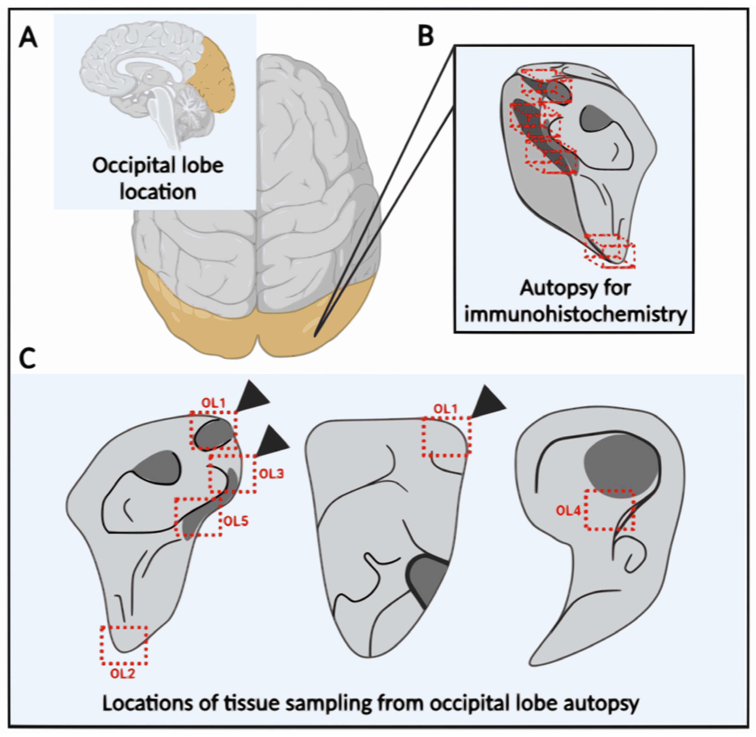
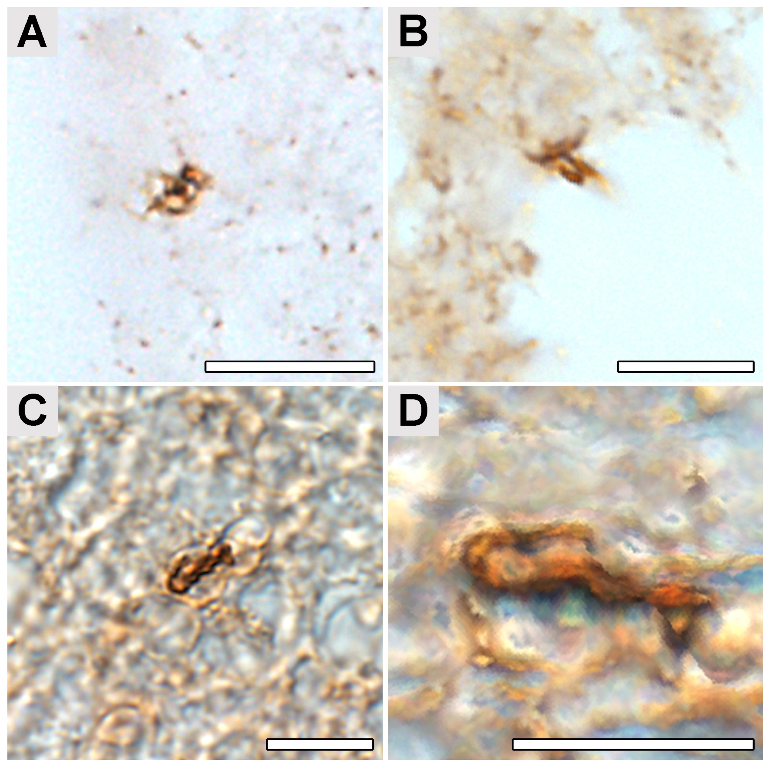
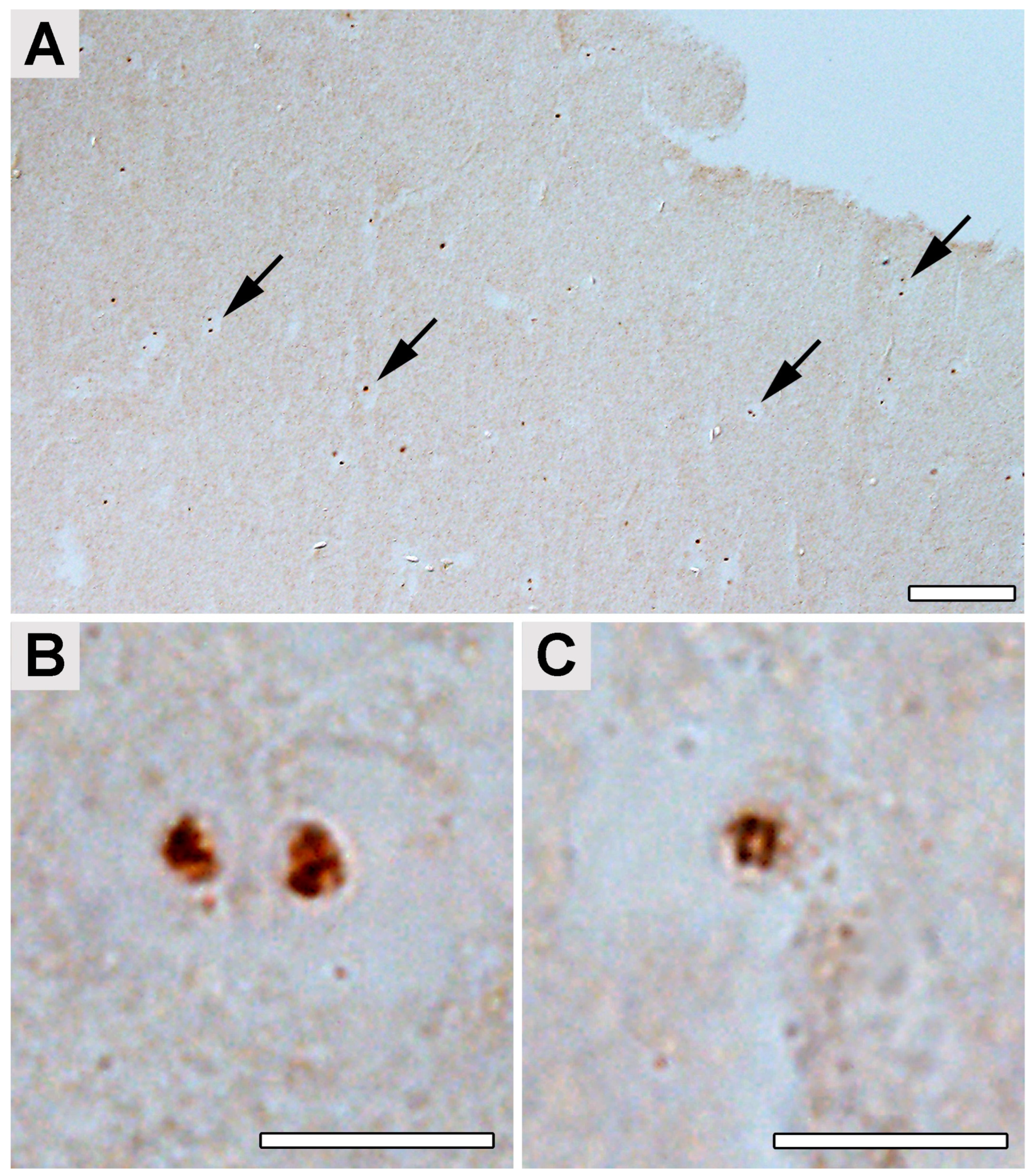
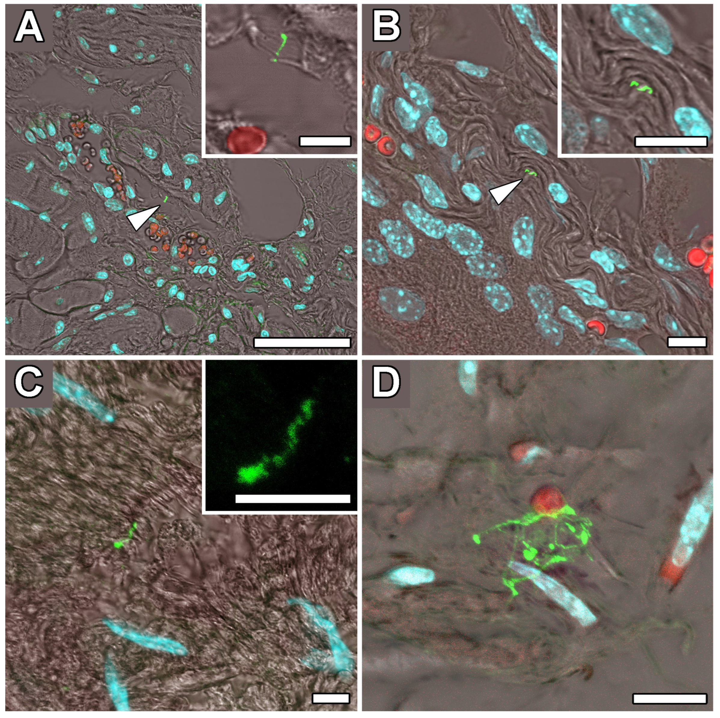
2.4. Occipital Lobe Tissue Exhibited Structures Consistent with Borrelia

Immunohistochemical (IHC) investigation of paraffin sections of the patient occipital lobe samples (Figure 1) revealed the presence of structures reactive with Borrelia-specific rabbit polyclonal antibodies (Figure 2 and Figure 3). Polyclonal, rather than monoclonal, antibodies were selected as they react with a broader spectrum of Borrelia antigens, and postmortem degradation might limit the number of borrelial antigens.
The size and morphology of these structures resembled the Borrelia cells with the atypical morphology detected previously as well from cultured spirochetes treated with doxycycline or amoxicillin, used as an in vitro test of antibody specificity (see Supplementary Material and Methods and Supplementary Figure S1). In Borrelia populations treated by both low (50 μg/mL) and high (100 μg/mL) concentrations of either doxycycline or amoxicillin, these antibodies stained both spiral and atypical B. burgdorferi forms (Supplementary Figure S1A,C,E,G,I).
The presence of multiple forms of Borrelia in the culture was verified using transmission electron microscopy (TEM) (Supplementary Figure S1B,D,F,H,J). The structures detected in the samples of the patient’s occipital lobe had a diameter of about 1–10 μm, and presumed protoplasmic cylinders ranged between 0.2 and 0.4 µm in diameter. These structures were detected with an estimated frequency of 0.16–0.3 per 1 mm3 of tissue typically located near the capillaries (Figure 2A,B). Borrelia cyst-like structures were also detected in the paraplast section of a sample from the patient’s occipital lobe (Figure 3). Anti-B.burgdorferi polyclonal antibodies visualized by the horseradish peroxidase-conjugated secondary antibodies were used for the detection. Our findings on human brain tissues were in accordance with the observation of tissue samples from mice artificially infected with B. burgdorferi s.s. and subsequently treated with antibiotics that we used as spiked controls (Figure 4). The structures corresponding to the atypical forms of Borrelia in the mouse tissues were mainly found in samples from bladders (Figure 4A,B) and knee joints (Figure 4C,D).

3. Discussion

Detection of Borrelia in multiple organs of infected animals, including humans, demonstrates the ability of these spirochetes to disseminate into the secondary sites of infection (to review, see [12] (citations 17–49). Detection of intact spirochetes at the sites of secondary infection in both laboratory animals and humans even after aggressive antibiotic treatments further demonstrates the ability of the spirochetes to persist ([22]; to review, see [12] (citations 50–85)). The Centers for Disease Control and Prevention (CDC, USA) indicate that up to 20% of patients continue to suffer from neurological manifestations such as persistent fatigue and joint or muscle pain [45] after seemingly successful antibiotic treatment; however, recent large-scale testing revealed that the real number of antibiotic-treated patients that develop symptoms of chronic disease is between 36 and 63% [46]. The role of biofilm and stationary persister forms of Borrelia in persistent LD is not clear; however, such morphologies are an example of bacterial adaptation to a changing environment. As complex structures highly resistant to environmental and therapeutic pressure, biofilms are recognized as an essential part of the mechanism of the establishment of chronic infection [12,47,48,49,50,51,52,53,54,55]. The ability of spirochetes to survive antibiotic treatment to instigate reoccurrence of the disease is strong evidence that chronic LD could arise from persisting and re-occurring infection, either caused by persisting forms hidden in biofilms, cell-wall deficient forms, round bodies, or other alternative spirochete morphologies [12]. These previous findings are consistent with our finding of spirochetes in a human brain despite extended antibiotic treatment. The detection of intact spirochetes in autopsy brain specimens of humans after extended treatments and those with a diverse history of disease manifestations, including widely recognized neurocognitive disorders, anxiety, depression, memory loss, brain atrophy, and progressive dementia [31,56,57,58,59,60], illustrates that persistent Borrelia infection can lead to a persistent disorder of the central nervous system (CNS) and the development of LNB [22,61,62]. It is thought that LNB does not reoccur frequently without reinfection. Nevertheless, cases of reoccurrence of LNB might suggest the formation of biofilm-protective structures and may explain the low rate of spirochete detection in blood in patients with persistent infection [63]. The neurotropic nature of LD spirochetes helps ensure their survival in the CNS [64] for an extended period of time in alternative morphological forms [65,66,67], including antibiotic-resistant biofilms [34]. In general, dissemination of spirochetes occurs through the bloodstream, where the spirochetes remain for a short period of time before moving to the extracellular matrix of various internal organs, where they can remain shielded from the host’s immune system and antibiotics [16,68,69,70,71,72,73]. However, LD spirochetes were found to also be present in cardiac myocytes, endothelial cells, and synovial cells [19,74,75]. Dissemination of spirochetes into the CNS is thought to occur via passage along the peripheral nerves [35]. Based on the distinct clinical manifestations of LNB in Europe and North America, ascribed to the different Borrelia species, B. garinii and B. burgdorferi s.s., respectively, the mechanisms of spirochete dissemination into the CNS might be species-dependent and rely on the ability of Borrelia to cross the BBB [39,40,76]. Some Borrelia genotypes may be able to disseminate through the host more effectively than the others or have an advantage competing with less efficient types due to the ospC genotypes they possess [41,77]. An association between ospC genotypes and the severity of LD in patients has been reported in multiple studies [41,78,79,80]. To date, out of 35 recognized ospC genotypes, four, A, B, I, and K, are responsible for systemic LD in humans around the world [77,78,81], although genotypes C, D, N, F, H, E, G, and M have been found in the secondary sites of infection [81,82,83,84]. While some Borrelia ospC genotypes cause the typical erythema migrans and the others disseminate through the bloodstream or CNS, only certain strains may exhibit neurotropism [41].
Our results confirmed the presence of two species of spirochetes from the B. burgdorferi sensu lato complex, B. garinii and B. burgdorferi s.s., in different areas of the human brain. Importantly, the DNAs of the two spirochete species were detected in distinct areas of the brain; in no case did we find infection with both in the same brain region. Multiple repeated PCR amplifications with different sets of primers, followed by sequence confirmation of Borrelia spp., always provided clear and definite chromatograms indicating the presence of DNA from only one species. Based on the analysis of a single brain tissue infected with spirochetes, it is not yet possible to conclude whether this finding represents biological restriction in brain colonization, the outcome of separate and rare colonization of the brain, or is just a coincidence. The specific mechanisms by which Borrelia crosses the blood-brain barrier are not yet fully understood, and an animal model allowing examination of the full pathogenic process has not yet been established, although a recent model using inbred mice has shown colonization of the dura mater during acute and late spirochete infection [85,86]. In this model, a decrease in the inflammatory and immune pathways was observed in dura mater over time [85]. It seems that the post-treatment stage of LNB is not triggered by either immune or inflammatory response pathways, which requires new mechanistic insight to identify ways of clinical intervention [41]. The development of LNB depends on the ability of spirochetes to cross the BBB. This invasion can occur via breaching either physical (tight junctions) or metabolic (enzymes, transport systems) barriers [87]. The role of the BBB in neurodegenerative and neuropsychiatric disorders is crucial; its failure plays an important role in the pathogenesis of many diseases of the CNS that are caused by bacteria or protozoa [88,89,90]. Recent studies show that, in the case of some neurocognitive disorders that lead to the development of dementia, as well as during normal aging, the “leakage” of the BBB is increased [91,92]. Whether Borrelia enter the brain by direct transmigration or are carried across the BBB hidden inside non-phagocyting leukocytes using the “Trojan horse” mechanism as in the case of other pathogens is not yet known [89,90]. It is tempting to speculate that some kind of competition might occur between the spirochete species in the process of crossing the BBB, and this might be the basis of our observations of species-segregated Borrelia loci in the brain. A recent study by Adams and colleagues [40] showed that different species of LD spirochetes are able to enter BBB-organoids with different success. The spirochetes that successfully invaded the organoids remained viable inside the BBB-organoids, initiating the loss of tight junctions and changes in the organoids’ gross morphology and integrity [40].
Unambiguous attribution of persistent neurological symptoms to LD and Borrelia persistence is limited by several factors, including the presence of comorbid disease with overlapping symptoms or reinfection with Borrelia. Horowitz has described up to 16 different overlapping factors that can account for persistent symptoms [10,54,93,94]. The multiple results of studies of relapsing fever (RF) in humans and animals confirmed that RF spirochetes cause neurological abnormalities as often as LD spirochetes, infecting the brain and other nervous tissues and persisting in the brain after treatment with antibiotics that do not readily penetrate the blood-brain barrier. Among the wide spectrum of neurological manifestations of tick-borne relapsing fever (i.e., meningismus, facial palsy, encephalitis, myelitis, radiculitis, and deafness), the neuropsychiatric abnormalities in patients manifested by the acute onset of mental confusion or excitement, persistent hallucinations, signs of mania, extreme irritability, flight of ideas, persistent fatigue, or neurasthenia [95]. Possibly, the so-called “residual brain infection” is a common phenomenon for all spirochetal diseases. In experimental animals, spirochete infection persisted in the brain for up to 3 years [96,97,98,99,100]. Among the nervous system tissues in which the spirochetes were detected were the cerebral hemispheres, cerebellum, spinal cord, medulla, and cortex. Interestingly, during the residual brain infection, the animals were protected against re-infection with the same strain [101,102]; spirochetes that cause residual brain infection were susceptible to the serum of the animal from which they were recovered earlier [102,103]. It is obvious that the immune response cannot effectively reach the spirochetes in the brain. The brain remains infectious, while the blood of the animal is not. Inoculation of the brain suspension in susceptible experimental animals results in the development of spirochetemia [104]. The strong neurotropism of spirochetes is widely recognized. Multiple pieces of evidence have confirmed the fact that spirochetes can invade the brain and cause persistent infection [58,105,106]. They are able to escape destruction by the host immune system and initiate a chronic inflammatory process. Evidence of infection of the brain in diseases with clear spirochete etiology, such as Lyme disease, relapsing fever, syphilis, and leptospirosis, for example, shows the strong association between the spirochetes and diseases. In addition, spirochetal etiology in other neurological diseases, such as Alzheimer’s disease (AD), has been postulated [31]. B. burgdorferi-specific antigens were detected in the brain of an AD patient with concurrent LNB. These findings were supported by positive PCR detection of B. burgdorferi DNA in the brain [107,108]. B. burgdorferi was also cultivated from the brain in AD patients by MacDonald and Miranda [109], MacDonald [110], and Miklossy [31].
Since the recognition of Lyme disease, thousands of studies have been published, yet optimal therapy is still a matter of debate [111]. The complexity of the pathophysiology of the Borrelia pathogen and the clinical uncertainty surrounding the signs and symptoms of Lyme and other tick-borne diseases make the choice of treatment a complex one that relies on the clinical judgment of the treating physician. Recognized persistent bacterial infections may require prolonged antibiotic therapy and seem reasonable and justifiable in some situations when considering patients with persistent LD symptoms [111]. However, recent scientific studies on the biofilm and persister forms of Borrelia and associated co-infections, along with early clinical trials, provide hope that shorter-term protocols with anti-persister drug regimens might lead to more effective treatments for those suffering from neuropsychiatric and other symptoms of chronic Lyme disease or post-treatment Lyme disease symptoms (PTLDS) [52,53,54,55,94].
To our knowledge, this is the first study that confirms the presence of B. garinii and B. burgdorferi s.s. structures in the human brain with strict separation of invaded brain loci. The question of what defines the distribution of spirochete species in brain tissues remains unanswered but is one of increasing importance as the number of human Borrelia infections increases. The possible pathological effect of persistent forms of Borrelia detected in the present study is also discussed from the point of view of the potential induction of an immunopathological reaction associated with chronic inflammation and/or autoreactive immune response, where non-replicative structures can be recognized as long-term antigenic stimuli [13].

4. Materials and Methods

4.1. Ethical Statement

Samples that were analyzed in this study originate from a young adult male who committed suicide in August 2019. The processing of post-mortem samples was performed based on informed consent provided by him before suicide in the form of a letter, which was accepted by the Ethical Committee of University Hospital Olomouc, 102/18 (NV19-05-00191).

4.2. CSF and Blood Collection and Analysis

The autopsy was performed at the Institute of Forensic Medicine and Medical Law University Hospital Olomouc and Faculty of Medicine and Dentistry, Palacky University Olomouc, Czech Republic, two days after the suicide. Peripheral blood and cerebrospinal fluid (CSF) samples were collected in a 2 mL volume. The CSF was markedly stained by the presence of blood. Part of the CSF was aseptically removed for the cultivation of spirochetes (Supplementary Materials and Methods). The remaining volume of CSF was centrifuged (2.000× g, 10 min, 4 °C) to remove cells, and the supernatant was collected and stored at −80 °C. The blood was centrifuged (2.000× g, 10 min, 4 °C), clarified serum was collected, transferred to a new tube, and stored at −80 °C. Serological tests were performed by standard protocols using the Anti-Borrelia EUROLINE-RN-AT, EUROLINE Autoimmune Inflammatory Myopathies 16 Ag (IgG), and EUROLINE ANA Profile 3 plus DSF70 (IgG) (EUROIMMUN, Lübeck, Germany) blot diagnostic kit with evaluation by a flatbed scanner and the software EUROLineScan Software 3.4 (EUROIMMUN, Lübeck, Germany).

4.3. Brain Tissue Collection (Post-Mortem)

Brain tissue samples of about 9 cm3 from seven different parts of the brain were collected post-mortem by certified pathologists at the Institute of Forensic Medicine and Medical Law, University Hospital Olomouc. Brain samples were collected from: 1—temporal lobe (right); 2—choroid plexus (left); 3—occipital lobe (left); 4—frontal lobe (left); 5—parietal lobe (right); 6—basal ganglia (right); and 7—cerebellum (right). All tissue samples were divided into three parts. One part was used for the cultivation of potentially live spirochetes. This sample was immediately aseptically transferred to BSK-H medium supplemented with 6% rabbit serum and antibiotics. The second part was used for PCR analyses. This portion was frozen at −80 °C until use. The third part was used for immunohistochemical analyses. This part was fixed in 10% buffered formalin (4% paraformaldehyde) and stored at 4 °C.
Based on PCR results, 5 samples of 125 mm3 in size were subsequently taken from the fixed autopsied occipital lobe sample for immunohistochemical staining (the sample position within the occipital lobe autopsy is shown in Figure 1).

4.4. Analysis of Total DNA from Human Brain Tissues: Polymerase Chain Reactions (PCR)

The DNA purification steps (Supplementary Material and Methods), PCR, and post-amplification analyses were all performed in separate areas with all precautions against contamination. The presence of Borrelia burgdorferi s. l. DNA in the samples was assessed by PCR amplification of partial genes encoding outer surface protein C (OspC) and flagellin, followed by amplification of eight housekeeping genes according to the previously described MLST protocol [112]. To reduce the inhibition of the reaction from the excess of human DNA in the template DNA, the PCR reactions were conducted as nested PCRs under conditions previously used with human samples [113,114]. To assure the absence of possible contamination during the DNA purification or cross-reactivity with the human brain tissue, negative extraction controls were included. The partial ospC and flagellin genes were amplified by nested PCR using the previously described primers (Supplementary Table S1), and the conditions of the reaction were as follows: 30 cycles of denaturation at 95 °C for 30 sec, annealing at 50 °C and 52 °C for 30 s for the external and internal rounds of PCR, respectively, and extension at 72 °C for 30 s [115,116]. Two-step amplification of eight housekeeping genes included: clpA (BB0369), clpX (BB0612), pyrG (BB0575), uvrA (BB0837), pepX (BB0627), recG (BB0581), rplB (BB0481, seminested PCR), and nifS (BB0084, seminested PCR) (Supplementary Table S1). The PCR conditions for the housekeeping genes, except for recG, were as follows: initial denaturation at 95 °C for 15 min, cyclic denaturation at 94 °C for 30 s, annealing temperature from 55 °C to 48 °C (touchdown PCR, decreasing 1 °C each cycle) for 30 s, and the extension step at 72 °C for 60 s. An additional 20 cycles were run using denaturation at 94 °C for 30 s, annealing at 48 °C for 30 s, and extension at 72 °C for 60 s. After a final extension step at 72 °C for 5 min, the samples were kept at 14 °C until the second (nested) PCR. The conditions for the second PCR were as follows: 95 °C for 7 min, followed by 35 cycles of [denaturation at 94 °C for 30 s, annealing at 50 °C for 30 s, extension at 72 °C for 60 s]. After a final extension step for 5 min at 72 °C, the samples were kept at 14 °C.
For recG, the PCR conditions for the first set of cycles consisted of initial denaturation at 95 °C for 15 min, followed by 30 cycles of denaturation at 94 °C for 30 s, annealing at 55 °C for 30 s, extension at 72 °C for 60 s, and final extension at 72 °C for 5 min. The conditions for the second set of cycles were identical for all the primers used.
The PCR reactions were carried out in a final volume of 20 μL using 2× HotStarTaq Plus Master Mix (Qiagen, Hilden, Germany). Amplicons were visualized by electrophoresis in a 1.5% agarose gel (1 × TAE, pH 8.0). In all cases, a reaction mix with water instead of a DNA template was used as the negative control. Borrelia carolinensis DNA was used as a positive control in all PCR reactions.

4.5. Immunohistochemical Detection of Borrelia in the Paraplast Section of a Human Brain Autopsy and Infected Mouse Tissues

Mice were infected with B. burgdorferi s.s. administered simultaneously by intradermal and intraperitoneal routes (Supplementary Material and Methods). The fixed, autopsied human occipital lobe samples were washed several times in phosphate-buffered saline (PBS). Organs dissected from euthanized mice were submerged in a fixative of saturated picric acid, 4% formaldehyde, and 2.3% copper acetate supplemented with mercuric chloride (Bouin-Hollande solution) [117] overnight at 4 °C. The fixative was then thoroughly washed with 70% ethanol. Standard techniques were used for both human autopsies and mouse tissue samples, including dehydration, embedding in paraplast, sectioning to 10 μm, deparaffinization, and rehydration. The sections were treated with Lugol’s iodine, followed by a 7.5% solution of sodium thiosulphate to remove residuals of heavy metal ions, and then washed in distilled water and PBS supplemented with 0.3% Tween 20 (PBS-Tw). The nonspecific binding sites were blocked with 5% normal goat serum in PBS-Tw (blocking solution) for 30 min at room temperature (RT). Incubation with rabbit polyclonal B. burgdorferi antibodies specific to a pool of B. burgdorferi s.l. complex proteins (cat.# PA1-73004, Invitrogen, Carlsbad, CA, USA) diluted at 1:200 in the blocking solution was carried out in a humidified chamber overnight at 4 °C, followed by washing the samples by thorough rinsing with PBS-Tw (three times for 10 min at RT) [118,119]. For enzymatic staining, the sections were further incubated in the cross-adsorbed horseradish peroxidase-labeled goat anti-rabbit secondary antibodies (Invitrogen, Carlsbad, USA) diluted at 1:500 in the blocking solution for 90 min in RT, washed in PBS-Tw (three times for 10 min at RT), in 0.05 M Tris-HCl pH 7.5 (for 10 min at RT), and stained in 10% 3,3’ diaminobenzidine in 0.05 M Tris-HCl pH 7.5 with 0.005% H2O2 for 10 min at RT. The reaction was stopped by rinsing in distilled water, dehydrating, and mounting in DPX mounting medium (Fluka, Buchs, Switzerland). The samples were examined under a BX51 microscope equipped with a DP80 CCD camera and cellSens software (Ver. 5174) (Olympus, Tokyo, Japan), and the images were reconstructed by stitching several Z stack series. The further 3D image analyses were performed in the FIJI ImageJ software (ImageJ 1.54f; http://imagej.org) [120] using the Iterative Deconvolution 3D plugin.
For fluorescence staining, the samples treated with primary antibody and washed in PBS-Tw were incubated with goat anti-rabbit IgG conjugated Alexa Fluor 488 (Life Technologies, Carlsbad, CA, USA) diluted 1:500 in the blocking solution for 90 min in RT, followed by rinsing with PBS-Tw (3 times for 10 min at RT in the dark). The samples were dehydrated and mounted in DPX mounting medium (Fluka, Buchs, Switzerland). The fluorescence signal was examined under the laser scanning confocal microscope FLUOVIEW FV3000 (Olympus, Tokyo, Japan) using the IMARIS 10.1 software (Oxford Instrument, Abingdon, UK) for 3D reconstruction of the Z-stack series. Examining the autofluorescence of human brain autopsy samples, enzymatic detection of bound primary antibodies was preferentially used for the detection of Borrelia in these samples.
The specificity of the primary antibodies to recognize both spiral and atypical forms of B. burgdorferi was verified by the application of antibodies to the paraplast sections of antibiotic-treated B. burgdorferi cultures mounted in agar (Supplementary Figure S1).

Supplementary Materials

The following supporting information can be downloaded at: https://www.mdpi.com/article/10.3390/ijms242316906/s1. Reference [121] is cited in the supplementary materials.

Author Contributions

Conceptualization, N.R., M.V., H.S., M.K. and M.R.; methodology, M.G., J.O., M.V., H.S., M.K., M.R. and N.R.; validation, M.G., N.R., M.V., H.S., M.K. and M.R.; formal analysis, M.G., J.O., V.K., M.D. and K.S.; samples collection, V.K. and M.D.; resources, M.K., M.R. and N.R.; data curation, M.G., N.R., M.V. and H.S.; writing—original draft preparation, N.R., M.G., M.V., H.S., M.K. and M.R.; visualization, J.O., H.S. and M.V.; supervision, N.R.; project administration, N.R.; funding acquisition, M.K., M.R. and N.R. All authors have read and agreed to the published version of the manuscript.

Funding

This research was funded by grant NV19-05-00191 from the Ministry of Health of the Czech Republic. Partial analysis of the samples was supported by project RVO 60077344 of the Biology Center CAS, Institute of Entomology.

Institutional Review Board Statement

This study was conducted according to the guidelines of the Declaration of Helsinki and approved by the Institutional Ethics Committee of University Hospital Olomouc (reference number 102/18 June 2018). The protocol of this study, including the informed consent of the patients, was approved by the Ethics Committee of the Olomouc University Hospital (reference number 102/18 June 2018).

Informed Consent Statement

Samples that were analyzed in this study originate from a young adult male who committed suicide in August 2019. The processing of post-mortem samples was performed based on informed consent provided by him before suicide in the form of a letter, which was accepted by the Ethical Committee of University Hospital Olomouc, 102/18 (NV19-05-00191).

Data Availability Statement

All data obtained during our study and supporting reported results can be found in this manuscript or Supplementary Materials file.

Acknowledgments

We acknowledge the Biology Center CAS core facility Laboratory of Electron Microscopy supported by MEYS CR (LM2023050 Czech-BioImaging and OP VVV CZ.02.1.01/0.0/0.0/18_046/0016045). We acknowledge the support of M.K., M.R. and K.S by grant IGA_LF_2023_10 from Palacky University Olomouc. The authors are indebted to Vett Lloyd and Richard Horowitz for their helpful discussion, valuable advice, and English editing of this manuscript.

Conflicts of Interest

The authors declare no conflict of interest.

References

  1. Steere, A. Lyme disease. N. Engl. J. Med. 1989, 321, 586–596. [Google Scholar] [CrossRef]
  2. Bransfield, R.C. Neuropsychiatric Lyme borreliosis: An overview with a focus on a specialty psychiatrist’s clinical practice. Healthcare 2018, 6, 104. [Google Scholar] [CrossRef] [PubMed]
  3. Bransfield, R.C.; Aidlen, D.M.; Cook, M.J.; Javia, S. A clinical diagnostic system for late-stage neuropsychiatric Lyme borreliosis based upon an analysis of 100 patients. Healthcare 2020, 8, 13. [Google Scholar] [CrossRef]
  4. Fallon, B.A.; Madsen, T.; Erlangsen, A.; Benros, M.E. Lyme borreliosis and associations with mental disorders and suicidal behavior: A nationwide danish cohort study. Am. J. Psychiatry 2021, 178, 921–931. [Google Scholar] [CrossRef] [PubMed]
  5. Barthold, S.W.; Hodzic, E.; Imai, D.M.; Feng, S.; Yang, X.; Luft, B.J. Ineffectiveness of tigecycline against persistent Borrelia burgdorferi. Antimicrob. Agents Chemother. 2010, 54, 643–651. [Google Scholar] [CrossRef]
  6. Preac-Mursic, V.; Wilske, B.; Gross, B.; Weber, K.; Pfister, H.W.; Baumann, A.; Prokop, J. Survival of Borrelia burgdorferi in antibiotically treated patients with Lyme borreliosis. Infection 1989, 17, 355–359. [Google Scholar] [CrossRef] [PubMed]
  7. Shadick, N.A.; Phillips, C.B.; Logigian, E.L.; Steere, A.C.; Kaplan, R.F.; Berardi, V.P.; Duray, P.H.; Larson, M.G.; Wright, E.A.; Ginsburg, K.S.; et al. The long-term clinical outcomes of Lyme disease: A population-based retrospective cohort study. Ann. Intern. Med. 1994, 121, 560–567. [Google Scholar] [CrossRef] [PubMed]
  8. Fauvart, M.; De Groote, V.N.; Michiels, J. Role of persister cells in chronic infections: Clinical relevance and perspectives on anti-persister therapies. J. Med. Microbiol. 2011, 60 Pt 6, 699–709. [Google Scholar] [CrossRef]
  9. Nemeth, J.; Bernasconi, E.; Heininger, U.; Abbas, M.; Nadal, D.; Strahm, C.; Erb, S.; Zimmerli, S.; Furrer, H.; Delaloye, J.; et al. Update of the Swiss guidelines on post-treatment Lyme disease syndrome. Swiss Med. Wkly. 2016, 146, w14353. [Google Scholar] [CrossRef]
  10. Horowitz, R.I.; Freeman, P.R. Precision medicine: The role of the MSIDS model in defining, diagnosing, and treating chronic lyme disease/post treatment Lyme disease syndrome and other chronic illness: Part 2. Healthcare 2018, 6, 129. [Google Scholar] [CrossRef]
  11. Hastey, C.J.; Elsner, R.A.; Barthold, S.W.; Baumgarth, N. Delays and diversions mark the development of B cell responses to Borrelia burgdorferi Infection. J. Immunol. 2012, 188, 5612–5622. [Google Scholar] [CrossRef]
  12. Rudenko, N.; Golovchenko, M.; Kybicova, K.; Vancova, M. Metamorphoses of Lyme disease spirochetes: Phenomenon of Borrelia persisters. Parasites Vectors 2019, 12, 237. [Google Scholar] [CrossRef]
  13. Sloupenska, K.; Koubkova, B.; Horak, P.; Hutyrova, B.; Racansky, M.; Mares, J.; Miklusova, M.; Schovanek, J.; Zapletalova, J.; Raska, M.; et al. Myositis autoantibodies in patients with suspected post-treatment Lyme disease syndrome. Life 2023, 13, 527. [Google Scholar] [CrossRef] [PubMed]
  14. Straubinger, R.K.; Straubinger, A.F.; Jacobson, R.H.; Chang, Y.; Summers, B.A.; Erb, H.N.; Appel, M.J.G. Two lessons from the canine model of Lyme disease: Migration of Borrelia burgdorferi in tissues and persistence after antibiotic treatment. J. Spirochetal Tick Borne Dis. 1997, 4, 24–31. [Google Scholar]
  15. Petzke, M.; Schwartz, I. Borrelia burgdorferi pathogenesis and the immune response. Clin. Lab. Med. 2015, 35, 745–764. [Google Scholar] [CrossRef]
  16. Tsao, J.I. Reviewing molecular adaptations of Lyme borreliosis spirochetes in the context of reproductive fitness in natural transmission cycles. Vet. Res. 2009, 40, 36. [Google Scholar] [CrossRef] [PubMed]
  17. Hyde, J.A. Borrelia burgdorferi keeps moving and carries on: A review of borrelial dissemination and invasion. Front. Immunol. 2017, 8, 114. [Google Scholar] [CrossRef]
  18. Livengood, J.A.; Gilmore, R.D. Invasion of human neuronal and glial cells by an infectious strain of Borrelia burgdorferi. Microbes Infect. 2006, 8, 2832–2840. [Google Scholar] [CrossRef]
  19. Ma, Y.; Sturrock, A.; Weis, J.J. Intracellular localization of Borrelia burgdorferi within human endothelial cells. Infect. Immun. 1991, 59, 671–678. [Google Scholar] [CrossRef]
  20. Montgomery, R.R.; Malawista, E.S. Entry of Borrelia burgdorferi into macrophages is end-on and leads to degradation in lysosomes. Infect. Immun. 1996, 64, 2867–2872. [Google Scholar] [CrossRef]
  21. Klempner, M.S.; Noring, R.; Rogers, R.A. Invasion of human skin fibroblasts by the Lyme disease spirochete, borrelia burgdorferi. J. Infect. Dis. 1993, 167, 1074–1081. [Google Scholar] [CrossRef]
  22. Gadila, S.K.G.; Rosoklija, G.; Dwork, A.J.; Fallon, B.A.; Embers, M.E. Detecting borrelia spirochetes: A case study with validation among autopsy specimens. Front. Neurol. 2021, 12, 707. [Google Scholar] [CrossRef]
  23. Hodzic, E.; Feng, S.; Holden, K.; Freet, K.J.; Barthold, S.W. Persistence of Borrelia burgdorferi following antibiotic treatment in mice. Antimicrob. Agents Chemother. 2008, 52, 1728–1736. [Google Scholar] [CrossRef] [PubMed]
  24. Hodzic, E.; Imai, D.; Feng, S.; Barthold, S.W. Resurgence of persisting non-cultivable Borrelia burgdorferi following antibiotic treatment in mice. PLoS ONE 2014, 9, e86907. [Google Scholar] [CrossRef]
  25. Straubinger, R.K.; Summers, A.B.; Chang, Y.F.; Appel, M.J. Persistence of Borrelia burgdorferi in experimentally infected dogs after antibiotic treatment. J. Clin. Microbiol. 1997, 35, 111–116. [Google Scholar] [CrossRef] [PubMed]
  26. Embers, M.E.; Barthold, S.W.; Borda, J.T.; Bowers, L.; Doyle, L.; Hodzic, E.; Jacobs, M.B.; Hasenkampf, N.R.; Martin, D.S.; Narasimhan, S.; et al. Persistence of Borrelia burgdorferi in rhesus macaques following antibiotic treatment of disseminated infection. PLoS ONE 2012, 7, e29914. [Google Scholar] [CrossRef]
  27. Rizzoli, A.; Hauffe, H.C.; Carpi, G.; Vourc’h, I.G.; Neteler, M.; Rosà, R. Lyme borreliosis in europe. Eurosurveillance 2011, 16, 19906. [Google Scholar] [CrossRef] [PubMed]
  28. Hubálek, Z. Epidemiology of Lyme Borreliosis. In Lyme Borreliosis, Current Problems in Dermatology Volume 37; Lipsker, D., Jaulhac, B., Eds.; Karger: Basel, Switzerland, 2009; pp. 31–50. [Google Scholar]
  29. Gade, A.; Matin, T.; Rubenstein, R.; Robinson, C.A. Acrodermatitis Chronica Atrophicans. In StatPearls; StatPearls Publishing: Treasure Island, FL, USA, 2023. [Google Scholar]
  30. Janson, C.G. Blocking Borrelia in the brain. Sci. Transl. Med. 2016, 8, 324ec18. [Google Scholar] [CrossRef]
  31. Miklossy, J.; Khalili, K.; Gern, L.; Ericson, R.L.; Darekar, P.; Bolle, L.; Hurlimann, J.; Paster, B.J. Borrelia burgdorferi persists in the brain in chronic lyme neuroborreliosis and may be associated with Alzheimer disease. J. Alzheimers Dis. 2004, 6, 639–649; discussion 673–681. [Google Scholar] [CrossRef]
  32. Miklossy, J.; Kis, A.; Radenovic, A.; Miller, L.; Forro, L.; Martins, R.; Reiss, K.; Darbinian, N.; Darekar, P.; Mihaly, L.; et al. Beta-amyloid deposition and Alzheimer’s type changes induced by Borrelia spirochetes. Neurobiol. Aging 2006, 27, 228–236. [Google Scholar] [CrossRef]
  33. Liegner, K.B.; Rosenkilde, C.; Campbell, G.; Quan, T.; Dennis, D. Culture confirmed treatment failure of cefotaxime and minocycline in a case of Lyme meningoencephalomyelitis in the United States. In Proceedings of the Program and Abstracts of the 5th International Conference on Lyme Borreliosis, Arlington, VA, USA, 30 May–2 June 1992. [Google Scholar]
  34. Sapi, E.; Kasliwala, R.S.; Ismail, H.; Torres, J.P.; Oldakowski, M.; Markland, S.; Gaur, G.; Melillo, A.; Eisendle, K.; Liegner, K.B.; et al. The long-term persistence of Borrelia burgdorferi antigens and DNA in the tissues of a patient with Lyme disease. Antibiotics 2019, 8, 183. [Google Scholar] [CrossRef] [PubMed]
  35. Miklossy, J.; Kasas, S.; Zurn, A.D.; McCall, S.; Yu, S.; McGeer, P.L. Persisting atypical and cystic forms of Borrelia burgdorferi and local inflammation in Lyme neuroborreliosis. J. Neuroinflamm. 2008, 5, 40. [Google Scholar] [CrossRef] [PubMed]
  36. Piesman, J.; Gern, L. Lyme borreliosis in Europe and North America. Parasitology 2004, 129, S191–S220. [Google Scholar] [CrossRef]
  37. Rudenko, N.; Golovchenko, M.; Grubhoffer, L.; Oliver, J.H., Jr. Updates on Borrelia burgdorferi sensu lato complex with respect to public health. Ticks Tick-Borne Dis. 2011, 2, 123–128. [Google Scholar] [CrossRef]
  38. Omotosho, Y.B.; Sherchan, R.; Ying, G.W.; Shayuk, M. A unique case of bannwarth syndrome in early disseminated Lyme disease. Cureus 2021, 13, e14680. [Google Scholar] [CrossRef]
  39. Garcia-Monco, J.C.; Benach, J.L. Lyme neuroborreliosis: Clinical outcomes, controversy, pathogenesis, and polymicrobial infections. Ann. Neurol. 2018, 85, 21–31. [Google Scholar] [CrossRef]
  40. Adams, Y.; Clausen, A.S.; Jensen, P.; Lager, M.; Wilhelmsson, P.; Henningson, A.J.; Lindgren, P.-E.; Faurholt-Jepsen, D.; Mens, H.; Kraiczy, P.; et al. 3D blood-brain barrier-organoids as a model for Lyme neuroborreliosis highlighting genospecies dependent organotropism. iScience 2022, 26, 105838. [Google Scholar] [CrossRef] [PubMed]
  41. Ford, L.; Tufts, D.M. Lyme neuroborreliosis: Mechanisms of B. burgdorferi infection of the nervous system. Brain Sci. 2021, 11, 789. [Google Scholar] [CrossRef] [PubMed]
  42. Embers, M.E.; Hasenkampf, N.R.; Jacobs, M.B.; Tardo, A.C.; Doyle-Meyers, L.A.; Philipp, M.T.; Hodzic, E. Variable manifestations, diverse seroreactivity and post-treatment persistence in non-human primates exposed to Borrelia burgdorferi by tick feeding. PLoS ONE 2017, 12, e0189071. [Google Scholar] [CrossRef]
  43. Marques, A.; Telford, S.R., 3rd; Turk, S.-P.; Chung, E.; Williams, C.; Dardick, K.; Krause, P.J.; Brandeburg, C.; Crowder, C.D.; Carolan, H.E.; et al. Xenodiagnosis to detect Borrelia burgdorferi infection: A first-in-human study. Clin. Infect. Dis. 2014, 58, 937–945. [Google Scholar] [CrossRef]
  44. Middelveen, M.J.; Sapi, E.; Burke, J.; Filush, K.R.; Franco, A.; Fesler, M.C.; Stricker, R.B. Persistent borrelia infection in patients with ongoing symptoms of Lyme disease. Healthcare 2018, 6, 33. [Google Scholar] [CrossRef]
  45. CDC Post-Treatment Lyme Disease Syndrome. 2017. Available online: https://www.cdc.gov/lyme/postLDS/index.html (accessed on 7 March 2018).
  46. Adrion, E.R.; Aucott, J.; Lemke, K.W.; Weiner, J.P. Health care costs, utilization and patterns of care following Lyme disease. PLoS ONE 2015, 10, e0116767. [Google Scholar] [CrossRef]
  47. Zhang, Y. Persisters, persistent infections and the Yin–Yang model. Emerg. Microbes Infect. 2014, 3, e3. [Google Scholar] [CrossRef]
  48. Feng, J.; Li, T.; Yee, R.; Yuan, Y.; Bai, C.; Cai, M.; Shi, W.; Embers, M.; Brayton, C.; Saeki, H.; et al. Stationary Phase Persister/Biofilm Microcolony of Borrelia burgdorferi Causes More Severe Disease in a Mouse Model of Lyme Arthritis: Implications for understanding persistence, Post-Treatment Lyme Disease Syndrome (PTLDS), and Treatment Failure. Published in Discovery Medicine on 28 March 2019. Available online: http://www.discoverymedicine.com/Jie-Feng/2019/03/persister-biofilm-microcolony-borrelia-burgdorferi-causes-severe-lyme-arthritis-in-mouse-model/ (accessed on 9 October 2023).
  49. Feng, J.; Auwaerter, P.G.; Zhang, Y. Drug combinations against Borrelia burgdorferi Persisters in vitro: Eradication achieved by using daptomycin, cefoperazone and doxycycline. PLoS ONE 2015, 10, e0117207. [Google Scholar] [CrossRef]
  50. Feng, J.; Shi, W.; Zhang, S.; Zhang, Y. Identification of new compounds with high activity against stationary phase Borrelia burgdorferi from the NCI compound collection. Emerg. Microbes Infect. 2015, 4, 1–15. [Google Scholar] [CrossRef]
  51. Lewis, K. Persister cells, dormancy and infectious disease. Nat. Rev. Microbiol. 2006, 5, 48–56. [Google Scholar] [CrossRef]
  52. Horowitz, R.I.; Freeman, P.R. The use of dapsone as a novel “persister” drug in the treatment of chronic Lyme disease/post treatment Lyme disease syndrome. J. Clin. Exp. Dermatol. 2016, 7, 345. [Google Scholar] [CrossRef]
  53. Horowitz, R.I.; Murali, K.; Gaur, G.; Freeman, P.R.; Sapi, E. Effect of dapsone alone and in combination with intracellular antibiotics against the biofilm form of B. burgdorferi. BMC Res. Notes 2020, 13, 455. [Google Scholar] [CrossRef] [PubMed]
  54. Horowitz, R.I.; Freeman, P.R. Efficacy of short-term high dose pulsed dapsone combination therapy in the treatment of chronic Lyme disease/post-treatment Lyme disease syndrome (PTLDS) and associated co-infections: A report of three cases and literature review. Antibiotics 2022, 11, 912. [Google Scholar] [CrossRef] [PubMed]
  55. Gao, J.; Gong, Z.; Montesano, D.; Glazer, E.; Liegner, K. “Repurposing” disulfiram in the treatment of Lyme disease and babesiosis: Retrospective review of first 3 years’ experience in one medical practice. Antibiotics 2020, 9, 868. [Google Scholar] [CrossRef] [PubMed]
  56. MacDonald, A.B. Borrelia in the brains of patients dying with dementia. JAMA 1986, 256, 2195–2196. [Google Scholar] [CrossRef]
  57. Fallon, A.B.; Nields, A.J. Lyme disease: A neuropsychiatric illness. Am. J. Psychiatry 1994, 151, 1571–1583. [Google Scholar] [CrossRef]
  58. Miklossy, J. Biology and neuropathology of dementia in syphilis and Lyme disease. Handb. Clin. Neurol. 2008, 89, 825–844. [Google Scholar]
  59. Blanc, F.; Philippi, N.; Cretin, B.; Kleitz, C.; Berly, L.; Jung, B.; Kremer, S.; Namer, I.J.; Sellal, F.; Jaulhac, B.; et al. Lyme neuroborreliosis and dementia. J. Alzheimers Dis. 2014, 41, 1087–1093. [Google Scholar] [CrossRef]
  60. Kristoferitsch, W.; Aboulenein-Djamshidian, F.; Jecel, J.; Rauschka, H.; Rainer, M.; Stanek, G.; Fischer, P. Secondary dementia due to Lyme neuroborreliosis. Wien. Klin. Wochenschr. 2018, 130, 468–478. [Google Scholar] [CrossRef]
  61. Garcia-Toro, M.; Aguirre, I. Biopsychosocial model in depression revisited. Med. Hypotheses 2007, 68, 683–691. [Google Scholar] [CrossRef] [PubMed]
  62. Senejani, A.G.; Maghsoudlou, J.; El-Zohiry, D.; Gaur, G.; Wawrzeniak, K.; Caravaglia, C.; Khatri, V.A.; MacDonald, A.; Sapi, E. Borrelia burgdorferi co-localizing with amyloid markers in Alzheimer’s disease brain tissues. J. Alzheimers Dis. 2022, 85, 889–903. [Google Scholar] [CrossRef] [PubMed]
  63. Di Domenico, E.G.; Cavallo, I.; Bordignon, V.; D’Agosto, G.; Pontone, M.; Trento, E.; Gallo, M.T.; Prignano, G.; Pimpinelli, F.; Toma, L.; et al. The emerging role of microbial biofilm in Lyme neuroborreliosis. Front. Neurol. 2018, 9, 1048. [Google Scholar] [CrossRef] [PubMed]
  64. Hansen, K.; Lebec, A.-M. The clinical and epidemiological profile of Lyme neuroborreliosis in Denmark 1985-1990: A prospective study of 187 patients with Borrelia burgdorferi specific intrathecal antibody production. Brain 1992, 115, 399–423. [Google Scholar] [CrossRef] [PubMed]
  65. MacDonald, A.B. Spirochetal cyst forms in neurodegenerative disorders,…hiding in plain sight. Med. Hypotheses 2006, 67, 819–832. [Google Scholar] [CrossRef]
  66. Gruntar, I.; Malovrh, T.; Murgia, R.; Cinco, M. Conversion of Borrelia garinii cystic forms to motile spirochetes in vivo note. APMIS 2001, 109, 383–388. [Google Scholar] [CrossRef] [PubMed]
  67. Murgia, R.; Cinco, M. Induction of cystic forms by different stress conditions in Borrelia burgdorferi. APMIS 2004, 112, 57–62. [Google Scholar] [CrossRef] [PubMed]
  68. Cabello, F.C.; Godfrey, H.P.; Newman, S.A. Hidden in plain sight: Borrelia burgdorferi and the extracellular matrix. Trends Microbiol. 2007, 15, 350–354. [Google Scholar] [CrossRef] [PubMed]
  69. Tilly, K.; Rosa, P.A.; Stewart, P.E. Biology of Infection with Borrelia burgdorferi. Infect. Dis. Clin. N. Am. 2008, 22, 217–234. [Google Scholar] [CrossRef]
  70. Liang, F.T.; Brown, E.L.; Wang, T.; Iozzo, R.V.; Fikrig, E. Protective niche for Borrelia burgdorferi to evade humoral immunity. Am. J. Pathol. 2004, 165, 977–985. [Google Scholar] [CrossRef]
  71. Nordstrand, A.; Barbour, A.G.; Bergström, S. Borrelia pathogenesis research in the post-genomic and post-vaccine era. Curr. Opin. Microbiol. 2000, 3, 86–92. [Google Scholar] [CrossRef]
  72. Berndtson, K. Review of evidence for immune evasion and persistent infection in Lyme disease. Int. J. Gen. Med. 2013, 6, 291–306. [Google Scholar] [CrossRef] [PubMed]
  73. Liang, F.T.; Yan, J.; Mbow, M.L.; Sviat, S.L.; Gilmore, R.D.; Mamula, M.; Fikrig, E. Borrelia burgdorferi changes its surface antigenic expression in response to host immune responses. Infect. Immun. 2004, 72, 5759–5767. [Google Scholar] [CrossRef]
  74. Pachner, A.; Basta, J.; Delaney, E.; Hulinska, D. Localisation of Borrelia burgdorferi in murine Lyme borreliosis by electron microscopy. Am. J. Trop. Med. Hyg. 1995, 52, 128–133. [Google Scholar] [CrossRef]
  75. Girschick, H.J.; Huppertz, H.I.; Krenn, V.; Karch, H.; Rüssman, H. Intracellular persistence of Borrelia burgdorferi in human synovial cells. Rheumatol. Int. 1996, 16, 125–132. [Google Scholar] [CrossRef]
  76. Rupprecht, T.A.; Koedel, U.; Fingerle, V.; Pfister, H.-W. The pathogenesis of Lyme neuroborreliosis: From infection to inflammation. Mol. Med. 2008, 14, 205–212. [Google Scholar] [CrossRef]
  77. Rudenko, N.; Golovchenko, M.; Hönig, V.; Mallátová, N.; Krbková, L.; Mikulášek, P.; Fedorova, N.; Belfiore, N.M.; Grubhoffer, L.; Lane, R.S.; et al. Detection of Borrelia burgdorferi sensu stricto ospC alleles associated with human Lyme borreliosis worldwide in non-human-biting tick Ixodes affinis and rodent hosts in Southeastern United States. Appl. Environ. Microbiol. 2013, 79, 1444–1453. [Google Scholar] [CrossRef]
  78. Qiu, W.-G.; Bruno, J.F.; McCaig, W.D.; Xu, Y.; Livey, I.; Schriefer, M.E.; Luft, B.J. Wide Distribution of a High-Virulence Borrelia burgdorferi clone in Europe and North America. Emerg. Infect. Dis. 2008, 14, 1097–1104. [Google Scholar] [CrossRef] [PubMed]
  79. Hanincovaá, K.; Liveris, D.; Sandigursky, S.; Wormser, G.P.; Schwartz, I. Borrelia burgdorferi sensu stricto is clonal in patients with early lyme borreliosis. Appl. Environ. Microbiol. 2008, 74, 5008–5014. [Google Scholar] [CrossRef] [PubMed]
  80. Strle, K.; Jones, K.L.; Drouin, E.E.; Li, X.; Steere, A.C. Borrelia burgdorferi RST1 (OspC type A) genotype is associated with greate rinflammation and more severe Lyme disease. Am. J. Pathol. 2011, 178, 2726–2739. [Google Scholar] [CrossRef] [PubMed]
  81. Seinost, G.; Dykhuizen, D.E.; Dattwyler, R.J.; Golde, W.T.; Dunn, J.J.; Wang, I.-N.; Wormser, G.P.; Schriefer, M.E.; Luft, B.J. Four clones of Borrelia burgdorferi sensu stricto cause invasive infection in humans. Infect. Immun. 1999, 67, 3518–3524. [Google Scholar] [CrossRef] [PubMed]
  82. Brisson, D.; Baxamusa, N.; Schwartz, I.; Wormser, G.P. Biodiversity of Borrelia burgdorferi strains in tissues of Lyme disease patients. PLoS ONE 2011, 6, e22926. [Google Scholar] [CrossRef]
  83. Dykhuizen, D.E.; Sandigursky, S.; Nadelman, R.B.; Nowakowski, J.; Brisson, D.; Wormser, G.P.; Schwartz, I. The propensity of different Borrelia burgdorferi sensu stricto genotypes to cause disseminated infections in humans. Am. J. Trop. Med. Hyg. 2008, 78, 806–810. [Google Scholar] [CrossRef]
  84. Ivanova, L.; Christova, I.; Neves, V.; Aroso, M.; Meirelles, L.; Brisson, D.; Gomes-Solecki, M. Comprehensive seroprofiling of sixteen B. burgdorferi OspC: Implications for Lyme disease diagnostics design. Clin. Immunol. 2009, 132, 393–400. [Google Scholar] [CrossRef]
  85. Casselli, T.; Divan, A.; Vomhof-DeKrey, E.E.; Tourand, Y.; Pecoraro, H.L.; Brissette, C.A. A murine model of Lyme disease demonstrates that Borrelia burgdorferi colonizes the dura mater and induces inflammation in the central nervous system. PLoS Pathog. 2021, 17, e1009256. [Google Scholar] [CrossRef]
  86. Divan, A.; Casselli, T.; Narayanan, S.A.; Mukherjee, S.; Zawieja, D.C.; Watt, J.A.; Brissette, C.A.; Newell-Rogers, M.K. Borrelia burgdorferi adhere to blood vessels in the dura mater and are associated with increased meningeal T cells during murine disseminated borreliosis. PLoS ONE 2018, 13, e0196893. [Google Scholar] [CrossRef] [PubMed]
  87. Persidsky, Y.; Ramirez, S.H.; Haorah, J.; Kanmogne, G.D. Blood–brain barrier: Structural components and function under physiologic and pathologic conditions. J. Neuroimmune Pharmacol. 2006, 1, 223–236. [Google Scholar] [CrossRef] [PubMed]
  88. Dupin, N.; Lecuyer, H.; Carlotti, A.; Poyart, C.; Coureuil, M.; Chanal, J.; Schmitt, A.; Vacher-Lavenu, M.-C.; Taha, M.-K.; Nassif, X.; et al. Chronic meningococcemia cutaneous lesions involve meningococcal perivascular invasion through the remodeling of endothelial barriers. Clin. Infect. Dis. 2012, 54, 1162–1165. [Google Scholar] [CrossRef]
  89. Adams, Y.; Olsen, R.W.; Bengtsson, A.; Dalgaard, N.; Zdioruk, M.; Satpathi, S.; Behera, P.K.; Sahu, P.K.; Lawler, S.E.; Qvortrup, K.; et al. Plasmodium falciparum erythrocyte membrane protein 1 variants induce cell swelling and disrupt the blood–brain barrier in cerebral malaria. J. Exp. Med. 2021, 218, e20201266. [Google Scholar] [CrossRef]
  90. Disson, O.; Lecuit, M. Targeting of the central nervous system by Listeria monocytogenes. Virulence 2012, 3, 213–221. [Google Scholar] [CrossRef]
  91. Hussain, B.; Fang, C.; Chang, J. Blood–brain barrier breakdown: An emerging biomarker of cognitive impairment in normal aging and dementia. Front. Neurosci. 2021, 15, 688090. [Google Scholar] [CrossRef]
  92. Al-Bachari, S.; Naish, J.H.; Parker, G.J.M.; Emsley, H.C.A.; Parkes, L.M. Blood–brain barrier leakage is increased in parkinson’s disease. Front. Physiol. 2020, 11, 593026. [Google Scholar] [CrossRef]
  93. Horowitz, I.R.; Freeman, P.R. Precision medicine: Retrospective chart review and data analysis of 200 patients on dapsone combination therapy for chronic Lyme disease/post-treatment Lyme disease syndrome: Part 1. Int. J. Gen. Med. 2019, 12, 101–119. [Google Scholar] [CrossRef] [PubMed]
  94. Horowitz, R.I.; Freeman, P.R. Efficacy of double-dose dapsone combination therapy in the treatment of chronic Lyme disease/post-treatment Lyme disease syndrome (PTLDS) and associated co-infections: A report of three cases and retrospective chart review. Antibiotics 2020, 9, 725. [Google Scholar] [CrossRef] [PubMed]
  95. Cadavid, D.; Barbour, A.G. Neuroborreliosis during relapsing fever: Review of the clinical manifestations, pathology, and treatment of infections in humans and experimental animals. Clin. Infect. Dis. 1998, 26, 151–164. [Google Scholar] [CrossRef]
  96. Sergent, A.; Richard, H. Spirochaeta hispanica peut persister plus de deux ans dans le cerveau d’un cobaye inocule experimentalement. Arch. Inst. Pasteur Alger. 1942, 20, 293–297. [Google Scholar]
  97. Sergent, E. Persistence de Spirochaete hispanica pendant trois ans dans le cerveau d’un cobaye. 3e note. Arch. Inst. Pasteur Alger. 1945, 23, 245–248. [Google Scholar] [PubMed]
  98. Schauder, H. Zur frage der spirochatenpersistenz in zentralnervensystem und ihrer chemotherapeutischen beeinflussbarkeit bei experimenteller rekurrens. Arch. Fuer Schiffs-Und Trop. Hyg. 1928, 32, 1–13. [Google Scholar]
  99. Adler, S.; Ashbel, R. Observations on Spirochaete sogdianum Nicolle and Anderson, 1928, in laboratory animals. Ann Trop Med Parasitol 1937, 31, 89–104. [Google Scholar] [CrossRef]
  100. Steiner, G.; Steinfeld, J.; Schauder, H. Zur frage der spirochatenpersistenzim zentralnervensystem bei experimenteller recurrens. Klin. Wochenschr. 1925, 4, 2288–2289. [Google Scholar] [CrossRef]
  101. Sergent, A. Fievre recurrente a Spirochaeta hispanicum en Algerie. Transmission par le rhipicephale du chien. Premunition. Serum de convalescents. Ann. Parasitol. Hum. Comparée 1938, 61, 217–254. [Google Scholar]
  102. Najera, L. Sobre la conservacion del Spirocheta hispanica. Boletın Real. Soc. Esp. Hist. Nat. Actas 1943, 41, 371–374. [Google Scholar]
  103. Steiner, G.; Steinfeld, J. Experimentelle untersuchungen zur pathologie und therapie der spirochatenkrankheiten. I. Die immunitatsverhaltnisse des gehirns und des serums in ihren beziehungen zueinander bei experimenteller recurrens. Klin. Wochenschr. 1925, 4, 1995–1997. [Google Scholar] [CrossRef]
  104. Cadavid, D.; Bundoc, V.; Barbour, A.G. Experimental infection of the mouse brain by a relapsing fever borrelia species: A molecular analysis. J. Infect. Dis. 1993, 168, 143–151. [Google Scholar] [CrossRef]
  105. Merritt, H.H.; Adams, R.D.; Solomon, H.C. Neurosyphilis; Oxford University Press: London, UK, 1946. [Google Scholar]
  106. Vinken, P.J.; Bruyn, G.W. Handbook of Neurology; Elsevier: Amsterdam, The Netherlands; New York, NY, USA, 1978. [Google Scholar]
  107. Miklossy, J. Alzheimer’s disease—A spirochetosis? Neuroreport 1993, 4, 841–848. [Google Scholar] [CrossRef]
  108. Loa, C.C.; Adelson, M.E.; Mordechai, E.; Lobrinus, J.A.; Fallon, B.A.; Tilton, R.C. Lyme disease associated with Alzheimer’s disease. Curr. Microbiol. 2006, 52, 330–332. [Google Scholar] [CrossRef]
  109. MacDonald, A.B.; Miranda, J.M. Concurrent neocortical borreliosis and Alzheimer’s disease. Hum. Pathol. 1987, 18, 759–761. [Google Scholar] [CrossRef] [PubMed]
  110. McDonald, A.B. Concurrent neocortical borreliosis and Alzheimer’s disease. Demonstration of a spirochetal cyst form. Ann. N. Y. Acad. Sci. 1988, 539, 468–470. [Google Scholar] [CrossRef]
  111. Stricker, R.B. Counterpoint: Long-term antibiotic therapy improves persistent symptoms associated with lyme disease. Clin. Infect. Dis. 2007, 45, 149–157. [Google Scholar] [CrossRef] [PubMed]
  112. Margos, G.; Gatewood, A.G.; Aanensen, D.M.; Hanincová, K.; Terekhova, D.; Vollmer, S.A.; Cornet, M.; Piesman, J.; Donaghy, M.; Bormane, A.; et al. MLST of housekeeping genes captures geographic population structure and suggests a European origin of Borrelia burgdorferi. Proc. Natl. Acad. Sci. USA 2008, 105, 8730–8735. [Google Scholar] [CrossRef]
  113. Golovchenko, M.; Sima, R.; Hajdusek, O.; Grubhoffer, L.; Oliver, J.H.; Rudenko, N. Invasive potential of Borrelia burgdorferi sensu stricto ospC type L strains increases the possible disease risk to humans in the regions of their distribution. Parasites Vectors 2014, 7, 538. [Google Scholar] [CrossRef] [PubMed]
  114. Rudenko, N.; Golovchenko, M.; Vancova, M.; Clark, K.; Grubhoffer, L.; Oliver, J. Isolation of live Borrelia burgdorferi sensu lato spirochaetes from patients with undefined disorders and symptoms not typical for Lyme borreliosis. Clin. Microbiol. Infect. 2015, 22, 267.e9–267.e15. [Google Scholar] [CrossRef]
  115. Bunikis, J.; Garpmo, U.; Tsao, J.; Berglund, J.; Fish, D.; Barbour, A.G. Sequence typing reveals extensive strain diversity of the Lyme borreliosis agents Borrelia burgdorferi in north america and borrelia afzelii in europe. Microbiology 2004, 150 Pt 6, 1741–1755. [Google Scholar] [CrossRef]
  116. Clark, K.; Hendricks, A.; Burge, D. Molecular identification and analysis of Borrelia burgdorferi sensu lato in lizards in the southeastern united states. Appl. Environ. Microbiol. 2005, 71, 2616–2625. [Google Scholar] [CrossRef]
  117. Levine, J.D.; Sauman, I.; Imbalzano, M.; Reppert, S.M.; Jackson, F. Period protein from the giant silkmoth antheraea pernyi functions as a circadian clock element in drosophila melanogaster. Neuron 1995, 15, 147–157. [Google Scholar] [CrossRef]
  118. Yuste, R.A.; Muenkel, M.; Axarlis, K.; Benito, M.J.G.; Reuss, A.; Blacker, G.; Tal, M.C.; Kraiczy, P.; Bastounis, E.E. Borrelia burgdorferi modulates the physical forces and immunity signaling in endothelial cells. iScience 2022, 25, 104793. [Google Scholar] [CrossRef]
  119. Sapiro, A.L.; Hayes, B.M.; Volk, R.F.; Zhang, J.Y.; Brooks, D.M.; Martyn, C.; Radkov, A.; Zhao, Z.; Kinnersley, M.; Secor, P.R.; et al. Longitudinal map of transcriptome changes in the Lyme pathogen Borrelia burgdorferi during tick-borne transmission. eLife 2023, 12, RP86636. [Google Scholar] [CrossRef] [PubMed]
  120. Schindelin, J.; Arganda-Carreras, I.; Frise, E.; Kaynig, V.; Longair, M.; Pietzsch, T.; Preibisch, S.; Rueden, C.; Saalfeld, S.; Schmid, B.; et al. Fiji: An open-source platform for biological-image analysis. Nat. Methods 2012, 9, 676–682. [Google Scholar] [CrossRef] [PubMed]
  121. Preac-Mursic, V.; Wilske, B.; Schierz, G. European Borrelia burgdorferi isolated from humans and ticks. Culture conditions and antibiotic susceptibility. Zentralbl. Bakteriol. Mikrobiol. Hyg. 1986, 263, 112–118. [Google Scholar] [CrossRef] [PubMed]
Figure 1. The scheme of the occipital lobe autopsy includes a description of sampling for immunohistochemistry detection. (A) Location of the occipital lobe (yellow) in the human brain. (B) The location of the collected autopsy samples (6 × 4 × 2 cm, approximately). (C) The red boxes and cubes indicate the five screening sites (each about 125 mm3) used for the immunohistochemical investigation. Arrow heads point to the locations where a positive signal was found.
Ijms 24 16906 g001
Figure 2. Atypical Borrelia-like structures in the paraplast section of a sample from the patient’s occipital lobe. Anti-B. burgdorferi polyclonal antibodies visualized by horseradish peroxidase-conjugated secondary antibodies were used for the detection. (AC) Images from a light microscope show structures resembling atypical forms of Borrelia. (D) The image “(C)”was edited by image analysis using the FIJI ImageJ software (ImageJ 1.54f; http://imagej.org). Scale bar: 10 μm.
Ijms 24 16906 g002
Figure 3. Borrelia cyst-like structures in the paraplast section of a sample from the patient’s occipital lobe. Anti-B. burgdorferi polyclonal antibodies visualized by horseradish peroxidase-conjugated secondary antibodies were used for the detection. The 3,3′ diaminobenzidine was used as a substrate for horseradish peroxidase. (A) Arrows pointing to the brown spots resemble Borrelia cyst-like structures. (B,C) High magnification of the Borrelia cyst-like structures. Scale bar: (A) 100 μm; (B,C) 10 μm.
Ijms 24 16906 g003
Figure 4. Laser scanning confocal images of persistent forms of B. burgdorferi s.s. in paraplast tissue sections of the C3H/HeN mouse treated with doxycycline and amoxicillin after establishment of infection. (A,B) Persistent form of Borrelia in the bladder (arrowheads). (C,D) Persistent form of Borrelia in the knee joint. Insets show close-up views of atypical forms of Borrelia. Borrelia was detected by B. burgdorferi polyclonal antibodies and visualized by fluorescently labeled (Alexa Fluor 488) secondary antibodies (in green), nuclei stained by DAPI (in blue), and autofluorescence of erythrocytes (in red). Scale bar: (A) 100 μm, (BD) and insets 10 μm.
Ijms 24 16906 g004
Table 1. Results of PCR amplification of Borrelia genes in different brain loci.
Gene
Sample
fla B
388 bp
ospC
617 bp
clpA
706 bp
clpX
721 bp
nifS
629 bp
rplB
720 bp
pepX
666 bp
pyrG
687 bp
uvrA
677 bp
1—temporal lobeBb s.s.Bb s.s.+ +++ +
2—choroid plexusBb s.s. + +++++
3—occipital lobe Bb s.s.+ +++++
4—frontal lobeBb s.s.Bb s.s.+ +++++
5—parietal lobeBb s.s.Bb s.s.+++++++
6—basal gangliaB. garB. gar +++++
7—cerebellumB. gar ++++
Disclaimer/Publisher’s Note: The statements, opinions and data contained in all publications are solely those of the individual author(s) and contributor(s) and not of MDPI and/or the editor(s). MDPI and/or the editor(s) disclaim responsibility for any injury to people or property resulting from any ideas, methods, instructions or products referred to in the content.

