Review

Assessing the Role of Aquaporin 4 in Skeletal Muscle Function

1
Neuroscience and Mental Health Institute, Faculty of Medicine and Dentistry, University of Alberta, 116 St. and 85 Ave., Edmonton, AB T6G 2E1, Canada
2
Department of Medical Genetics, Faculty of Medicine and Dentistry, University of Alberta, 116 St. and 85 Ave., Edmonton, AB T6G 2E1, Canada
3
The Friends of Garret Cumming Research and Muscular Dystrophy Canada HM Toupin Neurological Science Research Chair, 8812 112 St., Edmonton, AB T6G 2H7, Canada
*
Author to whom correspondence should be addressed.
Int. J. Mol. Sci. 2023, 24(2), 1489; https://doi.org/10.3390/ijms24021489
Submission received: 16 December 2022 / Revised: 9 January 2023 / Accepted: 9 January 2023 / Published: 12 January 2023
(This article belongs to the Collection Feature Paper Collection in Biochemistry)

Abstract

:
Water transport across the biological membranes is mediated by aquaporins (AQPs). AQP4 and AQP1 are the predominantly expressed AQPs in the skeletal muscle. Since the discovery of AQP4, several studies have highlighted reduced AQP4 levels in Duchenne muscular dystrophy (DMD) patients and mouse models, and other neuromuscular disorders (NMDs) such as sarcoglycanopathies and dysferlinopathies. AQP4 loss is attributed to the destabilizing dystrophin-associated protein complex (DAPC) in DMD leading to compromised water permeability in the skeletal muscle fibers. However, AQP4 knockout (KO) mice appear phenotypically normal. AQP4 ablation does not impair physical activity in mice but limits them from achieving the performance demonstrated by wild-type mice. AQP1 levels were found to be upregulated in DMD models and are thought to compensate for AQP4 loss. Several groups investigated the expression of other AQPs in the skeletal muscle; however, these findings remain controversial. In this review, we summarize the role of AQP4 with respect to skeletal muscle function and findings in NMDs as well as the implications from a clinical perspective

1. Introduction

The skeletal muscle is of utmost importance in the osmotic equilibrium of the human body because it holds the largest volumes of both intracellular and interstitial fluids in the body [1]. Under physiological stress during activity, there is a shift in the fluid volumes occurring in and out of the muscle. The force required to facilitate this volume change comes from intracellular osmolyte production associated with mechanical stress or activity [1,2]. Water movement across membranes in mammalian cells occurs by simple diffusion. Unfortunately, this speed is insufficient to promote rapid water fluxes to regulate homeostasis. A family of water channels, including aquaporin-1 (AQP1) and AQP4, plays an important role here.
The passive transport of water, a substance that makes up a substantial percentage of the human body, is undertaken by AQPs down the gradient throughout biological membranes [3]. AQPs are a group of selective integral membrane proteins that facilitate water and non-ionic molecule (i.e., glycerol and urea) transport across the membranes of a diverse group of cells in organisms [4]. Historically, AQP1, found in erythrocytes, was the first to be cloned and purified by Agre and colleagues, and its cDNA was subsequently sequenced [5,6]. This paved the way for later discoveries and a characterization of the water-transport function of AQPs [7,8]. Today, AQPs are functionally categorized into water-only transport channels named orthodox AQPs, and channels that selectively transport water alongside glycerol and urea, also known as aquaglyceroporins. In total, there are 13 known AQPs ranging from AQP0 to AQP12 with AQPs 0, 1, 2, 4, 5, 6, and 8 categorized as orthodox AQPs and AQPs 3, 7, 9, and 10 making up the aquaglyceroporin group [8,9]. AQPs in the membrane form a homotetrameric structure with each monomer containing an independent channel for water transport [3,10]. The membrane-spanning AQPs have an expected weight between 27 to 37 kDa and consist of six transmembrane domains connected via five loops, two of which have half helix components that contain the family’s Asn-Pro-Ala (NPA) motif that lines up the surface of the pore and is involved in hydrogen bond interactions [3].
Given the role of AQPs in regulating water permeability and maintaining homeostasis throughout body tissues and cells, the expression of different AQPs in varying proportions, depending on their localization, serves to help the proper functioning of many tissues [11]. For instance, members of the AQP family, such as AQP1, are expressed in major organs such as the brain to help manage the flow of fluids through processes of secretion and reabsorption, and similarly in kidneys, where AQP1 is involved in water reabsorption necessary for urine concentration [12,13,14,15]. AQPs are also expressed in the eyes, neuromuscular system and have been reported in red blood cells [13]. On the other hand, AQP1 is abundant in the endothelial microvascular system and is expressed in the microvessels of many tissues such as kidneys, lungs, pancreas, salivary glands, and skeletal muscle [16,17,18,19,20,21,22,23]. In the kidney, AQP1 found in the proximal tubules and descending thin limbs of Henle plays a role in the process of water reabsorption and concentration of urine [24,25]. In addition, AQP1 is associated with the formation of cerebrospinal fluid (CSF) [24,26,27]. AQP4 on the other hand is richly expressed in the perivascular astrocytic end-feet projections in the brain and other parts of the central nervous system (CNS) where it is thought to play a crucial role in maintaining the osmotic balance as well as transporting water from blood to the brain [28]. The expression of AQP4 has also been reported in the sarcolemma of skeletal muscles, especially in the fast-twitch fibers [29]. Slow-twitch muscle fibres do not express AQP4 [29].
Despite the growing evidence of AQP1 and AQP4 expression in skeletal muscles, their physiological roles remain elusive. Growing data have also indicated their possible involvement in muscle diseases such as Duchenne muscular dystrophy (DMD). The focus of this review is to present developments in assessing the function of AQP1 and AQP4 in the skeletal muscle, localization, and their role in DMD and neuromuscular disorders.

2. Discovery of Orthogonal Array Particles (OAPs)

Freeze-fracture electron microscopy (F-F) identified an assembly of orthogonal arrays (OAs) in the plasma membrane. The F-F technique cleaves the biological membrane into two leaflets known as the protoplasmic (P) face and the extracellular (E) face on which the presence of OAs has been reported in the membranes of a variety of cells including the epithelia of the small intestine, kidneys, skeletal and cardiac muscle cells, satellite cells, etc. [8,30]. The significance of the OA assembly was brought to light by Verkman and colleagues when they discovered its function as a water channel [8,18,31,32,33]. Frigeri et al., 1995, hypothesized the possibility of a connection between OAs and AQP4 channels [8,32]. When Chinese hamster ovary cells were transfected with AQP4 cDNA isolated from a rat, the cells demonstrated the formation of OAs on the P-face which was absent in control cells [33]. This, alongside F-F electron microscopy, provided direct evidence that AQP4 with a unique structure assembles in the OAs [31,34]. Several earlier groups provided evidence supporting the above theory. Verbavatz et al. demonstrated the absence of OAs in an AQP4 knockout murine model, while Rash et al. showed the direct immunogold labeling of AQP4 in the OAs residing in the astrocytes, brain, and spinal cord of a rat [31,35]. In the skeletal muscle, positive anti-AQP4 staining of the OAs in the plasma membrane of rat skeletal myofiber was reported by Shibuya et al. [36].
With respect to the skeletal muscles, the OAs were found to be expressed predominantly in the P face of the plasma membrane, while the E face had pits of OAs [8]. The OA numbers were almost nil at birth, followed by a steady increase until day 27 in normal rat muscle, beyond which the numbers slightly decreased and plateaued [34]. In the murine skeletal muscle, the OA density was highest two months after birth, followed by a gradual decrease [37]. The majority of the human skeletal muscle myofibers demonstrated the presence of OAs on the plasma membrane, while the murine skeletal muscles showed a selective preference for OAs in fast-twitch type 2 myofibers over slow-twitch type 1 myofibers [29,38]. Age-related trends in OA density in humans have not yet been studied.

3. Characterizing Aquaporin 4 in the Skeletal Muscle

AQP4 is a mercurial insensitive water channel (MIWC) that was initially cloned from a rat lung and identified as a non-glycosylated protein with six transmembrane domains lacking cysteine at the sites sensitive to mercury [29,39]. AQP4 is most abundantly localized in the neuromuscular system and is expressed in relatively high concentrations in the brain and astrocytes [29,39,40,41].
AQP4 expression was first reported by Frigeri A et al. in skeletal muscle fiber [32]. In skeletal muscles, AQP4 regulates the water flow of myofibers, maintains homeostasis, and is involved in the metabolic and fatigue resistance processes of skeletal muscles [1,29,42,43]. A dramatic difference in the expression levels was observed between the slow and fast-twitch fibers, indicating selective preference of the latter and its subsequent role in exercise and endurance [29]. A study carried out using normal human skeletal muscle biopsies revealed that 64–99% of the muscle fibers were positive for fast myosin heavy chain (MHC) expressed AQP4, while the fibers positive for slow MHC had a variable pattern of AQP4 expression (6–72%). Accounting for the variability, it was interpreted that the correlation between the MHC fast and slow isoforms with AQP4 expression may not always be true [44].
AQP4 exists as two major isoforms differing in length at the N-terminus, shorter AQP4-M23 and longer AQP4-M1, arising from their alternative translation initiation [45]. The ratio of AQP4-M1 and M23 determines the OA number and size. Six other isoforms were also reported in the rat that also arose from alternative splicing [46]. Another AQP4 isoform lacking exon 4 (AQP4 Δ 4) was recently identified in humans, arising from the alternative splicing of exon 4. In the majority of the skeletal muscles, the mRNA expression of AQP4 Δ 4 correlates inversely with AQP4 protein levels, suggesting a regulatory mechanism through which the cell-surface expression and activity of AQP4 are altered [47]. For example, the EDL muscle, which is a fast-twitch muscle group, had high levels of the protein translated from the full-length AQP4 gene. On the other hand, the flexor digitorum brevis (FDB) muscle, which is an oxidative fast-twitch muscle group, has low AQP4 protein levels due to the higher expression of the AQP4 Δ 4 isoform, suggesting its role in the active regulation of AQP4 in the skeletal muscle. The predicted weight of this isoform is 30 kDa, making it indistinguishable from the AQP4-M23 isoform. The translated protein from this isoform localizes intracellularly in the endoplasmic reticulum and not in the plasma membrane. Thus, due to improper trafficking and localization, the protein is rapidly degraded due to its high turnover [47]. The expressions of these isoforms alter depending on the homeostatic requirement of the cells [48].

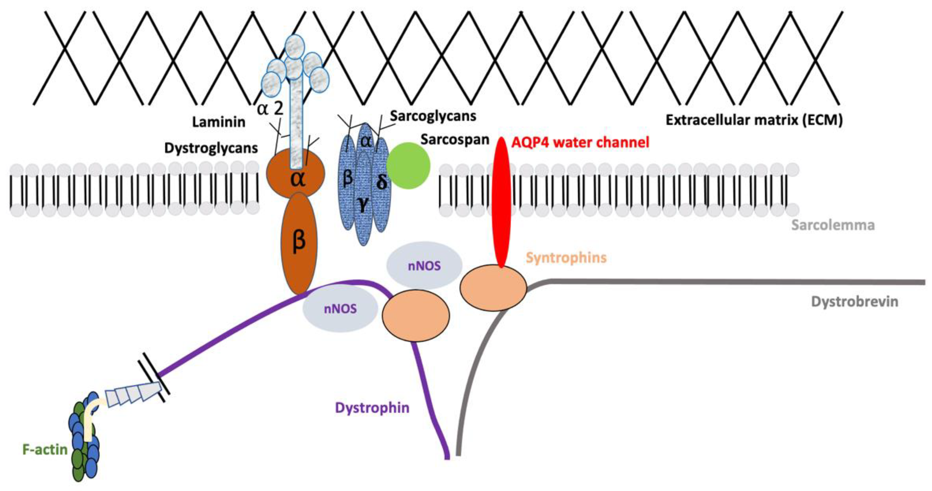
Association of AQP4 with the Dystrophin-Associated Glycoprotein Complex (DAPC)

The dystrophin-associated glycoprotein complex (DAPC) has a crucial role in the maturation and maintenance of the skeletal muscle sarcolemma and the neuromuscular junction (NMJ) in vertebrates [49]. The DAPC is made up of dystroglycans and sarcoglycans that span the plasma membrane, out of which α-dystroglycans bind to the extra-cellular matrix (ECM). The cytoplasmic components of the DAPC include dystrobrevin, syntrophin, and dystrophin, binding directly to the cytoskeletal protein actin [49]. AQP4 is predominantly localized to the sarcolemma type II fast-twitch glycolytic fibers and subsequently interacts with the DAPC, mediated by a key player: α-syntrophin (also called α1-syntrophin) [50,51]. The DAPC complex has been illustrated in Figure 1. Due to its close association with α-syntrophin, which in turn facilitates interaction with the DAPC, several groups studied the expression patterns of α-syntrophin and AQP4 in Duchenne muscular dystrophy (DMD), which is caused by mutations in the dystrophin gene.

4. Relevance of Aquaporins in Duchenne Muscular Dystrophy (DMD) and Other Neuromuscular Disorders (NMDs)

4.1. Duchenne Muscular Dystrophy (DMD)

With a prevalence of 1 in 3500 to 5000 male births every year, Duchenne muscular dystrophy (DMD) is one of the most common inherited neuromuscular disorders affecting the musculoskeletal system [52,53,54]. The causal DMD gene located on the X chromosome is the largest in the human genome, spanning over 79 exons, and is a mutational hotspot for insertions, deletions, mislocations, duplications, point mutations, and complex rearrangements that produce an out-of-frame transcript in DMD patients [53,55,56]. This ultimately leads to the absence of a 427 kDa functional translated protein called dystrophin. The lack of dystrophin results in progressive muscle damage, compromised muscle regeneration, and the replacement of muscle fibers with fibrotic adipose tissue [57,58,59]. As a result, patients are often wheelchair-bound in early adolescence and die prematurely in their twenties due to cardiorespiratory complications [60,61].
The full-length translated dystrophin protein comprises four domains: an actin-binding domain at the N-terminus, a central rod domain made up of 24 spectrin-like repeats (SLRs), a cysteine-rich domain, and a C-terminal domain [53]. Dystrophin protein acts as a stabilizer in the sarcolemma, by bridging the cytoskeletal actin to the extracellular matrix (ECM) with the help of the DAPC complex [62,63]. The absence of dystrophin in DMD destabilizes DAPC moiety, leading to muscle membrane rupture and subsequent necrosis during contractile activity [49,64].

4.2. Findings in DMD Models and Patients

Immunofluorescence staining of the skeletal muscle of mdx mice, a model for DMD with a nonsense mutation in exon 23, revealed a significant reduction in AQP4 [29,65,66]. Frigeri A et al. hypothesized that this reduction was unrelated to muscle degeneration in DMD. This laid the foundation for several groups to investigate the role of AQP4 in DMD and its involvement in the biochemical alteration of muscle fibers [29].
The mdx model is ideal for studying the relevance of AQP4 in DMD pathology. The implication of AQP4 reduction in DMD was studied further using mdx mice. Since the CNS is enriched in AQP4, especially in the astrocytic end feet and ependymal cells, it is thought to be a key player in maintaining the osmotic balance of the brain [26,32]. Biochemical analysis of the mdx brain showed increased extracellular and decreased intracellular brain volume, but the alterations in AQP4 levels were not reported at that time [67]. Hence, Frigeri et al. investigated AQP4 levels in the mdx brain [68]. They reported swollen glial processes, indicative of altered water balance in the mdx brains with a concomitant decrease in AQP4 levels in the dystrophic model. This provides evidence that the role of AQP4 is strictly associated with the reabsorption of water from the extracellular fluid to the blood and CSF [68]. This would possibly result in the slower drainage of water from the brain, resulting in edema and swelling [68]. Densitometric analysis of mdx brain homogenates revealed a 30% reduction in AQP4 protein levels in 1-month-old mdx mice and 70% in 1-year-old mice compared to age-matched controls [68]. In the tibialis anterior (TA) muscle of 1-month-old mdx mice, they observed AQP4 level differences even between the fast-twitch fibers. Type II B fibers were the first to manifest a reduction in AQP4, compared to type II A. In adult mdx mice (1-year old), the AQP4 reduction was more general. Interestingly, they observed no differences between mdx and control mice at the transcript level in the brain and skeletal muscle, indicating that protein degradation occurs at the posttranslational level [68]. Additionally, they also observed that the reduction in sarcolemmal staining of AQP4 was not accompanied by an increase in the intracellular compartmentalization of the mistargeted protein.
The same group also provided the first evidence of AQP4 reduction in DMD and Becker’s muscular dystrophy (BMD) patients with different mutations in the dystrophin gene [69]. Muscle biopsies from DMD patients aged 6 months-5 years were collected and analyzed for AQP4 expression. AQP4 immunoreactivity was nearly absent in the sarcolemma with no intracellular immunofluorescence. These findings were independent of the age and mutations in the patients [69]. Another interesting aspect of the study was that the reduction in AQP4 levels was not because of a decrease in fast-twitch type IIA muscle fibers, as the patient biopsy revealed several intact fibers [69]. The dystrophic muscles show a predominance of slow-twitch type I fibers that contain fewer OAs. The slow contraction of voluntary muscles in these patients may be related to decreased AQP4 expression [8].
Previously, certain studies suggested the dependence of AQP4 expression on the presence of α-syntrophin [50,70,71]. Upon performing double immunofluorescence on the BMD patient biopsies, Frigeri A. obtained mixed results, arguing that this correlation does not always hold true [69]. However, an α-syntrophin knockout mouse model exhibited an obvious decrease in AQP4 from the sarcolemma, demonstrating the correlation between AQP4 and α-syntrophin expression [50,70,71]. Studies carried out later also revealed that AQP4 reduction is associated with an actual concomitant decrease in α-syntrophin levels in patients [72]. Another group investigated whether the decrease in AQP4 protein levels was a result of reduced mRNA transcripts [73]. Contradicting the mouse model findings, Wakayama et al. observed a marked decrease in AQP4 mRNA expression using quantitative reverse transcriptase PCR (qRT-PCR) in muscle biopsies from DMD patients [73]. Further studies are required to determine the molecular basis of AQP4 reduction in DMD, and the subsequent pathological changes in dystrophic muscle.

4.3. Findings in Patients of Other NMDs

In addition to DMD, patients with other NMDs, including Fukuyama-type congenital muscular dystrophy (FCMD), dysferlinopathy, and sarcoglycanopathy, also revealed reduced levels of AQP4, although its roles in pathogenesis and pathophysiology are unclear [74]. FCMD is an early onset childhood progressive muscular dystrophy and is autosomal recessive, caused by mutations in the fukutin gene [75,76]. Although fukutin is ubiquitously expressed, it has stronger expression in the skeletal muscle, heart, pancreas, and brain [75]. It is characterized by joint contracture and mental retardation, cardiomyopathy, and muscle atrophy [76,77,78]. The prevalence rate of FCMD in comparison to DMD is 1:2.1 [76]. The absence of fukutin alters the structure of the basal lamina in the CNS [77,78]. AQP4 expression was found to be significantly reduced in FCMD myofibers. A possible reason for the AQP4 reduction may be attributed to the denervation of the skeletal muscle, which is discussed in the subsequent section.
Studies have also investigated the expression pattern of AQP4 in another group of muscle disorders. Dysferlinopathy includes a spectrum of muscle disorders characterized by two major phenotypes, limb-girdle muscular dystrophy type 2B (LGMDR2) and Miyoshi muscular dystrophy (MMD), characterized by muscle weakness and proximal muscle atrophy [79]. While AQP4 reduction in FCMD correlates with reduced α-syntrophin in the muscle, immunostaining for α1-syntrophin is unchanged in dysferlinopathy patients, suggesting a distinct mechanism involved [80,81].
AQP4 expression was also assessed in patient muscle biopsies affected by sarcoglycanopathies, namely LGMD 2C-F caused by deficiencies of α, β, γ, and δ-sarcoglycan. Sarcoglycanopathies comprise severe forms of autosomal LGMD, with muscle paralysis and wheelchair confinement in early childhood [82]. Knockout mice deficient in sarcoglycans α- γ also showed reduced AQP4 levels [83]. Immunoblot analysis revealed a reduction in overall AQP4 levels, especially the M23 isoform [84]. In conclusion, AQP4 reduction in sarcoglycanopathies is associated with a corresponding decrease in α-syntrophin [51].

5. Effects of Exercise on AQP4 Expression in the Skeletal Muscle

AQP4 together with AQP1 has been known to facilitate water exchange between muscle fibers and blood to ensure the volume changes during muscle activity are sustained. This is associated with muscle swelling and intracellular osmolyte production during exercise [85,86,87,88]. To shed light on the relevance of AQP4 in skeletal muscle function, Basco et al. examined the levels of AQP4 in rats when subjected to endurance training [85]. Moreover, they also looked at the contractile properties and endurance performance in mice with ablated AQP4 when compared to wild-type (WT) counterparts. Ten days after endurance activity following treadmill exercise, the levels of AQP4 in the fast-twitch muscles: TA, extensor digitorum longus (EDL), and quadriceps increased significantly compared to day 1 of treadmill activity in rats. However, it was surprising to see the levels not differ significantly on day 30 when compared to day 10 [85]. The increase in AQP4 levels also corroborated the exercise time. For example, the rats that ran on the treadmill for over 30 min exhibited a greater increase in AQP4 compared to the rats that ran for 15 min. This was the first immunoblotting demonstration in which AQP4 accumulation in fast-twitch fibers correlated with the extent of exercise [85]. Thus, the extent of physical endurance determines the increase in AQP4 to keep the water exchange in balance. AQP4 accumulation allows for a rapid change in the fiber volume which is vital to accommodate changes in intracellular concentration and the intracapillary hydrostatic pressure of osmotically active solutes present during prolonged exercise [88].
Basco et al. also investigated the effects of endurance training on the performance in mice deficient in AQP4. When the AQP4 KO mice were subjected to a treadmill-based endurance test, their running capacity was one-third of the WT counterparts on day 1 of the exercise, but they exhibited significant improvement on days 10 and 30, albeit lower than the WT running distance [85]. In a voluntary running wheel exercise, the WT mice still ran a greater distance each day than the KO mice. Nevertheless, the KO mice still showed an improvement in the voluntary running exercise, albeit significantly lower than the WT mice. These experiments demonstrated that ablation of AQP4 impairs muscle strength which ultimately causes the physical performance in daily running to plummet [51]. When they studied the contractile properties of the fast-twitch EDL muscle, they observed no significant difference in the fatigue index, time to peak, or the force generated, indicating that the contraction kinetics of the skeletal muscle was well-preserved [85]. The important takeaway from this study is that AQP4 ablation does not prevent physical activity in mice but limits the KO mice to reach the same performance as their WT counterparts. It also does not hamper the fiber distribution in the muscles. The researchers involved in this study hypothesized that AQP4 ablation did not affect the contractile properties because the duration of the experiment was much shorter, which did not need AQP4-dependent activation, compared to that of the endurance tests where AQP4 accumulation became more evident at day 10 and 30 [85].

6. AQP1 Acts as a Potential Compensator for AQP4 Loss

AQP1 is the only other aquaporin apart from AQP4 to have confirmed expression in the skeletal muscle. AQP1 expression is enriched in the majority of the tissues, including the blood cells, endothelial, and smooth muscle cells [1,51,89]. In the skeletal muscle, AQP1 is expressed by the endothelial cells in the capillaries arranged between the myofibers, and not on the plasma membrane or the cytoplasm [1]. AQP1 localization in the skeletal muscle fibers was demonstrated by Au et al. and Jimi T et al. [90,91]. Expressions of sarcolemmal AQP4 coupled with vascular AQP1 were thought to facilitate the quick transport of water from the blood into the muscle during intense physical activity [1].
Western blot analysis revealed elevated levels of AQP1 in DMD patient biopsies compared to age-matched healthy controls [51]. AQP1 distribution in the sarcolemma was weak in the control muscle cross-sections, but the intensity increased in the DMD biopsies [51]. Unlike AQP4, AQP1 expression was consistent in the sarcolemma of all fiber types. Interestingly, primary cultures of patient-derived human myoblasts showed altered expression of neither AQP4 nor AQP1. Primary cultures are not subjected to mechanical stress or disease modifiers such as increased cytokine expression and are therefore good models to characterize the expression patterns associated with DMD mutations. It is also worthwhile to mention that AQP1 upregulation may not always correlate with AQP4 loss. For example, limb-girdle muscular dystrophy type 2B (LGMDR2) patient biopsies that showed a reduction in AQP4 levels, did not express a concomitant increase in AQP1 [51]. AQP1 KO mice analysis revealed the increased migration of endothelial cells alongside vascular growth, which has implications for increased AQP1 expression in the endothelia of malignant tumors [92,93,94,95]. Since mdx mice have also demonstrated enhanced vascular growth, it is possible that upregulated AQP1 expression in DMD endothelia may be attributed to the regenerative capacity of DMD capillaries [90,96].

7. Effects of Skeletal Muscle Atrophy and Denervation on the Expressions of AQP4 and AQP1

Whenever there is a demand for increased muscle use, AQP4 expression is upregulated to respond to the change in muscle volume and therefore is considered to play a part in the maintenance of muscle hypertrophy [97]. It was recently demonstrated that AQP4 and a non-selective cation channel activated by osmotic stress, called transient receptor potential vanilloid4 (TRPV4), together regulate cell volume in astrocytes [98,99]. TRPV4 is also detected in the skeletal muscle and is therefore thought to also regulate muscle atrophy or hypertrophy along with AQP4 [100,101]. Ishido M. showed that TRPV4 accumulation increases in denervation-induced muscle atrophy [102]. Since AQP4 levels are maintained in muscular hypertrophy, it is possible that the balance between AQP4 and muscle volume is impaired in atrophy arising from denervation [97]. Additionally, the same group also provided evidence that AQP4 reduction in the denervated muscle is independent of α1-syntrophin levels. In muscular atrophies, there is a reduction in muscle mass and apparent denervation [103]. Muscle atrophy may be triggered by an interplay of several factors including injury, inflammation, metabolic stress, and glucocorticoid levels [104,105]. This also leads to upregulated levels of Atrogin-1, a marker for denervation [106]. A group reported reduced AQP4 levels in humans and mice with rotator cuff tears (RCT), a form of muscle injury that comprises fatty infiltration and musculoskeletal issues [107]. Muscle atrophy associated with RCT injury in a mouse model also saw a decrease in AQP4 levels. AQP4 loss was attributed to atrogin-1-mediated degradation, mediated by the ubiquitination pathway [108]. It is also worthwhile to mention increased AQP4 levels as a result of atrophy from hindlimb unloading [68,109]. The discrepancy in these findings may be due to the difference in atrophy induction, where the latter technique does not hamper innervation, but the former induces denervation by cutting the sciatic nerve [102]. While neurological disorders such as amyotrophic lateral sclerosis (ALS) show reduced mRNA AQP4 expression, as well as protein levels in the skeletal muscle [110], it would be intriguing to examine these findings in spinal muscular atrophy (SMA), one of the most common genetic neuromuscular disorder affecting infants worldwide [111]. SMA arises from a genetic mutation in the survival of the motor neuron 1 (SMN1) gene that causes the loss of motor neurons in the anterior horn of the spinal cord, ultimately leading to the denervation of the skeletal muscle [112,113]. The effects of nerve supply on AQP4 transcription during the early stages of denervation need to be elucidated in the future.
In response to hypertrophy or atrophy, the capillary supply is facilitated or declined [114,115]. AQP1, which is predominantly present in the endothelial cells of the blood capillaries, does not have altered expression post-denervation induced by sciatic nerve freezing [43,116]. Thus, the compensatory nature of AQP1 for AQP4 loss induced by muscle denervation and subsequent hypertrophy may not hold true. The regulation of AQP1 expression occurs independently of the nerve supply to the skeletal muscle [116]

8. Expression of Other AQPs in the Skeletal Muscle

To investigate the compensatory roles of AQPs in the skeletal muscle, several groups evaluated the presence of other AQPs. Wang et al. reported the expressions of AQP 3, 4, 5, 7, 8, and 9 in the human masseter muscle, and that of AQP 1, 3, 4, and 10 in the human infrahyoid muscle using RT-PCR [117]. However, the expression of these AQPs was linked to the possible contamination and infiltration by erythrocytes (AQP1), adipocytes (AQP7), and leucocytes (AQP9). To rule out this possibility, Wakayama et al. performed immunocytochemical tests and confirmed the expression of AQP 3, 5, 7, and 9 in the skeletal muscle fibers [4,118,119,120].
AQP3 is a glycerol-transporting integral protein (GLIP) that was cloned from a rat kidney [118,121,122,123]. AQP3 is expressed in the basolateral membrane of collecting duct cells in the kidney, pancreas, small intestine, liver, etc. [124]. Wakayama et al. showed the presence of AQP3 in the plasma membrane of skeletal muscle myofibers in addition to AQP4 using immunoblots and immunostaining, with no apparent preferential expression in the fiber types [118]. The co-expression of AQP3 and AQP4 resorted to the transport of both water and small non-ionic solutes in the skeletal muscle. However, no other studies have demonstrated AQP3 expression in the muscle.
AQP7 and AQP9 are glycerol channels expressed predominantly in the adipocytes and hepatocytes, respectively [4,119]. The weak expression of AQP9 was shown in the myofiber surface membrane [119]. Decreased feeding and increased fasting upregulate glycerol levels that in turn increase AQP7 and AQP9 expression [125]. In the skeletal muscle, the critical role of AQP9 is to facilitate the uptake of glycerol for glucose production during fasting. AQP9 and glycerol kinase mRNA expressions are downregulated by insulin, which is indicative of normal liver processing. However, in endocrine myopathies such as insulin resistance, AQP7 and AQP9 levels are elevated [126,127]. Wakayama et al. demonstrated the presence of AQP7 predominantly in both fast- and slow-twitch myofibers in human skeletal muscle and only in the fast-twitch type II myofibers in murine skeletal muscle [4]. The presence of AQP7 in the skeletal myofiber remains controversial because the immunostaining carried out by another group was negative for AQP7 [128]. Glucose and energy metabolism are essential for energy production in the skeletal muscle, and conditions such as obesity reduce the metabolism, which may induce insulin resistance [129]. Insulin resistance and obesity increase the relative proportion of glycolytic fast-twitch type II fibers but reduce the number of oxidative type I fibers [130,131,132]. Upregulated levels of AQP7 were reported in a mouse model of obesity in the skeletal muscle, indicating its role in insulin resistance [133].
AQP5 was first cloned from salivary glands and was shown to be expressed in the lungs and eyes [134]. Hwang et al. showed the presence of AQP5 in C2C12 myoblasts and that its expression was upregulated upon differentiation into myotubes and also when subjected to hypertonic stress [120]. Western blotting and immunocytochemical staining of C2C12 cells confirmed the presence of AQP5 [120]. The transient upregulation of AQP5 post-differentiation of C2C12 cells may hint at a role in early-stage myogenic differentiation. If AQP5 is a potential compensator for AQP4, there exists a possibility that AQP5 is expressed in the fast-twitch fibers in adult skeletal muscle, and the expression corresponded with the number of fast-twitch fibers [120].

9. Concluding Remarks

AQPs facilitate water and non-ionic molecule transport across the membranes of a diverse group of cells in organisms and are therefore essential moieties to be characterized and studied in terms of osmotic regulation and cellular homeostasis. With 13 different AQPs identified so far, there is consensus on the predominance of AQP4 and AQP1 expression in the skeletal muscle. Although there have been a few studies demonstrating the expression of other AQPs such as AQP3, AQP5, AQP7, and AQP9 in the skeletal muscle, these findings are concluded to be controversial. Further evidence may be required to support the results.
AQP4, the most abundantly expressed aquaporin member in the skeletal muscle, is responsible for sarcolemmal permeability, as well as playing a key role in responding to changes in muscle volume during contraction. It has a preferential expression in the sarcolemma of fast-twitch muscle fibers. For the first time, Basco et al. showed a positive correlation between AQP4 levels and endurance activity [85]. An interesting finding put forward by the group was the increase in AQP4 protein levels, and not mRNA, suggesting the role of post-transcriptional regulatory mechanisms in response to cellular stress. On the contrary, Au et al. observed a reduction in AQP4 even at the mRNA level in DMD patients, but not mdx mice. Further studies are deemed essential to validate these findings.
When subjected to either forced or voluntary exercise, AQP4 KO mice displayed lesser activity than WT mice. However, there was an improvement in performance after a continued exercise regime. It, therefore, is possible for AQP4 ablation to not prevent activity but limit KO mice from performing similarly to WT mice. This theory was also supported by the contractile studies of the fast-twitch muscle gastrocnemius and EDL muscle, where they observed similar twitch force generation and fatigue resistance in both WT and KO muscles. Additionally, muscle atrophy is not directly associated with AQP4 ablation. Detailed studies are necessary to support this theory.
Sarcolemmal integrity is severely compromised due to dystrophin loss and the subsequent destabilizing of the DAPC complex in DMD patients, coupled with significantly reduced levels of AQP4. Despite being associated with α1-syntrophin, a few studies revealed normal levels of AQP4 in the absence of α1-syntrophin [51]. For example, dysferlinopathy patient biopsies revealed reduced AQP4 levels despite normal α1-syntrophin levels. The water permeability in the sarcolemmal vesicles of mdx mice was significantly reduced in one study, with no effect on water permeability in another study. One possible explanation for the permeability remaining unaffected could be the compensatory role of AQP1, which has been demonstrated to be upregulated in mdx mice and DMD patients. However, since no increase in AQP1 expression was seen in LGMDR2 patients, this correlation may not always hold true for every muscle pathological disease. Nevertheless, AQP1 probably takes over the role of water transport in the muscle in the absence of AQP4. Future studies can elucidate the role of increased AQP1 in the endothelia of DMD models.
From a clinical perspective, it is important to highlight the pathology presented by patients with neuromyelitis optica (NMO), which is an autoimmune inflammatory disorder characterized by recurring neuritis of the optic nerve [135]. NMO patients produce autoantibodies against AQP4 (AQP4-IgG), leading to reduced AQP4 levels. Suzuki et al. demonstrated elevated levels of CK prior to the onset of optic neuritis (ON) in patients, showing the involvement of skeletal muscle dysfunction in these patients [136]. Physical injury to the skeletal muscle may exacerbate the condition by generating more AQP4-IgG, leading to further loss of AQP4 (). However, minimal changes in muscle pathology were observed in conventional staining. Nevertheless, AQP4 loss and IgG-mediated complement activation on sarcolemma type II myofibers serve as a diagnostic feature in NMO patients [137]. Detailed studies in the future in NMO patients can provide insight into the effect of AQP4 loss on skeletal muscle function.
In conclusion, several studies have highlighted the reduction in AQP4 in muscular dystrophies despite having different causal genetic mutations. The key player responsible for AQP4 reduction is DAPC complex destabilization or loss of α1-syntrophin. Further studies can delve deeper into the relationship of AQP4 loss with the DAPC complex, and validate previous findings. AQP4 and AQP1 are the predominantly expressed AQPs in the skeletal muscle, while the expression of AQPs 3, 5, 7, and 9 remain controversial.

Author Contributions

T.A., A.A.-a., and T.Y. prepared the manuscript. All authors have read and agreed to the published version of the manuscript.

Funding

This review article is supported by the Natural Sciences and Engineering Research Council of Canada (NSERC).

Institutional Review Board Statement

Not applicable.

Informed Consent Statement

Not applicable.

Data Availability Statement

No new data were created or analyzed in this study. Data sharing is not applicable to this article.

Acknowledgments

We thank the University of Alberta Faculty of Medicine and Dentistry, the Canadian Institutes of Health Research, the Friends of Garrett Cumming Research Funds, HM Toupin Neurological Science Research Funds, Muscular Dystrophy Canada, and National Sciences and Engineering Research Council of Canada.

Conflicts of Interest

T.Y. is a co-founder and shareholder of OligomicsTx Inc., which aims to commercialize antisense technology.

References

  1. Frigeri, A.; Nicchia, G.P.; Balena, R.; Nico, B.; Svelto, M. Aquaporins in skeletal muscle: Reassessment of the functional role of aquaporin-4. FASEB J. 2004, 18, 905–907. [Google Scholar] [CrossRef] [PubMed]
  2. Neering, I.R.; Quesenberry, L.A.; Morris, V.A.; Taylor, S.R. Nonuniform volume changes during muscle contraction. Biophys. J. 1991, 59, 926–933. [Google Scholar] [CrossRef] [PubMed] [Green Version]
  3. Markou, A.; Unger, L.; Abir-Awan, M.; Saadallah, A.; Halsey, A.; Balklava, Z.; Conner, M.; Tornroth-Horsefield, S.; Greenhill, S.D.; Conner, A.; et al. Molecular mechanisms governing aquaporin relocalisation. Biochim. Biophys. Acta Biomembr. 2022, 1864, 183853. [Google Scholar] [CrossRef]
  4. Wakayama, Y.; Inoue, M.; Kojima, H.; Jimi, T.; Shibuya, S.; Hara, H.; Oniki, H. Expression and localization of aquaporin 7 in normal skeletal myofiber. Cell Tissue Res. 2004, 316, 123–129. [Google Scholar] [CrossRef]
  5. Denker, B.M.; Smith, B.L.; Kuhajda, F.P.; Agre, P. Identification, purification, and partial characterization of a novel Mr 28,000 integral membrane protein from erythrocytes and renal tubules. J. Biol. Chem. 1988, 263, 15634–15642. [Google Scholar] [CrossRef]
  6. Preston, G.M.; Agre, P. Isolation of the cDNA for erythrocyte integral membrane protein of 28 kilodaltons: Member of an ancient channel family. Proc. Natl. Acad. Sci. USA 1991, 88, 11110–11114. [Google Scholar] [CrossRef] [PubMed] [Green Version]
  7. Preston, G.M.; Carroll, T.P.; Guggino, W.B.; Agre, P. Appearance of water channels in Xenopus oocytes expressing red cell CHIP28 protein. Science 1992, 256, 385–387. [Google Scholar] [CrossRef] [PubMed] [Green Version]
  8. Wakayama, Y. Aquaporin expression in normal and pathological skeletal muscles: A brief review with focus on AQP4. J. Biomed. Biotechnol. 2010, 2010, 731569. [Google Scholar] [CrossRef] [Green Version]
  9. Madeira, A.; Moura, T.F.; Soveral, G. Detecting Aquaporin Function and Regulation. Front. Chem. 2016, 4, 3. [Google Scholar] [CrossRef] [Green Version]
  10. Shi, L.B.; Skach, W.R.; Verkman, A.S. Functional independence of monomeric CHIP28 water channels revealed by expression of wild-type mutant heterodimers. J. Biol. Chem. 1994, 269, 10417–10422. [Google Scholar] [CrossRef]
  11. Gomes, D.; Agasse, A.; Thiebaud, P.; Delrot, S.; Geros, H.; Chaumont, F. Aquaporins are multifunctional water and solute transporters highly divergent in living organisms. Biochim. Biophys. Acta 2009, 1788, 1213–1228. [Google Scholar] [CrossRef] [Green Version]
  12. Aaij, R.; Adeva, B.; Adinolfi, M.; Ajaltouni, Z.; Akar, S.; Albrecht, J.; Alessio, F.; Alexander, M.; Alfonso Albero, A.; Ali, S.; et al. Search for Dark Photons Produced in 13 TeV pp Collisions. Phys. Rev. Lett. 2018, 120, 061801. [Google Scholar] [CrossRef] [PubMed] [Green Version]
  13. Bondy, C.; Chin, E.; Smith, B.L.; Preston, G.M.; Agre, P. Developmental gene expression and tissue distribution of the CHIP28 water-channel protein. Proc. Natl. Acad. Sci. USA 1993, 90, 4500–4504. [Google Scholar] [CrossRef] [PubMed] [Green Version]
  14. Ma, T.; Yang, B.; Gillespie, A.; Carlson, E.J.; Epstein, C.J.; Verkman, A.S. Severely impaired urinary concentrating ability in transgenic mice lacking aquaporin-1 water channels. J. Biol. Chem. 1998, 273, 4296–4299. [Google Scholar] [CrossRef] [PubMed] [Green Version]
  15. King, L.S.; Choi, M.; Fernandez, P.C.; Cartron, J.P.; Agre, P. Defective urinary concentrating ability due to a complete deficiency of aquaporin-1. N. Engl. J. Med. 2001, 345, 175–179. [Google Scholar] [CrossRef]
  16. Verkman, A.S. Physiological importance of aquaporin water channels. Ann. Med. 2002, 34, 192–200. [Google Scholar] [CrossRef]
  17. Nielsen, S.; Smith, B.L.; Christensen, E.I.; Agre, P. Distribution of the aquaporin CHIP in secretory and resorptive epithelia and capillary endothelia. Proc. Natl. Acad. Sci. USA 1993, 90, 7275–7279. [Google Scholar] [CrossRef] [Green Version]
  18. Verkman, A.S. Aquaporin water channels and endothelial cell function. J. Anat. 2002, 200, 617–627. [Google Scholar] [CrossRef]
  19. Folkesson, H.G.; Matthay, M.A.; Hasegawa, H.; Kheradmand, F.; Verkman, A.S. Transcellular water transport in lung alveolar epithelium through mercury-sensitive water channels. Proc. Natl. Acad. Sci. USA 1994, 91, 4970–4974. [Google Scholar] [CrossRef] [Green Version]
  20. Hasegawa, H.; Lian, S.C.; Finkbeiner, W.E.; Verkman, A.S. Extrarenal tissue distribution of CHIP28 water channels by in situ hybridization and antibody staining. Am. J. Physiol. 1994, 266, C893–C903. [Google Scholar] [CrossRef]
  21. Effros, R.M.; Darin, C.; Jacobs, E.R.; Rogers, R.A.; Krenz, G.; Schneeberger, E.E. Water transport and the distribution of aquaporin-1 in pulmonary air spaces. J. Appl. Physiol. 1997, 83, 1002–1016. [Google Scholar] [CrossRef] [PubMed] [Green Version]
  22. Gresz, V.; Kwon, T.H.; Hurley, P.T.; Varga, G.; Zelles, T.; Nielsen, S.; Case, R.M.; Steward, M.C. Identification and localization of aquaporin water channels in human salivary glands. Am. J. Physiol. Gastrointest. Liver Physiol. 2001, 281, G247–G254. [Google Scholar] [CrossRef] [PubMed] [Green Version]
  23. Hurley, P.T.; Ferguson, C.J.; Kwon, T.H.; Andersen, M.L.; Norman, A.G.; Steward, M.C.; Nielsen, S.; Case, R.M. Expression and immunolocalization of aquaporin water channels in rat exocrine pancreas. Am. J. Physiol. Gastrointest. Liver Physiol. 2001, 280, G701–G709. [Google Scholar] [CrossRef] [Green Version]
  24. Benga, G. The first discovered water channel protein, later called aquaporin 1: Molecular characteristics, functions and medical implications. Mol. Asp. Med. 2012, 33, 518–534. [Google Scholar] [CrossRef] [PubMed]
  25. Holmes, R.P. The role of renal water channels in health and disease. Mol. Asp. Med. 2012, 33, 547–552. [Google Scholar] [CrossRef]
  26. Nielsen, S.; Nagelhus, E.A.; Amiry-Moghaddam, M.; Bourque, C.; Agre, P.; Ottersen, O.P. Specialized membrane domains for water transport in glial cells: High-resolution immunogold cytochemistry of aquaporin-4 in rat brain. J. Neurosci. 1997, 17, 171–180. [Google Scholar] [CrossRef] [Green Version]
  27. Longatti, P.L.; Basaldella, L.; Orvieto, E.; Fiorindi, A.; Carteri, A. Choroid plexus and aquaporin-1: A novel explanation of cerebrospinal fluid production. Pediatr. Neurosurg. 2004, 40, 277–283. [Google Scholar] [CrossRef] [PubMed]
  28. Wen, H.; Nagelhus, E.A.; Amiry-Moghaddam, M.; Agre, P.; Ottersen, O.P.; Nielsen, S. Ontogeny of water transport in rat brain: Postnatal expression of the aquaporin-4 water channel. Eur. J. Neurosci. 1999, 11, 935–945. [Google Scholar] [CrossRef] [PubMed]
  29. Frigeri, A.; Nicchia, G.P.; Verbavatz, J.M.; Valenti, G.; Svelto, M. Expression of aquaporin-4 in fast-twitch fibers of mammalian skeletal muscle. J. Clin. Investig. 1998, 102, 695–703. [Google Scholar] [CrossRef] [Green Version]
  30. Branton, D. Fracture faces of frozen membranes. Proc. Natl. Acad. Sci. USA 1966, 55, 1048–1056. [Google Scholar] [CrossRef]
  31. Verbavatz, J.M.; Ma, T.; Gobin, R.; Verkman, A.S. Absence of orthogonal arrays in kidney, brain and muscle from transgenic knockout mice lacking water channel aquaporin-4. J. Cell Sci. 1997, 110 Pt 22, 2855–2860. [Google Scholar] [CrossRef]
  32. Frigeri, A.; Gropper, M.A.; Umenishi, F.; Kawashima, M.; Brown, D.; Verkman, A.S. Localization of MIWC and GLIP water channel homologs in neuromuscular, epithelial and glandular tissues. J. Cell Sci. 1995, 108 Pt 9, 2993–3002. [Google Scholar] [CrossRef]
  33. Yang, B.; Brown, D.; Verkman, A.S. The mercurial insensitive water channel (AQP-4) forms orthogonal arrays in stably transfected Chinese hamster ovary cells. J. Biol. Chem. 1996, 271, 4577–4580. [Google Scholar] [CrossRef] [PubMed] [Green Version]
  34. Sirken, S.M.; Fischbeck, K.H. Freeze-fracture studies of denervated and tenotomized rat muscle. J. Neuropathol. Exp. Neurol. 1985, 44, 147–155. [Google Scholar] [CrossRef] [PubMed]
  35. Rash, J.E.; Yasumura, T.; Hudson, C.S.; Agre, P.; Nielsen, S. Direct immunogold labeling of aquaporin-4 in square arrays of astrocyte and ependymocyte plasma membranes in rat brain and spinal cord. Proc. Natl. Acad. Sci. USA 1998, 95, 11981–11986. [Google Scholar] [CrossRef] [PubMed] [Green Version]
  36. Shibuya, S.W.Y.; Inoue, M. Identification of aquaporin 4 molecule in the replica of normal rat sarcolemma: Fracture-label immnoelectron microscopic study. Ann. Neurol. 1999, 46, 460. [Google Scholar]
  37. Shibuya, S.; Wakayama, Y. Changes in muscle plasma membranes in young mice with X chromosome-linked muscular dystrophy: A freeze-fracture study. Neuropathol. Appl. Neurobiol. 1991, 17, 335–344. [Google Scholar] [CrossRef]
  38. Wakayama, Y.; Okayasu, H.; Shibuya, S.; Kumagai, T. Duchenne dystrophy: Reduced density of orthogonal array subunit particles in muscle plasma membrane. Neurology 1984, 34, 1313–1317. [Google Scholar] [CrossRef]
  39. Hasegawa, H.; Ma, T.; Skach, W.; Matthay, M.A.; Verkman, A.S. Molecular cloning of a mercurial-insensitive water channel expressed in selected water-transporting tissues. J. Biol. Chem. 1994, 269, 5497–5500. [Google Scholar] [CrossRef]
  40. Lu, M.; Lee, M.D.; Smith, B.L.; Jung, J.S.; Agre, P.; Verdijk, M.A.; Merkx, G.; Rijss, J.P.; Deen, P.M. The human AQP4 gene: Definition of the locus encoding two water channel polypeptides in brain. Proc. Natl. Acad. Sci. USA 1996, 93, 10908–10912. [Google Scholar] [CrossRef] [Green Version]
  41. Jung, J.S.; Bhat, R.V.; Preston, G.M.; Guggino, W.B.; Baraban, J.M.; Agre, P. Molecular characterization of an aquaporin cDNA from brain: Candidate osmoreceptor and regulator of water balance. Proc. Natl. Acad. Sci. USA 1994, 91, 13052–13056. [Google Scholar] [CrossRef] [PubMed]
  42. Basco, D.; Nicchia, G.P.; D′Alessandro, A.; Zolla, L.; Svelto, M.; Frigeri, A. Absence of aquaporin-4 in skeletal muscle alters proteins involved in bioenergetic pathways and calcium handling. PLoS ONE 2011, 6, e19225. [Google Scholar] [CrossRef] [PubMed] [Green Version]
  43. Ishido, M.; Nakamura, T. The expression of aquaporin-4 is regulated based on innervation in skeletal muscles. J. Muscle Res. Cell Motil. 2018, 39, 17–23. [Google Scholar] [CrossRef] [PubMed]
  44. Vizzaccaro, E.; Terracciano, C.; Rastelli, E.; Massa, R. Aquaporin 4 expression in human skeletal muscle fiber types. Muscle Nerve 2018, 57, 856–858. [Google Scholar] [CrossRef]
  45. Rossi, A.; Pisani, F.; Nicchia, G.P.; Svelto, M.; Frigeri, A. Evidences for a leaky scanning mechanism for the synthesis of the shorter M23 protein isoform of aquaporin-4: Implication in orthogonal array formation and neuromyelitis optica antibody interaction. J. Biol. Chem. 2010, 285, 4562–4569. [Google Scholar] [CrossRef] [Green Version]
  46. Moe, S.E.; Sorbo, J.G.; Sogaard, R.; Zeuthen, T.; Petter Ottersen, O.; Holen, T. New isoforms of rat Aquaporin-4. Genomics 2008, 91, 367–377. [Google Scholar] [CrossRef] [Green Version]
  47. De Bellis, M.; Pisani, F.; Mola, M.G.; Basco, D.; Catalano, F.; Nicchia, G.P.; Svelto, M.; Frigeri, A. A novel human aquaporin-4 splice variant exhibits a dominant-negative activity: A new mechanism to regulate water permeability. Mol. Biol. Cell 2014, 25, 470–480. [Google Scholar] [CrossRef]
  48. De Bellis, M.; Pisani, F.; Mola, M.G.; Rosito, S.; Simone, L.; Buccoliero, C.; Trojano, M.; Nicchia, G.P.; Svelto, M.; Frigeri, A. Translational readthrough generates new astrocyte AQP4 isoforms that modulate supramolecular clustering, glial endfeet localization, and water transport. Glia 2017, 65, 790–803. [Google Scholar] [CrossRef]
  49. Grady, R.M.; Zhou, H.; Cunningham, J.M.; Henry, M.D.; Campbell, K.P.; Sanes, J.R. Maturation and maintenance of the neuromuscular synapse: Genetic evidence for roles of the dystrophin--glycoprotein complex. Neuron 2000, 25, 279–293. [Google Scholar] [CrossRef] [Green Version]
  50. Neely, J.D.; Amiry-Moghaddam, M.; Ottersen, O.P.; Froehner, S.C.; Agre, P.; Adams, M.E. Syntrophin-dependent expression and localization of Aquaporin-4 water channel protein. Proc. Natl. Acad. Sci. USA 2001, 98, 14108–14113. [Google Scholar] [CrossRef] [Green Version]
  51. Au, C.G.; Butler, T.L.; Egan, J.R.; Cooper, S.T.; Lo, H.P.; Compton, A.G.; North, K.N.; Winlaw, D.S. Changes in skeletal muscle expression of AQP1 and AQP4 in dystrophinopathy and dysferlinopathy patients. Acta Neuropathol. 2008, 116, 235–246. [Google Scholar] [CrossRef] [PubMed]
  52. Mendell, J.R.; Shilling, C.; Leslie, N.D.; Flanigan, K.M.; al-Dahhak, R.; Gastier-Foster, J.; Kneile, K.; Dunn, D.M.; Duval, B.; Aoyagi, A.; et al. Evidence-based path to newborn screening for Duchenne muscular dystrophy. Ann. Neurol. 2012, 71, 304–313. [Google Scholar] [CrossRef] [PubMed]
  53. Ervasti, J.M.; Ohlendieck, K.; Kahl, S.D.; Gaver, M.G.; Campbell, K.P. Deficiency of a glycoprotein component of the dystrophin complex in dystrophic muscle. Nature 1990, 345, 315–319. [Google Scholar] [CrossRef] [PubMed]
  54. Aslesh, T.; Erkut, E.; Yokota, T. Restoration of dystrophin expression and correction of Duchenne muscular dystrophy by genome editing. Expert Opin. Biol. Ther. 2021, 21, 1049–1061. [Google Scholar] [CrossRef]
  55. Koenig, M.; Hoffman, E.P.; Bertelson, C.J.; Monaco, A.P.; Feener, C.; Kunkel, L.M. Complete cloning of the Duchenne muscular dystrophy (DMD) cDNA and preliminary genomic organization of the DMD gene in normal and affected individuals. Cell 1987, 50, 509–517. [Google Scholar] [CrossRef]
  56. Juan-Mateu, J.; Gonzalez-Quereda, L.; Rodriguez, M.J.; Baena, M.; Verdura, E.; Nascimento, A.; Ortez, C.; Baiget, M.; Gallano, P. DMD Mutations in 576 Dystrophinopathy Families: A Step Forward in Genotype-Phenotype Correlations. PLoS ONE 2015, 10, e0135189. [Google Scholar] [CrossRef]
  57. Hoffman, E.P.; Brown, R.H., Jr.; Kunkel, L.M. Dystrophin: The protein product of the Duchenne muscular dystrophy locus. Cell 1987, 51, 919–928. [Google Scholar] [CrossRef]
  58. Marden, F.A.; Connolly, A.M.; Siegel, M.J.; Rubin, D.A. Compositional analysis of muscle in boys with Duchenne muscular dystrophy using MR imaging. Skelet. Radiol. 2005, 34, 140–148. [Google Scholar] [CrossRef]
  59. Zhou, L.; Lu, H. Targeting fibrosis in Duchenne muscular dystrophy. J. Neuropathol. Exp. Neurol. 2010, 69, 771–776. [Google Scholar] [CrossRef]
  60. Veltrop, M.; van Vliet, L.; Hulsker, M.; Claassens, J.; Brouwers, C.; Breukel, C.; van der Kaa, J.; Linssen, M.M.; den Dunnen, J.T.; Verbeek, S.; et al. A dystrophic Duchenne mouse model for testing human antisense oligonucleotides. PLoS ONE 2018, 13, e0193289. [Google Scholar] [CrossRef]
  61. Manzur, A.Y.; Kinali, M.; Muntoni, F. Update on the management of Duchenne muscular dystrophy. Arch. Dis. Child. 2008, 93, 986–990. [Google Scholar] [CrossRef] [PubMed]
  62. Zubrzycka-Gaarn, E.E.; Bulman, D.E.; Karpati, G.; Burghes, A.H.; Belfall, B.; Klamut, H.J.; Talbot, J.; Hodges, R.S.; Ray, P.N.; Worton, R.G. The Duchenne muscular dystrophy gene product is localized in sarcolemma of human skeletal muscle. Nature 1988, 333, 466–469. [Google Scholar] [CrossRef] [PubMed]
  63. Petrof, B.J.; Shrager, J.B.; Stedman, H.H.; Kelly, A.M.; Sweeney, H.L. Dystrophin protects the sarcolemma from stresses developed during muscle contraction. Proc. Natl. Acad. Sci. USA 1993, 90, 3710–3714. [Google Scholar] [CrossRef] [PubMed] [Green Version]
  64. Carlson, C.G. The dystrophinopathies: An alternative to the structural hypothesis. Neurobiol. Dis. 1998, 5, 3–15. [Google Scholar] [CrossRef] [PubMed] [Green Version]
  65. Wakayama, Y.; Jimi, T.; Misugi, N.; Kumagai, T.; Miyake, S.; Shibuya, S.; Miike, T. Dystrophin immunostaining and freeze-fracture studies of muscles of patients with early stage amyotrophic lateral sclerosis and Duchenne muscular dystrophy. J. Neurol. Sci. 1989, 91, 191–205. [Google Scholar] [CrossRef]
  66. Bulfield, G.; Siller, W.G.; Wight, P.A.; Moore, K.J. X chromosome-linked muscular dystrophy (mdx) in the mouse. Proc. Natl. Acad. Sci. USA 1984, 81, 1189–1192. [Google Scholar] [CrossRef] [Green Version]
  67. Tracey, I.; Dunn, J.F.; Radda, G.K. Brain metabolism is abnormal in the mdx model of Duchenne muscular dystrophy. Brain 1996, 119 Pt 3, 1039–1044. [Google Scholar] [CrossRef] [Green Version]
  68. Frigeri, A.; Nicchia, G.P.; Nico, B.; Quondamatteo, F.; Herken, R.; Roncali, L.; Svelto, M. Aquaporin-4 deficiency in skeletal muscle and brain of dystrophic mdx mice. FASEB J. 2001, 15, 90–98. [Google Scholar] [CrossRef] [Green Version]
  69. Frigeri, A.; Nicchia, G.P.; Repetto, S.; Bado, M.; Minetti, C.; Svelto, M. Altered aquaporin-4 expression in human muscular dystrophies: A common feature? FASEB J. 2002, 16, 1120–1122. [Google Scholar] [CrossRef]
  70. Adams, M.E.; Mueller, H.A.; Froehner, S.C. In vivo requirement of the alpha-syntrophin PDZ domain for the sarcolemmal localization of nNOS and aquaporin-4. J. Cell Biol. 2001, 155, 113–122. [Google Scholar] [CrossRef] [Green Version]
  71. Yokota, T.; Miyagoe, Y.; Hosaka, Y.; Tsukita, K.; Kameya, S.; Shibuya, S.; Matsuda, R.; Wakayama, Y.; Takeda, S.I. Aquaporin-4 is absent at the sarcolemma and at perivascular astrocyte endfeet in α1-syntrophin knockout mice. Proc. Japan. Acad. 2000, 76, 22–27. [Google Scholar] [CrossRef]
  72. Compton, A.G.; Cooper, S.T.; Hill, P.M.; Yang, N.; Froehner, S.C.; North, K.N. The syntrophin-dystrobrevin subcomplex in human neuromuscular disorders. J. Neuropathol. Exp. Neurol. 2005, 64, 350–361. [Google Scholar] [CrossRef] [Green Version]
  73. Wakayama, Y.; Jimi, T.; Inoue, M.; Kojima, H.; Murahashi, M.; Kumagai, T.; Yamashita, S.; Hara, H.; Shibuya, S. Reduced aquaporin 4 expression in the muscle plasma membrane of patients with Duchenne muscular dystrophy. Arch. Neurol. 2002, 59, 431–437. [Google Scholar] [CrossRef] [PubMed] [Green Version]
  74. Wakayama, Y.; Jimi, T.; Inoue, M.; Kojima, H.; Yamashita, S.; Kumagai, T.; Murahashi, M.; Hara, H.; Shibuya, S. Altered aquaporin 4 expression in muscles of Fukuyama-type congenital muscular dystrophy. Virchows Arch. 2003, 443, 761–767. [Google Scholar] [CrossRef] [PubMed]
  75. Kobayashi, K.; Nakahori, Y.; Miyake, M.; Matsumura, K.; Kondo-Iida, E.; Nomura, Y.; Segawa, M.; Yoshioka, M.; Saito, K.; Osawa, M.; et al. An ancient retrotransposal insertion causes Fukuyama-type congenital muscular dystrophy. Nature 1998, 394, 388–392. [Google Scholar] [CrossRef]
  76. Fukuyama, Y.; Osawa, M.; Suzuki, H. Congenital progressive muscular dystrophy of the Fukuyama type-clinical, genetic and pathological considerations. Brain Dev. 1981, 3, 1–29. [Google Scholar] [CrossRef]
  77. Nakano, I.; Funahashi, M.; Takada, K.; Toda, T. Are breaches in the glia limitans the primary cause of the micropolygyria in Fukuyama-type congenital muscular dystrophy (FCMD)? Pathological study of the cerebral cortex of an FCMD fetus. Acta Neuropathol. 1996, 91, 313–321. [Google Scholar] [CrossRef]
  78. Yamamoto, T.; Shibata, N.; Kanazawa, M.; Kobayashi, M.; Komori, T.; Kondo, E.; Saito, K.; Osawa, M. Early ultrastructural changes in the central nervous system in Fukuyama congenital muscular dystrophy. Ultrastruct. Pathol. 1997, 21, 355–360. [Google Scholar] [CrossRef]
  79. Liu, J.; Aoki, M.; Illa, I.; Wu, C.; Fardeau, M.; Angelini, C.; Serrano, C.; Urtizberea, J.A.; Hentati, F.; Hamida, M.B.; et al. Dysferlin, a novel skeletal muscle gene, is mutated in Miyoshi myopathy and limb girdle muscular dystrophy. Nat. Genet. 1998, 20, 31–36. [Google Scholar] [CrossRef]
  80. Wakayama, Y.; Inoue, M.; Kojima, H.; Yamashita, S.; Shibuya, S.; Jimi, T.; Hara, H.; Matsuzaki, Y.; Oniki, H.; Kanagawa, M.; et al. Reduced expression of sarcospan in muscles of Fukuyama congenital muscular dystrophy. Histol. Histopathol. 2008, 23, 1425–1438. [Google Scholar] [CrossRef]
  81. Wakayama, Y.; Inoue, M.; Kojima, H.; Jimi, T.; Yamashita, S.; Kumagai, T.; Shibuya, S.; Hara, H.; Oniki, H. Altered alpha1-syntrophin expression in myofibers with Duchenne and Fukuyama muscular dystrophies. Histol. Histopathol. 2006, 21, 23–34. [Google Scholar] [CrossRef] [PubMed]
  82. Laval, S.H.; Bushby, K.M. Limb-girdle muscular dystrophies--from genetics to molecular pathology. Neuropathol. Appl. Neurobiol. 2004, 30, 91–105. [Google Scholar] [CrossRef] [PubMed]
  83. Crosbie, R.H.; Dovico, S.A.; Flanagan, J.D.; Chamberlain, J.S.; Ownby, C.L.; Campbell, K.P. Characterization of aquaporin-4 in muscle and muscular dystrophy. FASEB J. 2002, 16, 943–949. [Google Scholar] [CrossRef] [PubMed] [Green Version]
  84. Assereto, S.; Mastrototaro, M.; Stringara, S.; Gazzerro, E.; Broda, P.; Nicchia, G.P.; Svelto, M.; Bruno, C.; Nigro, V.; Lisanti, M.P.; et al. Aquaporin-4 expression is severely reduced in human sarcoglycanopathies and dysferlinopathies. Cell Cycle 2008, 7, 2199–2207. [Google Scholar] [CrossRef] [Green Version]
  85. Basco, D.; Blaauw, B.; Pisani, F.; Sparaneo, A.; Nicchia, G.P.; Mola, M.G.; Reggiani, C.; Svelto, M.; Frigeri, A. AQP4-dependent water transport plays a functional role in exercise-induced skeletal muscle adaptations. PLoS ONE 2013, 8, e58712. [Google Scholar] [CrossRef]
  86. Crenshaw, A.G.; Thornell, L.E.; Friden, J. Intramuscular pressure, torque and swelling for the exercise-induced sore vastus lateralis muscle. Acta Physiol. Scand. 1994, 152, 265–277. [Google Scholar] [CrossRef] [PubMed]
  87. Greenleaf, J.E.; Van Beaumont, W.; Brock, P.J.; Morse, J.T.; Mangseth, G.R. Plasma volume and electrolyte shifts with heavy exercise in sitting and supine positions. Am. J. Physiol. 1979, 236, R206–R214. [Google Scholar] [CrossRef]
  88. Lindinger, M.I.; Spriet, L.L.; Hultman, E.; Putman, T.; McKelvie, R.S.; Lands, L.C.; Jones, N.L.; Heigenhauser, G.J. Plasma volume and ion regulation during exercise after low- and high-carbohydrate diets. Am. J. Physiol. 1994, 266, R1896–R1906. [Google Scholar] [CrossRef]
  89. Leinonen, H.; Juntunen, J.; Somer, H.; Rapola, J. Capillary circulation and morphology in Duchenne muscular dystrophy. Eur. Neurol. 1979, 18, 249–255. [Google Scholar] [CrossRef]
  90. Jimi, T.; Wakayama, Y.; Inoue, M.; Kojima, H.; Oniki, H.; Matsuzaki, Y.; Shibuya, S.; Hara, H.; Takahashi, J. Aquaporin 1: Examination of its expression and localization in normal human skeletal muscle tissue. Cells Tissues Organs 2006, 184, 181–187. [Google Scholar] [CrossRef]
  91. Au, C.G.; Cooper, S.T.; Lo, H.P.; Compton, A.G.; Yang, N.; Wintour, E.M.; North, K.N.; Winlaw, D.S. Expression of aquaporin 1 in human cardiac and skeletal muscle. J. Mol. Cell. Cardiol. 2004, 36, 655–662. [Google Scholar] [CrossRef] [PubMed]
  92. Endo, M.; Jain, R.K.; Witwer, B.; Brown, D. Water channel (aquaporin 1) expression and distribution in mammary carcinomas and glioblastomas. Microvasc. Res. 1999, 58, 89–98. [Google Scholar] [CrossRef] [PubMed]
  93. Saadoun, S.; Papadopoulos, M.C.; Davies, D.C.; Bell, B.A.; Krishna, S. Increased aquaporin 1 water channel expression in human brain tumours. Br. J. Cancer 2002, 87, 621–623. [Google Scholar] [CrossRef] [PubMed] [Green Version]
  94. Saadoun, S.; Papadopoulos, M.C.; Hara-Chikuma, M.; Verkman, A.S. Impairment of angiogenesis and cell migration by targeted aquaporin-1 gene disruption. Nature 2005, 434, 786–792. [Google Scholar] [CrossRef] [PubMed]
  95. Vacca, A.; Frigeri, A.; Ribatti, D.; Nicchia, G.P.; Nico, B.; Ria, R.; Svelto, M.; Dammacco, F. Microvessel overexpression of aquaporin 1 parallels bone marrow angiogenesis in patients with active multiple myeloma. Br. J. Haematol. 2001, 113, 415–421. [Google Scholar] [CrossRef]
  96. Straino, S.; Germani, A.; Di Carlo, A.; Porcelli, D.; De Mori, R.; Mangoni, A.; Napolitano, M.; Martelli, F.; Biglioli, P.; Capogrossi, M.C. Enhanced arteriogenesis and wound repair in dystrophin-deficient mdx mice. Circulation 2004, 110, 3341–3348. [Google Scholar] [CrossRef] [Green Version]
  97. Ishido, M.; Nakamura, T. Aquaporin-4 Protein Is Stably Maintained in the Hypertrophied Muscles by Functional Overload. Acta Histochem. Cytochem. 2016, 49, 89–95. [Google Scholar] [CrossRef] [Green Version]
  98. Benfenati, V.; Caprini, M.; Dovizio, M.; Mylonakou, M.N.; Ferroni, S.; Ottersen, O.P.; Amiry-Moghaddam, M. An aquaporin-4/transient receptor potential vanilloid 4 (AQP4/TRPV4) complex is essential for cell-volume control in astrocytes. Proc. Natl. Acad. Sci. USA 2011, 108, 2563–2568. [Google Scholar] [CrossRef] [Green Version]
  99. Mola, M.G.; Sparaneo, A.; Gargano, C.D.; Spray, D.C.; Svelto, M.; Frigeri, A.; Scemes, E.; Nicchia, G.P. The speed of swelling kinetics modulates cell volume regulation and calcium signaling in astrocytes: A different point of view on the role of aquaporins. Glia 2016, 64, 139–154. [Google Scholar] [CrossRef] [Green Version]
  100. Kunert-Keil, C.; Bisping, F.; Kruger, J.; Brinkmeier, H. Tissue-specific expression of TRP channel genes in the mouse and its variation in three different mouse strains. BMC Genom. 2006, 7, 159. [Google Scholar] [CrossRef] [Green Version]
  101. Pritschow, B.W.; Lange, T.; Kasch, J.; Kunert-Keil, C.; Liedtke, W.; Brinkmeier, H. Functional TRPV4 channels are expressed in mouse skeletal muscle and can modulate resting Ca2+ influx and muscle fatigue. Pflug. Arch. 2011, 461, 115–122. [Google Scholar] [CrossRef]
  102. Ishido, M.; Nakamura, T. Marked decrease of aquaporin-4 protein is independent of the changes in alpha1-syntrophin and TRPV4 levels in response to denervation-induced muscle atrophy in vivo. J. Muscle Res. Cell Motil. 2017, 38, 175–181. [Google Scholar] [CrossRef] [PubMed]
  103. Fanzani, A.; Conraads, V.M.; Penna, F.; Martinet, W. Molecular and cellular mechanisms of skeletal muscle atrophy: An update. J. Cachexia Sarcopenia Muscle 2012, 3, 163–179. [Google Scholar] [CrossRef] [PubMed]
  104. Kalyani, R.R.; Corriere, M.; Ferrucci, L. Age-related and disease-related muscle loss: The effect of diabetes, obesity, and other diseases. Lancet Diabetes Endocrinol. 2014, 2, 819–829. [Google Scholar] [CrossRef] [Green Version]
  105. Wing, S.S.; Goldberg, A.L. Glucocorticoids activate the ATP-ubiquitin-dependent proteolytic system in skeletal muscle during fasting. Am. J. Physiol. 1993, 264, E668–E676. [Google Scholar] [CrossRef]
  106. Bodine, S.C.; Baehr, L.M. Skeletal muscle atrophy and the E3 ubiquitin ligases MuRF1 and MAFbx/atrogin-1. Am. J. Physiol. Endocrinol. Metab. 2014, 307, E469–E484. [Google Scholar] [CrossRef] [PubMed] [Green Version]
  107. Chung, S.W.; Kim, J.Y.; Yoon, J.P.; Suh, D.W.; Yeo, W.J.; Lee, Y.S. Atrogin1-induced loss of aquaporin 4 in myocytes leads to skeletal muscle atrophy. Sci. Rep. 2020, 10, 14189. [Google Scholar] [CrossRef]
  108. Dibas, A.; Yang, M.H.; He, S.; Bobich, J.; Yorio, T. Changes in ocular aquaporin-4 (AQP4) expression following retinal injury. Mol. Vis. 2008, 14, 1770–1783. [Google Scholar] [PubMed]
  109. Basco, D.; Nicchia, G.P.; Desaphy, J.F.; Camerino, D.C.; Frigeri, A.; Svelto, M. Analysis by two-dimensional Blue Native/SDS-PAGE of membrane protein alterations in rat soleus muscle after hindlimb unloading. Eur. J. Appl. Physiol. 2010, 110, 1215–1224. [Google Scholar] [CrossRef]
  110. Jimi, T.; Wakayama, Y.; Matsuzaki, Y.; Hara, H.; Inoue, M.; Shibuya, S. Reduced expression of aquaporin 4 in human muscles with amyotrophic lateral sclerosis and other neurogenic atrophies. Pathol. Res. Pr. 2004, 200, 203–209. [Google Scholar] [CrossRef]
  111. Munsat, T.L.; Davies, K.E. International SMA consortium meeting. (26–28 June 1992, Bonn, Germany). Neuromuscul. Disord. 1992, 2, 423–428. [Google Scholar] [CrossRef] [PubMed]
  112. Melki, J.; Lefebvre, S.; Burglen, L.; Burlet, P.; Clermont, O.; Millasseau, P.; Reboullet, S.; Benichou, B.; Zeviani, M.; Le Paslier, D.; et al. De novo and inherited deletions of the 5q13 region in spinal muscular atrophies. Science 1994, 264, 1474–1477. [Google Scholar] [CrossRef] [PubMed]
  113. Lefebvre, S.; Burglen, L.; Reboullet, S.; Clermont, O.; Burlet, P.; Viollet, L.; Benichou, B.; Cruaud, C.; Millasseau, P.; Zeviani, M.; et al. Identification and characterization of a spinal muscular atrophy-determining gene. Cell 1995, 80, 155–165. [Google Scholar] [CrossRef] [Green Version]
  114. Egginton, S.; Hudlicka, O.; Brown, M.D.; Walter, H.; Weiss, J.B.; Bate, A. Capillary growth in relation to blood flow and performance in overloaded rat skeletal muscle. J. Appl. Physiol. 1998, 85, 2025–2032. [Google Scholar] [CrossRef] [Green Version]
  115. Wagatsuma, A.; Osawa, T. Time course of changes in angiogenesis-related factors in denervated muscle. Acta Physiol. 2006, 187, 503–509. [Google Scholar] [CrossRef]
  116. Ishido, M.; Nakamura, T. Time course changes in AQP4 expression patterns in progressive skeletal muscle atrophy during the early stage of denervation. J. Musculoskelet. Neuronal. Interact. 2020, 20, 114–120. [Google Scholar] [PubMed]
  117. Wang, W.; Hart, P.S.; Piesco, N.P.; Lu, X.; Gorry, M.C.; Hart, T.C. Aquaporin expression in developing human teeth and selected orofacial tissues. Calcif. Tissue Int. 2003, 72, 222–227. [Google Scholar] [CrossRef]
  118. Wakayama, Y.; Jimi, T.; Inoue, M.; Kojima, H.; Shibuya, S.; Murahashi, M.; Hara, H.; Oniki, H. Expression of aquaporin 3 and its localization in normal skeletal myofibres. Histochem. J. 2002, 34, 331–337. [Google Scholar] [CrossRef] [PubMed]
  119. Inoue, M.; Wakayama, Y.; Kojima, H.; Shibuya, S.; Jimi, T.; Hara, H.; Iijima, S.; Masaki, H.; Oniki, H.; Matsuzaki, Y. Aquaporin 9 expression and its localization in normal skeletal myofiber. J. Mol. Histol. 2009, 40, 165–170. [Google Scholar] [CrossRef]
  120. Hwang, S.M.; Lee, R.H.; Song, J.M.; Yoon, S.; Kim, Y.S.; Lee, S.J.; Kang, S.K.; Jung, J.S. Expression of aquaporin-5 and its regulation in skeletal muscle cells. Exp. Mol. Med. 2002, 34, 69–74. [Google Scholar] [CrossRef] [Green Version]
  121. Echevarria, M.; Windhager, E.E.; Tate, S.S.; Frindt, G. Cloning and expression of AQP3, a water channel from the medullary collecting duct of rat kidney. Proc. Natl. Acad. Sci. USA 1994, 91, 10997–11001. [Google Scholar] [CrossRef] [PubMed]
  122. Ishibashi, K.; Sasaki, S.; Fushimi, K.; Uchida, S.; Kuwahara, M.; Saito, H.; Furukawa, T.; Nakajima, K.; Yamaguchi, Y.; Gojobori, T.; et al. Molecular cloning and expression of a member of the aquaporin family with permeability to glycerol and urea in addition to water expressed at the basolateral membrane of kidney collecting duct cells. Proc. Natl. Acad. Sci. USA 1994, 91, 6269–6273. [Google Scholar] [CrossRef] [PubMed] [Green Version]
  123. Ma, T.; Frigeri, A.; Hasegawa, H.; Verkman, A.S. Cloning of a water channel homolog expressed in brain meningeal cells and kidney collecting duct that functions as a stilbene-sensitive glycerol transporter. J. Biol. Chem. 1994, 269, 21845–21849. [Google Scholar] [CrossRef] [PubMed]
  124. Inase, N.; Fushimi, K.; Ishibashi, K.; Uchida, S.; Ichioka, M.; Sasaki, S.; Marumo, F. Isolation of human aquaporin 3 gene. J. Biol. Chem. 1995, 270, 17913–17916. [Google Scholar] [CrossRef] [PubMed] [Green Version]
  125. Carbrey, J.M.; Gorelick-Feldman, D.A.; Kozono, D.; Praetorius, J.; Nielsen, S.; Agre, P. Aquaglyceroporin AQP9: Solute permeation and metabolic control of expression in liver. Proc. Natl. Acad. Sci. USA 2003, 100, 2945–2950. [Google Scholar] [CrossRef] [Green Version]
  126. O’Brien, R.M.; Granner, D.K. Regulation of gene expression by insulin. Physiol. Rev. 1996, 76, 1109–1161. [Google Scholar] [CrossRef]
  127. O’Brien, R.M.; Streeper, R.S.; Ayala, J.E.; Stadelmaier, B.T.; Hornbuckle, L.A. Insulin-regulated gene expression. Biochem. Soc. Trans. 2001, 29, 552–558. [Google Scholar] [CrossRef] [Green Version]
  128. Skowronski, M.T.; Lebeck, J.; Rojek, A.; Praetorius, J.; Fuchtbauer, E.M.; Frokiaer, J.; Nielsen, S. AQP7 is localized in capillaries of adipose tissue, cardiac and striated muscle: Implications in glycerol metabolism. Am. J. Physiol. Ren. Physiol. 2007, 292, F956–F965. [Google Scholar] [CrossRef] [Green Version]
  129. Qiu, S.; Mintz, J.D.; Salet, C.D.; Han, W.; Giannis, A.; Chen, F.; Yu, Y.; Su, Y.; Fulton, D.J.; Stepp, D.W. Increasing muscle mass improves vascular function in obese (db/db) mice. J. Am. Heart Assoc. 2014, 3, e000854. [Google Scholar] [CrossRef] [Green Version]
  130. Acevedo, L.M.; Raya, A.I.; Rios, R.; Aguilera-Tejero, E.; Rivero, J.L. Obesity-induced discrepancy between contractile and metabolic phenotypes in slow- and fast-twitch skeletal muscles of female obese Zucker rats. J. Appl. Physiol. 2017, 123, 249–259. [Google Scholar] [CrossRef] [Green Version]
  131. Holmang, A.; Brzezinska, Z.; Bjorntorp, P. Effects of hyperinsulinemia on muscle fiber composition and capitalization in rats. Diabetes 1993, 42, 1073–1081. [Google Scholar] [CrossRef] [PubMed]
  132. Klueber, K.M.; Feczko, J.D.; Schmidt, G.; Watkins, J.B., 3rd. Skeletal muscle in the diabetic mouse: Histochemical and morphometric analysis. Anat. Rec. 1989, 225, 41–45. [Google Scholar] [CrossRef] [PubMed]
  133. Wakayama, Y.; Hirako, S.; Ohtaki, H.; Arata, S.; Jimi, T.; Honda, K. Histopathological and aquaporin7 mRNA expression analyzes in the skeletal and cardiac muscles of obese db/db mice. J. Vet. Med. Sci. 2021, 83, 1155–1160. [Google Scholar] [CrossRef] [PubMed]
  134. Raina, S.; Preston, G.M.; Guggino, W.B.; Agre, P. Molecular cloning and characterization of an aquaporin cDNA from salivary, lacrimal, and respiratory tissues. J. Biol. Chem. 1995, 270, 1908–1912. [Google Scholar] [CrossRef] [PubMed] [Green Version]
  135. Zhang, W.; Dong, M.; Dong, H.; Wang, W.; Sun, W.; Hao, Y.; Jiao, Y.; Cui, L.; Jiao, J. Reduced sarcolemmal aquaporin 4 expression can support the differential diagnosis of neuromyelitis optica spectrum disorders. J. Neuroimmunol. 2020, 339, 577121. [Google Scholar] [CrossRef] [PubMed]
  136. Suzuki, N.; Takahashi, T.; Aoki, M.; Misu, T.; Konohana, S.; Okumura, T.; Takahashi, H.; Kameya, S.; Yamaki, K.; Kumagai, T.; et al. Neuromyelitis optica preceded by hyperCKemia episode. Neurology 2010, 74, 1543–1545. [Google Scholar] [CrossRef]
  137. He, D.; Li, Y.; Dai, Q.; Zhang, Y.; Xu, Z.; Li, Y.; Cai, G.; Chu, L. Myopathy associated with neuromyelitis optica spectrum disorders. Int. J. Neurosci. 2016, 126, 863–866. [Google Scholar] [CrossRef]
Figure 1. Representation of the DAPC complex in the skeletal muscle. The DAPC complex is a multicomponent system divided into subcomplexes: dystroglycans and sarcoglycans. The cytoplasmic components such as syntrophins and dystrobrevin serve as scaffolds for signalling proteins. AQP4 is localized in the sarcolemma. There are also other signalling molecules (ex. neuronal nitric oxide synthase, nNOS), kinases, ions, etc. Dystrophin, the largest protein in humans, stabilizes the sarcolemma by bridging the cytoskeletal actin to the extracellular matrix (ECM).
Ijms 24 01489 g001
Disclaimer/Publisher’s Note: The statements, opinions and data contained in all publications are solely those of the individual author(s) and contributor(s) and not of MDPI and/or the editor(s). MDPI and/or the editor(s) disclaim responsibility for any injury to people or property resulting from any ideas, methods, instructions or products referred to in the content.

