Article

Bta-miR-484 Targets SFRP1 and Affects Preadipocytes Proliferation, Differentiation, and Apoptosis

Key Laboratory of Ruminant Molecular and Cellular Breeding of Ningxia Hui Autonomous Region, College of Animal Science and Technology, Ningxia University, Yinchuan 750021, China
*
Author to whom correspondence should be addressed.
These authors contributed equally to this work.
Int. J. Mol. Sci. 2023, 24(16), 12710; https://doi.org/10.3390/ijms241612710
Submission received: 7 June 2023 / Revised: 2 August 2023 / Accepted: 10 August 2023 / Published: 11 August 2023
(This article belongs to the Special Issue Molecular Genetics and Breeding Mechanisms in Domestics Animals)

Abstract

:
MicroRNAs (miRNAs) are essential regulators of numerous biological processes in animals, including adipogenesis. Despite the abundance of miRNAs associated with adipogenesis, their exact mechanisms of action remain largely unknown. Our study highlights the role of bta-miR-484 as a major regulator of adipocyte proliferation, apoptosis, and differentiation. Here, we demonstrated that the expression of bta-miR-484 initially increased during adipogenesis before decreasing. Overexpression of bta-miR-484 in adipocytes ultimately inhibited cell proliferation and differentiation, reduced the number of EdU fluorescence-stained cells, increased the number of G1 phase cells, reduced the number of G2 and S phase cells, and downregulated the expression of proliferation markers (CDK2 and PCNA) and differentiation markers (CEBPA, FABP4, and LPL). Additionally, overexpression of bta-miR-484 promoted the expression of apoptosis-related genes (Caspase 3, Caspase 9, and BAX), and increased the number of apoptotic cells observed via flow cytometry. In contrast, bta-miR-484 inhibition in adipocytes yielded opposite effects to those observed during bta-miR-484 overexpression. Moreover, luciferase reporter assays confirmed SFRP1 as a target gene of bta-miR-484, and revealed that bta-miR-484 downregulates SFRP1 mRNA expression. These findings offer compelling evidence that bta-miR-484 targets SFRP1, inhibits proliferation and differentiation, and promotes apoptosis. Therefore, these results offer novel insights into the bta-miR-484 regulation of adipocyte growth and development.

1. Introduction

Lipogenesis was the process by which preadipocytes differentiate into mature adipocytes, characterized by an increased number of adipocytes and increased lipid storage in adipocytes, which controls the renewal of adipocytes throughout the life cycle. Adipogenesis was a complex network regulatory process mediated by a series of regulatory factors. Current studies have demonstrated that microRNA (miRNA) was involved in lipid metabolism and adipogenesis. Specifically, bta-miR-193b [1] and miRNA-204-5p [2] both regulate adipocyte differentiation, proliferation, and apoptosis. miR-146a [3], miR-222-3p [4], and microRNA-27b [5] promote preadipocyte differentiation by targeting recombinant natriuretic peptide receptor 3 (NPR3), DNA-damage-inducible transcript 4 (DDIT4), and acyl-CoA thioesteras 2 (ACOT2). Additionally, miR-26 [6] and miR-107 [7] inhibit adipocyte differentiation and adipogenesis by targeting F-box and leucine-rich repeat protein 19 (FBXL19) and cyclin-dependent kinase 6 (CDK6). Alternatively, bta-miR-150 [8] and bta-miR-2400 [9] have been determined to promote bovine adipocyte proliferation and inhibit differentiation. Bta-miR-149-5p [10] and bta-miR-376a [11] target CREB regulated transcription coactivator (CRTC) and KLF transcription factor 15 (KLF15), which inhibits the proliferation and differentiation of bovine adipocytes. Despite this progress, there remain several miRNAs that may participate in adipocyte differentiation and fat deposition, warranting further exploration into the mechanisms of these miRNAs in adipogenesis.
miR-484 has been implicated in the regulation of a wide range of biological processes, including dengue virus replication [12], acute coronary syndrome [13], multiple sclerosis [14], liver cirrhosis and fibrosis [15], pulmonary fibrosis [16], prostate cancer [17], ovarian cancer [18], metastatic renal cell carcinoma [19], gastric cancer [20], and mental illnesses [21,22]. Furthermore, bta-miR-484 has been predicted to possibly down-regulate the human heart fatty acid binding protein 3 (FABP3) gene and thus intramuscular fat deposition [23]. In the present study, we evaluate the potential relationship between bta-miR-484 and adipocyte differentiation in the laboratory. Bta-miR-484 expression was detected at different differentiation stages in bovine preadipocytes; this expression exhibited an initial upward trend before a subsequent downward trend, which was hypothesized to be associated with the adipocyte differentiation process. However, the function and mechanism of action of bta-miR-484 in bovine adipocyte proliferation, apoptosis, and differentiation remain largely unexplored. Therefore, in this study, bta-miR-484 was overexpressed or knocked down in bovine preadipocytes to explore the regulatory mechanisms of bta-miR-484 in these biological processes. Further, a dual-luciferase reporter assay verified whether bta-miR-484 has a targeting relationship with the secreted frizzled-related protein 1 (SFRP1) gene, providing a theoretical basis for further perfecting the molecular network of miRNA regulation of adipogenesis.

2. Results

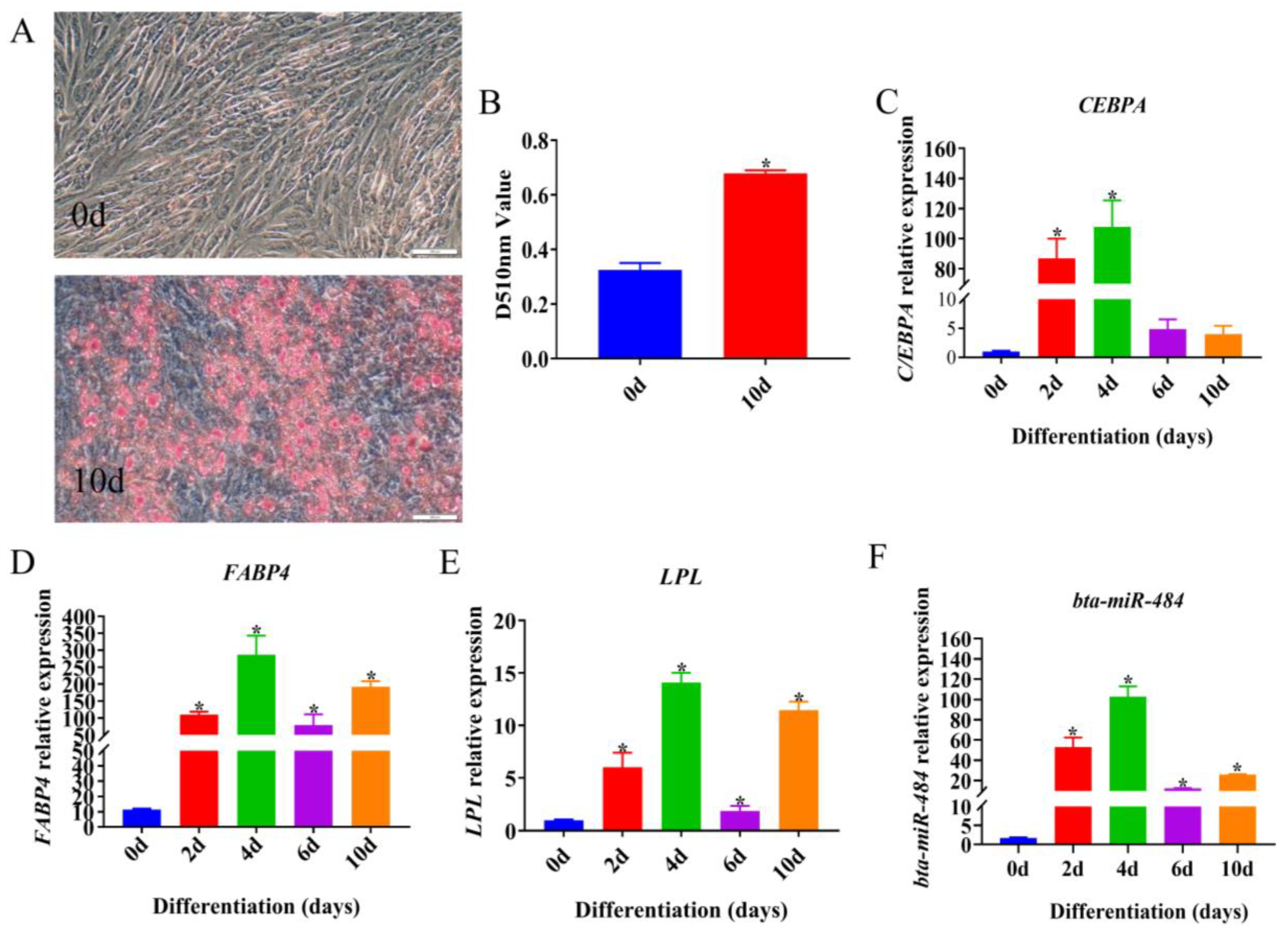
2.1. Temporal Expression Profile of bta-miR-484 during Adipose Differentiation

In order to verify whether the isolated primary adipocytes could be successfully induced to differentiate, the expression levels of adipocyte differentiation marker genes CCAAT/enhancer binding protein alpha (CEBPA), Fatty acid-binding protein (FABP4), and lipoproteinlipase (LPL) were detected at different times of induction differentiation, and the cells were stained with Oil Red O 10 d after induction differentiation. The results demonstrated that the differentiation induced promoted the accumulation of lipid droplets within adipocytes, increased the absorption value of the Oil Red O staining solution eluted by isopropanol (Figure 1A,B), and promoted the expression of CEBPA, FABP4, and LPL, which were lipid markers (Figure 1C–E). These results revealed successful differentiation of the preadipocytes used in this study, affirming their suitability for subsequent experiments. The expression of bta-miR-484 was then detected at different time points during the induction of preadipocyte differentiation. With an increase in induction time, the expression of bta-miR-484 initially increased before decreasing, reaching its highest value on day 4 (Figure 1F).

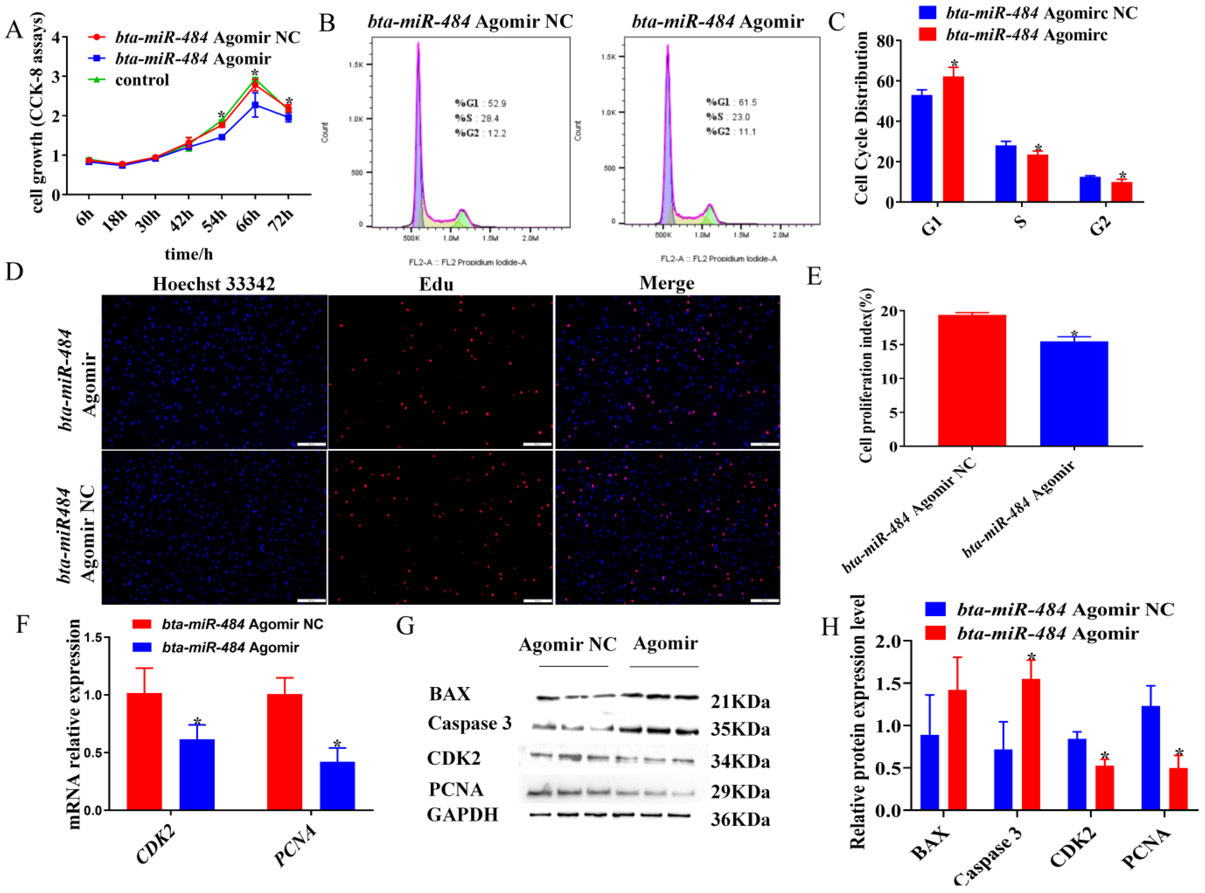
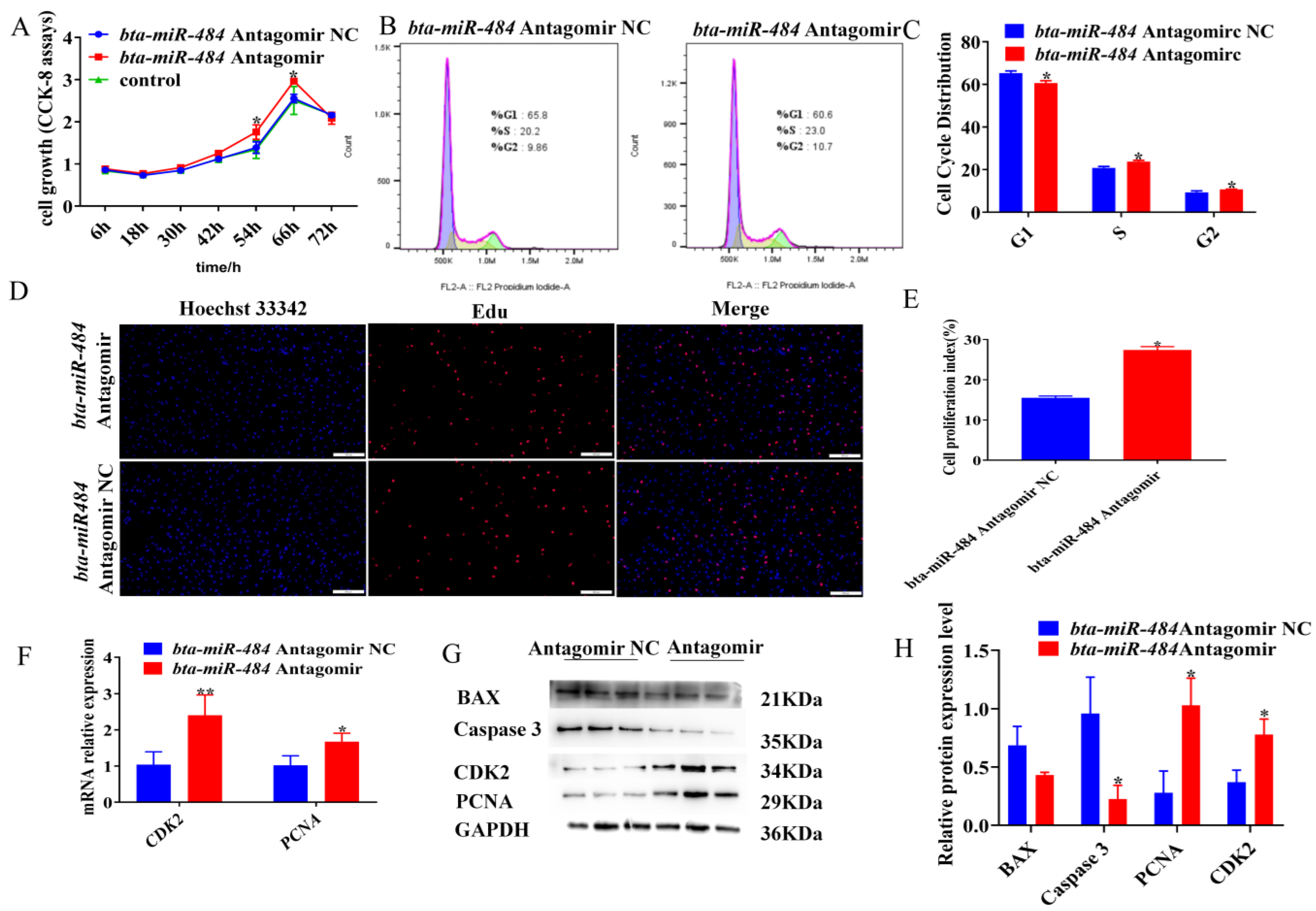
2.2. Bta-miR-484 Inhibits Adipocyte Proliferation

To investigate the role of bta-miR-484 in adipocyte proliferation, preadipocytes were transfected with a bta-miR-484 agomir, bta-miR-484 agomir negative control (agomir NC), bta-miR-484 antagomir, or bta-miR-484 antagomir negative control (antagomir NC). Subsequently, the cell counting kit-8 (CCK-8) assay, 5-ethynyl-2’-deoxyuridine (EdU) assay, flow cytometry, real-time quantitative PCR detecting system (qPCR), and western blotting (WB) were used to assess the effect of bta-miR-484 on the proliferation of adipocytes. Overall, transfection with the bta-miR-484 agomir reduced the proliferative activity of adipocytes (Figure 2A), increased the number of cells in the G1 phase cells, reduced the number of S and G2 phase cells (Figure 2B,C), decreased the number of cells in a proliferative state (Figure 2D,E), and inhibited the gene and protein expression of cyclin-dependent kinase 2 (CDK2) and proliferating cell nuclear antigen (PCNA) (Figure 2F–H). In contrast, the bta-miR-484 antagomir enhanced the proliferative activity of adipocytes (Figure 3A), decreased the number of G1 phase cells, increased the number of S and G2 phase cells (Figure 3B,C), increased the number of proliferative cells (Figure 3D,E), and promoted the genes and protein expression of CDK2 and PCNA (Figure 3F–H). Collectively, these findings indicated that bta-miR-484 inhibited adipocyte proliferation.

2.3. Bta-miR-484 Promotes Adipocyte Apoptosis

We then examined the effect of bta-miR-484 on adipocyte apoptosis. First, we performed an annexin V-FITC/PI staining assay. Overall, overexpression of bta-miR-484 was found to increase the apoptosis index of adipocytes (Figure 4A–C). Subsequently, we evaluated the mRNA expression of genes associated with cell survival by qPCR. The corresponding results demonstrated that overexpression of bta-miR-484 increased BAX, Caspase 3, and cysteine-dependent aspartate-specific proteases 9 (Caspase 9) expression (Figure 4D). Finally, WB analysis showed enhanced expressions of BAX and Caspase 3 proteins in bta-miR-484-overexpressing adipocytes (Figure 2G,H). In contrast, silencing bta-miR-484 reduced the apoptotic index of adipocytes, inhibited the expressions of the apoptosis-related marker genes BAX, Caspase 3, and Caspase 9, and decreased the expressions of BAX and Caspase 3 proteins (Figure 3G,H and Figure 4E–H). These results demonstrated that bta-miR-484 promotes adipocyte apoptosis.

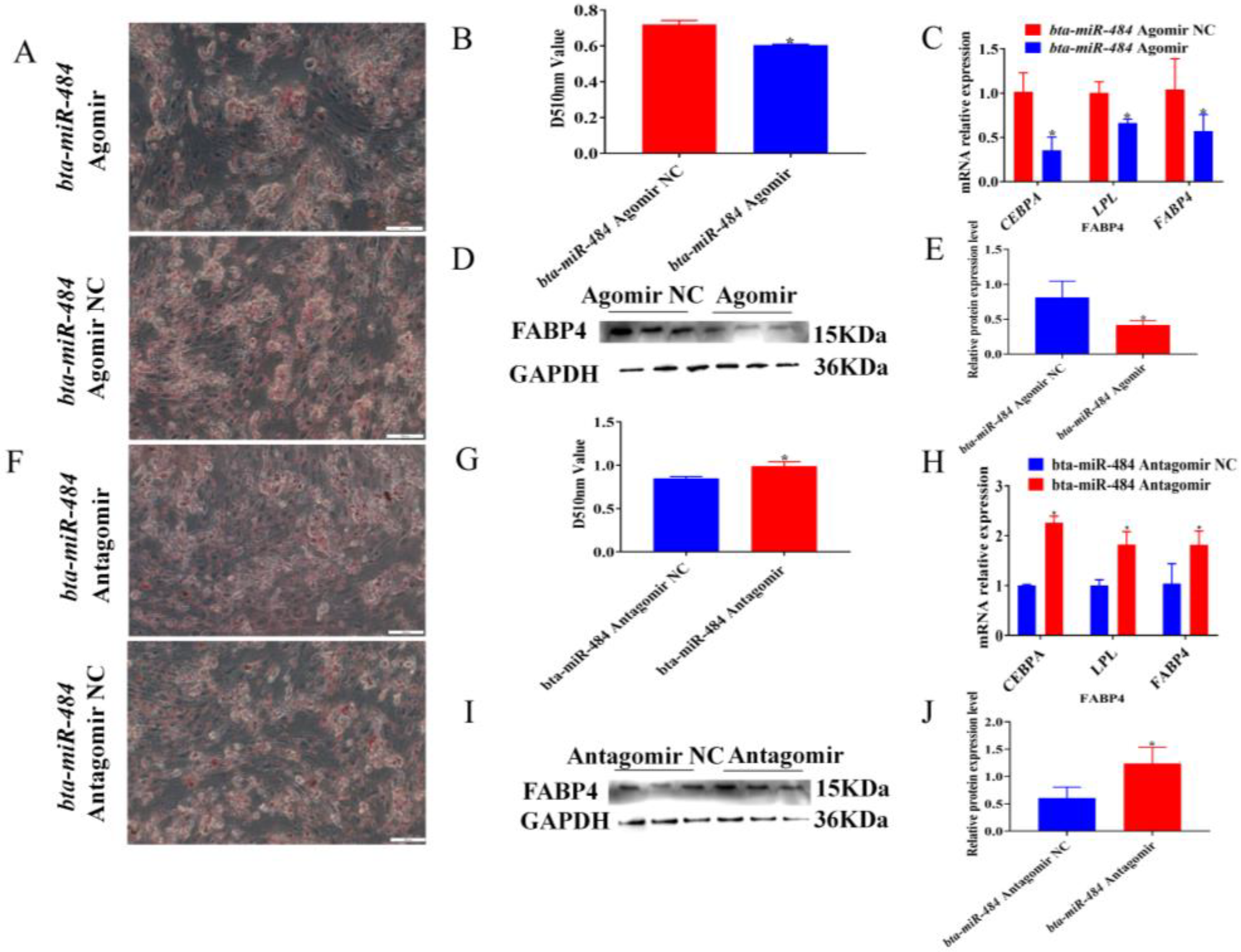
2.4. Bta-miR-484 Inhibits Adipocyte Differentiation

To explore the role of bta-miR-484 in adipocyte differentiation, bta-miR-484 agomir, bta-miR-484 agomir NC, bta-miR-484 antagomir, or bta-miR-484 antagomir NC were transfected into preadipocytes; then, 6 d after differentiation induction, the effects of bta-miR-484 on lipid droplet accumulation and the expression of adipogenesis-associated genes and proteins were assessed using Oil Red O staining, qPCR, and WB. The Oil Red O staining results indicated that the bta-miR-484 agomir inhibited the accumulation of lipid droplets and reduced the absorbance value of the Oil Red O dye eluted by isopropanol at 510 nm (Figure 5A,B). Additionally, the qPCR results demonstrated that the bta-miR-484 agomir reduced the expression of CEBPA, LPL, and FABP4 (Figure 5C). WB results established that the protein expression of FABP4 was also inhibited following adipocyte transfection with the bta-miR-484 agomir (Figure 5D,E). On the contrary, the bta-miR-484 antagomir promoted the expression of CEBPA, LPL, and FABP4 genes, upregulated the expression of FABP4 protein, and increased lipid droplet accumulation (Figure 5F,J). These results suggested that bta-miR-484 suppresses adipocyte differentiation.

2.5. RNA Sequencing Analysis

To further elucidate the role of bta-miR-484 in adipocytes, we compared the mRNA expression profiles in control and bta-miR-484 overexpressing adipocytes using RNA sequencing. In total, 51 upregulated and 77 downregulated genes were detected in adipocytes overexpressing bta-miR-484 (Figure 6A,B). The RNA sequencing results were confirmed by using real-time PCR (Figure 6C,D). Gene ontology (GO) annotation indicated that differentially expressed genes (DEGs) were enriched in the positive regulation of glycogen biosynthetic processes, glucose import, and other biological processes (Figure 6E). The clusters of orthologous groups (COG) classification revealed that these DEGs were rich in amino acid transport and metabolism, posttranslational modification, protein turnover, chaperones, secondary metabolite biosynthesis, transport and catabolism, general function prediction only, signal transduction mechanisms, and other processes (Figure 6F). Kyoto encyclopedia of genes and genomes (KEGG) enrichment analysis indicated that these DEGs were enriched in the apoptosis (2.13%), AMPK (2.13%), calcium (2.13%), Ras (4.26%), MAPK (4.26%), Wnt (4.26%), PI3K-Akt (8.51%), adipocytokin (2.13%), insulin secretion (4.26%), and PPAR signaling pathways (6.38%) (Figure 6G).

2.6. Bta-miR-484 Targeted Binding of SFRP1

To further understand how bta-miR-484 exerts its biological function, we utilized two bioinformatics tools, TargetScan and RNAhybrid, to predict its potential targets; we then combined these results with prior literature reports to screen for candidate target genes of bta-miR-484. We determined that the 3’UTR of SFRP1 possessed a target sequence of bta-miR-484; further, the free-pairing energy between bta-miR-484 and SFRP1 was calculated as -38.6 kcal/mol. Moreover, RNA sequencing results demonstrated that overexpression of bta-miR-484 down-regulated the expression level of SFRP1. Therefore, SFRP1 was selected as a candidate target gene of bta-miR-484. To determine the targeted regulatory relationship between bta-miR-484 and SFRP1, 3’UTR wild-type and mutant vectors of SFRP1 were constructed, and the nucleotide mismatch between bta-miR-484 and SFRP1 was introduced by mutation (Figure 7A). A dual luciferase reporter assay demonstrated the cotransfection of SFRP1 wild-type 3’UTR (SFRP1-3’UTR-wt) and bta-miR-484 down-regulated relative luciferase activity. However, cotransfection of SFRP1 mutant 3’UTR (SFRP1-3’UTR-mut) and bta-miR-484 could no longer inhibit relative luciferase activity (Figure 7B). Furthermore, after the overexpression or inhibition of bta-miR-484, the expression levels of SFRP1 and bta-miR-484 exhibited opposite trends (Figure 7C,D). The expression of SFRP1 was then detected at different time points during the induction of preadipocyte differentiation. With an increase in induction time, the expression of SFRP1 initially decreased before increasing. (Figure 7E). Thus, bta-miR-484 was determined to target SFRP1 and negatively regulate its expression.

3. Discussion

The fundamental property of preadipocytes was their ability to proliferate and differentiate into mature adipocytes. CEBPA and PPARγ are key regulators of adipogenesis; in particular, the primary role of CEBPA in this process is to induce the expression of PPARγ [24]. Then, activated PPARγ acts as a transcription factor for FABP4 and LPL genes expressed in mature adipocytes [25]. In the present study, isolated preadipocytes exhibited increased accumulation of lipid droplets and upregulated CEBPA, FABP4, and LPL gene expression following induction of differentiation. The temporal expression patterns of these genes were similar to those previously reported [26], indicating that the isolated preadipocytes were successfully transformed into mature adipocytes and suggesting that this model could be utilized in further bovine adipogenesis experiments. Subsequently, we evaluated the expression of bta-miR-484 at different time points during bovine adipogenesis. Overall, the bta-miR-484 expression levels initially increased before decreasing during adipocyte differentiation; this indicated that bta-miR-484 may be involved in the regulation of adipogenesis.
Cell proliferation and apoptosis were essential biological processes. E-type cyclins bind to CDK2 to promote G1/S transition during the normal cell cycle [27]. Additionally, PCNA plays a key role in DNA replication and replication-related processes [28]. Alternatively, activation of cysteine proteases (caspases) is the most widely recognized biochemical marker of early and late apoptosis. Specifically, the detection of Caspase-3 and Caspase-9 expression in cells is important for determining the induction of apoptosis [29,30]. BAX, a pro-apoptotic protein involved at the mitochondrial level, was a critical protein for enhancing apoptosis as it has the potential to induce necrotic cell death in some cases, even when caspase activation was inhibited [31]. In previous studies, miR-484 has been shown to inhibit cell viability and promote apoptosis of granulosa cells by directly targeting Yes-associated protein 1 (YAP1) [32]. Additionally, miR-484 has been observed to target CC chemokine ligand 18 (CCL-18) through the PI3K/AKT signaling pathway, thereby inducing G1 phase cell cycle arrest, inhibiting cell proliferation, and promoting apoptosis of gastric cancer cells [33]. These findings were consistent with the results of this study, which indicated that bta-miR-484 overexpression in bovine adipocytes reduces cell viability, inhibited mRNA and protein expression of proliferation marker genes (PCNA and CDK2), induced G1 phase cell cycle arrest, promoted the mRNA expression of apoptotic marker genes (BAX, Caspase 3, and Caspase 9) mRNA, promoted the protein expression of BAX and Caspase 3, and increased the number of apoptotic cells. Based on these results, we conclude that bta-miR-484 plays an important role in the proliferation and apoptosis of bovine preadipocytes.
Recently, various miRNAs have been implicated in the regulation of adipocyte differentiation. miR-424(322)/503 targets γ-synuclein (SNCG), thereby regulating adipocyte differentiation [34]. Additionally, bta-miR-149-5p regulates bovine adipogenesis by downregulating the mRNA levels of adipogenic marker genes, such as KLF6, ACSL1, SCD, SIK2, and ZEB1 in adipocytes [35]. Overexpression of miR-19a and miR-19b suppresses the expression of PPARγ and CEBPA in 3T3-L1 cells. [36]. Further, overexpression of miR-146b promotes the expression of genes associated with adipocyte differentiation, such as CEPBA, PPARγ, and AP2 [37]. In the current study, we further explored the effects of bta-miR-484 on adipocyte differentiation. Overexpression of bta-miR-484 reduced lipid droplet formation and correspondingly decreased the expression of lipogenic markers (CEBPA, FABP4, and LPL). Conversely, inhibition of bta-miR-484 expression increased the accumulation of lipid droplet and promoted the expression of lipogenic markers. Simultaneous sequencing indicated that overexpression of bta-miR-484 downregulated expression of FABP4, a gene positively associated with adipocyte differentiation, and upregulated apolipoprotein A 1 (APOA1), a gene negatively associated with adipocyte differentiation. Ultimately, these results suggest that bta-miR-484 was involved in the differentiation of bovine adipocytes, and has a negative regulatory effect on the formation and lipid accumulation of bovine preadipocytes.
SFRP1 was a Wnt antagonist involved in the regulation of adipogenesis and was expressed in both mouse and human mature adipocytes. Further, it has been determined to promote adipogenesis and inhibit the Wnt/β-catenin signaling pathway in vitro [38]. SFRP1 deficiency can increase fat weight and adipocyte size, and was regulated during adipogenesis and obesity itself. SFRP1 was upregulated in the early stages of obesity, thereby promoting adipose tissue expansion [39]. In addition, SFRP1 affects the secretion of interleukin 6 (IL-6), monocyte chemoattractant protein-1 (MCP-1), and adiponectin, which were positively correlated with insulin sensitivity [40]. SFRP1 has been previously identified as a direct target of miR-542-3p using a luciferase reporting assay. Additionally, miR-542-3p can alleviate the increase in adipogenesis after methotrexate treatment by inhibiting SFRP1 [41]. However, the involvement of SFRP1 as a direct target of bta-miR-484 in the regulation of adipocyte differentiation remained unclear. Nonetheless, the current study identified SFRP1 as a direct target of bta-miR-484 using a double-luciferase reporter assay. Additionally, the mRNA expression of SFRP1 was determined to be reduced in preadipocytes overexpressing bta-miR-484; in contrast, SFRP1 expression was increased in adipocytes after bta-miR-484 was silenced. Meanwhile, the corresponding RNA-seq results demonstrated that overexpression of bta-miR-484 downregulated the expression of the SFRP1 gene enriched in the Wnt signaling pathway. In addition, the expression of SFRP1 and bta-miR-484 showed an opposite trend with increasing time of adipocyte-induced differentiation. These results indicate that SFRP1 is a target of bta-miR-484-mediated adipocyte differentiation. In conclusion, bta-miR-484 negatively regulates adipocyte differentiation; this provides a novel molecular target for elucidating the regulatory mechanisms of adipocyte differentiation and adipogenesis in cattle.

4. Materials and Methods

4.1. Materials

Subcutaneous adipose tissue was collected from three premature calf. The adipose tissue was washed 4 times with PBS containing 2% penicillin-streptomycin (Pen-Strep, Hyclone, UT, USA). The visible nerves, blood vessels, and connective tissues on the surface were removed. Then, the adipose tissue was transferred to PBS containing 1% Pen-Strep and transported to the laboratory within 2 h at room temperature to isolate adipocytes. HEK293T cells were purchased from Fenghui Biological Corporation; these cells were resuscitated and cultured in the Key Laboratory of Ruminant Molecular Cell Breeding of Ningxia Hui Autonomous Region, before subculturing and cryopreservation.

4.2. Adipocyte Isolation, Culture and Differentiation

Preadipocytes were isolated from bovine adipose tissue using 1 mg/mL collagenase I (Sigma-Aldrich, St. Louis, MO, USA). Subsequently, these adipocytes were cultured in DMEM (Hyclone, Logan, UT, USA) containing 10% fetal bovine serum (FBS; Biological Industries, Kibbutz, Israel) and 1% Pen-Strep. After the adipocytes reached 100% confluence, the medium was replaced with induced differentiation medium. After 2 d of induction, the differentiation induction medium was discarded and maintenance medium was added. The maintenance medium was changed every 2 d before adipocytes were collected [42].

4.3. Oil Red O Staining

When adipocyte differentiation was complete, the culture medium was discarded, and the cells were washed with PBS three times and fixed with 4% paraformaldehyde at room temperature for 60 min, after which the excess paraformaldehyde solution was discarded and the cells were cleaned with 60% isopropanol. After ensuring the Petri dish was completely dry, the Oil Red O working solution was added and allowed to incubate at room temperature for 30 min. Next, Oil Red O was discarded, and the cells were washed four times with PBS. Then, images were captured using an inverted fluorescence microscope. Finally, Oil Red O was eluted from the stained cells with 100% isopropanol; then, absorbance was quantified at 510 nm using a multifunctional enzyme marker to analyze lipid droplet aggregation.

4.4. CCK-8 Assay and EdU Staining

Adipocytes were seeded in 96-well plates (Corning, New York, NY, USA) and allowed to adhere overnight. On the second day of incubation, adipocytes were transfected with bta-miR-484 agomir, bta-miR-484 agomir NC, bta-miR-484 antagomir, or bta-miR-484 antagomir NC; the medium was then replaced with fresh medium 6 h post-transfection. Subsequently, in each well, cells that were transfected for 6, 18, 30, 42, 54, 66, and 72 h were combined with 10 μL of cell counting kit-8 (CCK-8; Plomag, Beijing, China) reagents, and incubated for an additional hour. Sample absorbance was measured at 450 nm by using a multifunctional enzyme marker. A blank control was used to detect the CCK-8 absorbance of adipocytes without transfection. Finally, we analyzed the effect of bta-miR-484 on adipocyte viability across these different time points of transfection.

4.5. Flow Cytometery

Adipocytes transfected with bta-miR-484 agomir, bta-miR-484 agomir NC, bta-miR-484 antagomir, or bta-miR-484 antagomir NC for 48 h were collected via trypsin digestion. Cell cycle phases were detected using the cell cycle and apoptosis analysis kit (Beyotime, Shanghai, China) and apoptosis was detected using the annexin V-FITC apoptosis detection kit (Beyotime). Flow cytometry was performed using an accuri C6 plus cytometer (BD Biosciences, San Jose, CA, USA), and the fraction of cells in each cell cycle phase and apoptosis stage were analyzed using FlowJo software (version v10.6.2; Tree Star Inc., Ashland, OR, USA).

4.6. RNA Extraction and Quantitative Real-Time PCR (qPCR)

Total RNA was harvested from adipocytes using Trizol reagent according to the manufacturer’s instructions (Invitrogen, Carlsbad, CA, USA). RNA concentration was then determined using multifunctional enzyme markers. Next, the total RNA was reverse-transcribed into cDNA using the PrimeScript RT kit (Takara, Kusatsu, Shiga, Japan); the cDNA obtained by replacing universal primers with specific stem ring primers was then used as a template for miRNA quantification. Then, qPCR was performed using a CFX96 system (Bio-Rad, Hercules, CA, USA) with SYBR Green Supermix (Takara). The sequences of the oligonucleotide primers used in this study were listed in Table S1. Glyceraldehyde-3-phosphate dehydrogenase (GAPDH)-normalized mRNA, U6-normalized miRNA, and 2−ΔΔCT were used to analyze the relative mRNA abundance of each gene [43].

4.7. Western Blotting

Adipocytes were transfected with a bta-miR-484 agomir, bta-miR-484 agomir NC, bta-miR-484 antagomir, or bta-miR-484 antagomir NC before differentiation was induced. Adipocytes were collected using trypsin digestion (Hyclone) 2 d after transfection and 6 d after induction of differentiation. Proteins were isolated from adipocytes using a whole protein extraction kit (KeyGEN BioTec, Jiangsu, China) and total protein was quantified using a BCA protein quantification kit (KeyGEN BioTec). Denatured protein samples were then added to a 10% gel tank before electrophoresis at 80 v for 2 h, until they had transferred to the bottom layer of the separated gel. Using the wet transfer method, protein strips were electrotransferred to a PDVF membrane; then, the PDVF membrane was transferred to 5% skimmed milk powder and sealed for 2 h. Then, the milk powder was discarded, the membrane was washed 1–2 times with TBST, and the primary antibody was added before incubation overnight at 4 °C. Next, the membrane was washed 3 times with TBST for 10 min each time, the waste solution was discarded, the secondary antibody was added, and the membra ne was incubated for 2 h at a constant temperature. Finally, the PVDF membrane was treated with 200 µL of ultra-sensitive luminescent solution; the corresponding images were collected and stored by an imager. Image J software (version v10.6.2). was then used for quantitative analysis of protein bands. Antibody information is shown in Table S2.

4.8. RNA-Seq

Adipocytes transfected with bta-miR-484 agomir or bta-miR-484 agomir NC 48 h were collected using TRIzol (n = 3); then, second-generation transcriptome sequencing was performed at BioMarker Technology (Qingdao, Shandong, China) using an Illumina HiSeq TM sequencing platform. The primer used to verify the sequencing results were shown in Table S3. BMKCloud (www.biocloud.net, accessed on 21 June 2022) was used for differential expression screening, GO functional annotation, COG classification, and KEGG pathway enrichment analyses.

4.9. Target Gene Prediction

TargetScan databases (http://www.targetscan.org/vert_72/; accessed on 9 October 2021) were used to predict the bta-miR-484 target mRNA, and the RNAhybrid database (https://bibiserv.cebitec.uni-bielefeld.de/mahybrid/; accessed on 9 October 2021) was applied to the analysis of bta-miR-484 candidate targets and to calculate the free energy of interaction between bta-miR-484 and these candidate genes [44]. At the same time, combined with the literature reports, the candidate target genes of bta-miR-484 were screened.

4.10. Dual Luciferase Reporter Gene Assay

Dual luciferase reporter vector with SFRP1-3’UTR-wt and SFRP1-3’UTR-mut binding sites were constructed by General Biol (Anhui, China). SFRP1-3’UTR-wt or SFRP1-3’UTR-mut were co-transfected with bta-miR-484 agomir or bta-miR-484 agomir NC into HEK293T cells cultured to 70% confluence. Lipofectamine 3000 (Invitrogen) was used as the transfection reagent in this protocol. Then, these HEK293T Cells were harvested 48 h after transfection, and the relative fluorescence activity was measured using the Dual-Glo Luciferase Assay System (Promega, Madison, WI, USA) according to the manufacturer’s instruction.

4.11. Statistical Analyses

Differences between the groups were analyzed using a Student’s t-test when comparing two groups or a one-way analysis of variance (ANOVA) when comparing two groups. All data were presented as mean  ±  standard deviation (SD). * p < 0.05 was considered statistically significant. GraphPad Prism 7 (GraphPad, San Diego, CA, USA) was used for graphical representation of the data.

5. Conclusions

In the present study, we performed a series of assays to characterize and evaluate the function of bta-miR-484; these suggested that bta-miR-484 targets SFRP1 and regulates proliferation, differentiation, and apoptosis. Overall, these results provide a strong reference for further elucidation of the mechanism of action of bta-miR-484 in bovine adipogenesis.

Supplementary Materials

The following supporting information can be downloaded at: https://www.mdpi.com/article/10.3390/ijms241612710/s1.

Author Contributions

Y.M., M.Y. and X.G. conceived and designed the research. M.Y. and X.G. analyzed the data and conducted the experiments. M.Y. wrote the manuscript. Y.M., C.H., S.W. and H.S. modified the manuscript. All the authors read and approved the final manuscript. All authors have read and agreed to the published version of the manuscript.

Funding

This study was funded by the National Natural Science Foundation of China (U22A20506, 32072720), the Key R & D projects in Ningxia Hui Autonomous Region (2023BCF01006, 2021BEF01002, 2021NXZD1), Leading Talents Fund in Science and Technology Innovation in Ningxia Hui Autonomous Region (2020GKLRLX02).

Institutional Review Board Statement

Animal experiments were conducted according to the Regulations for the Administration of Affairs Concerning Experimental Animals (Ministry of Science and Technology, China, 2004). All animal protocols were approved by the Animal Ethics Committee of Ningxia University (permit number NXUC20200618) and Zerui Ecological Breeding Farm (permit number ZR20200615) [45].

Informed Consent Statement

Informed consent was obtained from all subjects involved in the study.

Data Availability Statement

The RNA-seq data (GSE238109) were uploaded to GEO (https://www.ncbi.nlm.nih.gov/gds/?term=GSE238109, accessed on 31 July 2023).

Conflicts of Interest

The authors have declared that no conflict of interest exists.

References

  1. Kang, Z.; Zhang, S.; Jiang, E.; Wan, F.; Lan, X.; Liu, M. Mir-193b Regulates the Differentiation, Proliferation, and Apoptosis of Bovine Adipose Cells by Targeting the ACSS2/AKT Axis. Animals 2020, 10, 1265. [Google Scholar] [CrossRef] [PubMed]
  2. Du, J.; Zhang, P.; Gan, M.; Zhao, X.; Xu, Y.; Li, Q.; Jiang, Y.; Tang, G.; Li, M.; Wang, J.; et al. MicroRNA-204-5p regulates 3T3-L1 preadipocyte proliferation, apoptosis and differentiation. Gene 2018, 668, 1–7. [Google Scholar] [CrossRef] [PubMed]
  3. Roos, J.; Dahlhaus, M.; Funcke, J.B.; Kustermann, M.; Strauss, G.; Halbgebauer, D.; Boldrin, E.; Holzmann, K.; Möller, P.; Trojanowski, B.M.; et al. miR-146a regulates insulin sensitivity via NPR3. Cell Mol. Life Sci. 2021, 78, 2987–3003. [Google Scholar] [CrossRef] [PubMed]
  4. Yamaguchi, S.; Zhang, D.; Katayama, A.; Kurooka, N.; Sugawara, R.; Albuayjan, H.H.H.; Nakatsuka, A.; Eguchi, J.; Wada, J. Adipocyte-Specific Inhibition of Mir221/222 Ameliorates Diet-Induced Obesity Through Targeting Ddit4. Front. Endocrinol. 2022, 12, 750261. [Google Scholar] [CrossRef]
  5. Murata, Y.; Yamashiro, T.; Kessoku, T.; Jahan, I.; Usuda, H.; Tanaka, T.; Okamoto, T.; Nakajima, A.; Wada, K. Up-Regulated MicroRNA-27b Promotes Adipocyte Differentiation via Induction of Acyl-CoA Thioesterase 2 Expression. Biomed. Res. Int. 2019, 2019, 2916243. [Google Scholar] [CrossRef] [Green Version]
  6. Acharya, A.; Berry, D.C.; Zhang, H.; Jiang, Y.; Jones, B.T.; Hammer, R.E.; Graff, J.M.; Mendell, J.T. miR-26 suppresses adipocyte progenitor differentiation and fat production by targeting Fbxl19. Genes Dev. 2019, 33, 1367–1380. [Google Scholar] [CrossRef] [Green Version]
  7. Ahonen, M.A.; Haridas, P.A.N.; Mysore, R.; Wabitsch, M.; Fischer-Posovszky, P.; Olkkonen, V.M. miR-107 inhibits CDK6 expression, differentiation, and lipid storage in human adipocytes. Mol. Cell Endocrinol. 2019, 479, 110–116. [Google Scholar] [CrossRef] [Green Version]
  8. Chen, X.; Raza, S.H.A.; Ma, X.; Wang, J.; Wang, X.; Liang, C.; Yang, X.; Mei, C.; Suhail, S.M.; Zan, L. Bovine Pre-adipocyte Adipogenesis Is Regulated by bta-miR-150 Through mTOR Signaling. Front. Genet. 2021, 12, 636550. [Google Scholar] [CrossRef]
  9. Zhang, Y.; Ma, L.; Gu, Y.; Chang, Y.; Liang, C.; Guo, X.; Bao, P.; Chu, M.; Ding, X.; Yan, P. Bta-miR-2400 Targets SUMO1 to Affect Yak Preadipocytes Proliferation and Differentiation. Biology 2021, 10, 949. [Google Scholar] [CrossRef]
  10. Khan, R.; Raza, S.H.A.; Junjvlieke, Z.; Wang, X.; Wang, H.; Cheng, G.; Mei, C.; Elsaeid Elnour, I.; Zan, L. Bta-miR-149-5p inhibits proliferation and differentiation of bovine adipocytes through targeting CRTCs at both transcriptional and posttranscriptional levels. J. Cell Physiol. 2020, 235, 5796–5810. [Google Scholar] [CrossRef]
  11. Chen, X.; Raza, S.H.A.; Cheng, G.; Ma, X.; Wang, J.; Zan, L. Bta-miR-376a Targeting KLF15 Interferes with Adipogenesis Signaling Pathway to Promote Differentiation of Qinchuan Beef Cattle Preadipocytes. Animals 2020, 10, 2362. [Google Scholar] [CrossRef]
  12. Castrillón-Betancur, J.C.; Urcuqui-Inchima, S. Overexpression of miR-484 and miR-744 in Vero cells alters Dengue virus replication. Mem. Inst. Oswaldo Cruz 2017, 112, 281–291. [Google Scholar] [CrossRef] [PubMed] [Green Version]
  13. Masoodi Khabar, P.; Ghydari, M.E.; Vazifeh Shiran, N.; Shirazy, M.; Hamidpour, M. Platelet MicroRNA-484 as a Novel Diagnostic Biomarker for Acute Coronary Syndrome. Lab. Med. 2023, 54, 256–261. [Google Scholar] [CrossRef]
  14. Mohamed, D.A.W.; Selim, H.M.; Elmazny, A.; Genena, A.; Nabil, M.M. Apoptotic protease activating factor-1 gene and MicroRNA-484: A possible interplay in relapsing remitting multiple sclerosis. Mult. Scler. Relat. Disord. 2022, 58, 103502. [Google Scholar] [CrossRef] [PubMed]
  15. El-Maraghy, S.A.; Adel, O.; Zayedm, N.; Yosry, A.; El-Nahaas, S.M.; Gibriel, A.A. Circulatory miRNA-484, 524, 615 and 628 expression profiling in HCV mediated HCC among Egyptian patients; implications for diagnosis and staging of hepatic cirrhosis and fibrosis. J. Adv. Res. 2019, 22, 57–66. [Google Scholar] [CrossRef]
  16. Konaka, T.; Kawami, M.; Yamamoto, A.; Yumoto, R.; Takano, M. miR-484: A Possible Indicator of Drug-Induced Pulmonary Fibrosis. J. Pharm. Pharm. Sci. 2020, 23, 486–495. [Google Scholar] [CrossRef] [PubMed]
  17. Lee, D.; Tang, W.; Dorsey, T.H.; Ambs, S. miR-484 is associated with disease recurrence and promotes migration in prostate cancer. Biosci. Rep. 2020, 40, BSR20191028. [Google Scholar] [CrossRef] [PubMed]
  18. Vecchione, A.; Belletti, B.; Lovat, F.; Volinia, S.; Chiappetta, G.; Giglio, S.; Sonego, M.; Cirombella, R.; Onesti, E.C.; Pellegrini, P.; et al. A microRNA signature defines chemoresistance in ovarian cancer through modulation of angiogenesis. Proc. Natl. Acad. Sci. USA 2013, 110, 9845–9850. [Google Scholar] [CrossRef]
  19. Merhautova, J.; Hezova, R.; Poprach, A.; Kovarikova, A.; Radova, L.; Svoboda, M.; Vyzula, R.; Demlova, R.; Slaby, O. miR-155 and miR-484 Are Associated with Time to Progression in Metastatic Renal Cell Carcinoma Treated with Sunitinib. Biomed. Res. Int. 2015, 2015, 941980. [Google Scholar] [CrossRef] [Green Version]
  20. Zare, A.; Ahadi, A.; Larki, P.; Omrani, M.D.; Zali, M.R.; Alamdari, N.M.; Ghaedi, H. The clinical significance of miR-335, miR-124, miR-218 and miR-484 downregulation in gastric cancer. Mol. Biol. Rep. 2018, 45, 1587–1595. [Google Scholar] [CrossRef]
  21. Allach, E.l.; Khattabi, L.; Heide, S.; Caberg, J.H.; Andrieux, J.; Doco Fenzy, M.; Vincent-Delorme, C.; Callier, P.; Chantot-Bastaraud, S.; Afenjar, A.; et al. 16p13.11 microduplication in 45 new patients: Refined clinical significance and genotype-phenotype correlations. J. Med. Genet. 2020, 57, 301–307. [Google Scholar] [CrossRef]
  22. Bradshaw, N.J.; Ukkola-Vuoti, L.; Pankakoski, M.; Zheutlin, A.B.; Ortega-Alonso, A.; Torniainen-Holm, M.; Sinha, V.; Therman, S.; Paunio, T.; Suvisaari, J.; et al. The NDE1 genomic locus can affect treatment of psychiatric illness through gene expression changes related to microRNA-484. Open Biol. 2017, 7, 170153. [Google Scholar] [CrossRef] [PubMed]
  23. Du, X. Molecular Characteristics of H-FABP Gene and Identification of miRNAs Associated with Intramuscular Fat Deposition in Beef Cattle; Shenyang Agricultural University: Shenyang, China, 2016. [Google Scholar]
  24. Rosen, E.D. The transcriptional basis of adipocyte development. Prostaglandins Leukot. Essent. Fat. Acids 2005, 73, 31–34. [Google Scholar] [CrossRef] [PubMed]
  25. Tontonoz, P.; Spiegelman, B.M. Fat and beyond: The diverse biology of PPARgamma. Annu. Rev. Biochem. 2008, 77, 289–312. [Google Scholar] [CrossRef] [PubMed]
  26. Zhang, Y.; Wu, X.; Liang, C.; Bao, P.; Ding, X.; Chu, M.; Jia, C.; Guo, X.; Yan, P. MicroRNA-200a regulates adipocyte differentiation in the domestic yak Bos grunniens. Gene 2018, 650, 41–48. [Google Scholar] [CrossRef]
  27. Fagundes, R.; Teixeira, L.K. Cyclin E/CDK2: DNA Replication, Replication Stress and Genomic Instability. Front. Cell Dev. Biol. 2021, 9, 774845. [Google Scholar] [CrossRef]
  28. González-Magaña, A.; Blanco, F.J. Human PCNA Structure, Function and Interactions. Biomolecules 2020, 10, 570. [Google Scholar] [CrossRef] [Green Version]
  29. Eskandari, E.; Eaves, C.J. Paradoxical roles of caspase-3 in regulating cell survival, proliferation, and tumorigenesis. J. Cell Biol. 2022, 221, e202201159. [Google Scholar] [CrossRef]
  30. Araya, L.E.; Soni, I.V.; Hardy, J.A.; Julien, O. Deorphanizing Caspase-3 and Caspase-9 Substrates in and out of Apoptosis with Deep Substrate Profiling. ACS Chem. Biol. 2021, 16, 2280–2296. [Google Scholar] [CrossRef]
  31. Jensen, K.; WuWong, D.J.; Wong, S.; Matsuyama, M.; Matsuyama, S. Pharmacological inhibition of Bax-induced cell death: Bax-inhibiting peptides and small compounds inhibiting Bax. Exp. Biol. Med. 2019, 244, 621–629. [Google Scholar] [CrossRef] [Green Version]
  32. Li, H.; Wang, X.; Mu, H.; Mei, Q.; Liu, Y.; Min, Z.; Zhang, L.; Su, P.; Xiang, W. Mir-484 contributes to diminished ovarian reserve by regulating granulosa cell function via YAP1-mediated mitochondrial function and apoptosis. Int. J. Biol. Sci. 2022, 18, 1008–1021. [Google Scholar] [CrossRef] [PubMed]
  33. Liu, J.; Li, S.M. MiR-484 suppressed proliferation, migration, invasion and induced apoptosis of gastric cancer via targeting CCL-18. Int. J. Exp. Pathol. 2020, 101, 203–214. [Google Scholar] [CrossRef]
  34. Rodríguez-Barrueco, R.; Latorre, J.; Devis-Jáuregui, L.; Lluch, A.; Bonifaci, N.; Llobet, F.J.; Olivan, M.; Coll-Iglesias, L.; Gassner, K.; Davis, M.L.; et al. A microRNA Cluster Controls Fat Cell Differentiation and Adipose Tissue Expansion by Regulating SNCG. Adv. Sci. 2022, 9, e2104759. [Google Scholar] [CrossRef] [PubMed]
  35. Guo, H.; Khan, R.; Abbas Raza, S.H.; Suhail, S.M.; Khan, H.; Khan, S.B.; Abd El-Aziz, A.H.; Zan, L. RNA-Seq Reveals Function of Bta-miR-149-5p in the Regulation of Bovine Adipocyte Differentiation. Animals 2021, 11, 1207. [Google Scholar] [CrossRef]
  36. Juiz-Valiña, P.; Varela-Rodríguez, B.M.; Outeiriño-Blanco, E.; García-Brao, M.J.; Mena, E.; Cordido, F.; Sangiao-Alvarellos, S. MiR-19 Family Impairs Adipogenesis by the Downregulation of the PPARγ Transcriptional Network. Int. J. Mol. Sci. 2022, 13, 15792. [Google Scholar] [CrossRef] [PubMed]
  37. Ahn, J.; Lee, H.; Jung, C.H.; Jeon, T.I.; Ha, T.Y. MicroRNA-146b promotes adipogenesis by suppressing the SIRT1-FOXO1 cascade. EMBO Mol. Med. 2013, 5, 1602–1612. [Google Scholar] [CrossRef] [PubMed]
  38. Lagathu, C.; Christodoulides, C.; Tan, C.Y.; Virtue, S.; Laudes, M.; Campbell, M.; Ishikawa, K.; Ortega, F.; Tinahones, F.J.; Fernández-Real, J.M.; et al. Secreted frizzled-related protein 1 regulates adipose tissue expansion and is dysregulated in severe obesity. Int. J. Obes. 2010, 34, 1695–1705. [Google Scholar] [CrossRef] [Green Version]
  39. Martínez-Uña, M.; Varela-Rey, M.; Cano, A.; Fernández-Ares, L.; Beraza, N.; Aurrekoetxea, I.; Martínez-Arranz, I.; García-Rodríguez, J.L.; Buqué, X.; Mestre, D.; et al. Excess S-adenosylmethionine reroutes phosphatidylethanolamine towards phosphatidylcholine and triglyceride synthesis. Hepatology 2013, 58, 1296–1305. [Google Scholar] [CrossRef] [Green Version]
  40. Ehrlund, A.; Mejhert, N.; Lorente-Cebrián, S.; Aström, G.; Dahlman, I.; Laurencikiene, J.; Rydén, M. Characterization of the Wnt Inhibitors Secreted Frizzled-Related Proteins (SFRPs) in Human Adipose Tissue. J. Clin. Endocrinol. Metab. 2013, 98, E503–E508. [Google Scholar] [CrossRef]
  41. Zhang, Y.L.; Liu, L.; Su, Y.W.; Xian, C.J. miR-542-3p Attenuates Bone Loss and Marrow Adiposity Following Methotrexate Treatment by Targeting sFRP-1 and Smurf2. Int. J. Mol. Sci. 2021, 22, 10988. [Google Scholar] [CrossRef]
  42. Yang, M.; Gao, X.; Ma, Y.; Wang, X.; Lei, Z.; Wang, S.; Hu, H.; Tang, L.; Ma, Y. Bta-miR-6517 promotes proliferation and inhibits differentiation of pre-adipocytes by targeting PFKL. J. Anim. Physiol. Anim. Nutr. 2022, 106, 1197–1207. [Google Scholar] [CrossRef] [PubMed]
  43. Livak, K.J.; Schmittgen, T.D. Analysis of relative gene expression data using real-time quantitative PCR and the 2(-Delta Delta C(T)) Method. Methods 2001, 25, 402–408. [Google Scholar] [CrossRef] [PubMed]
  44. Riffo-Campos, Á.L.; Riquelme, I.; Brebi-Mieville, P. Tools for Sequence-Based miRNA Target Prediction: What to Choose? Int. J. Mol. Sci. 2016, 9, 1987. [Google Scholar] [CrossRef] [Green Version]
  45. Pan, C.; Lei, Z.; Wang, S.; Wang, X.; Wei, D.; Cai, X.; Luoreng, Z.; Wang, L.; Ma, Y. Genome-wide identification of cyclin-dependent kinase (CDK) genes affecting adipocyte differentiation in cattle. BMC Genom. 2021, 22, 532. [Google Scholar] [CrossRef] [PubMed]
Figure 1. Induced preadipocytes differentiation. (A) Oil Red O staining at 0 d and 10 d after induced preadipocytes differentiation. Scale bar: 200 μm. (B) Oil Red O absorption value detected at 510 nm. (CF) Expression of CEBPA, FABP4, LPL, and bta-miR-484 during induction of preadipocyte differentiation. Data are presented as mean ± SD. n = 3. * p < 0.05.
Ijms 24 12710 g001
Figure 2. Bta-miR-484 agomir inhibited the proliferation of preadipocytes. (A) CCK-8 assay to determine adipocyte viability. (B,C) Cell cycle phase analysis of adipocytes by flow cytometry. (D,E) Adipocyte proliferation was examined by EdU immunofluorescent staining. Red represents EdU staining; blue represents cell nuclei stained with Hoechst 33342. Scale bar: 500 μm. (F) Relative mRNA expression of CDK2 and PCNA genes in preadipocytes 48 h after transfection with a bta-miR-484 agomir or bta-miR-484 agomir NC. (G,H) Protein expression levels of CDK2, PCNA, Bcl-2-associated X protein (BAX), and cysteine-dependent aspartate-specific proteases 3 (Caspase 3) were detected in bta-miR-484 over-expressing adipocytes using WB. GAPDH was used as an internal reference. Data are presented as mean ± SD. n = 3. * p < 0.05.
Ijms 24 12710 g002
Figure 3. Inhibition of bta-miR-484 promotes adipocyte proliferation. (A) CCK-8 assay to determine adipocyte viability. (B,C) Cell cycle phase analysis of adipocytes using flow cytometry. (D,E) Adipocyte proliferation was examined by EdU immunofluorescent staining. Red represents EdU staining; blue represents cell nuclei stained with Hoechst 33342. Scale bar: 500 μm. (F) Relative mRNA expression of CDK2 and PCNA in preadipocytes 48 h after transfection with a bta-miR-484 antagomir or bta-miR-484 antagomir NC. (G,H) Protein expression levels of CDK2, PCNA, BAX, and Caspase3 were detected in bta-miR-484 down-regulated adipocytes using WB. GAPDH was used as an internal reference. Data are presented as mean ± SD. n = 3. * p < 0.05. ** p < 0.01.
Ijms 24 12710 g003
Figure 4. The effect of bta-miR-484 overexpression or silencing on adipocyte apoptosis. (AC,EG) The apoptotic phase of adipocytes was analyzed using flow cytometry. (D,H) Relative mRNA expression of BAX, Caspase 3, and Caspase 9 genes in bta-miR-484 overexpressing or bta-miR-484 inhibited adipocytes. Data are presented as mean ± SD. n = 3. * p < 0.05.
Ijms 24 12710 g004
Figure 5. Bta-miR-484 inhibits the differentiation of preadipocytes. (A,F) Graphs show brightfield microscopy of differentiated adipocytes stained by Oil Red O for bta-miR-484 overexpression or inhibition adipocytes. Scale bars: 500 μm. (B,G) The Oil Red O dye was eluted with 100% isopropanol and the absorbance was detected at 510 nm. (C,H) Expression of CEBPA, LPL, and FABP4 genes was measured 6 d after induction of differentiation in bta-miR-484- overexpressing or inhibiting adipocytes. (D,E,I,J) Protein levels of FABP4 were detected by the WB of adipocytes with bta-miR-484 overexpression or inhibition. GAPDH was used as an internal reference. Data are presented as mean ± SD. n = 3. * p < 0.05.
Ijms 24 12710 g005
Figure 6. RNA sequencing analysis of bta-miR-484 overexpression in adipocytes. (A) Differential expression volcano plot. (B) Heat map of differentially expressed mRNAs in control and bta-miR-484 overexpressing adipocytes. A484 = “bta-miR-484 agomir”, NC484 = “bta-miR-484 agomir NC” (C,D) qPCR analysis of DEGs in control and bta-miR-484-overexpressing adipocytes. (E) GO enrichment histogram of differentially expressed genes. (F) COG annotation classification statistics of differentially expressed genes. (G) KEGG classification map of differentially expressed genes. Data are presented as mean ± SD. n = 3. * p < 0.05.
Ijms 24 12710 g006
Figure 7. Verification of the bta-miR-484 target gene, SFRP1. (A) Bta-miRNA-484 and SFRP1 3’UTR binding sites. (B) Dual-luciferase reporter assay to evaluate the targeting relationship between SFRP1 and bta-miR-484. (C,D) The expression level of SFRP1 in adipocytes was detected after overexpression or inhibition of bta-miRNA-484. (E) Expression of SFRP1 during induction of preadipocyte differentiation. Data are presented as mean ± SD. n = 3. * p < 0.05.
Ijms 24 12710 g007
Disclaimer/Publisher’s Note: The statements, opinions and data contained in all publications are solely those of the individual author(s) and contributor(s) and not of MDPI and/or the editor(s). MDPI and/or the editor(s) disclaim responsibility for any injury to people or property resulting from any ideas, methods, instructions or products referred to in the content.

