Article

Synthesis and Properties of (1R(S),5R(S),7R(S),8R(S))-1,8-Bis(hydroxymethyl)-6-azadispiro[4.1.4.2]tridecane-6-oxyl: Reduction-Resistant Spin Labels with High Spin Relaxation Times

by
Yulia V. Khoroshunova
1,2,
Denis A. Morozov
1,
Danil A. Kuznetsov
2,
Tatyana V. Rybalova
1,
Yurii I. Glazachev
3,
Elena G. Bagryanskaya
1 and
Igor A. Kirilyuk
1,*
1
N.N. Vorozhtsov Institute of Organic Chemistry SB RAS, Academician Lavrentiev Ave. 9, 630090 Novosibirsk, Russia
2
Department of Physics, Novosibirsk State University, Pirogova Str. 1, 630090 Novosibirsk, Russia
3
Voevodsky Institute of Chemical Kinetics and Combustion SB RAS, Institutskaya 3, 630090 Novosibirsk, Russia
*
Author to whom correspondence should be addressed.
Int. J. Mol. Sci. 2023, 24(14), 11498; https://doi.org/10.3390/ijms241411498
Submission received: 22 June 2023 / Revised: 12 July 2023 / Accepted: 13 July 2023 / Published: 15 July 2023

Abstract

:
Site-directed spin labeling followed by investigation using Electron Paramagnetic Resonance spectroscopy is a rapidly expanding powerful biophysical technique to study structure, local dynamics and functions of biomolecules using pulsed EPR techniques and nitroxides are the most widely used spin labels. Modern trends of this method include measurements directly inside a living cell, as well as measurements without deep freezing (below 70 K), which provide information that is more consistent with the behavior of the molecules under study in natural conditions. Such studies require nitroxides, which are resistant to the action of biogenic reductants and have high spin relaxation (dephasing) times, Tm. (1R(S),5R(S),7R(S),8R(S))-1,8-bis(hydroxymethyl)-6-azadispiro[4.1.4.2]tridecane-6-oxyl is a unique nitroxide that combines these features. We have developed a convenient method for the synthesis of this radical and studied the ways of its functionalization. Promising spin labels have been obtained, the parameters of their spin relaxation T1 and Tm have been measured, and the kinetics of reduction with ascorbate have been studied.

1. Introduction

Site-directed spin labeling coupled to Electron Paramagnetic Resonance spectroscopy (SDSL–EPR) is a rapidly expanding powerful biophysical technique to study biomolecules in physiologically relevant environments [1,2,3,4,5,6]. These methods are based on site-specific introductions of paramagnetic labels (unpaired electrons) into biomolecules of interest with subsequent investigation using various EPR techniques. The latter give inter-spin distances, solvent accessibility, the polarity of its immediate environment, and the dynamics of the labeled region, which are complimentary to information obtained by other methods of structural biology, such as NMR, X-ray crystallography, and cryo-electron microscopy. This helps to identify biologically important conformations of large biomolecules, especially membrane and intrinsically disordered proteins. Nitroxides are the most widely used spin labels [7]. Unlike other spin labels, they can be used both to study local dynamics in biomolecules at physiological temperatures and to measure inter-spin distances [5]. Examples of specific applications of nitroxide spin labels to structural biology studies of protein can be found in hundreds of publications [2].
The best way to obtain correct information about the native structure and functions of biomolecules implies their study in natural conditions—in a living cell. Currently, a set of evidence has been accumulated for the difference in protein structure and function in model and natural conditions [8,9,10]. For this reason, SDSL–EPR experiments in living cells attract more and more attention. A serious obstacle to the use of nitroxide spin labels in living cells is the rapid reduction of the nitroxide into diamagnetic compounds by low molecular weight reductants and enzymatic systems, typically found within cells [11]. Resistance of nitroxides to bioreduction can be strongly improved via introduction of several bulky alkyl (larger than methyl) substituents to the α-carbon atom of the nitroxide group [12]. A number of reduction-resistant spin labels for in-cell application have been developed based on tetraethyl nitroxides [13,14,15,16,17,18,19].
Several EPR methods have been developed for inter-spin distance measurement [20]. All of them are dependent on electron spin relaxation parameters of spin labels [2,21,22]. In Pulsed Electron–Electron Double Resonance (PELDOR or DEER) technique, which is currently the most popular approach for distance measurements, the maximal distance one can measure and the precision of the distance distribution are determined by the phase memory time (Tm) of the spin label [22]. The Tm parameter is temperature-dependent and must be as high as possible. To achieve optimal performance using conventional tetramethyl or tetraethyl spin labels, the measurements must be carried out at 40–65 K, because higher temperature rotation of the alkyl groups leads to a decrease in Tm. This rotation is impossible in nitroxides with spirocyclic moieties at α-carbons of nitroxide group, and these spirocyclic spin labels can be used for measurements at much higher temperatures (125 K) and even at room temperature [23]. Regretfully, the spirocyclic spin labels demonstrate much lower resistance to bioreduction compared to tetraethyl nitroxides [24].
According to expert estimates, labels that can eliminate the need for data acquisition at cryogenic temperatures and labels that can enter and survive in the cellular environment are of current and future interest [2]. The attempts to improve reduction resistance of spirocyclic nitroxides have been a subject of recent research [25]. The literature data on the rate constants of chemical reduction of representative dispirocyclic nitroxides and tetraethyl nitroxides are listed in the Figure 1. The data demonstrate that reduction resistance of the nitroxide 1 by far exceeds those of other spirocyclic nitroxides. The nitroxide group in 1 is stabilized with two spiro-(2-hydroxymethyl)cyclopentane moieties with the hydroxymethyl groups directed towards N-O. In addition, this nitroxide showed the highest longitudinal relaxation time T1 among a broad set of various nitroxides [26]. This parameter is very important for distance measurement using Saturation Recovery (SR) method, another pulsed EPR technique widely used in structural biology [2,21].
The nitroxide 1 was prepared from 3,4-di-tert-butoxypyrroline 1-oxide [31] via repetitive sequence of procedures: pent-4-en-1-ylmagnesium bromide addition, intramolecular 1,3-dipolar cycloaddition, isoxazolidine ring opening and oxidation [27,30]. The literature protocol implies multi-step synthesis with low overall yield (ca. 5%) from commercially available L-tartaric acid, making 1 unfavorable object for further chemical transformations [27,30].
Recent advances in condensation, cyclization, and dipolar cycloaddition cascade chemistry [32] allowed us to develop convenient and scalable protocol for synthesis of dispirocyclic nitroxide 5, a 3,4-unsubstituted analog of 1, with overall yield 43% from commercially available 4-chlorobutyryl chloride (6). Chemical properties of the nitroxide 5 were studied and several dispirocyclic spin labels were prepared. The new nitroxides showed improved resistance to reduction compared to 1. The spin relaxation times T1 and Tm were measured for some of the nitroxides.

2. Results and Discussion
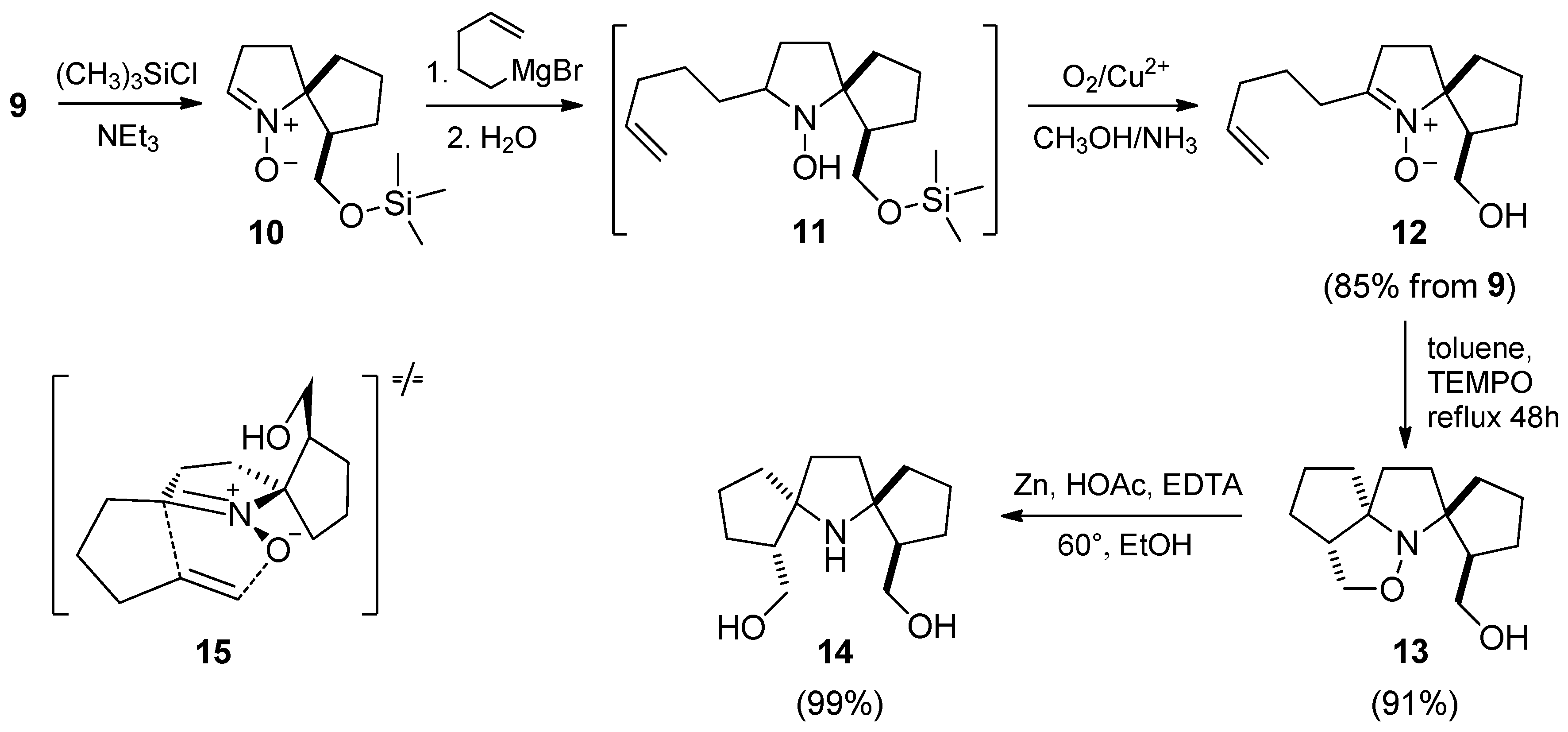
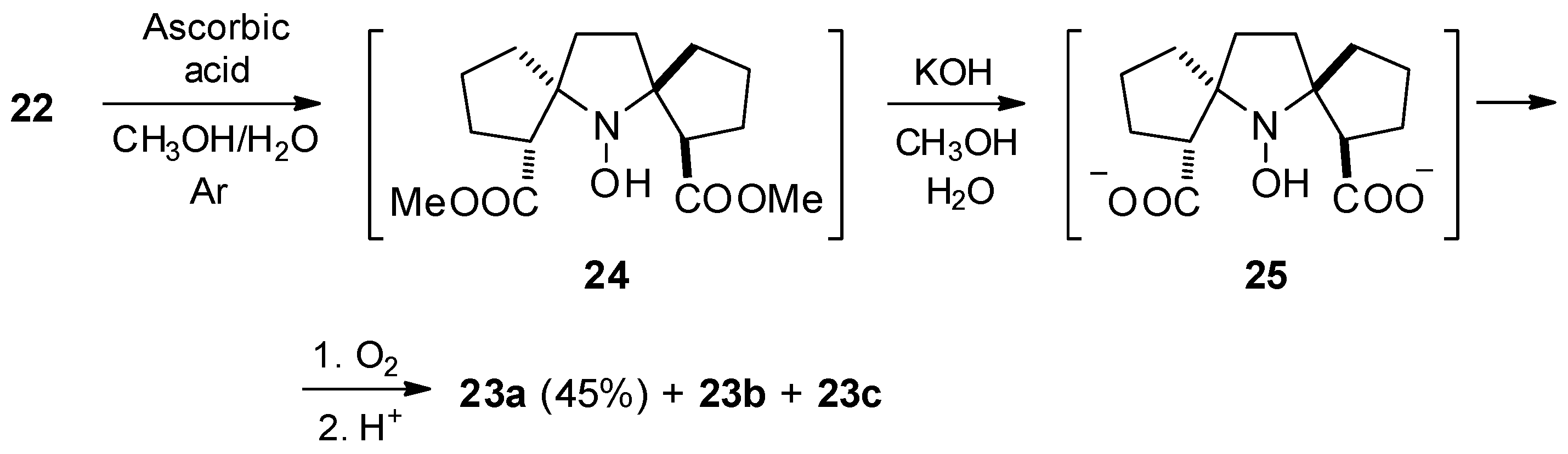
2.1. Synthesis

The (1R(S),8R(S))-6-oxa-5-azatricyclo[6.3.0.01,5]undecane (8) (6aR(S),9aR(S))-hexahydro-1H,6H-cyclopenta[c]pyrrolo[1,2-b]isoxazole) was prepared in two steps as enantiomeric mixture according to the literature protocol [32] (Scheme 1). This allowed us to avoid multi-step synthesis.
Subsequent oxidative isoxazolidine ring opening with m-cloroperoxybenzoic acid (m-CPBA) afforded aldonitrone 9. The reaction was carried out in dry dichlorometane (DCM) using dry reagent to prevent formation of hydroxamic acid. The structure of 9 was confirmed with IR, UV, 1H, and 13C NMR (see Section 3 and Figure S1, S24, S31 and S32) and X-ray diffraction data (Figure 2).
We have previously published a set of procedures to convert 1-pyrroline 1-oxides into pyrrolidine nitroxides with spiro-(2-hydroxymethyl)cyclopentane moiety [30]. Here, we used trimethylsilyl protection of the hydroxy group in order to avoid unproductive consumption of the organometallic reagent (Scheme 2). The crude silylated nitrone 10 was treated with 1.5-fold excess of pent-4-en-1-ylmagnesium bromide to give hydroxylamine 11 and the latter was in situ oxidized with air oxygen in presence of Cu2+.
The nitrone 12 easily undergoes intramolecular 1,3-dipolar cycloaddition affording single product. The best yield was achieved upon heating to reflux in toluene in presence of 2,2,6,6-tetramethylpiperidine-1-oxyl (TEMPO). Intramolecular cycloaddition in 2-pent-4-en-1-ylpyrroline 1-oxides was reported to give a single regioisomer [27,30,33]. Similarly, the 1H and 13C NMR spectra and 1H-1H and 1H-13C correlations revealed the signals of two different CH-CH2O moieties (see Section 3 and Figures S35, S36 and S64–S67), confirming formation of (hexahydro-1H-spiro[cyclopenta[c]pyrrolo[1,2-b]isoxazole-3,1′-cyclopentane]-2′-yl) methanol system. However, one could not exclude the possibility of C=C bond approaching the plane of the nitrone group from different sides with the formation of cis- and trans-isomers relative to the position of the hydroxymethyl group. To assign a structure to the cycloadduct 1H-1H NOESY NMR spectra were recorded (Figure S67). 1H-1H NOESY correlation showed a cross-peak between protons of 6CH2 and 15CH2 groups, while no cross-peak was observed between 11CH2 and 6CH2 protons (Figure 3). Apparently, the hydroxymethyl group prevents C=C from approaching the nitrone group, and the formation of a cycloadduct is possible only through the transition state 15 (Scheme 2).
Reductive scission of the isoxazolidine ring N-O bond with Zn-AcOH system afforded 14, which was isolated as a colorless crystalline solid. Half of the signal set was observed in 1H and 13C NMR spectra of 14, indicating symmetrical structure. Single-crystal X-ray diffraction data of this compound showed C2-symmetry of the molecule, which confirms our previous assignment of structure to 13. Thus, despite the absence of bulky substituents at positions 3 and 4 of the pyrroline ring, the intramolecular 1,3-dipolar cycloaddition reaction in 12 proceeds stereospecifically.
Previously, we reported that oxidation of 2,2-disubstituted 1-azaspiro[4.4]nonan-6-ylmethanols to nitroxides with m-CPBA may be accompanied with conversion of hydroxymethyl group into aldehyde, and acylation of the hydroxy group(s) before oxidation increases the nitroxide yield [30]. Following this procedure, 14 was heated with acetic anhydride (Ac2O) to give 16, which was then oxidized with m-CPBA (Scheme 3). The reaction afforded two nitroxides. The main product 17 was isolated as yellow crystals with 86% yield. The structure of 17 was confirmed by single-crystal X-ray diffraction data (Figure 2).
The structure of the minor product 18 was assigned on the basis of 1H and 13C NMR, and 1H-1H COSY, 1H-13C HSQC, and 1H-13C HMBC spectra were acquired after reduction of the nitroxide to corresponding amine with Zn in presence of trifluoroacetic acid according to the earlier described procedure [18] (see Section 3 and Figures S41, S42 and S68–S70) and confirmed with high-resolution mass-spectrum and IR spectral data (Figure S17). We have previously observed the formation of similar dehydrogenated nitroxide upon oxidation of (2,2-dimethyl-1-azaspiro[4.4]nonan-6-yl)methyl acetate, and proposed a mechanism implying proton abstraction from intermediate oxoammonium cation [30].
Nitroxide 17 was then subjected to alkaline hydrolysis to give 5 with nearly quantitative yield. The structure of 5 was confirmed by single-crystal X-ray diffraction data (Figure 4). The overall yield of this nitroxide starting from commercially available 4-chlorobutyryl chloride and 5-bromo-1-pentene exceeds 40%, which makes it an attractive material for the synthesis of spin labels.
Modification of the hydroxymethyl groups seems to be the simplest way to functional derivatives capable of binding to biomolecules. In our recent study, we demonstrated that activation of the hydroxyl group to nucleophilic substitution in 1-unsubstituted pyrrolidines and corresponding alkoxyamines, acyloxyamines, or nitroxides with spiro-(2-hydroxymethyl)cyclopentane moiety always leads to cyclization, which may be followed by rearrangement [34]. Here, we studied the oxidation of hydroxymethyl groups to carboxylic ones, and the alkylation/acylation of hydroxyl groups.
Attempts to oxidize the hydroxymethyl groups in 5 using TEMPO—Sodium chlorite system [35] were unsuccessful, leading to strong tarring. This may occur due to the formation of an oxoammonium cation, which can cause oxidative transformations in the side chains (cf. [30] and above). Therefore, we used the diamagnetic precursor 14 to prepare the desired nitroxide with carboxylate groups. Direct oxidation of 14 with Jones reagent afforded crude 20 as a colorless viscous oil (Scheme 4). The NMR spectra showed half of the signal set, showing that the compound remained a racemic mixture (no other diastereomers formed). The pure crystalline sample was isolated via column chromatography and crystallization from methanol-ethyl acetate mixture 50:1, and the structure was confirmed by single-crystal X-ray diffraction data (Figure 4).
Attempts to oxidize of 20 either with m-CPBA or H2O2/Na2WO4 were not successful, so the crude 20 was dissolved in methanol saturated with HCl and diester 21 was isolated. Oxidation of 21 with m-CPBA afforded 22, which was isolated as a yellow crystalline solid. The 1H NMR spectrum acquired after reduction of freshly prepared nitroxide with Zn/CF3COOH showed half of the signal set with significant downfield shift (due to protonation) as compared to spectrum of 21. The structure of 22 was confirmed by single-crystal X-ray diffraction data (Figure 4). Alkaline hydrolysis of 22 gave an inseparable mixture of structurally related compounds with total yield 42% (Scheme 5). The element analysis of the mixture corresponded to the formula C14H20NO5, which allowed us to assume that the product was a mixture of isomers. The 13C NMR spectrum of the mixture after reduction with Zn/CF3COOH corresponded to a mixture of three isomers, two symmetric and one asymmetric dicarboxylic acids (Figure S51). This picture corresponds to a mixture of three isomers, two symmetric, and one asymmetric dicarboxylic acids. The ratio of the isomers was estimated using integrals of the signals of methine hydrogens at 3.02–3.30 ppm in 1H NMR spectrum.
Inversion of the asymmetric center adjacent to the ester group may result from C-H acidity. TLC analysis of samples of 22 after long-term storage showed the emergence of two compounds with close Rf, probably the isomers. The formation of these compounds accelerates in the presence of a base or LiI; however, these reactions were accompanied by tarring. Presumably, in alkaline solution, isomerization proceeds faster than hydrolysis, resulting in nearly statistical ratio of isomers.
It was shown that nitroxide group can decrease pKa of the acidic center in two σ-bond distance by 2.5 orders of magnitude compared to corresponding methoxyamine derivative [36]. Thus, reduction of the nitroxide group may slow down the isomerization. To verify this hypothesis, the freshly prepared nitroxide 22 was reduced to corresponding hydroxylamine with ascorbic acid in oxygen-free conditions, and then, potassium hydroxide was added (Scheme 6). After the hydrolysis was complete, the products were oxidized with air oxygen, acidified, and extracted. Dicarboxylic acid 23a was a major component of the resulting mixture and it was isolated with the yield 45%. The structure of 23a was confirmed by single-crystal X-ray diffraction data (Figure 5).
Inversion of the asymmetric center at the ester group in 22 leads to a loss of configuration that provides higher hindrance to the nitroxide group. Despite succeeding in isolating pure 23a, it is obvious that activated esters capable of binding to biomolecules, which could be prepared from 23a, will behave similarly to 22. Thus, the nitroxides with spiro-(2-carboxy)cyclopentane moieties are not optimal for spin labeling.
Exploring the possible ways to functional derivatives of 5, we tried several alkylation and acylation reactions. A reaction of nitroxide alcohols with carbonyldiimidazole (CDI) was used for binding to primary amino groups [37,38]. A reaction of 5 with CDI afforded the nitroxide 26 with an excellent yield (Scheme 7). The structure of 26 was confirmed by single-crystal X-ray diffraction data (Figure 5). A reaction of 26 with N,N-dimethyl-1,3-diaminopropane gave 27 with 55% yield.
Another carbamate derivative 28 was prepared via treatment of 5 with methyl 3-isocyanatopropionate with quantitative yield. After alkaline hydrolysis of the ester groups corresponding dicarboxylic acid 29 was isolated. The structure of the nitroxides 27, 28, and 29 were confirmed by element analyses, and IR spectra data and 1H and 13C NMR spectra were acquired after reduction with Zn/CF3COOH, which showed half of the signal set (See Supporting Information).
Recently, copper-catalyzed azide-alkyne cycloaddition (CuAAC) was successfully used for the site-directed spin labeling of the protein in vivo [39]. The spin labels capable of binding azides were prepared via alkylation of 5 with propargyl bromide (Scheme 8). Two nitroxides 30 and 31 were isolated from the reaction mixture. The proposed structures were confirmed with IR spectra, HRMS, and element analysis data. The ability of the spin label 31 to bind to azide-containing biomolecules was demonstrated by the reaction with 2,3,4,6-tetra-O-acetyl-β-d-galactopyranosyl azide in analogy to the literature protocols [40,41,42]. The reaction of enantiomerically pure galactose derivative with racemic mixture 31 expectedly gave a mixture of diastereomers 32, which was not separated. The structure of 32 was confirmed by element analysis, and IR spectral data and 1H and 13C NMR spectra were acquired after reduction with Zn/CF3COOH (See SI). The NMR spectra of two diastereomers are very close: only few signals of carbon atoms of the spirocyclic system differ and it is impossible to assign them to a specific diastereomer. Ammonolysis of 32, in analogy with the literature procedure [42], afforded spin-labelled galactose 33, which was isolated as a glassy solid. To acquire 1H and 13C NMR spectra, the nitroxide sample was reduced with Zn in presence of formic and oxalic acids mixture. Similarly to the above-mentioned for 32, most of the signals of the two diastereomers were not resolved, and very few slightly differed in chemical shift (see Section 3 and Figures S60 and S61).

2.2. CW EPR Spectra

CW EPR spectra of nitroxides 5 and 33 are shown in Figure 6 and their parameters obtained from simulations of experimental spectra using EasySpin software are given in Table 1. For comparison spectra of 3-carboxy-Proxyl (34) and 2,2,5,5-tetramethyl-3-(((1-((2R,3R,4S,5R,6R)-3,4,5-trihydroxy-6-(hydroxymethyl)tetrahydro-2H-pyran-2-yl)-1H-1,2,3-triazol-4-yl)methoxy)methyl)pyrrolidine-1-oxyl (35) (Table 1) (see Section 3 for synthesis and Figure S21 and S63 for spectra). Measurements were carried out in aqueous solutions, and in addition for 5 and 34, the spectra were recorded in toluene (33 and 35 are poorly soluble in toluene, partition coefficients octanol/water (Kp) for 5, 29, and 33 were 11, 6, and 0.1, respectively (see Section 3 and Figure S73 for details). Broadening of the spectral lines is a typical feature of spirocyclic nitroxides (cf. [28,43]). This broadening is caused by the contribution of hyperfine interactions of an unpaired electron with hydrogen nuclei on substituents. In the spectra of 34 and 35 at room temperature, the rotation of methyl groups leads to averaging of hyperfine interactions.

2.3. Electron Spin Relaxation Time

Electron spin relaxation times were measured for four radicals 5, 33, 34, and 35 at two temperatures of 80 K and 120 K in water−glycerol solution (1:1); the data are shown on Figure 7 and are listed in Table 2. In Figure 7a, the non-exponential decay of spin echo for both radicals is clearly visible. A similar effect was described by Eaton and coworkers [22,24,26] for the phase memory time of the spin echo of nitroxyl radicals with spirocyclohexyl moieties—the positions 2 and 6 of the piperidine ring. It was shown that the observed spin echo decay curves in a water–glycerole solution (1:1) are described by the following formula: I = I0 × exp(−(t/T2)n, where n > 1, and are determined by the nuclear spin diffusion of solvent protons [22]. This effect has been studied in detail by G. Jeschke et al. in recent years [44,45]. An increase in the contribution to the spin−spin relaxation mechanism of various dynamic processes, such as the rotation of methyl groups, leads to a decrease in the value of n to 1. As expected, spirocyclic nitroxides 5 and 33 showed a less pronounced dependence of relaxation time Tm on temperature than radicals with methyl substituents 34 and 35. An increase in temperature from 80 to 120 K did not lead to a change in the time Tm for 33 within the measurement accuracy, while for radical 35 it decreased more than 3-fold from 3.4 to 1.1 μs. This effect is caused by the manifestation of an additional mechanism of electron spin relaxation due to faster rotation of methyl groups with increasing temperature. It can be seen from Figure 7b that, already at 120 K, an exponential decay of spin echo signal is observed for 35 (n ≈ 2.1), whereas for 33 there is no additional contribution to relaxation and n ≈ 2 is retained due to the absence of methyl substituents.
Curiously, binding to galactose does not cause large changes in T1, but in all cases leads to a significant increase in Tm. This effect may be associated with another mechanism of electron spin relaxation due to modulation of hfi and g-tensor anisotropy by libration motion of nitroxide molecule [46,47]. The influence of the solvent matrix to the libration motion of nitroxide ring was discussed previously and it was shown that the attachment of long chains to nitroxide ring can affect the libration motion.

2.4. Reduction Rate Constants

The kinetics of reduction of nitroxides 5, 29, and 33 with ascorbate was studied. The reaction was carried out under argon using large excess of ascorbate in presence of glutathione to suppress possible reverse processes [48] and followed by EPR. The second-order rate constants were calculated from exponential decay of the nitroxide signal (see Section 3 and Figure S71 and S72). The resulting values (kred, M−1s−1) for 5, 29, and 33 were as follows: (3.2 ± 0.2) × 10−3, (1.40 ± 0.06) × 10−3 and (2.70 ± 0.06) × 10−3, respectively. All the new radicals demonstrate higher resistance to reduction as compared to 1 (Figure 1). Interestingly, removal of electron-withdrawing tert-butoxy groups produces only minor effect on the reduction rate (cf. kred for 1 and 5). Presumably, the electron effect of t-BuO-groups is compensated by their influence on the conformation of spirocyclic fragments in analogy to [25]. An increase in steric demand of the substituents adjacent to nitroxide group is known to stabilize the radicals against reduction, enlargement of all neighboring substituents being most efficient [49]. Comparison of the reduction rates for 5, 33, and 29 shows similar effect, with 29 being the most resistant to reduction. Presumably, an increase in substituent size in the positions 1 and 8 of the 6-azadispiro[4.1.4.2]tridecane-6-oxyl system stabilize the nitroxide due to proximity of the bulky groups to the radical center. One can expect much higher stabilization effect upon binding of similar nitroxides to large biomolecules.

3. Materials and Methods

3.1. General

The IR spectra were recorded on a Bruker Vector 22 FT-IR spectrometer (Bruker, Billerica, MA, USA) in KBr pellets (1:150 ratio) or in neat samples (see the Supplementary Information in this article pp. 6–17, Figures S1–S23) and are reported in wave numbers (cm−1). UV spectra were acquired on a HP Agilent 8453 spectrometer (Agilent Technologies, Santa Clara, CA, USA) in ethanol solutions (concentration ~10−4) (see the Supplementary Information in this article pp. 18–21, Figures S24–S30). 1H NMR spectra were recorded on a Bruker AV 300 (300.132 MHz), AV 400(400.134 MHz), DRX 500 (500.130 MHz), and Bruker AV 600 (600.300 MHz) spectrometers (Bruker, Billerica, MA, USA). 13C NMR spectra were recorded on a Bruker AV 300 (75.467 MHz), AV 400 (100.614 MHz), DRX 500 (125.758 MHz), and Bruker AV 600 (150.945 MHz) spectrometers (see the Supplementary Information in this article pp. 19–42, Figures S31–S70). All the NMR spectra were acquired for 5–10% solutions in CDCl3 or CD3OD at 300 K using the signal of the solvent as a standard. NMR spectra of nitroxides for analysis and structure assignment were recorded after reduction with Zn in CD3OD-CF3COOH (or CD3OD-HCOOH-HOOCCOOH mixture) at 65 °C as described in [19] or with Zn and ND4Cl in CD3OD at 5 °C. Atoms numbering are shown in figures placed on spectra in SI. HRMS analyses were performed using a High-Resolution Mass Spectrometer DFS (Thermo Electron, Waltham, MA, USA).
Reactions were monitored by TLC on precoated TLC sheets ALUGRAM Xtra SIL G/UV254 (Macherey-Nagel GmbH & Co. KG, Düren, Germany) using UV light 254 nm, 1% aqueous permanganate, 10% solution of phosphomolybdic acid in ethanol and Dragendorff’s reagent as visualizing agents. Kieselgel 60 (Macherey-Nagel GmbH & Co. KG) or neutral alumina were utilized as an adsorbent for column chromatography.
The X-ray diffraction experiments were carried out on a Bruker KAPPA APEX II diffractometer (graphite-monochromated Mo Kα radiation). Reflection intensities were corrected for absorption by SADABS2016/2 program [50] except of 14, 17, 20 treated by SADABS2008/1 version [50]. The structures were solved by direct methods using the SHELXS-97 (Sheldrick, 2008) program [51] of 14, 17, 20, and SHELXT 2014/5 (Sheldrick, 2014) [52] for the rest ones. All compounds were refined by anisotropic (isotropic for all H atoms) full-matrix least-squares method against F2 of all reflections by SHELXL2018/3 [53]. The positions of the hydrogen atoms were calculated geometrically and refined in riding model except of hydrogenes in OH and NH-groups of 14 and 23a localized from difference map and refined independently with restriction of bond lengths. The presence of two hydrogen atoms on N1 in 20 was proved from the electron density difference map. The asymmetric units of 17, 22, and 26 include a half of molecule, the same one of 5 and 14 consists of three and two molecules, respectively. Note that cyclopentane and azalidine cycles in 22 and 23 are statistically disordered due to conformational mobility in approximate ratio 3:1 and 7:1, respectively. The imidazole cycles of 26 are also disordered due to rotational mobility in approximate ratio 1:1. For experimental details see Table S1.
Crystallographic data for 5, 9, 14, 17, 20, 22, 23a, and 26 have been deposited at the Cambridge Crystallographic Data Centre as supplementary publication no. CCDC 2269369–2269376. Copy of the data can be obtained, free of charge, on application to CCDC, 12 Union Road, Cambridge CB21EZ, UK (fax: +44-1223336033 or e-mail: deposit@ccdc.cam.ac.uk; internet: www.ccdc.cam.ac.uk).

3.2. EPR

CW EPR spectra were recorded at X-band frequencies (~9.4 GHz) on a commercial Bruker spectrometer, Elexsys E 540 (Bruker Corporation, Billerica, MA, USA). Electron spin resonance spectra were recorded with the following settings: frequency, 9.25 GHz; microwave power, 2.0 mW; modulation amplitude, 0.02–0.10 mT; time constant, 40.96 ms; and conversion time, 20.8 ms. Simulations of solution electron spin resonance lines were carried out in the EasySpin software (5.2.35), which is available at http://www.easyspin.org (accessed on 18 May 2023).
Electron spin relaxation times were measured using home-made pulse EPR spectrometers equipped with a flow helium cryostat and temperature control system [54]. Tm was measured using a two-pulse electron spin echo (ESE) sequence; T1 was measured by an inversion recovery technique with inversion π-pulse and detection of a two-pulse ESE sequence. The π-pulse length was 40 ns.

3.3. Kinetic Measurements and Partition Coefficients

For kinetic measurements, stock solutions were prepared in phosphate-citrate-borate buffer (0.5 mM of each): (1) 1 M solution of ascorbic acid and (2) 5 mM solution of glutathione (GSH). Nitroxides were dissolved in the same buffer and diluted to a concentration of 0.2–0.3 mM. The pH was adjusted to 7.5 with NaOH and the solutions were deoxygenated via bubbling with argon. The solutions carefully and quickly mixed in appropriate proportions in a small tube and placed into EPR capillary (50 μL). Oxygen-free conditions were kept permanently. Capillaries were sealed and placed into EPR resonator. EPR experiments were performed on CW EPR X-band spectrometer Bruker ER-200D (9.87 GHz). Spectra were recorded in oxygen-free conditions using the following settings: microwave power 5 mW, modulations amplitude 0.1 or 0.2 mT; time constant 100 ms; conversion time 50.12 ms. The decay of amplitude of low field component of the EPR spectrum was followed in kinetics measurements. Kinetics of decay were fitted with monoexponential function to calculate the first order rate constants. The kinetics measurements were performed at ascorbate concentrations of 100, 200, and 300 mM. The calculated first reaction constants were plotted versus ascorbate concentration; data were fitted with linear dependence. The slope corresponds to second order reaction (See Figures S71 and S72).
For partition coefficients measurements nitroxides were dissolved in 1 mL of distilled water in plastic tube. At this step, the EPR signal was measured as a zero point. Then, the fraction of octanol was added to this solution and shacked for a few minutes to achieve the stationary distribution of radical in the mixture. The tube was shortly centrifuged to separate the octanol and water fractions. The aliquot of radical solution (water phase) was carefully taken with capillary from the bottom of the tube and new EPR spectrum was recorded with the same settings. This procedure was repeated three times for three different octanol contents. The inverse normalized decrease in the EPR signal was plotted versus octanol/water ratio and the slope of the linear fit was used to determine the partition coefficient (see Figure S73).

3.4. Synthesis

2,3,4,6-Tetra-O-acetyl-β-d-galactopyranosyl azide [40], 8 [32], and 3-hydroxymethyl-2,2,5,5-tetramethylpyrrolidine-1-oxyl (36) [55] were prepared according to the literature procedures.
((1R(S),5R(S),7R(S),8R(S))-1,8-Bis(hydroxymethyl)-6-azadispiro[4.1.4.2]tridecane-6-oxyl (5). Aqueous 10% solution of NaOH (8 mL) was added to a solution of nitroxide 17 (0.335 g, 0.99 mmol) in MeOH (8 mL), and allowed to stand at room temperature until the reaction was complete (up to 12 h). The progress of the reaction was monitored by TLC (silica gel, hexane:Et2O = 1:2, visualization with UV-254). The solution was evaporated under reduced pressure. The residue was dissolved in the saturated solution of NaCl (10 mL) and extracted with Et2O (4 × 10 mL). The extract was dried with Na2SO4. After evaporation of the solvent, the crude residue was purified via column chromatography (silica gel, EtOAc) and recrystallized from Et2O to give 5 as yellow crystals. Yield: 0.236 g (94%). m.p. 75–77 °C. UV-Vis (λmax (nm)/lg(ε)): 240 (3.24). IR (KBr, cm−1): 3276 (O-H). Analysis: found C 65.85, H 9.90, N 5.45; calcd. for C14H24NO3 C 66.11, H 9.51, N 5.51. HRMS (EI/DFS) m/z [M+]: found: 254.1748; calcd. for C14H24NO3: 254.1751.
[(5R(S),6R(S))-1-Oxido-1-azaspiro[4.4]non-1-en-6-yl]methanol (9). The solution of MCPBA (20.29 g; 117.6 mmol) in dry CH2Cl2 (200 mL) was added portion-wise within 1 h to the solution of 8 (18.0 g; 117.6 mmol) in dry CH2Cl2 (100 mL) under argon at −50 °C. The mixture was allowed to warm up to room temperature (TLC control on silica gel, CHCl3:CH3OH = 10:1, Rf = 0.4, visualization with UV-254 and Dragendorff’s reagent). Then the mixture was concentrated in a vacuum, and the residue was separated via column chromatography (Al2O3, CH2Cl2) to give 9 as colorless crystals, m.p. 67–68 °C (from Et2O). Yield 15.9 g (80%). UV-Vis (λmax (nm)/lg(ε)): 232 (3.91). IR (KBr, cm−1): 3222 (O-H), 3074.1 (H-C=), 1593 (C=N). Analysis: found C 63.90, H 8.44, N 8.27; calcd. for C9H14NO C 63.88, H 8.93, N 8.28. HRMS (EI/DFS) m/z [M-17] +: found: 152.1069; calcd. for C9H14NO: 152.1070. 1H NMR (400 MHz, CDCl3, δ): 1.55 (dddd, Jd1 = 3.9, Jd2 = 8.1, Jd3 = 10.2, Jd4 = 16.5, 1H 7CH2); 1.69 (ddd, Jd1 = 2.8, Jd2 = 8.1, Jd3 = 13.1, 1H 6CH2); 1.78 (dddd, Jd1 = 3.6, Jd2 = 3.9, Jd3 = 8.1, Jd4 = 12.6, 1H 8CH2); 1.88 (ddd, Jd1 = 7.3, Jd2 = 7.9, Jd3 = 12.6, 1H 8CH2); 1.93 (ddddd, Jd1 = 2.8, Jd2 = 3.6, Jd3 = 7.9, Jd4 = 8.5 Jd5 = 16.5, 1H 7CH2); 2.01 (dddd, Jd1 = 3.7, Jd2 = 5.5, Jd3 = 7.3, Jd4 = 8.1, 1H 9CH); 2.13 (ddd, Jd1 = 6.7, Jd2 = 8.9, Jd3 = 13.0, 1H 4CH2); 2.19 (ddd, Jd1 = 4.9, Jd2 = 8.3, Jd3 = 13.0, 1H 4CH2); 2.52 (ddd, Jd1 = 8.5, Jd2 = 10.2, Jd3 = 13.1, 1H 6CH2); 2.53 (dddd, Jd1 = 2.6, Jd2 = 4.9, Jd3 = 8.9, Jd4 = 18.6, 1H 3CH2); 2.63 (dddd, Jd1 = 2.6, Jd2 = 6.7, Jd3 = 8.3, Jd4 = 18.6, 1H 3CH2); 3.62 (ddd, Jd1 = 5.5, Jd2 = 5.8, Jd3 = 13.7, 1H 10CH2); 3.65 (ddd, Jd1 = 3.7, Jd2 = 7.4, Jd3 = 13.7, 1H 10CH2); 4.81 (dd, Jd1 = 5.8, Jd2 = 7.4, 1H OH); 6.99 (dd, Jd1 = 2.6, Jd2 = 2.6, 1H 2CH=). 13C NMR (125 MHz, CDCl3, δ): 22.87, 25.34, 27.60, 35.55, 36.61 (3CH2, 4CH2, 6CH2, 7CH2, 8CH2), 51.22 (9CH), 61.84 (10CH2), 84.39 (5C), 137.33 (2CH=N).
[(5R(S),6R(S))-1-Oxido-2-pent-4-en-1-yl-1-azaspiro[4.4]non-1-en-6-yl]methanol (12). To the cooled-on-the-ice-bath solution of triethylamine (5.95 g; 59.0 mmol) and nitrone 9 (7.66 g; 45.3 mmol) in dry THF (40 mL), the solution of Me3SiCl (5.88 g, 54.4 mmol) in dry THF (15 mL) was added dropwise. The mixture was stirred at room temperature until the reaction complete (TLC control on silica gel, CHCl3:CH3OH = 10:1, Rf = 0.75, visualization with UV-254). The reaction mixture was concentrated in a vacuum. The crude mixture was diluted with dry Et2O (40 mL) and insoluble precipitate was filtered off. The resulting solution contained (5R,6R)-6-(((trimethylsilyl)oxy)methyl)-1-azaspiro[4.4]non-1-ene 1-oxide (10), 1H NMR (400 MHz, CDCl3, δ): 0.04 (s, 9H)(11CH3, 12CH3, 13CH3); 1.50–1.64 (m, 1H); 1.69–1.82 (m, 3H); 1.88–2.00 (m, 1H); 2.00–2.11 (m, 1H); 2.14–2.24 (m, 1H); 2.31–2.46 (m, 2H); 2.48–22.70 (m, 2H); 2.67 (d, Jd = 7.0, 2H 10CH2); 6.75 (t, Jt = 2.5, 1H 2CH=). The solution of crude 10 without further purification was added dropwise to the solution of pent-4-en-1-ylmagnesium bromide which was prepared via slow addition of a 5-bromopentene-1 (10.13 g, 68 mmol) and Et2O (15 mL) mixture to a suspension of Mg chips (1.74 g, 72.5 mmol) in dry Et2O (15 mL). The reaction mixture was stirred for 2 h, quenched with water (10 mL), and filtered. The filtrate was concentrated in vacuum and the residue was dissolved in MeOH (80 mL). Then, the solution of CuSO4·5 H2O (10 mg) in 1 mL of NH3 (aq.) was added to the mixture, and the air was bubbled until the solution turned dark blue (TLC control on silica gel, CHCl3:CH3OH = 15:1, Rf = 0.4, visualization with UV-254, KMnO4 in water). Methanol was evaporated in a vacuum. Then, the mixture was diluted with brine (20 mL) with the addition of 25% aqueous NH3 (5 mL) and was extracted with CHCl3 (3 × 20 mL). The organic phase was evaporated in a vacuum, and the residue was separated via column chromatography on silica gel (CHCl3:CH3OH = 15:1) to give 12 as colorless oil. Yield: 9.13 g (85%). UV-Vis (λmax (nm)/lg(ε)): 236 (3.97). IR (neat, cm−1): 3310 (O-H), 3076 (H-C=), 1641 (C=C), 1600 (C=N). Analysis: found C 70.90, H 9.77, N 5.78; calcd. for C14H23NO2 C 70.85, H 9.77, N 5.90. HRMS (EI/DFS) m/z [M+]: found: 237.1722; calcd. for C14H23NO2: 237.1723. 1H NMR (400 MHz, CDCl3, δ): 1.46–1.56 (m, 1H); 1.56–1.66 (m, 3H); 1.70–1.81 (m, 1H); 1.83–2.10 (m, 7H); 2.37–2.67 (m, 5H); 3.52 (ddd, Jd1 = 2.7, Jd2 = 4.6, Jd3 = 12.2, 1H 10CH2); 3.58 (ddd, Jd1 = 6.1, Jd2 = 8.0, Jd3 = 12.2, 1H 10CH2); 4.93 (tdd, Jt = 1.1, Jd1 = 1.9, Jd2 = 10.2, 1H 15bCH); 4.98 (tdd, Jt = 1.6, Jd1 = 1.9, Jd2 = 17.1, 1H 15aCH); 5.36 (dd, Jd1 = 4.6, Jd2 = 8.0, 1H OH); 5.74 (tdd, Jt = 6.7, Jd1 = 10.2, Jd2 = 17.1, 1H 14CH=). 13C NMR (100 MHz, CDCl3, δ): 22.72, 24.19, 26.79, 27.16, 28.41, 33.63, 33.92, 36.09 (3CH2, 4CH2, 6CH2, 7CH2, 8CH2, 11CH2, 12CH2, 13CH2), 50.99 (9CH), 61.87 (10CH2), 84.45 (5C), 115.62 (15CH2=), 137.52 (14CH=), 151.46 (2C=N).
((1R(S),2R(S),6a′R(S),9a′R(S))-Hexahydro-6′H-spiro[cyclopentan-1,3′-cyclopenta[c]pyrrolo[1,2-b]isoxazol]-2-yl)methanol (13). A solution of 12 (0.823 g; 3.42 mmol) and TEMPO (2–3 mg) in toluene (10 mL) was bubbled with Ar and the mixture was heated at 100 °C for 48 h. The resulting solution was concentrated in vacuum, and the residue was separated via column chromatography on silica gel (hexane:EtOAc = 1:1) to give 13 as colorless oil. Yield: 0.75 g (91%). IR (neat, cm−1): 3330 (O-H). Analysis: found C 71.15, H 9.78 N 5.65; calcd. for C14H23NO2 C 70.85, H 9.77, N 5.90. HRMS (EI/DFS) m/z [M+]: found: 237.1720; calcd. for C14H23NO2: 237.1723. 1H NMR (600 MHz, CDCl3, δ): 1.13–1.20 (m, 1H 8CH2); 1.36–1.43 (m, 2H)(one from 6CH2 and one from 7CH2); 1.44–1.50 (m, 1H 13CH2); 1.51–1.61 (m, 3H)(one from 4CH2, one from 11CH2 and one from 12CH2); 1.61–1.71 (m, 4H one from 4CH2, one from 7CH2, one from 8CH2 and one from 12CH2); 1.73–1.79 (m, 1H 13CH2); 1.79–1.84 (m, 1H 9CH); 1.84 (ddd, Jd1 = 6.5, Jd2 = 9.3, Jd3 = 12.9, 1H 3CH2); 1.92 (ddd, Jd1 = 5.0, Jd2 = 6.8, Jd3 = 12.9, 1H 3CH2); 1.98–2.03 (m, 1H 11CH2); 2.10–2.18 (m, 1H 6CH2); 2.45 (ddd, Jd1 = 3.3, Jd2 = 7.0, Jd3 = 12.1, 1H 14CH); 3.36 (dd, Jd1 = 3.9, Jd2 = 11.4, 1H 10CH2); 3.50 (dd, Jd1 = 3.3, Jd2 = 8.5, 1H 15CH2); 3.66 (dd, Jd1 = 10.0, Jd2 = 11.4, 1H 10CH2); 3.81 (dd, Jd1 = 7.0, Jd2 = 8.5, 1H)(15CH2); 5.40–5.75 (br. s, 1H OH). 13C NMR (150 MHz, CDCl3, δ): 21.43 (7CH2), 25.76 (12CH2), 25.98 (8CH2), 30.83 (6CH2), 32.58 (13CH2), 34.40 (3CH2), 38.56 (4CH2), 39.44 (11CH2), 48.16 (9CH), 55.01 (14CH), 65.49 (10CH2), 76.30 (15CH2), 80.07 (5C), 83.41 (2C).
(1R(S),5R(S),7R(S),8R(S))-6-Azadispiro[4.1.4.2]tridecane-1,8-diyldimethanol (14). Zn powder (1.08 g, 16.62 mmol) was added in one portion to a warm (60 °C) stirred solution containing 13 (0.394 g, 1.66 mmol), EtOH (3 mL), 10 M AcOH (10 mL), and EDTA disodium salt (2.4 g). The reaction mixture was stirred at 60 °C for 1 h and then cooled down to room temperature. The mixture was basified to pH 10 with NaOH solution and extracted with EtOAc (4 × 15 mL). The organic extract was dried with Na2CO3. After evaporation of the solvent, the crude residue was purified via recrystallisation (hexane:EtOAc = 10:1) to give 14 as colorless crystals. Yield: 0.393 g (99%). M.p. 73–77 °C. IR (KBr, cm−1): 3288 (OH, N-H). Analysis: found C 70.39, H 10.28, N 5.87; calcd. for C14H25NO2 C 70.25, H 10.53, N 5.85. HRMS (EI/DFS) m/z [M+]: found: 239.1883; calcd. for C14H25NO2: 239.1880. 1H NMR (400 MHz, CDCl3, δ): 1.33–1.43 (m, 2H); 1.43–1.55 (m, 4H); 1.55–1.63 (m, 2H); 1.63–1.81 (m, 10H); 3.59 (dd, Jd1 = 4.4, Jd2 = 11.0, 2H) (one from 10CH2 and one from 15CH2); 3.63 (dd, Jd1 = 7.0, Jd2 = 11.0, 2H one from 10CH2 and one from 15CH2); 4.16–4.54 (br. s, 2H OH). 13C NMR (100 MHz, CDCl3, δ): 21.49, 26.53, 37.69, 39.28 (3CH2 and 4CH2, 6CH2 and 11CH2, 7CH2 and 12CH2, 8CH2 and 13CH2), 47.88 (9CH and 14CH), 64.01 (10CH2 and 15CH2), 72.32 (2C and 5C).
((1R(S),5R(S),7R(S),8R(S))-6-Azadispiro[4.1.4.2]tridecane-1,8-diyl)bis(methylene) diacetate (16). Acetic anhydride (11.73 g; 115 mmol) was added to a solution of 14 (2.75 g; 11.5 mmol) in dry chloroform (40 mL) and the mixture heated under reflux until the reaction was complete (TLC control on silica gel, CHCl3:CH3OH = 10:1, Rf = 0.75, visualization with Dragendorff’s reagent). The reaction mixture was washed with a saturated solution of Na2CO3 and dried with Na2CO3. After evaporation of the solvent, the crude residue was separated via column chromatography (silica gel, hexane:EtOAc = 2:1) to give 16 as colorless crystals. Yield: 3.60 g (97%). M.p. 36–37 °C. IR (KBr, cm−1): 1739 (C=O). Analysis: found C 66.83, H 9.02, N 4.30; calcd. for C18H29NO4 C 66.84, H 9.04, N 4.33. HRMS (EI/DFS) m/z [M+]: found: 323.2088; calcd. for C18H29NO4: 323.2088. 1H NMR (500 MHz, CDCl3, δ): 1.34–1.44 (m, 2H); 1.44–1.53 (m, 4H); 1.53–1.67 (m, 6H); 1.69–1.80 (m, 4H); 1.83–1.92 (m, 2H); 1.99 (s, 6H 17CH3 and 19CH3); 3.94 (dd, Jd1 = 7.4, Jd2 = 11.0, 2H one from 10CH2 and one from 15CH2); 4.17 (dd, Jd1 = 6.1, Jd2 = 11.0, 2H one from 10CH2 and one from 15CH2). 13C NMR (125 MHz, CDCl3, δ): 21.14 (17CH3 и 19CH3), 21.16, 27.85, 37.78, 40.99 (3CH2 and 4CH2, 6CH2 and 11CH2, 7CH2 and 12CH2, 8CH2 and 13CH2), 46.48 (9CH and 14CH), 66.09 (10CH2 and 15CH2), 70.88 (2C and 5C), 171.18 (16C=O and 18C=O).
((1R(S),5R(S),7R(S),8R(S))-1,8-Bis(acetyloxymethyl)-6-azadispiro[4.1.4.2]tridecane-6-oxyl (17). A solution of 16 (3.49 g, 10.8 mmol) in dry CHCl3 (30 mL) was cooled to ca. -50 °C with liquid nitrogen and MCPBA (2.8 g, 16.2 mmol) was added upon stirring. The mixture was allowed to warm up to room temperature (TLC control on silica gel, hexane:EtOAc = 3:1, Rf = 0.6, visualization with UV-254 and Dragendorff’s reagent). The reaction mixture was washed with a saturated solution of Na2CO3, water, and dried with Na2SO4. After evaporation of the solvent, the crude residue was purified via column chromatography (silica gel, hexane:EtOAc = 4:1) and recrystallized from hexane to give 17 as yellow crystals. Yield: 3.14 g (86%). m.p. 70.8–70.9 °C. UV-Vis (λmax (nm)/lg(ε)): 241 (3.18). IR (KBr, cm−1): 1720 (C=O), 1243 (C-OAc). Analysis: found C 63.59, H 8.94, N 4.14; calcd. for C18H28NO5 C 63.88, H 8.34, N 4.30. HRMS (EI/DFS) m/z [M+]: found: 338.1960; calcd. for C18H28NO5: 338.1958
(4R(S),5S(R),7R(S),8R(S))-4,8-Bis[(acetyloxy)methyl]-6-azoniadispiro[4.1.4.2]tridec-1-ene-6-oxyl (18). Yield: 0182 g (5%). Yellow oil. UV-Vis (λmax (nm)/lg(ε)): 241 (3.17). IR (neat, cm−1): 3054 (H-C=), 1739 (C=O), 1618 (C=C). HRMS (EI/DFS) m/z [M+]: found: 336.1802; calcd. for C18H26NO5: 336.1806. To confirm the structure 18 was reduced with Zn-CF3COOH to give (4R(S),5S(R),7R(S),8R(S))-4,8-Bis[(acetyloxy)methyl]-6-azadispiro[4.1.4.2]tridec-1-ene trifluoroacetate (19). 1H NMR (400 MHz, CD3OD + CF3COOH, δ: 1.60–1.80 (m, 3H); 1.81–2.21 (m, 7H); 2.01 and 2.04 (s, both 3H 17CH3 and 19CH3); 2.28–2.43 (m, 2H 14CH and one from 8CH2); 2.52–2.62 (m, 1H 8CH2); 2.69–2.78 (m, 1H 9CH); 4.13–4.16 (m, 2H 15CH2); 4.32 (dd, Jd1 = 8.1, Jd2 = 12.0, 1H 10CH2); 4.39 (dd, Jd1 = 6.3, Jd2 = 12.0, 1H 10CH2); 5.93–5.96 (m, 1H 6CH=); 6.14–6.18 (m, 1H 7CH=). 13C NMR (100 MHz, CD3OD + CF3COOH, δ): 20.70 and 20.77 (17CH3 and 19CH3), 27.31 (13CH2), 35.53 (8CH2), 20.90, 36.23, 37.03, 37.17 (3CH2, 4CH2, 11CH2, 12CH2), 46.41 (9CH), 47.37 (14CH), 63.82 (10CH2), 64.74 (15CH2), 77.16 (2C), 81.66 (5C), 132.27 (6CH=), 137.88 (7CH=), 172.23 and 172.46 (16C=O and 18C=O).
Dimethyl (1R(S),5R(S),7R(S),8R(S))-6-azadispiro[4.1.4.2]tridecane-1,8-dicarboxylate (21). CrO3 (2.16 g, 21.6 mmol) was dissolved in H2SO4 (4.31 g, 41.1 mmol) and H2O (33 mL). The resulting mixture was added dropwise to a stirring cooled in an ice bath solution of 14 (1.0 g, 4.18 mmol) in acetone (20 mL). The mixture was stirred at room temperature until the reaction was complete (20 h. TLC control on silica gel, CH3OH:EtOAc = 1:2, visualization with I2(vap.)). Acetone was evaporated and Ba(OH)2 water solution was added to the residue to pH = 6. The precipitate formed was filtered off, washed with water and EtOH. The filtrate was evaporated to dryness under reduced pressure and extra pumped out on a high vacuum pump to give crude (1R(S),5R(S),7R(S),8R(S))-6-azadispiro[4.1.4.2]tridecane-1,8-dicarboxylic acid (20). A small portion of 20 was isolated using column chromatography on silica gel, eluted with gradient from EtOAc:MeOH = 4:1 to pure MeOH. The pure fractions were collected, evaporated, triturated in the mixture CHCl3:CCl4 = 1:2 and crystallized from the mixture MeOH:EtOAc = 50:1. M.p. 190.6–191.9 °C. IR (KBr, cm−1): 3423, 2765, 2700, 2588, 2514, 2468 (O-H, N-H), 1654 (C=O). Analysis: found C 62.91, H 7.89, N 5.31; calcd. for C14H21NO4 C 62.90, H 7.92, N 5.24. 1H NMR (400 MHz, CD3OD, δ): 1.77–1.97 (m, 6H); 1.97–2.10 (m, 4H); 2.15–2.35 (m, 6H); 2.88 (t, Jt = 9.0, 2H 9CH and 14CH). 13C NMR (75 MHz, CD3OD, δ): 20.77, 28.50, 34.88, 36.45 (4CH2 and 3CH2, 6CH2 and 11CH2, 7CH2 and 12CH2, 8CH2 and 13CH2), 50.20 (9CH and 14CH), 74.64 (2C and 5C), 177.89 (10COOH and 15COOH). The crude 20 was dissolved in methanol (50 mL), saturated with gaseous HCl and left for 5 days at room temperature to complete the reaction (TLC control on silica gel, CH3OH:EtOAc = 1:5, visualization with Dragendorff’s reagent). The methanol was distilled off in vacuum and brine (20 mL) was added to the residue, the mixture was basified to pH 10–11 with Na2CO3 solution and extracted with EtOAc (3 × 20 mL). The organic extract was dried with Na2SO4. After evaporation of the solvent, the crude residue was purified via column chromatography (silica gel, hexane:EtOAc = 4:1) to give 21 as colorless oil. Yield: 0.778 g (63%). IR (neat, cm−1): 1729 (C=O). Analysis: found C 64.90, H 8.76, N 4.81; calcd. for C16H25NO4 C 65.06, H 8.53, N 4.74. HRMS (EI/DFS) m/z [M+]: found: 295.1777; calcd. for C16H25NO4: 295.1778. 1H NMR (300 MHz, CDCl3, δ): 1.37–1.51 (m, 4H); 1.60–1.73 (m, 6H); 1.73–1.82 (m, 2H); 1.83–2.01 (m, 4H); 2.57 (dd, Jd1 = 7.5 Jd2 = 8.5, 2H 9CH and 14CH); 3.61 (s, 6H 16CH3 and 17CH3). 13C NMR (75 MHz, CDCl3, δ): 22.24, 27.52, 37.59, 41.02 (3CH2 and 4CH2, 6CH2 and 11CH2, 7CH2 and 12CH2, 8CH2 and 13CH2), 51.09 (9CH and 14CH), 53.63 (16CH3 and 17CH3), 72.50 (2C and 5C), 175.03 (10C=O and 15C=O).
(1R(S),5R(S),7R(S),8R(S))-1,8-Bis(methoxycarbonyl)-6-azadispiro[4.1.4.2]tridecane-6-oxyl (22). A solution of 21 (2.98 g, 10.8 mmol) in dry CHCl3 (30 mL) was cooled in liquid nitrogen to ca. −50 °C and MCPBA (2.17 g, 12.6 mmol) was added upon stirring. The mixture was allowed to warm up to room temperature (TLC control on silica gel, hexane:EtOAc = 3:1, Rf = 0.4, visualization with UV-254 and Dragendorff’s reagent). The reaction mixture was washed with a saturated solution of Na2CO3, water and dried with Na2SO4. After evaporation of the solvent, the crude residue was purified via column chromatography (silica gel, hexane:EtOAc = 5:1) and recrystallized from hexane:Et2O = (10:1) mixture to give 22 as yellow crystals. Yield: 2.6 g (83%). M.p. 84.4–88.8 °C. UV-Vis (λmax (nm)/lg(ε)): 241 (3.26). IR (KBr, cm−1): 1731 (C=O). Analysis: found C 61.88, H 7.72, N 4.58; calcd. for C16H24NO5 C 61.92, H 7.79, N 4.51. HRMS (EI/DFS) m/z [M+]: found: 310.1649; calcd. for C16H24NO5: 310.1646. 1H NMR (300 MHz, CD3OD + CF3COOH, δ): 1.80–2.00 (m, 6H); 2.00–2.37 (m, 10H); 3.15 (dd, Jd1 = 7.1 Jd2 = 8.6, 2H 9CH and 14CH); 3.81 (s, 6H 16CH3 and 17CH3).
1,8-Dicarboxy-6-azadispiro[4.1.4.2]tridecane-6-oxyl (23a–c) (mixture of isomers). A solution of KOH (10%) in water:methanol = 1:1mixture (8 mL) was added to a solution of 22 (0.11 g, 0.32 mmol) in MeOH (5 mL) and allowed to stand at r.t. until the reaction was complete (up to 24 h). The progress of the reaction was monitored by TLC (silica gel, EtOAc:AcOH = 100:1, visualization with UV-254). The solution was evaporated under reduced pressure. The residue was dissolved in water, the pH was adjusted to 3 with NaHSO4 10% solution and the mixture was extracted with EtOAc (3 × 8 mL). The extract was dried with Na2SO4 and evaporated to give 23a-c as a yellow powder with 1:2:1 ratio of isomers according to 1H NMR after reduction with Zn/CF3COOH (see Figure S50).
(1R(S),5R(S),7R(S),8R(S))-1,8-Dicarboxy-6-azadispiro[4.1.4.2]tridecane-6-oxyl (23a). A solution of ascorbic acid (0.109 g, 6.21 mmol) in water (3 mL) was stirred under argon and pH was adjusted to ca. 5 with Na2CO3. Then, a solution of 22 (0.175 g, 5.65 mmol) in methanol (2 mL) was added and the mixture was stirred for 1.5 h. The resulting mixture was extracted with Et2O (3 × 1 mL) under argon via injecting, mixing, and taking the upper layer with a syringe. The extract was placed into separate vial and ether was removed in the stream of argon. A solution of KOH (10%) in water–methanol mixture 1:1, (8 mL) was added and the solution was allowed to stand at room temperature for 48 h under argon. Then, air was bubbled through the reaction mixture for 8 h. The pH was adjusted to 3 with NaHSO4 10% solution and the mixture was extracted with EtOAc (3 × 2 mL). The extract was dried with Na2SO4 and concentrated in vacuum; the residue was separated via column chromatography (silica gel, hexane:EtOAc:AcOH = 10:100:1) to give pure 23a as yellow crystals. Total yield: 0.072 g (45%). M.p. 152–153 °C. UV-Vis (λmax (nm)/lg(ε)): 239 (3.23). IR (KBr, cm−1): 3400, 3078, 2669, 2570 (O-H), 1704 (C=O). Analysis: found C 59.58, H 6.90, N 4.87; calcd. for C14H20NO5 C 59.56, H 7.14, N 4.96. HRMS (EI/DFS) m/z [M+]: found: 282.1333; calcd. for C14H20NO5: 282.1336. 1H NMR (500 MHz, CD3OD + CF3COOH, δ): 1.81–1.96 (m, 6H); 2.04–2.14 (m, 4H); 2.16–2.23 (m, 2H); 2.23–2.32 (m, 4H); 3.06 (t, Jt = 8.3, 2H 9CH and 14CH). 13C NMR (125 MHz, CD3OD + CF3COOD, δ): 21.79, 29.38, 36.08, 37.28 (3CH2 and 4CH2, 6CH2 and 11CH2, 7CH2 and 12CH2, 8CH2 and 13CH2), 50.46 (9CH and 14CH), 76.22 (2C and 5C), 178.53 (10C=O and 15C=O).
(1R(S),5R(S),7R(S),8R(S))-1,8-Bis(((1H-imidazol-1-ylcarbonyl)oxy)methyl)-6-azadispiro[4.1.4.2]tridecane-6-oxyl (26). The solution of CDI (0.359 g, 2.216 mmol) in dry THF (3 mL) was added dropwise to the solution of 5 (0.268 g; 1.055 mmol) in dry THF (4 mL). The mixture was stirred at room temperature until the reaction was complete (about 12 h. TLC control on silica gel, hexane:Et2O = 1:2, visualization with UV-254). The reaction mixture was diluted with dry Et2O and cooled in a refrigerator for 2 h. The precipitate formed was filtered off, washed with dry Et2O, dried in air, and recrystallized from CH2Cl2. Yellow crystals. Yield: 0.448 g (96%). M.p. 128.5–128.8 °C. UV-Vis (λmax (nm)/lg(ε)): 230 (3.95). IR (KBr, cm−1): 3149, 3130, 3112, 3101 (H-C=), 1766 (C=O). Analysis: found C 60.01, H 5.99, N 15.87; calcd. for C22H28N5O5 C 59.72, H 6.38, N 15.83. HRMS (EI/DFS) m/z [M+]: found: 442.2087; calcd. for C22H28N5O5: 442.2085.
(1R(S),5R(S),7R(S),8R(S))-1,8-Bis[(([{3-(dimethylamino)propyl}amino]carbonyl)oxy)methyl]-6-azadispiro[4.1.4.2]tridecane-6-oxyl (27). N,N-Dimethylpropane-1,3-diamine (0.217 g, 2.12 mmol) was added to the solution of 26 (0.313 g; 0.708 mmol) in dry CH2Cl2 (6 mL). The mixture was stirred at room temperature until the reaction was complete (about 100 h. TLC control on silica gel, hexane:Et2O = 1:2, visualization with UV-254). The reaction mixture was evaporated in a vacuum, and the residue was separated via column chromatography on Al2O3 (CH2Cl2:CH3OH = 10:1) to give 27 as yellow oil. Yield: 0.199 g (55%). IR (neat, cm−1): 3332 (N-H), 1714, 1699 (C=O). Analysis: found C 61.00, H 9.47, N 13.91; calcd. for C26H48N5O5 C 61.15, H 9.70, N 13.71. HRMS (EI/DFS) m/z [M+]: found: 510.3648; calcd. for C26H48N5O5: 510.3650. 1H NMR (500 MHz, CD3OD + CF3COOD, δ): 1.70–1.84 (m, 4H); 1.85–2.03 (m, 10H); 2.03–2.15 (m, 4H); 2.16–2.26 (m, 2H); 2.36–2.43 (m, 2H 9CH and 14CH); 2.89 (s, 12H 20CH3, 21CH3, 26CH3, 27CH3); 3.13–3.19 (m, 4H 17CH2 and 23CH2); 3.23 (t, Jt = 6.5, 4H 19CH2 and 25CH2); 4.22 (dd, Jd1 = 5.4 Jd2 = 11.9, 2H one from 10CH2 and one from 15CH2); 4.28 (dd Jd1 = 6.3 Jd2 = 11.9, 2H one from 10CH2 and one from 15CH2). 13C NMR (125 MHz, CD3OD + CF3COOD, δ): 20.92, 26.19, 27.33, 36.41, 37.22, 38.61 (3CH2 and 4CH2, 6CH2 and 11CH2, 7CH2 and 12CH2, 8CH2 and 13CH2, 17CH2 and 23CH2, 18CH2 and 24CH2), 43.42 and 43.47 (20CH3, 21CH3, 26CH3, 27CH3), 47.68 (9CH and 14CH), 56.55 (19CH2 and 25CH2), 65.26 (10CH2 and 15CH2), 77.62 (2C and 5C), 158.75 (16C=O and 22C=O).
1R(S),5R(S),7R(S),8R(S)-1,8-Bis[(([{3-methoxy-3-oxopropyl}amino]carbonyl)oxy)methyl]-6-azadispiro[4.1.4.2]tridecane-6-oxyl (28). Methyl 3-isocyanatopropanoate (0.754 g, 5.846 mmol) was added to the solution of 5 (0.675 g; 2.567 mmol) in dry THF (8 mL). The mixture was heated to reflux until the reaction was complete (ca. 72 h, TLC control on silica gel, hexane:Et2O = 1:5, visualization with UV-254). The reaction mixture was evaporated in a vacuum, and the residue was purified via column chromatography (silica gel, Et2O) to give 28 as yellow crystals. Yield: 1.35 g (99%). M.p.: 74.2–80.1 °C. IR (neat, cm−1): 3349 (N-H), 1735, 1720, (C=O). Analysis: found C 56.10, H 7.50, N 8.09; calcd. for C24H38N3O9 C 56.24, H 7.47, N 8.20. 1H NMR (500 MHz, CD3OD + CF3COOD, δ): 1.70–1.85 (m, 4H); 1.87–2.23 (m, 12H); 2.36–2.44 (m, 2H 9CH and 14CH); 2.56 (t, Jt = 6.5, 4H 18CH2 and 23CH2); 3.42 (t, Jt = 6.5, 4H 17CH2 and 22CH2); 3.68 (s, 6H 20CH3 and 25CH3); 4.21–4.30 (m, 4H 10CH2 and 15CH2). 13C NMR (125 MHz, CD3OD + CF3COOD, δ): 20.91, 27.41, 34.98, 36.58, 36.86, 37.76 (3CH2 and 4CH2, 6CH2 and 11CH2, 7CH2 and 12CH2, 8CH2 and 13CH2, 17CH2 and 22CH2, 18CH2 and 23CH2), 47.86 (9CH and 14CH), 52.21 (20CH3 and 25CH3), 65.35 (10CH2 and 15CH2), 77.37 (2C and 5C), 158.50 (16C=O and 21C=O), 173.90 (19C=O and 24C=O).
1R(S),5R(S),7R(S),8R(S)-1,8-Bis[(([{2-carboxyethyl}amino]carbonyl)oxy)methyl]-6-azadispiro[4.1.4.2]tridecane-6-oxyl (29). An aqueous solution of NaOH (1%, 100 mL) was added to a solution of nitroxide 28 (1.21 g, 2.36 mmol) in MeOH (100 mL), and the mixture was allowed to stand at room temperature until the reaction was complete (ca. 24 h). The progress of the reaction was monitored by TLC (silica gel, EtOAc, visualization with UV-254). The methanol was distilled off under reduced pressure and the pH was adjusted to 4 with the saturated solution of NaHSO4 and extracted with EtOAc (5 × 15 mL). The organic phase was concentrated in a vacuum, and the residue was separated via column chromatography on silica gel (EtOAc:AcOH = 50:1) to give 29 as yellow crystals. Yield: 1.05 g (92%). M.p.: 116–119 °C. IR (KBr, cm−1): 3369 (N-H, O-H), 1718 (C=O). Analysis: found C 54.10, H 6.87, N 8.63; calcd. for C22H34N3O9 C 54.54, H 1.07, N 8.67. 1H NMR (400 MHz, CD3OD + CF3COOD, δ): 1.60–1.78 (m, 4H); 1.78–2.15 (m, 12H); 2.28–2.37 (m, 2H 9CH and 14CH); 2.47 (t, Jt = 6.5, 4H 18CH2 and 22CH2); 3.34 (t, Jt = 6.5, 4H 17CH2 and 21CH2); 4.19 (d, Jd = 5.5, 4H 10CH2 and 15CH2). 13C NMR (150 MHz, CD3OD + CF3COOD, δ): 20.96, 27.47, 34.99, 36.63, 36.99, 37.83 (3CH2 and 4CH2, 6CH2 and 11CH2, 7CH2 and 12CH2, 8CH2 and 13CH2, 17CH2 and 21CH2, 18CH2 and 22CH2), 47.88 (9CH and 14CH), 65.36 (10CH2 and 15CH2), 77.47 (2C and 5C), 158.45 (16C=O and 20C=O), 175.37 (19COOH and 23COOH).
(1R(S),5R(S),7R(S),8R(S))-1,8-Bis[(prop-2-yn-1-yloxy)methyl]-6-azadispiro[4.1.4.2]tridecane-6-oxyl (30) and (1R(S),5R(S),7R(S),8R(S))-1-(Hydroxymethyl)-8-[(prop-2-yn-1-yloxy)methyl]-6-azadispiro[4.1.4.2]tridecane-6-oxyl (31). To the solution of 5 (1.015 g; 3.99 mmol) in dry THF (16 mL), the NaH (50% suspension in oil, 0.228 g, 5.99 mmol) was added. The mixture was stirred at room temperature for 1 h. Then, the solution of propargyl bromide in toluene (80%, 0.713 g; 5.99 mmol) was added. The mixture was stirred at room temperature until the reaction was complete (about 12 h. TLC control on silica gel, hexane:Et2O = 3:4, visualization with UV-254). The mixture was diluted with AcOH (pH = 5–6) and Et2O (15 mL) and washed with brine (2 × 15 mL). The organic phase was separated, and after evaporation of the solvent, the crude residue was separated using column chromatography on silica gel (hexane:Et2O from 3:1 to 1:4) to give 30 and 31. 30: yellow crystals, yield 30 0.475 g (36%), m.p.: 54.1–54.9 °C. UV-Vis (λmax (nm)/lg(ε)): 240 (3.26). IR (KBr, cm−1): 3293, 3251 (H-C≡), 2113 (C≡C). Analysis: found C 73.11, H 8.96, N 4.27; calcd. for C20H28NO3 C 73.69, H 8.54, N 4.24. HRMS (EI/DFS) m/z [M+]: found: 330.2062; calcd. for C20H28NO3: 330.2064. 31: yellow oil, yield 31 0.467 g (40%). UV-Vis (λmax (nm)/lg(ε)): 232 (3.17). IR (neat, cm−1): 3421 (O-H), 3305, 3253 (H-C≡), 2111 (C≡C). Analysis: found C 69.60, H 8.64, N 4.58; calcd. C 69.83, H 8.96, N 4.79. HRMS (EI/DFS) m/z [M+]: found: 292.1907; calcd. for C17H26NO3: 292.1902.
(1R(S),5R(S),7R(S),8R(S))-1-(Hydroxymethyl-8-({(1-[2,3,4,6-tetra-O-acetyl-β-d-galactopyranosyl]-1H-1,2,3-triazol-4-yl)methoxy}methyl)-6-azadispiro[4.1.4.2]tridecane-6-oxyl (32). The solution of 2,3,4,6-tetra-O-acetyl-β-D-galactopyranosyl azide (0.344 g, 0.921 mmol) in EtOH (4 mL) was added dropwise to the solution of 31 (0.269 g; 0.921 mmol) in EtOH (2 mL). A solution of CuSO4 in H2O (10%, 300 μL, 0.19 mmol) was mixed with a solution of ascorbic acid (190 mg, 1.08 mmol) in water (2 mL) and immediately poured into the above solution of azide and alkyne. The mixture was stirred at room temperature until the reaction was complete (ca. 12 h, TLC control on silica gel, hexane:Et2O = 1:2, visualization with UV-254). EtOH was distilled off in vacuum, brine (8 mL) was added to the residue and the mixture was extracted with CHCl3 (2 × 15 mL). The organic phase was evaporated in a vacuum, and the residue was separated via column chromatography on silica gel (CHCl3:CH3OH = 20:1) to give 32 as yellow glassy solid. Yield: 10.21 g (68%). IR (KBr, cm−1): 3434 (O-H), 3145 (H-C=), 1755 (C=O), 1226 (C-OAc). Analysis: found C 56.07, H 6.80, N 8.07; calcd. for C31H45N4O12 C 55.93, H 6.81, N 8.42. 1H NMR (400 MHz, CD3OD + CF3COOD, δ): 1.55–2.25 (m, 17H); 1.87, 1.97, 2.01, 2.20 (s, all 3H 29CH3, 30CH3, 31CH3, 32CH3); 2.27–2.37 (m, 1H); 3.71 (ddd, Jd1 = 4.2 Jd2 = 7.3 Jd3 = 10.3, 1H 10CH2 or 15CH2); 3.81 (ddd, Jd1 = 6.6 Jd2 = 6.8 Jd3 = 11.5, 1H 10CH2 or 15CH2); 3.86 (ddd, Jd1 = 3.6 Jd2 = 10.2 Jd3 = 10.3, 1H 10CH2 or 15CH2); 3.94 (ddd, Jd1 = 0.9 Jd2 = 3.5 Jd3 = 11.5, 1H 10CH2 or 15CH2); 4.15 (ddd, Jd1 = 0.7 Jd2 = 7.0 Jd3 = 11.4, 1H); 4.24 (dd, Jd1 = 5.6 Jd2 = 11.4, 1H); 4.50 (tdd, Jt = 0.9 Jd1 = 6.0 Jd2 = 6.9, 1H); 4.71 (m, 2H); 5.44 (ddd, Jd1 = 1.0 Jd2 = 3.4 Jd3 = 10.3, 1H); 5.56–5.63 (m, 2H); 6.11 (dd, Jd1 = 1.9 Jd2 = 9.1, 1H 19CH); 8.29 (d, Jd = 5.4, 1H 18CH=). 13C NMR (100 MHz, CD3OD + CF3COOD, δ): 20.15, 20.37, 20.41, 20.46 (29CH3, 30CH3, 31CH3, 32CH3), 21.16, 21.51, 26.22, 26.55, 35.77, 35.81, 36.00, 36.86, 36.96, 37.70 (3CH2, 4CH2, 6CH2, 7CH2, 8CH2, 11CH2, 12CH2, 13CH2), 46.32 and 47.31 (9CH and 14CH), 62.12, 62.59, [64.74 and 64.82], [70.64 and 70.72], [77.76 and 77.87], [78.81 and 78.82], 68.58, 69.75, 72.10, 74.99 (20CH, 21CH, 22CH, 23CH), 87.09 (19CH), 124.41 (18CH=), 145.23 (17C), 170.67, 171.36, 171.89, 172.15 (25C=O, 26C=O, 27C=O, 28C=O).
(1R(S),5R(S),7R(S),8R(S))-1-(Hydroxymethyl)-8-({(1-[β-d-galactopyranosyl]-1H-1,2,3-triazol-4-yl)methoxy}methyl)-6-azadispiro[4.1.4.2]tridecane-6-oxyl (33). Nitroxide 32 (0.26 g; 0.39 mmol) was dissolved in saturated solution of NH3 in MeOH (5 mL) and allowed to stand at r.t. until the reaction was complete (up to 72 h). The progress of the reaction was monitored by TLC (silica gel, CHCl3:MeOH = 7:1, visualization with UV-254). The solution was evaporated under reduced pressure and the residue was separated using column chromatography on silica gel (SiO2, CHCl3:CH3OH from 3:1 to 2:1) to give 33 as yellow glassy solid. Yield: 0.154 g (79%). IR (neat, cm−1): 3440 (O-H). Analysis: found C 55.87, H 7.80, N 11.07; calcd. for C23H37N4O8 C 55.52, H 7.50, N 11.26. 1H NMR (500 MHz, CD3OD + CF3COOD, δ): 1.53–1.72 (m, 4H); 1.74–2.08 (m, 9H); 2.09–2.23 (m, 4H); 2.31–2.38 (m, 1H); 3.75–3.84 (m, 5H); 3.88–3.96 (m, 3H); 4.04–4.08 (m, 1H); 4.17–4.23 (m, 1H); 4.73 (d, Jd = 12.5, 1H 16CH2); 4.73 (d, Jd = 12.5, 1H 16CH2); 5.65 (d, Jd = 9.2, 1H 19CH); 8.29–8.31 (m, 1H 18CH=). 13C NMR (125 MHz, CD3OD + CF3COOD, δ): [21.20 and 21.22], 21.42, 26.15, 26.54, 35.73, [35.94 and 35.97], 36.88, [37.57 and 37.59] (3CH2, 4CH2, 6CH2, 7CH2, 8CH2, 11CH2, 12CH2, 13CH2), 46.23 and 47.19 (9CH and 14CH), 62.14, 62.30, 64.82, 70.89 (10CH2, 15CH2, 16CH2, 24CH2), 77.78 and 78.75 (2C и 5C), 70.22, 71.33, 75.10, 79.70 (20CH, 21CH, 22CH, 23CH), 89.95 (19CH), [124.26 and 124.31] (18CH), [144.75 and 144.78] (17C).
2,2,5,5-Tetramethyl-3-(((1-((β-d-galactopyranosyl)-1H-1,2,3-triazol-4-yl)methoxy)methyl)pyrrolidine-1-oxyl (35) was prepared according to the Scheme 9.
2,2,5,5-Tetramethyl-3-((prop-2-ynyloxy)methyl)pyrrolidine-1-oxyl (37). Sodium hydride (50% in oil, 170 mg, 3.54 mmol) was added portion-wise under argon to a stirred solution of 36 (580 mg, 3.37 mmol) in dry THF (5 mL). The mixture was stirred at room temperature under argon for 1 h; then, the solution of propargyl bromide in toluene (80%, 0.5 g, 4.2 mmol) was added. The mixture was stirred at room temperature overnight, quenched with HOAc (0.5 mL), diluted with brine (5 mL), the resulting nitroxide was extracted with ether:hexane = 1:1 mixture and the extract was dried with MgSO4. The solution was concentrated in vacuum and the residue was separated using column chromatography on silica gel (CH2Cl2) to give 37 as a yellow oil. Yield 0.45 g (64%). IR (KBr, cm−1): 3289, 3232 (H-C≡), 2112 (C≡C). Analysis: found C 68.46, H 9.55, N 6.67; calcd. for C12H20NO2 C 68.54, H 9.59, N 6.66. HRMS (EI/DFS) m/z [M+]: found: 210.1488; calcd. for C12H20NO2: 210.1489.
2,2,5,5-Tetramethyl-3-(((1-(2,3,4,6-tetra-O-acetyl-β-d-galactopyranosyl)-1H-1,2,3-triazol-4-yl)methoxy)methyl)pyrrolidine-1-oxyl (38). 2,3,4,6-Tetra-O-acetyl-β-d-galactopyranosyl azide (0.570 g, 1.53 mmol) and 2,2,5,5-tetramethyl-3-((prop-2-ynyloxy)methyl)pyrrolidine 1-oxyl (330 mg, 1.57 mmol) were dissolved in a mixture of EtOH (5 mL) and H2O (1 mL) at 40 °C. A solution of CuSO4 in H2O (10%, 300 μL, 0.19 mmol) was mixed with a solution of ascorbic acid (100 mg, 0.57 mmol) in water (1 mL) and immediately poured into the above solution of azide and alkyne. The mixture was left overnight; then, MnO2 (0.5 g, 5.7 mmol) was added, the mixture was stirred for 0.5 h, the precipitate was filtered off, washed with EtOH and CHCl3, the combined filtrates were concentrated in vacuum and the residue was separated using column chromatography on silica gel (CHCl3 + 2% MeOH), and fractions were evaporated in vacuum and triturated with dry diethyl ether to give 38 as a yellow glassy solid, which was dried in high vacuum. Yield 0.79 g (89%). IR (KBr, cm−1): 3143 (H-C=), 1755 (C=O), 1227 (C-OAc). Analysis: found C 53.11, H 6.96, N 6.27; calcd. for C26H39N4O11 C 53.51, H 6.74, N 9.60. 1H NMR (300 MHz, CD3OD + CF3COOH, δ): 1.34, 1.48, 1.51, 1.53 (each s, 3H)(1CH3, 2CH3, 3CH3, 4CH3); 1.89, 2.01, 2.04, 2.24 (each s, 3H 5CH3, 6CH3, 7CH3, 8CH3); 1.88 (m, 1H) and 2.09 (m, 1H) (9CH2); 2.58 (ddd, Jd1 = 6.7 Jd2 = 6.6 Jd3 = 12.4, 1H 10CH); 3.54 (m, 2H 11CH2); 4.64 (m, 2H 12CH2); 4.21 (m, 2H 19CH2); 4.35 (t, J = 6.4, 1H 18CH); 5.38 (dd, Jd1 = 3.1 Jd2 = 10.2, 1H 16CH); 5.57 (m, 2H 14CH and 15CH); 6.0 (m, 1H 17CH); 8.07 (m, 1H 13CH).
2,2,5,5-Tetramethyl-3-(((1-(β-d-galactopyranosyl)-1H-1,2,3-triazol-4-yl)methoxy)methyl)pyrrolidine-1-oxyl (35). The nitroxide 38 (0.6 g, 1.03 mmol) was dissolved in the saturated solution of NH3 in dry methanol (25 mL). The solution was left overnight; then, evaporated in vacuum and the residue was separated using column chromatography on silica gel (CHCl3 + 20% MeOH). The pure fractions were collected, evaporated in vacuum and the residue was triturated with diethyl ether and dried in high vacuum to give 35 as light-yellow glassy powder. Yield 0.32 g (77%). IR (KBr, cm−1): 3410 (O-H), 3153 (H-C=). Analysis: found C 52.11, H 7.96, N 13.27; calcd. for C18H31N4O7 C 52.04, H 7.52, N 13.49. 1H NMR (300 MHz, CD3OD, reduced with Zn/NH4Cl at 5 °C, and then, filtered and acidified with few drops of CF3COOH, a mixture of invertamers and diastereomers, δ): 1.2–1.6 (m, 12H CH3 groups); 1.88 (m), 1.99 (m), 2.11 (m), 2.30 (m), 2.46 (m) and 2.65 (m), (total 3H pyrrolidine ring, two invertamers); 3.63 (m, 2H OCH2-); 4.66 (m, 2H CH2-C=); 3.7–3.84 (m, 3H), 3.89 (m, 1H), 4.03 (m, 1H), 4.18 (m, 1H), 5.62 (d, J = 8.8 Hz) (Galactose); 8.28 (s, 1H CH=).

4. Conclusions

In this work, we described a new family of dispirocyclic nitroxides that show very attractive properties for SDSL/EPR studies in biological media and in cells. The unique feature of these radicals is the combination of high resistance to reduction with improved spin relaxation characteristics typical of nitroxides with two spirocyclic moieties adjacent to N-O group. These nitroxides can be prepared from commercially available chemicals using a set of simple procedures with an overall yield over 40%. The resulting dispirocyclic core contains two hydroxymethyl groups near the radical center, which opens up the possibility of synthesizing mono- and bifunctional spin labels. It is important that binding to large biomolecules may further increase the resistance to reduction due to proximity of the functional groups to nitroxide moiety.

Supplementary Materials

The following supporting information can be downloaded at: https://www.mdpi.com/article/10.3390/ijms241411498/s1, IR and NMR spectra of all new compounds, X-ray diffraction data for 5, 9, 14, 17, 20, 22, 23a and 26, EPR signal decay curves in presence of ascorbate, reduction rate constants and partition coefficients determination.

Author Contributions

Conceptualization, I.A.K. and E.G.B.; methodology, I.A.K.; software, T.V.R., Y.I.G. and E.G.B.; validation, D.A.M., Y.V.K. and E.G.B.; formal analysis, Y.I.G. and D.A.K.; investigation, Y.V.K., D.A.M., D.A.K., T.V.R., Y.I.G. and E.G.B.; resources, D.A.M.; data curation, Y.V.K. and E.G.B.; writing—original draft preparation, Y.V.K., D.A.M. and I.A.K.; writing—review and editing, I.A.K. and E.G.B.; visualization, Y.V.K., E.G.B. and T.V.R.; supervision, E.G.B. and I.A.K.; project administration, D.A.M.; funding acquisition, D.A.M. All authors have read and agreed to the published version of the manuscript.

Funding

This work has been supported by the grants the Russian Science Foundation, RSF 23-23-00617 (https://rscf.ru/project/23-23-00617/ (accessed on 12 July 2023)).

Institutional Review Board Statement

Not applicable.

Informed Consent Statement

Not applicable.

Data Availability Statement

All the data are in the text and the Supplementary Information in this article.

Acknowledgments

We thank the personnel of the Multi-Access Center of SB RAS for recording the IR, UV, NMR, and HRMS spectra and elemental analyses.

Conflicts of Interest

The authors declare no conflict of interest.

References

  1. Pierro, A.; Drescher, M. Dance with Spins: Site-Directed Spin Labeling Coupled to Electron Paramagnetic Resonance Spectroscopy Directly inside Cells. Chem. Commun. 2023, 59, 1274–1284. [Google Scholar] [CrossRef]
  2. Klug, C.S.; Lerch, M.T.; Feix, J.B. Chapter 10—Applications of Nitroxide Spin Labels to Structural Biology. In Nitroxides: Synthesis, Properties and Applications; Ouari, O., Gigmes, D., Eds.; The Royal Society of Chemistry: Cambridge, UK, 2021; pp. 392–419. [Google Scholar] [CrossRef]
  3. Sahu, I.D.; Lorigan, G.A. Site-Directed Spin Labeling EPR for Studying Membrane Proteins. BioMed Res. Int. 2018, 2018, 3248289. [Google Scholar] [CrossRef]
  4. Klare, J.P. Site-directed spin labeling EPR spectroscopy in protein research. Biol. Chem. 2013, 394, 1281–1300. [Google Scholar] [CrossRef]
  5. Bonucci, A.; Ouari, O.; Guigliarelli, B.; Belle, V.; Mileo, E. In-Cell EPR: Progress towards Structural Studies Inside Cells. ChemBioChem 2020, 21, 451–460. [Google Scholar] [CrossRef]
  6. Galazzo, L.; Bordignon, E. Electron paramagnetic resonance spectroscopy in structural-dynamic studies of large protein complexes. Prog. Nucl. Magn. Reson. Spectrosc. 2023, 134–135, 1–19. [Google Scholar] [CrossRef]
  7. Haugland, M.M.; Anderson, E.A.; Lovett, J.E. Tuning the properties of nitroxide spin labels for use in electron paramagnetic resonance spectroscopy through chemical modification of the nitroxide framework. In Electron Paramagnetic Resonance; Chechik, V., Murphy, D.M., Eds.; The Royal Society of Chemistry: Cambridge, UK, 2017; Volume 25, pp. 1–34. [Google Scholar] [CrossRef] [Green Version]
  8. Nilaweera, T.D.; Nyenhuis, D.A.; Nakamoto, R.K.; Cafiso, D.S. Disulfide Chaperone Knockouts Enable In Vivo Double Spin Labeling of an Outer Membrane Transporter. Biophys. J. 2019, 117, 1476–1484. [Google Scholar] [CrossRef]
  9. Wang, Y.; Sarkar, M.; Smith, A.E.; Krois, A.S.; Pielak, G.J. Macromolecular Crowding and Protein Stability. J. Am. Chem. Soc. 2012, 134, 16614–16618. [Google Scholar] [CrossRef] [PubMed]
  10. Kyne, C.; Crowley, P.B. Grasping the nature of the cell interior: From Physiological Chemistry to Chemical Biology. FEBS J. 2016, 283, 3016–3028. [Google Scholar] [CrossRef] [PubMed] [Green Version]
  11. Singewald, K.; Lawless, M.J.; Saxena, S. Increasing nitroxide lifetime in cells to enable in-cell protein structure and dynamics measurements by electron spin resonance spectroscopy. J. Magn. Res. 2019, 299, 21–27. [Google Scholar] [CrossRef] [PubMed]
  12. Jagtap, A.P.; Krstic, I.; Kunjir, N.C.; Hänsel, R.; Prisner, T.F.; Sigurdsson, S.T. Sterically Shielded Spin Labels for In-Cell EPR Spectroscopy: Analysis of Stability in Reducing Environment. Free Radic. Res. 2015, 49, 78–85. [Google Scholar] [CrossRef] [PubMed]
  13. Jana, S.; Evans, E.G.B.; Jang, H.S.; Zhang, S.; Zhang, H.; Rajca, A.; Gordon, S.E.; Zagotta, W.N.; Stoll, S.; Mehl, R.A. Ultra-Fast Bioorthogonal Spin-Labeling and Distance Measurements in Mammalian Cells Using Small, Genetically Encoded Tetrazine Amino Acids. bioRxiv 2023. [Google Scholar] [CrossRef]
  14. Karthikeyan, G.; Bonucci, A.; Casano, G.; Gerbaud, G.; Abel, S.; Thomé, V.; Kodjabachian, L.; Magalon, A.; Guigliarelli, B.; Belle, V.; et al. Bioresistant Nitroxide Spin Label for In-Cell EPR Spectroscopy: In Vitro and In Oocytes Protein Structural Dynamics Studies. Angew. Chem. Int. Ed. Engl. 2018, 57, 1366–1370. [Google Scholar] [CrossRef] [PubMed]
  15. Saha, S.; Jagtap, A.P.; Sigurdsson, S.T. Site-directed spin labeling of 20-amino groups in RNA with isoindoline nitroxides that are resistant to reduction. Chem. Commun. 2015, 51, 13142. [Google Scholar] [CrossRef] [PubMed]
  16. Bleicken, S.; Assafa, T.E.; Zhang, H.; Elsner, C.; Ritsch, I.; Pink, M.; Rajca, S.; Jeschke, G.; Rajca, A.; Bordignon, E. gem-Diethyl Pyrroline Nitroxide Spin Labels: Synthesis, EPR Characterization, Rotamer Libraries and Biocompatibility. ChemistryOpen 2019, 8, 1035. [Google Scholar] [CrossRef] [Green Version]
  17. Braun, T.S.; Widder, P.; Osswald, U.; Groß, L.; Williams, L.; Schmidt, M.; Helmle, I.; Summerer, D.; Drescher, M. Isoindoline-Based Nitroxides as Bioresistant Spin Labels for Protein Labeling through Cysteines and Alkyne-Bearing Noncanonical Amino Acids. Chembiochem 2020, 21, 958–962. [Google Scholar] [CrossRef]
  18. Ketter, S.; Dajka, M.; Rogozhnikova, O.; Dobrynin, S.A.; Tormyshev, V.M.; Bagryanskaya, E.G.; Joseph, B. In situ distance measurements in a membrane transporter using maleimide functionalized orthogonal spin labels and 5-pulse electron-electron double resonance spectroscopy. J. Magn. Reson. Open (Companion Title J. Magn. Reson.) 2022, 10–11, 100041. [Google Scholar] [CrossRef]
  19. Dobrynin, S.A.; Usatov, M.S.; Zhurko, I.F.; Morozov, D.A.; Polienko, Y.F.; Glazachev, Y.I.; Parkhomenko, D.A.; Tyumentsev, M.A.; Gatilov, Y.V.; Chernyak, E.I.; et al. A simple method of synthesis of 3-carboxy-2,2,5,5-tetraethylpyrrolidine-1-oxyl and preparation of reduction-resistant spin labels and probes of pyrrolidine series. Molecules 2021, 26, 5761. [Google Scholar] [CrossRef] [PubMed]
  20. Jeschke, G. The contribution of modern EPR to structural biology. Emerg. Top. Life Sci. 2018, 2, 9–18. [Google Scholar] [CrossRef] [Green Version]
  21. Eaton, S.S.; Eaton, G.R. Measurement of Distances Between Electron Spins Using Pulsed EPR. In Biomedical EPR, Part B: Methodology, Instrumentation, and Dynamics; Eaton, S.R., Eaton, G.R., Berliner, L.J., Eds.; Biological Magnetic Resonance Series; Springer: Boston, MA, USA, 2005; Volume 24/B, pp. 223–236. [Google Scholar] [CrossRef]
  22. Rajca, A.; Kathirvelu, V.; Roy, S.K.; Pink, M.; Rajca, S.; Sarkar, S.; Eaton, S.S.; Eaton, G.R. A spirocyclohexyl nitroxide amino acid spin label for pulsed EPR spectroscopy distance measurements. Chemistry 2010, 16, 5778–5782. [Google Scholar] [CrossRef] [Green Version]
  23. Meyer, V.; Swanson, M.A.; Clouston, L.J.; Boratyński, P.J.; Stein, R.A.; Mchaourab, H.S.; Rajca, A.; Eaton, S.S.; Eaton, G.R. Room-temperature distance measurements of immobilized spin-labeled protein by DEER/PELDOR. Biophys. J. 2015, 108, 1213–1219. [Google Scholar] [CrossRef] [Green Version]
  24. Paletta, J.T.; Pink, M.; Foley, B.; Rajca, S.; Rajca, A. Synthesis and Reduction Kinetics of Sterically Shielded Pyrrolidine Nitroxides. Org. Lett. 2012, 14, 5322–5325. [Google Scholar] [CrossRef] [PubMed] [Green Version]
  25. Sowiński, M.P.; Gahlawat, S.; Warnke, A.-L.; Lund, B.A.; Hopmann, K.H.; Lovett, J.E.; Haugland, M.M. Conformational tuning improves the stability of spirocyclic nitroxides with long paramagnetic relaxation times. Commun. Chem. 2023, 6, 111. [Google Scholar] [CrossRef]
  26. Kuzhelev, A.A.; Strizhakov, R.K.; Krumkacheva, O.A.; Polienko, Y.F.; Morozov, D.A.; Shevelev, G.Y.; Pyshnyi, D.V.; Kirilyuk, I.A.; Fedin, M.V.; Bagryanskaya, E.G. Room-temperature electron spin relaxation of nitroxides immobilized in trehalose: Effect of substituents adjacent to NO-group. J. Magn. Reson. 2016, 266, 1–7. [Google Scholar] [CrossRef]
  27. Morozov, D.A.; Kirilyuk, I.A.; Komarov, D.A.; Goti, A.; Bagryanskaya, I.Y.; Kuratieva, N.V.; Grigor’ev, I.A. Synthesis of a Chiral C2-Symmetric Sterically Hindered Pyrrolidine Nitroxide Radical via Combined Iterative Nucleophilic Additions and Intramolecular 1,3-Dipolar Cycloadditions to Cyclic Nitrones. J. Org. Chem. 2012, 77, 10688–10698. [Google Scholar] [CrossRef] [PubMed]
  28. Kirilyuk, I.A.; Polienko, Y.F.; Krumkacheva, O.A.; Strizhakov, R.K.; Gatilov, Y.V.; Grigor’ev, I.A.; Bagryanskaya, E.G. Synthesis of 2,5-Bis(spirocyclohexane)-Substituted Nitroxides of Pyrroline and Pyrrolidine Series, Including Thiol-Specific Spin Label: An Analogue of MTSSL with Long Relaxation Time. J. Org. Chem. 2012, 77, 8016–8027. [Google Scholar] [CrossRef] [PubMed]
  29. Wang, Y.; Paletta, J.T.; Berg, K.; Reinhart, E.; Rajca, S.; Rajca, A. Synthesis of unnatural amino acids functionalized with sterically shielded pyrroline nitroxides. Org. Lett. 2014, 16, 5298–5300. [Google Scholar] [CrossRef]
  30. Khoroshunova, Y.V.; Morozov, D.A.; Taratayko, A.I.; Gladkikh, P.D.; Glazachev, Y.I.; Kirilyuk, I.A. Synthesis of 1-azaspiro[4.4]nonan-1-oxyls via intramolecular 1,3-dipolar cycloaddition. Beilstein J. Org. Chem. 2019, 15, 2036–2042. [Google Scholar] [CrossRef] [Green Version]
  31. Cicchi, S.; Hold, I.; Brandi, A.J. New Synthesis of Five-Membered Cyclic Nitrones from Tartaric Acid. J. Org. Chem. 1993, 58, 5274–5275. [Google Scholar] [CrossRef]
  32. Saruengkhanphasit, R.; Collier, D.; Coldham, I. Synthesis of Spirocyclic Amines by Using Dipolar Cycloadditions of Nitrones. J. Org. Chem. 2017, 82, 6489–6496. [Google Scholar] [CrossRef]
  33. Grigg, R.; Markandu, J.; Surendrakumar, S.; Thornton-Pett, M.; Warnock, W.J. X = Y − ZH Systems as potential 1,3-dipoles. Part 37 generation of nitrones from oximes. Tandem intramolecular 1,3-azaprotio cyclotransfer—Intramolecular 1,3-dipolar cycloaddition reactions. Class 4 processes. Tetrahedron 1992, 48, 10399–10432. [Google Scholar] [CrossRef]
  34. Khoroshunova, Y.V.; Morozov, D.A.; Taratayko, A.I.; Dobrynin, S.A.; Eltsov, I.V.; Rybalova, T.V.; Sotnikova, Y.S.; Polovyanenko, D.N.; Asanbaeva, N.B.; Kirilyuk, I.A. The Reactions of 6-(Hydroxymethyl)-2,2-dimethyl-1-azaspiro[4.4]nonanes with Methanesulfonyl Chloride or PPh3-CBr4. Molecules 2021, 26, 6000. [Google Scholar] [CrossRef] [PubMed]
  35. Zhao, M.; Li, J.; Mano, E.; Song, Z.; Tschaen, D.M.; Grabowski, E.J.J.; Reider, P.J. Oxidation of Primary Alcohols to Carboxylic Acids with Sodium Chlorite Catalyzed by TEMPO and Bleach. J. Org. Chem. 1999, 64, 2564–2566. [Google Scholar] [CrossRef]
  36. Khramtsov, V.V.; Weiner, L.M.; Eremenko, S.I.; Belchenko, O.I.; Scastnev, P.V.; Grigir’ev, I.A.; Reznikov, V.A. Proton exchange in stable nitroxyl radicals of the imidazoline and imidazolidine series. J. Magn. Reson. 1985, 61, 397–408. [Google Scholar] [CrossRef]
  37. Hankovszky, O.H.; Hideg, K.; Lex, L.; Tigiy, J. Nitroxyls; IV. Synthesis of Spin-Labelled N-(4-Piperidinyloxycarbonyl)-imidazole and 4-Piperidinyloxycarbonyl Azides and Their Reaction with Amino Acid Derivatives. Synthesis 1979, 1979, 530–531. [Google Scholar] [CrossRef]
  38. Trofimov, D.G.; Glazachev, Y.I.; Gorodetsky, A.A.; Komarov, D.A.; Rybalova, T.V.; Kirilyuk, I.A. 4-Dialkylamino-2,5-dihydroimidazol-1-oxyls with Functional Groups at the Position 2 and at the Exocyclic Nitrogen: The pH-Sensitive Spin Labels. Gels 2022, 8, 11. [Google Scholar] [CrossRef]
  39. Widder, P.; Schuck, J.; Summerer, D.; Drescher, M. Combining site-directed spin labeling in vivo and in-cell EPR distance determination. Phys. Chem. Chem. Phys. 2020, 22, 4875–4879. [Google Scholar] [CrossRef] [Green Version]
  40. Rajaram, H.; Palanivelu, M.K.; Arumugam, T.V.; Rao, V.M.; Shaw, P.N.; McGeary, R.P.; Ross, B.P. ‘Click’ assembly of glycoclusters and discovery of a trehalose analogue that retards Aβ40 aggregation and inhibits Aβ40-induced neurotoxicity. Bioorg. Med. Chem. Lett. 2014, 24, 4523–4528. [Google Scholar] [CrossRef] [Green Version]
  41. Shukla, A.K.; Shrivash, M.K.; Pandey, A.; Pandey, J. Synthesis, in vitro and computational studies of novel glycosyl-1, 2, 3-1H-triazolyl methyl benzamide derivatives as potential α-glucosidase inhibitory activity. Bioorganic Chem. 2021, 109, 104687. [Google Scholar] [CrossRef]
  42. El-Sayed, W.A.; Alminderej, F.M.; Mounier, M.M.; Nossier, E.S.; Saleh, S.M.; Kassem, A.F. Novel 1,2,3-Triazole-Coumarin Hybrid Glycosides and Their Tetrazolyl Analogues: Design, Anticancer Evaluation and Molecular Docking Targeting EGFR, VEGFR-2 and CDK-2. Molecules 2022, 27, 2047. [Google Scholar] [CrossRef]
  43. Okazaki, S.; Mannan, M.A.; Sawai, K.; Masumizu, T.; Miura, Y.; Takeshita, K. Enzymatic reduction-resistant nitroxyl spin probes with spirocyclohexyl rings. Free. Radic. Res. 2007, 41, 1069–1077. [Google Scholar] [CrossRef]
  44. Soetbeer, J.; Hülsmann, M.; Godt, A.; Polyhach, Y.; Jeschke, G. Dynamical decoupling of nitroxides in o-terphenyl: A study of temperature, deuteration and concentration effects. Phys. Chem. Chem. Phys. 2018, 20, 1615–1628. [Google Scholar] [CrossRef] [PubMed]
  45. Soetbeer, J.; Millen, M.; Zouboulis, K.; Hülsmann, M.; Godt, A.; Polyhach, Y.; Jeschke, G. Dynamical decoupling in water–glycerol glasses: A comparison of nitroxides, trityl radicals and gadolinium complexes. Phys. Chem. Chem. Phys. 2021, 23, 5352–5369. [Google Scholar] [CrossRef] [PubMed]
  46. Kirilina, E.P.; Dzuba, S.A.; Maryasov, A.G.; Tsvetkov, Y.D. Librational dynamics of nitroxide molecules in a molecular glass studied by echo-detected EPR. Appl. Magn. Reson. 2001, 21, 203–221. [Google Scholar] [CrossRef]
  47. Dzuba, S.A.; Kirilina, E.P.; Salnikov, E.S. On the possible manifestation of harmonic-anharmonic dynamical transition in glassy media in electron paramagnetic resonance of nitroxide spin probes. J. Chem. Phys. 2006, 125, 054502. [Google Scholar] [CrossRef]
  48. Bobko, A.A.; Kirilyuk, I.A.; Grigor’ev, I.A.; Zweier, J.L.; Khramtsov, V.V. Reversible reduction of nitroxides to hydroxylamines: Roles for ascorbate and glutathione. Free Radic. Biol. Med. 2007, 42, 404–412. [Google Scholar] [CrossRef] [Green Version]
  49. Kirilyuk, I.A.; Bobko, A.A.; Semenov, S.V.; Komarov, D.A.; Irtegova, I.G.; Grigor’ev, I.A.; Bagryanskaya, E. Effect of Sterical Shielding on the Redox Properties of Imidazoline and Imidazolidine Nitroxides. J. Org. Chem. 2015, 80, 9118–9125. [Google Scholar] [CrossRef]
  50. Sheldrick, G.M. SADABS, v. 2008/1; Bruker AXS, Inc.: Madison, WI, USA, 2008. [Google Scholar]
  51. Sheldrick, G.M. A Short History of SHELX. Acta Crystallogr. Sect. A 2008, 64, 112–122. [Google Scholar] [CrossRef] [Green Version]
  52. Sheldrick, G.M. SHELXT—Integrated space-group and crystal-structure determination. Acta Crystallogr. Sect. A 2015, A71, 3–8. [Google Scholar] [CrossRef] [Green Version]
  53. Sheldrick, G.M. Crystal Structure Refinement with SHELXL. Acta Crystallogr. Sect. C 2015, C71, 3–8. [Google Scholar] [CrossRef] [Green Version]
  54. Isaev, N.P.; Melnikov, A.R.; Lomanovich, K.A.; Dugin, M.V.; Ivanov, M.Y.; Polovyanenko, D.N.; Veber, S.L.; Bowman, M.K.; Bagryanskaya, E.G. A broadband pulse EPR spectrometer for high-throughput measurements in the X-band. J. Magn. Reson. Open 2023, 14–15, 100092. [Google Scholar] [CrossRef]
  55. Hideg, K.; Hankovszky, H.O.; Lex, L.; Kulcsár, G. Nitroxyls; VI1. Synthesis and Reactions of 3-Hydroxymethyl-2,2,5,5-tetramethyl-2,5-dihydropyrrole-1-oxyl and 3-Formyl Derivatives. Synthesis 1980, 1980, 911–914. [Google Scholar] [CrossRef]
Figure 1. Structures of nitroxides 15 and the rate constants of reduction with ascorbate (kred, M−1s−1) according to the literature data [25,27,28,29,30].
Ijms 24 11498 g001
Scheme 1. Synthesis of (5R(S),6R(S))-6-(hydroxymethyl)-1-azaspiro[4.4]non-1-ene 1-oxide (9).
Ijms 24 11498 sch001
Figure 2. The structure of 9 (left), 14 (middle) and 17 (right) according to single-crystal X-ray diffraction data (hydrogen atoms are not shown).
Ijms 24 11498 g002
Scheme 2. Synthesis of 14 and estimated structure of the transition state 15.
Ijms 24 11498 sch002
Figure 3. The analysis of some correlations in 1H-1H NOESY spectrum of 13.
Ijms 24 11498 g003
Scheme 3. Synthesis of nitroxides 17 and 5.
Ijms 24 11498 sch003
Figure 4. The structure of 5 (left), 20 (middle), and 22 (right) according to single-crystal X-ray diffraction data (hydrogen atoms are not shown).
Ijms 24 11498 g004
Scheme 4. Synthesis of 22.
Ijms 24 11498 sch004
Scheme 5. Alkaline hydrolysis of 22 and isomer ratio of 23ac mixture.
Ijms 24 11498 sch005
Scheme 6. Synthesis of 23a.
Ijms 24 11498 sch006
Figure 5. The structure of 23a (left) and 26 (right) according to single-crystal X-ray diffraction data (hydrogen atoms are not shown).
Ijms 24 11498 g005
Scheme 7. Synthesis of carbamate derivatives of 5.
Ijms 24 11498 sch007
Scheme 8. Synthesis of spin-labelled galactose 33.
Ijms 24 11498 sch008
Figure 6. Experimental EPR spectra of 0.1 mM solutions of nitroxides 33 (A), 5 (B), 35 (C), and 34 (D) in water solution. Spectrometer settings were as follows: frequency, 9.87 GHz; mw power, 10 mW; modulation frequency, 100 kHz; modulation amplitude, 0.1 mT; conversion time, 58.6 ms.
Ijms 24 11498 g006
Figure 7. Above: echo-detected EPR spectrum of 33. The red line shows the position of magnetic field where electron spin relaxation times Tm and T1 were measured. Experimental envelopes of the decay of the spin echo signal as a result of phase relaxation for solutions of 33 and 35 in water−glycerol (1:1) at a concentration of 0.5 mM) and temperatures: (a) 80 K, (b) 120 K; mw-pulses with a duration of π/2 pulse 20 ns and π pulse 40 ns, with a frequency of 9.70 GHz, were used with a magnetic field of 354.8 mT corresponding to the maximum of the spectrum; solid lines correspond to simulation data using formula I = I0 × exp(−(t/T2)n, with correspondent values of n listed in Table 2.
Ijms 24 11498 g007
Scheme 9. Synthesis of 35.
Ijms 24 11498 sch009
Table 1. Values of the line width (Gaussian shape, ωg, and Lorentzian shape, ωl,); nitrogen hfc constants, aN, in water and toluene, mT.
Nitroxideωg (mT)
(±0.02)
ωl (mT)
(±0.002)
aN (H2O)
(mT), (±0.05)
aN (Toluene)
(mT), (±0.05)
Ijms 24 11498 i0010.230.0161.521.44
Ijms 24 11498 i0020.230.0191.52
Ijms 24 11498 i0030.100.0051.62 1.42
Ijms 24 11498 i0040.210.0761.61
Table 2. Electron spin echo dephasing time (Tm), electron relaxation time (T1), and n-parameter in water−glycerol (1:1) solution for investigated radicals at 80 and 120 K.
Nitroxide80 K120 K
T1, ms
(±0.01)
Tm, µs
(±0.1)
n
(±0.03)
T1, ms
(±0.01)
Tm, µs
(±0.1)
n
(±0.03)
50.94 3.0 1.700.34 2.8 1.67
331.01 3.91.970.34 3.9 2.1
340.51 3.11.700.20 0.9 1.0
350.53 3.4 1.97 0.221.1 1.0
Disclaimer/Publisher’s Note: The statements, opinions and data contained in all publications are solely those of the individual author(s) and contributor(s) and not of MDPI and/or the editor(s). MDPI and/or the editor(s) disclaim responsibility for any injury to people or property resulting from any ideas, methods, instructions or products referred to in the content.

