Article

Transcriptional Readthrough Interrupts Boundary Function in Drosophila

1
Department of the Control of Genetic Processes, Institute of Gene Biology Russian Academy of Sciences, 34/5 Vavilov St., Moscow 119334, Russia
2
Center for Precision Genome Editing and Genetic Technologies for Biomedicine, Institute of Gene Biology, Russian Academy of Sciences, 34/5 Vavilov St., Moscow 119334, Russia
3
Department of Molecular Biology, Princeton University, Princeton, NJ 08544, USA
*
Authors to whom correspondence should be addressed.
Int. J. Mol. Sci. 2023, 24(14), 11368; https://doi.org/10.3390/ijms241411368
Submission received: 3 June 2023 / Revised: 7 July 2023 / Accepted: 10 July 2023 / Published: 12 July 2023
(This article belongs to the Collection Feature Papers in Molecular Genetics and Genomics)

Abstract

:
In higher eukaryotes, distance enhancer-promoter interactions are organized by topologically associated domains, tethering elements, and chromatin insulators/boundaries. While insulators/boundaries play a central role in chromosome organization, the mechanisms regulating their functions are largely unknown. In the studies reported here, we have taken advantage of the well-characterized Drosophila bithorax complex (BX-C) to study one potential mechanism for controlling boundary function. The regulatory domains of BX-C are flanked by boundaries, which block crosstalk with their neighboring domains and also support long-distance interactions between the regulatory domains and their target gene. As many lncRNAs have been found in BX-C, we asked whether readthrough transcription (RT) can impact boundary function. For this purpose, we took advantage of two BX-C boundary replacement platforms, Fab-7attP50 and F2attP, in which the Fab-7 and Fub boundaries, respectively, are deleted and replaced with an attP site. We introduced boundary elements, promoters, and polyadenylation signals arranged in different combinations and then assayed for boundary function. Our results show that RT can interfere with boundary activity. Since lncRNAs represent a significant fraction of Pol II transcripts in multicellular eukaryotes, it is therefore possible that RT may be a widely used mechanism to alter boundary function and regulation of gene expression.

1. Introduction

The chromosomes of multicellular animals are organized into a series of looped domains called TADs (topologically associated domains) [1,2,3,4]. While a variety of elements contribute to folding the chromatin fiber (e.g., the tethering elements that help link enhancers to promoters [5]), this three-dimensional organization depends, in part, on special elements called boundaries or insulators [1,6,7]. Although boundary elements have now been identified in many different species, they have been most thoroughly characterized in Drosophila [1,7,8].
Fly boundaries are 150–1500 base pairs (bp) in length and span one or more nucleosome-free nuclease hypersensitive regions that are formed by different combinations of chromosomal architectural proteins, including Drosophila CTCF (dCTCF) [9,10,11].
Functional studies using transgene assays indicate that fly boundary elements function as insulators [7,12,13]. When placed between an enhancer or silencer and a reporter, they prevent regulatory interactions. When a reporter is bracketed by boundary elements, they protect against chromosomal position effects. With some exceptions, boundary function in these assays is “constitutive”—i.e., it is observed throughout development and is independent of cell type. The likely reasons for this constitutive activity are that most of the fly architectural proteins are ubiquitously expressed [8] and that different combinations of these proteins are deployed to generate the activity of individual boundaries [10,14].
Since multiple functionally redundant architectural proteins contribute to the functions of individual fly boundaries in flies, it seems unlikely that boundaries will undergo genome-wide reorganization during cellular differentiation as this would require a change in the patterns of expression of multiple chromosomal proteins. Rather, one might expect that boundary organization would be subject to local alterations by modulating the insulating functions of specific boundary elements. In the studies reported here, we have used the Drosophila bithorax complex (BX-C) to identify potential mechanisms for modulating local boundary function.
BX-C is responsible for specifying the nine posterior-most parasegments (PS5–PS14 in embryos, segments T3–A9 in adults) of flies [15,16,17,18]. Since there are three homeotic genes in BX-C: Ultrabithorax (Ubx), abdominal-A (abd-A), and Abdominal-B (Abd-B) (Figure 1A), the regulation of their expression in each parasegment determines the correct segment differentiation. This is accomplished by subdividing the complex into nine cis-regulatory domains. Each domain has tissue- and stage-specific enhancers responsible for directing a unique parasegment-specific pattern of expression of one of the homeotic genes [16,19,20,21,22,23]. Ubx is responsible for specifying PS5 (T3) and PS6 (A1), and its expression in these two parasegments is controlled by the bx/abx and bxd/pbx regulatory domains, respectively. The infra-abdominal (iab) domains regulate the transcription of abd-A and Abd-B. The abd-A gene is controlled by iab-2, iab-3, and iab-4 in PS7 (A2), PS8 (A3), and PS9 (A4), respectively. Four domains, iab-5, iab-6, iab-7, and iab-8,9, regulate Abd-B expression in PS10 (A5), PS11 (A6), PS12 (A7), and PS13,14 (A8 (♀), A9 (♂)), respectively (Figure 1A).
The regulatory domains are activated sequentially in successive parasegments along the anterior–posterior axis [17]. The activity state, on or off, of each regulatory domain is set early in development by maternal, gap, and pair-rule gene proteins, which bind to initiation elements in each domain [24,25,26]. For example, (Figure 1B), in PS11 (A5), the iab-5 initiator turns on the iab-5 domain, while the adjacent iab-6 and other more distal (relative to the centromere) domains remain in the off state. In PS11 (A6), the initiator in iab-6 turns the iab-6 domain on. While iab-5 is also active in PS11, iab-7 is off. Once the activity state is set, it is remembered during the remainder of development by mechanisms that depend upon trithorax and Polycomb group proteins [27,28,29,30]. Each domain is flanked by boundary elements which function to block crosstalk between initiation elements in adjacent regulatory domains [26,27,31,32,33,34,35,36]. For example, the Fab-7 boundary in the Abd-B region of BX-C separates the iab-6 and iab-7 domains. When Fab-7 is deleted, iab-6 and iab-7 fuse into a single domain, and the iab-6 initiation element inappropriately activates iab-7 in PS11 (A6) (Figure 1C). As a result, iab-7 drives Abd-B expression not only in PS12 (A7) but also in PS11 (A6), inducing a gain-of-function (GOF) phenotype, manifested in the transformation of transforming PS11 (A6) into a copy of PS12 (A7).
Some BX-C boundaries also have a second function, which is boundary “bypass” [37]. For example, the Abd-B regulatory domains iab-5, iab-6, and iab-7 are separated from their gene target by one or more boundaries (Figure 1A). In order for these domains to regulate Abd-B, there must be a mechanism that enables the enhancers in each domain to bypass the intervening boundaries. Recent studies have shown that the Fab-7 and Fab-8 boundaries have subelements that confer bypass activity and enable the domain immediately distal to the boundary to “jump over” the intervening boundaries and activate Abd-B expression [38,39,40]. This function is important in understanding the phenotypes that are observed when Fab-7 is replaced by a heterologous boundary lacking bypass activity. Typically, the heterologous boundary can block the iab-6 initiators from activating iab-7 in PS11/A6. This rescues the GOF transformation that is observed when the Fab-7 boundary is deleted. However, because the heterologous boundary lacks bypass activity, it interferes with interactions between the iab-6 regulatory domain and Abd-B in PS11/A6. As a result, iab-6 is not able to drive the appropriate level of Abd-B expression in PS11/A6, and it takes on morphological features characteristic of the more anterior parasegment (segment) PS10 (A5).
In previous studies [41,42], the Fab-7 boundary was replaced with two versions of the scs insulator from the 87A7 heat shock locus, scs, and scsmin [43,44,45]. The larger version, scs, is a complex insulator containing the Cad87A and CG31211 promoters (Figure 2A). The smaller fragment, scsmin, lacks most of the Cad87A promoter [45] (Figure 2A). Hogga et al., 2002, showed that when the Cad87A promoter in the larger scs replacement is oriented towards iab-6, it disrupts the functioning of the iab-6 and iab-5 regulatory domains inducing a loss-of-function (LOF) phenotype in which the A6 and A5 cuticle had morphological features like A4 [41]. They suggested that RT from the Cad87 promoter inactivates enhancers in iab-5 and iab-6 required for the development of the adult cuticle. However, a completely different result was observed when the Cad87 promoter in the scs replacement was oriented towards iab-7. In this case, a GOF phenotype was induced: A6 was converted into a copy of A7. To explain this result, it was suggested that transcription into iab-7 disrupted Polycomb-dependent silencing but did not impact the activity of the iab-7 tissue/stage-specific enhancers [45]. As the explanations for the phenotypic effects of the scs replacement in the two different orientations seemed to be inconsistent, we decided to reinvestigate the functioning of both scs and scsmin replacements in Fab-7. In the course of our studies, we discovered that RT appears to be a general mechanism for turning boundary and enhancer functions off and changing patterns of gene regulation.

2. Results

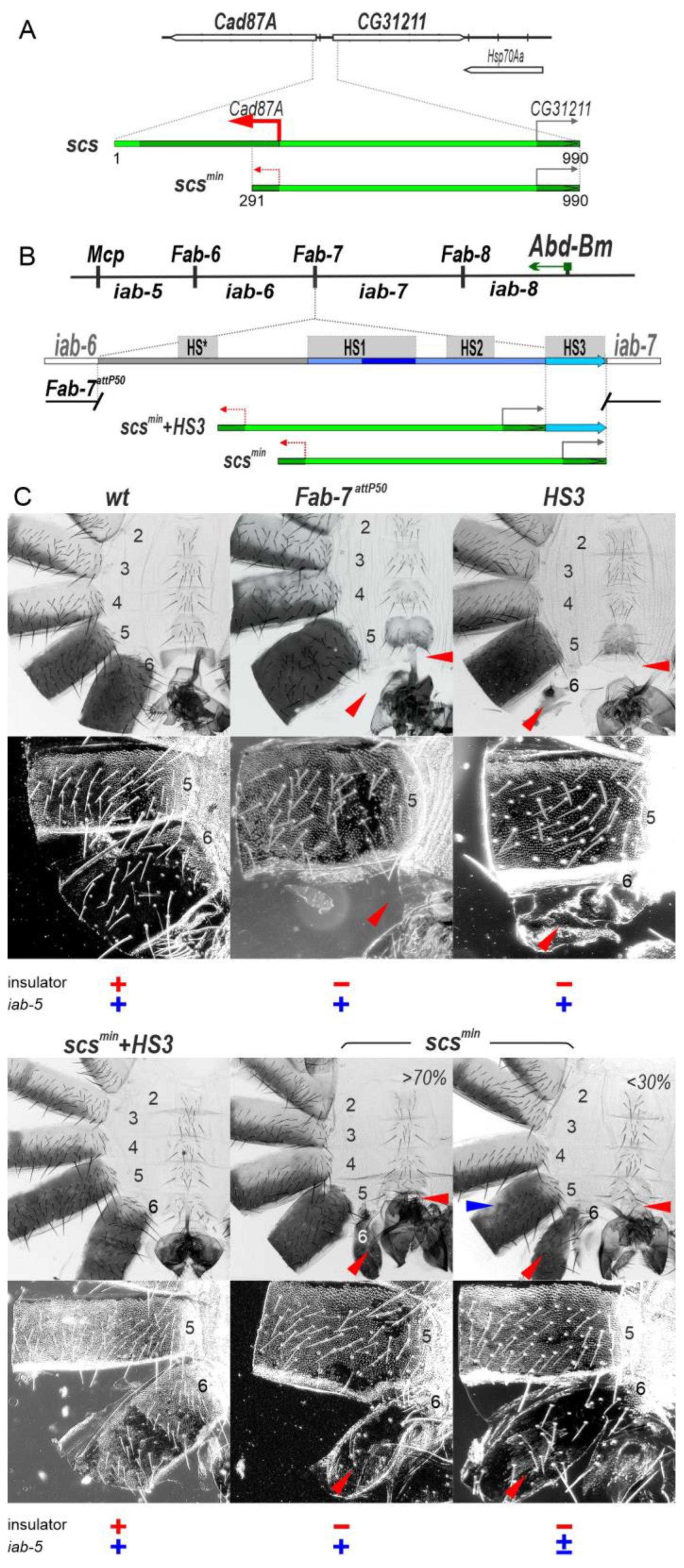
2.1. The scsmin Insulator Can Block Crosstalk between the iab-5 and iab-6 Domains Only in Cooperation with the iab-7 PRE

In the boundary replacement experiments of Hogga et al. [41,42], the scsmin insulator was introduced into a Fab-7 deletion which removes three of the four nuclease hypersensitive sites associated with the Fab-7 boundary, HS*, HS1, and H2. This deletion results in an incomplete GOF transformation of A6 (PS11) into A7 (PS12) [36]. When scsmin replaced this deletion, it blocked crosstalk between the iab-6 and iab-7 initiation elements and rescued the GOF phenotype of the starting deletion. However, since scsmin does not support boundary bypass, the A6 segment was transformed towards A5 [42].
In the Fab-7 deletion used by Hogga et al. [41,42], the HS3 sequence was still present because it was thought at that time that HS3 corresponded to the iab-7 Polycomb response element (iab-7 PRE) [36,47,48] and that its only function was Polycomb-dependent silencing. However, recent work has shown that HS3 has insulating activity [46] and that this, not silencing, is its primary function. In fact, a fully functional Fab-7 boundary can be generated by combining HS3 with the distal half of HS1 (dHS1) [38,46]. This finding made us wonder whether scsmin would be able to block crosstalk between iab-6 and iab-7 in the absence of HS3.
To address this question, we used a previously characterized Fab-7attP50 replacement platform in which the HS*, HS1, HS2, and HS3 are substituted by an attP site [49] (Figure 2B). In the starting Fab-7attP50 deletion, iab-7 is inappropriately activated in A6 (PS11), and this results in the transformation of A6 into a copy of A7 so that both the A6 and A7 segments are absent in adult males (Figure 2C).
To test for scsmin function with and without HS3, we introduced two replacements, scsmin+HS3 and scsmin, into the Fab-7attP50 platform. Though scsmin+HS3 has a slightly different sequence composition compared to the scsmin replacement published by Hogga et al. [42] due to the use of different replacement platforms, their activity is similar. As shown in Figure 2C, the scsmin+HS3 combination rescues the GOF transformation of A6 in adult males, and the A6 segment is present. However, because this combination lacks bypass activity, the iab-6 regulatory domain is blocked from regulating Abd-B expression in A6. As a consequence, the morphology of the A6 segment resembles that of A5. Instead of a banana shape without any bristles, the sternite has a quadrilateral shape and is covered in bristles, and it resembles the sternite in A5. In wild type (wt), the trichome hairs on the A6 tergite are restricted to the anterior and ventral margin, while the trichome hairs cover almost all of the A5 tergite (Figure 2C). As can be seen in the darkfield image, the A6 tergite in scsmin+HS3 males is covered with trichome hairs just like A5. In addition, there are patches of unpigmented tergite in A6 and also A5; these transformations will be considered further below. As was observed for Fab-7 (class II) deletions that retained HS3 [36], the HS3 replacement alone has only a limited ability to block crosstalk between iab-6 and iab-7 [46]. In HS3 males, the A6 tergite is greatly reduced in size, and the A6 sternite is completely missing (Figure 2C).
Like HS3, the scsmin replacement only partially blocks crosstalk between iab-6 and iab-7 (Figure 2C). However, it differs from the HS3 replacement in that there is a range of phenotypes in adult scsmin males. In all scsmin males, there is a residual A6 tergite, while the A6 sternite is absent. The residual tergite has patches of cells with trichome hairs indicating that in these cells, there is a LOF transformation in parasegment/segment identity from PS11/A6 to PS10/A5. In about 30% of the males, the morphogenesis of A5 is also affected. As shown in Figure 2C, there are patches of tissue in the A5 tergite that are not fully pigmented. This phenotype indicates that the iab-5 regulatory domain is not fully functional in a subset of scsmin males, and we will return to this issue below.

2.2. Transcription Induced by the Cad87A Promoter in scs Can Affect the Activity of the iab-7 Domain

The finding that scsmin must be combined with HS3 to block crosstalk between iab-6 and iab-7 efficiently prompted us to examine the blocking activity of the larger scs fragment, which contains the Cad87A promoter (Figure 3A).
Initially, we inserted scs in the reverse orientation in the Fab-7attP50 platform (scsR). As shown in Figure 3B, scsR males have an unusual LOF phenotype in which A7 is transformed towards A6. Unlike wt males, which lack an A7 segment, scsR males have an A7 tergite and sternite. The tergite is fully pigmented, and the trichome hairs are largely (but not completely) restricted to the anterior and lateral edges. The sternite has a banana shape like the sternite in A6; however, it is malformed and has bristles. One explanation for this unexpected phenotype is that transcription from the Cad87A promoter into the iab-7 domain may lead to an inactivation of the iab-7 enhancers. To test this possibility, we inserted a transcription termination/polyadenylation signal (PAS) in between scsR and the iab-7 domain. Figure 3B shows that scsR+PAS males lack an A7 segment. This finding indicates that when readthrough transcription from Cad87A is blocked, the iab-7 enhancers are functional (Figure 3B). On the other hand, the scs insulator is at most partially functional: there is a rudimentary A6 tergite with patches of ectopic trichome hairs, while the A6 sternite is absent.

2.3. Transcription Disrupts Boundary Function

The findings in the previous section indicate that, like scsmin, scsR is unable to effectively block crosstalk between iab-6 and iab-7 on its own. We wondered whether the larger scsR element would be functional when combined with HS3 or whether Cad87A-dependent transcription would disrupt the insulator function of HS3 just like it does for the iab-7 enhancers. For this purpose, we generated a tripartite combination of scsR+HS3+PAS (Figure 3A). The PAS sequence was included to protect the iab-7 enhancers from transcriptional readthrough. Figure 3B shows that male flies carrying the scsR+HS3+PAS combination have a GOF transformation of A6 towards A7, just like that observed for scsmin, indicating inactivation of the insulator function. This result suggests that transcription initiated at the Cad87 promoter adversely impacts the insulator activity of HS3 (Figure 3B).
To further test the idea that RT can disrupt boundary function, we generated a quadripartite replacement, 5′P+scsmin+HS3+PAS, including the P-element promoter (5′P). As shown in Figure 3B, the inclusion of the P-element promoter disrupts the boundary activity of scsmin+HS3. While scsmin+HS3 on its own rescues the boundary between the iab-6 and iab-7 (Figure 2C), this is not true for 5′P+scsmin+HS3+PAS (Figure 3B). When the P-element promoter is included in the replacement, the A6 segment is almost completely absent, as would be expected if RT disrupts insulator function.

2.4. Transcription Disrupts the Functioning of the iab-6 and iab-5 Regulatory Domains

The phenotypic abnormalities of scsR are not restricted to A7 (Figure 3B). The A6 tergite has ectopic patches of trichome hairs, while the A6 sternite has bristles and is misshapen. Though A6 shows evidence of a LOF transformation in portions of the adult cuticle, the opposite effect is observed in A5. The A5 tergite of scsR is partially devoid of trichome hairs, while A5 sternite has an abnormal banana-like shape. In scsR+PAS and scsR+HS3+PAS lines (Figure 3B), the male A5 segment is also affected: the sternite is reduced in size, while tergite has variable pigmentation and patches of ectopic trichome hairs (mix LOF/GOF phenotype). The effects of scsR on A5 (and also A6) suggest that transcription from the CG31211 promoter is disrupting the functioning of iab-5 (and iab-6). To test this idea, we generated a PAS+scsR+HS3+PAS transgenic line in which a second PAS sequence was inserted between the iab-6 domain and scsR (Figure 3B). In PAS+scsR+HS3+PAS males, the A5 segment displays a nearly wt phenotype. This finding suggests that transcription initiated from the CG31211 promoter perturbs the proper development of the A5 segment.
It seemed possible that a similar mechanism, namely transcription from the residual part of the Cad87A promoter, might account for the variable and unexpected LOF defects in the development of A5 that we observed (see above) in the scsmin replacement (Figure 2C). To further explore the effects of transcription on the functioning of the iab-5 and iab-6 regulatory domains, we inserted scs in the forward orientation. We generated three different insertions, scs alone, scs plus HS3 (scs+HS3), and scs plus the three major Fab-7 hypersensitive sites, HS1, HS2, and HS3 (F7HS1+2+3) (Figure 4A). The scs+HS3 and scs+F7HS1+2+3 combinations have blocking activity and rescue the GOF transformations evident in the starting F7attP50 platform. On the other hand, consistent with the idea that transcription can disrupt the activity of the iab-5 and iab-6 enhancers, we find that in both replacements, A6 and A5 have an A4-like phenotype. This is most clearly seen in the pattern of pigmentation and in the dense trichome hairs in the A5 and A6 tergites (compare A5 and A6 with A4 in Figure 4B).
A more complicated phenotype is observed with scs alone (Figure 4B). As expected, blocking activity is not complete, and A6 shows evidence of GOF transformations. The A6 tergite is reduced in size, while there is only a small patch of sternite tissue. In both cases, the residual A6 tissue has a phenotype indicative of a transformation towards A4 identity: there are bristles on the patch of sternite tissue, while the residual tergite is depigmented and has patches of ectopic trichome hairs. Interestingly a mixed GOF and LOF phenotype is also observed in A5: both the sternite and tergite are reduced in size as expected for a GOF transformation, while the tergite is depigmented, and there are patches of densely packed trichome hairs. The GOF transformations in the scs replacement resemble those seen when both Fab-7 and Fab-6 boundaries are deleted (see F6attP+F7attP in Figure 4B). There is also evidence of a weak GOF transformation of A4 in the double boundary deletion. However, unlike scs, the double boundary deletion shows no evidence of LOF transformations of A5 and A6.
These findings indicate that transcription from the Cad87 promoter in scs directed towards iab-6 and iab-5 disrupts not only the boundaries but also the functioning of enhancers of these domains. They also suggest that the variable LOF phenotypes in A5 evident in males carrying the scsmin replacement might be due to a low level of transcription from the truncated Cad87A promoter. Since the LOF phenotypes in A5 varied between individuals and were seen in only about 30% of the scsmin males, one plausible explanation is that stochastic differences in promoter activity between individuals might account for incomplete penetrance. To test this possibility, we generated a PAS+scsmin replacement (Figure 4C). Unlike scsmin, the A5 tergite in PAS+scsmin males is fully pigmented in all adult males, which would suggest that transcription from the clipped Cad87 promoter is likely responsible for the pigmentation defects in A5. However, this does not seem to be true for the trichome hairs on the tergite, as they are still densely packed like those in A4. This finding indicates that the trichome hair phenotype is likely due to the blocking activity of the scsmin element, which prevents iab-5 from regulating Abd-B in cells that can give rise to trichome hairs.
To further investigate the effects of RT, we placed HS3 upstream of scsmin in HS3+scsmin. Unlike scsmin+HS3, HS3+scsmin is unable to prevent crosstalk between iab-6 and iab-7, and A6 is transformed towards A7 (compare scsmin+HS3 with HS3+scsmin in Figure 4C). However, HS3 is able to complement scsmin when RT is blocked by an interposed PAS sequence (HS3+PAS+scsmin, Figure 4C). Thus, a low level of transcription from the truncated Cad87A promoter is apparently sufficient to impact the boundary activity of HS3.

2.5. Readthrough Transcription Disrupts the Functioning of a Minimal Fub (pHS2) Replacement Boundary

We wondered whether RT would also impact the functioning of other boundary elements. To investigate this possibility, we chose the BX-C Fub boundary. Fub marks the border between the Ubx regulatory domain, bxd/pbx, and the abd-A gene and its regulatory domain, iab-2 [32]. As illustrated in Figure 5A, there are two Fub hypersensitive regions, HS1 and HS2. The larger Fub hypersensitive region HS2 contains motifs for several known chromosomal architectural proteins. The distal 177 bp HS2 sequence (dHS1) has binding sites for dCTCF and Su(Hw), and we found that it can function as an effective boundary [14,50]. The proximal 450 bp HS2 sequence (pHS2, Figure 5A) contains binding sites for Pita and Su(Hw) [51,52].
We first tested whether pHS2 is able to function as a boundary when introduced into the Fab-7attP50 platform. As shown in Figure 5B, Fub pHS2 has an insulator function and rescues the GOF phenotype of the Fab-7attP50 deletion. Like most other heterologous replacements, pHS2 blocks crosstalk but does not support bypass: an A6 segment is present in the pHS2 replacement; however, its morphological features indicate that it has an A5 rather than an A6 identity: the sternite is misshapen and is covered in bristles. While the A5 tergite is fully pigmented, the trichome hairs are densely packed, much like the A4 tergite (consistent with the idea that trichome morphology in A5 is more sensitive to blocking activity by replacement boundaries than pigmentation).
A different result is obtained when the P-element promoter is placed upstream of pHS2 in the Fab-7 replacement (Figure 5B). As was observed for the P-element combination 5′P+scsmin+HS3, pHS2 boundary activity is lost in 5′P+pHS2, and the A6 segment is missing. To test whether this is due to RT from the P-element promoter, we generated two additional replacement combinations. In the first, the PAS element was placed downstream of the pHS2 boundary to give 5′P+pHS2+PAS, while in the second, the PAS element was placed between the P-element promoter and pHS2 to give 5′P+PAS+pHS2. As would be expected if RT disrupts boundary function in the 5′P+pHS2+PAS replacement, there is only a residual A6 tergite, while the insulator function is rescued when the PAS element is placed between the P-element promoter and pHS2 (5′P+PAS+pHS2) (Figure 5B).

2.6. Readthrough Transcription Disrupts Fub Function in Its Endogenous Context

Bender and Fizgerald (2002) [53] generated a series of imprecise hop outs of a P-element transgene inserted near the distal end of the bxd/pbx regulatory domain close to the sequences that were subsequently found to correspond to the Fub boundary [32,53]. These hop-out events induced an anterior-to-posterior transformation of A1 towards A2 identity. Molecular characterization of one of the hop outs that had a particularly strong GOF phenotype, UabHH1, revealed that it was a truncated P-element transgene that retained only the P-element promoter and 65 bp of lacZ coding sequence. The P-element transgene was also inverted so that the promoter was pointing towards the Fub boundary and the abd-A gene. Several potential mechanisms were proposed to account for the transformation of A1 to A2 induced by P-element transcription [53]. One was that transcription disrupted the functioning of an as-yet unidentified boundary that blocked crosstalk between the Ubx bxd/pbx and abd-A iab-2 regulatory domains. A second was that transcription interfered with the functioning of an element in iab-2 that is required to keep the iab-2 domain silenced in A1.
To test the boundary model, we took advantage of a Fub replacement platform F2attP (Figure 6A) that removes a 2106 bp sequence containing the two nuclease hypersensitive sites associated with the Fub boundary and replacing it with an attP site (F2attP) [14]. As shown in Figure 6B, the A1 tergite in wt is narrower than the A2 tergite, lacks bristles, and has less pigmentation, while the A1 sternite is absent. In F2attP males, the A1 segment is transformed into a copy of A2: the tergite is larger, and it has a pigmentation and bristle pattern like A2, and there is also a sternite that is covered in bristles. These phenotypic transformations in the adult cuticle resemble those previously reported [53] for the P-element hop-out mutants.
The GOF transformations evident in the starting F2attP deletion platform can be fully rescued by a 1587 bp fragment, F2, which includes both HS1 and HS2 (Figure 6A). Consistent with the previous results [53], we find that rescuing activity is disrupted when the P-element promoter, 5′P, is placed upstream of the F2 fragment (5′P+F2). In this replacement, the A1 segment resembles A2, just like the starting deletion platform (Figure 6B). Since introducing the PAS element downstream of F2 in the 5′P+F2+PAS combination does not rescue the GOF transformation, it would appear that boundary function rather than a downstream silencing element is the critical target for transcription inactivation.

3. Discussion

Blocking the activity of scs is context dependent: Our results indicate that the scs boundary has only a limited ability to block crosstalk between the iab-6 and iab-7 regulatory domains. This result is unexpected, as in transgene assays, scs was found to have one of the “stronger” insulator activities [43,44,45,54,55]. It seems likely that scs is a poor match with the neighboring Fab-6 and Fab-8 boundaries [56,57]. Both depend upon CTCF, while scs does not [58]. Also, when placed in the context of BX-C, scs seems to have a cell and/or an enhancer-specific blocking activity. For example, the phenotype of the A5 tergite in PAS+scsmin males (Figure 4C) suggests that scs is unable to block the regulatory interactions between iab-5 and Abd-B required for wt pigmentation, while its insulating activity is sufficient to block the interactions needed to inhibit the formation of trichomes.
Transcription disrupts enhancer activity: We found that when scsR was introduced into a larger Fab-7 deletion that lacks HS3, transcription from the Cad87A promoter interferes with the functioning of the iab-7 domain, inducing a LOF transformation. The suggestion that RT can disrupt the activity of tissue-specific enhancers in this region of BX-C is supported by the effects of inserting scs in the direct orientation. In this case, it induces a LOF transformation of both A6 (PS11) and A5 (PS10) towards A4 (PS9). The effects of transcription from the Cad87A promoter on these regulatory domains are most clear-cut when scs is combined with HS3 or F7HS1+2+3. In both of these replacements, the combination of scs with HS3 or F7HS1+2+3 suppresses the GOF phenotype of the Fab-7attP50 deletion platform, making the LOF transformations in A6 (PS11) and A5 (PS10) more obvious.
Transcription disrupts boundary function: The enhancers in the Abd-B regulatory domains are not the only elements whose function is disrupted by RT. We find that boundary activity can also be abrogated by RT. In the case of our Fab-7 replacements, this is most directly demonstrated when boundaries are placed downstream of a P-element promoter. The scsmin+HS3 combination not only rescues the GOF transformation of A6 (PS11) in the Fab-7attP50 deletion platform but also prevents iab-6 and, to a lesser extent, the iab-5 domain from regulating Abd-B expression. However, if the P-element promoter is placed upstream of scs+HS3 as in the 5′P+scs+HS3+PAS combination, blocking activity is largely lost, and A6 is transformed towards an A7 identity.
The effects of transcription on boundary activity are not limited to scs and HS3 (Fab-7), as transcription also interferes with the functioning of the Fub boundary fragment pHS2. On its own, it rescues the GOF phenotype of Fab-7attP50; however, when placed downstream of the P-element promoter, blocking activity is lost. Transcription also inactivates the 1587 bp Fub boundary in its native context (Figure 6). Moreover, as was the case for a combination in which the truncated Cad87A promoter in scsmin is pointing towards HS3, the disruption in the pHS2 boundary function by the P-element promoter can be rescued by placing the PAS element in between the promoter and the boundary. These findings argued that boundary function is disrupted by RT rather than some other properties of the promoter.
While previous studies have shown that RT can suppress the activity of enhancers and promoters, how this happens is not fully understood [59,60,61]. One idea is that RNA Pol II transiently displaces DNA-binding proteins as it passes [62,63]. In the case of boundary elements, it seems possible that even a transient displacement of factors important for their activity could have a significant impact on boundary function. Fly boundaries link distant sequences together to form looped domains or TADs by boundary: boundary pairing interactions [5,10,12]. In this mechanism, TADs are formed when proteins associated with one boundary element physically interact in a stable fashion with proteins associated with a second boundary element. This means that a transient displacement of boundary-associated proteins from one of the elements would disrupt the TAD as it would uncouple the physical linkage between the distant sequences that define the endpoints of the loop. Consistent with this idea, a low level of transcription from the truncated Cad87A promoter can disrupt the boundary functions of HS3.
While our experimental paradigm is artificial, there are contexts in which RT provides a mechanism for coordinating higher-order chromosome organization with regulating gene activity. For example, the blocking activity of the Fub-1 boundary is turned off by RT of a lncRNA from a promoter that is activated by the Ubx regulatory domain bxd/pbx [64] in PS6/A1 and more posterior parasegments. Inactivation of the Fub-1 boundary enables enhancers in the bxd/pbx domain to regulate Ubx expression. MicroC experiments suggest that RT of the Fub-1 boundary is likely accompanied by a switch from one TAD configuration to another configuration. Since transcription is not continuous but instead occurs in bursts that can differ both in their length and frequency depending on the specific enhancer–promoter combinations, an RT mechanism would result in only a transient remodeling of the TAD organization. Moreover, this remodeling would also be subject to regulation. In this respect, it is interesting to note that lncRNAs are thought to account for a vast majority of the transcripts in mammalian genomes [65,66,67,68]. It would be reasonable to suppose that some of these lncRNAs span boundaries and “tethering” elements that might also be sensitive to RT. RT of these lncRNAs would then alter the local chromatin organization and, in doing so, generate new combinations of regulatory elements and potential target genes.

4. Materials and Methods

4.1. Generation of the Replacement Lines

The strategy of the Fab-7 replacement lines is described in detail in [49] and is based on the Fab-7attB50 landing platform in which the 1950 bp Fab-7 region was deleted, as shown in Figure 1A. Fab-7attB50 landing platform contains an attP site for the integration of the tested constructs; lox and frt sites were used for the excision of the plasmid body and the rosy marker gene. The plasmid that was injected into the Fab-7attp50 line contains an attB site for integration, frt sites for excision of the rosy gene, and lox sites for excision of the plasmid body. Testing elements were inserted just in front of the attB site. After the integration of the plasmid within Fab-7attp50, ry+ transformants were selected. Then, rosy and plasmid cassettes were excised by FLP-recombinase to remove about 10.2 kb additional sequence between the tested element and iab-7 in the ry+ line.
In the F2attP platform [14], the 2106 bp within the Fub region (genome release R6.22: 3R:16,797,757 to 16,799,862 or complete sequence of BX-C in SEQ89E numbering: 183,576 to 185,681) was substituted with [attP]-[lox] sites. For the F2attP replacement, the recombination plasmid contains several genetic elements in the following order: [attB]-[polylinker]-[lox]-[3P3-mCherry]-[mini-y]. All elements were assembled within the pBluescript SK vector. loxP site is located after the polylinker and, in combination with the second site, which is located in the platform, is used for the excision of marker genes and plasmid body. DNA fragments used for the replacement experiments were generated by PCR amplification and verified by sequencing. The sequences of the used fragments are shown in Supporting Table S1. The polyadenylation signal (PAS) corresponds to 700 bp XbaI-BamHI fragment of main SV40 termination sequences from the pUAST base vector.

4.2. Cuticle Preparations

Three-day adult flies were collected in Eppendorf tubes and stored in 70% ethanol for at least 1 day. Then, ethanol was replaced with 10% KOH, and flies were heated at 70 °C for 1 h. After, the heating flies were washed with dH2O two times and heated again in dH2O for 45 min. Then, the digested flies were washed with 70% ethanol and stored in 70% ethanol. The abdomen cuticles were cut from the rest of the digested fly using a fine tweezer and a needle of an insulin syringe and put in a droplet of glycerol on a glass slide. Then, the abdomens were cut longitudinally on the dorsal side through all of the tergites with the syringe. To spread the cuticles flat on the slides, cuts may be carried out between the tergites. Then, the cuticles were flattened with a coverslip. Photographs in the bright or dark field were taken on the using Nikon DS-Ri2 digital camera (Nikon microscope products, Tokyo, Japan, 2015), processed with ImageJ 1.50c4 and Fiji bundle 2.0.0-rc-46.

Supplementary Materials

The supporting information can be downloaded at: https://www.mdpi.com/article/10.3390/ijms241411368/s1.

Author Contributions

Conceptualization, P.G. and O.K.; data curation, O.K.; investigation, O.K., V.S., M.T. and G.M.; methodology, O.K. and M.T.; project administration, P.S., O.K. and P.G.; resources, P.S. and O.K.; supervision, P.S., P.G. and O.K.; validation, O.K., P.S. and P.G.; visualization, O.K. and V.S.; writing—original draft, P.S., O.K. and P.G.; writing—review and editing, P.S., O.K. and P.G. All authors have read and agreed to the published version of the manuscript.

Funding

This work (all functional and morphological analysis) was supported by the Russian Science Foundation (19-14-00103 to O.K.). Part of this work (study of Fub substitutions) was supported by grant 075-15-2019-1661 from the Ministry of Science and Higher Education of the Russian Federation. P.S. acknowledges support from the National Institutes of Health (R35 GM126975).

Institutional Review Board Statement

Not applicable.

Informed Consent Statement

Not applicable.

Data Availability Statement

Data are contained within the article or Supplementary Materials.

Acknowledgments

We thank Farhod Hasanov for the fly injections.

Conflicts of Interest

The authors declare no conflict of interest.

References

  1. Cavalheiro, G.R.; Pollex, T.; Furlong, E.E. To Loop or Not to Loop: What Is the Role of TADs in Enhancer Function and Gene Regulation? Curr. Opin. Genet. Dev. 2021, 67, 119–129. [Google Scholar] [CrossRef]
  2. Hafner, A.; Boettiger, A. The Spatial Organization of Transcriptional Control. Nat. Rev. Genet. 2022, 24, 53–68. [Google Scholar] [CrossRef]
  3. Jerkovic, I.; Cavalli, G. Understanding 3D Genome Organization by Multidisciplinary Methods. Nat. Rev. Mol. Cell Biol. 2021, 22, 511–528. [Google Scholar] [CrossRef]
  4. Mir, M.; Bickmore, W.; Furlong, E.E.M.; Narlikar, G. Chromatin Topology, Condensates and Gene Regulation: Shifting Paradigms or Just a Phase? Development 2019, 146, dev182766. [Google Scholar] [CrossRef] [Green Version]
  5. Batut, P.J.; Bing, X.Y.; Sisco, Z.; Raimundo, J.; Levo, M.; Levine, M.S. Genome Organization Controls Transcriptional Dynamics during Development. Science 2022, 375, 566–570. [Google Scholar] [CrossRef]
  6. Kyrchanova, O.; Georgiev, P. Mechanisms of Enhancer-Promoter Interactions in Higher Eukaryotes. Int. J. Mol. Sci. 2021, 22, 671. [Google Scholar] [CrossRef]
  7. Matthews, N.E.; White, R. Chromatin Architecture in the Fly: Living without CTCF/Cohesin Loop Extrusion? Alternating Chromatin States Provide a Basis for Domain Architecture in Drosophila. Bioessays 2019, 41, e1900048. [Google Scholar] [CrossRef] [Green Version]
  8. Melnikova, L.S.; Georgiev, P.G.; Golovnin, A.K. The Functions and Mechanisms of Action of Insulators in the Genomes of Higher Eukaryotes. Acta Naturae 2020, 12, 15–33. [Google Scholar] [CrossRef]
  9. Chen, D.; Lei, E.P. Function and Regulation of Chromatin Insulators in Dynamic Genome Organization. Curr. Opin. Cell Biol. 2019, 58, 61–68. [Google Scholar] [CrossRef]
  10. Kyrchanova, O.V.; Bylino, O.V.; Georgiev, P.G. Mechanisms of Enhancer-Promoter Communication and Chromosomal Architecture in Mammals and Drosophila. Front. Genet. 2022, 13, 1081088. [Google Scholar] [CrossRef]
  11. Maksimenko, O.G.; Fursenko, D.V.; Belova, E.V.; Georgiev, P.G. CTCF As an Example of DNA-Binding Transcription Factors Containing Clusters of C2H2-Type Zinc Fingers. Acta Naturae 2021, 13, 31–46. [Google Scholar] [CrossRef]
  12. Chetverina, D.; Fujioka, M.; Erokhin, M.; Georgiev, P.; Jaynes, J.B.; Schedl, P. Boundaries of Loop Domains (Insulators): Determinants of Chromosome Form and Function in Multicellular Eukaryotes. Bioessays 2017, 39, 1600233. [Google Scholar] [CrossRef] [Green Version]
  13. Matzat, L.H.; Lei, E.P. Surviving an Identity Crisis: A Revised View of Chromatin Insulators in the Genomics Era. Biochim. Biophys. Acta 2014, 1839, 203–214. [Google Scholar] [CrossRef] [Green Version]
  14. Kyrchanova, O.; Maksimenko, O.; Ibragimov, A.; Sokolov, V.; Postika, N.; Lukyanova, M.; Schedl, P.; Georgiev, P. The Insulator Functions of the Drosophila Polydactyl C2H2 Zinc Finger Protein CTCF: Necessity versus Sufficiency. Sci. Adv. 2020, 6, eaaz3152. [Google Scholar] [CrossRef] [Green Version]
  15. Kyrchanova, O.; Mogila, V.; Wolle, D.; Magbanua, J.P.; White, R.; Georgiev, P.; Schedl, P. The Boundary Paradox in the Bithorax Complex. Mech. Dev. 2015, 138 Pt 2, 122–132. [Google Scholar] [CrossRef]
  16. Lewis, E.B. A Gene Complex Controlling Segmentation in Drosophila. Nature 1978, 276, 565–570. [Google Scholar] [CrossRef]
  17. Maeda, R.K.; Karch, F. The ABC of the BX-C: The Bithorax Complex Explained. Development 2006, 133, 1413–1422. [Google Scholar] [CrossRef] [Green Version]
  18. Maeda, R.K.; Karch, F. The Open for Business Model of the Bithorax Complex in Drosophila. Chromosoma 2015, 124, 293–307. [Google Scholar] [CrossRef] [Green Version]
  19. Celniker, S.E.; Sharma, S.; Keelan, D.J.; Lewis, E.B. The Molecular Genetics of the Bithorax Complex of Drosophila: Cis-Regulation in the Abdominal-B Domain. EMBO J. 1990, 9, 4277–4286. [Google Scholar] [CrossRef]
  20. Duncan, I. The Bithorax Complex. Annu. Rev. Genet. 1987, 21, 285–319. [Google Scholar] [CrossRef]
  21. Garaulet, D.L.; Lai, E.C. Hox MiRNA Regulation within the Drosophila Bithorax Complex: Patterning Behavior. Mech. Dev. 2015, 138 Pt 2, 151–159. [Google Scholar] [CrossRef]
  22. Hagstrom, K.; Muller, M.; Schedl, P. Fab-7 Functions as a Chromatin Domain Boundary to Ensure Proper Segment Specification by the Drosophila Bithorax Complex. Genes Dev. 1996, 10, 3202–3215. [Google Scholar] [CrossRef] [Green Version]
  23. Sánchez-Herrero, E. Control of the Expression of the Bithorax Complex Genes Abdominal-A and Abdominal-B by Cis-Regulatory Regions in Drosophila Embryos. Development 1991, 111, 437–449. [Google Scholar] [CrossRef]
  24. Casares, F.; Sánchez-Herrero, E. Regulation of the Infraabdominal Regions of the Bithorax Complex of Drosophila by Gap Genes. Development 1995, 121, 1855–1866. [Google Scholar] [CrossRef]
  25. Drewell, R.A.; Nevarez, M.J.; Kurata, J.S.; Winkler, L.N.; Li, L.; Dresch, J.M. Deciphering the Combinatorial Architecture of a Drosophila Homeotic Gene Enhancer. Mech. Dev. 2014, 131, 68–77. [Google Scholar] [CrossRef]
  26. Iampietro, C.; Gummalla, M.; Mutero, A.; Karch, F.; Maeda, R.K. Initiator Elements Function to Determine the Activity State of BX-C Enhancers. PLoS Genet. 2010, 6, e1001260. [Google Scholar] [CrossRef] [Green Version]
  27. Bowman, S.K.; Deaton, A.M.; Domingues, H.; Wang, P.I.; Sadreyev, R.I.; Kingston, R.E.; Bender, W. H3K27 Modifications Define Segmental Regulatory Domains in the Drosophila Bithorax Complex. eLife 2014, 3, e02833. [Google Scholar] [CrossRef]
  28. Cheutin, T.; Cavalli, G. The Multiscale Effects of Polycomb Mechanisms on 3D Chromatin Folding. Crit. Rev. Biochem. Mol. Biol. 2019, 54, 399–417. [Google Scholar] [CrossRef]
  29. Chiang, A.; O’Connor, M.B.; Paro, R.; Simon, J.; Bender, W. Discrete Polycomb-Binding Sites in Each Parasegmental Domain of the Bithorax Complex. Development 1995, 121, 1681–1689. [Google Scholar] [CrossRef]
  30. Kassis, J.A.; Kennison, J.A.; Tamkun, J.W. Polycomb and Trithorax Group Genes in Drosophila. Genetics 2017, 206, 1699–1725. [Google Scholar] [CrossRef] [Green Version]
  31. Barges, S.; Mihaly, J.; Galloni, M.; Hagstrom, K.; Müller, M.; Shanower, G.; Schedl, P.; Gyurkovics, H.; Karch, F. The Fab-8 Boundary Defines the Distal Limit of the Bithorax Complex Iab-7 Domain and Insulates Iab-7 from Initiation Elements and a PRE in the Adjacent Iab-8 Domain. Development 2000, 127, 779–790. [Google Scholar] [CrossRef] [PubMed]
  32. Bender, W.; Lucas, M. The Border between the Ultrabithorax and Abdominal-A Regulatory Domains in the Drosophila Bithorax Complex. Genetics 2013, 193, 1135–1147. [Google Scholar] [CrossRef] [Green Version]
  33. Galloni, M.; Gyurkovics, H.; Schedl, P.; Karch, F. The Bluetail Transposon: Evidence for Independent Cis-Regulatory Domains and Domain Boundaries in the Bithorax Complex. EMBO J. 1993, 12, 1087–1097. [Google Scholar] [CrossRef]
  34. Gyurkovics, H.; Gausz, J.; Kummer, J.; Karch, F. A New Homeotic Mutation in the Drosophila Bithorax Complex Removes a Boundary Separating Two Domains of Regulation. EMBO J. 1990, 9, 2579–2585. [Google Scholar] [CrossRef]
  35. Karch, F.; Galloni, M.; Sipos, L.; Gausz, J.; Gyurkovics, H.; Schedl, P. Mcp and Fab-7: Molecular Analysis of Putative Boundaries of Cis-Regulatory Domains in the Bithorax Complex of Drosophila Melanogaster. Nucleic Acids Res. 1994, 22, 3138–3146. [Google Scholar] [CrossRef] [PubMed] [Green Version]
  36. Mihaly, J.; Hogga, I.; Gausz, J.; Gyurkovics, H.; Karch, F. In Situ Dissection of the Fab-7 Region of the Bithorax Complex into a Chromatin Domain Boundary and a Polycomb-Response Element. Development 1997, 124, 1809–1820. [Google Scholar] [CrossRef]
  37. Kyrchanova, O.; Sokolov, V.; Georgiev, P. Mechanisms of Interaction between Enhancers and Promoters in Three Drosophila Model Systems. Int. J. Mol. Sci. 2023, 24, 2855. [Google Scholar] [CrossRef]
  38. Kyrchanova, O.; Sabirov, M.; Mogila, V.; Kurbidaeva, A.; Postika, N.; Maksimenko, O.; Schedl, P.; Georgiev, P. Complete Reconstitution of Bypass and Blocking Functions in a Minimal Artificial Fab-7 Insulator from Drosophila Bithorax Complex. Proc. Natl. Acad. Sci. USA 2019, 116, 13462–13467. [Google Scholar] [CrossRef] [PubMed] [Green Version]
  39. Kyrchanova, O.; Wolle, D.; Sabirov, M.; Kurbidaeva, A.; Aoki, T.; Maksimenko, O.; Kyrchanova, M.; Georgiev, P.; Schedl, P. Distinct Elements Confer the Blocking and Bypass Functions of the Bithorax Fab-8 Boundary. Genetics 2019, 213, 865–876. [Google Scholar] [CrossRef]
  40. Postika, N.; Metzler, M.; Affolter, M.; Müller, M.; Schedl, P.; Georgiev, P.; Kyrchanova, O. Boundaries Mediate Long-Distance Interactions between Enhancers and Promoters in the Drosophila Bithorax Complex. PLoS Genet. 2018, 14, e1007702. [Google Scholar] [CrossRef] [Green Version]
  41. Hogga, I.; Karch, F. Transcription through the Iab-7 Cis-Regulatory Domain of the Bithorax Complex Interferes with Maintenance of Polycomb-Mediated Silencing. Development 2002, 129, 4915–4922. [Google Scholar] [CrossRef]
  42. Hogga, I.; Mihaly, J.; Barges, S.; Karch, F. Replacement of Fab-7 by the Gypsy or Scs Insulator Disrupts Long-Distance Regulatory Interactions in the Abd-B Gene of the Bithorax Complex. Mol. Cell 2001, 8, 1145–1151. [Google Scholar] [CrossRef]
  43. Kuhn, E.J.; Hart, C.M.; Geyer, P.K. Studies of the Role of the Drosophila Scs and Scs’ Insulators in Defining Boundaries of a Chromosome Puff. Mol. Cell Biol. 2004, 24, 1470–1480. [Google Scholar] [CrossRef]
  44. Kyrchanova, O.; Leman, D.; Parshikov, A.; Fedotova, A.; Studitsky, V.; Maksimenko, O.; Georgiev, P. New Properties of Drosophila Scs and Scs’ Insulators. PLoS ONE 2013, 8, e62690. [Google Scholar] [CrossRef] [PubMed] [Green Version]
  45. Vazquez, J.; Schedl, P. Sequences Required for Enhancer Blocking Activity of Scs Are Located within Two Nuclease-Hypersensitive Regions. EMBO J. 1994, 13, 5984–5993. [Google Scholar] [CrossRef] [PubMed]
  46. Kyrchanova, O.; Kurbidaeva, A.; Sabirov, M.; Postika, N.; Wolle, D.; Aoki, T.; Maksimenko, O.; Mogila, V.; Schedl, P.; Georgiev, P. The Bithorax Complex Iab-7 Polycomb Response Element Has a Novel Role in the Functioning of the Fab-7 Chromatin Boundary. PLoS Genet. 2018, 14, e1007442. [Google Scholar] [CrossRef] [PubMed] [Green Version]
  47. Hagstrom, K.; Muller, M.; Schedl, P. A Polycomb and GAGA Dependent Silencer Adjoins the Fab-7 Boundary in the Drosophila Bithorax Complex. Genetics 1997, 146, 1365–1380. [Google Scholar] [CrossRef]
  48. Mishra, R.K.; Mihaly, J.; Barges, S.; Spierer, A.; Karch, F.; Hagstrom, K.; Schweinsberg, S.E.; Schedl, P. The Iab-7 Polycomb Response Element Maps to a Nucleosome-Free Region of Chromatin and Requires Both GAGA and Pleiohomeotic for Silencing Activity. Mol. Cell Biol. 2001, 21, 1311–1318. [Google Scholar] [CrossRef] [Green Version]
  49. Wolle, D.; Cleard, F.; Aoki, T.; Deshpande, G.; Schedl, P.; Karch, F. Functional Requirements for Fab-7 Boundary Activity in the Bithorax Complex. Mol. Cell Biol. 2015, 35, 3739–3752. [Google Scholar] [CrossRef] [Green Version]
  50. Holohan, E.E.; Kwong, C.; Adryan, B.; Bartkuhn, M.; Herold, M.; Renkawitz, R.; Russell, S.; White, R. CTCF Genomic Binding Sites in Drosophila and the Organisation of the Bithorax Complex. PLoS Genet. 2007, 3, e112. [Google Scholar] [CrossRef] [Green Version]
  51. Sabirov, M.; Kyrchanova, O.; Pokholkova, G.V.; Bonchuk, A.; Klimenko, N.; Belova, E.; Zhimulev, I.F.; Maksimenko, O.; Georgiev, P. Mechanism and Functional Role of the Interaction between CP190 and the Architectural Protein Pita in Drosophila Melanogaster. Epigenet. Chromatin 2021, 14, 16. [Google Scholar] [CrossRef] [PubMed]
  52. Schwartz, Y.B.; Linder-Basso, D.; Kharchenko, P.V.; Tolstorukov, M.Y.; Kim, M.; Li, H.-B.; Gorchakov, A.A.; Minoda, A.; Shanower, G.; Alekseyenko, A.A.; et al. Nature and Function of Insulator Protein Binding Sites in the Drosophila Genome. Genome Res. 2012, 22, 2188–2198. [Google Scholar] [CrossRef] [PubMed] [Green Version]
  53. Bender, W.; Fitzgerald, D.P. Transcription Activates Repressed Domains in the Drosophila Bithorax Complex. Development 2002, 129, 4923–4930. [Google Scholar] [CrossRef] [PubMed]
  54. Majumder, P.; Cai, H.N. The Functional Analysis of Insulator Interactions in the Drosophila Embryo. Proc. Natl. Acad. Sci. USA 2003, 100, 5223–5228. [Google Scholar] [CrossRef]
  55. Kuhn, E.J.; Viering, M.M.; Rhodes, K.M.; Geyer, P.K. A Test of Insulator Interactions in Drosophila. EMBO J. 2003, 22, 2463–2471. [Google Scholar] [CrossRef] [Green Version]
  56. Gohl, D.; Aoki, T.; Blanton, J.; Shanower, G.; Kappes, G.; Schedl, P. Mechanism of Chromosomal Boundary Action: Roadblock, Sink, or Loop? Genetics 2011, 187, 731–748. [Google Scholar] [CrossRef]
  57. Kyrchanova, O.; Chetverina, D.; Maksimenko, O.; Kullyev, A.; Georgiev, P. Orientation-Dependent Interaction between Drosophila Insulators Is a Property of This Class of Regulatory Elements. Nucleic Acids Res. 2008, 36, 7019–7028. [Google Scholar] [CrossRef] [Green Version]
  58. Gaszner, M.; Vazquez, J.; Schedl, P. The Zw5 Protein, a Component of the Scs Chromatin Domain Boundary, Is Able to Block Enhancer-Promoter Interaction. Genes Dev. 1999, 13, 2098–2107. [Google Scholar] [CrossRef] [Green Version]
  59. Castro Alvarez, J.J.; Revel, M.; Carrasco, J.; Cléard, F.; Pauli, D.; Hilgers, V.; Karch, F.; Maeda, R.K. Repression of the Hox Gene Abd-A by ELAV-Mediated Transcriptional Interference. PLoS Genet. 2021, 17, e1009843. [Google Scholar] [CrossRef]
  60. Erokhin, M.; Davydova, A.; Parshikov, A.; Studitsky, V.M.; Georgiev, P.; Chetverina, D. Transcription through Enhancers Suppresses Their Activity in Drosophila. Epigenet. Chromatin 2013, 6, 31. [Google Scholar] [CrossRef] [Green Version]
  61. Fujioka, M.; Nezdyur, A.; Jaynes, J.B. An Insulator Blocks Access to Enhancers by an Illegitimate Promoter, Preventing Repression by Transcriptional Interference. PLoS Genet. 2021, 17, e1009536. [Google Scholar] [CrossRef] [PubMed]
  62. Pande, A.; Brosius, J.; Makalowska, I.; Makalowski, W.; Raabe, C.A. Transcriptional Interference by Small Transcripts in Proximal Promoter Regions. Nucleic Acids Res. 2018, 46, 1069–1088. [Google Scholar] [CrossRef] [PubMed] [Green Version]
  63. Pande, A.; Makalowski, W.; Brosius, J.; Raabe, C.A. Enhancer Occlusion Transcripts Regulate the Activity of Human Enhancer Domains via Transcriptional Interference: A Computational Perspective. Nucleic Acids Res. 2020, 48, 3435–3454. [Google Scholar] [CrossRef] [PubMed] [Green Version]
  64. Ibragimov, A.; Bing, X.; Shidlovskii, Y.; Levine, M.; Georgiev, P.; Schedl, P. The Insulating Activity of the Drosophila BX-C Chromatin Boundary Fub-1 Is Parasegmentally Regulated by LncRNA Read-Through. bioRxiv 2023. [Google Scholar] [CrossRef]
  65. Herman, A.B.; Tsitsipatis, D.; Gorospe, M. Integrated LncRNA Function upon Genomic and Epigenomic Regulation. Mol. Cell 2022, 82, 2252–2266. [Google Scholar] [CrossRef]
  66. Núñez-Martínez, H.N.; Recillas-Targa, F. Emerging Functions of LncRNA Loci beyond the Transcript Itself. Int. J. Mol. Sci. 2022, 23, 6258. [Google Scholar] [CrossRef]
  67. Sartorelli, V.; Lauberth, S.M. Enhancer RNAs Are an Important Regulatory Layer of the Epigenome. Nat. Struct. Mol. Biol. 2020, 27, 521–528. [Google Scholar] [CrossRef]
  68. Statello, L.; Guo, C.-J.; Chen, L.-L.; Huarte, M. Gene Regulation by Long Non-Coding RNAs and Its Biological Functions. Nat. Rev. Mol. Cell Biol. 2021, 22, 96–118. [Google Scholar] [CrossRef]
Figure 1. Boundaries organize regulatory region of the BX-C. (A) Map of the BX-C showing the location of the three homeotic genes and the parasegment-specific regulatory domains. There are nine cis-regulatory domains (shown as colored boxes) that are responsible for the regulation of the BX-C genes and the specification of parasegments 5 to 13, which correspond to T3-A8 segments (shown on the diagram of the adult fly using the same color code). The abx/bx (yellow) and bxd/pbx (orange) domains activate Ubx, iab-2–iab-4 (shades of blue)—abd-A and iab-5–9 (shades of green)—Abd-B. Lines with colored circles mark chromatin boundaries. The dCTCF and Pita binding sites at the boundaries are shown as red and blue circles, respectively. (B) Schematic presentation of Abd-B activation in A5(PS11) and A6(PS12) segments (parasegments). (C) Deletion of the Fab-7 boundary results in premature activation of the iab-7 domain in A6 (PS12).
Ijms 24 11368 g001
Figure 2. HS3 (iab-7 PRE) is required for the boundary activity of the scsmin insulator. (A) Schematic presentation of the scs insulator (990 bp) and scsmin (700 bp) marked as green lines. TSS is the transcription start sites of Cad87A and CG31211 are marked by red and grey arrows, respectively, indicating the orientation of the promoters. The truncated Cad87A promoter is marked by the dotted red arrow. Exons of Cad87A and CG31211 are marked as dark green boxes. (B) Schematic representation of the abd-AAbd-B regulatory regions and Fab-7attP50 platform in which four hypersensitive sites, HS*, HS1, HS2, and HS3 (marked as grey boxes), are deleted. The endpoints of the deletion are indicated by breaks in the line. HS3 marked as azure arrow. Replacement fragments are shown below the map. (C) Morphology of the male abdominal segments (numbered) in wild type (wt), Fab-7attP50, HS3 [46], scsmin+HS3, and scsmin transgenic lines. Bright-field (top) and dark-field (bottom) images of cuticles prepared from males. The filled red arrowheads show morphological features indicative of boundary inactivation (GOF phenotype). The blue arrowheads show signs of the iab enhancer inactivation (LOF phenotype). The insulator function is indicated by red “+” (normal) and “−” (moderate or strong inactivation). The function of the iab-5 enhancers is shown by blue “+” (normal), “+/−” (weak inactivation), “−” (strong inactivation). In wild type, the A5 and A6 tergites (on the dorsal side of the fly) are fully pigmented, while the A2–4 tergites have only a stripe of pigmentation. The trichome hairs (shown in dark field) on the A6 tergite are restricted to the anterior and ventral margins, while A5 tergite is typically covered in uniform lawn trichomes (although there are small patches of cuticle that have only a few trichomes). The more anterior tergites are fully covered in trichome hairs, and the hairs are much more densely packed. The A2–A5 sternites on the ventral side have a quadrilateral shape with multiple bristles. The A6 sternite is different in that it lacks bristles and has a wide and curved shape. In males, the A7 segment is absent. Descriptions of other phenotypes are found in the text.
Ijms 24 11368 g002
Figure 3. The Cad87A promoter in scs inserted in the reverse orientation (scsR) is responsible for inactivation of the iab-7 domain. (A) Schemes of Fab-7 boundary replacement with scs inserted in reverse orientation. The P-element promoter is shown as blue arrow. Polyadenylation signal from SV40 (PAS) is shown as red line with stop signal. Other designations are as in Figure 2. (B) Morphology of the male abdominal segments (numbered) in wt, scsR, scsR+PAS, scsR+HS3+PAS, PAS+scsR+HS3+PAS, and 5′P+scsmin+HS3+PAS. The filled red arrowheads show morphological features indicative of boundary inactivation (GOF phenotype). The blue arrowheads show signs of the iab enhancer inactivation (LOF phenotype).
Ijms 24 11368 g003
Figure 4. The Cad87A promoter in the scs inserted in the direct orientation (scs) is responsible for inactivation of the iab-5 and iab-6 domains. (A) Fab-7 boundary replacement schemes in which scs and scsmin were inserted in direct orientation in different combinations with the Fab-7 fragments and PAS. Designations are as in Figure 2 and Figure 3. (B) Bright-field (top) and dark-field (bottom) images of cuticles prepared from males of wt, scs, scs+HS3, scs+F7, and F6attP+F7attP. (C) Comparing images of cuticles prepared from males of scsmin, PAS+scsmin, HS3+scsmin and HS3+PAS+scsmin. Bright-field (top) and dark-field (bottom) The filled red arrowheads show morphological features indicative of boundary inactivation (GOF phenotype). The blue arrowheads show signs of the iab enhancer inactivation (LOF phenotype).
Ijms 24 11368 g004
Figure 5. Transcription from the P-element promoter suppresses the activity of the Fub subfragment pHS2 when it replaces the Fab-7 boundary. (A) Fab-7 boundary replacement schemes, in which pHS2 was inserted in different combinations with the P-element promoter and PAS. Designations are as in Figure 2 and Figure 3. (B) Bright-field (top) and dark-field (bottom) images of cuticles prepared from males of wt, Fab-7attP50, pHS2, 5′P+pHS2+PAS, and 5′P+PAS+pHS2. The filled red arrowheads show morphological features indicative of boundary inactivation (GOF phenotype).
Ijms 24 11368 g005
Figure 6. Transcription from the P-element promoter suppresses the functional activity of the Fub boundary and the Fub subelement pHS2 in its endogenous location between the bxd/pbx (the Ubx regulatory region) and iab-2 (the abd-A regulatory region) domains. (A) Fub boundary replacement schemes, in which Fub was inserted in different combinations with the P-element promoter and PAS. Designations are as in Figure 2 and Figure 3. (B) Morphology of the male abdominal segments (numbered) in wt, F2attP, F2, P+F2, and 5′P+F2+PAS. The filled red arrowheads show morphological features indicative of boundary inactivation (GOF phenotype).
Ijms 24 11368 g006
Disclaimer/Publisher’s Note: The statements, opinions and data contained in all publications are solely those of the individual author(s) and contributor(s) and not of MDPI and/or the editor(s). MDPI and/or the editor(s) disclaim responsibility for any injury to people or property resulting from any ideas, methods, instructions or products referred to in the content.

