Article

Tissue-Specific Hormone Signalling and Defence Gene Induction in an In Vitro Assembly of the Rapeseed Verticillium Pathosystem

1
Department of Agriculture, Ecotrophology, and Landscape Development, Anhalt University of Applied Sciences, 06406 Bernburg, Germany
2
Institute of Crop Sciences, University of Hohenheim, 70593 Stuttgart, Germany
*
Author to whom correspondence should be addressed.
Int. J. Mol. Sci. 2023, 24(13), 10489; https://doi.org/10.3390/ijms241310489
Submission received: 20 April 2023 / Revised: 11 June 2023 / Accepted: 20 June 2023 / Published: 22 June 2023
(This article belongs to the Special Issue Signal Transduction Mechanism in Plant Disease and Immunity)

Abstract

:
Priming plants with beneficial microbes can establish rapid and robust resistance against numerous pathogens. Here, compelling evidence is provided that the treatment of rapeseed plants with Trichoderma harzianum OMG16 and Bacillus velezensis FZB42 induces defence activation against Verticillium longisporum infection. The relative expressions of the JA biosynthesis genes LOX2 and OPR3, the ET biosynthesis genes ACS2 and ACO4 and the SA biosynthesis and signalling genes ICS1 and PR1 were analysed separately in leaf, stem and root tissues using qRT-PCR. To successfully colonize rapeseed roots, the V. longisporum strain 43 pathogen suppressed the biosynthesis of JA, ET and SA hormones in non-primed plants. Priming led to fast and strong systemic responses of JA, ET and SA biosynthesis and signalling gene expression in each leaf, stem and root tissue. Moreover, the quantification of plant hormones via UHPLC-MS analysis revealed a 1.7- and 2.6-fold increase in endogenous JA and SA in shoots of primed plants, respectively. In roots, endogenous JA and SA levels increased up to 3.9- and 2.3-fold in Vl43-infected primed plants compared to non-primed plants, respectively. Taken together, these data indicate that microbial priming stimulates rapeseed defence responses against Verticillium infection and presumably transduces defence signals from the root to the upper parts of the plant via phytohormone signalling.

1. Introduction

As sessile organisms, plants are exposed to diverse microbial communities in natural environments and have evolved a complex, multi-layered defence system to protect themselves against pathogen invasions in both local and distal tissues [1]. The root endophytic Verticillium longisporum is a soil-borne fungal pathogen from the Ascomycota phylum. V. longisporum is an amphidiploid hybrid that originated from four different ancestors via interspecific hybridization. It has a very restricted host range that mainly comprises Brassicaceae crops, including the economically important crop oil seed rape (Brassica napus) [2,3,4]. V. longisporum hyphae infect the rapeseed roots either through wounds or by the direct penetration of root hairs [5,6]. After entering the host system, hyphae colonize the root cortex both intercellularly and intracellularly, and invade the central cylinder [7]. Such colonization of the vascular system by V. longisporum hyphae results in the blockage of the vessels, which causes the obstruction of the sap stream in the xylem [8]. At the initial disease stage, dark unilateral stripes become visible on the stems of healthy plants in the late growing season due to cortical tissue necrosis caused by V. longisporum [9], and in later stages microsclerotia are formed in the stem cortex. After the decomposition of plant debris, microsclerotia enter the soil and reside there to start a new infection cycle upon the availability of suitable environmental conditions [10]. Disease symptoms develop with an increasing amount of pathogen-colonized rapeseed root and shoot tissues [11,12]. However, it is difficult to distinguish the disease symptoms from natural senescence due to premature crop ripening caused by V. longisporum. Since current disease control approaches are unable to provide appropriate protection, V. longisporum infestation management in rapeseed is very challenging [5]. Resistance breeding is impeded by the lack of effective resistance (R) genes against V. longisporum. However, recently, useful genetic resources capable of decreasing the susceptibility of brassicaceous plants to V. longisporum infection have been found. For example, the Enhancer of vascular Wilt Resistance 1 (EWR1) increased the resistance of A. thaliana against V. longisporum infestation significantly [13,14]. Nevertheless, no monogenic resistance has been yet detected against V. longisporum. In the C-genome of parental B. oleracea lines, three quantitative trait loci (QTLs) that notably correlate with V. longisporum resistance were identified. However, the understanding on actual mechanisms of this quantitative resistance is still insufficient [15]. Therefore, it is of high importance to develop alternative, cost-effective and efficient strategies to control V. longisporum infection in rapeseed.
In response to pathogen attacks, plants have developed a number of inducible defence mechanisms for protection [16,17], including induced systemic resistance (ISR), triggered by beneficial fungi, e.g., Trichoderma harzianum [18,19,20], and rhizobacteria, e.g., Bacillus velezensis [21,22], and systemic acquired resistance (SAR), triggered by pathogenic fungi, e.g., V. longisporum [23], bacteria or insects [24,25]. Although ISR and SAR share several signalling components, these two phenomena are different from each other based on elicitors and the regulatory pathways. In systemic tissues, the initiation of ISR requires the activation of jasmonic acid (JA) and ethylene (ET)-regulated pathways [26,27]. In contrast, SAR is characterized by increased salicylic acid (SA) levels, which subsequently enhance the expression of pathogenesis-related (PR)-protein coding genes through the activation of the redox-regulated protein Non-Expressor of PR genes 1 (NPR1) [28,29,30,31,32,33,34,35]. In contrast to SAR, in ISR, plant-beneficial microbes stimulate the defence system of the whole plant body for fast and strong responses against pathogen attack instead of directly activating local defence responses. This phenomenon is called defence priming [28,32,36,37,38,39]. To enter the plant cells and subsequently establish the symbiotic relationship, beneficial rhizosphere-associated microbes suppress the host defence system by modulating plant hormone signalling pathways [30]. Induced defence responses of plants against pathogenic infection rely on the inception of ET/JA and SA phytohormone-mediated signalling pathways [40,41]. In nature, the crosstalk of phytohormones is the backbone of plants’ defence responses against biotrophic and necrotrophic pathogens [42,43]. A detailed analysis of A. thaliana mutants impaired in SA, JA and ET signalling, singly or in combination, revealed that defence signalling is not mediated individually, but rather orchestrated in a complex network in which SA, ET and JA hormones extensively crosstalk [44,45,46,47,48]. Studies also reported both synergistic and antagonistic functions of the SA-, ET- and JA-dependent defence signalling pathways [32,49,50,51,52].
In this study, we investigated the role of T. harzianum OMG16 and B. velezensis FZB42 priming in rapeseed growth responses and defence activation against V. longisporum 43 (Vl43) infection. Therefore, the relative expressions of ET, JA and SA phytohormone biosynthesis, as well as signalling genes, were analysed separately in root, stem and leaf tissues of Vl43-infected primed and non-primed rapeseed plants by applying qRT-PCR. Moreover, the contents of the endogenous phytohormones JA and SA were quantified in both shoot and root tissues after Vl43 infection in both primed and non-primed rapeseed using UHPLC-MS analysis.

2. Results

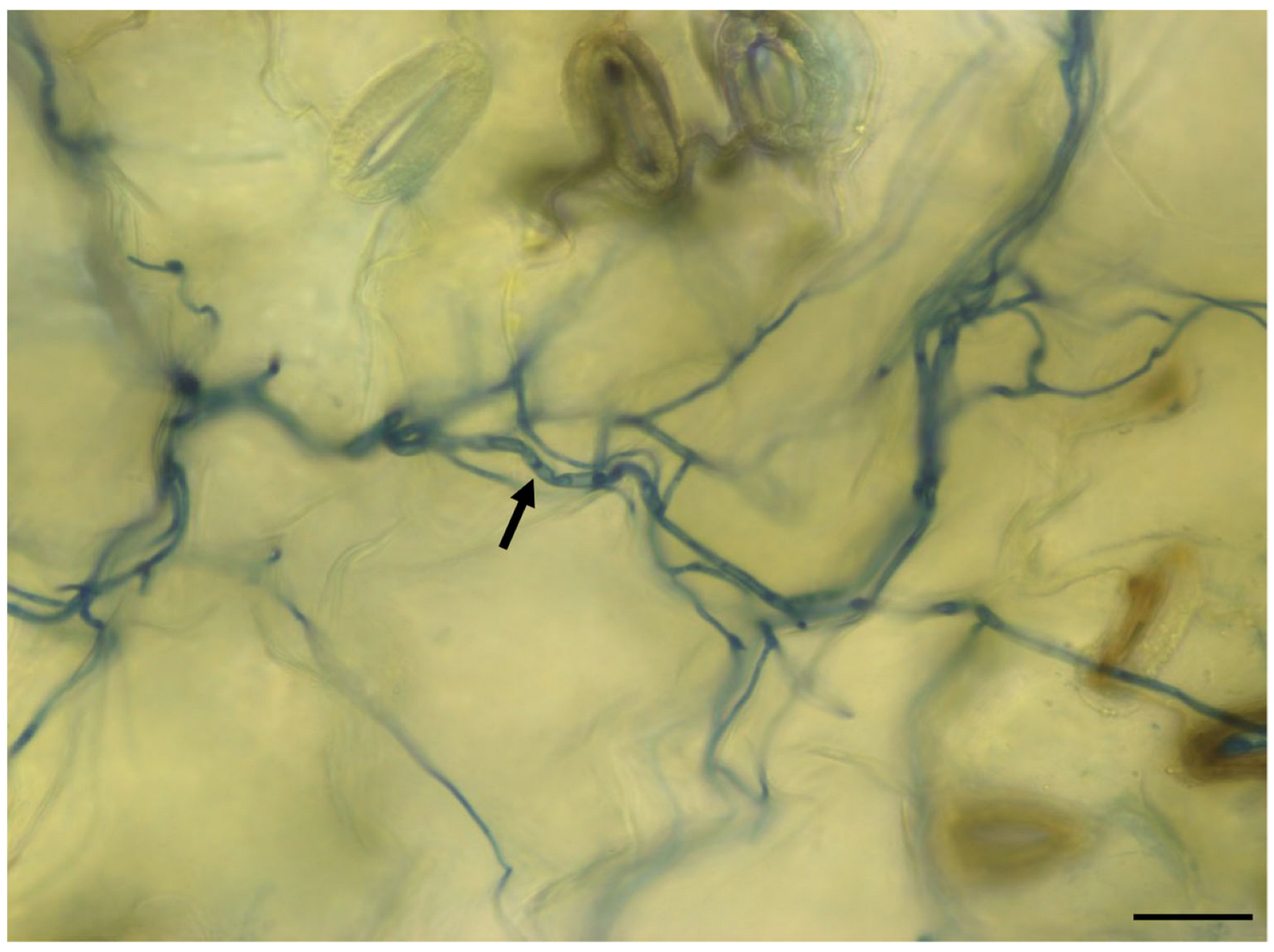
2.1. Analysis of V. longisporum Growth in Rapeseed

To determine the substantial virulent interaction between Verticillium and rapeseed, roots of two-week-old rapeseed plants were infected with the spores of the pathogenic fungus Vl43 strain. A microscopic analysis of royal blue ink-stained leaves 8 days after root infection with Vl43 revealed a net of hyphae colonizing the rapeseed leaves (Figure 1). This confirmed that spores germinated, and hyphae were able to penetrate rapeseed roots, colonize the xylem and move into the leaves via the vascular system.

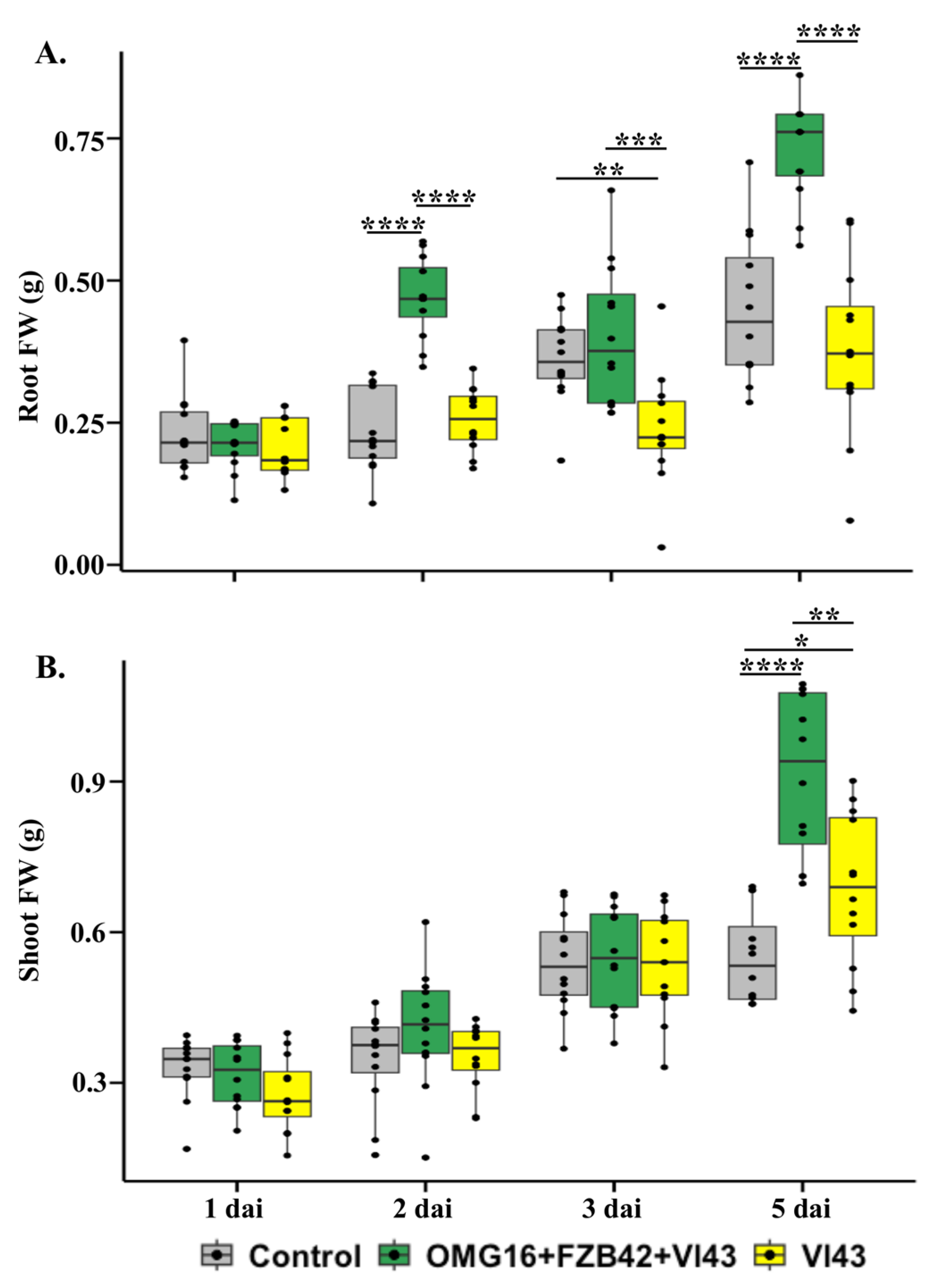
2.2. Effects of Priming on Rapeseed Growth Performance

To determine the effects of microbial priming on plant growth and development, the fresh weights of root and shoot tissues of primed and non-primed rapeseed seedlings were measured. In Vl43-infected primed plants, the fresh weight of roots was increased 2 days after infection (dai) and 5 dai (p < 0.0001 each) compared to the untreated controls. In contrast, the root weight was decreased on 3 dai (p < 0.01) in the Vl43-infected non-primed plants compared to the untreated controls. In Vl43-infected primed plants, the root weight was highly enhanced on 2 dai (p < 0.0001), 3 dai (p < 0.001) and 5 dai (p < 0.0001) compared to the non-primed plants (Figure 2A).
In addition, the shoot weight of Vl43-infected non-primed rapeseed plants slightly (p < 0.05) increased as late as on 5 dai compared to the untreated control plants. Microbial priming also enhanced the shoot weight 5 days after Vl43 infection (p < 0.0001), compared to the untreated controls. In addition, the shoot fresh weight increased in Vl43-infected primed plants on 5 dai (p < 0.01) compared to the non-primed plants (Figure 2B).

2.3. Effects of Priming on V. longisporum 43 Root Infection

The effects of OMG16 plus FZB42 priming on reducing the Vl43 infection rate in rapeseed roots were analysed using qPCR (absolute quantification). The average amount of Vl43 DNA was substantially lower in primed plants than in non-primed plants (Table 1). Vl43 was detectable in Vl43-infected non-primed roots as early as on 1 dai, where an amount of 40.8 ng Vl43 DNA in 15 ng total root DNA was observed. In the following days, the amount continued to increase until 5 dai, the end point of this experiment, where an amount as high as 271.7 ng Vl43 DNA in 15 ng total root DNA was reached. In sharp contrast, the average amount of Vl43 DNA in roots of primed plants was significantly lower, and reached only 15.2 ng Vl43 DNA in 15 ng total root DNA, even at 5 dai. These data demonstrate the high efficiency of priming for restricting infection by V. longisporum, and are in line with our previous findings in Hafiz et al. (2022) [53].

2.4. Enhanced Rapeseed Defence upon Priming

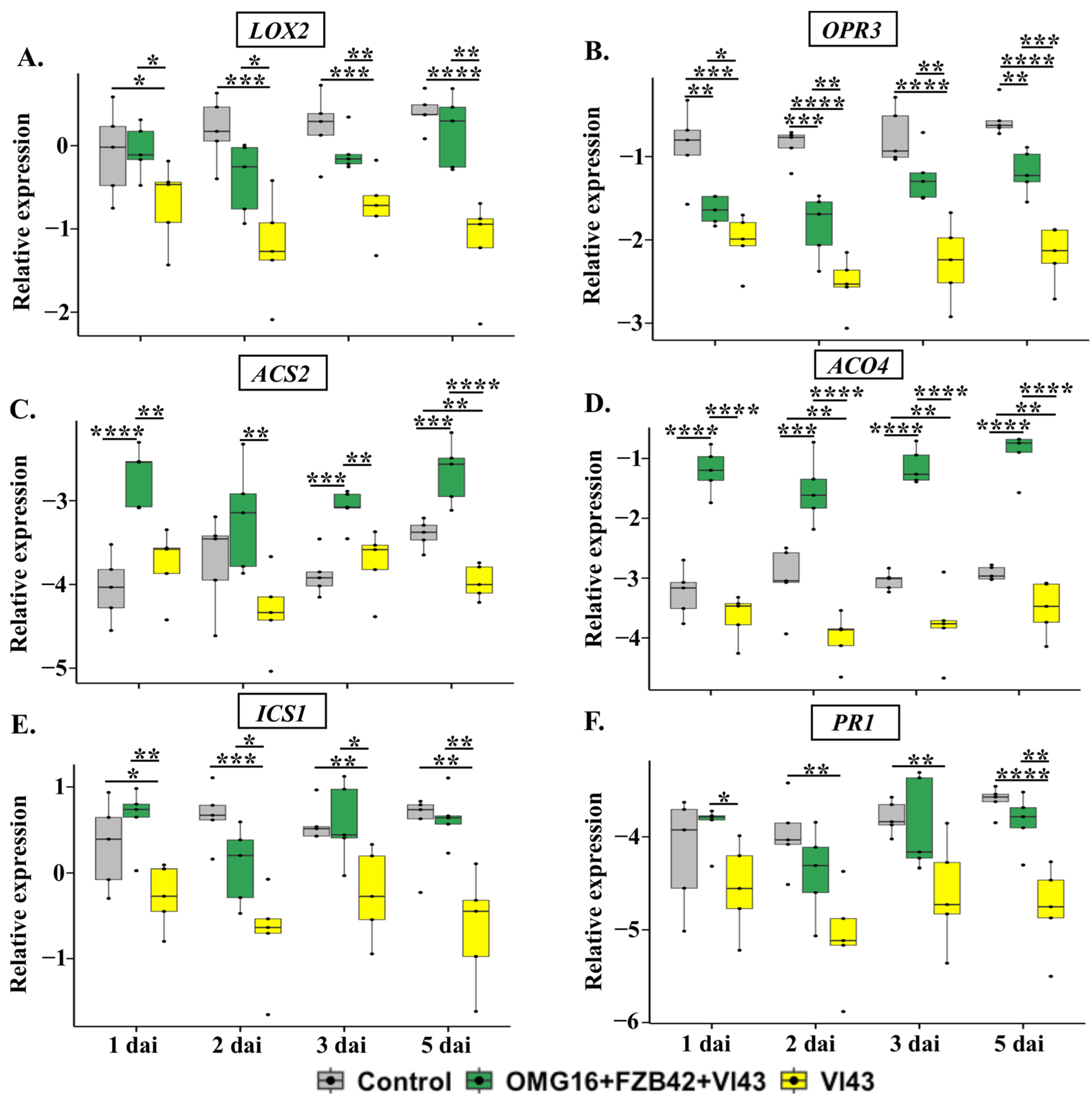
2.4.1. The Expression of Defence-Related Rapeseed Genes in Roots

To investigate the local defence responses of the plants, the relative transcript abundances of the JA biosynthesis genes Lipoxygenase 2 (LOX2) and 12-Oxophytodienoic acid reductase 3 (OPR3), the ET biosynthesis genes 1-Aminocyclopropane-1-carboxylate (ACC) synthase 2 (ACS2) and ACC oxidase 4 (ACO4), the SA biosynthesis gene Isochorismate synthase 1 (ICS1) and the SA-specific marker pathogenesis-related gene 1 (PR1) were analysed in root tissue using qRT-PCR. Prior to the pathogen infection, there was no significant induction of these defence genes in roots of OMG16- and FZB42-primed plants compared to the untreated controls (Supplementary Figure S2).
The expression of both LOX2 and OPR3, involved in JA biosynthesis, was rapidly downregulated in roots of Vl43-infected non-primed plants during the whole experimental period in comparison with non-infected controls. However, microbial priming enhanced LOX2 and OPR3 expression as early as on 1 dai until the end point of this experiment, compared to the Vl43-infected non-primed plants (Figure 3A,B).
The analysis of ET biosynthesis gene expression in the roots of Vl43-infected non-primed plants revealed that the mRNA abundances of ACS2 and ACO4 were downregulated compared to the untreated controls. However, this effect was completely reverted by priming the roots with the selected OMG16 and FZB42 inoculants prior to Vl43 infection (Figure 3C,D).
Similarly, the expressions of SA biosynthesis ICS1 and SA response PR1 were suppressed in roots of Vl43-infected non-primed plants compared to the untreated control plants. However, both ICS1 and PR1 expressions were higher in Vl43-infected primed than in the Vl43-infected non-primed plants (Figure 3E,F).

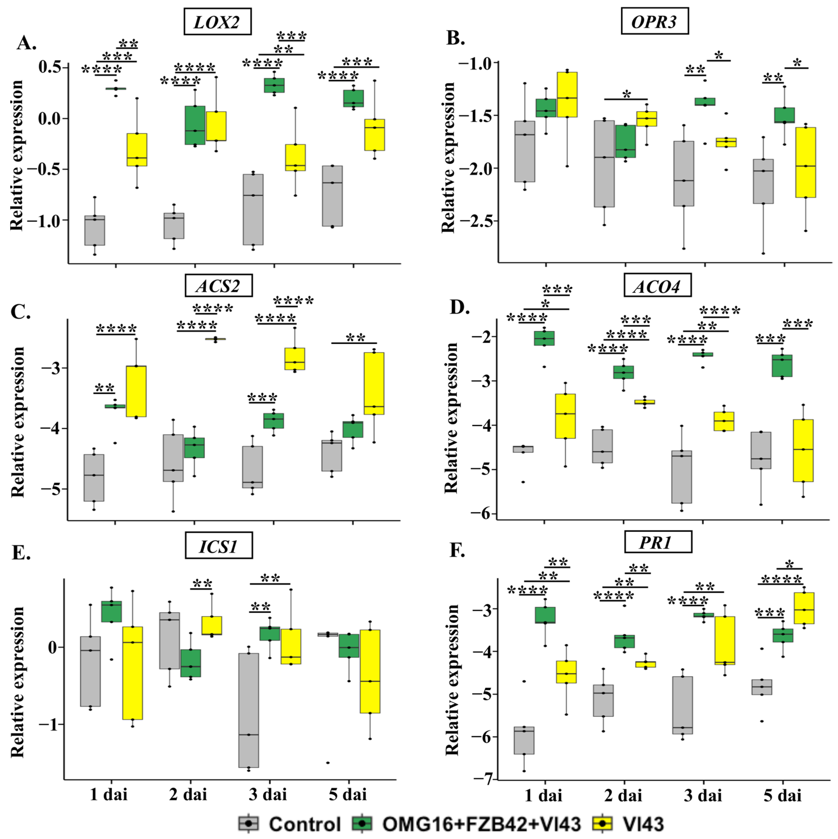
2.4.2. The Expression of Defence-Related Genes in Stems

In addition, to investigate the systemic defence responses, the relative transcript abundances of the previously studied genes were analysed in stem tissue using qRT-PCR. Prior to infection, OMG16 and FZB42 priming significantly increased the relative expression of LOX2, OPR3 and ICS1 in stems compared to the untreated controls (Supplementary Figure S3).
Unlike in roots, the expression of the JA biosynthesis gene LOX2 was highly enhanced in stems of Vl43-infected non-primed plants as early as 1 dai until 5 dai, the end point of the experiment, compared to the untreated controls. In contrast, the OPR3 expression increased only on 2 dai. Moreover, both LOX2 and OPR3 expression increased in stems of Vl43-infected primed plants compared to Vl43-infected non-primed plants (Figure 4A,B).
In the stems of non-primed plants, the expression of the ET biosynthesis genes ACS2 and ACO4 behaved differently in primed and non-primed Vl43-infected plants. In primed Vl43-infected plants, the expression of ACS2 remained, at most time points, at relatively low values, similar to control plants, while the transcript level was clearly increased in non-primed, Vl43-infected plants (Figure 4C). On the other hand, the expression of ACO4 was massively increased in primed, Vl43-infected plants compared to the untreated controls, while in Vl43-infected non-primed plants it was only slightly enhanced or remained even at a level comparable to control plants (Figure 4D).
In both primed and non-primed plants, the expression of the SA biosynthesis gene ICS1 only increased on 3 dai (p < 0.01 each) after infection. Interestingly, the expression of ICS1 was higher on 2 dai (p < 0.01) in Vl43-infected non-primed plants compared to the primed plants (Figure 4E).
The expression of PR1 in stems of Vl43-infected non-primed plants was already elevated on 1 dai and remained upregulated until 5 dai compared to the untreated controls. However, microbial priming enhanced the PR1 expression on 1 dai and 2 dai in Vl43-infected primed plants compared to the non-primed ones. In contrast, on 5 dai, PR1 was higher (p < 0.05) in Vl43-infected non-primed plants compared to the primed plants (Figure 4F).

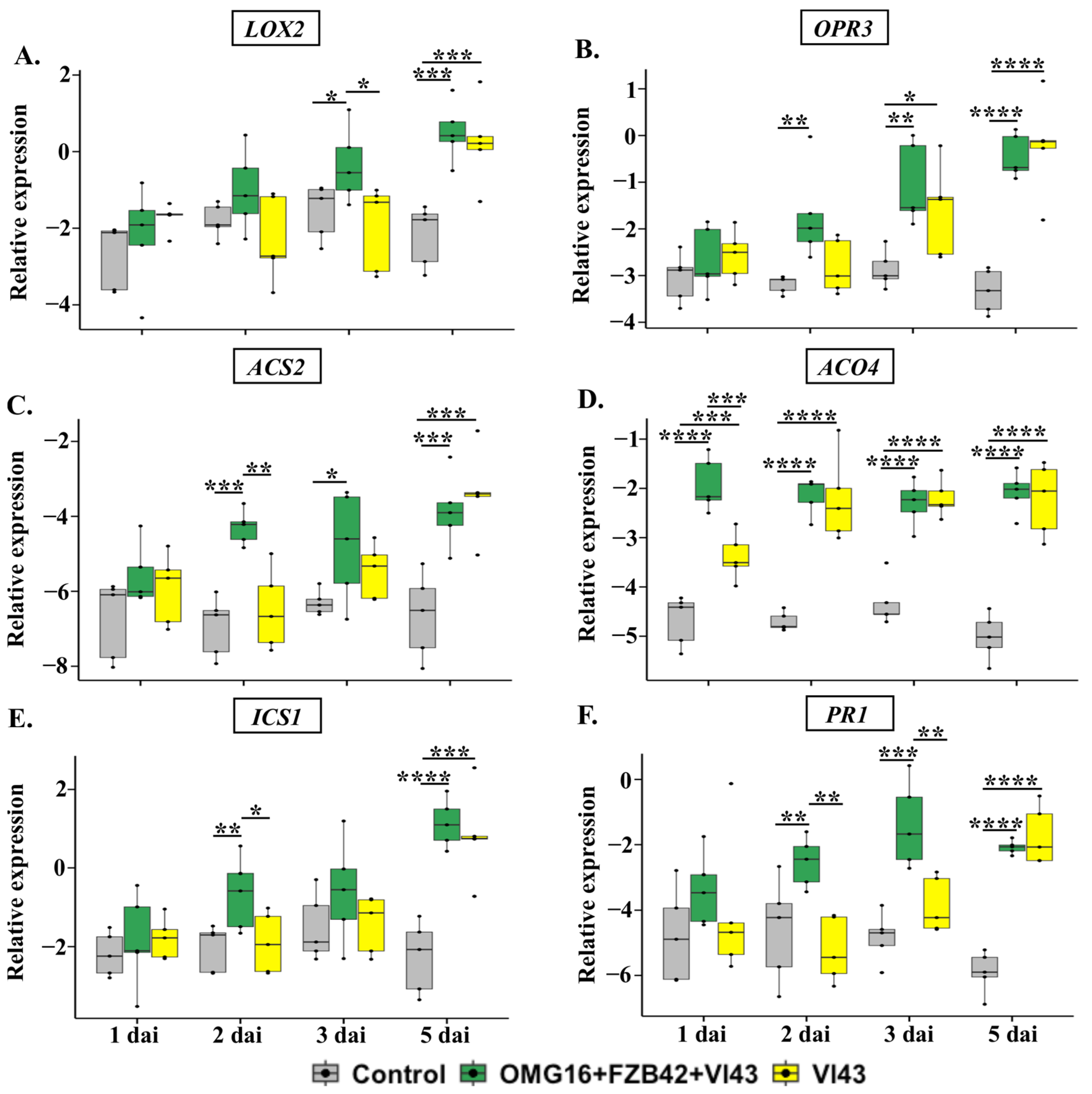
2.4.3. The Expression of Defence-Related Genes in Leaves

To assess the systemic defence responses of rapeseed, the relative expressions of the previously analysed genes were measured in leaf tissue using qRT-PCR. Upon priming, the relative expression of OPR3, ACO4 and PR1 significantly enhanced in leaves of OMG16- and FZB42-primed plants prior to Vl43 infection, compared to the untreated controls (Supplementary Figure S4).
Vl43 infection delayed the induction of LOX2 and OPR3 expression in leaves compared to the untreated controls. Priming with OMG16 and FZB42 led to a higher transcript abundance of these two JA biosynthesis genes in leaves after Vl43 root infection than the Vl43-infected non-primed plants (Figure 5A,B).
The late induction of ACS2 was observed in Vl43-infected non-primed plants compared to the untreated controls (Figure 5C). In contrast, Vl43 infection enhanced ACO4 expression in non-primed plants during the whole experimental period (Figure 5D). Priming increased the expression of both ACS2 and ACO4 after Vl43 infection more rapidly than in Vl43-infected non-primed plants.
The expression of ICS1 and PR1 was only enhanced as late as on 5 dai in Vl43-infected non-primed plants compared to the untreated controls. For both investigated genes, this effect was accelerated by priming the roots with the beneficial microorganisms, OMG16 and FZB42 (Figure 5E,F).

2.5. Determination of Stress-Related Phytohormonal Homeostasis

To analyse the variation in endogenous phytohormone content after priming, the amounts of plant hormones present in rapeseed seedlings were determined in shoots (both in leaves and stems) and roots. The 14-day-old B. napus seedlings were primed with OMG16 plus FZB42. Two days after priming, roots were infected with the pathogenic Vl43 fungal strain. The amounts of endogenous phytohormones JA and SA were measured with UHPLC-MS.

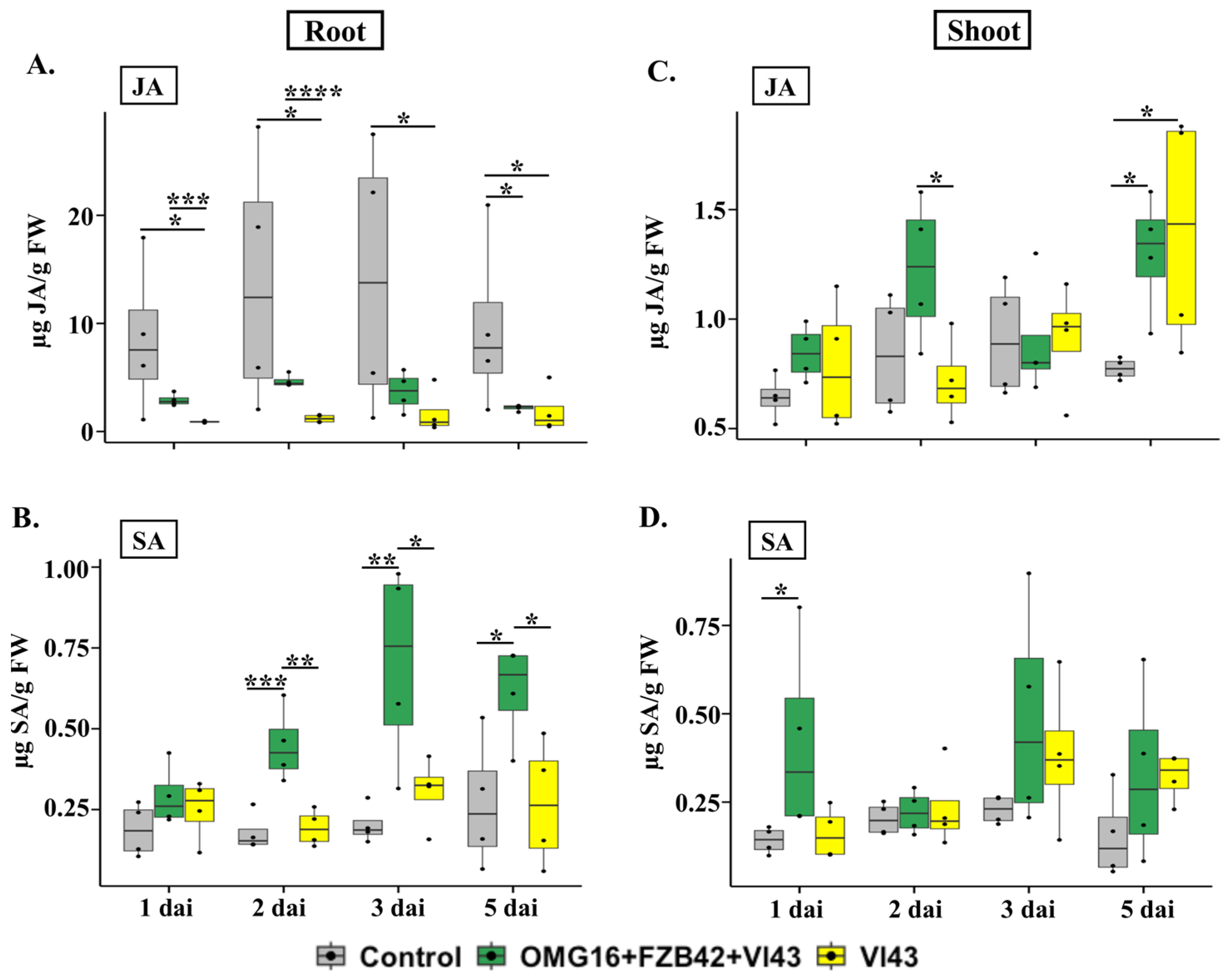
2.5.1. Phytohormone Contents in Root Tissue

In root tissue, the JA content was only significantly lower on 5 dai (p < 0.05) in Vl43 infected primed plants compared to the untreated controls. In contrast, in Vl43-infected non-primed plants, JA production was reduced on 1 dai, 2 dai, 3 dai and 5 dai (p < 0.05 each) compared to the untreated controls. Nevertheless, the JA level highly increased in the roots of Vl43-infected primed plants on 1 dai (p < 0.001) and 2 dai (p < 0.0001) compared to the non-primed ones (Figure 6A).
The content of SA increased on 2 dai (p < 0.001), 3 dai (p < 0.01) and 5 dai (p < 0.05) in Vl43-infected primed plants compared to the untreated controls. In contrast, there was no induction of SA in Vl43-infected non-primed plants compared to the untreated control plants. The level of endogenous SA was enhanced in roots of Vl43-infected primed plants on 2 dai (p < 0.01), 3 dai (p < 0.05) and 5 dai (p < 0.05) compared to the non-primed controls (Figure 6B).

2.5.2. Phytohormone Contents in Shoot Tissues

The content of JA increased as late as on 5 dai (p < 0.05 each) in shoots of both Vl43-infected primed and non-primed plants compared to the untreated controls. The JA content also increased in Vl43-infected primed plants on 2 dai (p < 0.05) compared to the Vl43-infected non-primed plants (Figure 6C).
The endogenous content of SA was enhanced only on 1 dai (p < 0.05) in shoots of Vl43-infected primed plants compared to the untreated controls (Figure 6D). There was no induction of the SA level in Vl43-infected primed plants compared to the non-primed ones.

3. Discussion

3.1. Microscopic Observation of V. longisporum Growth in Rapeseed Leaves

The micrograph presented in Figure 1 shows the hyphal net of Vl43 endophytically colonizing the rapeseed leaves 8 days after root infection. After entering the rapeseed roots, Vl43 hyphae spread to the leaves through the vascular system. This hyphal migration process was previously described by Eynck et al. (2007) and Zheng et al., 2019 [7,54]. This observation also confirmed that the Vl43 spores used were vital and virulent.

3.2. Priming Effects on Plant Growth

The application of combined OMG16 plus FZB42 inocula to the rapeseed roots prior to Vl43 infection improved plant growth. Data indicated that the fresh weight of rapeseed root increased up to 1.9-fold in primed plants compared to the non-primed seedlings after Vl43 infection (Figure 2A), while the shoot fresh weight of primed plants enhanced up to 1.3-fold compared to the non-primed rapeseed followed by Vl43 infection (Figure 2B). In line with this result, previous investigations demonstrated that V. longisporum infection reduced the biomass of rapeseed, and the root biomass was more severely decreased than the shoot. Interestingly, the root colonization by OMG16 with five different Bacillus strains improved the growth and morphology of maize roots [55]. Similar effects were also reported for a Bacillus consortium, where PGPR treatment induced plant growth and development, which in turn increased the biomass and phenotype of maize leaves [56].

3.3. Priming Effects on Reducing Root Infection by Vl43

The priming of rapeseed roots with OMG16 plus FZB42 successfully reduced Vl43 root infection. Approximately 10 to 100 times less infection as measured by presence of pathogen DNA was recorded in the primed roots compared to the non-primed (Table 1). The underlying mechanisms of such disease resistance might include the physical barrier formed by root endophytic OMG16 and FZB42 microbes, restricting the entry and growth of Vl43 hyphae into the rapeseed roots. Here, it is worth mentioning that our previous study demonstrated OMG16-formed endophytic hyphae inside rapeseed root hairs to develop distinct intracellular structures [53]. Other reasons for Vl43 growth suppression might include competition for space and nutrition [57], the production of hydrolytic enzymes by the Trichoderma strain to break down the cell wall of the pathogen and the release of secondary antimicrobial metabolites [58,59].

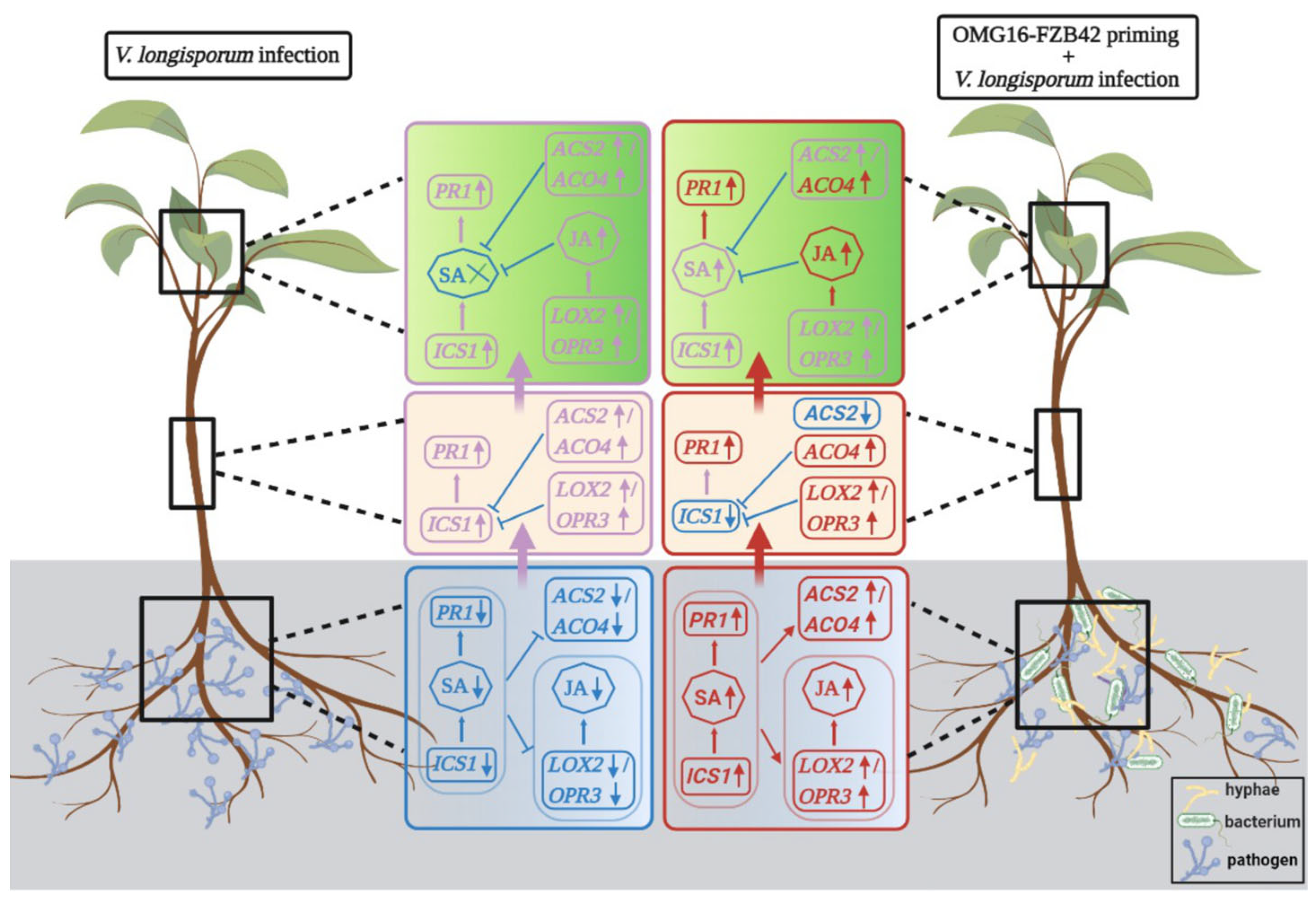
3.4. Priming Effects on Rapeseed-Enhanced Defence Activation after Vl43 Pathogen Challenge

Our previous study demonstrated that OMG16 and FZB42 priming reduced Vl43 growth up to 100-fold in rapeseed root tissue [53]. As we show here, priming also enhanced the plants’ systemic defence responses against Vl43 infection (Figure 3, Figure 4 and Figure 5). Priming the rapeseed roots significantly enhanced the JA and ET biosynthesis, as well as the SA signalling pathways in leaves, by inducing the corresponding biosynthesis and signalling genes, OPR3, LOX2 and PR1 (Supplementary Figure S4). This observation corroborates previous studies reporting that T. virens and T. atroviride activated the JA/ET pathway by inducing the expression of LOX-1 in A. thaliana. The same fungi also induced the expression of PR1, thereby activating the SAR pathway [60,61]. In addition, T. asperellum DQ-1 enhanced the expression of PR2 and LOX1 in both Botrytis cinerea challenged and non-challenged inoculation treatments, and increased resistance against grey mould disease in tomato [62].
Similarly, in stems, priming also showed fast and strong induction of the JA biosynthesis pathway and relatively slower and weaker induction of SA biosynthesis (Supplementary Figure S3). There was no significant induction of JA, ET and SA biosynthesis or signalling in roots (Supplementary Figure S2). These data indicate that beneficiary fungal and bacterial strains can prime the whole rapeseed plant body and transduce the ISR defence signal into the upper part of plants, presumably via the phytohormones JA and ET or through some as-yet-unknown mobile messenger. Subsequently, when the rapeseed roots were infected with Vl43 spores 2 days after priming, such molecular memory of immunization enhanced rapeseed defence against the pathogen challenge. Similar effects were previously observed after the combined application of T. harzianum Tr6 and Pseudomonas sp. Ps14, which significantly increased the resistance level in cucumber against Fusarium oxysporum infection through the elevated expression of a set of defence-related genes [63].
The analysis of gene expression using qRT-PCR revealed a fast and strong induction of JA, ET and SA biosynthesis and signalling genes separately in root, stem and leaf tissues of the OMG16 plus FZB42 primed plants. Moreover, non-primed Vl43 infection also induced rapeseed defence through the SAR pathway by enhancing the expression of JA, ET and SA biosynthesis and signalling genes. Such Verticillium-mediated defence activation was relatively weaker and slower.

3.4.1. Priming Effects on Roots

qRT-PCR analysis showed that the expression of LOX2 and OPR3 was downregulated in the roots of Vl43-infected non-primed plants (Figure 3A,B). This indicates that for the successful colonization of rapeseed roots, Vl43 suppressed the JA biosynthesis pathway shortly after infection (1 dai) and kept it attenuated at least until 5 dai, the end point of this experiment. A previous study demonstrated that Vl43 requires the functional activity of JA receptor, coronatine insensitive 1 (COI1), in the roots of Arabidopsis thaliana to efficiently colonize its roots and elicit disease symptoms in the shoots [64]. In contrast, rapeseed plants primed with OMG16 and FZB42 were able to successfully overcome the suppression of JA biosynthesis in the roots after Vl43 infection. The upregulation of both LOX2 and OPR3 in the primed plants was identified as fast as 1 dai, and remained activated until 5 dai compared to the non-primed plants. Nevertheless, the expression of OPR3 still remained downregulated in Vl43-infected primed plants compared to the untreated controls. However, OPR3 expression was remarkably higher in the Vl43-infected primed plants than in the non-primed plants.
In addition, endogenous JA contents in rapeseed roots were measured using UHPLC-MS. The JA level was approximately 10-fold less in the roots of Vl43-infected non-primed plants than in the untreated controls (Figure 6A). Vl43 repressed JA production in rapeseed roots as early as 1 dai, and this continued until 5 dai. This observation again demonstrated that Vl43 infection impaired JA biosynthesis during the early root colonization process. In Vl43-infected primed plants, the JA content was 3.9-fold higher within 1 dai and 2 dai than in Vl43-infected non-primed plants. This points to the evidence that priming with OMG16 and FZB42 enabled the plants to overcome the suppression of JA biosynthesis caused by Vl43.
Vl43 infection also inhibited the ET biosynthesis pathway at the site of infection by downregulating the expression of ACS2 and ACO4 in non-primed plants (Figure 3C,D). Compared to the untreated controls, ACS2 expression was suppressed as late as 5 dai, while the downregulation of ACO4 was detected as early as on 2 dai and remained suppressed until 5 dai. The phytohormone ET was reported earlier to be necessary for V. longisporum to efficiently colonize plant roots. The A. thaliana mutants, ein2-1, ein4-1 and ein6-1, impaired in the ET signalling pathway, displayed an enhanced susceptibility to V. longisporum compared to wild-type plants [65,66]. Rapeseed roots previously treated with OMG16 and FZB42 suspensions showed fast and strong induction of the ET biosynthesis pathway by enhancing the expression of ACS2 and ACO4 in root tissues after Vl43 infection. In addition, the expression of the ET biosynthesis genes ACS2 and ACO4 also reacted faster and more strongly to Vl43 infection in primed compared to the non-primed plants. This points to the fact that priming with beneficial microbes enabled rapeseed roots to overcome the adverse effects of ET biosynthesis caused by Vl43 infection and enhanced the generation of ET.
The expression of ICS1 and PR1 was drastically reduced after Vl43 infection in the non-primed roots, indicating that the Vl43 pathogen suppressed both SA biosynthesis and signalling pathways to successfully colonize the rapeseed roots (Figure 3E,F). In accordance with this result, no induction of PR1 was found in the Vl43-infected rapeseed roots at 3 dai [67]. In a COI1-dependent manner, V. longisporum suppressed the SA-dependent defence in rapeseed by generating JA-isoleucine-mimic coronatine [64]. In our study, we showed that primed plants were able to quickly enhance the expression of both ICS1 and PR1, thus mitigating the downregulation of SA biosynthesis and signalling caused by Vl43 infection. Moreover, the endogenous SA content was also enhanced in Vl43-infected primed roots compared to both Vl43-infected non-primed plants and untreated control plants (Figure 6B). The synthesis of SA in the roots of primed rapeseed increased to 2.3-fold on 2 dai and remained induced until 5 dai. Ratzinger et al. (2009) reported that SA is involved in the susceptibility of B. napus to V. longisporum infection [68]. Priming may reduce the susceptibility by increasing SA accumulation in rapeseed roots.
Taken together, these results suggest that Vl43 hijacks the defence system of rapeseed by inhibiting the biosynthesis of JA, ET and SA phytohormones, and successfully enters into the roots to establish a long-lasting compatible interaction with the host. Rapeseed roots primed with OMG16 and FZB42 microbes revealed a rapid reprogramming of gene expression to activate the biosynthetic pathways of the hormones JA, ET and SA, and restricted the entry of the Vl43 pathogen into the root cells. In a similar study, the inoculation of watermelon roots with the biocontrol agent F1-35 induced the JA and ET pathways by elevating the expression of LOX1, AOS and JAR1 in local root tissue. Inoculated plants showed improved resistance against Fusarium oxysporum f. sp. Niveum [69].

3.4.2. Priming Effects on Stems

After penetrating the root, the pathogen Vl43 systemically spread through the xylem and generated microsclerotia in the stem of oil seed rape to develop “Verticillium stem striping” disease [5,7]. Gene expression analysis with qRT-PCR revealed a fast and strong induction of LOX2 and OPR3 in the stem tissue (Figure 4A,B). The expression of these two JA biosynthesis genes indicated the enhanced production of JA in both Vl43-infected primed and non-primed plants. Notably, the expressions of LOX2 and OPR3 were higher in the stems of Vl43-infected primed plants compared to the non-primed controls.
Similarly, the expressions of ET biosynthesis genes ACS2 and ACO4 were highly enhanced in stems of both Vl43-infected primed and non-primed plants (Figure 4C,D). The ACO4 expression was higher in Vl43-infected primed than non-primed plants. Interestingly, after the Vl43 challenge, the ACS2 expression increased in the non-primed plants compared to the primed ones.
In Vl43-infected stems, SA biosynthesis and signalling increased both in primed and non-primed plants. PR1 expression was higher in the primed stems for the first 2 days after infection, while the expression increased on 5 dai in the non-primed plants (Figure 4F). This indicates that SA signalling increased in the stem tissue with the gradual progress of Vl43 infection. In line with this result, a previous study documented an elevated level of SA and its metabolite, salicylic acid glucoside (SAG), in the xylem sap of rapeseed shoots after V. longisporum infection. A strong correlation was also reported between the content of V. longisporum DNA in hypocotyls and SAG levels in the shoot [68].

3.4.3. Priming Effects on Leaves

The analysis of gene expression with qRT-PCR revealed the faster and stronger induction of LOX2 and OPR3 in leaves of Vl43-infected primed plants compared to non-primed plants (Figure 5A,B). The expression of these two biosynthesis genes in leaves indicated the activation of the JA biosynthesis pathway in both Vl43-infected primed and non-primed plants. Moreover, endogenous JA levels were up to 1.7-fold higher in the shoots of Vl43-infected primed than in non-primed plants (Figure 6C). Therefore, these data indicated that the presence of OMG16 and FZB42 in rapeseed roots induced the JA biosynthesis pathway in leaves.
The expression of ET biosynthesis genes ACS2 and ACO4 was highly enhanced in the leaves of primed plants after Vl43 infection (Figure 5C,D). Therefore, these results demonstrated that OMG16 and FZB42 priming induced the ET biosynthesis pathway in rapeseed leaves to increase the resistance against Vl43 infection.
In addition, the expression of the SA biosynthesis gene ICS1 and the SA signalling gene PR1 was remarkably enhanced in leaves of Vl43-infected primed plants (Figure 5E,F). Moreover, the level of endogenous SA was elevated up to 2.6-fold in shoots of Vl43-infected primed plants shortly after infection (1 dai) (Figure 6D), which indicates that both SA biosynthesis and signalling pathways were induced in leaves after priming the rapeseed roots. In a previous study, seed-coating by Trichoderma atroviride SG3403 and Bacillus subtilis 22 co-culture enhanced the expression of defence genes, including PR1 and ACS, which promoted tolerance against F. graminearum infection in wheat leaves [70].
Similarly, the pre-inoculation of peanut with the root endophytic fungus Phomopsis liquidambaris B3 triggered SA signalling by enhancing the expression of PRs and increasing the SA level, which in turn reduced F. oxysporum root colonization and disease severity both locally and systemically. However, such resistance was absent in the untreated roots [71].
Taken together, our data indicate that the priming of rapeseed roots with the beneficial microbes OMG16 and FZB42 can lead to rapid and strong induction of plant defence hormone biosynthesis and signalling in roots, stems and leaves. Moreover, a root-borne mobile signal transduces from roots via stems to leaves, mediated by the phytohormones JA, ET and SA, to enhance the defence system in response to Vl43 infection. Similarly, B. amyloliquefaciens ssp. plantarum UCMB5113 rhizobacterium enhanced plant defence priming and conferred resistance to airborne pathogens without any direct interaction [72]. In a similar study, rhizobacterium P. alvei K165 triggered ISR to protect A. thaliana from V. dahliae infestation. K165 induced the SA-dependent defence pathway by increasing the PR1 expression in leaves [73]. On the other hand, the transcriptome analysis of Trichoderma atroviride T11 showed increased expression levels of genes involved in catalytic, hydrolytic and transporter activities, and secondary metabolism in response to V. dahliae infection [74]. Therefore, the orchestrated transcriptional reprogramming of JA, ET and SA pathways upon priming induced rapeseed defence and restricted V. longisporum proliferation.

4. Materials and Methods

4.1. Plant Materials and Growth Conditions

Oilseed rape (B. napus) seedlings, genotype AgP4 (seeds were provided by the NPZ Innovation GmbH, Hohenlieth, Germany), were used for all the experiments in this study. Details of the cultivar were discussed in Hafiz et al. (2022) [53]. For surface sterilization, B. napus seeds were incubated in 3% sodium hypochlorite solution (Carl Roth, Karlsruhe, Germany) for 15 min with gentle shaking followed by rigorous washing with sterile, deionised water. Surface-sterilized seeds were germinated on soaked filter papers (grade MN 619 eh; Macherey-Nagel, Düren, Germany) placed on glass Petri dishes (150 × 30 mm), placed in a growth chamber at 20 °C with 16 h light (80 µmol m−2s−1) and 60% relative humidity. Six days after germination, the seedlings were robust and shifted onto sterile filter papers soaked with half-strength Murashige and Skoog (MS) medium (Sigma-Aldrich, St. Louis, MO, USA) (supplemented with 0.5% sucrose and 5.4 µM 1-naphthylacetic acid), inserted into sterilized plant boxes and incubated in a growth chamber set to the conditions mentioned above until the seedlings were two weeks old.

4.2. Microbial Priming and Pathogen Infection

To analyse the effects of microbial priming in rapeseed defence activation, two-week-old rapeseed roots were primed with a mixed suspension of Trichoderma harzianum OMG16 (received from the strain collection of Anhalt University of Applied Sciences, Bernburg, Germany) plus Bacillus velezensis FZB42 syn. Bacillus amyloliquefaciens subsp. plantarum FZB42 (obtained from ABiTEP GmbH, Berlin, Germany). Microbial growth conditions and inoculation procedures were carried out according to the protocol mentioned in Hafiz et al. (2022) [53]. Briefly, roots of rapeseed seedlings were dipped in a combined inocula of OMG16 (2000 spores per mL) plus FZB42 (OD600 = 0.275, corresponding to approximately 2.2 × 10⁸ cells per mL) or in deionized water (control). Two days after priming, the same roots were infected with Verticillium longisporum 43 (provided by NPZ Innovation GmbH, Hohenlieth, Germany) by dipping into Vl43 spore suspension (106 spores per mL) for 30 min or into deionized water (control). Roots of non-primed seedlings were also infected with Vl43 as positive controls. For each treatment, there were five biological replicates and three plants were pooled to obtain one biological replicate.

4.3. Microscopic Analysis of Vl43-Infected Rapeseed Leaf Tissue

Roots of two-week-old rapeseed seedlings were infected with Vl43 according to the procedure mentioned above. Eight days after infection, cotyledons were separated from the stem using a sharp scalpel. Prior to staining, leaves were thoroughly washed with sterile water. Then, the leaves were dipped in blue staining solution (25% acetic acid and 10% royal blue ink from Rohrer and Klingner Leipzig-Co., Zella-Mehlis, Germany) for 1 min. Stained leaves were briefly washed with sterile deionized water and examined under a Zeiss Axio Observer 7 inverse microscope (Carl Zeiss, Jena, Germany).

4.4. Absolute Quantification of Vl43 DNA in Root Samples via qPCR

Roots were harvested from both Vl43-infected primed and non-primed plants. Approximately 50 mg of root segments were weighed to isolate total DNA with the DNeasy plant mini kit (Qiagen, Venlo, The Netherlands) according to the manufacturer instructions. Then, the concentrations of the isolated DNA were measured with Qubit 3.0 fluorometer using the Qubit dsDNA assay kit (Thermo Fisher Scientific, Waltham, MA, USA). A qPCR reaction of 20 μL total volume contained 15 ng of isolated DNA from infected roots, 10 μL of PowerUp SYBR Green master mix (Applied Biosystems, Waltham, MA, USA) and the V. longisporum-specific primer pairs OLG70 and OLG71 [7]. The following qPCR program was used in a QuantStudio 5 real-time PCR system (Applied Biosystems, Waltham, MA, USA): 50 °C for 2 min, 95 °C for 2 min, and 36 cycles of amplification (denaturation, 95 °C for 20 s; annealing, 59 °C for 30 s and elongation, 72 °C for 30 s). A melting curve was included with initial denaturation at 95 °C for 15 s, annealing at 60 °C for 1 min, and finally a linear increase in the temperature from 60 to 95 °C with a rate of 0.1 °C s−1. All primer sequences used in this study are listed in Supplementary Table S1. The precise annealing temperature and efficiency of each primer pair was determined by gradient PCR and applied in qPCR (Supplementary Figure S1).

4.5. Gene Expression Studies with Total RNA Extraction, cDNA Synthesis and qRT-PCR

To analyse the tissue-specific priming effects, root, stem and leaf tissues were separated with a sharp scalpel and immediately transferred to −80 °C at 1 day and 2 days after OMG16 and FZB42 priming. Moreover, the same tissue types were also collected and immediately stored in −80 °C at 1 day, 2 days, 3 days and 5 days after Vl43 infection. Approximately 60 mg of tissues were weighed to isolate total RNA by using the RNeasy plant mini kit (Qiagen, Venlo, The Netherlands), according to the manufacturer’s instructions. Then, 1.2 µg of total RNA was taken to synthesize cDNA by using the SuperScript IV first-strand synthesis system (Thermo Fisher Scientific, Waltham, MA, USA). The concentrations of isolated cDNAs were quantified with a Qubit 3.0 fluorometer, using the Qubit ssDNA assay kit (Thermo Fisher Scientific, Waltham, MA, USA). The expression of rapeseed defence-related genes was analysed using qRT-PCR performed in a QuantStudio 5 real-time PCR system (Applied Biosystems, Waltham, MA, USA). qRT-PCR reactions were prepared in quadruplicate, each in 20 µL of total volume containing 15 ng of cDNA, 10 µL of PowerUp SYBR Green master mix (Applied Biosystems, Waltham, MA, USA) and 5 pmol each of forward and reverse primer. The cycling instructions for qRT-PCR included UGD activation at 50 °C for 2 min, Dual-LockTM DNA polymerase at 95 °C for 2 min and 40 cycles of amplification (denaturation, 95 °C for 1 s; annealing and elongation, 60 °C for 30 s). A melting curve was recorded after amplification with initial denaturation at 95 °C for 15 s, annealing at 60 °C for 1 min and finally a linear increase in the temperature from 60 °C to 95 °C with a rate of 0.1 °C s−1. For normalization, BnaUbiquitin11, BnaActin and BnaTubulin were selected to serve as endogenous controls. To calculate the relative transcript abundances of genes, the efficiency-corrected Cq of three endogenous controls was averaged. ∆Cq was calculated as follows: ∆Cq = Cq reference genes—Cq target gene [75,76,77].

4.6. Determination of Phytohormones Using UHPLC-MS Analysis

After microbial inoculation onto rapeseed roots, as mentioned above, roots and shoots were harvested by cutting with a sharp scalpel and tissues were frozen separately into liquid nitrogen. There were four biological replicates, and three plants were pooled for each biological replicate. Phytohormone contents of rapeseed shoots and roots were determined with UHPLC-MS analysis according to the procedure mentioned in Moradtalab et al. (2018), with slight modifications [78]. Briefly, 1 g of frozen tissue samples (shoots, roots) was ground to fine powder with liquid N2 and extracted with 80% methanol (5 mL). Afterwards, the ground tissues were further homogenized by ultrasonication (Micra D-9 homogenizer, Art, Müllheim, Germany) for 1 min and 15 s at 10,000 rpm, and then 2 mL methanol extracts were added to microtubes and centrifuged at 14,000× g for 10 min. Later, the supernatant (400 µL) was mixed with same amount of ultra-pure water and again centrifuged at 14,000× g for 5 min. The supernatant was cleaned with membrane filtration (Chromafil R O20/15 MS) and transferred to HPLC vials. UHPLC-MS analysis was conducted in a Velos LTQ System (Thermo Fisher Scientific, Waltham, MA, USA) fitted with a Synergi Polar column, 4 µL, 150 × 3.0 mm (Phenomenex, Torrance, CA, USA), for the measurement of JA and SA phytohormones. The injection volume was set to 3 µL. The flow rate was adjusted to 0.5 mL min−1 for gradient elution with mobile phase A (water/acetonitrile = 95/5 (v/v)) and mobile phase B (pure acetonitrile), and a gradient profile of 0–1 min, 95% A, 5% B, 11–13 min, 10% A, 90% B, 13.1 min, 95% A, 5% B, 16 min 95% A and 5% B. All standard reference substances were purchased from Sigma Aldrich (St. Louis, MO, USA).

4.7. Statistical Analysis

In this study, statistical analysis was performed using R software, version 4.0.2 [79]. The significance of rapeseed defence gene expression after Vl43 pathogen challenge was calculated using Dunnett’s test with multivariate testing (mvt) adjustment at p < 0.05 compared to the untreated control plants. The significance of the relative expression of genes between Vl43-challenged primed and non-primed treatments was calculated using an unpaired t-test at p < 0.05. The significance of endogenous plant hormone concentrations was calculated using Dunnett’s test with mvt adjustment at p < 0.05 compared to the untreated controls.

5. Conclusions

Our studies conducted so far demonstrated that priming rapeseed roots with T. harzianum OMG16 and B. velezensis FZB42 activated the plant defence system and systematically transduced that defence signals went from the root to the upper part of the plant body through the activation of a set of phytohormones. In Vl43-infected non-primed plants, SA biosynthesis and signalling was downregulated in roots compared to the untreated controls. Evidently, weaker and slower JA/ET signals were transduced from the roots to the leaves in Vl43-infected non-primed plants compared to the primed plants. On the other hand, in primed plants, SA biosynthesis and signalling were upregulated in roots after Vl43 infection compared to the non-primed plants. Activated SA simultaneously triggered both JA and ET biosynthetic pathways. In stems of primed plants, both JA and ET acted antagonistically with the SA biosynthesis pathway. Similarly, in leaves, elevated levels of JA suppressed SA production. In primed plants, the expression of the JA and ET marker genes was stronger and faster after Vl43 infection than in the non-primed plants. Here, remarkably intense SA, JA and ET signals were systematically transduced from the root to the shoot of the primed plants. A conceptional model summarizing the events of signal transduction involved in resistance induction against Vl43 in oilseed rape seedlings is presented in Figure 7. Our results suggest an alternative approach to develop enhanced rapeseed tolerance against Verticillium root infection. Such an induction of defence mechanisms upon beneficial microbial treatments could also be transferred to field conditions to further investigate the control of Verticillium stem stripping disease in rapeseed.

Supplementary Materials

The following supporting information can be downloaded at: https://www.mdpi.com/article/10.3390/ijms241310489/s1.

Author Contributions

Conceptualisation, W.R., J.G. and F.B.H.; methodology, F.B.H. and J.G.; validation, G.N. and W.R.; formal analysis, F.B.H. and A.A.M.; investigation, F.B.H.; writing—original draft preparation, F.B.H. and J.G.; writing—review and editing, W.R. and G.N.; supervision, W.R. and I.S.; project administration, J.G.; funding acquisition, J.G. All authors have read and agreed to the published version of the manuscript.

Funding

This research work was funded by International Graduate Schools in Agricultural and Polymer Sciences (AGRIPOLY), European Commission–European Social Fund (EU-ESF), Funding-ID: ZS/2016/08/80644; German Federal Ministry of Education and Research (BMBF) through the program BonaRes II (Project DiControl), Funding-ID: 031B0514B and D; the German Research Foundation (Deutsche Forschungsgemeinschaft DFG), project number 491460386; and the Open Access Publishing Fund of Anhalt University of Applied Sciences.

Institutional Review Board Statement

Not applicable.

Informed Consent Statement

Not applicable.

Data Availability Statement

Data will be made available upon request.

Acknowledgments

The authors would like to thank NPZ Innovation GmbH, Hohenlieth, Germany, for kindly providing the rapeseed seeds and the spores of V. longisporum strain 43.

Conflicts of Interest

The authors declare no conflict of interest.

References

  1. Torrens-Spence, M.P.; Bobokalonova, A.; Carballo, V.; Glinkerman, C.M.; Pluskal, T.; Shen, A.; Weng, J.K. PBS3 and EPS1 Complete Salicylic Acid Biosynthesis from Isochorismate in Arabidopsis. Mol. Plant 2019, 12, 1577–1586. [Google Scholar] [CrossRef] [PubMed] [Green Version]
  2. Leonard, M.; Kühn, A.; Harting, R.; Maurus, I.; Nagel, A.; Starke, J.; Kusch, H.; Valerius, O.; Feussner, K.; Feussner, I.; et al. Verticillium longisporum Elicits Media-Dependent Secretome Responses With Capacity to Distinguish Between Plant-Related Environments. Front. Microbiol. 2020, 11, 1876. [Google Scholar] [CrossRef] [PubMed]
  3. Novakazi, F.; Inderbitzin, P.; Sandoya, G.; Hayes, R.J.; von Tiedemann, A.; Subbarao, K.V. The Three Lineages of the Diploid Hybrid Verticillium longisporum Differ in Virulence and Pathogenicity. Phytopathology 2015, 105, 662–673. [Google Scholar] [CrossRef] [PubMed] [Green Version]
  4. Harting, R.; Starke, J.; Kusch, H.; Pöggeler, S.; Maurus, I.; Schlüter, R.; Landesfeind, M.; Bulla, I.; Nowrousian, M.; de Jonge, R.; et al. A 20-kb lineage-specific genomic region tames virulence in pathogenic amphidiploid Verticillium longisporum. Mol. Plant Pathol. 2021, 22, 939–953. [Google Scholar] [CrossRef]
  5. Depotter, J.R.; Deketelaere, S.; Inderbitzin, P.; Tiedemann, A.V.; Höfte, M.; Subbarao, K.V.; Wood, T.A.; Thomma, B.P. Verticillium longisporum, the invisible threat to oilseed rape and other brassicaceous plant hosts. Mol. Plant Pathol. 2016, 17, 1004–1016. [Google Scholar] [CrossRef] [Green Version]
  6. Taylor, J.T.; Harting, R.; Shalaby, S.; Kenerley, C.M.; Braus, G.H.; Horwitz, B.A. Adhesion as a Focus in Trichoderma-Root Interactions. J. Fungi 2022, 8, 372. [Google Scholar] [CrossRef]
  7. Eynck, C.; Koopmann, B.; Grunewaldt-Stoecker, G.; Karlovsky, P.; Von Tiedemann, A. Differential interactions of Verticillium longisporum and V. dahliae with Brassica napus detected with molecular and histological techniques. Eur. J. Plant Pathol. 2007, 118, 259–274. [Google Scholar] [CrossRef]
  8. Kamble, A.; Koopmann, B.; Von Tiedemann, A. Induced resistance to Verticillium longisporum in Brassica napus by β-aminobutyric acid. Plant Pathol. 2013, 62, 552–561. [Google Scholar] [CrossRef]
  9. Heale, J.B.; Karapapa, V.K. The Verticillium threat to Canada’s major oilseed crop: Canola. Can. J. Plant Pathol. 1999, 21, 1–7. [Google Scholar] [CrossRef]
  10. Sarenqimuge, S.; Rahman, S.; Wang, Y.; von Tiedemann, A. Dormancy and germination of microsclerotia of Verticillium longisporum are regulated by soil bacteria and soil moisture levels but not by nutrients. Front. Microbiol. 2022, 13, 979218. [Google Scholar] [CrossRef]
  11. Dunker, S.; Keunecke, H.; Steinbach, P.; Von Tiedemann, A. Impact of Verticillium longisporum on yield and morphology of winter oilseed rape (Brassica napus) in relation to systemic spread in the plant. J. Phytopathol. 2008, 156, 698–707. [Google Scholar] [CrossRef]
  12. Wang, Y.; Strelkov, S.E.; Hwang, S.F. Blackleg Yield Losses and Interactions with Verticillium Stripe in Canola (Brassica napus) in Canada. Plants 2023, 12, 434. [Google Scholar] [CrossRef]
  13. Yadeta, K.A.; Hanemian, M.; Smit, P.; Hiemstra, J.A.; Pereira, A.; Marco, Y.; Thomma, B.P. The Arabidopsis thaliana DNA-binding protein AHL19 mediates Verticillium wilt resistance. Mol. Plant Microbe Interact. 2011, 24, 1582–1591. [Google Scholar] [CrossRef] [PubMed] [Green Version]
  14. Yadeta, K.A.; Valkenburg, D.J.; Hanemian, M.; Marco, Y.; Thomma, B.P. The Brassicaceae-specific EWR1 gene provides resistance to vascular wilt pathogens. PLoS ONE 2014, 9, e88230. [Google Scholar] [CrossRef] [Green Version]
  15. Obermeier, C.; Hossain, M.A.; Snowdon, R.; Knüfer, J.; von Tiedemann, A.; Friedt, W. Genetic analysis of phenylpropanoid metabolites associated with resistance against Verticillium longisporum in Brassica napus. Mol. Breed. 2013, 31, 347–361. [Google Scholar] [CrossRef]
  16. Wu, L.; Huang, Z.; Li, X.; Ma, L.; Gu, Q.; Wu, H.; Liu, J.; Borriss, R.; Wu, Z.; Gao, X. Stomatal Closure and SA-, JA/ET-Signaling Pathways Are Essential for Bacillus amyloliquefaciens FZB42 to Restrict Leaf Disease Caused by Phytophthora nicotianae in Nicotiana benthamiana. Front. Microbiol. 2018, 9, 847. [Google Scholar] [CrossRef] [Green Version]
  17. Yu, Y.; Gui, Y.; Li, Z.; Jiang, C.; Guo, J.; Niu, D. Induced Systemic Resistance for Improving Plant Immunity by Beneficial Microbes. Plants 2022, 11, 386. [Google Scholar] [CrossRef]
  18. Alkooranee, J.T.; Aledan, T.R.; Ali, A.K.; Lu, G.; Zhang, X.; Wu, J.; Fu, C.; Li, M. Detecting the Hormonal Pathways in Oilseed Rape behind Induced Systemic Resistance by Trichoderma harzianum TH12 to Sclerotinia sclerotiorum. PLoS ONE 2017, 12, e0168850. [Google Scholar] [CrossRef] [Green Version]
  19. Tyśkiewicz, R.; Nowak, A.; Ozimek, E.; Jaroszuk-Ściseł, J. Trichoderma: The Current Status of Its Application in Agriculture for the Biocontrol of Fungal Phytopathogens and Stimulation of Plant Growth. Int. J. Mol. Sci. 2022, 23, 2329. [Google Scholar] [CrossRef]
  20. Yadav, M.; Dubey, M.K.; Upadhyay, R.S. Systemic Resistance in Chilli Pepper against Anthracnose (Caused by Colletotrichum truncatum) Induced by Trichoderma harzianum, Trichoderma asperellum and Paenibacillus dendritiformis. J. Fungi 2021, 7, 307. [Google Scholar] [CrossRef]
  21. Chowdhury, S.P.; Hartmann, A.; Gao, X.; Borriss, R. Biocontrol mechanism by root-associated Bacillus amyloliquefaciens FZB42—A review. Front. Microbiol. 2015, 6, 780. [Google Scholar] [CrossRef] [PubMed] [Green Version]
  22. Lam, V.B.; Meyer, T.; Arias, A.A.; Ongena, M.; Oni, F.E.; Höfte, M. Bacillus Cyclic Lipopeptides Iturin and Fengycin Control Rice Blast Caused by Pyricularia oryzae in Potting and Acid Sulfate Soils by Direct Antagonism and Induced Systemic Resistance. Microorganisms 2021, 9, 1441. [Google Scholar] [CrossRef] [PubMed]
  23. Zheng, X.; Koopmann, B.; von Tiedemann, A. Role of Salicylic Acid and Components of the Phenylpropanoid Pathway in Basal and Cultivar-Related Resistance of Oilseed Rape (Brassica napus) to Verticillium longisporum. Plants 2019, 8, 491. [Google Scholar] [CrossRef] [PubMed] [Green Version]
  24. Van Wees, S.C.; Van der Ent, S.; Pieterse, C.M. Plant immune responses triggered by beneficial microbes. Curr. Opin. Plant Biol. 2008, 11, 443–448. [Google Scholar] [CrossRef] [Green Version]
  25. Kamle, M.; Borah, R.; Bora, H.; Jaiswal, A.K.; Singh, R.K.; Kumar, P. Systemic acquired resistance (SAR) and induced systemic resistance (ISR): Role and mechanism of action against phytopathogens. In Fungal Biotechnology and Bioengineering; Springer: Cham, Switzerland, 2020; pp. 457–470. [Google Scholar] [CrossRef]
  26. Coatsworth, P.; Gonzalez-Macia, L.; Collins, A.S.P.; Bozkurt, T.; Güder, F. Continuous monitoring of chemical signals in plants under stress. Nat. Rev. Chem. 2022, 7, 7–25. [Google Scholar] [CrossRef]
  27. Salwan, R.; Sharma, M.; Sharma, A.; Sharma, V. Insights into Plant Beneficial Microorganism-Triggered Induced Systemic Resistance. Plant Stress 2023, 7, 100140. [Google Scholar] [CrossRef]
  28. Choudhary, D.K.; Prakash, A.; Johri, B.N. Induced systemic resistance (ISR) in plants: Mechanism of action. Indian J. Microbiol. 2007, 47, 289–297. [Google Scholar] [CrossRef] [Green Version]
  29. Pieterse, C.; Van Wees, S.; Ton, J.; Van Pelt, J.; Van Loon, L. Signalling in rhizobacteria-induced systemic resistance in Arabidopsis thaliana. Plant Biology 2002, 4, 535–544. [Google Scholar] [CrossRef]
  30. Pieterse, C.M.; Van der Does, D.; Zamioudis, C.; Leon-Reyes, A.; Van Wees, S.C. Hormonal modulation of plant immunity. Annu. Rev. Cell Dev. Biol. 2012, 28, 489–521. [Google Scholar] [CrossRef] [Green Version]
  31. Pieterse, C.M.; van Wees, S.C.; van Pelt, J.A.; Knoester, M.; Laan, R.; Gerrits, H.; Weisbeek, P.J.; van Loon, L.C. A novel signaling pathway controlling induced systemic resistance in Arabidopsis. Plant Cell 1998, 10, 1571–1580. [Google Scholar] [CrossRef] [Green Version]
  32. Pieterse, C.M.; Zamioudis, C.; Berendsen, R.L.; Weller, D.M.; Van Wees, S.C.; Bakker, P.A. Induced systemic resistance by beneficial microbes. Annu. Rev. Phytopathol. 2014, 52, 347–375. [Google Scholar] [CrossRef] [PubMed] [Green Version]
  33. Romera, F.J.; García, M.J.; Lucena, C.; Martínez-Medina, A.; Aparicio, M.A.; Ramos, J.; Alcántara, E.; Angulo, M.; Pérez-Vicente, R. Induced Systemic Resistance (ISR) and Fe Deficiency Responses in Dicot Plants. Front. Plant Sci. 2019, 10, 287. [Google Scholar] [CrossRef] [PubMed] [Green Version]
  34. Van Loon, L.C.; Bakker, P.A.; Pieterse, C.M. Systemic resistance induced by rhizosphere bacteria. Annu. Rev. Phytopathol. 1998, 36, 453–483. [Google Scholar] [CrossRef] [PubMed] [Green Version]
  35. Dhar, N.; Chen, J.Y.; Subbarao, K.V.; Klosterman, S.J. Hormone Signaling and Its Interplay With Development and Defense Responses in Verticillium-Plant Interactions. Front. Plant Sci. 2020, 11, 584997. [Google Scholar] [CrossRef] [PubMed]
  36. Berendsen, R.L.; Pieterse, C.M.; Bakker, P.A. The rhizosphere microbiome and plant health. Trends Plant Sci. 2012, 17, 478–486. [Google Scholar] [CrossRef] [PubMed]
  37. Jung, S.C.; Martinez-Medina, A.; Lopez-Raez, J.A.; Pozo, M.J. Mycorrhiza-induced resistance and priming of plant defenses. J. Chem. Ecol. 2012, 38, 651–664. [Google Scholar] [CrossRef]
  38. Martinez-Medina, A.; Flors, V.; Heil, M.; Mauch-Mani, B.; Pieterse, C.M.J.; Pozo, M.J.; Ton, J.; van Dam, N.M.; Conrath, U. Recognizing Plant Defense Priming. Trends Plant Sci. 2016, 21, 818–822. [Google Scholar] [CrossRef] [Green Version]
  39. Timmermann, T.; González, B.; Ruz, G.A. Reconstruction of a gene regulatory network of the induced systemic resistance defense response in Arabidopsis using boolean networks. BMC Bioinform. 2020, 21, 142. [Google Scholar] [CrossRef] [Green Version]
  40. Yang, J.; Duan, G.; Li, C.; Liu, L.; Han, G.; Zhang, Y.; Wang, C. The Crosstalks Between Jasmonic Acid and Other Plant Hormone Signaling Highlight the Involvement of Jasmonic Acid as a Core Component in Plant Response to Biotic and Abiotic Stresses. Front. Plant Sci. 2019, 10, 1349. [Google Scholar] [CrossRef] [Green Version]
  41. Meena, M.; Yadav, G.; Sonigra, P.; Nagda, A.; Mehta, T.; Swapnil, P.; Marwal, A. Role of elicitors to initiate the induction of systemic resistance in plants to biotic stress. Plant Stress 2022, 5, 100103. [Google Scholar] [CrossRef]
  42. He, X.; Jiang, J.; Wang, C.Q.; Dehesh, K. ORA59 and EIN3 interaction couples jasmonate-ethylene synergistic action to antagonistic salicylic acid regulation of PDF expression. J. Integr. Plant Biol. 2017, 59, 275–287. [Google Scholar] [CrossRef] [PubMed] [Green Version]
  43. Li, N.; Han, X.; Feng, D.; Yuan, D.; Huang, L.J. Signaling Crosstalk between Salicylic Acid and Ethylene/Jasmonate in Plant Defense: Do We Understand What They Are Whispering? Int. J. Mol. Sci. 2019, 20, 671. [Google Scholar] [CrossRef] [PubMed] [Green Version]
  44. Hillmer, R.A.; Tsuda, K.; Rallapalli, G.; Asai, S.; Truman, W.; Papke, M.D.; Sakakibara, H.; Jones, J.D.G.; Myers, C.L.; Katagiri, F. The highly buffered Arabidopsis immune signaling network conceals the functions of its components. PLoS Genet. 2017, 13, e1006639. [Google Scholar] [CrossRef] [PubMed] [Green Version]
  45. Kim, Y.; Tsuda, K.; Igarashi, D.; Hillmer, R.A.; Sakakibara, H.; Myers, C.L.; Katagiri, F. Mechanisms underlying robustness and tunability in a plant immune signaling network. Cell Host Microbe 2014, 15, 84–94. [Google Scholar] [CrossRef] [Green Version]
  46. Mine, A.; Nobori, T.; Salazar-Rondon, M.C.; Winkelmüller, T.M.; Anver, S.; Becker, D.; Tsuda, K. An incoherent feed-forward loop mediates robustness and tunability in a plant immune network. EMBO Rep. 2017, 18, 464–476. [Google Scholar] [CrossRef] [PubMed] [Green Version]
  47. Tsuda, K.; Mine, A.; Bethke, G.; Igarashi, D.; Botanga, C.J.; Tsuda, Y.; Glazebrook, J.; Sato, M.; Katagiri, F. Dual regulation of gene expression mediated by extended MAPK activation and salicylic acid contributes to robust innate immunity in Arabidopsis thaliana. PLoS Genet. 2013, 9, e1004015. [Google Scholar] [CrossRef]
  48. Tsuda, K.; Sato, M.; Stoddard, T.; Glazebrook, J.; Katagiri, F. Network properties of robust immunity in plants. PLoS Genet. 2009, 5, e1000772. [Google Scholar] [CrossRef] [Green Version]
  49. Caarls, L.; Pieterse, C.M.; Van Wees, S.C. How salicylic acid takes transcriptional control over jasmonic acid signaling. Front. Plant Sci. 2015, 6, 170. [Google Scholar] [CrossRef]
  50. De Vleesschauwer, D.; Xu, J.; Höfte, M. Making sense of hormone-mediated defense networking: From rice to Arabidopsis. Front. Plant Sci. 2014, 5, 611. [Google Scholar] [CrossRef]
  51. Shigenaga, A.M.; Argueso, C.T. No hormone to rule them all: Interactions of plant hormones during the responses of plants to pathogens. Semin. Cell Dev. Biol. 2016, 56, 174–189. [Google Scholar] [CrossRef]
  52. Shigenaga, A.M.; Berens, M.L.; Tsuda, K.; Argueso, C.T. Towards engineering of hormonal crosstalk in plant immunity. Curr. Opin. Plant Biol. 2017, 38, 164–172. [Google Scholar] [CrossRef] [PubMed]
  53. Hafiz, F.B.; Moradtalab, N.; Goertz, S.; Rietz, S.; Dietel, K.; Rozhon, W.; Humbeck, K.; Geistlinger, J.; Neumann, G.; Schellenberg, I. Synergistic Effects of a Root-Endophytic Trichoderma Fungus and Bacillus on Early Root Colonization and Defense Activation Against Verticillium longisporum in Rapeseed. Mol. Plant Microbe Interact. 2022, 35, 380–392. [Google Scholar] [CrossRef]
  54. Zheng, X.; Lopisso, D.T.; Eseola, A.B.; Koopmann, B.; von Tiedemann, A. Potential for Seed Transmission of Verticillium longisporum in Oilseed Rape (Brassica napus). Plant Dis. 2019, 103, 1843–1849. [Google Scholar] [CrossRef] [PubMed] [Green Version]
  55. Mpanga, I.K.; Gomez-Genao, N.; Moradtalab, N.; Wanke, D.; Chrobaczek, V.; Ahmed, A.; Windisch, S.; Geistlinger, J.; Hafiz, F.B.; Walker, F.J.; et al. The role of N form supply for PGPM-host plant interactions in maize. J. Plant Nutr. Soil Sci. 2019, 182, 908–920. [Google Scholar] [CrossRef]
  56. Lephatsi, M.; Nephali, L.; Meyer, V.; Piater, L.A.; Buthelezi, N.; Dubery, I.A.; Opperman, H.; Brand, M.; Huyser, J.; Tugizimana, F. Molecular mechanisms associated with microbial biostimulant-mediated growth enhancement, priming and drought stress tolerance in maize plants. Sci. Rep. 2022, 12, 10450. [Google Scholar] [CrossRef]
  57. Segarra, G.; Casanova, E.; Avilés, M.; Trillas, I. Trichoderma asperellum strain T34 controls Fusarium wilt disease in tomato plants in soilless culture through competition for iron. Microb. Ecol. 2010, 59, 141–149. [Google Scholar] [CrossRef]
  58. Karlsson, M.; Atanasova, L.; Jensen, D.F.; Zeilinger, S. Necrotrophic Mycoparasites and Their Genomes. Microbiol. Spectr. 2017, 5, 1005–1026. [Google Scholar] [CrossRef]
  59. Köhl, J.; Kolnaar, R.; Ravensberg, W.J. Mode of Action of Microbial Biological Control Agents Against Plant Diseases: Relevance Beyond Efficacy. Front. Plant Sci. 2019, 10, 845. [Google Scholar] [CrossRef] [Green Version]
  60. Contreras-Cornejo, H.A.; Macías-Rodríguez, L.; Beltrán-Peña, E.; Herrera-Estrella, A.; López-Bucio, J. Trichoderma-induced plant immunity likely involves both hormonal- and camalexin-dependent mechanisms in Arabidopsis thaliana and confers resistance against necrotrophic fungi Botrytis cinerea. Plant Signal. Behav. 2011, 6, 1554–1563. [Google Scholar] [CrossRef] [Green Version]
  61. Salas-Marina, M.A.; Silva-Flores, M.A.; Uresti-Rivera, E.E.; Castro-Longoria, E.; Herrera-Estrella, A.; Casas-Flores, S. Colonization of Arabidopsis roots by Trichoderma atroviride promotes growth and enhances systemic disease resistance through jasmonic acid/ethylene and salicylic acid pathways. Eur. J. Plant Pathol. 2011, 131, 15–26. [Google Scholar] [CrossRef]
  62. Wang, R.; Chen, D.; Khan, R.A.A.; Cui, J.; Hou, J.; Liu, T. A novel Trichoderma asperellum strain DQ-1 promotes tomato growth and induces resistance to gray mold caused by Botrytis cinerea. FEMS Microbiol. Lett. 2021, 368, fnab140. [Google Scholar] [CrossRef] [PubMed]
  63. Alizadeh, H.; Behboudi, K.; Ahmadzadeh, M.; Javan-Nikkhah, M.; Zamioudis, C.; Pieterse, C.M.; Bakker, P.A. Induced systemic resistance in cucumber and Arabidopsis thaliana by the combination of Trichoderma harzianum Tr6 and Pseudomonas sp. Ps14. Biol. Control. 2013, 65, 14–23. [Google Scholar] [CrossRef]
  64. Ralhan, A.; Schöttle, S.; Thurow, C.; Iven, T.; Feussner, I.; Polle, A.; Gatz, C. The vascular pathogen Verticillium longisporum requires a jasmonic acid-independent COI1 function in roots to elicit disease symptoms in Arabidopsis shoots. Plant Physiol. 2012, 159, 1192–1203. [Google Scholar] [CrossRef] [Green Version]
  65. Johansson, A.; Staal, J.; Dixelius, C. Early responses in the Arabidopsis-Verticillium longisporum pathosystem are dependent on NDR1, JA- and ET-associated signals via cytosolic NPR1 and RFO1. Mol. Plant Microbe Interact. 2006, 19, 958–969. [Google Scholar] [CrossRef] [PubMed] [Green Version]
  66. Veronese, P.; Narasimhan, M.L.; Stevenson, R.A.; Zhu, J.K.; Weller, S.C.; Subbarao, K.V.; Bressan, R.A. Identification of a locus controlling Verticillium disease symptom response in Arabidopsis thaliana. Plant J. 2003, 35, 574–587. [Google Scholar] [CrossRef] [PubMed]
  67. Behrens, F.H.; Schenke, D.; Hossain, R.; Ye, W.; Schemmel, M.; Bergmann, T.; Häder, C.; Zhao, Y.; Ladewig, L.; Zhu, W.; et al. Suppression of abscisic acid biosynthesis at the early infection stage of Verticillium longisporum in oilseed rape (Brassica napus). Mol. Plant Pathol. 2019, 20, 1645–1661. [Google Scholar] [CrossRef]
  68. Ratzinger, A.; Riediger, N.; von Tiedemann, A.; Karlovsky, P. Salicylic acid and salicylic acid glucoside in xylem sap of Brassica napus infected with Verticillium longisporum. J. Plant Res. 2009, 122, 571–579. [Google Scholar] [CrossRef] [Green Version]
  69. Dong, X.M.; Lian, Q.G.; Chen, J.; Jia, R.M.; Zong, Z.F.; Ma, Q.; Wang, Y. The Improved Biocontrol Agent, F1-35, Protects Watermelon against Fusarium Wilt by Triggering Jasmonic Acid and Ethylene Pathways. Microorganisms 2022, 10, 1710. [Google Scholar] [CrossRef]
  70. Liu, H.; Li, T.; Li, Y.; Wang, X.; Chen, J. Effects of Trichoderma atroviride SG3403 and Bacillus subtilis 22 on the Biocontrol of Wheat Head Blight. J. Fungi 2022, 8, 1250. [Google Scholar] [CrossRef]
  71. Sun, K.; Xie, X.-G.; Lu, F.; Zhang, F.-M.; Zhang, W.; He, W.; Dai, C.-C. Peanut preinoculation with a root endophyte induces plant resistance to soil-borne pathogen Fusarium oxysporum via activation of salicylic acid-dependent signaling. Plant Soil 2021, 460, 297–312. [Google Scholar] [CrossRef]
  72. Sarosh, B.R.; Danielsson, J.; Meijer, J. Transcript profiling of oilseed rape (Brassica napus) primed for biocontrol differentiate genes involved in microbial interactions with beneficial Bacillus amyloliquefaciens from pathogenic Botrytis cinerea. Plant Mol. Biol. 2009, 70, 31–45. [Google Scholar] [CrossRef] [PubMed]
  73. Gkizi, D.; Lehmann, S.; L’Haridon, F.; Serrano, M.; Paplomatas, E.J.; Métraux, J.P.; Tjamos, S.E. The Innate Immune Signaling System as a Regulator of Disease Resistance and Induced Systemic Resistance Activity Against Verticillium dahliae. Mol. Plant-Microbe Interact. 2016, 29, 313–323. [Google Scholar] [CrossRef] [PubMed] [Green Version]
  74. Morán-Diez, M.E.; Carrero-Carrón, I.; Rubio, M.B.; Jiménez-Díaz, R.M.; Monte, E.; Hermosa, R. Transcriptomic Analysis of Trichoderma atroviride Overgrowing Plant-Wilting Verticillium dahliae Reveals the Role of a New M14 Metallocarboxypeptidase CPA1 in Biocontrol. Front. Microbiol. 2019, 10, 1120. [Google Scholar] [CrossRef] [PubMed] [Green Version]
  75. Bustin, S.A.; Benes, V.; Garson, J.A.; Hellemans, J.; Huggett, J.; Kubista, M.; Mueller, R.; Nolan, T.; Pfaffl, M.W.; Shipley, G.L.; et al. The MIQE guidelines: Minimum information for publication of quantitative real-time PCR experiments. Clin. Chem. 2009, 55, 611–622. [Google Scholar] [CrossRef] [PubMed] [Green Version]
  76. Pfaffl, M.W. A new mathematical model for relative quantification in real-time RT-PCR. Nucleic Acids Res. 2001, 29, e45. [Google Scholar] [CrossRef]
  77. Vandesompele, J.; De Preter, K.; Pattyn, F.; Poppe, B.; Van Roy, N.; De Paepe, A.; Speleman, F. Accurate normalization of real-time quantitative RT-PCR data by geometric averaging of multiple internal control genes. Genome Biol. 2002, 3, Research0034. [Google Scholar] [CrossRef] [Green Version]
  78. Moradtalab, N.; Weinmann, M.; Walker, F.; Höglinger, B.; Ludewig, U.; Neumann, G. Silicon Improves Chilling Tolerance During Early Growth of Maize by Effects on Micronutrient Homeostasis and Hormonal Balances. Front. Plant Sci. 2018, 9, 420. [Google Scholar] [CrossRef]
  79. R Core Team. R: A Language and Environment for Statistical Computing; R Foundation for Statistical Computing: Vienna, Austria, 2013. [Google Scholar]
Figure 1. Endophytic colonization of rapeseed leaf with Verticillium hyphae observed after root infection. Roots of two-week-old plants were infected with Vl43 spores and leaves were stained with royal blue ink 8 days after infection. Arrows point to hyphae stained with royal blue ink in a leaf. Scale bar, 20 μm.
Ijms 24 10489 g001
Figure 2. Effects of OMG16 and FZB42 priming and Vl43 infection on root and shoot development. Two-week-old rapeseed roots were primed with OMG16 plus FZB42 inocula. Two days later, the roots of the primed plants were infected with Vl43 fungal pathogen. As positive controls, roots of non-primed seedlings were also challenged with Vl43. Box and whisker plots show results of twelve biological replicates, where statistical significance was calculated using Dunnett’s test with multivariate testing (mvt) adjustment at p < 0.05 compared to the untreated control plants. Root (A) and shoot (B) fresh weight (FW) between OMG16 plus FZB42 plus Vl43 and single Vl43 treatments were calculated with Wilcoxon rank-sum tests at p < 0.05. * p < 0.05; ** p < 0.01; *** p < 0.001; **** p < 0.0001. OMG16, T. harzianum OMG16; FZB42, B. velezensis FZB42; Vl43, V. longisporum 43; dai, days after infection.
Ijms 24 10489 g002
Figure 3. Relative transcript abundances of defence-related rapeseed genes LOX2 (A), OPR3 (B), ACS2 (C), ACO4 (D), ICS1 (E) and PR1 (F) in roots upon Vl43 infection in primed (OMG16 + FZB42) and non-primed plants compared to untreated plants. Priming was performed by inoculating the roots of two-week-old rapeseed seedlings with OMG16 plus FZB42. Two days later, the roots of the primed plants were challenged with pathogenic Vl43. To assess local responses, the relative transcript abundance of the rapeseed defence genes involved in JA, ET and SA biosynthesis and signalling pathways were analysed using qRT-PCR. As endogenous reference genes, BnaActin, BnaTubulin and BnaUbiquitin11 were applied for normalization. ΔCq values were calculated from five biological replicates with quadruplicate qRT-PCRs and represented in box and whisker plots. Dunnett’s test was applied with mvt adjustment at p < 0.05 to enumerate the significance of changes in gene expression compared to the untreated control plants. An unpaired t-test was also applied at p < 0.05 to calculate the significances of differences in relative gene expression between OMG16 plus FZB42 plus Vl43 and single Vl43 treatments. * p < 0.05; ** p < 0.01; *** p < 0.001; **** p < 0.0001. OMG16, T. harzianum OMG16; FZB42, B. velezensis FZB42; Vl43, V. longisporum 43; dai, days after infection.
Ijms 24 10489 g003
Figure 4. Relative expression of rapeseed defence genes LOX2 (A), OPR3 (B), ACS2 (C), ACO4 (D), ICS1 (E) and PR1 (F) in stems upon OMG16 plus FZB42 plus Vl43 and Vl43 inoculation alone. Priming was performed by inoculating the roots of two-week-old rapeseed seedlings with OMG16 plus FZB42. Two days later, the roots of the primed plants were challenged with pathogenic Vl43. To assess systemic responses, the relative transcript abundance of the rapeseed defence genes involved in JA, ET and SA biosynthesis and signalling pathways were analysed with qRT-PCR in stems of the same plants tested in Figure 3. As endogenous reference genes, BnaActin, BnaTubulin and BnaUbiquitin11 were applied for normalization. ΔCq values were calculated from five biological replicates with quadruplicate qRT-PCRs and represented in box and whisker plots. Dunnett’s test was applied with mvt adjustment at p < 0.05 to enumerate the significance of changes in gene expression compared to the untreated control plants. An unpaired t-test was applied at p < 0.05 to calculate the significances of differences in relative gene expression between OMG16 plus FZB42 plus Vl43 and single Vl43 treatments. * p < 0.05; ** p < 0.01; *** p < 0.001; **** p < 0.0001. OMG16, T. harzianum OMG16; FZB42, B. velezensis FZB42; Vl43, V. longisporum 43; dai, days after infection.
Ijms 24 10489 g004
Figure 5. Relative expressions of rapeseed defence genes LOX2 (A), OPR3 (B), ACS2 (C), ACO4 (D), ICS1 (E) and PR1 (F) in leaves upon OMG16 plus FZB42 plus Vl43 and Vl43 inoculation alone. Priming was performed by inoculating the roots of two-week-old rapeseed seedlings with OMG16 plus FZB42. Two days later, the roots of the primed plants were challenged with pathogenic Vl43. To assess systemic responses, the relative transcript abundance of the rapeseed defence genes involved in JA, ET and SA biosynthesis and signalling pathways were analysed using qRT-PCR in leaves of the same plants tested in Figure 3. As endogenous reference genes, BnaActin, BnaTubulin and BnaUbiquitin11 were applied for normalization. ΔCq values were calculated from five biological replicates with quadruplicate qRT-PCRs and represented in box and whisker plots. Dunnett’s test was applied with mvt adjustment at p < 0.05 to enumerate the significance of changes in gene expression compared to the untreated control plants. An unpaired t-test was applied at p < 0.05 to calculate the significances of differences in relative gene expression between OMG16 plus FZB42 plus Vl43 and single Vl43 treatments. * p < 0.05; ** p < 0.01; *** p < 0.001; **** p < 0.0001. OMG16, T. harzianum OMG16; FZB42, B. velezensis FZB42; Vl43, V. longisporum 43; dai, days after infection.
Ijms 24 10489 g005
Figure 6. Endogenous contents of JA (A,C) and SA (B,D) in root and shoot tissues of rapeseed plants at different time points upon OMG16 plus FZB42 plus Vl43 and Vl43 inoculation alone. Priming was performed by inoculating the roots of two-week-old rapeseed seedlings with OMG16 plus FZB42. Two days later, the roots of the primed plants were challenged with pathogenic Vl43. To assess the phytohormone content, roots and shoots (both leaves and stems) of the same plants were separately analysed using UHPLC-MS. Box and whisker plots show the phytohormone levels of five biological replicates where significance was calculated by Dunnett’s test with mvt adjustment at p < 0.05 compared to the untreated control plants. Phytohormone content between OMG16 plus FZB42 plus Vl43 and single Vl43 treatments was calculated by unpaired t-test at p < 0.05. * p < 0.05; ** p < 0.01; *** p < 0.001; **** p < 0.0001. OMG16, T. harzianum OMG16; FZB42, B. velezensis FZB42; Vl43, V. longisporum 43; dai, days after infection.
Ijms 24 10489 g006
Figure 7. Schematic representation of molecular mechanisms involved in rapeseed defence signal transduction from root to stem to leaf tissues through crosstalk among the SA, ET and JA hormonal pathways in response to Verticillium-rapeseed interaction and Trichoderma-Bacillus-rapeseed priming, followed by Verticillium infection. The red colour represents faster and stronger responses, while purple represents slower and weaker responses. Moreover, the blue colour represents down-regulation or suppression. Arrows indicate positive regulation (activation) and blunt-ended lines indicate negative regulation (repression), while upward arrows (↑) indicate upregulation and downward arrows (↓) indicate downregulation.
Ijms 24 10489 g007
Table 1. Quantification of Vl43 DNA in root tissue of Vl43-infected primed and non-primed plants with qPCR.
TreatmentVl43 Infection OMG16-FZB42 Priming + Vl43 Infection
Days after Infection (dai)ng Vl43 DNA from 15 ng Root DNAng Vl43 DNA from 15 ng Root DNA
140.8 (±4.8)2.1 (±0.3) ****
2111.4 (±21.7)3.5 (±1.0) ***
3229.5 (±33.8)1.4 (±0.7) ****
5271.7 (±15.8)15.2 (±9.5) ****
Values are means of the amount of Vl43 DNA in roots, ±standard error (*** p < 0.001; **** p < 0.0001).
Disclaimer/Publisher’s Note: The statements, opinions and data contained in all publications are solely those of the individual author(s) and contributor(s) and not of MDPI and/or the editor(s). MDPI and/or the editor(s) disclaim responsibility for any injury to people or property resulting from any ideas, methods, instructions or products referred to in the content.

