Review

Overcoming Steroid Resistance in Pediatric Acute Lymphoblastic Leukemia—The State-of-the-Art Knowledge and Future Prospects

by
Kamil Kośmider
1,
Katarzyna Karska
2,
Agata Kozakiewicz
1,
Monika Lejman
3,† and
Joanna Zawitkowska
2,*,†
1
Student Scientific Society, Laboratory of Genetic Diagnostics, Medical University of Lublin, Gębali 6, 20-093 Lublin, Poland
2
Department of Pediatric Hematology, Oncology and Transplantology, Medical University of Lublin, Gębali 6, 20-093 Lublin, Poland
3
Laboratory of Genetic Diagnostics, Medical University of Lublin, Gębali 6, 20-093 Lublin, Poland
*
Author to whom correspondence should be addressed.
These authors contributed equally as senior and supervisor authors.
Int. J. Mol. Sci. 2022, 23(7), 3795; https://doi.org/10.3390/ijms23073795
Submission received: 22 January 2022 / Revised: 20 March 2022 / Accepted: 28 March 2022 / Published: 30 March 2022

Abstract

:
Acute lymphoblastic leukemia (ALL) is the most common malignancy among children. Despite the enormous progress in ALL therapy, resulting in achieving a 5-year survival rate of up to 90%, the ambitious goal of reaching a 100% survival rate is still being pursued. A typical ALL treatment includes three phases: remission induction and consolidation and maintenance, preceded by a prednisone prephase. Poor prednisone response (PPR) is defined as the presence of ≥1.0 × 10⁹ blasts/L in the peripheral blood on day eight of therapy and results in significantly frequent relapses and worse outcomes. Hence, identifying risk factors of steroid resistance and finding methods of overcoming that resistance may significantly improve patients’ outcomes. A mitogen-activated protein kinase/extracellular signal-regulated kinase (MAPK-ERK) pathway seems to be a particularly attractive target, as its activation leads to steroid resistance via a phosphorylating Bcl-2-interacting mediator of cell death (BIM), which is crucial in the steroid-induced cell death. Several mutations causing activation of MAPK-ERK were discovered, notably the interleukin-7 receptor (IL-7R) pathway mutations in T-cell ALL and rat sarcoma virus (Ras) pathway mutations in precursor B-cell ALL. MAPK-ERK pathway inhibitors were demonstrated to enhance the results of dexamethasone therapy in preclinical ALL studies. This report summarizes steroids’ mechanism of action, resistance to treatment, and prospects of steroids therapy in pediatric ALL.

1. Introduction

Acute lymphoblastic leukemia (ALL) is by far the most common childhood cancer, accounting for 25% of all malignances and 75% of leukemias among children and adolescents [1]. ALL is characterized by abnormal lymphoid cell growth caused by a variety of factors, such as chromosomal abnormalities, transcription factor alterations, and/or chromosomal aneuploidy [2]. The ALL lineage divides the disease into two broad, clinically and physiologically significant categories: precursor B-cell ALL (pre-B ALL) and precursor T-cell ALL (T-ALL). However, numerous new ALL subtypes are currently being found and examined. Over the last few decades, remarkable progress has been made in the treatment of ALL. The Associazione Italiana di Ematologia Oncologia Pediatrica-Berlin-Frankfurt-Münster Acute Lymphoblastic Leukemia (AIEOP-BFM ALL) 2000 study found that the 7-year survival estimates (SE) for 1855 BFM patients were 92.8% [3,4].
Glucocorticoids, such as prednisone or dexamethasone, are essential components of chemotherapy regimens for ALL.
The substantial link between primary glucocorticoid resistance and poor prognosis in ALL emphasizes the significance of glucocorticoid therapy.
In the AIEOP-BFM ALL 2000 protocols, the absolute blast count in the peripheral blood on day eight is critical for classifications. These are (1) Prednisolone good response (PGR): absolute blast count in peripheral blood of <1000/µL; (2) Prednisolone poor response (PPR): absolute blast count in peripheral blood of ≥1000/µL [5,6].
PPR is significantly linked to an increased risk of relapse and unfavorable clinical patients’ outcomes; hence, glucocorticoid resistance poses a significant challenge in the treatment of ALL [7]. However, the precise mechanisms of this resistance have yet to be fully understood. Glucocorticoid treatment may consequently put leukemic cells under selection pressure to acquire genetic changes that diminish a functional steroid response, leading to therapy failure and relapse. On the other hand, subclones harboring mutations responsible for glucocorticoid resistance can be present at the time of diagnosis. Thus, eliminating glucocorticoid-sensitive cells causes the resistant subpopulation to become a dominant clone [8].
Given the significantly worse outcomes in PPR patients, there is a need to develop efficient methods of augmenting the response to glucocorticoids and overcome resistance to the steroid’s treatment. This report summarizes glucocorticoids’ mechanism of action, resistance to treatment, and prospects of steroids therapy in the pediatric ALL.

2. Concepts of Glucocorticoid Resistance in Pediatric ALL

2.1. Activation of Glucocorticoid Receptor—First Things First

Glucocorticoid receptor (GR, NR3C1) is a protein that is widely expressed and binds glucocorticoid hormones to mediate cellular and tissue-specific effects in development, metabolism, and immune response [9,10]. The human GR is encoded by the NR3C1 gene (locus 5q31.3) and comprises 9 exons (GR protein is encoded by exons 2–9). Several NR3C1 isoforms are produced as a result of alternative splicing events and translation initiation. The primary splice variants of GR that result from alternative splicing of exon 9 are GRα (777 AA) and GRβ (724 AA). Because of its shorter ligand-binding domain, GRβ is unable to bind glucocorticoids and is thought to be an inhibitor of GRα by competing with GRα at the DNA-binding site, and for coregulators. The GRβ isoform contributes to glucocorticoid resistance in the ALL treatment. This resistance can be produced by its antagonism towards GR, as well as the transcriptome changes caused by its presence [11,12,13]. Webster et al. found and demonstrated the function of a tumor-necrosis-factor (TNF)-responsive NF-kB DNA: the consensus-binding element located in the 59-flanking regions of the GR promoter. NF-kB DNA binds to site 5′ of the GR promoter, causing levels of the GRβ protein isoform to be more stable than the GRα protein isoform, making GRβ the dominant endogenous receptor isoform [14]. Levels of TNF-alpha and interleukin 6 (IL-6) were significantly higher in ALL patients at the baseline and before therapy, compared with controls and later study time points, indicating the presence of an inflammatory state in these patients [15]. This could exacerbate GRβ and contribute to the development of steroid resistance. Other GR splice variants, GRγ, GR-A, and GR-P, were discovered to change GR sensibility. GRγ acts as a transcription factor, although it only has 50% of the activity of GRα for conventional glucocorticoid target genes (GTGs). GRγ expression has been linked to resistance to dexamethasone treatment in ALL [13]. Beger et al. established target selectivity of GRγ amplification using sequence-specific primers. Using GRγ-specific amplification in comparison to GR-total (all isoforms) expression in leukemic blasts from patients with either a PGR or a PPR in vivo, researchers discovered that relative GRγ expression was lower in cells from PGR patients compared to PPR patients. These findings were linked to cell survival, with PGR patients’ cells showing a greater activation of apoptosis than PPR patients’ cells [16].
Furthermore, lower overall GR expression may play an important role in the resistance to glucocorticoid treatment. Decreased NR3C1 expression is linked to a poor prognosis and tumor development, and may influence the steroid response [7]. Kuster et al. found that NR3C1 deletions are prevalent in patients with the ETV6::RUNX1 fusion gene. NR3C1-deleted subjects with minimal residual disease (MRD) had insufficient blast cell clearance in the bone marrow after two months of therapy. Although it is uncertain when the NR3C1 deletions arose, the fact that they were discovered only during relapse does not rule out the potential that they existed in a cell fraction previously [17]. Other studies have also confirmed that NR3C1 deletions are frequent in the ETV6::RUNX1-positive pre-B ALL [18,19,20,21]. A recent study by Liu et al., involving 333 newly diagnosed and 18 relapsed ALL cases, found NR3C1 mutations in 3 (0.9%) of the newly diagnosed patients and in 2 (11.1%) of the relapsed participants. Four out of five samples with NR3C1 alterations had loss of function mutations (p. R477H, p. Y478C, p. P530fs, and p. H726P). Next, it was found that the loss of function mutations contributes to resistance to glucocorticoid treatment during in vitro assessments. NR3C1 mutations caused upregulation of anti-apoptotic members of the B-cell lymphoma 2 (Bcl-2) family and downregulation of the pro-apoptotic Bcl-2 proteins (view Section 2.2) [22]. As reported by van der Zwet et al., 7% of juvenile T-ALL patients had recurrent inactivating NR3C1 aberrations, such as deletions, missense, and nonsense mutations at the time of diagnosis. However, relative NR3C1 messenger RNA (mRNA) expression in primary diagnostic patient samples did not correlate with steroid response [8].
Transfection of Reh (pre-B ALL cells), and Jurkat (T-ALL cells) cell lines with a pLentiC-Myc-DDK-NR3C1 lentiviral vector, carrying wild-type NR3C1, forces NR3C1 expression in dexamethasone-resistant Reh (pre-B ALL cells), and Jurkat (T-ALL cells) cell lines. This ultimately caused significant reduction of proliferation rate and increase in apoptosis, as compared to Reh and Jurkat cells transfected with an empty control vector. This demonstrates the association of the NR3C1 expression and glucocorticoid resistance in vitro [23].

2.2. Bcl-2 Protein Family as a Critical Mediator of Glucocorticoid-Induced Apoptosis

Nevertheless, effective activation of GR is only a partial success, as these receptors exert multiple effects in leukemic cells. Glucocorticoids bind to the GR in the cytoplasm, resulting in NR3C1 homodimerization. These homodimers are subsequently transferred to the nucleus, where they regulate GTGs expressions by binding with specific DNA sequences, called glucocorticoid response elements (GREs) [24,25]. In most ALL cases, GR activation leads to the apoptosis of leukemic cells and consequently reduces the blasts count. Unfortunately, steroid-resistant clones can be occasionally encountered, significantly worsening the prognosis [26]. Therefore, in recent years, cellular consequences of GR activation and their malfunction were the subject of intensive research. Although GR has been demonstrated to regulate a multitude of genes, this review focuses on the most important signaling pathways affected by GR in the context of ALL treatment.
The Bcl-2 protein family is a group of proteins sharing Bcl-2 homology (BH1, BH2, BH3, and BH4) domains and taking part in the regulation of apoptosis. Among proteins promoting apoptosis are the BH3-only proteins, transmitting signals from different pathways, the inter alia Bcl-2-interacting mediator of cell death (BIM), the BH3-interacting-domain death agonist (Bid), the p53-upregulated modulator of apoptosis (Puma), the Bcl-2-associated agonist of cell death (Bad), phorbol-12-myristate-13-acetate-induced protein 1 (Noxa), and activator of apoptosis harakiri (Hrk). The critical mediators Bcl-2-associated X-protein 4 (Bax) and Bcl-2 homologous antagonist/killer (Bak) initiate apoptosis by causing permeabilization of the outer mitochondrial membrane. Bax and Bak activation is suppressed by the anti-apoptotic members of the BCL-2 protein family, such as Bcl-2, B-cell lymphoma extra-large (Bcl-XL), B-cell lymphoma-w (Bcl-w), and the induced myeloid leukemia cell differentiation protein (Mcl-1). Suppression of Bax and Bak synthesis is overcome when the BH3-only proteins bind to the anti-apoptotic members of the Bcl-2 family, causing caseation of Bax and Bak blockage [27]. However, the results of several studies indicate that the BH3-only proteins may also be able to initiate apoptosis via direct interaction with Bak or Bax [28,29,30,31,32,33,34,35].
Interestingly, anti-apoptotic proteins are basally overexpressed in ALL, whereas pro-apoptotic members of the Bcl-2 family (for instance, Bax) are shown to be downregulated, pushing the balance between pro and anti-apoptotic factors towards the cell’s survival, which significantly contributes to treatment resistance [36,37,38,39,40,41,42,43]. The Bcl-2 protein family has been demonstrated to be regulated via GR activation. One of the most important pro-apoptotic proteins involved in steroid-induced apoptosis of leukemic cells is BIM, encoded by the BCL2L11 gene, located at 2q13 [44,45,46,47,48,49]. Wang et al. found that BIM was significantly induced after dexamethasone treatment of glucocorticoid-sensitive T-cell lymphoma cell lines (S49.A2 and WEHI7.2). It has been observed that, in the case of inhibited transcription or protein synthesis, BIM level drops to a level that is difficult to detect. Thus, it was concluded that dexamethasone treatment results in de novo transcription and translation of BCL2L11. Furthermore, BIM was also induced in glucocorticoid-sensitive T-ALL line CEM-C7 after dexamethasone treatment [46]. Zhao and colleagues also detected that BIM was up-regulated in CEM-C7 cells after their incubation with dexamethasone, which caused dose-dependent and time-dependent apoptosis of those cells. However, when glucocorticoid-resistant CEM-C1 cells were incubated with dexamethasone, not only did it not cause apoptosis, but it also did not manage to significantly induce BIM [45]. Erlacher et al. described that 20 h after a single intraperitoneal dexamethasone injection in wild-type mice, the reduction of thymic immature CD4+8+ double-positive cells count, as well as a decrease of pre-B-cell numbers in the bone marrow, occurred. However, when dexamethasone was administered in the BIM-deficient or Puma-deficient mice, the observed reductions of thymocytes and immature B-cells were significantly lower, indicating that pro-apoptotic members of the Bcl-2 family BIM and Puma are critical in the glucocorticoid-induced apoptosis of ALL cells [47]. Schmidt et al. obtained peripheral blood lymphocytes of 13 glucocorticoid-sensitive children (3 T-ALL, 10 pre-B ALL) prior to prednisolone therapy initiation and at 6- to 8-h intervals after the beginning of glucocorticoid treatment. BCL2L11 was up-regulated in two of the T-ALL patients and in four of the pre-B ALL children. This indicates that although BIM induction is the most recognizable mechanism of glucocorticoid related apoptosis in ALL, there is a considerable group of glucocorticoid-sensitive patients, in which apoptosis is initiated by different actions [49].
Another important protein involved in GR-induced apoptosis is the anti-apoptotic Bcl-2, encoded by the Bcl-2 gene (18q21.3); its role in apoptosis was discovered as early as 1988 [50]. Laane et al. performed an analysis of Bcl-2 proteins’ expression in cells obtained from the bone marrow of 12 children (9 with pre-B ALL, 3 suffering from T-ALL), and in the pre-B ALL cell lines RS4 and Reh, as well as in T-ALL line CCRF-CEM. Cell cultures were divided into the untreated control and dexamethasone-treated groups and incubated for 72 h. It turned out that in the highly sensitive RS4 line, Bcl-2 was significantly suppressed. This down-regulation of Bcl-2 correlated with apoptosis. Furthermore, Bcl-XL (encoded by BCL2L1 gene) was also significantly suppressed. Importantly, when apoptosis was induced by doxorubicin, no changes in the Bcl-2 or BCL2L1 expression occurred, indicating that Bcl-2 protein family is critically important, specifically in the glucocorticoid-induced apoptosis of ALL cells. Suppression of Bcl-2 and Bcl-XL was also found in the CCRF-CEM cells, whereas in the resistant Reh cells, levels of the proteins were not changed [51]. In 2015, Jing et al. published the results of their study on the effects of dexamethasone treatment of pre-B ALL patient-derived xenografts. Microarray analysis of gene expression in 10 xenografts (5 glucocorticoid-sensitive and 5 glucocorticoid-resistant) after dexamethasone administration into the engrafted mouse revealed up-regulation of BCL2L11 in the sensitive cells, whereas in the resistant xenografts, BCL2L11 expression was distinctly lower. It is noteworthy that Bcl-2 was expressed exactly opposite. Interestingly, there was a significant correlation between Bcl-2 down-regulation and BIM induction (p < 0.05), indicating the significant role of coordinated Bcl-2 and BCL2L11 regulation in the glucocorticoid-induced apoptosis of leukemic cells. Further analysis led to the discovery of novel BIM IGR (intronic GR binding region), at which GR binding was detected following dexamethasone administration in vivo. Moreover, two GREs were identified within BIM IGR, and GR’s binding to these elements was detected, thus revealing the direct influence of dexamethasone on BIM expression [52].

2.3. Role of Proteasomal Degradation in Resistance to Glucocorticoid Treatment in Pediatric ALL

F-Box and WD Repeat Domain-Containing 7 (FBXW7) inactivation was found to be associated with PGR and overall better outcomes in the ALL by numerous studies [53,54,55,56,57,58,59]. According to Malyukova et al.’s investigation, in a glycogen-synthase-kinase-3 (GSK3)-dependent mechanism, FBXW7 mediates ubiquitylation and proteasomal degradation of GR. Inactivation of FBXW7 increases GRα stability and activity, boosting transcription of GTGs, including pro-apoptotic genes. GSK3-mediated phosphorylation of GRα, at S404, is needed for FBXW7 binding to GRα, which eventually targets it for proteasomal degradation. The S404A GRα mutant was unable to undergo FBXW7-mediated ubiquitination, limiting its proteasome destruction. Hence, FBXW7 activity and expression have implications for glucocorticoid sensitivity through modulating glucocorticoid-mediated declines in the GR pool [60,61].
Dexamethasone significantly (p < 0.05) decreased pro-apoptotic Noxa levels in the T-ALL cell lines CEM-C7H2 and JurkatGR, as well as in the pre-B ALL line 697/EU-3. Further, glucocorticoid treatment repressed Noxa mRNA as demonstrated by real-time polymerase chain reaction (RT-PCR) analysis. Dexamethasone treatment of derivatives of CEM-C7H2-2C8 cells, in which Noxa expression can be triggered by doxycycline but cannot be modified by glucocorticoids, revealed regression of doxycycline-induced Noxa levels. Further, when these cells were cultured with proteasomal inhibitors (MG-132 or lactacystin) and incubated with dexamethasone, it resulted in an increased Noxa level, suggesting that glucocorticoids cause Noxa proteolysis via the proteasome. Moreover, in CEM-C7H2-2C8 cells modified to express BIMEL, after doxycycline administration, apoptosis was induced faster by doxycycline than by following the exposure to dexamethasone with end BIMEL levels similar in both groups, which may be explained by the fact that glucocorticoids induce both pro- and anti-apoptotic signaling. When the glucocorticoids could not decrease Noxa expression in the modified CEM-C7H2-2C8 cells, the dexamethasone-induced apoptosis was significantly enhanced in comparison to the unaltered CEM-C7H2-2C8 treated with dexamethasone. Importantly, cell death was increased to a similar level as in the group in which BIMEL was triggered by doxycycline alone. This indicates that glucocorticoids’ anti-apoptotic action exerted via proteasomal degradation of Noxa significantly weakens its pro-apoptotic effect on the leukemic cells [62]. Thus, proteasomal inhibitors might be beneficial in overcoming glucocorticoid resistance.

2.4. IKZF1 Alterations and Glucocorticoid Resistance

DNA-binding protein Ikaros is a transcriptional regulator belonging to the zinc finger protein family, encoded by the IKZF1 gene. Ikaros has been found to bind to target DNA sequences with a specific TGGGAA consensual motif, and subsequently regulate the gene’s transcription. The exact mechanism in which Ikaros exerts its action has not yet been found, however, it was described that it can trigger chromatin remodeling. Importantly, Ikaros was found to regulate the development of all lymphoid lineages and alterations in the IKZF1 gene, leading to the development of various lymphological malignancies [63,64,65,66]. An important subgroup, IKZF1plus, consists of patients with IKZF1 deletions, combined with deletions in CDKN2A or CDKN2B (only homozygous deletions), or the PAX5 or PAR1 region (P2RY8-CRLF2), in the absence of ERG deletion [67].
Marke et al. demonstrated that splenic B-cells obtained from IKZF1+/− mice are less prone to apoptosis after incubation for 48 h with prednisolone or dexamethasone than wild-type B-cells (p < 0.001). Furthermore, when IKZF1 was silenced in glucocorticoid-sensitive RS4;11 and NALM6 pre-B ALL cell lines, a significant glucocorticoid resistance was induced, which correlated with alterations in GTG’s expression. The 152 IKZF1-wild-type pre-B ALL samples and 37 pre-B ALL samples with mutation/deletion of the IKZF1 gene were incubated with prednisolone and dexamethasone. The median lethal concentration (LC50) after 96 h in samples with IKZF1 alteration was 10-fold higher for prednisolone (p = 0.004) and 20-fold higher for dexamethasone (p = 0.0001) than in wild-type samples [68].
Philadelphia chromosome-like (Ph-like) ALL is an ALL subtype characterized by the gene expression profile similar to Philadelphia chromosome-positive ALL, although without the BCR::ABL1 fusion gene. IKZF1 mutations are especially prevalent in the Ph-like ALL, as they are found in 68% of such patients. Further, 16% of patients with the BCR::ABL1-negative, pre-B ALL patients harbor mutations in the IKZF1 gene. IKZF1 malfunction is related to a significantly worse prognosis, both in Ph-like and in BCR::ABL1-negative, pre-B ALL patients [69,70,71,72]. The correlation between prednisone response on day eight and IKZF1-deletion status in 646 pre-B ALL pediatric patients was examined. It turned out that IKZF1 deletions were more prevalent in PPR (n = 52) patients than in PGR (n = 594) patients (27% vs. 14%; p = 0.015) [68]. Similar results were obtained during the Japan Association of Childhood Leukemia Study. Patients with pre-B ALL (n = 1174) were assigned to SR (standard risk), HR (high risk), and extremely high risk (ER) groups. ER group was characterized by initial prednisone resistance and poor response on day 15. Patients in the ER group had a significantly lower 5-year event-free survival (EFS) rate than SR and HR participants (66.0 ± 6.3% vs. 86.3 ± 2.2%; p = 0.0005). IKZF1 deletions were found in 15 of 71 patients in the ER group and 22 of 261 patients in the remaining groups (21% vs. 8.4%; p = 0.003). However, the difference in 5-year EFS, between IKZF1-mutated and IKZF1-wild-type patients in the ER group, was not statistically significant, indicating that among these patients, IKZF1 alterations are not the only factor that causes poor prognosis. On the contrary, among patients in the SR and HR groups, the presence of IKZF1 alteration significantly worsened the 5-year EFS rate (61.2 ± 10.8% in the HR+SR IKZF1-deletion patients vs. 85.9 ± 2.9% in the HR+SR IKZF1-wild-type participants; p = 0.0005) [73]. Recently, Braun et al. completed a study in which 373 Polish pediatric pre-B ALL patients were treated according to the ALL-IC BFM 2009 protocol. It turned out that patients with the IKZF1 deletion or IKZF1plus pattern are characterized by a higher PPR rate than IKZF1-wild-type participants (26.5% vs. 12.5% vs. 7.6%; p = 0.010) [74].

3. Signaling Pathways Contribute to Glucocorticoid Resistance in Pediatric ALL—Prospects for Future Treatment

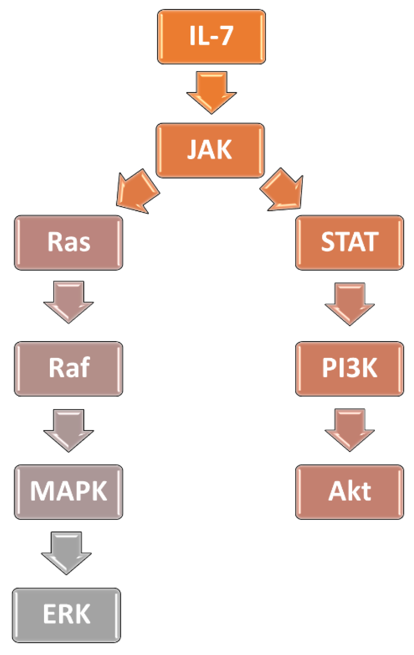
3.1. Interleukin-7 Signaling Pathway and Glucocorticoid Resistance in ALL

Interleukin-7 (IL-7) plays a crucial role in the T- and B-cell development. It is responsible for survival and regulating the functions of peripheral mature T-cells [75,76,77,78,79]. IL-7 acts on cells via a heterodimer IL-7 receptor (IL-7R) comprised of subunit alpha (IL-7Rα, CD127) and the common γ chain (γc) subunit [79]. Binding to IL-7R results in activation of associated tyrosine Janus kinases (JAK): JAK1 (by IL-7Rα) and JAK3 (by γc) [79,80]. JAK 1 and JAK3 induce phosphorylation of signal transducer and activator of transcription proteins (STAT) transcriptional factors, mainly STAT5A and STAT5B. Furthermore, STAT1 and STAT 3 are also activated. Next, STAT proteins translocate into the nucleus where they regulate cell-growth and survival via inter alia, increasing Bcl-2 expression. STAT5 proteins also activate the PI3K/AKT signaling pathway [79,80,81]. Increased activation of the IL-7 pathway causes leukemogenesis in multiple in vitro studies. Upregulation of IL-7 signaling pathway was observed in the leukemic cells obtained from T-ALL patients, as well as in the T-ALL cell lines, and was associated with increased cell survival and proliferation [81,82,83,84,85,86,87,88]. Furthermore, activation of the IL-7 signaling pathway was showed to be associated with T-ALL resistance to glucocorticoid treatment [89,90,91].
The findings described above are also important for the ALL treatment. Mutations in IL-7R/JAK signaling have been found in pre-B ALL, and thus are inhibitors of this pathway, as well as blockers of downstream signaling, which could be beneficial in pre-B ALL therapy [87,92,93].
Delgado-Martin et al. demonstrated that in vitro IL-7-related glucocorticoid resistance could be overcome by inhibiting the JAK/STAT pathway. IL-7-related resistance was induced by incubating the non-early T-cell progenitor (ETP) (n = 22) and ETP T-ALL (n = 10) xenograft samples with IL-7. Some of the ETP T-ALL samples were resistant to glucocorticoids irrespective of the IL-7 addition, however, others were resistant only when incubated with IL-7. Similarly, glucocorticoid resistance in non-ETP T-ALL samples was strongly IL-7-dependant (n = 10), partially dependent (n = 7) and IL-7-independent (n = 5). Ruxolitinib, a JAK1/2 inhibitor, in combination with dexamethasone, restored glucocorticoid sensitivity in the IL-7-dependent non-ETP and ETP T-ALL samples. Furthermore, when cells were incubated with the JAK3 inhibitor, a similar effect occurred. Importantly, the augmentation of apoptosis was significantly higher when JAK inhibitors were used in combination with dexamethasone, instead of a single treatment. Ruxolitinib and dexamethasone co-treatment was found to decrease the Bcl-2 level, which was elevated in the IL-7-dependent cells. In contrast, there was no augmentation of Bcl-2 level in IL-7-independent samples. Interestingly, venetoclax (Bcl-2 inhibitor), in combination with dexamethasone, sensitized IL-7-dependent cells to glucocorticoid treatment; however, the effects of this drug combination were far worse than those of the ruxolitinib and dexamethasone combination [90].
In around half of the Ph-like ALL rearrangement, the cytokine receptor-like factor 2 (CRLF2) gene is found, which leads to CRLF2 overexpression. This, in turn, is frequently associated with activation of JAK/STAT cascade [94,95]. Ruxolitinib, in combination with vincristine, dexamethasone, and an L-asparaginase (VXL) induction-type treatment regimen, was found to be efficient in mice engrafted with Ph-like ALL xenografts harboring JAK activating mutations. This drug combination acted synergistically and prolonged disease remission [96].
It is worth noting that a phase I clinical study of a monoclonal antibody against IL-7Rα (GSK2618960) has been completed. As indicated by this double-blind study conducted in 18 healthy individuals, GSK2618960 treatment is tolerated and efficiently blocks IL-7 signaling. However, it did not induce any effect on healthy T-cells. Nevertheless, GSK2618960 can still decrease pro-survival cascade activation in leukemic cells; thus, further studies are needed [97]. Furthermore, another monoclonal antibody against IL-7Rα (named B12), which blocks both the wild-type and mutated IL-7Rα, was developed. B12 was found to block IL-7 and mutant IL-7Rα signaling, and to induce apoptosis in vitro. Further, B12 delayed T-ALL progression in vivo in the T-ALL engrafted mice. It was also found to potentialize dexamethasone-induced apoptosis in vitro [98]. Given the good tolerance of IL-7Rα inhibitors during the phase I clinical study, as well as promising results of preclinical research, efficiency of these drugs in ALL treatment should be evaluated as soon as possible.

3.2. Activation of PI3K/AKT/mTOR Signaling Cascade Prevents GR from Translocation to the Nucleus

An alternate underlying mechanism of glucocorticoid resistance dependent on NRC3C1 activation could be the NR3C1 inhibitory phosphorylation, which reduces its nuclear localization and transactivation ability to activate critical downstream GTGs. Serine/threonine kinase (AKT) 1 binds to and phosphorylates the NR3C1 protein, inhibiting its nuclear translocation, according to Piovan et al. This finding suggests that activation of AKT1 may play a role in the development of glucocorticoid resistance in ALL. In vitro and in vivo, pharmacological inhibition of AKT with MK2206 significantly restores glucocorticoid-induced NR3C1 translocation to the nucleus, increases the sensitivity of T-ALL cells to the glucocorticoid therapy, and successfully reverses glucocorticoid resistance [99].
AKT1 is a part of phosphatidylinositol 3-kinase (PI3K)/AKT/mammalian target of a rapamycin kinase (mTOR) signaling cascade. In leukemia, the PI3K/AKT/mTOR pathway is usually activated and plays a role in leukemogenesis, especially in T-ALL. Increased cell metabolism, proliferation, and decreased apoptosis are the result of continuous stimulation of this mechanism. Activating mutations in PI3K genes, as well as downstream effectors of the cascade, such as AKT and mTOR, causes overexpression of this pathway. Moreover, inactivating mutations in the PTEN gene have been reported in T-ALL patients. PTEN, in turn, is a crucial inhibitor of the PI3K/AKT cascade [100]. Activation of PI3K/AKT signaling has been also linked to the increase in the level of Bcl-2, which is an anti-apoptotic protein [88]. Furthermore, the FBXW7 seems to be regulated by the PI3K/AKT, thus, blocking this pathway may result in increasing the GRα level [101]. Therefore, this may be another mechanism of glucocorticoid resistance induced by the PI3K/AKT axis. However, this requires further laboratory studies, as so far, the influence of PI3K/AKT activation on FBXW7 in leukemic cells has not been clarified.
Wandler et al. used PI3K inhibitor pictilisib (GDC-0941) to treat primary murine, genetically heterogeneous T-ALLs, both in a single treatment or in combination with dexamethasone. Authors reported that 68% of T-ALL mice that relapsed after initially responding to glucocorticoid treatment in vivo had a decreased or missing GR protein expression. Adding GDC-0941 to glucocorticoid treatment resulted in modestly prolonged survival (median 31 versus 40 days; p = 0.0805). Importantly, GR protein levels were reduced in 40% of relapsed human T-ALL samples, implying that GR expression loss is a primary source of glucocorticoid resistance. This finding is consistent with clinical studies linking a poor response to glucocorticoid treatment during induction to a higher likelihood of relapse [102]. Pictilisib is demonstrated also by other studies to be efficient in the T-ALL with PI3K/AKT/mTOR pathway activation [103,104,105]. Several other PI3K inhibitors have been efficient in treating ALL during preclinical studies. Idelalisib successfully treated ex vivo pre-B ALL samples with TCF3::PBX1 gene fusion [106]. Further, idelalisib was also found to be effective ex vivo against T-cell leukemia-lymphoma samples [107]. Buparlisib treatment was beneficial in vitro in the T-ALL cell lines [108,109]. Evangelisti et al. found that PI3K p110 inhibitors (ZSTK-474, AS-605240, CAL-101, and IPI-145) induce apoptosis both in pre-B ALL cell lines, as well as ex vivo in the samples obtained from pre-B ALL patients. Reh6 and Nalm6 cells were incubated with human bone marrow mesenchymal stem cells, which induce glucocorticoid resistance in vitro. P110 inhibitors (ZSTK-474 and IPI-145) were found to increase the effects of dexamethasone therapy in these cell lines, thus overcoming glucocorticoid resistance. Furthermore, PI3K inhibitor (IPI-145 or ZSTK-474) treatment of KOPN8 and Nalm6 reversed the AKT-induced impartment of GR translocation to the nucleus. Dexamethasone, in co-treatment with PI-145 or ZSTK-474, was found to be more beneficial than a single drug treatment against pre-B ALL patient samples [110].
MTOR inhibitors might be beneficial in treating primary human ALL. In their studies, Teachey et al. demonstrated the activity of mTOR inhibitors in preclinical models of ALL [111]. In addition, mTOR inhibitors improve methotrexate sensitivity by downregulating dihydrofolate reductase expression [112]. In particular, the combined inhibition of PI3K and the mTOR complex may provide an effective treatment for acute leukemia. In fact, they had a much stronger cytostatic effect on ALL cells than everolimus, according to Wong et al. [113]. Dactolisib (BEZ235) is an imidazoquinoline derivative that is a potent dual pan-class I PI3K and mTOR inhibitor. It inhibits downstream PI3K effectors in numerous preclinical models, including cell lines and xenografts, resulting in efficient reduction of tumor proliferation and growth [114]. As showed by the phase I clinical study performed by Lang et al., dual inhibition of PI3K and mTOR by dactolisib induces responses in 30% of ALL cases. Dactolisib, at the dose of 400 mg/day, was poorly tolerated and severe toxicity events occurred, especially gastrointestinal (mainly stomatitis). Considering that most dose-limiting toxicities such as fatigue, diarrhea, nausea, and mucositis were noted with both PI3K and mTOR inhibitors, it is not unexpected that pan-PI3K and mTOR inhibition resulted in a high prevalence of adverse events at the dose of 400 mg/day. On the other hand, 300 mg/day was far better tolerated by the patients; therefore, this dose was established as recommended for phase II studies [115]. It is worth noting that dactolisib was found to increase the incidence of grade 3–4 adverse events in evaluable patients in several other clinical studies [116]. Regrettably, currently there is no ongoing clinical study of dactolisib, according to clinicaltrials.gov.

3.3. The MAPK-ERK Pathway

Glucocorticoid resistance of T-ALL leukemic cells, related to upregulation of the IL-7 pathway, was mainly associated with STAT5-induced increase in the anti-apoptotic Bcl-2. Furthermore, it was observed that glucocorticoid treatment results in upregulation of IL-7Rα, thus creating a vicious cycle [89,90]. However, recently, additional mechanisms of IL-7-pathway-related resistance have been described.
Li et al. discovered mutations of IL-7 pathway genes in 47 (32%) samples obtained from 146 pediatric T-ALL patients. These mutations affected IL7R, JAK1, JAK3, NF1, NRAS, KRAS, and AKT genes. In 28 out of 97 prednisolone-treated samples, the IL-7 pathway mutations were associated with resistance to glucocorticoid treatment (p = 0.033). Interestingly, patients with IL-7 pathway mutations were characterized by significantly (p = 0.009) worse clinical outcomes, as compared to children without these mutations. Furthermore, those mutations caused activation of the PI3K/AKT pathway, resulting in an increase in Mcl-1 and Bcl-XL levels. Furthermore, higher levels of inactivated GSK3B, which is an important kinase that regulates BIM’s function, were found. Consequently, a higher ratio of phosphorylated/unphosphorylated BIM was observed. These changes were associated with mitogen-activated protein kinase (MAPK)—extracellular signal-regulated kinase (ERK) pathway activation [89]. The MAPK-ERK pathway takes part in controlling cells’ growth, proliferation, survival, and division. Upon MAPK-ERK activation, ERK migrates to the nucleus where it directly phosphorylates target proteins or controls other kinase activity [117]. When glucocorticoid-resistant cell lines were treated with the MAPK inhibitor (CI1040), there was a significant enhancement in GSK3B activation, as well as an increase in the non-phosphorylated BIM level [89].
A recently published study by van der Zwet et al. demonstrated that SUPT-1 cells expressing cysteine mutants IL-7RαPILLT240−244RFCPH, IL-7R-αPIL240−242QSPSC, and IL-7RαLT243−244LMCP, exhibited glucocorticoid resistance and activation of the downstream MAPK-ERK. In contrast, in the cells expressing the wild-type and the non-cysteine IL-7Rα mutant, MAPK-ERK upregulation and glucocorticoid resistance were not found. The MAPK-ERK pathway was also upregulated in the glucocorticoid-resistant SUPT-1 cells expressing JAK1R724H, JAK1T901A, NRASWT, or NRASG12D, whereas in the glucocorticoid-sensitive SUPT-1 cells (expressing wild-type JAK1) MAPK-ERK was not activated. It was demonstrated that MAPK-ERK upregulation causes increase in the phosphorylation of BIMEL and BIML isoforms. Further, it was showed that ERK is responsible for the direct phosphorylation of BIM. BIM’s phosphorylation was found to cause impaired BIM’s binding to Bcl-2, Mcl-1, and Bcl-XL, which prevents their inactivation and, thus, promotes cells’ survival. Furthermore, in the JAK1T901A-and-NRASG12D-expressing SUPT-1 cells, MAPK 1/2 inhibitors selumetinib and trametinib prevented BIM’s phosphorylation in a dose-dependent manner. A similar effect was achieved in SUPT-1 cells expressing JAK1T901A treated with JAK1/JAK2 inhibitor ruxolitinib. The response to the glucocorticoid treatment in the 46 T-ALL patient-derived xenografts with or without IL-7 was assessed. IL-7 addition induced glucocorticoid resistance in 12 (26%) of all xenografts. Interestingly, in those samples, MAPK-ERK signaling was activated by IL-7, indicating that physiological IL-7 signaling may activate the downstream MAPK-ERK pathway in T-ALL. Thus, in T leukemic cells, MAPK-ERK can be upregulated both by mutant and physiological IL-7 signaling [118]. Interestingly, the MAPK-ERK signaling pathway is not activated by the IL-7 signaling in the healthy T-cells [119]. Xenografts, in the absence of IL-7, were treated using MAPK inhibitors (selumetinib, trametinib, and binimetinib) and ruxolitinib. Ruxolitinib did not cause therapeutic effects, in contrast to MAPK inhibitors, which induced cytotoxicity in most of the samples. Six samples with IL-7-induced glucocorticoid resistance were then treated with ruxolitinib. The significant relationship (p = 0.0039) between sensitivity to ruxolitinib treatment and IL-7-enhanced cells viability was found. Combined treatment with selumetinib and prednisolone was highly synergic, both in xenografts with IL-7-induced glucocorticoid resistance, and in the samples with not-IL-7-related glucocorticoid resistance. Furthermore, such a synergistic effect was also found in the glucocorticoid-sensitive xenografts. Ruxolitinib and prednisolone co-treatment was efficient only in one xenograft, with not- IL-7-related glucocorticoid resistance in the presence of IL-7, and in one xenograft with IL-7-induced glucocorticoid resistance without IL-7. In both samples, there was a significant STAT5 overexpression, which explains ruxolitinib efficiency. Therefore, ruxolitinib has limited clinical application, as it may efficiently decrease MAPK-ERK activation caused by IL-7R/JAK mutations or physiological IL-7 transmission, whereas it is not useful in the alterations occurring downstream of IL-7R/JAK. Such limitations are not encountered when using MAPK inhibitors [118].
Mutations of genes encoding the rat sarcoma virus (Ras) protein family are found in around 30% of all cancers in humans, making them the most common genetical alterations occurring in cancer [117,119]. Ras’s proteins are involved in the Ras/rapidly accelerated fibrosarcoma (Raf)/MAPK/ERK signaling cascade; thus, Ras activation leads to ERK induction [119]. Irving et al. examined 54 samples obtained from children with pre-B ALL at the time of diagnosis in search for activating mutations of Ras proteins (KRAS and NRAS), mutations of Ras regulators (PTPN11), and alterations in genes encoding upstream signaling proteins (FLT3). In 28 (51.9%) children, such mutations have been found. Ras/Raf/MAPK/ERK pathway activation was estimated in 80 pre-B ALL patients. Among 32 cases with Ras-related mutations, 27 (84.3%) had relevant Ras/Raf/MAPK/ERK signaling cascade activation. In the remaining patients without Ras mutations, activation of this pathway was found in 9 of 48 (18.7%) samples. The cells with Ras/Raf/MAPK/ERK activation were significantly more sensitive to selumetinib, regardless of the presence of Ras-related mutations [120]. Jerchel et al. found Ras-related mutations (NRAS, KRAS, FLT3, PTPN11, and others) in 44.2% of 461 samples obtained from children with pre-B ALL at the time of diagnosis. Overall, the Ras-related mutations were most frequent in the high hyperdiploid (72.6%) and t (4;11)-rearranged (73.3%) pre-B ALL. Ras-related mutations were associated with significantly worse clinical outcomes in the HR patients. It was further established that Ras mutations are associated with glucocorticoid resistance, as samples with Ras-related mutations were 3-fold more resistant to prednisolone treatment than samples without these mutations (p = 0.024). In this context, the clonal/subclonal KRAS G13 were most harmful, as blasts with this mutation were characterized by more than 2000-fold-higher glucocorticoid resistance, as compared to wild-type leukemic cells. In contrast, NRAS and KRAS G12 were found not to increase glucocorticoid resistance in a significant manner. The treatment of Ras-mutated samples using MAPK inhibitor trametinib induced cytotoxicity, whereas wild-type blasts were unaffected (p = 0.001) [121]. Signaling cascades involved in the glucocorticoid resistance in ALL have been schematically presented on Figure 1.
Matheson et al. demonstrated, that selemutinib and dexamethasone co-treatment is highly synergic, both in the in vitro and in vivo studies. In vitro, selemutinib and dexamethasone co-treatment exerted a highly synergic impact (mean combination index (CI) of 0.1) on primagrafts with Ras-related mutations (affecting NRAS, KRAS, and CBL/FLT3). Combined therapy resulted in elevation of BIM levels, a decreased Mcl-1 level, and ERK phosphorylation. In vivo, it was demonstrated that selumetinib and dexamethasone co-treatment acts highly synergic in nonobese diabetic (NOD) SCID (severe combined immunodeficiency) gamma mice engrafted with a Ras-mutated, patient-derived xenograft. KRAS G13D, KRAS G12D, and NRAS Q61R mice treated with drug combination had a spleen weight at the end of the therapy comparable with the healthy mice spleen (p < 0.001) [122]. Polak et al. obtained blast samples from 22 adults with newly diagnosed pre-B ALL. These cells were incubated with dexamethasone or dexamethasone in combination with selumetinib. Combined therapy managed to augment the dexamethasone-induced apoptosis in 17 samples [123].
Ras-activating mutations have been also found in the relapsed T-ALL patients and were associated with highly unfavorable outcomes [89,124,125,126,127]. Kerstjens et al. treated Ras mutant and Ras wild-type t (4;11)+ infant ALL blasts with salirasib (Ras inhibitor), vemurafenib (serine/threonine-protein kinase B-raf inhibitor), sorafenib (pan-kinase inhibitor), temsirolimus (mTOR inhibitor), and MAPK inhibitors trametinib, selumetinib and binimetinib. MAPK inhibitors were significantly more efficient than the other tested drugs [128]. This is particularly important, as Ras-mutated mixed lineage leukemia (MLL)-rearranged leukemic cells are likely to be resistant to glucocorticoid treatment [129]. MLL-rearranged ALL is also associated with far worse clinical outcomes as compared to patients without MLL translocations [130].
Combining JAK inhibitors with blockers of the downstream signaling also seems to be a promising strategy for the ALL treatment. Ba/F3 cells harboring JAK3(L857Q) and JAK3(M511I) mutations were treated with tofacitinib (JAK1/JAK3 inhibitor) and selumetinib at various concentrations. All combinations were characterized by synergic, dose-dependent action (CI < 0.1–0.9). A tofacitinib and venetoclax combination was also synergistic, however to a lesser extent. Co-treatment with tofacitinib and buparlisib (PI3K inhibitor) also provided mediocre effects. Furthermore, in the blasts harboring a JAK3 (M511I) mutation, obtained from the T-ALL patient, the combination of tofacitinib and trametinib was highly synergic (CI from <0.1 to 1, depending on the drugs concentrations). Further, tofacitinib and venetoclax co-treatment at low concentrations was also effective (CI < 0.1). Oral treatment consisting of venetoclax (20 mg/kg/day) and ruxolitinib (40 mg/kg/day) was effective in treating mice engrafted with blasts carrying the JAK3 (M511I) mutation [131]. Inhibiting the JAK/STAT pathway, combined with blocking MAPK/ERK signaling, seems to be a rational strategy. MAPK/ERK can be also activated by Ras-activating mutations, therefore blocking JAK/STAT signaling may not be enough to overcome resistance to the treatment [118,119,120,121,122,123]. Furthermore, combining JAK/STAT inhibitors with Bcl-2 inhibitors is also rational, as STAT proteins induce Bcl-2. Unfortunately, MAPK inhibitors have not been used in co-treatment with venetoclax in this study, as this combination could also be beneficial. Further, using all of these drugs simultaneously could hypothetically provide the best results, as it would target various mechanisms of treatment resistance. Thus, further preclinical studies are required.
Agents capable of blocking signaling pathways involved in the ALL glucocorticoid resistance have been systematized in the Table 1.

4. Methods of Enhancing the Results of Glucocorticoid Therapy in Pediatric ALL

4.1. Enhancing Effects of GR Activation

Roderick et al. showed that NR3C1 mRNA and GR protein levels are increased by cyclic adenosine monophosphate-dependent protein kinase (cAMP-PKA) signaling in mouse and human T-ALL cells. Furthermore, when the cAMP-activating Gnas gene was silenced, it caused resistance to the dexamethasone treatment, both in vitro and in vivo. Further, cAMP activation induced by 6,16-dimethyl-prostaglandin E2 (dmPGE2) and dexamethasone co-treatment was found to overcome glucocorticoid resistance in the T-ALL patient samples. Therefore, it was concluded that the cAMP-PKA-induced increase in GR level is the reason for its beneficial effects in co-treatment with dexamethasone [134]. This interesting and novel mechanism requires further laboratory research.
The microRNAs (miRNAs) miR-100 and miR-99a were shown to be downregulated in childhood ALL patients, and their expression levels were linked to the ALL patients’ prognosis, as demonstrated by the investigation by Li et al. MiR-100 and miR-99a were found to be important in the regulation of cell proliferation and dexamethasone-induced apoptosis in ALL cell lines in vitro. The researchers discovered that the FK506-binding protein 51 (FKBP51) is a novel target of miR-100 and miR-99a. FKBP51, in turn, is responsible for inhibiting GR’s nuclear translocation. The findings of the study demonstrate that miR-100 and miR-99a are tumor suppressors, and that reinstating them could be a treatment option for ALL patients [135]. A study by Liang et al. showed that miR-124 expression is significantly increased in children suffering from glucocorticoid-resistant ALL, as compared to the glucocorticoid-sensitive samples. Moreover, miR-124 causes dexamethasone resistance and suppresses glucocorticoid-induced apoptosis in the sensitive cell lines CCRF-CEM and CEM/C1. miR-124 targets NR3C1, suppressing its expression, consequently lowering efficiency of glucocorticoid treatment. Thus, Liang et al. proposed a unique mechanism for GC resistance in ALL, which could be potentially targeted in order to overcome treatment resistance [136].
Paugh et al. discovered that the recombinant caspase 1 (CASP1) cleaves the GR at its transactivation region, and that forced overexpression of CASP1 combined with the NLR family pyrin domain containing three inflammasome activations causes human leukemia cells to become resistant to glucocorticoid therapy. Persistently inhibiting CASP1 expression or lowering CASP1 activity with an inhibitory protein (CrmA) in CASP1-overexpressing leukemia cells raises cellular GR levels and significantly enhances glucocorticoid sensitivity [137]. Therefore, CASP1 inhibitors (e.g., VX 765 and Ac-FLTD-CMK), or even pan-caspase inhibitors such as emricasan, which is clinically used in the treatment of non-alcoholic fatty liver disease, could potentially be implemented in the ALL therapy.
Gallagher et al. discovered that the orphan nuclear receptor estrogen-related receptor-b (ESRRB), is an important transcription factor that collaborates with the GR to mediate the GTGs expression signature in mice and human ALL cells using a genome-wide, survival-based short hairpin RNA (shRNA) screen. They found that ESRRB knockdown inhibits dexamethasone-induced gene expression, implying that ESRRB cooperates with GR to generate optimal dexamethasone transcriptional responses. Furthermore, ESRRB agonist (GSK4716), in combination with dexamethasone, is highly synergic—a CI of <1 in treating human T-ALL cell lines KOPTK1 and DND-41 [138].

4.2. The BH3 Mimetics—Targeting the Primary Mechanism of Glucocorticoid-Induced Apoptosis of ALL Cells

The BH3 mimetics, which are the drugs that initiate apoptosis by targeting pro-survival Bcl-2 proteins, show great potential in the treatment of ALL. Venetoclax (previously ABT-199), a discovered 2013 Bcl-2 inhibitor, is currently the only BH3 mimetic that has been clinically approved [139,140]. Both Food and Drug Administration (FDA) and European Medicines Agency (EMA)-approved venetoclax for the treatment of chronic lymphocytic leukemia and acute myeloid leukemia (AML) [140,141,142]. Navitoclax (ABT-263) is a BH3 mimetic which inhibits not only Bcl-2, but also Bcl-XL and Bcl-w. Unfortunately, navitoclax has not been approved yet by the FDA and EMA for clinical use. However, navitoclax in co-treatment with venetoclax is available for expanded access (NCT03592576). Therefore, there are prospects of navitoclax approbation for co-treatment with different BH3 agonists.
A particularly interesting study has been conducted by Ni Chonghaile et al., who demonstrated that leukemic cells in different maturation stages have dissimilar levels of Bcl-2 proteins. Cell lines underwent Bcl-2 profiling, which was based on the strong Bcl-2 binding to Bad (but not to Hrk), whereas Bcl-XL interact equally with Bad and Hrk. Most T-ALL cell lines (CEM-CCRF, PF382, Molt4, P12-Itchikawa, Jurkat, KOPTKI, RPMI-1640, and CEM-C1) were characterized by a strong Bcl-XL dependence, which means that Bcl-XL was mainly responsible for anti-apoptotic action and, consequently, cells’ survival. In contrast, ETP, the Loucy cell line, was found to be Bcl-2 dependent. Venetoclax sufficiently induced apoptosis in Loucy cells, however, navitoclax was also effective. Unsurprisingly, Bcl-XL-dependent cells were killed more efficiently by navitoclax. Furthermore, 26 samples obtained from primary pediatric T-ALL (10 with ETP-ALL) at the time of diagnosis were examined. The ETP-ALL cells’ survival was definitely Bcl-2 dependent, in contrast with typical T-ALL, which was found to be Bcl-XL dependent, thus confirming the results obtained in vitro. Moreover, healthy cells at the earliest double negative (DN) intrathymic stage of differentiation (DN1) were also found to be Bcl-2 dependent, corresponding to ETP-ALL leukemic cells. Cells during the CD4+ and CD8+ stages of differentiation were, in turn, dependent of Bcl-XL, thus indicating that there is a significant change in expression of Bcl-2 family proteins during the lymphocyte development. A Bcl-2 profiling of the T-ALL samples obtained during the Dana-Farber Cancer Institute trials additionally confirmed that ETP-ALL cells are dependent on Bcl-2, whereas typical T-ALL was characterized by the Bcl-XL dependence. Similarly, to the previous outcomes, in this case, ETP-ALL was also sensitive both to venetoclax and navitoclax, whereas primary T-ALL cells responded well to navitoclax. Patient-derived xenografts from mature T-ALL and ETP-ALL patients were established, and mice with at least 65% CD45+ blasts were given venetoclax, navitoclax, or a vehicle for 14 days. The results were comparable to previous tests, as ETP-ALL xenografts were sensitive to both venetoclax and navitoclax, whereas typical T-ALL xenografts responded better to navitoclax than venetoclax [143]. A study conducted by Peirs et al. evaluated in vitro Bcl-2 expression in different T-ALL molecular subgroups, as well as their response to venetoclax treatment. Mature T-ALL cell lines ALL-SIL, CUTLL1, TALL-1, KOPTK1, DND-41, PF-382, KARPAS-45, PEER, Jurkat, and CCRF-CEM were characterized by intermediate responses to venetoclax treatment, whereas Loucy cells were highly sensitive to venetoclax, which confirms the results obtained by Ni Chonghaile et al. Seventeen pediatric primary T-ALL bone marrow samples, with different maturation states and genetical profiles, were selected, and the cells were treated with venetoclax. Cells in the earlier stages of maturation were characterized by half maximal inhibitory concentration (IC50) < 300 nM, whereas in most of the mature cells, the treatment outcome was worse [144]. Venetoclax is currently being tested in combination with C10403 chemotherapy for the newly diagnosed adult pre-B ALL during the phase I study (NCT05157971). The future prospects of BH3 mimetics used in ALL have been summarized in Table 2.

4.3. Proteasome Inhibitors—Multitude Mechanism of Action Benefiting the Glucocorticoid Treatment

Proteasome inhibitors showed great potential in treating cancer in multiple studies. Currently, three drugs belonging to this group have been approved for clinical use by the FDA and EMA: bortezomib, ixazomib, and carfilzomib [148]. Bortezomib is an efficient reversible inhibitor of the β5 chymotrypsin-like subunit of 20S proteolytic site of the 26S proteasome. Bortezomib also reversibly blocks the β1 caspase-like subunit and β2 trypsin-like subunit when used in higher doses [148,149]. Inhibition of proteasomes results in higher p27 and p53 levels. Additionally, proteasomes are crucial in activation of nuclear factor-κB (NF-κB), which ceases transcription of pro-apoptotic factors [150]. Moreover, bortezomib activates c-Jun N-terminal kinase and cause aggregation of incorrect proteins, which also promotes apoptosis [151]. Results of the study on the effects of prednisolone and bortezomib co-treatment of pre-B leukemic cells have been published. Both glucocorticoid-resistant (MHH-cALL-2) and sensitive (MHH-cALL-3) cells underwent treatment with single agents or co-treatment with both drugs. Bortezomib alone induced apoptosis in both cell lines in a dose-dependent manner. In both cell lines, bortezomib and prednisolone co-treatment caused additional increase in cell death, mostly in MHH-cALL-2, indicating that bortezomib may be useful in overcoming glucocorticoid resistance [152]. It should be noted that bortezomib may also increase Noxa levels, contributing to enhancing apoptosis in glucocorticoid-resistant cells via decreasing glucocorticoid-induced proteasomal degradation of Noxa [62]. This hypothesis is supported by results of several in vitro studies, in which bortezomib was found to increase Noxa levels [153,154,155,156]. Nevertheless, this potential effect of bortezomib and other proteasome inhibitors’ administration in ALL cells requires further research.
Carfilzomib is a drug belonging to a second generation of proteasome inhibitors, which is an irreversible blocker of β5/β5i subunits [149]. Carfilzomib in a single treatment exhibited an antileukemic action in the Molt4 cells (a glucocorticoid-resistant human T-ALL-derived cell line). It has also sensitized the Molt4 cell line to dexamethasone-induced apoptosis. Interestingly, carfilzomib caused the generation of reactive oxygen species (ROS), thus generating the state of oxidative stress in the Molt4 cells [157]. ROS generation caused forkhead box O3 (FOXO3a) induction, which causes cell death via up-regulation of autophagy-related genes [157,158]. Carfilzomib-generated ROS production also elevates the stress induced sirtuin 1 (SIRT1) level, which plays a role in the endoplasmic reticulum’s (RER) stress-induced apoptosis and autophagy [157,159]. Another interesting mechanism of the carfilzomib action is increasing the C/EBP Homologous Protein (CHOP) protein transcription, which is a downstream target of protein kinase R (PKR)-like endoplasmic reticulum kinase (PERK) and the cyclic AMP-dependent transcription factor (ATF4) [157]. CHOP acts as a critical pro-apoptotic mediator in the RER stress, and when activated, it regulates the Bcl-2 family proteins. In the case of RER stress, CHOP decreases Bcl-2, Bcl-XL, and Mcl-1 levels, whereas it raises the BIM expression. Additionally, CHOP induce tribbles homolog 3 (TRB3), which in turn inhibits AKT activity. CHOP has been also demonstrated to up-regulate expression of death receptor (DR) 4 and DR5, which are the components of the DR pathway [160]. CHOP was also found to activate GADD34, a DNA damage protein. CHOP may also be activated in the course of bortezomib-induced RER stress in different cancer cell lines [161,162,163].
Unfortunately, for carfilzomib combined with dexamethasone, mitoxantrone, methotrexate, pegylated L-asparaginase, and vincristine (UKALLR3), induction therapy was found to be excessively toxic [164]. Interestingly, phase I of the carfilzomib and hyperfractionated cyclophosphamide, vincristine, doxorubicin, and dexamethasone (HyperCVAD) co-treatment in newly diagnosed ALL has been recently completed. Ten patients with Philadelphia negative ALL (eight with pre-B ALL and two with T-ALL), aged 18–64, were enrolled. The treatment was well-tolerated and resulted in a complete response (CR) rate of 90% after the first cycle; the last patient achieved CR after the fourth cycle. Moreover, MRD negativity was achieved in seven (70%) patients [165].
Ixazomib is a drug belonging to the third generation of proteasome inhibitors, which acts mainly by reversibly blocking β5/β5i subunits; thus, its action is similar to bortezomib. However, it lasts significantly shorter. Ixazomib also reversibly inhibits β1 and β2 subunits [148,149]. This drug has been found to inhibit growth of the wild-type CCRF-CEM T-ALL; however, bortezomib was found to be 10 times more potent. Furthermore, the leukemic cells obtained from nine patients suffering from primary ALL were significantly more sensitive to bortezomib as compared to ixazomib (p < 0.0001) [166]. Nevertheless, it was suggested that this difference in effective concentration of these drugs during in vitro studies may not be present in the in vivo studies. Ixazomib is characterized by excellent pharmacokinetics properties; it is quickly absorbed from the digestive tract and its tissue distribution is 5-fold better than bortezomib’s [148,166]. Therefore, the in vivo results of ixazomib treatment may be more promising [166].
The high potential of bortezomib and carfilzomib in the ALL treatment is reflected by the considerable number of currently ongoing clinical trials (Table 3).

5. Other Prospects of Enhancing Glucocorticoid Sensitivity in ALL

5.1. Tigecycline

Tigecycline is the first in a new class of antibiotics, the glycylcyclines, which are structurally related to tetracyclines. Tigecycline inhibits the protein translation in bacterial cells by binding the 30S ribosome subunit and blocking the attachment of the aminoacyl-tRNA molecule to the ribosomal A-site [172]. It is highly active against many gram-positive and gram-negative pathogens, both aerobic and anaerobic. As bacterial and mitochondrial ribosomes are similar, tigecycline also inhibits the synthesis of mitochondria-encoded proteins involved in oxidative phosphorylation (OXPHOS). It has been reported that such a mechanism of action of tigecycline is responsible for its properties of inhibiting development of cancers, such as chronic myeloid leukemia stem cells, ALL, non-small cell lung cancer, ovarian cancer, and hepatocellular carcinoma [173,174]. Enhanced oxidative phosphorylation is associated with steroid resistance in ALL cells. Studies revealed that inhibitors of OXPHOS pathways increase sensitivity to steroids in T-ALL in vitro [175]. Therefore, research on the use of tigecycline in ALL therapeutic protocols are suggested. So far, Xuedong et al. reported preclinical evidence that tigecycline suppresses mitochondrial respiration, induces apoptosis of cancer cells, and also intensifies the effect of chemotherapeutic drugs—doxorubicin and vincristine [176]. These conclusions may be a starting point for further studies.

5.2. Tamoxifen

Tamoxifen is a synthetic anti-estrogenic medicine used mainly in the treatment of luminal breast cancer. Recently, new targets of tamoxifen activity other than estrogen receptors have been discovered. In the literature, attention is paid to its influence on important mediators of signaling pathways initiating cell proliferation, conditioning the aggressive course of disease, or cancer sensitivity to chemotherapy [177]. Taking these findings into account, the possibility of using tamoxifen in the therapy of neoplasms other than breast cancer should be considered.
As it was mentioned previously (Section 3.1), resistance to steroids correlates with increased activity of the anti-apoptotic proteins of the Bcl-2 family. Tamoxifen has been demonstrated to stimulate apoptosis by reducing the expression of Bcl-2, as well as increasing the expression of the pro-apoptotic Bax protein [177]. Another reported effect of tamoxifen is the activation of autophagy, confirmed by experiments conducted on various tumor cell lines [178]. Autophagy is a natural process in which defective or dispensable intracellular proteins or cell organelles are delivered to lysosomes for degradation. In cancer cells, autophagy plays a double role—it may be responsible for the development of resistance to the applied therapy, as well as for cell death. On the one hand, it can be activated in response to stress factors such as radiation therapy or chemotherapy and lead to tumor growth and survival. Autophagy can inhibit neoplastic transformation and induce apoptosis, and consequently increase the effectiveness of the treatment [179]. Since glucocorticoids induces cell death through the autophagic machinery activation, tamoxifen, which enhances this process, may be an effective medicine in partial reversing steroid resistance [180].

5.3. Cannabidiol

For many years, cannabidiol (CBD) has been used in the treatment of drug-resistant epilepsy [181]. The FDA has approved it for the treatment of Lennox–Gaustat and Dravet syndromes. Compared to Δ9-tetrahydrocannabinol (Δ9-THC), CBD has a weak affinity for cannabinoid receptor type 1 and type 2 receptors, and, therefore, does not cause undesirable side effects on the central nervous system (CNS). In oncology, cannabidiol is an aid in palliative treatment. It alleviates the symptoms associated with cancer and chemotherapy, such as pain, nausea, vomiting, loss of appetite, or even anxiety. Recently, however, cytotoxic effects of cannabinoids have been discussed. They have been shown to have pro-apoptotic and antiproliferative effects [182]. Rimmerman et al., in their study, suggest that cannabinoids may cause cancer cell death by modulating the voltage-dependent anion channel 1 (VDAC) located in the outer mitochondrial membrane [183]. VDAC enables the transfer of ions and metabolites between the cytoplasm and the mitochondria, thus regulating many processes, such as apoptosis and cell metabolism. Besides, it anchors many proteins, including hexokinase. This combination is responsible for the regulation of glycolysis and protection against cell death [184]. CBD binds to mitochondrial membranes containing VDAC, thereby reducing the conductivity of the channels, weakening cell viability and ultimately leading to apoptosis [183]. VDAC plays a key role in the coordination between the aforementioned oxygen glycolysis and mitochondrial OXPHOS. It is worth considering the use of a combination of glucocorticoids with CBD in anti-cancer therapy, which could increase sensitivity to steroid therapy [185].

5.4. Mebendazole

Anthelmintics are given to children suffering from ALL, as they are more prone to parasitic infections while receiving chemotherapy. However, their use may also be justified by their anti-cancer activity. Mebendazole has been shown to inhibit the neurogenic locus notch homolog protein (Notch) signalization consisting of four receptors (Notch 1–4) [186]. Arresting Notch signaling can reverse glucocorticoid resistance in T-ALL by restoring autoregulation of the GR and induction of the gene-encoding Bcl-2 [187].

5.5. Demethylating Agents

BIM IGR exhibits inactive chromatin configuration in non-lymphoid cells, whereas the lymphoblasts have accessible chromatin at this site, making BIM IGR a lymphocyte-specific target of GR activation. Moreover, glucocorticoid-resistant lymphoblasts are characterized by the high BIM IGR methylation, and, consequently, low chromatin accessibility impairing GR’s binding, in contrary to glucocorticoid-sensitive cells, which have a low level of BIM IGR methylation and are well accessible. Furthermore, treatment of ALL-7CL-resistant cells with demethylating drug 5-azacitidine resulted in a time-dependent decrease in BIM IGR methylation. Moreover, co-treatment of ALL-7CL with dexamethasone and 5-azacitidine caused a significant increase in BIM expression after 48 h, compared to ALL-7CL treated only with dexamethasone (however, it was pretreated with 5-azacitidine for 3 days prior to dexamethasone). When ALL-7R-engrafted, NOD SCID gamma mice underwent 14 days of treatment with dexamethasone combined with decitabine, a clinically used demethylating agent, there was a significant reduction in leukemic cells in bone marrow combined with longer EFS, as compared to the mice in which only one agent was used. This indicates the critical importance of opposing DNA methylation in alleviating glucocorticoid resistance in ALL [188].

6. Conclusions and Future Directions

In conclusion, most of the preclinical studies involve novel agents that are capable of enhancing the results of glucocorticoid therapy. It should be emphasized that the described drugs could potentially be included in the induction phase in the newly diagnosed ALL patients. This, ultimately, could prevent relapse and provide better outcomes. Furthermore, incorporating into the diagnostics BH3 profiling and screening for activating mutations in the IL7/JAK/STAT cascade, the Ras pathway, downstream PI3K/AKT/mTOR, and MAPK/ERK pathways could potentially help to identify the patients that could benefit from targeting those signaling cascades. Nevertheless, the described findings still require further research.

Author Contributions

K.K. (Kamil Kośmider), M.L. and J.Z. were responsible for the conception of the study; M.L., K.K. (Kamil Kośmider) and J.Z. were responsible for the design of the study; K.K. (Kamil Kośmider), K.K. (Katarzyna Karska) and A.K. were responsible for the acquisition of the literature for the manuscript; K.K. (Kamil Kośmider), K.K. (Katarzyna Karska) and A.K. wrote the original draft of the manuscript; M.L. and J.Z. reviewed and edited the manuscript; M.L. and J.Z. supervised the study. All authors have read and agreed to the published version of the manuscript.

Funding

This research received no external funding.

Institutional Review Board Statement

Not applicable.

Informed Consent Statement

Not applicable.

Data Availability Statement

No new data were created or analyzed in this study. Data sharing is not applicable to this article.

Conflicts of Interest

The authors declare no conflict of interest.

Abbreviations

ALLacute lymphoblastic leukemia
AIEOP-BFM ALLAssociazione Italiana di Ematologia Oncologia Pediatrica-Berlin-Frankfurt-Münster Acute Lymphoblastic Leukemia
AKTserine/threonine kinase
AMLacute myeloid leukemia
ATF4cyclic AMP-dependent transcription factor
BadBcl-2-associated agonist of cell death
BakBcl-2 homologous antagonist/killer
BaxBcl-2-associated X-protein 4
Bcl-2B-cell lymphoma 2
Bcl-wB-cell lymphoma-w
Bcl-XLB-cell lymphoma extra-large
BidBH3 interacting-domain death agonist
BIMBcl-2-interacting mediator of cell death
cAMP-PKAcyclic adenosine monophosphate-dependent protein kinase
CASP1caspaze 1
CBDcannabidiol
CHOPC/EBP Homologous Protein
CIcombination index
CNScentral nervous system
CRcomplete response
CRLF2cytokine receptor-like factor 2
dmPGE26,16-dimethyl-prostaglandin E2
DNdouble negative
DRdeath receptor
EFSevent-free survival
EMAEuropean Medicines Agency
ERextreme risk
ERKextracellular signal-regulated kinase
ESRRBorphan nuclear receptor estrogen-related receptor-b
ETPearly T-cell progenitor
FBXW7F-Box and WD Repeat Domain-Containing 7
FDAFood and Drug Administration
FKBP51FK506-binding protein 51
FOXO3aforkhead box O3
GRglucocorticoid receptor
GREglucocorticoid response element
GSK3glycogen synthase kinase-3
GTGglucocorticoid target gene
HRhigh risk
Hrkactivator of apoptosis harakiri
HyperCVADhyperfractionated cyclophosphamide, vincristine, doxorubicin, dexamethasone
IC50half maximal inhibitory concentration
IGRintronic GR binding region
IL-6interleukin 6
IL-7interleukin-7
IL-7RIL-7 receptor
JAKJanus kinases
LC50median lethal concentration
MAPKmitogen-activated protein kinase
Mcl-1induced myeloid leukemia cell differentiation protein
miRNAmicroRNA
MLLmixed lineage leukemia
MRDminimal residual disease
mRNAmessenger RNA
mTORmammalian target of rapamycin kinase
NF-κBnuclear factor-κB
NODnonobese diabetic
Notchneurogenic locus notch homolog protein
Noxaphorbol-12-myristate-13-acetate-induced protein 1
OXPHOSoxidative phosphorylation
PERKprotein kinase R -like endoplasmic reticulum kinase
PGRprednisolone good response
Ph-likePhiladelphia chromosome-like
PI3Kphosphatidylinositol 3-kinase
PKRprotein kinase R
PPRprednisolone poor response
pre-B ALLprecursor-B-cell ALL
Pumap53-upregulated modulator of apoptosis
Rafrapidly accelerated fibrosarcoma
Rasrat sarcoma virus
RERendoplasmic reticulum
ROSreactive oxygen species
RT-PCRreal-time polymerase chain reaction
SCIDsevere combined immunodeficiency
SEsurvival estimates
shRNAshort hairpin RNA
SIRT1sirtuin 1
SRstandard risk
STATsignal transducer and activator of transcription
T-ALLprecursor T-cell ALL
T-LLT-lymphoblastic lymphoma
TNFtumor-necrosis-factor
TRB3tribbles homolog 3
VDACvoltage-dependent anion channel 1
VXLvincristine, dexamethasone and L-asparaginase
γcγ chain
Δ9-THCΔ9-tetrahydrocannabinol

References

  1. Stiller, C.A. Epidemiology and Genetics of Childhood Cancer. Oncogene 2004, 23, 6429–6444. [Google Scholar] [CrossRef] [Green Version]
  2. Shah, D.S.; Kumar, R. Steroid Resistance in Leukemia. World J. Exp. Med. 2013, 3, 21–25. [Google Scholar] [CrossRef] [PubMed]
  3. Hunger, S.P.; Lu, X.; Devidas, M.; Camitta, B.M.; Gaynon, P.S.; Winick, N.J.; Reaman, G.H.; Carroll, W.L. Improved Survival for Children and Adolescents With Acute Lymphoblastic Leukemia Between 1990 and 2005: A Report From the Children’s Oncology Group. J. Clin. Oncol. 2012, 30, 1663–1669. [Google Scholar] [CrossRef] [PubMed]
  4. Conter, V.; Bartram, C.R.; Valsecchi, M.G.; Schrauder, A.; Panzer-Grümayer, R.; Möricke, A.; Aricò, M.; Zimmermann, M.; Mann, G.; de Rossi, G.; et al. Molecular Response to Treatment Redefines All Prognostic Factors in Children and Adolescents with B-Cell Precursor Acute Lymphoblastic Leukemia: Results in 3184 Patients of the AIEOP-BFM ALL 2000 Study. Blood 2010, 115, 3206–3214. [Google Scholar] [CrossRef] [PubMed]
  5. Hunger, S. P Glucocorticoid Selection for Pediatric ALL. Blood 2016, 127, 2049–2051. [Google Scholar] [CrossRef] [Green Version]
  6. Inaba, H.; Pui, C.-H. Glucocorticoid Use in Acute Lymphoblastic Leukaemia. Lancet Oncol. 2010, 11, 1096–1106. [Google Scholar] [CrossRef] [Green Version]
  7. Gao, J.; Liu, W.J. Prognostic Value of the Response to Prednisone for Children with Acute Lymphoblastic Leukemia: A Meta-Analysis. Eur. Rev. Med. Pharmacol. Sci. 2018, 22, 7858–7866. [Google Scholar] [CrossRef]
  8. van der Zwet, J.C.G.; Smits, W.; Buijs-Gladdines, J.G.C.A.M.; Pieters, R.; Meijerink, J.P.P. Recurrent NR3C1 Aberrations at First Diagnosis Relate to Steroid Resistance in Pediatric T-Cell Acute Lymphoblastic Leukemia Patients. HemaSphere 2020, 5, e513. [Google Scholar] [CrossRef]
  9. Caratti, G.; Matthews, L.; Poolman, T.; Kershaw, S.; Baxter, M.; Ray, D. Glucocorticoid Receptor Function in Health and Disease. Clin. Endocrinol. 2015, 83, 441–448. [Google Scholar] [CrossRef]
  10. Vettorazzi, S.; Nalbantoglu, D.; Gebhardt, J.C.M.; Tuckermann, J. A Guide to Changing Paradigms of Glucocorticoid Receptor Function—A Model System for Genome Regulation and Physiology. FEBS J. 2021, 2, febs.16100. [Google Scholar] [CrossRef]
  11. Nick, Z.L.U.; Cidlowski, J.A. The Origin and Functions of Multiple Human Glucocorticoid Receptor Isoforms. Ann. N. Y. Acad. Sci. 2004, 1024, 102–123. [Google Scholar] [CrossRef]
  12. Lu, N.Z.; Cidlowski, J.A. Translational Regulatory Mechanisms Generate N-Terminal Glucocorticoid Receptor Isoforms with Unique Transcriptional Target Genes. Mol. Cell 2005, 18, 331–342. [Google Scholar] [CrossRef] [PubMed]
  13. Cain, D.W.; Cidlowski, J.A. Specificity and Sensitivity of Glucocorticoid Signaling in Health and Disease. Best Pract. Res. Clin. Endocrinol. Metab. 2015, 29, 545–556. [Google Scholar] [CrossRef] [PubMed] [Green Version]
  14. Webster, J.C.; Oakley, R.H.; Jewell, C.M.; Cidlowski, J.A. Proinflammatory Cytokines Regulate Human Glucocorticoid Receptor Gene Expression and Lead to the Accumulation of the Dominant Negative Beta Isoform: A Mechanism for the Generation of Glucocorticoid Resistance. Proc. Natl. Acad. Sci. USA 2001, 98, 6865–6870. [Google Scholar] [CrossRef] [Green Version]
  15. Giordano, P.; Molinari, A.C.; del Vecchio, G.C.; Saracco, P.; Russo, G.; Altomare, M.; Perutelli, P.; Crescenzio, N.; Santoro, N.; Marchetti, M.; et al. Prospective Study of Hemostatic Alterations in Children with Acute Lymphoblastic Leukemia. Am. J. Hematol. 2010, 85, 325–330. [Google Scholar] [CrossRef]
  16. Beger, C.; Gerdes, K.; Lauten, M.; Tissing, W.J.E.; Fernandez-Munoz, I.; Schrappe, M.; Welte, K. Expression and Structural Analysis of Glucocorticoid Receptor Isoform Gamma in Human Leukaemia Cells Using an Isoform-Specific Real-Time Polymerase Chain Reaction Approach. Br. J. Haematol. 2003, 122, 245–252. [Google Scholar] [CrossRef]
  17. Kuster, L.; Grausenburger, R.; Fuka, G.; Kaindl, U.; Krapf, G.; Inthal, A.; Mann, G.; Kauer, M.; Rainer, J.; Kofler, R.; et al. ETV6/RUNX1-Positive Relapses Evolve from an Ancestral Clone and Frequently Acquire Deletions of Genes Implicated in Glucocorticoid Signaling. Blood 2011, 117, 2658–2667. [Google Scholar] [CrossRef]
  18. Bokemeyer, A.; Eckert, C.; Meyr, F.; Koerner, G.; von Stackelberg, A.; Ullmann, R.; Türkmen, S.; Henze, G.; Seeger, K. Copy Number Genome Alterations Are Associated with Treatment Response and Outcome in Relapsed Childhood ETV6/RUNX1-Positive Acute Lymphoblastic Leukemia. Haematologica 2014, 99, 706–714. [Google Scholar] [CrossRef] [Green Version]
  19. Grausenburger, R.; Bastelberger, S.; Eckert, C.; Kauer, M.; Stanulla, M.; Frech, C.; Bauer, E.; Stoiber, D.; von Stackelberg, A.; Attarbaschi, A.; et al. Genetic Alterations in Glucocorticoid Signaling Pathway Components Are Associated with Adverse Prognosis in Children with Relapsed ETV6/RUNX1-Positive Acute Lymphoblastic Leukemia. Leuk. Lymphoma 2016, 57, 1163–1173. [Google Scholar] [CrossRef]
  20. Sun, C.; Chang, L.; Zhu, X. Pathogenesis of ETV6/RUNX1-Positive Childhood Acute Lymphoblastic Leukemia and Mechanisms Underlying Its Relapse. Oncotarget 2017, 8, 35445–35459. [Google Scholar] [CrossRef] [Green Version]
  21. Irving, J.A.E.; Enshaei, A.; Parker, C.A.; Sutton, R.; Kuiper, R.P.; Erhorn, A.; Minto, L.; Venn, N.C.; Law, T.; Yu, J.; et al. Integration of Genetic and Clinical Risk Factors Improves Prognostication in Relapsed Childhood B-Cell Precursor Acute Lymphoblastic Leukemia. Blood 2016, 128, 911–922. [Google Scholar] [CrossRef] [PubMed] [Green Version]
  22. Liu, H.; Li, Z.; Qiu, F.; Li, C.; Lin, X.; He, Y.; Qian, M.; Song, Y.; Zhang, H. Association Between NR3C1 Mutations and Glucocorticoid Resistance in Children With Acute Lymphoblastic Leukemia. Front. Pharmacol. 2021, 12, 634956. [Google Scholar] [CrossRef] [PubMed]
  23. Xiao, H.; Ding, Y.; Gao, Y.; Wang, L.M.; Wang, H.; Ding, L.; Li, X.; Yu, X.; Huang, H. Haploinsufficiency of NR3C1 Drives Glucocorticoid Resistance in Adult Acute Lymphoblastic Leukemia Cells by Down-Regulating the Mitochondrial Apoptosis Axis, and Is Sensitive to Bcl-2 Blockage. Cancer Cell Int. 2019, 19, 218. [Google Scholar] [CrossRef] [PubMed] [Green Version]
  24. Heitzer, M.D.; Wolf, I.M.; Sanchez, E.R.; Witchel, S.F.; DeFranco, D.B. Glucocorticoid Receptor Physiology. Rev. Endocr. Metab. Disord. 2007, 8, 321–330. [Google Scholar] [CrossRef] [PubMed]
  25. Schoneveld, O.J.L.M.; Gaemers, I.C.; Lamers, W.H. Mechanisms of Glucocorticoid Signalling. Biochim. Biophys. Acta 2004, 1680, 114–128. [Google Scholar] [CrossRef] [PubMed]
  26. Goossens, S.; van Vlierberghe, P. Overcoming Steroid Resistance in T Cell Acute Lymphoblastic Leukemia. PLoS Med. 2016, 13, e1002208. [Google Scholar] [CrossRef] [Green Version]
  27. Willis, S.N.; Fletcher, J.I.; Kaufmann, T.; van Delft, M.F.; Chen, L.; Czabotar, P.E.; Ierino, H.; Lee, E.F.; Fairlie, W.D.; Bouillet, P.; et al. Apoptosis Initiated When BH3 Ligands Engage Multiple Bcl-2 Homologs, Not Bax or Bak. Science 2007, 315, 856–859. [Google Scholar] [CrossRef] [Green Version]
  28. Gavathiotis, E.; Suzuki, M.; Davis, M.L.; Pitter, K.; Bird, G.H.; Katz, S.G.; Tu, H.C.; Kim, H.; Cheng, E.H.Y.; Tjandra, N.; et al. BAX Activation Is Initiated at a Novel Interaction Site. Nature 2008, 455, 1076–1081. [Google Scholar] [CrossRef] [Green Version]
  29. Kim, H.; Rafiuddin-Shah, M.; Tu, H.C.; Jeffers, J.R.; Zambetti, G.P.; Hsieh, J.J.D.; Cheng, E.H.Y. Hierarchical Regulation of Mitochondrion-Dependent Apoptosis by BCL-2 Subfamilies. Nat. Cell Biol. 2006, 8, 1348–1358. [Google Scholar] [CrossRef]
  30. Cartron, P.F.; Gallenne, T.; Bougras, G.; Gautier, F.; Manero, F.; Vusio, P.; Meflah, K.; Vallette, F.M.; Juin, P. The First Alpha Helix of Bax Plays a Necessary Role in Its Ligand-Induced Activation by the BH3-Only Proteins Bid and PUMA. Mol. Cell 2004, 16, 807–818. [Google Scholar] [CrossRef]
  31. Harada, H.; Quearry, B.; Ruiz-Vela, A.; Korsmeyer, S.J. Survival Factor-Induced Extracellular Signal-Regulated Kinase Phosphorylates BIM, Inhibiting Its Association with BAX and Proapoptotic Activity. Proc. Natl. Acad. Sci. USA 2004, 101, 15313–15317. [Google Scholar] [CrossRef] [PubMed] [Green Version]
  32. Kuwana, T.; Bouchier-Hayes, L.; Chipuk, J.E.; Bonzon, C.; Sullivan, B.A.; Green, D.R.; Newmeyer, D.D. BH3 Domains of BH3-Only Proteins Differentially Regulate Bax-Mediated Mitochondrial Membrane Permeabilization Both Directly and Indirectly. Mol. Cell 2005, 17, 525–535. [Google Scholar] [CrossRef] [PubMed]
  33. Kuwana, T.; Mackey, M.R.; Perkins, G.; Ellisman, M.H.; Latterich, M.; Schneiter, R.; Green, D.R.; Newmeyer, D.D. Bid, Bax, and Lipids Cooperate to Form Supramolecular Openings in the Outer Mitochondrial Membrane. Cell 2002, 111, 331–342. [Google Scholar] [CrossRef] [Green Version]
  34. Marani, M.; Tenev, T.; Hancock, D.; Downward, J.; Lemoine, N.R. Identification of Novel Isoforms of the BH3 Domain Protein Bim Which Directly Activate Bax to Trigger Apoptosis. Mol. Cell. Biol. 2002, 22, 3577–3589. [Google Scholar] [CrossRef] [Green Version]
  35. Walensky, L.D.; Pitter, K.; Morash, J.; Oh, K.J.; Barbuto, S.; Fisher, J.; Smith, E.; Verdine, G.L.; Korsmeyer, S.J. A Stapled BID BH3 Helix Directly Binds and Activates BAX. Mol. Cell 2006, 24, 199–210. [Google Scholar] [CrossRef]
  36. Maung, Z.T.; MacLean, F.R.; Reid, M.M.; Pearson, A.D.J.; Proctor, S.J.; Hamilton, P.J.; Hall, A.G. The Relationship between Bcl-2 Expression and Response to Chemotherapy in Acute Leukaemia. Br. J. Haematol. 1994, 88, 105–109. [Google Scholar] [CrossRef]
  37. Coustan-Smith, E.; Kitanaka, A.; Pui, C.H.; McNinch, L.; Evans, W.E.; Raimondi, S.C.; Behm, F.G.; Aricò, M.; Campana, D. Clinical Relevance of BCL-2 Overexpression in Childhood Acute Lymphoblastic Leukemia. Blood 1996, 87, 1140–1146. [Google Scholar] [CrossRef] [Green Version]
  38. Klobusická, M.; Kusenda, J.; Babusíkova, O. Expression of P53 and Bcl-2 Proteins in Acute Leukemias: An Immunocytochemical Study. Neoplasma 2001, 48, 489–495. [Google Scholar]
  39. Brown, L.M.; Hanna, D.T.; Khaw, S.L.; Ekert, P.G. Dysregulation of BCL-2 Family Proteins by Leukemia Fusion Genes. J. Biol. Chem. 2017, 292, 14325. [Google Scholar] [CrossRef] [Green Version]
  40. Prokop, A.; Wieder, T.; Sturm, I.; Emann, F.; Seeger, K.; Wuchter, C.; Ludwig, W.D.; Henze, G.; Dörken, B.; Daniel, P.T. Relapse in Childhood Acute Lymphoblastic Leukemia Is Associated with a Decrease of the Bax/Bcl-2 Ratio and Loss of Spontaneous Caspase-3 Processing in Vivo. Leukemia 2000, 14, 1606–1613. [Google Scholar] [CrossRef] [Green Version]
  41. Wojcik, I.; Szybka, M.; Golanska, E.; Rieske, P.; Blonski, J.Z.; Robak, T.; Bartkowiak, J. Abnormalities of the P53, MDM2, BCL2 and BAX genes in acute leukemias. Neoplasma 2005, 52, 318–324. [Google Scholar] [PubMed]
  42. Ferrando, A.A.; Neuberg, D.S.; Staunton, J.; Loh, M.L.; Huard, C.; Raimondi, S.C.; Behm, F.G.; Pui, C.H.; Downing, J.R.; Gilliland, D.G.; et al. Gene Expression Signatures Define Novel Oncogenic Pathways in T Cell Acute Lymphoblastic Leukemia. Cancer Cell 2002, 1, 75–87. [Google Scholar] [CrossRef] [Green Version]
  43. Sarang, Z.; Gyurina, K.; Scholtz, B.; Kiss, C.; Szegedi, I. Altered Expression of Autophagy-Related Genes Might Contribute to Glucocorticoid Resistance in Precursor B-Cell-Type Acute Lymphoblastic Leukemia. Eur. J. Haematol. 2016, 97, 453–460. [Google Scholar] [CrossRef]
  44. Wang, Z.; Malone, M.H.; He, H.; McColl, K.S.; Distelhorst, C.W. Microarray Analysis Uncovers the Induction of the Proapoptotic BH3-Only Protein Bim in Multiple Models of Glucocorticoid-Induced Apoptosis. J. Biol. Chem. 2003, 278, 23861–23867. [Google Scholar] [CrossRef] [Green Version]
  45. Zhao, Y.N.; Guo, X.; Ma, Z.G.; Gu, L.; Ge, J.; Li, Q. Pro-Apoptotic Protein BIM in Apoptosis of Glucocorticoid-Sensitive and -Resistant Acute Lymphoblastic Leukemia CEM Cells. Med. Oncol. 2011, 28, 1609–1617. [Google Scholar] [CrossRef] [PubMed]
  46. Abrams, M.T.; Robertson, N.M.; Yoon, K.; Wickstrom, E. Inhibition of Glucocorticoid-Induced Apoptosis by Targeting the Major Splice Variants of BIM MRNA with Small Interfering RNA and Short Hairpin RNA. J. Biol. Chem. 2004, 279, 55809–55817. [Google Scholar] [CrossRef] [PubMed] [Green Version]
  47. Erlacher, M.; Michalak, E.M.; Kelly, P.N.; Labi, V.; Niederegger, H.; Coultas, L.; Adams, J.M.; Strasser, A.; Villunger, A. BH3-Only Proteins Puma and Bim Are Rate-Limiting for Gamma-Radiation- and Glucocorticoid-Induced Apoptosis of Lymphoid Cells in Vivo. Blood 2005, 106, 4131–4138. [Google Scholar] [CrossRef] [Green Version]
  48. Bachmann, P.S.; Gorman, R.; MacKenzie, K.L.; Lutze-Mann, L.; Lock, R.B. Dexamethasone Resistance in B-Cell Precursor Childhood Acute Lymphoblastic Leukemia Occurs Downstream of Ligand-Induced Nuclear Translocation of the Glucocorticoid Receptor. Blood 2005, 105, 2519–2526. [Google Scholar] [CrossRef]
  49. Schmidt, S.; Rainer, J.; Riml, S.; Ploner, C.; Jesacher, S.; Achmüller, C.; Presul, E.; Skvortsov, S.; Crazzolara, R.; Fiegl, M.; et al. Identification of Glucocorticoid-Response Genes in Children with Acute Lymphoblastic Leukemia. Blood 2006, 107, 2061–2069. [Google Scholar] [CrossRef]
  50. Vaux, D.L.; Cory, S.; Adams, J.M. Bcl-2 Gene Promotes Haemopoietic Cell Survival and Cooperates with c-Myc to Immortalize Pre-B Cells. Nature 1988, 335, 440–442. [Google Scholar] [CrossRef]
  51. Laane, E.; Panaretakis, T.; Pokrovskaja, K.; Buentke, E.; Corcoran, M.; Söderhäll, S.; Heyman, M.; Mazur, J.; Zhivotovsky, B.; Porwit, A.; et al. Dexamethasone-Induced Apoptosis in Acute Lymphoblastic Leukemia Involves Differential Regulation of Bcl-2 Family Members. Haematologica 2007, 92, 1460–1469. [Google Scholar] [CrossRef] [PubMed] [Green Version]
  52. Jing, D.; Bhadri, V.A.; Beck, D.; Thoms, J.A.I.; Yakob, N.A.; Wong, J.W.H.; Knezevic, K.; Pimanda, J.E.; Lock, R.B. Opposing Regulation of BIM and BCL2 Controls Glucocorticoid-Induced Apoptosis of Pediatric Acute Lymphoblastic Leukemia Cells. Blood 2015, 125, 273–283. [Google Scholar] [CrossRef] [PubMed] [Green Version]
  53. Asnafi, V.; Buzyn, A.; le Noir, S.; Baleydier, F.; Simon, A.; Beldjord, K.; Reman, O.; Witz, F.; Fagot, T.; Tavernier, E.; et al. NOTCH1/FBXW7 Mutation Identifies a Large Subgroup with Favorable Outcome in Adult T-Cell Acute Lymphoblastic Leukemia (T-ALL): A Group for Research on Adult Acute Lymphoblastic Leukemia (GRAALL) Study. Blood 2009, 113, 3918–3924. [Google Scholar] [CrossRef] [PubMed]
  54. Abdelali, R.B.; Asnafi, V.; Leguay, T.; Boissel, N.; Buzyn, A.; Chevallier, P.; Thomas, X.; Lepretre, S.; Huguet, F.; Vey, N.; et al. Pediatric-Inspired Intensified Therapy of Adult T-ALL Reveals the Favorable Outcome of NOTCH1/FBXW7 Mutations, but Not of Low ERG/BAALC Expression: A GRAALL Study. Blood 2011, 118, 5099–5107. [Google Scholar] [CrossRef] [Green Version]
  55. Breit, S.; Stanulla, M.; Flohr, T.; Schrappe, M.; Ludwig, W.D.; Tolle, G.; Happich, M.; Muckenthaler, M.U.; Kulozik, A.E. Activating NOTCH1 Mutations Predict Favorable Early Treatment Response and Long-Term Outcome in Childhood Precursor T-Cell Lymphoblastic Leukemia. Blood 2006, 108, 1151–1157. [Google Scholar] [CrossRef] [Green Version]
  56. Clappier, E.; Collette, S.; Grardel, N.; Girard, S.; Suarez, L.; Brunie, G.; Kaltenbach, S.; Yakouben, K.; Mazingue, F.; Robert, A.; et al. NOTCH1 and FBXW7 Mutations Have a Favorable Impact on Early Response to Treatment, but Not on Outcome, in Children with T-Cell Acute Lymphoblastic Leukemia (T-ALL) Treated on EORTC Trials 58881 and 58951. Leukemia 2010, 24, 2023–2031. [Google Scholar] [CrossRef] [Green Version]
  57. Yang, T.J. Parvovirus-Induced Regression of Canine Transmissible Venereal Sarcoma. Am. J. Vet. Res. 1987, 48, 799–800. [Google Scholar]
  58. Malyukova, A.; Dohda, T.; von der Lehr, N.; Akhondi, S.; Corcoran, M.; Heyman, M.; Spruck, C.; Grandér, D.; Lendahl, U.; Sangfelt, O. The Tumor Suppressor Gene HCDC4 Is Frequently Mutated in Human T-Cell Acute Lymphoblastic Leukemia with Functional Consequences for Notch Signaling. Cancer Res. 2007, 67, 5611–5616. [Google Scholar] [CrossRef] [Green Version]
  59. Park, M.J.; Taki, T.; Oda, M.; Watanabe, T.; Yumura-Yagi, K.; Kobayashi, R.; Suzuki, N.; Hara, J.; Horibe, K.; Hayashi, Y. FBXW7 and NOTCH1 Mutations in Childhood T Cell Acute Lymphoblastic Leukaemia and T Cell Non-Hodgkin Lymphoma. Br. J. Haematol. 2009, 145, 198–206. [Google Scholar] [CrossRef]
  60. Malyukova, A.; Brown, S.; Papa, R.; O’Brien, R.; Giles, J.; Trahair, T.N.; Dalla Pozza, L.; Sutton, R.; Liu, T.; Haber, M.; et al. FBXW7 Regulates Glucocorticoid Response in T-Cell Acute Lymphoblastic Leukaemia by Targeting the Glucocorticoid Receptor for Degradation. Leukemia 2013, 27, 1053–1062. [Google Scholar] [CrossRef] [Green Version]
  61. Wilkinson, L.; Verhoog, N.J.D.; Louw, A. Disease- and Treatment-Associated Acquired Glucocorticoid Resistance. Endocr. Connect. 2018, 7, R328–R349. [Google Scholar] [CrossRef] [PubMed] [Green Version]
  62. Ploner, C.; Rainer, J.; Lobenwein, S.; Geley, S.; Kofler, R. Repression of the BH3-Only Molecule PMAIP1/Noxa Impairs Glucocorticoid Sensitivity of Acute Lymphoblastic Leukemia Cells. Apoptosis 2009, 14, 821–828. [Google Scholar] [CrossRef]
  63. Georgopoulos, K.; Bigby, M.; Wang, J.H.; Molnar, A.; Wu, P.; Winandy, S.; Sharpe, A. The Ikaros Gene Is Required for the Development of All Lymphoid Lineages. Cell 1994, 79, 143–156. [Google Scholar] [CrossRef]
  64. Kastner, P.; Chan, S. Role of Ikaros in T-Cell Acute Lymphoblastic Leukemia. World J. Biol. Chem. 2011, 2, 108. [Google Scholar] [CrossRef] [PubMed]
  65. Joshi, I.; Yoshida, T.; Jena, N.; Qi, X.; Zhang, J.; van Etten, R.A.; Georgopoulos, K. Ikaros Mutation Confers Integrin-Dependent Pre-B Cell Survival and Progression to Acute Lymphoblastic Leukemia. Nat. Immunol. 2014, 15, 294. [Google Scholar] [CrossRef]
  66. Oliveira, V.C.; Lacerda, M.P.; Moraes, B.B.; Gomes, C.P.; Maricato, J.T.; Souza, O.F.; Schenkman, S.; Pesquero, J.B.; Moretti, N.S.; Rodrigues, C.A.; et al. Deregulation of Ikaros Expression in B-1 Cells: New Insights in the Malignant Transformation to Chronic Lymphocytic Leukemia. J. Leukoc. Biol. 2019, 106, 581–594. [Google Scholar] [CrossRef]
  67. Stanulla, M.; Dagdan, E.; Zaliova, M.; Möricke, A.; Palmi, C.; Cazzaniga, G.; Eckert, C.; te Kronnie, G.; Bourquin, J.P.; Bornhauser, B.; et al. IKZF1 plus Defines a New Minimal Residual Disease-Dependent Very-Poor Prognostic Profile in Pediatric b-Cell Precursor Acute Lymphoblastic Leukemia. J. Clin. Med. 2018, 36, 1240–1249. [Google Scholar] [CrossRef] [Green Version]
  68. Marke, R.; Havinga, J.; Cloos, J.; Demkes, M.; Poelmans, G.; Yuniati, L.; van Ingen Schenau, D.; Sonneveld, E.; Waanders, E.; Pieters, R.; et al. Tumor Suppressor IKZF1 Mediates Glucocorticoid Resistance in B-Cell Precursor Acute Lymphoblastic Leukemia. Leukemia 2015, 30, 1599–1603. [Google Scholar] [CrossRef]
  69. Roberts, K.G.; Li, Y.; Payne-Turner, D.; Harvey, R.C.; Yang, Y.-L.; Pei, D.; McCastlain, K.; Ding, L.; Lu, C.; Song, G.; et al. Targetable Kinase-Activating Lesions in Ph-like Acute Lymphoblastic Leukemia. N. Engl. J. Med. 2014, 371, 1005. [Google Scholar] [CrossRef] [Green Version]
  70. Collins-Underwood, J.R.; Mullighan, C.G. Genomic Profiling of High-Risk Acute Lymphoblastic Leukemia. Leukemia 2010, 24, 1676–1685. [Google Scholar] [CrossRef] [Green Version]
  71. Kuiper, R.P.; Waanders, E.; van der Velden, V.H.J.; van Reijmersdal, S.v.; Venkatachalam, R.; Scheijen, B.; Sonneveld, E.; van Dongen, J.J.M.; Veerman, A.J.P.; van Leeuwen, F.N.; et al. IKZF1 Deletions Predict Relapse in Uniformly Treated Pediatric Precursor B-ALL. Leukemia 2010, 24, 1258–1264. [Google Scholar] [CrossRef] [PubMed]
  72. Cario, G.; Leoni, V.; Conter, V.; Baruchel, A.; Schrappe, M.; Biondi, A. BCR-ABL1-like Acute Lymphoblastic Leukemia in Childhood and Targeted Therapy. Haematologica 2020, 105, 2200. [Google Scholar] [CrossRef] [PubMed]
  73. Imamura, T.; Yano, M.; Asai, D.; Moriya-Saito, A.; Suenobu, S.I.; Hasegawa, D.; Deguchi, T.; Hashii, Y.; Kawasaki, H.; Hori, H.; et al. IKZF1 Deletion Is Enriched in Pediatric B-Cell Precursor Acute Lymphoblastic Leukemia Patients Showing Prednisolone Resistance. Leukemia 2016, 30, 1801–1803. [Google Scholar] [CrossRef]
  74. Braun, M.; Pastorczak, A.; Sędek, Ł.; Taha, J.; Madzio, J.; Jatczak-Pawlik, I.; Wypyszczak, K.; Matysiak, M.; Derwich, K.; Lejman, M.; et al. Prognostic Significance of IKZF1 Deletions and IKZF1 plus Profile in Children with B-Cell Precursor Acute Lymphoblastic Leukemia Treated According to the ALL-IC BFM 2009 Protocol. Hematol. Oncol. 2022. [Google Scholar] [CrossRef] [PubMed]
  75. Grabstein, K.H.; Waldschmidt, T.J.; Finkelman, F.D.; Hess, B.W.; Alpert, A.R.; Boiani, N.E.; Namen, A.E.; Morrissey, P.J. Inhibition of Murine B and T Lymphopoiesis in Vivo by an Anti-Interleukin 7 Monoclonal Antibody. J. Exp. Med. 1993, 178, 257–264. [Google Scholar] [CrossRef] [Green Version]
  76. Sudo, T.; Nishikawa, S.; Ohno, N.; Akiyama, N.; Tamakoshi, M.; Yoshida, H.; Nishikawa, S.I. Expression and Function of the Interleukin 7 Receptor in Murine Lymphocytes. Proc. Natl. Acad. Sci. USA 1993, 90, 9125–9129. [Google Scholar] [CrossRef] [Green Version]
  77. Chazen, G.D.; Pereira, G.M.B.; LeGros, G.; Gillis, S.; Shevach, E.M. Interleukin 7 Is a T-Cell Growth Factor. Proc. Natl. Acad. Sci. USA 1989, 86, 5923–5927. [Google Scholar] [CrossRef] [Green Version]
  78. Schluns, K.S.; Kieper, W.C.; Jameson, S.C.; Lefrançois, L. Interleukin-7 Mediates the Homeostasis of Naïve and Memory CD8 T Cells in Vivo. Nat. Immunol. 2000, 1, 426–432. [Google Scholar] [CrossRef]
  79. Kittipatarin, C.; Khaled, A.R. Interlinking Interleukin-7. Cytokine 2007, 39, 75–83. [Google Scholar] [CrossRef] [Green Version]
  80. Suzuki, K.; Nakajima, H.; Saito, Y.; Saito, T.; Leonard, W.J.; Iwamoto, I. Janus Kinase 3 (Jak3) Is Essential for Common Cytokine Receptor Gamma Chain (Gamma(c))-Dependent Signaling: Comparative Analysis of Gamma(c), Jak3, and Gamma(c) and Jak3 Double-Deficient Mice. Int. Immunol. 2000, 12, 123–132. [Google Scholar] [CrossRef] [Green Version]
  81. Ribeiro, D.; Melão, A.; van Boxtel, R.; Santos, C.I.; Silva, A.; Silva, M.C.; Cardoso, B.A.; Coffer, P.J.; Barata, J.T. STAT5 Is Essential for IL-7-Mediated Viability, Growth, and Proliferation of T-Cell Acute Lymphoblastic Leukemia Cells. Blood Adv. 2018, 2, 2199–2213. [Google Scholar] [CrossRef] [PubMed]
  82. Barata, J.T.; Cardoso, A.A.; Boussiotis, V.A. Interleukin-7 in T-Cell Acute Lymphoblastic Leukemia: An Extrinsic Factor Supporting Leukemogenesis? Leuk. Lymphoma 2005, 46, 483–495. [Google Scholar] [CrossRef] [PubMed]
  83. Zenatti, P.P.; Ribeiro, D.; Li, W.; Zuurbier, L.; Silva, M.C.; Paganin, M.; Tritapoe, J.; Hixon, J.A.; Silveira, A.B.; Cardoso, B.A.; et al. Oncogenic IL7R Gain-of-Function Mutations in Childhood T-Cell Acute Lymphoblastic Leukemia. Nat. Genet. 2011, 43, 932–941. [Google Scholar] [CrossRef] [PubMed]
  84. Abraham, N.; Ma, M.C.; Snow, J.W.; Miners, M.J.; Herndier, B.G.; Goldsmith, M.A. Haploinsufficiency Identifies STAT5 as a Modifier of IL-7-Induced Lymphomas. Oncogene 2005, 24, 5252–5257. [Google Scholar] [CrossRef] [PubMed] [Green Version]
  85. Silva, A.; Laranjeira, A.B.A.; Martins, L.R.; Cardoso, B.A.; Demengeot, J.; Andrés Yunes, J.; Seddon, B.; Barata, J.T. IL-7 Contributes to the Progression of Human T-Cell Acute Lymphoblastic Leukemias. Cancer Res. 2011, 71, 4780–4789. [Google Scholar] [CrossRef] [Green Version]
  86. Vicente, C.; Schwab, C.; Broux, M.; Geerdens, E.; Degryse, S.; Demeyer, S.; Lahortiga, I.; Elliott, A.; Chilton, L.; la Starza, R.; et al. Targeted Sequencing Identifies Associations between IL7R-JAK Mutations and Epigenetic Modulators in T-Cell Acute Lymphoblastic Leukemia. Haematologica 2015, 100, 1301–1310. [Google Scholar] [CrossRef] [Green Version]
  87. Shochat, C.; Tal, N.; Bandapalli, O.R.; Palmi, C.; Ganmore, I.; te Kronnie, G.; Cario, G.; Cazzaniga, G.; Kulozik, A.E.; Stanulla, M.; et al. Gain-of-Function Mutations in Interleukin-7 Receptor-α (IL7R) in Childhood Acute Lymphoblastic Leukemias. J. Exp. Med. 2011, 208, 901–908. [Google Scholar] [CrossRef]
  88. Barata, J.T.; Silva, A.; Brandao, J.G.; Nadler, L.M.; Cardoso, A.A.; Boussiotis, V.A. Activation of PI3K Is Indispensable for Interleukin 7-Mediated Viability, Proliferation, Glucose Use, and Growth of T Cell Acute Lymphoblastic Leukemia Cells. J. Exp. Med. 2004, 200, 659–669. [Google Scholar] [CrossRef] [Green Version]
  89. Li, Y.; Buijs-Gladdines, J.G.C.A.M.; Canté-Barrett, K.; Stubbs, A.P.; Vroegindeweij, E.M.; Smits, W.K.; van Marion, R.; Dinjens, W.N.M.; Horstmann, M.; Kuiper, R.P.; et al. IL-7 Receptor Mutations and Steroid Resistance in Pediatric T Cell Acute Lymphoblastic Leukemia: A Genome Sequencing Study. PLoS Med. 2016, 13, e1002200. [Google Scholar] [CrossRef]
  90. Delgado-Martin, C.; Meyer, L.K.; Huang, B.J.; Shimano, K.A.; Zinter, M.S.; Nguyen, J.V.; Smith, G.A.; Taunton, J.; Winter, S.S.; Roderick, J.R.; et al. JAK/STAT Pathway Inhibition Overcomes IL7-Induced Glucocorticoid Resistance in a Subset of Human T-Cell Acute Lymphoblastic Leukemias. Leukemia 2017, 31, 2568–2576. [Google Scholar] [CrossRef]
  91. Meyer, L.K.; Huang, B.J.; Delgado-Martin, C.; Roy, R.P.; Hechmer, A.; Wandler, A.M.; Vincent, T.L.; Fortina, P.; Olshen, A.B.; Wood, B.L.; et al. Glucocorticoids Paradoxically Facilitate Steroid Resistance in T Cell Acute Lymphoblastic Leukemias and Thymocytes. J. Clin. Investig. 2020, 130, 863–876. [Google Scholar] [CrossRef] [PubMed] [Green Version]
  92. Barata, J.T.; Durum, S.K.; Seddon, B. Flip the Coin: IL-7 and IL-7R in Health and Disease. Nat. Immunol. 2019, 20, 1584–1593. [Google Scholar] [CrossRef] [PubMed]
  93. Maude, S.L.; Tasian, S.K.; Vincent, T.; Hall, J.W.; Sheen, C.; Roberts, K.G.; Seif, A.E.; Barrett, D.M.; Chen, I.M.; Collins, J.R.; et al. Targeting JAK1/2 and MTOR in Murine Xenograft Models of Ph-like Acute Lymphoblastic Leukemia. Blood 2012, 120, 3510–3518. [Google Scholar] [CrossRef] [PubMed] [Green Version]
  94. Harvey, R.C.; Mullighan, C.G.; Chen, I.M.; Wharton, W.; Mikhail, F.M.; Carroll, A.J.; Kang, H.; Liu, W.; Dobbin, K.K.; Smith, M.A.; et al. Rearrangement of CRLF2 Is Associated with Mutation of JAK Kinases, Alteration of IKZF1, Hispanic/Latino Ethnicity, and a Poor Outcome in Pediatric B-Progenitor Acute Lymphoblastic Leukemia. Blood 2010, 115, 5312–5321. [Google Scholar] [CrossRef] [PubMed] [Green Version]
  95. Roll, J.D.; Reuther, G.W. CRLF2 and JAK2 in B-Progenitor Acute Lymphoblastic Leukemia: A Novel Association in Oncogenesis. Cancer Res. 2010, 70, 7347–7352. [Google Scholar] [CrossRef] [Green Version]
  96. Böhm, J.W.; Sia, K.C.S.; Jones, C.; Evans, K.; Mariana, A.; Pang, I.; Failes, T.; Zhong, L.; Mayoh, C.; Landman, R.; et al. Combination Efficacy of Ruxolitinib with Standard-of-Care Drugs in CRLF2-Rearranged Ph-like Acute Lymphoblastic Leukemia. Leukemia 2021, 35, 3101–3112. [Google Scholar] [CrossRef]
  97. Ellis, J.; van Maurik, A.; Fortunato, L.; Gisbert, S.; Chen, K.; Schwartz, A.; McHugh, S.; Want, A.; Santos Franco, S.; Oliveira, J.J.; et al. Anti-IL-7 Receptor α Monoclonal Antibody (GSK2618960) in Healthy Subjects—A Randomized, Double-Blind, Placebo-Controlled Study. Br. J. Clin. Pharmacol. 2019, 85, 304–315. [Google Scholar] [CrossRef] [Green Version]
  98. Akkapeddi, P.; Fragoso, R.; Hixon, J.A.; Ramalho, A.S.; Oliveira, M.L.; Carvalho, T.; Gloger, A.; Matasci, M.; Corzana, F.; Durum, S.K.; et al. A Fully Human Anti-IL-7Rα Antibody Promotes Antitumor Activity against T-Cell Acute Lymphoblastic Leukemia. Leukemia 2019, 33, 2155–2168. [Google Scholar] [CrossRef]
  99. Piovan, E.; Yu, J.; Tosello, V.; Herranz, D.; Ambesi-Impiombato, A.; DaSilva, A.C.; Sanchez-Martin, M.; Perez-Garcia, A.; Rigo, I.; Castillo, M.; et al. Direct Reversal of Glucocorticoid Resistance by AKT Inhibition in Acute Lymphoblastic Leukemia. Cancer Cell 2013, 24, 766–776. [Google Scholar] [CrossRef] [Green Version]
  100. Montaño, A.; Forero-Castro, M.; Marchena-Mendoza, D.; Benito, R.; Hernández-Rivas, J.M. New Challenges in Targeting Signaling Pathways in Acute Lymphoblastic Leukemia by NGS Approaches: An Update. Cancers 2018, 10, 110. [Google Scholar] [CrossRef] [Green Version]
  101. Schülein, C.; Eilers, M.; Popov, N. PI3K-Dependent Phosphorylation of Fbw7 Modulates Substrate Degradation and Activity. FEBS Lett. 2011, 585, 2151–2157. [Google Scholar] [CrossRef] [PubMed] [Green Version]
  102. Wandler, A.M.; Huang, B.J.; Craig, J.W.; Hayes, K.; Yan, H.; Meyer, L.K.; Scacchetti, A.; Monsalve, G.; Dail, M.; Li, Q.; et al. Loss of Glucocorticoid Receptor Expression Mediates in Vivo Dexamethasone Resistance in T-Cell Acute Lymphoblastic Leukemia. Leukemia 2020, 34, 2025–2037. [Google Scholar] [CrossRef] [PubMed]
  103. Bressanin, D.; Evangelisti, C.; Ricci, F.; Tabellini, G.; Chiarini, F.; Tazzari, P.L.; Melchionda, F.; Buontempo, F.; Pagliaro, P.; Pession, A.; et al. Harnessing the PI3K/Akt/MTOR Pathway in T-Cell Acute Lymphoblastic Leukemia: Eliminating Activity by Targeting at Different Levels. Oncotarget 2012, 3, 811–823. [Google Scholar] [CrossRef] [PubMed] [Green Version]
  104. Ksionda, O.; Mues, M.; Wandler, A.M.; Donker, L.; Tenhagen, M.; Jun, J.; Ducker, G.S.; Matlawska-Wasowska, K.; Shannon, K.; Shokat, K.M.; et al. Comprehensive Analysis of T Cell Leukemia Signals Reveals Heterogeneity in the PI3 Kinase-Akt Pathway and Limitations of PI3 Kinase Inhibitors as Monotherapy. PLoS ONE 2018, 13, e0193849. [Google Scholar] [CrossRef]
  105. Dail, M.; Wong, J.; Lawrence, J.; O’Connor, D.; Nakitandwe, J.; Chen, S.C.; Xu, J.; Lee, L.B.; Akagi, K.; Li, Q.; et al. Loss of Oncogenic Notch1 with Resistance to a PI3K Inhibitor in T-Cell Leukaemia. Nature 2014, 513, 512–516. [Google Scholar] [CrossRef] [PubMed]
  106. Eldfors, S.; Kuusanmäki, H.; Kontro, M.; Majumder, M.M.; Parsons, A.; Edgren, H.; Pemovska, T.; Kallioniemi, O.; Wennerberg, K.; Gökbuget, N.; et al. Idelalisib Sensitivity and Mechanisms of Disease Progression in Relapsed TCF3-PBX1 Acute Lymphoblastic Leukemia. Leukemia 2017, 31, 51–57. [Google Scholar] [CrossRef]
  107. Katsuya, H.; Cook, L.B.M.; Rowan, A.G.; Satou, Y.; Taylor, G.P.; Bangham, C.R.M. Phosphatidylinositol 3-Kinase-δ (PI3K-δ) Is a Potential Therapeutic Target in Adult T-Cell Leukemia-Lymphoma. Biomark. Res. 2018, 6, 24. [Google Scholar] [CrossRef] [Green Version]
  108. Pereira, J.K.N.; Machado-Neto, J.A.; Lopes, M.R.; Morini, B.C.; Traina, F.; Costa, F.F.; Saad, S.T.O.; Favaro, P. Molecular Effects of the Phosphatidylinositol-3-Kinase Inhibitor NVP-BKM120 on T and B-Cell Acute Lymphoblastic Leukaemia. Eur. J. Cancer 2015, 51, 2076–2085. [Google Scholar] [CrossRef]
  109. Lonetti, A.; Antunes, I.L.; Chiarini, F.; Orsini, E.; Buontempo, F.; Ricci, F.; Tazzari, P.L.; Pagliaro, P.; Melchionda, F.; Pession, A.; et al. Activity of the Pan-Class I Phosphoinositide 3-Kinase Inhibitor NVP-BKM120 in T-Cell Acute Lymphoblastic Leukemia. Leukemia 2014, 28, 1196–1206. [Google Scholar] [CrossRef]
  110. Evangelisti, C.; Cappellini, A.; Oliveira, M.; Fragoso, R.; Barata, J.T.; Bertaina, A.; Locatelli, F.; Simioni, C.; Neri, L.M.; Chiarini, F.; et al. Phosphatidylinositol 3-Kinase Inhibition Potentiates Glucocorticoid Response in B-Cell Acute Lymphoblastic Leukemia. J. Cell. Physiol. 2018, 233, 1796–1811. [Google Scholar] [CrossRef]
  111. Teachey, D.T.; Obzut, D.A.; Cooperman, J.; Fang, J.; Carroll, M.; Choi, J.K.; Houghton, P.J.; Brown, V.I.; Grupp, S.A. The MTOR Inhibitor CCI-779 Induces Apoptosis and Inhibits Growth in Preclinical Models of Primary Adult Human ALL. Blood 2006, 107, 1149–1155. [Google Scholar] [CrossRef] [Green Version]
  112. Teachey, D.T.; Sheen, C.; Hall, J.; Ryan, T.; Brown, V.I.; Fish, J.; Reid, G.S.D.; Seif, A.E.; Norris, R.; Chang, Y.J.; et al. MTOR Inhibitors Are Synergistic with Methotrexate: An Effective Combination to Treat Acute Lymphoblastic Leukemia. Blood 2008, 112, 2020–2023. [Google Scholar] [CrossRef] [PubMed] [Green Version]
  113. Wong, J.; Welschinger, R.; Hewson, J.; Bradstock, K.F.; Bendall, L.J. Efficacy of Dual PI-3K and MTOR Inhibitors in Vitro and in Vivo in Acute Lymphoblastic Leukemia. Oncotarget 2014, 5, 10460. [Google Scholar] [CrossRef] [PubMed] [Green Version]
  114. Brachmann, S.M.; Hofmann, I.; Schnell, C.; Fritsch, C.; Wee, S.; Lane, H.; Wang, S.; Garcia-Echeverria, C.; Maira, S.M. Specific Apoptosis Induction by the Dual PI3K/MTor Inhibitor NVP-BEZ235 in HER2 Amplified and PIK3CA Mutant Breast Cancer Cells. Proc. Natl. Acad. Sci. USA 2009, 106, 22299–22304. [Google Scholar] [CrossRef] [PubMed] [Green Version]
  115. Lang, F.; Wunderle, L.; Badura, S.; Schleyer, E.; Brüggemann, M.; Serve, H.; Schnittger, S.; Gökbuget, N.; Pfeifer, H.; Wagner, S.; et al. A Phase i Study of a Dual PI3-Kinase/MTOR Inhibitor BEZ235 in Adult Patients with Relapsed or Refractory Acute Leukemia. BMC Pharmacol. Toxicol. 2020, 21, 70. [Google Scholar] [CrossRef] [PubMed]
  116. Pongas, G.; Fojo, T.; Peter, J.J. BEZ235: When Promising Science Meets Clinical Reality. Oncologist 2016, 21, 1033–1034. [Google Scholar] [CrossRef] [Green Version]
  117. Guo, Y.; Pan, W.; Liu, S.; Shen, Z.; Xu, Y.; Hu, L. ERK/MAPK Signalling Pathway and Tumorigenesis. Exp. Ther. Med. 2020, 19, 1997–2007. [Google Scholar] [CrossRef] [Green Version]
  118. van der Zwet, J.C.G.; Buijs-Gladdines, J.G.C.A.M.; Cordo’, V.; Debets, D.O.; Smits, W.K.; Chen, Z.; Dylus, J.; Zaman, G.J.R.; Altelaar, M.; Oshima, K.; et al. MAPK-ERK Is a Central Pathway in T-Cell Acute Lymphoblastic Leukemia That Drives Steroid Resistance. Leukemia 2021, 35, 3394–3405. [Google Scholar] [CrossRef]
  119. Khan, A.Q.; Kuttikrishnan, S.; Siveen, K.S.; Prabhu, K.S.; Shanmugakonar, M.; Al-Naemi, H.A.; Haris, M.; Dermime, S.; Uddin, S. RAS-Mediated Oncogenic Signaling Pathways in Human Malignancies. Semin. Cancer Biol. 2019, 54, 1–13. [Google Scholar] [CrossRef]
  120. Irving, J.; Matheson, E.; Minto, L.; Blair, H.; Case, M.; Halsey, C.; Swidenbank, I.; Ponthan, F.; Kirschner-Schwabe, R.; Groeneveld-Krentz, S.; et al. Ras Pathway Mutations Are Prevalent in Relapsed Childhood Acute Lymphoblastic Leukemia and Confer Sensitivity to MEK Inhibition. Blood 2014, 124, 3420–3430. [Google Scholar] [CrossRef]
  121. Jerchel, I.S.; Hoogkamer, A.Q.; Ariës, I.M.; Steeghs, E.M.P.; Boer, J.M.; Besselink, N.J.M.; Boeree, A.; van de Ven, C.; de Groot-Kruseman, H.A.; de Haas, V.; et al. RAS Pathway Mutations as a Predictive Biomarker for Treatment Adaptation in Pediatric B-Cell Precursor Acute Lymphoblastic Leukemia. Leukemia 2018, 32, 931–940. [Google Scholar] [CrossRef] [PubMed]
  122. Matheson, E.C.; Thomas, H.; Case, M.; Blair, H.; Jackson, R.K.; Masic, D.; Veal, G.; Halsey, C.; Newell, D.R.; Vormoor, J.; et al. Glucocorticoids and Selumetinib Are Highly Synergistic in RAS Pathway-Mutated Childhood Acute Lymphoblastic Leukemia through Upregulation of BIM. Haematologica 2019, 104, 1804–1811. [Google Scholar] [CrossRef] [Green Version]
  123. Polak, A.; Kiliszek, P.; Sewastianik, T.; Szydłowski, M.; Jabłońska, E.; Białopiotrowicz, E.; Górniak, P.; Markowicz, S.; Nowak, E.; Grygorowicz, M.A.; et al. MEK Inhibition Sensitizes Precursor B-Cell Acute Lymphoblastic Leukemia (B-ALL) Cells to Dexamethasone through Modulation of MTOR Activity and Stimulation of Autophagy. PLoS ONE 2016, 11, e0155893. [Google Scholar] [CrossRef] [PubMed] [Green Version]
  124. Richter-Pechańska, P.; Kunz, J.B.; Hof, J.; Zimmermann, M.; Rausch, T.; Bandapalli, O.R.; Orlova, E.; Scapinello, G.; Sagi, J.C.; Stanulla, M.; et al. Identification of a Genetically Defined Ultra-High-Risk Group in Relapsed Pediatric T-Lymphoblastic Leukemia. Blood Cancer J. 2017, 7, e523. [Google Scholar] [CrossRef] [Green Version]
  125. Oshima, K.; Khiabanian, H.; da Silva-Almeida, A.C.; Tzoneva, G.; Abate, F.; Ambesi-Impiombato, A.; Sanchez-Martin, M.; Carpenter, Z.; Penson, A.; Perez-Garcia, A.; et al. Mutational Landscape, Clonal Evolution Patterns, and Role of RAS Mutations in Relapsed Acute Lymphoblastic Leukemia. Proc. Natl. Acad. Sci. USA 2016, 113, 11306–11311. [Google Scholar] [CrossRef] [Green Version]
  126. Gianfelici, V.; Chiaretti, S.; Demeyer, S.; di Giacomo, F.; Messina, M.; la Starza, R.; Peragine, N.; Paoloni, F.; Geerdens, E.; Pierini, V.; et al. RNA Sequencing Unravels the Genetics of Refractory/Relapsed T-Cell Acute Lymphoblastic Leukemia. Prognostic and Therapeutic Implications. Haematologica 2016, 101, 941–950. [Google Scholar] [CrossRef] [PubMed]
  127. Petit, A.; Trinquand, A.; Chevret, S.; Ballerini, P.; Cayuela, J.M.; Grardel, N.; Touzart, A.; Brethon, B.; Lapillonne, H.; Schmitt, C.; et al. Oncogenetic Mutations Combined with MRD Improve Outcome Prediction in Pediatric T-Cell Acute Lymphoblastic Leukemia. Blood 2018, 131, 289–300. [Google Scholar] [CrossRef] [Green Version]
  128. Kerstjens, M.; Driessen, E.M.C.; Willekes, M.; Pinhanços, S.S.; Schneider, P.; Pieters, R.; Stam, R.W. MEK Inhibition Is a Promising Therapeutic Strategy for MLL-Rearranged Infant Acute Lymphoblastic Leukemia Patients Carrying RAS Mutations. Oncotarget 2017, 8, 14835–14846. [Google Scholar] [CrossRef] [Green Version]
  129. Driessen, E.M.C.; van Roon, E.H.J.; Spijkers-Hagelstein, J.A.P.; Schneider, P.; de Lorenzo, P.; Valsecchi, M.G.; Pieters, R.; Stam, R.W. Frequencies and Prognostic Impact of RAS Mutations in MLL-Rearranged Acute Lymphoblastic Leukemia in Infants. Haematologica 2013, 98, 937–944. [Google Scholar] [CrossRef]
  130. Pieters, R.; Schrappe, M.; de Lorenzo, P.; Hann, I.; de Rossi, G.; Felice, M.; Hovi, L.; LeBlanc, T.; Szczepanski, T.; Ferster, A.; et al. A Treatment Protocol for Infants Younger than 1 Year with Acute Lymphoblastic Leukaemia (Interfant-99): An Observational Study and a Multicentre Randomised Trial. Lancet 2007, 370, 240–250. [Google Scholar] [CrossRef]
  131. Degryse, S.; de Bock, C.E.; Demeyer, S.; Govaerts, I.; Bornschein, S.; Verbeke, D.; Jacobs, K.; Binos, S.; Skerrett-Byrne, D.A.; Murray, H.C.; et al. Mutant JAK3 Phosphoproteomic Profiling Predicts Synergism between JAK3 Inhibitors and MEK/BCL2 Inhibitors for the Treatment of T-Cell Acute Lymphoblastic Leukemia. Leukemia 2018, 32, 788–800. [Google Scholar] [CrossRef] [PubMed]
  132. Schult, C.; Dahlhaus, M.; Glass, A.; Fischer, K.; Lange, S.; Freund, M.; Junghanss, C. The dual kinase inhibitor NVP-BEZ235 in combination with cytotoxic drugs exerts anti-proliferative activity towards acute lymphoblastic leukemia cells. Anticancer. Res. 2012, 32, 463–474. [Google Scholar] [PubMed]
  133. Tasian, S.K.; Assad, A.; Hunter, D.S.; Du, Y.; Loh, M.L. A Phase 2 Study of Ruxolitinib with Chemotherapy in Children with Philadelphia Chromosome-like Acute Lymphoblastic Leukemia (INCB18424-269/AALL1521): Dose-Finding Results from the Part 1 Safety Phase. Blood 2018, 132, 555. [Google Scholar] [CrossRef]
  134. Roderick, J.E.; Gallagher, K.M.; Murphy, L.C.; O’Connor, K.W.; Tang, K.; Zhang, B.; Brehm, M.A.; Greiner, D.L.; Yu, J.; Zhu, L.J.; et al. Prostaglandin E2 Stimulates CAMP Signaling and Resensitizes Human Leukemia Cells to Glucocorticoid-Induced Cell Death. Blood 2021, 137, 500–512. [Google Scholar] [CrossRef]
  135. Li, X.J.; Luo, X.Q.; Han, B.W.; Duan, F.T.; Wei, P.P.; Chen, Y.Q. MicroRNA-100/99a, Deregulated in Acute Lymphoblastic Leukaemia, Suppress Proliferation and Promote Apoptosis by Regulating the FKBP51 and IGF1R/MTOR Signalling Pathways. Br. J. Cancer 2013, 109, 2189–2198. [Google Scholar] [CrossRef] [Green Version]
  136. Liang, Y.N.; Tang, Y.L.; Ke, Z.Y.; Chen, Y.Q.; Luo, X.Q.; Zhang, H.; Huang, L.B. MiR-124 Contributes to Glucocorticoid Resistance in Acute Lymphoblastic Leukemia by Promoting Proliferation, Inhibiting Apoptosis and Targeting the Glucocorticoid Receptor. J. Steroid Biochem. Mol. Biol. 2017, 172, 62–68. [Google Scholar] [CrossRef]
  137. Paugh, S.W.; Bonten, E.J.; Savic, D.; Ramsey, L.B.; Thierfelder, W.E.; Gurung, P.; Malireddi, R.K.S.; Actis, M.; Mayasundari, A.; Min, J.; et al. NALP3 Inflammasome Upregulation and CASP1 Cleavage of the Glucocorticoid Receptor Cause Glucocorticoid Resistance in Leukemia Cells. Nat. Genet. 2015, 47, 607–614. [Google Scholar] [CrossRef] [Green Version]
  138. Gallagher, K.M.; Roderick, J.E.; Tan, S.H.; Tan, T.K.; Murphy, L.; Yu, J.; Li, R.; O’Connor, K.W.; Zhu, J.; Green, M.R.; et al. ESRRB Regulates Glucocorticoid Gene Expression in Mice and Patients with Acute Lymphoblastic Leukemia. Blood Adv. 2020, 4, 3154–3168. [Google Scholar] [CrossRef]
  139. Croce, C.M.; Reed, J.C. Finally, An Apoptosis-Targeting Therapeutic for Cancer. Cancer Res. 2016, 76, 5914–5920. [Google Scholar] [CrossRef] [Green Version]
  140. Souers, A.J.; Leverson, J.D.; Boghaert, E.R.; Ackler, S.L.; Catron, N.D.; Chen, J.; Dayton, B.D.; Ding, H.; Enschede, S.H.; Fairbrother, W.J.; et al. ABT-199, a Potent and Selective BCL-2 Inhibitor, Achieves Antitumor Activity While Sparing Platelets. Nat. Med. 2013, 19, 202–208. [Google Scholar] [CrossRef]
  141. Thol, F. What to Use to Treat AML: The Role of Emerging Therapies. Hematology 2021, 2021, 16–23. [Google Scholar] [CrossRef] [PubMed]
  142. Held, L.; Siu, C.; Shadman, M. Venetoclax as a Therapeutic Option for the Treatment of Chronic Lymphocytic Leukemia: The Evidence so Far. Expert. Opin. Pharmacother. 2021, 22, 655–665. [Google Scholar] [CrossRef] [PubMed]
  143. Ni Chonghaile, T.; Roderick, J.E.; Glenfield, C.; Ryan, J.; Sallan, S.E.; Silverman, L.B.; Loh, M.L.; Hunger, S.P.; Wood, B.; DeAngelo, D.J.; et al. Maturation Stage of T-Cell Acute Lymphoblastic Leukemia Determines BCL-2 versus BCL-XL Dependence and Sensitivity to ABT-199. Cancer Discov. 2014, 4, 1074–1087. [Google Scholar] [CrossRef] [PubMed] [Green Version]
  144. Peirs, S.; Matthijssens, F.; Goossens, S.; van de Walle, I.; Ruggero, K.; de Bock, C.E.; Degryse, S.; Canté-Barrett, K.; Briot, D.; Clappier, E.; et al. ABT-199 Mediated Inhibition of BCL-2 as a Novel Therapeutic Strategy in T-Cell Acute Lymphoblastic Leukemia. Blood 2014, 124, 3738–3747. [Google Scholar] [CrossRef]
  145. El-Cheikh, J.; Moukalled, N.M.; el Darsa, H.; Massoud, R.; Kanj, S.S.; Mahfouz, R.; Bazarbachi, A. Feasibility of the Combination of Venetoclax and Asparaginase-Based Chemotherapy for Adult Patients With Relapsed/Refractory Acute Lymphoblastic Leukemia. Clin. Lymphoma Myeloma Leuk. 2018, 18, e441–e444. [Google Scholar] [CrossRef]
  146. Richard-Carpentier, G.; Jabbour, E.; Short, N.J.; Rausch, C.R.; Savoy, J.M.; Bose, P.; Yilmaz, M.; Jain, N.; Borthakur, G.; Ohanian, M.; et al. Clinical Experience With Venetoclax Combined With Chemotherapy for Relapsed or Refractory T-Cell Acute Lymphoblastic Leukemia. Clin. Lymphoma Myeloma Leuk. 2020, 20, 212–218. [Google Scholar] [CrossRef]
  147. Pullarkat, V.A.; Lacayo, N.J.; Jabbour, E.; Rubnitz, J.E.; Bajel, A.; Laetsch, T.W.; Leonard, J.; Colace, S.I.; Khaw, S.L.; Fleming, S.A.; et al. Venetoclax and Navitoclax in Combination with Chemotherapy in Patients with Relapsed or Refractory Acute Lymphoblastic Leukemia and Lymphoblastic Lymphoma. Cancer Discov. 2021, 11, 1440–1453. [Google Scholar] [CrossRef]
  148. Fogli, S.; Galimberti, S.; Gori, V.; del Re, M.; Danesi, R. Pharmacology Differences among Proteasome Inhibitors: Implications for Their Use in Clinical Practice. Pharmacol. Res. 2021, 167, 105537. [Google Scholar] [CrossRef]
  149. Zhang, X.; Adwal, A.; Turner, A.G.; Callen, D.F.; Abell, A.D. New Peptidomimetic Boronates for Selective Inhibition of the Chymotrypsin-like Activity of the 26S Proteasome. ACS Med. Chem. Lett. 2016, 7, 1039–1043. [Google Scholar] [CrossRef] [Green Version]
  150. Niewerth, D.; Kaspers, G.J.L.; Jansen, G.; van Meerloo, J.; Zweegman, S.; Jenkins, G.; Whitlock, J.A.; Hunger, S.P.; Lu, X.; Alonzo, T.A.; et al. Proteasome Subunit Expression Analysis and Chemosensitivity in Relapsed Paediatric Acute Leukaemia Patients Receiving Bortezomib-Containing Chemotherapy. J. Hematol. Oncol. 2016, 9, 82. [Google Scholar] [CrossRef] [Green Version]
  151. Robak, P.; Robak, T. Bortezomib for the Treatment of Hematologic Malignancies: 15 Years Later. Drugs RD 2019, 19, 73. [Google Scholar] [CrossRef] [PubMed] [Green Version]
  152. Junk, S.; Cario, G.; Wittner, N.; Stanulla, M.; Scherer, R.; Schlegelberger, B.; Schrappe, M.; von Neuhoff, N.; Lauten, M. Bortezomib Treatment Can Overcome Glucocorticoid Resistance in Childhood B-Cell Precursor Acute Lymphoblastic Leukemia Cell Lines. Klin. Padiatr. 2015, 227, 123–130. [Google Scholar] [CrossRef] [PubMed]
  153. Nguyen, T.; Parker, R.; Zhang, Y.; Hawkins, E.; Kmieciak, M.; Craun, W.; Grant, S. Homoharringtonine Interacts Synergistically with Bortezomib in NHL Cells through MCL-1 and NOXA-Dependent Mechanisms. BMC Cancer 2018, 18, 1129. [Google Scholar] [CrossRef]
  154. Rizzatti, E.G.; Mora-Jensen, H.; Weniger, M.A.; Gibellini, F.; Lee, E.; Daibata, M.; Lai, R.; Wiestner, A. Noxa Mediates Bortezomib Induced Apoptosis in Both Sensitive and Intrinsically Resistant Mantle Cell Lymphoma Cells and This Effect Is Independent of Constitutive Activity of the AKT and NF-KappaB Pathways. Leuk. Lymphoma 2008, 49, 798–808. [Google Scholar] [CrossRef] [PubMed]
  155. Ohshima-Hosoyama, S.; Davare, M.A.; Hosoyama, T.; Nelon, L.D.; Keller, C. Bortezomib Stabilizes NOXA and Triggers ROS-Associated Apoptosis in Medulloblastoma. J. Neurooncol. 2011, 105, 475–483. [Google Scholar] [CrossRef] [PubMed]
  156. Qin, J.Z.; Ziffra, J.; Stennett, L.; Bodner, B.; Bonish, B.K.; Chaturvedi, V.; Bennett, F.; Pollock, P.M.; Trent, J.M.; Hendrix, M.J.C.; et al. Proteasome Inhibitors Trigger NOXA-Mediated Apoptosis in Melanoma and Myeloma Cells. Cancer Res. 2005, 65, 6282–6293. [Google Scholar] [CrossRef] [PubMed] [Green Version]
  157. Hosseini, M.S.; Mohammadi, M.H.; Roudsari, R.V.; Jafari, L.; Mashati, P.; Gharehbaghian, A. Proteasome Inhibition by Carfilzomib Induced Apotosis and Autophagy in a T-Cell Acute Lymphoblastic Leukemia Cell Line. Iran. J. Pharm. Res. 2019, 18, 132–145. [Google Scholar] [CrossRef] [PubMed]
  158. Liu, J.W.; Chandra, D.; Rudd, M.D.; Butler, A.P.; Pallotta, V.; Brown, D.; Coffer, P.J.; Tang, D.G. Induction of Prosurvival Molecules by Apoptotic Stimuli: Involvement of FOXO3a and ROS. Oncogene 2005, 24, 2020–2031. [Google Scholar] [CrossRef] [Green Version]
  159. Koga, T.; Suico, M.A.; Shimasaki, S.; Watanabe, E.; Kai, Y.; Koyama, K.; Omachi, K.; Morino-Koga, S.; Sato, T.; Shuto, T.; et al. Endoplasmic Reticulum (ER) Stress Induces Sirtuin 1 (SIRT1) Expression via the PI3K-Akt-GSK3β Signaling Pathway and Promotes Hepatocellular Injury. J. Biol. Chem. 2015, 290, 30366–30374. [Google Scholar] [CrossRef] [Green Version]
  160. Hu, H.; Tian, M.; Ding, C.; Yu, S. The C/EBP Homologous Protein (CHOP) Transcription Factor Functions in Endoplasmic Reticulum Stress-Induced Apoptosis and Microbial Infection. Front. Immunol. 2019, 9, 3083. [Google Scholar] [CrossRef] [Green Version]
  161. Zhang, Y.; Bai, C.; Lu, D.; Wu, X.; Gao, L.; Zhang, W. Endoplasmic Reticulum Stress and Autophagy Participate in Apoptosis Induced by Bortezomib in Cervical Cancer Cells. Biotechnol. Lett. 2016, 38, 357–365. [Google Scholar] [CrossRef] [PubMed]
  162. Rao, R.; Nalluri, S.; Fiskus, W.; Savoie, A.; Buckley, K.M.; Ha, K.; Balusu, R.; Joshi, A.; Coothankandaswamy, V.; Tao, J.; et al. Role of CAAT/Enhancer Binding Protein Homologous Protein in Panobinostat-Mediated Potentiation of Bortezomib-Induced Lethal Endoplasmic Reticulum Stress in Mantle Cell Lymphoma Cells. Clin. Cancer Res. 2010, 16, 4742–4754. [Google Scholar] [CrossRef] [PubMed] [Green Version]
  163. Li, X.; Liang, M.; Jiang, J.; He, R.; Wang, M.; Guo, X.; Shen, M.; Qin, R. Combined Inhibition of Autophagy and Nrf2 Signaling Augments Bortezomib-Induced Apoptosis by Increasing ROS Production and ER Stress in Pancreatic Cancer Cells. Int. J. Biol. 2018, 14, 1291. [Google Scholar] [CrossRef] [PubMed] [Green Version]
  164. Burke, M.J.; Ziegler, D.S.; Bautista Sirvent, F.J.; Attarbaschi, A.; Gore, L.; Locatelli, F.; O’Brien, M.M.; Pauly, M.; Obreja, M.; Morris, C.L.; et al. Phase 1b Study of Carfilzomib in Combination with Induction Chemotherapy in Children with Relapsed or Refractory Acute Lymphoblastic Leukemia (ALL). Blood 2019, 134, 3873. [Google Scholar] [CrossRef]
  165. Jonas, B.A.; Fisch, S.C.; Rosenberg, A.S.; Hoeg, R.T.; Tuscano, J.M.; Abedi, M. Phase I Study of Escalating Doses of Carfilzomib with HyperCVAD in Patients with Newly Diagnosed Acute Lymphoblastic Leukemia. Am. J. Hematol. 2021, 96, E114–E117. [Google Scholar] [CrossRef]
  166. Roeten, M.S.F.; van Meerloo, J.; Kwidama, Z.J.; ter Huizen, G.; Segerink, W.H.; Zweegman, S.; Kaspers, G.J.L.; Jansen, G.; Cloos, J. Pre-Clinical Evaluation of the Proteasome Inhibitor Ixazomib against Bortezomib-Resistant Leukemia Cells and Primary Acute Leukemia Cells. Cells 2021, 10, 665. [Google Scholar] [CrossRef]
  167. Messinger, Y.; Gaynon, P.; Raetz, E.; Hutchinson, R.; DuBois, S.; Glade-Bender, J.; Sposto, R.; van der Giessen, J.; Eckroth, E.; Bostrom, B.C. Phase I Study of Bortezomib Combined with Chemotherapy in Children with Relapsed Childhood Acute Lymphoblastic Leukemia (ALL): A Report from the Therapeutic Advances in Childhood Leukemia (TACL) Consortium. Pediatr. Blood Cancer 2010, 55, 254–259. [Google Scholar] [CrossRef] [Green Version]
  168. Messinger, Y.H.; Gaynon, P.S.; Sposto, R.; van der Giessen, J.; Eckroth, E.; Malvar, J.; Bostrom, B.C. Bortezomib with Chemotherapy Is Highly Active in Advanced B-Precursor Acute Lymphoblastic Leukemia: Therapeutic Advances in Childhood Leukemia & Lymphoma (TACL) Study. Blood 2012, 120, 285–290. [Google Scholar] [CrossRef] [Green Version]
  169. Bertaina, A.; Vinti, L.; Strocchio, L.; Gaspari, S.; Caruso, R.; Algeri, M.; Coletti, V.; Gurnari, C.; Romano, M.; Cefalo, M.G.; et al. The Combination of Bortezomib with Chemotherapy to Treat Relapsed/Refractory Acute Lymphoblastic Leukaemia of Childhood. Br. J. Haematol. 2017, 176, 629–636. [Google Scholar] [CrossRef] [Green Version]
  170. Horton, T.M.; Whitlock, J.A.; Lu, X.; O’Brien, M.M.; Borowitz, M.J.; Devidas, M.; Raetz, E.A.; Brown, P.A.; Carroll, W.L.; Hunger, S.P. Bortezomib Reinduction Chemotherapy in High-Risk ALL in First Relapse: A Report from the Children’s Oncology Group. Br. J. Haematol. 2019, 186, 274–285. [Google Scholar] [CrossRef]
  171. Burke, M.J.; Ziegler, D.S.; Bautista, F.; Attarbaschi, A.; Gore, L.; Locatelli, F.; O’Brien, M.M.; Pauly, M.; Obreja, M.; Morris, C.L.; et al. Phase 1b Study of Carfilzomib in Combination with Induction Chemotherapy in Children with Relapsed or Refractory Acute Lymphoblastic Leukemia (ALL). Blood 2021, 138, 1235. [Google Scholar] [CrossRef]
  172. Townsend, M.L.; Pound, M.W.; Drew, R.H. Tigecycline: A New Glycylcycline Antimicrobial. Int. J. Clin. Pract. 2006, 60, 1662–1672. [Google Scholar] [CrossRef] [PubMed]
  173. Dong, Z.; Abbas, M.N.; Kausar, S.; Yang, J.; Li, L.; Tan, L.; Cui, H. Biological Functions and Molecular Mechanisms of Antibiotic Tigecycline in the Treatment of Cancers. Int. J. Mol. Sci. 2019, 20, 3577. [Google Scholar] [CrossRef] [PubMed] [Green Version]
  174. Kuntz, E.M.; Baquero, P.; Michie, A.M.; Dunn, K.; Tardito, S.; Holyoake, T.L.; Helgason, G.V.; Gottlieb, E. Targeting Mitochondrial Oxidative Phosphorylation Eradicates-Resistant Chronic Myeloid Leukemic Stem Cells. Nat. Med. 2017, 23, 1234. [Google Scholar] [CrossRef] [Green Version]
  175. Samuels, A.L.; Heng, J.Y.; Beesley, A.H.; Kees, U.R. Bioenergetic Modulation Overcomes Glucocorticoid Resistance in T-Lineage Acute Lymphoblastic Leukaemia. Br. J. Haematol. 2014, 165, 57–66. [Google Scholar] [CrossRef]
  176. Fu, X.; Liu, W.; Huang, Q.; Wang, Y.; Li, H.; Xiong, Y. Targeting Mitochondrial Respiration Selectively Sensitizes Pediatric Acute Lymphoblastic Leukemia Cell Lines and Patient Samples to Standard Chemotherapy. Am. J. Cancer Res. 2017, 7, 2395. [Google Scholar]
  177. Bogush, T.A.; Polezhaev, B.B.; Mamichev, I.A.; Bogush, E.A.; Polotsky, B.E.; Tjulandin, S.A.; Ryabov, A.B. Tamoxifen Never Ceases to Amaze: New Findings on Non-Estrogen Receptor Molecular Targets and Mediated Effects. Cancer Investig. 2018, 36, 211–220. [Google Scholar] [CrossRef]
  178. Bogush, T.; Dudko, E.; Bogush, E.; Polotsky, B.; Tjulandin, S.; Davydov, M. Tamoxifen Non-Estrogen Receptor Mediated Molecular Targets. Oncol. Rev. 2012, 6, 122–129. [Google Scholar] [CrossRef] [Green Version]
  179. Ho, C.J.; Gorski, S.M. Molecular Mechanisms Underlying Autophagy-Mediated Treatment Resistance in Cancer. Cancers 2019, 11, 1775. [Google Scholar] [CrossRef] [Green Version]
  180. Torres-López, L.; Maycotte, P.; Liñán-Rico, A.; Liñán-Rico, L.; Donis-Maturano, L.; Delgado-Enciso, I.; Meza-Robles, C.; Vásquez-Jiménez, C.; Hernández-Cruz, A.; Dobrovinskaya, O. Tamoxifen Induces Toxicity, Causes Autophagy, and Partially Reverses Dexamethasone Resistance in Jurkat T Cells. J. Leukoc. Biol. 2019, 105, 983–998. [Google Scholar] [CrossRef]
  181. Szaflarski, J.P.; Bebin, E.M.; Comi, A.M.; Patel, A.D.; Joshi, C.; Checketts, D.; Beal, J.C.; Laux, L.C.; de Boer, L.M.; Wong, M.H.; et al. Long-term Safety and Treatment Effects of Cannabidiol in Children and Adults with Treatment-resistant Epilepsies: Expanded Access Program Results. Epilepsia 2018, 59, 1540. [Google Scholar] [CrossRef] [PubMed] [Green Version]
  182. Massi, P.; Solinas, M.; Cinquina, V.; Parolaro, D. Cannabidiol as Potential Anticancer Drug. Br. J. Clin. Pharmacol. 2013, 75, 303–312. [Google Scholar] [CrossRef] [PubMed] [Green Version]
  183. Rimmerman, N.; Ben-Hail, D.; Porat, Z.; Juknat, A.; Kozela, E.; Daniels, M.P.; Connelly, P.S.; Leishman, E.; Bradshaw, H.B.; Shoshan-Barmatz, V.; et al. Direct Modulation of the Outer Mitochondrial Membrane Channel, Voltage-Dependent Anion Channel 1 (VDAC1) by Cannabidiol: A Novel Mechanism for Cannabinoid-Induced Cell Death. Cell Death Dis. 2013, 4, e949. [Google Scholar] [CrossRef] [PubMed]
  184. Mazure, N.M. VDAC in Cancer. Biochim. Biophys. Acta Bioenerg. 2017, 1858, 665–673. [Google Scholar] [CrossRef]
  185. Olivas-Aguirre, M.; Pottosin, I.; Dobrovinskaya, O. Mitochondria as Emerging Targets for Therapies against T Cell Acute Lymphoblastic Leukemia. J. Leukoc. Biol. 2019, 105, 935–946. [Google Scholar] [CrossRef]
  186. Wang, X.; Lou, K.; Song, X.; Ma, H.; Zhou, X.; Xu, H.; Wang, W. Mebendazole Is a Potent Inhibitor to Chemoresistant T Cell Acute Lymphoblastic Leukemia Cells. Toxicol. Appl. Pharmacol. 2020, 396, 115001. [Google Scholar] [CrossRef]
  187. Real, P.J.; Tosello, V.; Palomero, T.; Castillo, M.; Hernando, E.; de Stanchina, E.; Sulis, M.L.; Barnes, K.; Sawai, C.; Homminga, I.; et al. γ-Secretase Inhibitors Reverse Glucocorticoid Resistance in T Cell Acute Lymphoblastic Leukemia. Nat. Med. 2008, 15, 50–58. [Google Scholar] [CrossRef] [Green Version]
  188. Jing, D.; Huang, Y.; Liu, X.; Sia, K.C.S.; Zhang, J.C.; Tai, X.; Wang, M.; Toscan, C.E.; McCalmont, H.; Evans, K.; et al. Lymphocyte-Specific Chromatin Accessibility Pre-Determines Glucocorticoid Resistance in Acute Lymphoblastic Leukemia. Cancer Cell 2018, 34, 906–921.e8. [Google Scholar] [CrossRef] [Green Version]
Figure 1. Signaling cascades involved in glucocorticoid resistance.
Ijms 23 03795 g001
Table 1. Targeting signaling pathways in ALL treatment.
DrugsMechanism of
Action
Preclinical StudiesCompleted
Clinical Studies
Ongoing
Clinical Studies
MK2206Allosteric AKT 1 inhibition MK2206 and dexamethasone co-treatment of resistant ALL 2 cell lines in vitro and in vivo [99]Not applicableNot applicable
Pictilisib
Idelalisib
Buparlisib
ZSTK-474 AS-605240 CAL-101 Duvelisib
pan-PI3K 3 inhibitionPictilisib in T-ALL 4 treatment [102,103,104,105]
Idelalisib treatment of T-cell leukemia-lymphoma samples [107]
Not applicablePhase I, chimeric antigen receptor T-cell followed by duvelisib for ALL, LL 5, and lymphosarcoma (NCT05044039).
Phase I, duvelisib for relapsing/remitting T-LL 7
Buparlisib treatment of T-ALL cell lines [108,109]
ZSTK-474, AS-605240, CAL-101, and IPI-145 for the treatment of pre-B ALL 8 cell lines and ex vivo blasts from pre-B ALL patients [110]
Dactolisibpan-PI3K inhibition,
mTOR 9 inhibition
Dactolisib treatment of T-ALL and pre-B ALL cell lines [132]Phase I, relapsed/remitting ALL [115]Not applicable
RuxolitinibJAK1/2 inhibitionRuxolitinib and dexamethasone co-treatment of T-ALL cells [90]
Ruxolitinib and VXL 10 chemotherapy in mice engrafted with Ph-like 11 ALL [96]
Ruxolitinib treatment of cell lines with IL-7 12-signaling, mediated steroid resistance. Ruxolitinib alone or in co-treatment with prednisolone in the T-ALL xenografts ex vivo [118]
Venetoclax and ruxolitinib co-treatment in vivo in treating mice engrafted with blasts carrying JAK3 mutation [131]
Phase II—part 1, ruxolitinib and consolidation chemotherapy for pediatric Ph-like ALL [133]Phase I, newly diagnosed pediatric Ph-like ALL (NCT03571321)
Phase II—part 2, pediatric Ph-like ALL (NCT02723994)
Phase II, relapsing/remitting Ph-like ALL (NCT02420717)
Phase II/III, pediatric T-ALL/T-LL or pre-B ALL/B-LL 13 (NCT03117751)
CI1040MAPK 14 inhibitionCI1040 treatment of glucocorticoid-resistant cell lines [89]
CI1040 treatment of cell lines with IL-7-signaling, mediated steroid resistance [118]
Not applicableNot applicable
Selumetinib
Trametinib
Binimetinib
MAPK inhibitionSelumetinib and trametinib treatment of cell lines with IL-7-signaling, mediated steroid resistance. Selumetinib, trametinib and binimetinib alone in the T-ALL xenografts ex vivo. Selumetinib in co-treatment with prednisolone alone in the T-ALL xenografts ex vivo [118]
Selumetinib ex vivo treatment of pre-B ALL samples. Selumetinib in vivo treatment of xenografts with Ras 15 pathway mutant/wild-type ALL cells [120]
Selumetinib in vivo and in vitro treatment of xenografts with Ras pathway mutant ALL cells [122]
Selumetinib in vivo and in vitro treatment of xenografts with Ras pathway mutant/wild-type ALL cells [122]
Selumetinib in co-treatment with dexamethasone for pre-B ALL samples ex vivo and in vitro pre-B ALL and T-ALL cell lines [123]
Trametinib treatment ex vivo for pre-B ALL samples [121]
Selumetinib, trametinib and binimetinib treatment of Ras mutated/wild-type MLL 16 rearranged ALL cell lines [128]
Tofacitinib and MAPK inhibitors co-treatment for JAK 6-mutated ALL cell lines and ex vivo blasts from T-ALL patients [131]
Not applicablePhase I/II, selumetinib for the relapsing/remitting ALL (NCT03705507)
1 AKT, serine/threonine kinase; 2 ALL, acute lymphoblastic leukemia; 3 PI3K, phosphatidylinositol 3-kinase; 4 T-ALL, precursor T-cell acute lymphoblastic leukemia; 5 LL, lymphoblastic lymphoma; 6 JAK, Janus kinases; 7 T-LL, T-lymphoblastic lymphoma; 8 pre-B ALL, precursor-B-cell acute lymphoblastic leukemia; 9 mTOR, mammalian target of rapamycin kinase; 10 VXL, vincristine, dexamethasone and L-asparaginase; 11 Ph-like, Philadelphia chromosome-like; 12 IL-7, interleukin-7; 13 B-LL, B-lymphoblastic lymphoma; 14 MAPK, mitogen-activated protein kinase; 15 Ras, rat sarcoma virus; 16 MLL, mixed lineage leukemia.
Table 2. BH3 mimetics in ALL treatment.
DrugPreclinical StudiesCompleted Clinical StudiesOngoing Clinical Studies
VenetoclaxVenetoclax treatment of T-ALL 1 and ETP ALL 2 cell lines/blasts from patients [143]
Venetoclax treatment of T-ALL cell lines/blasts from patients [144]
Series of cases, relapsing/remitting ALL 3 [145]
Retrospective study, relapsing/remitting T-ALL [146]
Phase I, relapsing/remitting ALL [147]
Phase Ib-II, navitoclax and venetoclax co-treatment for pre-transplant and post-transplant treatment of adult T-ALL patients (NCT05054465)
Phase I, adult pre-B ALL 4 (NCT05157971)
Phase I/phase II, relapsing/remitting ALL (NCT03808610, NCT03504644, NCT03576547, NCT03319901, NCT04872790, NCT05016947, NCT03808610, NCT04752163, and NCT05149378)
Venetoclax and tofacitinib ex vivo co-treatment of JAK 5-mutated ALL cell lines and blasts from T-ALL patients [137]
NavitoclaxNavitoclax treatment of T-ALL and ETP ALL cell lines/blasts from patients [143]Phase I, relapsing/remitting ALL [147]Phase Ib-II, navitoclax and venetoclax co-treatment for pre-transplant and post-transplant treatment of adult T-ALL patients (NCT05054465)
1 T-ALL, precursor T-cell acute lymphoblastic leukemia; 2 ETP ALL, early T-cell progenitor acute lymphoblastic leukemia; 3 ALL, acute lymphoblastic leukemia; 4 pre-B ALL, precursor-B-cell acute lymphoblastic leukemia; 5 JAK, Janus kinases.
Table 3. Proteasome inhibitors in ALL treatment.
DrugPreclinical StudiesCompleted Clinical StudiesOngoing Clinical Studies
BortezomibBortezomib and prednisone co-treatment of resistant/sensitive ALL 1 cell lines [152]
Bortezomib treatment of sensitive T-ALL cell line and cells from primary ALL patients [166]
Phase I/II, pediatric relapsed ALL [167,168]
Prospective cohort study, pediatric relapsed ALL [169]
Phase II, relapsed ALL/T-LL [170]
Phase II, infants with newly diagnosed ALL (NCT02553460)
Phase II/III, pediatric T-ALL 2/T-LL 3 or pre-B ALL 4/B-LL 5 (NCT03117751)
Phase III, newly diagnosed ALL/LL 6 (NCT02112916)
AIEOP-BFM ALL 2017 study, early- HR 7 pre-B ALL (NCT03390387)
Phase II, relapsed/remitting ALL (NCT03136146, NCT03590171)
Phase IV, relapsed ALL (NCT05137860)
CarfilzomibCarfilzomib and dexamethasone co-treatment in resistant cell line [157]Phase I, relapsed/refractory ALL [171]Phase II, relapsed/refractory ALL (NCT02303821)
Phase I, newly diagnosed ALL [165]Phase I, relapsed/refractory solid tumors or leukemia (NCT02512926)
IxazomibIxazomib treatment of sensitive T-ALL cell line and cells from primary ALL patients [166]Not applicablePhase I/II, relapsed/remitting ALL (NCT03817320)
1 ALL, acute lymphoblastic leukemia; 2 T-ALL, precursor T-cell acute lymphoblastic leukemia; 3 T-LL, T-lymphoblastic lymphoma; 4 pre-B ALL, precursor-B-cell acute lymphoblastic leukemia; 5 B-LL, B-lymphoblastic lymphoma; 6 LL lymphoblastic lymphoma; 7 HR, high risk.
Publisher’s Note: MDPI stays neutral with regard to jurisdictional claims in published maps and institutional affiliations.

