
Comparative Proteomic Analysis of Transcriptional and Regulatory Proteins Abundances in S. lividans and S. coelicolor Suggests a Link between Various Stresses and Antibiotic Production


Abstract
:1. Introduction
2. Results
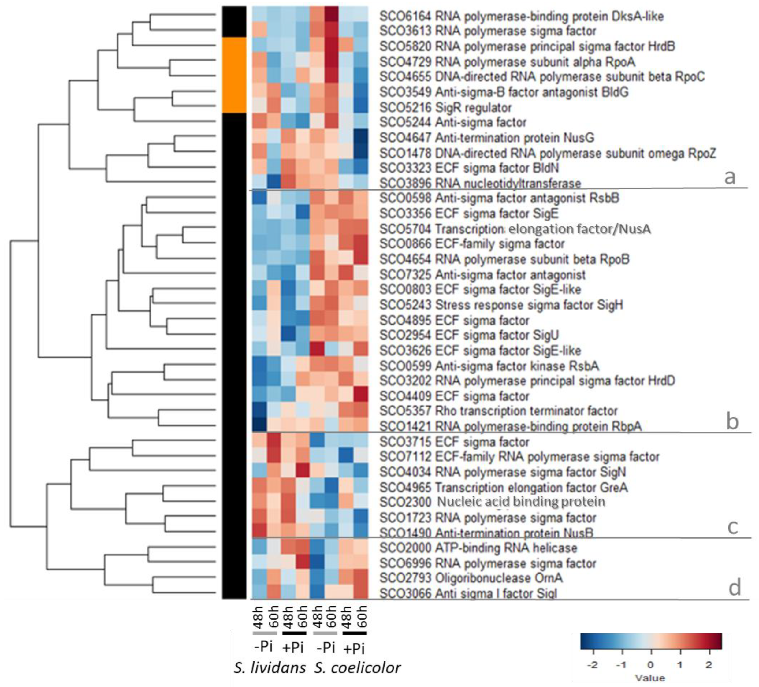
2.1. Components of the Transcriptional Apparatus

2.2. Transcriptional Regulators and Sensory Histidine Kinases
2.2.1. Cluster A: Regulators More Abundant in Pi Proficiency than in Pi Limitation in Both Strains
- -
- The response regulator (RR) OsaB/SCO5749 of TCS OsaA (HK)/OsaB whose expression is induced in conditions of OsS in a sigma B-dependent manner [66]. An OsaB mutant cannot erect aerial hyphae and produces up to 5-fold more antibiotic than the wild-type strain [66]. This TCS, involved in osmoadaptation, thus plays a negative role in the regulation of antibiotic production suggesting that high OsS contributes to the triggering of antibiotic production.
- -
- The sensory histidine kinase (SHK) DraK/SCO3062 of the TCS DraK/DraR. DraR was shown to directly activate the transcription of pathway-specific activator actII-ORF4 and thus ACT biosynthesis [67] whereas it represses CPK and RED biosynthesis via the repression it exerts on the expression of KasO, the specific activator of the CPK pathway, but its effect on RED biosynthesis is independent of the pathway-specific activators RedD and RedZ [67].
- -
- The regulator DmdR1/SCO4394 controls iron homeostasis via the repression it exerts on the expression of the desABCDE/sco2782-85 cluster that directs the biosynthesis of the siderophore, desferrioxamine. Such repression is relieved in conditions of iron deprivation. Our previous data indicated that DesABCE were more abundant in SL than in SC, especially in Pi proficiency, whereas, unexpectedly, the siderophore biosynthetic enzyme DesD, was highly abundant in both Pi conditions in SC (Figure S12B of [9]).
- -
- The SOS regulatory protein LexA/SCO5803 that positively controls the expression of the recombination protein RecA/SCO5769 [68]. Consistently, LexA and RecA were more abundant in Pi proficiency than in Pi limitation in SL but RecA was similarly and highly abundant in both Pi conditions in SC (Figure S8 of [9]) indicating a situation of stress.
- -
- The gene encoding the regulator SCO1614 that is located in divergence of an ethanolamine utilization pathway [69].

2.2.2. Cluster B: Regulatory Proteins More Abundant in Pi Limitation than in Pi Proficiency in Both Strains
- -
- The SHK CseC/SCO3359 of the TCS cseC/cseB belongs to an operon constituted also by cseA (lipoprotein) and sigE. This TCS activates the transcription of sigE/sco3356, in response to the alteration of the cell envelope [47,75]. Consistently, SigE was far more abundant in SC than in SL in both Pi conditions (Figure 1). This indicated that damages to the cell wall occur independently of Pi availability in SC.
- -
- AbrC1/SCO4598 is part of an atypical TCS constituted of 2 SHK (AbrC1/SCO4598 and AbrC2/SCO4597) and of the RR AbrC3/SCO4596 [76]. The genes encoding this TCS are located in divergence of genes encoding sub-units of NADH dehydrogenase (SCO4599-SCO4608) which were far more abundant in SC than in SL in both Pi conditions (Figure 5 of [9]). This TCS has a positive impact on antibiotic production and morphological differentiation in SC [76,77,78].
- -
- The regulator BldB/SCO5723 that has a positive impact on antibiotic production and morphological differentiation [79].
- -
- Three proteins involved in the regulation of phosphate metabolism: the TCS PhoR/PhoP as well as the regulatory protein PhoU/SCO4228. In condition of Pi limitation, PhoR/PhoP positively controls the expression of genes of the Pho regulon involved in Pi scavenging and up-take [80] and negatively those involved in nitrogen assimilation [81]. This TCS also plays a negative role in the control of morphological differentiation and antibiotic production [82,83]. PhoU, through its interaction with PstB, the ATP-binding cassette of the high affinity ABC Pi transporter PstSCAB, was proposed to sense environmental Pi and to transmit this signal to the SHK PhoR, promoting or inhibiting its auto-phosphorylation in Pi limitation or proficiency, respectively [84,85]. The difference in the abundance of proteins involved in Pi scavenging and up-take, in conditions of in Pi limitation or proficiency, was much greater in SL than in SC (Figure 6 of [9]).
- -
- The adenylyltransferase GlnE/SCO2234 that modulates the activity of the glutamine synthase GSI through adenylylation, in response to N availability [86] and is thus involved in the regulation of nitrogen metabolism.
- -
- The LacI regulator SCO4158 [87] whose encoding gene is located downstream of glnR/sco4159 encoding a major regulator of N metabolism (cluster G). SCO4158 was abundant in both strains in Pi limitation whereas GlnR was far more abundant in Pi proficiency than in Pi limitation in SL but poorly abundant in SC in both Pi conditions. Considering the proximity of sco4158 and glnR and their opposite regulatory features, SCO4158 might regulate glnR expression.

- -
- -
- The gene encoding the GntR regulator SCO1728 is located in divergence of a gene encoding a putative mycothiol synthase that might play a role in the resistance to OxS. In the condition of over-expression, SCO1728 was shown to have a negative impact on antibiotic production [91], which might be linked to its role in the resistance to OxS that was proposed to be an important trigger of ACT biosynthesis [8,12].
- -
- The MarR regulator SCO0487/CchL, whose encoding gene is present in the biosynthetic pathway of the peptide siderophore, coelichelin (sco0499/cchA—sco0489/cchK) [92]. The 11 proteins of the coelichelin cluster detected (Figure S12B of [9]) were, as SCO0487, all more abundant in Pi limitation than in Pi proficiency in SL, suggesting that SCO0487 positively regulates their expression in Pi limitation. Five of these proteins (SCO0493 to SCO0497) were also more abundant in Pi limitation than in Pi proficiency in SC whereas, unexpectedly, four others (SCO0492, SCO0498 to SCO0490) were more abundant in Pi proficiency than in Pi limitation in SC and two (SCO0499 and SCO0491) were poorly abundant in both Pi conditions in SC.
2.2.3. Cluster C: Regulators More Abundant in SC than in SL in Both Pi Conditions
- -
- Two regulatory systems able to sense nitric oxide (NO), the TCS OdsK/SCO0203-OdsR/SCO0204 [94] also called DevS/R [95], and NsrR/SCO7427 which positively controls the expression of NO-detoxifying flavohemoglobins hmpA1/SCO7428 and hpmA2/SCO7094 [96,97]. The RR OdsR regulates the expression of genes of the dormancy/survival regulon [94]. Proteins of the OdsR regulon fall into two groups, highly and poorly expressed in SC, and conversely in SL (Figure S11C of [9]). Intracellular NO is thought to inhibit the auto-phosphorylation of the SHK DevS/OdsK which becomes unable to phosphorylate the RR DevR/OdsR [95]. Un-phosphorylated OdsR is unable to interact with the promoter region of actII-ORF4 to activate its expression [95]. NO thus has an indirect negative effect on ACT biosynthesis. Since SC produces ACT abundantly, it might be legitimate to propose that little NO is generated in SC, and this would be consistent with the previously mentioned reduced N availability in this strain.
- -
- -
- The regulator WblA that is a target of the OrrA was identified as a down-regulator of the expression of genes involved in the resistance to OxS [101]. Since OxS was proposed to be an important trigger of ACT biosynthesis [8,12], this can explain the negative impact that WblA exerts on ACT biosynthesis [102].
- -

- -
- The regulator Rex SCO3320 [103] that responds to NAD+/NADH poise and represses the transcription of numerous genes, including its own and that of genes encoding subunits of the complex I/NADH dehydrogenase of the respiratory chain, (nuoABCDEFGHIJKLMN/sco4562-75) and of the ATP synthase (atpIBEFHAGDC/sco5366-74). The repressing effect of Rex is alleviated when the intracellular NADH concentration is high [103], as is likely to be the case in SC that is characterized by an oxidative metabolism [10]. Interestingly, the sub-units of the complex I of the respiratory chain were far more abundant in SC than in SL, in both Pi conditions (Figure 5 of [9]).
- -
- -
- -
2.2.4. Cluster D: Regulatory Proteins More Abundant in Pi Proficiency than in Pi Limitation in SC
- -
- The regulator DasR/SCO5231 which is known to repress the expression of genes involved in the up-take and catabolism of N-acetyl glucosamine [109]. Glucosamine-6-phosphate (GlcN-6P) and N-acetylglucosamine-6-phosphate (GlcNAc-6P) act as allosteric effectors of DasR impairing binding to operator sites and thus allowing the expression of the DasR-target genes. Since several enzymes involved in cell wall degradation were far more abundant in SC than in SL (Figure S10, cluster C of [9]), GlcN-6P as GlcNAc-6P are likely to result from the autolytic degradation of peptidoglycan that obviously takes place in SC but not in SL.
- -
- The RR MacR/SCO2120 of the TCS MarS/MarR positively controls the expression of several membrane proteins involved in maintenance of cell wall integrity of some proteins involved in primary carbon metabolism and of proteins of the Zur regulon which plays a role in zinc homeostasis [110,111]. Furthermore, MacR was shown to directly interact with the actII-orf4 promotor region and to have a positive impact on ACT, as well as CDA and RED biosynthesis [111].
- -
- The RR RapA1/SCO5403 of the TCS RapA2/RapA1, which has a positive impact on CPK and ACT biosynthesis [112].
- -
- -
- The ScbR-like regulator SlbR/SCO0608 which, in interaction with γ-butyrolactone SCB1, binds to the promoter regions of scbR/A and adpA [115] and inhibits the transcription of these genes that positively control morphological development and antibiotic production [116]. SlbR thus has an indirect negative impact on antibiotic production and morphological differentiation [117].
- -
- -
- -
- The gene encoding the regulator SCO2152 is located in divergence of the qcrCAB operon (sco2148—sco2151), which plays an important role in growth and development of SC in conditions of oxygen limitation [120,121]. SCO2152 was rather more abundant in SC than in SL in both Pi conditions, whereas QcrA/SCO2149 showed an opposite trend (Figure 5 of [9] and Figure S1) suggesting that SCO2152 might negatively regulate the expression of the qcrCAB operon.

2.2.5. Cluster E: Regulatory Proteins More Abundant in Pi Limitation than in Pi proficiency in SC
- -
- -
- The zinc-binding regulator HypR/SCO6294 [127], whose expression is induced by hydroxyproline. SCO6294 controls the expression of the Hyp operon (sco6289-sco6293) involved with L-hydroxyproline catabolism. Since hydroxyproline is known to enhance the activity of the peptidoglycan N-deacetylase [128], the induction of HypR expression in SC in Pi limitation might be related to cell wall alteration in SC.

- -
- The RR DraR/SCO3063 of the TCS DraK (sub-cluster A2)/DraR, which has a positive impact on ACT biosynthesis but a negative one on CPK and RED biosynthesis [62].
- -
- The ArsR-like regulator SCO6808, which has a negative impact on antibiotic biosynthesis [117]
- -
- -
- The KorSA-like regulator SCO3932 is located upstream of a pSAM2-like ICE element and has a positive impact CPK and ACT synthesis via its direct interaction with cpkD, a coelimycin biosynthetic gene, and actII-orf4 [129].
- -
- The RR SCO5785 of the TCS SCO5784/SCO5785 has a positive impact on the production of several extracellular proteins and on antibiotics biosynthesis, and a negative one on the expression of some ribosomal genes [130].
- -
- The MmyB-like regulator SCO4944 is a putative member of the A-factor signaling cascade [131] and is likely to have a positive impact on antibiotic biosynthesis.
- -
2.2.6. Cluster F: Regulators More Abundant in SL than in SC in Both Pi Conditions
- -
- Zur/SCO2508 which, in interaction with Zinc [140], positively controls the expression of the gene encoding the zinc exporter ZitB/SCO6751 [141] and negatively that of the genes encoding an ABC transporter involved in Zn uptake (SCO2505/SCO2506/SCO2507) [141,142], and of a cluster of genes directing the biosynthesis of the zincophore, coelibactin [143]. Consistently, ZitB/SCO6751 was poorly abundant in SC and in SL, and to a lesser extent in Pi proficiency (Figure S19 of [9]), whereas proteins of the coelibactin cluster and SCO2506 were more abundant in Pi proficiency than in Pi limitation in SC as in SL, to a lesser extent (Figure 10 of [9]). This indicated that Zur is involved in the regulation of Zn homeostasis, maintaining a low intracellular Zn concentration. In S. avermitilis, the deletion of zur resulted in decreased production of antibiotics and delayed morphological differentiation [144], as did the addition of Zn in the growth medium of SC [145] indicating that Zur plays a positive role in the regulation of antibiotic biosynthesis.

- -
- -
- SCO5464/CabB, a calmodulin-like protein that has a role in calcium homeostasis [149].
- -
- -
- Scr1/SCO4441, which has a strong positive impact on endogenous antibiotic production of SC, SL, and other Streptomyces strains [156].
- -
- -
- The regulator SCO1678, which negatively controls the expression of the divergent operon involved in gluconate metabolism [159].
- -
- -
- BldH/SCO2792/AdpA, which activates the transcription of ramR, an atypical response regulator that itself activates expression of the genes of the ramCSAB operon required for aerial mycelium formation [162].
- -
- NsdB/SCO7252 [163] and NsdA/SCO5582 [164,165]. NsdA was more abundant in Pi proficiency than in Pi limitation in SL. nsdA is located just upstream of a cluster of genes targets of GlnR/SCO4159, an important regulator of N metabolism [166]: amtB/sco5583 (ammonium transporter), glnK/sco5584 (regulatory protein P-II, cluster I), and glnD/sco5585 (protein PII adenylyltransferase) [167]. These proteins were more abundant in Pi proficiency than in Pi limitation in SL as well as in SC but to a far lesser extent (Figure 7 of [9]). Considering the proximity of ndsA with this cluster of genes and its differential abundance in SL and SC, NdsA might play a role in the regulation of the expression of genes of this cluster either directly or indirectly via the regulation of glnR expression.
- -
- -
- SCO5804/NrdR, which represses the expression of class I and class II ribonucleotide reductases encoding genes (RNRs, nrdAB/sco5225-26, and nrdJ/sco5805) involved in the conversion of ribonucleotides to deoxyribonucleotides [170,171]. NrdR was similarly abundant in both Pi conditions in SL whereas NrdAB were clearly more abundant in Pi proficiency than in Pi limitation in SL (Figure S7A of [9]) but far less abundant in SC than in SL in both Pi conditions (Figure S7A of [9]) suggesting a default in the activation of the expression of these genes in SC.
- -
- SCO2647 bears similarities with PecS from plant pathogens [172] and is responsive to urate that is generated by xanthine dehydrogenase at the same time as reactive oxygen species (ROS). SCO2647 might thus be induced in conditions of OxS.
- -
2.2.7. Cluster G: Regulatory Proteins More Abundant in Pi Proficiency than in Pi Limitation in SL but Not in SC
- -
- -
- The gene encoding the TetR regulator SCO2775 is located upstream of genes (sco2774-73) encoding proteins involved in the β-oxidation of fatty acids and in divergence of genes encoding acetyl/propionyl CoA carboxylase (α/SCO2777 and β/SCO2776 subunits) involved in the biosynthesis of malonyl or methyl-malonylCoA, possibly used for fatty acids and/or polyketide (ACT) biosynthesis. SCO2777 and SCO2776 were highly abundant in SC but poorly abundant in SL, in both Pi conditions (Figure S2A of [9]), suggesting that SCO2775 represses the expression of the corresponding encoding genes, consistently with high ACT production of SC.
- -

- -
- The gene encoding the regulator SCO4443 is located downstream of a gene encoding a glutathione peroxidase/SCO4444. SCO4444 was more abundant in Pi limitation than in Pi proficiency in SL (Figure S11A of [9]) (but it was the opposite in SC) suggesting that SCO4443 represses SCO4444 expression in Pi proficiency, at least in SL.
- -
- The gene encoding the regulator MalR/SCO2232 is located in divergence of the maltose transport/utilization operon [177,178] and mediates maltose induction and glucose repression of the maltose transport/utilization operon [179]. Consistently, SCO2231/MalE, putative maltose-binding protein, was mainly abundant in Pi limitation in SL (Figure S15B of [9]). This regulation is obviously altered in SC since MalE was abundant in both Pi conditions in SC. In contrast, the maltose permease SCO2229/MalG (Figure S15D of [9]) was mainly abundant in Pi limitation in SC.
- -
- The gene encoding the regulator SCO2182 is located upstream of sco2183/aceE1 encoding a putative component E1 of pyruvate dehydrogenase. SCO2183 was similarly abundant in the two strains in Pi proficiency (Figure S1 of [9]) suggesting that SCO2182 positively regulates its expression in Pi proficiency.
2.2.8. Cluster H: Regulatory Proteins More Abundant in Pi Limitation than in Pi Proficiency in SL but Not in SC
- -
- The response regulator MtrA/SCO3013 of the TCS SCO3013/SCO3012 (MtrB, HK) which, as PhoP, regulates both N and Pi metabolism. MtrA negatively auto-regulates its own expression and that of GlnR/SCO4159 [180,181]. MtrA represses, in condition of N proficiency, the genes that are activated by GlnR/SCO4159 in condition of N limitation [181]. MtrA has a direct, but either positive or negative, impact on the expression of PhoRP and of other genes of Pi metabolism depending on the composition of the growth medium [182]. MtrA also binds to sites located in the promoter regions of genes encoding pathway specific activators ActII-Orf-1, ActII-Orf4, and RedZ and has a negative impact on their expression and thus on ACT and RED biosynthesis [183]. It also represses the expression of bldD (sub-cluster D3) that plays a positive role in the regulation of antibiotic biosynthesis [125]. In contrast, MtrA activates the expression of genes involved in formation of aerial mycelium, including chp, rdl, and ram and regulatory genes of the Bld and Whi families [184]. MtrA thus positively controls morphological differentiation and negatively antibiotic production.

- -
- The regulator AbsC/SCO5405 plays a role in Zinc (Zn) homeostasis since it negatively controls, together with Zur (cluster F), the expression of the cluster directing the biosynthesis of the zincophore, coelibactin, and of another NRPS cluster with predicted siderophore-like activity [185]. The deletion of AbsC results in elevated expression of these clusters and thus higher Zn availability. Since Zn was shown to have a negative impact on antibiotic biosynthesis [145], the negative impact that AbsC exerts on antibiotic production is thought to be due to enhanced Zn availability [185]. Furthermore, numerous enzymes belonging to carbon and nitrogen metabolism were more abundant in an AbsC mutant suggesting that AbsC negatively regulates their expression [186]. Consistently, most of the enzymes of central carbon and nitrogen metabolism listed in [186] and thought to be under the negative control of AbsC were indeed more abundant in Pi proficiency, when the expression of AbsC is low [9], than in Pi limitation. The cause of the low abundance of AbsC in SC is unknown but is likely to have important consequences on the cellular metabolism of this strain.
- -
- The gene encoding the sensory HK SCO5829 of the TCS SCO5829/SC5828 (RR) is located downstream of a gene encoding sucrase/ferredoxin-like protein SCO5830 and SCO5831 a citrate synthase-like protein. SCO5831 was clearly more abundant in Pi proficiency than in Pi limitation in SL (Figure 4 of [9]) suggesting that this TCS might repress SCO5831 expression in Pi limitation in SL.
- -
- Finally, the three cold shock proteins (Csps) SCO0527/ScoF, SCO3731/ScoF1, and SCO4325/CspB were also highly expressed in SL in Pi limitation. Originally, Csps were viewed as nucleic acid chaperons preventing the formation of secondary structures in mRNA at low temperature and thus facilitating the initiation of translation but since the expression of some Csps was shown to be non-cold inducible, they are now viewed as involved in the adaptation to various stresses [187].
2.3. Eukaryotic-Like Serine or Threonine Protein Kinases

- -
- AfsK/SCO4423 which phosphorylates, together with other ESTPK [196], AfsR/SCO4426, a global regulator, that has a positive impact on antibiotic biosynthesis [153] and morphological differentiation [154,155]. AfsK was also shown to phosphorylate the DnaA initiator protein impairing its interaction with the replication origin and thus inhibiting replication [197].
- -
- PkaF/SCO2110, whose over-expression was shown to be correlated with a strong reduction of ACT (but not of RED) biosynthesis and an inhibition of morphological differentiation, suggesting that it plays a negative role in the regulation of these processes [198].
3. Discussion


| List of Transcriptionnal Regulators and Regulatory Protein Kinases | Impact on Act Biosynth |
|---|---|
| Regulation of Phosphate and/or Nitrogen up-take and assimilation | |
| PhoR/SCO4229-PhoP/SCO4230 | Neg [77,78] |
| MtrA/SCO3013 | Neg [181] |
| GlnR/SCO4159 | Neg [86] |
| DasR/SCO5231 (Regulation of N-acetylglucosamine metabolism) | ? |
| NnaR/SCO2958 (Regu of nitrate-nitrite up-take and assimilation) | (Neg?) |
| SCO7699/EshA (Stringent response) | Pos [159] |
| Cell wall stress | |
| SigE/SCO3356 | Neg [42,44] |
| SigU/ SCO2954 | ? |
| TCS CseB/CseC (SCO3358/SCO3359) | Neg [42] |
| TCS VanR/VanS | ? |
| HypR/SCO6294 (?) | ? |
| ? | |
| Osmotic stress | |
| (SigBND/SCO0600) | (Neg?) |
| SigH/SCO5243 | ? |
| SCO5244 (anti-sigma factor H) | ? |
| BldG/SCO3549 (anti-anti-sigma factor) | Pos [36] |
| TCS OhkA/OrrA (SCO1596/SCO3008) | Neg [94,95] |
| TCS OsaA/SCO5748 -OsaB/SCO5749 | Neg [61] |
| Prst/SCO3066 (Anti sigma I factor) | ? |
| Oxidative Stress | |
| WblA/SCO3579 | Neg [101] |
| SigR/SCO5216 | (Neg ?) |
| SCO2647 (MarR-like regulator) | (Neg?) |
| SCO4180 (Fur-like regulator) | (Neg?) |
| SigN/SCO4034 (general stress response) | Pos [49,50] |
| Metal homeostasis | |
| TCS CutR/SCO5862-CutS/SCO5863 (copper homeostasis) | Neg [104] |
| TCS RapA1/SCO5403- RapA2/SCO5404 (copper homeostasis?) | Pos [110] |
| SCO0487 (regulator of the coelibactin biosynthetic cluster/zinc homeostasis) | ? |
| DmdR1/SCO4394 (regulator of the desferrioxamine biosynthetic cluster/iron homeostasis) | ? |
| AbsC/SCO5405 (MarR-like regulator/zinc homeostasis) | Neg [183] |
| Zur /SCO2508 (zinc homeostasis) | Neg [141] |
| Butyrolactone | |
| CprB/SCO0671 (γ-butyrolactone-responsive regulator) | Neg [162,163] |
| SlbR/SCO0608 (γ-butyrolactone-responsive regulator) | Neg [113] |
| ScbA/SCO6266 (gamma-butyrolactone synthesis) | Neg [56,57] |
| AfsR/SCO4426 (γ-butyrolactone-responsive regulator) | Pos [152,156,157] |
| Others | |
| BldD/SCO5723 (c-di-GMP signaling) | Neg [123] |
| SCO6808 | Neg [115] |
| SCO1728 | Neg [85] |
| PkaF/SCO2110 (ESTPK) | Neg [155] |
| RpoZ/SCO1478 (omega sub-unit of RNA polymerase) | Pos [209] |
| OdsR/OdsK-SCO0204/SCO0203 (TCS dormancy/survival regulon) | Pos [91] |
| Crp/SCO3571 (Cyclic AMP receptor protein) | Pos [82] |
| AtrA/SCO4118 | Pos [84] |
| AbrC3/SCO4596 (TCS response regulator) | Pos [71,72,73] |
| AbrC1/ SCO4598 (TCS sensory histidine kinase) | Pos [71] |
| SCO6992 | Pos [102] |
| SCO3932 | Pos [127] |
| Spa/SCO7629 (starvation sensing protein) | Pos [117] |
| MacR/SCO2120 (TCS regulator) | Pos [109] |
| BldC/SCO4091 | Pos [111,112] |
| TCS DraR/Dra K-SCO3063/SCO3062 | Pos [62] |
| Rok7B7/SCO6008 (regulator of the xylose transport operon xylFGH) | Pos [105,106] |
| SCO5785 (TCS response regulator) | Pos [128] |
| AfsK/SCO4423 (ESTPK) | Pos [195] |
| Rex/SCO3320 (Sensor of NAD+/NADH poise) | ? |
| SCO5556 (HU DNA-binding protein) | ? |
| LexA/SCO5803 (SOS regulatory protein) | ? |
| OrnA/SCO2793 (Oligoribonuclease) | ? |
| SCO3209 (regulator of para-hydroxybenzoate catabolism) | ? |
| SCO1614 (regulator of the ethanolamine utilization pathway) | ? |
| PkaH/SCO4775 (ESTPK, regulation of sporulation) | ? |
| PglW/SCO6626 (phage resistance) | |
| SCO1678 | Pos [161] |
| ScrI/SCO4441 | Pos [158] |
| CprB/SCO6071 (regulator of γ-butyrolactone biosynthesis) | Neg [162,163] |
| NsdA/SCO5582 (Nitrogen metabolism?) | Neg [167,168] |
| NsdB/SCO7252 | Neg [164] |
| CabB/SCO5464 (calcium homeostasis) | ? |
| SCO2832 (regulator of probable amino acid ABC transporter protein) | ? |
| BldH/SCO2792 (Activator of aerial mycelium formation) | ? |
| SCO2775 | ? |
| SCO5804/NrdR (Regulator of ribonucleotide reductase) | ? |
| RamC/SCO6681 (ESTPK, activator of aerial mycelium formation) | ? |
4. Materials and Methods
4.1. Bacterial Strains, Media and Culture Conditions
4.2. Total Proteins Extraction and Digestion
4.3. Liquid Chromatography Tandem Mass Spectrometry Analysis
4.4. Protein Identifications
4.5. Label-Free Mass Spectroscopy-Based Relative Protein Quantification
4.6. Protein Abundance Changes and Statistical Analysis
Supplementary Materials
Author Contributions
Funding
Informed Consent Statement
Data Availability Statement
Conflicts of Interest
References
- Mast, Y.; Stegmann, E. Actinomycetes: The Antibiotics Producers. Antibiotics 2019, 8, 105. [Google Scholar] [CrossRef] [Green Version]
- Bibb, M.J. Regulation of secondary metabolism in Streptomycetes. Curr. Opin. Microbiol. 2005, 8, 208–215. [Google Scholar] [CrossRef] [PubMed]
- Liu, G.; Chater, K.F.; Chandra, G.; Niu, G.; Tan, H. Molecular Regulation of Antibiotic Biosynthesis in Streptomyces. Microbiol. Mol. Biol. Rev. 2013, 77, 112–143. [Google Scholar] [CrossRef] [Green Version]
- Kong, D.; Wang, X.; Nie, J.; Niu, G. Regulation of Antibiotic Production by Signaling Molecules in Streptomyces. Front. Microbiol. 2019, 10, 2927. [Google Scholar] [CrossRef] [PubMed] [Green Version]
- Romero-Rodríguez, A.; Robledo-Casados, I.; Sánchez, S. An overview on transcriptional regulators in Streptomyces. Biochim. Biophys. Acta 2015, 1849, 1017–1039. [Google Scholar] [CrossRef] [PubMed]
- Bentley, S.D.; Chater, K.F.; Cerdeño-Tárraga, A.-M.; Challis, G.L.; Thomson, N.R.; James, K.D.; Harris, D.E.; Quail, M.A.; Kieser, H.; Harper, D.; et al. Complete genome sequence of the model actinomycete Streptomyces coelicolor A3(2). Nature 2002, 417, 141–147. [Google Scholar] [CrossRef] [PubMed]
- Rückert, C.; Albersmeier, A.; Busche, T.; Jaenicke, S.; Winkler, A.; Friðjónsson, H.; Hreggviðsson, G.Ó.; Lambert, C.; Badcock, D.; Bernaerts, K.; et al. Complete genome sequence of Streptomyces lividans TKJ. Biotechnology 2015, 199, 21–22. [Google Scholar] [CrossRef]
- Millan-Oropeza, A.; Henry, C.; Lejeune, C.; David, M.; Virolle, M.-J. Expression of genes of the Pho regulon is altered in Streptomyces coelicolor. Sci. Rep. 2020, 10, 8492. [Google Scholar] [CrossRef]
- Lejeune, C.; Sago, L.; Cornu, D.; Redeker, V.; Virolle, M.-J. A Proteomic Analysis Indicates That Oxidative Stress Is the Common Feature Triggering Antibiotic Production in Streptomyces coelicolor and in the pptA Mutant of Streptomyces lividans. Front. Microbiol. 2022, 12, 813993. [Google Scholar] [CrossRef]
- Esnault, C.; Dulermo, T.; Smirnov, A.; Askora, A.; David, M.; Deniset-Besseau, A.; Holland, I.-B.; Virolle, M.-J. Strong antibiotic production is correlated with highly active oxidative metabolism in Streptomyces coelicolor M145. Sci. Rep. 2017, 7, 200. [Google Scholar] [CrossRef]
- Vinogradov, A.D.; Grivennikova, V.G. Oxidation of NADH and ROS production by respiratory complex I. Biochim. Biophys. Acta 2016, 1857, 863–871. [Google Scholar] [CrossRef] [PubMed]
- Virolle, M.J. A Challenging View: Antibiotics Play a Role in the Regulation of the Energetic Metabolism of the Producing Bacteria. Antibiotics 2020, 9, 83. [Google Scholar] [CrossRef] [PubMed] [Green Version]
- Romero-Rodríguez, A.; Maldonado-Carmona, N.; Ruiz-Villafán, B.; Koirala, N.; Rocha, D.; Sánchez, S. Interplay between carbon, nitrogen and phosphate utilization in the control of secondary metabolite production in Streptomyces. Antonie Leeuwenhoek 2018, 111, 761–781. [Google Scholar] [CrossRef]
- Doucette, C.D.; Schwab, D.J.; Wingreen, N.S.; Rabinowitz, J.D. α-ketoglutarate coordinates carbon and nitrogen utilization via enzyme I inhibition. Nat. Chem. Biol. 2011, 7, 894–901. [Google Scholar] [CrossRef] [PubMed] [Green Version]
- Buttner, M.; Chater, K.F.; Bibb, M.J. Cloning, disruption, and transcriptional analysis of three RNA polymerase sigma factor genes of Streptomyces coelicolor A3(2). J. Bacteriol. 1990, 172, 3367–3378. [Google Scholar] [CrossRef] [PubMed] [Green Version]
- Tetsuo, S.; Kan, T.; Hideo, T. Sequence of hrdB, an essential gene encoding sigma-like transcription factor of Streptomyces coelicolor A3(2): Homology to principal sigma factors. Gene 1991, 107, 145–148. [Google Scholar] [CrossRef]
- Šmídová, K.; Ziková, A.; Pospíšil, J.; Schwarz, M.; Bobek, J.; Vohradsky, J. DNA mapping and kinetic modeling of the HrdB regulon in Streptomyces coelicolor. Nucleic Acids Res. 2018, 47, 621–633. [Google Scholar] [CrossRef] [PubMed] [Green Version]
- Cho, E.-J.; Bae, J.-B.; Kang, J.-G.; Roe, J.-H. Molecular Analysis of RNA Polymerase Alpha Subunit Gene from Streptomyces coelicolor A3(2). Nucleic Acids Res. 1996, 24, 4565–4571. [Google Scholar] [CrossRef] [Green Version]
- Babcock, M.J.; Buttner, M.; Keler, C.H.; Clarke, B.R.; Morris, R.A.; Lewis, C.G.; Brawner, M.E. Characterization of the rpoC gene of Streptomyces coelicolor A3(2) and its use to develop a simple and rapid method for the purification of RNA polymerase. Gene 1997, 196, 31–42. [Google Scholar] [CrossRef]
- Paget, M.S.; Kang, J.; Roe, J.; Buttner, M. Sigma R, an RNA polymerase sigma factor that modulates expression of the thioredoxin system in response to oxidative stress in Streptomyces coelicolor A3(2). EMBO J. 1998, 17, 5776–5782. [Google Scholar] [CrossRef]
- Paget, M.S.B.; Molle, V.; Cohen, G.; Aharonowitz, Y.; Buttner, M.J. Defining the disulphide stress response in Streptomyces coelicolor A3(2): Identification of the sigmaR regulon. Mol. Microbiol. 2001, 42, 1007–1020. [Google Scholar] [CrossRef] [PubMed]
- Kallifidas, D.; Thomas, D.; Doughty, P.; Paget, M.S. The sigmaR regulon of Streptomyces coelicolor A32 reveals a key role in protein quality control during disulphide stress. Microbiology 2010, 156, 1661–1672. [Google Scholar] [CrossRef] [PubMed] [Green Version]
- Kim, M.S.; Dufour, Y.S.; Yoo, J.S.; Cho, Y.B.; Park, J.H.; Nam, G.B.; Kim, H.M.; Lee, K.L.; Donohue, T.J.; Roe, J.H. Conservation of thiol-oxidative stress responses regulated by SigR orthologues in actinomycetes. Mol. Microbiol. 2012, 85, 326–344. [Google Scholar] [CrossRef] [PubMed] [Green Version]
- Park, J.; Lee, J.; Roe, J. SigR, a hub of multilayered regulation of redox and antibiotic stress responses. Mol. Microbiol. 2019, 112, 420–431. [Google Scholar] [CrossRef] [Green Version]
- Mingyar, E.; Sevcikova, B.; Rezuchova, B.; Homerova, D.; Novakova, R.; Kormanec, J. The sigma(F)-specific anti-sigma factor RsfA is one of the protein kinases that phosphorylates the pleiotropic anti-anti-sigma factor BldG in Streptomyces coelicolor A3(2). Gene 2014, 538, 280–287. [Google Scholar] [CrossRef]
- Sevcikova, B.; Rezuchova, B.; Homerova, D.; Kormanec, J. The Anti-Anti-Sigma Factor BldG Is Involved in Activation of the Stress Response Sigma Factor σH in Streptomyces coelicolor A3(2). J. Bacteriol. 2010, 192, 5674–5681. [Google Scholar] [CrossRef] [Green Version]
- Sevcikova, B.; Rezuchova, B.; Mingyar, E.; Homerova, D.; Novakova, R.; Feckova, L.; Kormanec, J. Pleiotropic anti-anti-sigma factor BldG is phosphorylated by several anti-sigma factor kinases in the process of activating multiple sigma factors in Streptomyces coelicolor A3(2). Gene 2020, 755, 144883. [Google Scholar] [CrossRef]
- Aldridge, M.; Facey, P.; Francis, L.; Bayliss, S.; Del Sol, R.; Dyson, P. A novel bifunctional histone protein in Streptomyces: A candidate for structural coupling between DNA conformation and transcription during development and stress? Nucleic Acids Res. 2013, 41, 4813–4824. [Google Scholar] [CrossRef] [Green Version]
- Travis, B.A.; Schumacher, M.A. Diverse molecular mechanisms of transcription regulation by the bacterial alarmone ppGpp. Mol. Microbiol. 2021, 117, 252–260. [Google Scholar] [CrossRef]
- Bibb, M.J.; Molle, V.; Buttner, M.J. Sigma(BldN), an extracytoplasmic function RNA polymerase sigma factor required for aerial mycelium formation in Streptomyces coelicolor A3(2). J. Bacteriol. 2000, 182, 4606–4616. [Google Scholar] [CrossRef]
- Bibb, M.J.; Domonkos, Á.; Chandra, G.; Buttner, M.J. Expression of the chaplin and rodlin hydrophobic sheath proteins in Streptomyces venezuelae is controlled by sigma(BldN) and a cognate anti-sigma factor, RsbN. Mol. Microbiol. 2012, 84, 1033–1049. [Google Scholar] [CrossRef] [PubMed]
- Martín, J.F.; Sola-Landa, A.; Santos-Beneit, F.; Fernández-Martínez, L.T.; Prieto, C.; Rodríguez-García, A. Cross-talk of global nutritional regulators in the control of primary and secondary metabolism in Streptomyces. Microb. Biotechnol. 2010, 4, 165–174. [Google Scholar] [CrossRef] [PubMed] [Green Version]
- Santos-Beneit, F.; Barriuso-Iglesias, M.; Fernández-Martínez, L.T.; Martínez-Castro, M.; Sola-Landa, A.; Rodríguez-García, A.; Martín, J.F. The RNA polymerase omega factor RpoZ is regulated by PhoP and has an important role in antibiotic biosynthesis and morphological differentiation in Streptomyces coelicolor. Appl. Environ. Microbiol. 2011, 77, 7586–7594. [Google Scholar] [CrossRef] [PubMed] [Green Version]
- Puttikhunt, C.; Nihira, T.; Yamada, Y. Cloning, nucleotide sequence, and transcriptional analysis of the nusG gene of Streptomyces coelicolor A3(2), which encodes a putative transcriptional antiterminator. Mol. Gen. Genet. 1995, 247, 118–122. [Google Scholar] [CrossRef] [PubMed]
- Bralley, P.; Gust, B.; Chang, S.; Chater, K.F.; Jones, G.H. RNA 3′-tail synthesis in Streptomyces: In vitro and in vivo activities of RNase PH, the SCO3896 gene product and polynucleotide phosphorylase. Microbiology 2006, 152, 627–636. [Google Scholar] [CrossRef] [Green Version]
- YTanaka, Y.; Kasahara, K.; Hirose, Y.; Murakami, K.; Kugimiya, R.; Ochi, K. Activation and products of the cryptic sec-ondary metabolite biosynthetic gene clusters by rifampin resistance (rpoB) mutations in actinomycetes. J. Bacteriol. 2013, 195, 2959–2970. [Google Scholar] [CrossRef] [Green Version]
- Liu, B.; Tabib-Salazar1, A.; Doughty, P.; Lewis, R.; Ghosh, S.; Parsy, M.L.; Simpson, P.; O’Dwyer, K.; Matthews, S.; Paget, M. The actinobacterial transcription factor RbpA binds to the principal sigma subunit of RNA polymerase. Nucleic Acids Res. 2013, 41, 5679–5691. [Google Scholar] [CrossRef]
- Ingham, C.J.; Hunter, I.S.; Smith, M.C. Isolation and Sequencing of the rho Gene from Streptomyces lividans ZX7 and Characterization of the RNA-dependent NTPase Activity of the Overexpressed Protein. J. Biol. Chem. 1996, 271, 21803–21807. [Google Scholar] [CrossRef] [Green Version]
- Sun, D.; Liu, C.; Zhu, J.; Liu, W. Connecting Metabolic Pathways: Sigma Factors in Streptomyces spp. Front. Microbiol. 2017, 8, 2546. [Google Scholar] [CrossRef]
- Mazurakova, V.; Sevcikova, B.; Rezuchova, B.; Kormanec, J. Cascade of sigma factors in streptomycetes: Identification of a new extracytoplasmic function sigma factor sigmaJ that is under the control of the stress-response sigma factor sigmaH in Streptomyces coelicolor A3(2). Arch. Microbiol. 2006, 186, 435–446. [Google Scholar] [CrossRef]
- Bignell, D.R.D.; Warawa, J.L.; Strap, J.L.; Chater, K.F.; Leskiw, B.K. Study of the bldG locus suggests that an anti-anti-sigma factor and an anti-sigma factor may be involved in Streptomyces coelicolor antibiotic production and sporulation. Microbiology 2000, 146 Pt 9, 2161–2173. [Google Scholar] [CrossRef] [PubMed] [Green Version]
- Sevcikova, B.; Benada, O.; Kofronova, O.; Kormanec, J. Stress-response sigma factor sigma(H) is essential for morphological differentiation of Streptomyces coelicolor A3(2). Arch. Microbiol. 2001, 177, 98–106. [Google Scholar] [CrossRef]
- Viollier, P.; Weihofen, A.; Folcher, M.; Thompson, C.J. Post-transcriptional Regulation of the Streptomyces coelicolor Stress Responsive Sigma Factor, SigH, Involves Translational Control, Proteolytic Processing, and an Anti-sigma Factor Homolog. J. Mol. Biol. 2003, 325, 637–649. [Google Scholar] [CrossRef]
- Helmann, J.D. The extracytoplasmic function (ECF) sigma factors. Adv. Microb. Physiol. 2002, 46, 47–110. [Google Scholar] [CrossRef]
- Sineva, E.; Savkina, M.; Ades, S.E. Themes and variations in gene regulation by extracytoplasmic function (ECF) sigma factors. Curr. Opin. Microbiol. 2017, 36, 128–137. [Google Scholar] [CrossRef] [PubMed] [Green Version]
- Tanaka, K.; Shiina, T.; Takahashi, H. Nucleotide sequence of genes hrdA, hrdC, and hrdD from Streptomyces coelicolor A3(2) having similarity to rpoD genes. Mol. Gen. Genet. 1991, 229, 334–340. [Google Scholar] [CrossRef]
- Paget, M.S.B.; Leibovitz, E.; Buttner, M. A putative two-component signal transduction system regulates sigmaE, a sigma factor required for normal cell wall integrity in Streptomyces coelicolor A3(2). Mol. Microbiol. 1999, 33, 97–107. [Google Scholar] [CrossRef]
- Buttner, M.J.; Lewis, C.G. Construction and characterization of Streptomyces coelicolor A3(2) mutants that are multiply deficient in the nonessential hrd-encoded RNA polymerase sigma factors. J. Bacteriol. 1992, 174, 5165–5167. [Google Scholar] [CrossRef] [Green Version]
- Lonetto, M.A.; Brown, K.L.; Rudd, K.E.; Buttner, M.J. Analysis of the Streptomyces coelicolor sigE gene reveals the existence of a subfamily of eubacterial RNA polymerase sigma factors involved in the regulation of extracytoplasmic functions. Proc. Natl. Acad. Sci. USA 1994, 91, 7573–7577. [Google Scholar] [CrossRef] [Green Version]
- Gordon, N.D.; Ottaviano, G.L.; Connell, S.E.; Tobkin, G.V.; Son, C.H.; Shterental, S.; Gehring, A.M. Secreted-protein response to sigmaU activity in Streptomyces coelicolor. J. Bacteriol. 2008, 190, 894–904. [Google Scholar] [CrossRef] [PubMed]
- Gehring, A.M.; Yoo, N.J.; Losick, R. RNA Polymerase Sigma Factor That Blocks Morphological Differentiation by Streptomyces coelicolor. J. Bacteriol. 2001, 183, 5991–5996. [Google Scholar] [CrossRef] [PubMed] [Green Version]
- Lee, E.J.; Cho, Y.H.; Kim, H.S.; Ahn, B.E.; Roe, J.H. Regulation of sigmaB by an anti- and an anti-anti-sigma factor in Streptomyces coelicolor in response to osmotic stress. J. Bacteriol. 2004, 186, 8490–8498. [Google Scholar] [CrossRef] [PubMed] [Green Version]
- Cho, Y.-H.; Lee, E.-J.; Ahn, B.-E.; Roe, J.-H. SigB, an RNA polymerase sigma factor required for osmoprotection and proper differentiation of Streptomyces coelicolor. Mol. Microbiol. 2008, 42, 205–214. [Google Scholar] [CrossRef] [PubMed]
- Wang, C.; Long, X.; Mao, X.; Dong, H.; Xu, L.; Li, Y. SigN is responsible for differentiation and stress responses based on comparative proteomic analyses of Streptomyces coelicolor wild-type and sigN deletion strains. Microbiol. Res. 2010, 165, 221–231. [Google Scholar] [CrossRef] [PubMed]
- Dalton, K.A.; Thibessard, A.; Hunter, J.I.B.; Kelemen, G.H. A novel compartment, the “subapical stem” of the aerial hyphae, is the location of a sigN-dependent, developmentally distinct transcription in Streptomyces coelicolor. Mol. Microbiol. 2007, 64, 719–737. [Google Scholar] [CrossRef] [PubMed] [Green Version]
- Homerova, D.; Sevcikova, B.; Rezuchova, B.; Kormanec, J. Regulation of an alternative sigma factor sigmaI by a partner switching mechanism with an anti-sigma factor PrsI and an anti-anti-sigma factor ArsI in Streptomyces coelicolor A3(2). Gene 2012, 492, 71–80. [Google Scholar] [CrossRef]
- Ohnishi, Y.; Nishiyama, Y.; Sato, R.; Kameyama, S.; Horinouchi, S. An Oligoribonuclease Gene in Streptomyces griseus. J. Bacteriol. 2000, 182, 4647–4653. [Google Scholar] [CrossRef] [Green Version]
- Salerno, P.; Larsson, J.; Bucca, G.; Laing, E.; Smith, C.P.; Flärdh, K. One of the Two Genes Encoding Nucleoid-Associated HU Proteins in Streptomyces coelicolor is Developmentally Regulated and Specifically Involved in Spore Maturation. J. Bacteriol. 2009, 191, 6489–6500. [Google Scholar] [CrossRef] [Green Version]
- Hsiao, N.H.; Soding, J.; Linke, D.; Lange, C.; Hertweck, C.; Wohlleben, W.; Takano, E. ScbA from Streptomyces coelicolor A3(2) has homology to fatty acid synthases and is able to synthesize gamma-butyrolactones. Microbiology 2007, 153, 1394–1404. [Google Scholar] [CrossRef] [Green Version]
- Takano, E.; Chakraburtty, R.; Nihira, T.; Yamada, Y.; Bibb, M.J. A complex role for the gamma-butyrolactone SCB1 in regulating antibiotic production in Streptomyces coelicolor A3(2). Mol. Microbiol. 2001, 41, 1015–1028. [Google Scholar] [CrossRef]
- Mehra, S.; Charaniya, S.; Takano, E.; Hu, W.-S. A Bistable Gene Switch for Antibiotic Biosynthesis: The Butyrolactone Regulon in Streptomyces coelicolor. PLoS ONE 2008, 3, e2724. [Google Scholar] [CrossRef] [PubMed]
- Tsigkinopoulou, A.; Takano, E.; Breitling, R. Unravelling the gamma-butyrolactone network in Streptomyces coelicolor by computational ensemble modelling. PLoS Comput. Biol. 2020, 16, e1008039. [Google Scholar] [CrossRef] [PubMed]
- Huang, H.; Grove, A. The transcriptional regulator TamR from Streptomyces coelicolor controls a key step in central metabolism during oxidative stress. Mol. Microbiol. 2013, 87, 1151–1166. [Google Scholar] [CrossRef]
- Grandvalet, C.; Rapoport, G.; Mazodier, P. hrcA, encoding the repressor of the groEL genes in Streptomyces albus G, is associated with a second dnaJ gene. J. Bacteriol. 1998, 180, 5129–5134. [Google Scholar] [CrossRef] [PubMed] [Green Version]
- Servant, P.; Mazodier, P. Negative regulation of the heat shock response in Streptomyces. Arch. Microbiol. 2001, 176, 237–242. [Google Scholar] [CrossRef] [PubMed]
- Bishop, A.; Fielding, S.; Dyson, P.; Herron, P. Systematic Insertional Mutagenesis of a Streptomycete Genome: A Link between Osmoadaptation and Antibiotic Production. Genome Res. 2004, 14, 893–900. [Google Scholar] [CrossRef] [Green Version]
- Yu, Z.; Zhu, H.; Dang, F.; Zhang, W.; Qin, Z.; Yang, S.; Jiang, W. Differential regulation of antibiotic biosynthesis by DraR-K, a novel two-component system in Streptomyces coelicolor. Mol. Microbiol. 2012, 85, 535–556. [Google Scholar] [CrossRef]
- Vierling, S.; Weber, T.; Wohlleben, W.; Muth, G. Evidence that an Additional Mutation Is Required to Tolerate Insertional Inactivation of the Streptomyces lividans recA Gene. J. Bacteriol. 2001, 183, 4374–4381. [Google Scholar] [CrossRef] [Green Version]
- Krysenko, S.; Matthews, A.; Okoniewski, N.; Kulik, A.; Girbas, M.G.; Tsypik, O.; Meyners, C.S.; Hausch, F.; Wohlleben, W.; Bera, A. Initial Metabolic Step of a Novel Ethanolamine Utilization Pathway and Its Regulation in Streptomyces coelicolor M145. mBio 2019, 10, e00326-19. [Google Scholar] [CrossRef] [Green Version]
- Amin, R.; Reuther, J.; Bera, A.; Wohlleben, W.; Mast, Y. A novel GlnR target gene, nnaR, is involved in nitrate/nitrite assimilation in Streptomyces coelicolor. Microbiology 2012, 158, 1172–1182. [Google Scholar] [CrossRef]
- Hesketh, A.; Fink, D.; Gust, B.; Rexer, H.U.; Scheel, B.; Chater, K.; Wohlleben, W.; Engels, A. The GlnD and GlnK homologues of Streptomyces coelicolor A3(2) are functionally dissimilar to their nitrogen regulatory system counterparts from enteric bacteria. Mol. Microbiol. 2002, 46, 319–330. [Google Scholar] [CrossRef] [PubMed]
- Waldvogel, E.; Herbig, A.; Battke, F.; Amin, R.; Nentwich, M.; Nieselt, K.; Ellingsen, T.E.; Wentzel, A.; Hodgson, D.A.; Wohlleben, W.; et al. The PII protein GlnK is a pleiotropic regulator for morphological differentiation and secondary metabolism in Streptomyces coelicolor. Appl. Microbiol. Biotechnol. 2011, 92, 1219–1236. [Google Scholar] [CrossRef] [PubMed]
- Antoraz, S.; Rico, S.; Rodríguez, H.; Sevillano, L.; Alzate, J.F.; Santamaria, R.I.; Díaz, M. The Orphan Response Regulator Aor1 Is a New Relevant Piece in the Complex Puzzle of Streptomyces coelicolor Antibiotic Regulatory Network. Front. Microbiol. 2017, 8, 2444. [Google Scholar] [CrossRef] [PubMed]
- Lee, E.J.; Karoonuthaisiri, N.; Kim, H.S.; Park, J.H.; Cha, C.J.; Kao, C.M.; Roe, J.H. A master regulator sigmaB governs osmotic and oxidative response as well as differentiation via a network of sigma factors in Streptomyces coelicolor. Mol. Microbiol. 2005, 57, 1252–1264. [Google Scholar] [CrossRef]
- Hong, H.J.; Paget, M.S.; Buttner, M.J. A signal transduction system in Streptomyces coelicolor that activates the expression of a putative cell wall glycan operon in response to vancomycin and other cell wall-specific antibiotics. Mol. Microbiol. 2002, 44, 1199–1211. [Google Scholar] [CrossRef] [Green Version]
- Rodriguez, H.; Rico, S.; Yepes, A.; Franco-Echevarria, E.; Antoraz, S.; Santamaria, R.I.; Diaz, M. The two kinases, AbrC1 and AbrC2, of the atypical two-component system AbrC are needed to regulate antibiotic production and differentiation in Streptomyces coelicolor. Front. Microbiol. 2015, 6, 450. [Google Scholar] [CrossRef]
- Yepes, A.; Rico, S.; Rodríguez-García, A.; Santamaria, R.I.; Díaz, M. Novel Two-Component Systems Implied in Antibiotic Production in Streptomyces coelicolor. PLoS ONE 2011, 6, e19980. [Google Scholar] [CrossRef] [Green Version]
- Rico, S.; Yepes, A.; Rodriguez, H.; Santamaria, J.; Antoraz, S.; Krause, E.M.; Diaz, M.; Santamaria, R.I. Regulation of the AbrA1/A2 two-component system in Streptomyces coelicolor and the potential of its deletion strain as a heterologous host for antibiotic production. PLoS ONE 2014, 9, e109844. [Google Scholar] [CrossRef] [Green Version]
- Eccleston, M.; Ali, R.A.; Seyler, R.; Westpheling, J.; Nodwell, J. Structural and Genetic Analysis of the BldB Protein of Streptomyces coelicolor. J. Bacteriol. 2002, 184, 4270–4276. [Google Scholar] [CrossRef] [Green Version]
- Santos-Beneit, F.; Rodríguez-García, A.; Franco-Domínguez, E.; Martín, J.F. Phosphate-dependent regulation of the low- and high-affinity transport systems in the model actinomycete Streptomyces coelicolor. Microbiology 2008, 154, 2356–2370. [Google Scholar] [CrossRef]
- Barreiro, C.; Martínez-Castro, M. Regulation of the phosphate metabolism in Streptomyces genus: Impact on the secondary metabolites. Appl. Microbiol. Biotechnol. 2019, 103, 1643–1658. [Google Scholar] [CrossRef] [PubMed]
- Smirnov, A.; Esnault, C.; Prigent, M.; Holland, I.B.; Virolle, M.-J. Phosphate Homeostasis in Conditions of Phosphate Proficiency and Limitation in the Wild Type and the phoP Mutant of Streptomyces lividans. PLoS ONE 2015, 10, e0126221. [Google Scholar] [CrossRef] [PubMed] [Green Version]
- Rodriguez-Garcia, A.; Barreiro, C.; Santos-Beneit, F.; Sola-Landa, A.; Martin, J.F. Genome-wide transcriptomic and proteomic analysis of the primary response to phosphate limitation in Streptomyces coelicolor M145 and in a DeltaphoP mutant. Proteomics 2007, 7, 2410–2429. [Google Scholar] [CrossRef] [PubMed]
- Martin-Martin, S.; Rodriguez-Garcia, A.; Santos-Beneit, F.; Franco-Dominguez, E.; Sola-Landa, A.; Martin, J.F. Self-control of the PHO regulon: The PhoP-dependent protein PhoU controls negatively expression of genes of PHO regulon in Streptomyces coelicolor. J. Antibiot. 2017, 71, 113–122. [Google Scholar] [CrossRef]
- Vuppada, R.K.; Hansen, C.R.; Strickland, K.A.P.; Kelly, K.M.; McCleary, W.R. Phosphate signaling through alternate conformations of the PstSCAB phosphate transporter. BMC Microbiol. 2018, 18, 8. [Google Scholar] [CrossRef] [Green Version]
- Fink, D.; Weissschuh, N.; Reuther, J.; Wohlleben, W.; Engels, A. Two transcriptional regulators GlnR and GlnRII are involved in regulation of nitrogen metabolism in Streptomyces coelicolor A3(2). Mol. Microbiol. 2002, 46, 331–347. [Google Scholar] [CrossRef]
- Meng, L.; Yang, S.H.; Kim, T.J.; Suh, J.W. Effects of Two Putative LacI-family Transcriptional Regulators, SCO4158 and SCO7554, on Antibiotic Pigment Production of Streptomyces coelicolor and Streptomyces lividans. J. Korean Soc. Appl. Biol. Chem. 2012, 55, 737–741. [Google Scholar] [CrossRef]
- Gao, C.; Hindra; Mulder, D.; Yin, C.; Elliot, M.A. Crp Is a Global Regulator of Antibiotic Production in Streptomyces. mBio 2012, 3, e00407-12. [Google Scholar] [CrossRef] [Green Version]
- Derouaux, A.; Halici, S.; Nothaft, H.; Neutelings, T.; Moutzourelis, G.; Dusart, J.; Rigali, S. Deletion of a cyclic AMP receptor protein homologue diminishes germination and affects morphological development of Streptomyces coelicolor. J. Bacteriol. 2004, 186, 1893–1897. [Google Scholar] [CrossRef] [Green Version]
- Uguru, G.C.; Stephens, K.E.; Stead, J.A.; Towle, J.E.; Baumberg, S.; McDowall, K.J. Transcriptional activation of the pathway-specific regulator of the actinorhodin biosynthetic genes in Streptomyces coelicolor. Mol. Microbiol. 2005, 58, 131–150. [Google Scholar] [CrossRef]
- Tsypik, O.; Yushchuk, O.; Zaburannyi, N.; Flardh, K.; Walker, S.; Fedorenko, V.; Ostash, B. Transcriptional regulators of GntR family in Streptomyces coelicolor A3(2): Analysis in silico and in vivo of YtrA subfamily. Folia Microbiol. 2016, 61, 209–220. [Google Scholar] [CrossRef] [PubMed]
- Challis, G.L.; Ravel, J. Coelichelin, a new peptide siderophore encoded by the Streptomyces coelicolor genome: Structure prediction from the sequence of its non-ribosomal peptide synthetase. FEMS Microbiol. Lett. 2000, 187, 111–114. [Google Scholar] [CrossRef] [PubMed] [Green Version]
- Zhang, R.; Lord, D.M.; Bajaj, R.; Peti, W.; Page, R.; Sello, J.K. A peculiar IclR family transcription factor regulates para-hydroxybenzoate catabolism in Streptomyces coelicolor. Nucleic Acids Res. 2018, 46, 1501–1512. [Google Scholar] [CrossRef] [PubMed] [Green Version]
- Urem, M.; van Rossum, T.; Bucca, G.; Moolenaar, G.F.; Laing, E.; Swiatek-Polatynska, M.A.; Willemse, J.; Tenconi, E.; Rigali, S.; Goosen, N.; et al. OsdR of Streptomyces coelicolor and the Dormancy Regulator DevR of Mycobacterium tuberculosis Control Overlapping Regulons. mSystems 2016, 1, e00014-16. [Google Scholar] [CrossRef] [PubMed] [Green Version]
- Honma, S.; Ito, S.; Yajima, S.; Sasaki, Y. Nitric Oxide Signaling for Actinorhodin Production in Streptomyces coelicolor A3(2) via the DevS/R Two-Component System. Appl. Environ. Microbiol. 2021, 87, e0048021. [Google Scholar] [CrossRef] [PubMed]
- Crack, J.C.; Munnoch, J.; Dodd, E.L.; Knowles, F.; Al Bassam, M.M.; Kamali, S.; Holland, A.A.; Cramer, S.P.; Hamilton, C.J.; Johnson, M.K.; et al. NsrR from Streptomyces coelicolor is a Nitric Oxide-sensing [4Fe-4S] Cluster Protein with a Specialized Regulatory Function. J. Biol. Chem. 2015, 290, 12689–12704. [Google Scholar] [CrossRef] [Green Version]
- Tucker, N.P.; Hicks, M.G.; Clarke, T.A.; Crack, J.C.; Chandra, G.; Le Brun, N.E.; Dixon, R.; Hutchings, M.I. The Transcriptional Repressor Protein NsrR Senses Nitric Oxide Directly via a [2Fe-2S] Cluster. PLoS ONE 2008, 3, e3623. [Google Scholar] [CrossRef] [Green Version]
- Zhu, Y.; Wang, X.; Zhang, J.; Ni, X.; Zhang, X.; Tao, M.; Pang, X. The regulatory gene wblA is a target of the orphan response regulator OrrA in Streptomyces coelicolor. Environ. Microbiol. 2022, 24, 3081–3096. [Google Scholar] [CrossRef]
- Lu, Y.; He, J.; Zhu, H.; Yu, Z.; Wang, R.; Chen, Y.; Dang, F.; Zhang, W.; Yang, S.; Jiang, W. An orphan histidine kinase, OhkA, regulates both secondary metabolism and morphological differentiation in Streptomyces coelicolor. J. Bacteriol. 2011, 193, 3020–3032. [Google Scholar] [CrossRef] [Green Version]
- Zheng, G.; Liu, P.; He, W.; Tao, H.; Yang, Z.; Sun, C.; Wang, W.; Lu, Y.; Jiang, W. Identification of the cognate response regulator of the orphan histidine kinase OhkA involved in both secondary metabolism and morphological differentiation in Streptomyces coelicolor. Appl. Microbiol. Biotechnol. 2021, 105, 5905–5914. [Google Scholar] [CrossRef]
- Kim, J.S.; Lee, H.N.; Kim, P.; Lee, H.S.; Kim, E.S. Negative role of wblA in response to oxidative stress in Streptomyces coelicolor. J. Microbiol. Biotechnol. 2012, 22, 736–741. [Google Scholar] [CrossRef] [PubMed]
- Kang, S.-H.; Huang, J.; Lee, H.-N.; Hur, Y.-A.; Cohen, S.N.; Kim, E.-S. Interspecies DNA Microarray Analysis Identifies WblA as a Pleiotropic Down-Regulator of Antibiotic Biosynthesis in Streptomyces. J. Bacteriol. 2007, 189, 4315–4319. [Google Scholar] [CrossRef] [PubMed] [Green Version]
- Brekasis, D.; Paget, M.S. A novel sensor of NADH/NAD+ redox poise in Streptomyces coelicolor A3(2). EMBO J. 2003, 22, 4856–4865. [Google Scholar] [CrossRef] [PubMed]
- Jin, X.M.; Choi, M.Y.; Tsevelkhoroloo, M.; Park, U.; Suh, J.W.; Hong, S.K. SCO6992, a Protein with beta-Glucuronidase Activity, Complements a Mutation at the absR Locus and Promotes Antibiotic Biosynthesis in Streptomyces coelicolor. J. Microbiol. Biotechnol. 2021, 31, 1591–1600. [Google Scholar] [CrossRef]
- Tseng, H.C.; Chen, C.W. A cloned ompR-like gene of Streptomyces lividans 66 suppresses defective melC1, a putative copper-transfer gene. Mol. Microbiol. 1991, 5, 1187–1196. [Google Scholar] [CrossRef]
- Chang, H.M.; Chen, M.Y.; Shieh, Y.T.; Bibb, M.J.; Chen, C.W. The cutRS signal transduction system of Streptomyces lividans represses the biosynthesis of the polyketide antibiotic actinorhodin. Mol. Microbiol. 1996, 21, 1075–1085. [Google Scholar]
- Świątek, M.A.; Gubbens, J.; Bucca, G.; Song, E.; Yang, Y.-H.; Laing, E.; Kim, B.-G.; Smith, C.P.; Van Wezel, G.P. The ROK Family Regulator Rok7B7 Pleiotropically Affects Xylose Utilization, Carbon Catabolite Repression, and Antibiotic Production in Streptomyces coelicolor. J. Bacteriol. 2013, 195, 1236–1248. [Google Scholar] [CrossRef] [Green Version]
- Lu, X.; Liu, X.; Chen, Z.; Li, J.; van Wezel, G.P.; Chen, W.; Wen, Y. The ROK-family regulator Rok7B7 directly controls carbon catabolite repression, antibiotic biosynthesis, and morphological development in Streptomyces avermitilis. Environ. Microbiol. 2020, 22, 5090–5108. [Google Scholar] [CrossRef]
- Rigali, S.; Titgemeyer, F.; Barends, S.; Mulder, S.; Thomae, A.W.; Hopwood, D.A.; van Wezel, G.P. Feast or famine: The global regulator DasR links nutrient stress to antibiotic production by Streptomyces. EMBO Rep. 2008, 9, 670–675. [Google Scholar] [CrossRef] [Green Version]
- Liu, M.; Zhang, P.; Zhu, Y.; Lu, T.; Wang, Y.; Cao, G.; Shi, M.; Chen, X.L.; Tao, M.; Pang, X. Novel Two-Component System MacRS Is a Pleiotropic Regulator That Controls Multiple Morphogenic Membrane Protein Genes in Streptomyces coelicolor. Appl. Environ. Microbiol. 2019, 85, e02178-18. [Google Scholar] [CrossRef] [Green Version]
- Liu, M.; Xu, W.; Zhu, Y.; Cui, X.; Pang, X. The Response Regulator MacR and its Potential in Improvement of Antibiotic Production in Streptomyces coelicolor. Curr. Microbiol. 2021, 78, 3696–3707. [Google Scholar] [CrossRef] [PubMed]
- Lu, Y.; Wang, W.; Shu, D.; Zhang, W.; Chen, L.; Qin, Z.; Yang, S.; Jiang, W. Characterization of a novel two-component regulatory system involved in the regulation of both actinorhodin and a type I polyketide in Streptomyces coelicolor. Appl. Microbiol. Biotechnol. 2007, 77, 625–635. [Google Scholar] [CrossRef] [PubMed]
- Hunt, A.C.; Servín-González, L.; Kelemen, G.H.; Buttner, M.J. The bldC Developmental Locus of Streptomyces coelicolor Encodes a Member of a Family of Small DNA-Binding Proteins Related to the DNA-Binding Domains of the MerR Family. J. Bacteriol. 2005, 187, 716–728. [Google Scholar] [CrossRef] [PubMed] [Green Version]
- Schumacher, M.A.; den Hengst, C.D.; Bush, M.J.; Le, T.B.K.; Tran, N.T.; Chandra, G.; Zeng, W.; Travis, B.; Brennan, R.G.; Buttner, M.J. The MerR-like protein BldC binds DNA direct repeats as cooperative multimers to regulate Streptomyces development. Nat. Commun. 2018, 9, 1139. [Google Scholar] [CrossRef] [PubMed]
- Yang, Y.H.; Song, E.; Kim, J.N.; Lee, B.R.; Kim, E.J.; Park, S.H.; Kim, W.S.; Park, H.Y.; Jeon, J.M.; Rajesh, T.; et al. Characterization of a new ScbR-like gamma-butyrolactone binding regulator (SlbR) in Streptomyces coelicolor. Appl. Microbiol. Biotechnol. 2012, 96, 113–121. [Google Scholar] [CrossRef]
- Ohnishi, Y.; Yamazaki, H.; Kato, J.Y.; Tomono, A.; Horinouchi, S. AdpA, a central tran-scriptional regulator in the A-factor regulatory cascade that leads to morphological development and secondary metabolism in Streptomyces griseus. Biosci. Biotechnol. Biochem. 2005, 69, 431–439. [Google Scholar] [CrossRef] [Green Version]
- Yang, Y.-H.; Kim, J.-N.; Song, E.; Kim, E.; Oh, M.-K.; Kim, B.-G. Finding new pathway-specific regulators by clustering method using threshold standard deviation based on DNA chip data of Streptomyces coelicolor. Appl. Microbiol. Biotechnol. 2008, 80, 709–717. [Google Scholar] [CrossRef]
- Huisman, G.W.; Kolter, R. Sensing starvation: A homoserine lactone—Dependent signaling pathway in Escherichia coli. Science 1994, 265, 537–539. [Google Scholar] [CrossRef]
- Schneider, D.; Bruton, C.J.; Chater, K.F. Characterization of spaA, a Streptomyces coelicolor gene homologous to a gene involved in sensing starvation in Escherichia coli. Gene 1996, 177, 243–251. [Google Scholar] [CrossRef]
- Falke, D.; Fischer, M.; Biefel, B.; Ihling, C.; Hammerschmidt, C.; Reinefeld, K.; Haase, A.; Sinz, A.; Sawers, R.G. Cytochrome bcc-aa3 Oxidase Supercomplexes in the Aerobic Respiratory Chain of Streptomyces coelicolor A3(2). J. Mol. Microbiol. Biotechnol. 2018, 28, 255–268. [Google Scholar] [CrossRef]
- Fischer, M.; Falke, D.; Naujoks, C.; Sawers, R.G. Cytochrome bd Oxidase Has an Important Role in Sustaining Growth and Development of Streptomyces coelicolor A3(2) under Oxygen-Limiting Conditions. J. Bacteriol. 2018, 200, e00239-18. [Google Scholar] [CrossRef] [PubMed]
- Schumacher, M.A.; Zeng, W.; Findlay, K.C.; Buttner, M.; Brennan, R.G.; Tschowri, N. The Streptomyces master regulator BldD binds c-di-GMP sequentially to create a functional BldD2-(c-di-GMP)4 complex. Nucleic Acids Res. 2017, 45, 6923–6933. [Google Scholar] [CrossRef] [PubMed] [Green Version]
- Tschowri, N.; Schumacher, M.A.; Schlimpert, S.; Chinnam, N.B.; Findlay, K.C.; Brennan, R.G.; Buttner, M.J. Tetrameric c-di-GMP Mediates Effective Transcription Factor Dimerization to Control Streptomyces Development. Cell 2014, 158, 1136–1147. [Google Scholar] [CrossRef] [PubMed] [Green Version]
- Hengst, C.D.D.; Tran, N.T.; Bibb, M.J.; Chandra, G.; Leskiw, B.K.; Buttner, M.J. Genes essential for morphological development and antibiotic production in Streptomyces coelicolor are targets of BldD during vegetative growth. Mol. Microbiol. 2010, 78, 361–379. [Google Scholar] [CrossRef] [PubMed] [Green Version]
- Makitrynskyy, R.; Tsypik, O.; Nuzzo, D.; Paululat, T.; Zechel, D.L.; Bechthold, A. Secondary nucleotide messenger c-di-GMP exerts a global control on natural product biosynthesis in Streptomycetes. Nucleic Acids Res. 2020, 48, 1583–1598. [Google Scholar] [CrossRef] [Green Version]
- McLean, T.C.; Lo, R.; Tschowri, N.; Hoskisson, P.A.; Al Bassam, M.M.; Hutchings, M.I.; Som, N.F. Sensing and responding to diverse extracellular signals: An updated analysis of the sensor kinases and response regulators of Streptomyces species. Microbiology 2019, 165, 929–952. [Google Scholar] [CrossRef]
- Kotowska, M.; Swiat, M.; Zareba-Paslawska, J.; Jaworski, P.; Pawlik, K. A GntR-Like Transcription Factor HypR Regulates Expression of Genes Associated with L-Hydroxyproline Utilization in Streptomyces coelicolor A3(2). Front. Microbiol. 2019, 10, 1451. [Google Scholar] [CrossRef] [PubMed] [Green Version]
- Prejanò, M.; Romeo, I.; Sgrizzi, L.; Russo, N.; Marino, T. Why hydroxy-proline improves the catalytic power of the peptidoglycan N-deacetylase enzyme: Insight from theory. Phys. Chem. Chem. Phys. 2019, 21, 23338–23345. [Google Scholar] [CrossRef]
- Pawlik, K.J.; Zelkowski, M.; Biernacki, M.; Litwinska, K.; Jaworski, P.; Kotowska, M. GntR-like SCO3932 Protein Provides a Link between Actinomycete Integrative and Conjugative Elements and Secondary Metabolism. Int. J. Mol. Sci. 2021, 22, 11867. [Google Scholar] [CrossRef]
- Rozas, D.; Gullón, S.; Mellado, R.P. A Novel Two-Component System Involved in the Transition to Secondary Metabolism in Streptomyces coelicolor. PLoS ONE 2012, 7, e31760. [Google Scholar] [CrossRef] [Green Version]
- Xu, Q.; van Wezel, G.P.; Chiu, H.J.; Jaroszewski, L.; Klock, H.E.; Knuth, M.W.; Miller, M.D.; Lesley, S.A.; Godzik, A.; Elsliger, M.A.; et al. Structure of an MmyB-like regulator from C. aurantiacus, member of a new transcription factor family linked to antibiotic metabolism in actinomycetes. PLoS ONE 2012, 7, e41359. [Google Scholar]
- Bush, M.J.; Chandra, G.; Bibb, M.J.; Findlay, K.C.; Buttner, M.J. Genome-Wide Chromatin Immunoprecipitation Sequencing Analysis Shows that WhiB Is a Transcription Factor That Cocontrols Its Regulon with WhiA to Initiate Developmental Cell Division in Streptomyces. mBio 2016, 7, e00523-16. [Google Scholar] [CrossRef] [PubMed]
- Kaiser, B.K.; Stoddard, B.L. DNA recognition and transcriptional regulation by the WhiA sporulation factor. Sci. Rep. 2011, 1, 156. [Google Scholar] [CrossRef] [PubMed] [Green Version]
- Holley, T.A.; Stevenson, C.E.M.; Bibb, M.J.; Lawson, D.M. High resolution crystal structure of Sco5413, a widespread actinomycete MarR family transcriptional regulator of unknown function. Proteins 2012, 81, 176–182. [Google Scholar] [CrossRef] [PubMed]
- Stevenson, C.E.; Assaad, A.; Chandra, G.; Le, T.B.; Greive, S.J.; Bibb, M.J.; Lawson, D.M. Investigation of DNA sequence recognition by a streptomycete MarR family transcriptional regulator through surface plasmon resonance and X-ray crystallography. Nucleic Acids Res. 2013, 41, 7009–7022. [Google Scholar] [CrossRef] [PubMed]
- Hong, H.-J.; Hutchings, M.I.; Buttner, M.J. Vancomycin Resistance VanS/VanR Two-Component Systems. Adv. Exp. Med. Biol. 2008, 631, 200–213. [Google Scholar] [CrossRef] [PubMed]
- Hong, H.J.; Hutchings, M.I.; Neu, J.M.; Wright, G.D.; Paget, M.S.; Buttner, M.J. Characterization of an inducible vancomycin resistance system in Streptomyces coelicolor reveals a novel gene (vanK) required for drug resistance. Mol. Microbiol. 2004, 52, 1107–1121. [Google Scholar] [CrossRef]
- Santos-Beneit, F. Genome sequencing analysis of Streptomyces coelicolor mutants that overcome the phosphate-depending vancomycin lethal effect. BMC Genom. 2018, 19, 457. [Google Scholar] [CrossRef]
- Hutchings, M.I.; Hong, H.J.; Leibovitz, E.; Sutcliffe, I.C.; Buttner, M.J. The sigma(E) cell envelope stress response of Streptomyces coelicolor is influenced by a novel lipoprotein, CseA. J. Bacteriol. 2006, 188, 7222–7229. [Google Scholar] [CrossRef] [PubMed] [Green Version]
- Shin, J.-H.; Jung, H.J.; An, Y.J.; Cho, Y.-B.; Cha, S.-S.; Roe, J.-H. Graded expression of zinc-responsive genes through two regulatory zinc-binding sites in Zur. Proc. Natl. Acad. Sci. USA 2011, 108, 5045–5050. [Google Scholar] [CrossRef] [Green Version]
- Choi, S.-H.; Lee, K.-L.; Shin, J.-H.; Cho, Y.-B.; Cha, S.-S.; Roe, J.-H. Zinc-dependent regulation of zinc import and export genes by Zur. Nat. Commun. 2017, 8, 15812. [Google Scholar] [CrossRef] [PubMed] [Green Version]
- Shin, J.-H.; Oh, S.-Y.; Kim, S.-J.; Roe, J.-H. The Zinc-Responsive Regulator Zur Controls a Zinc Uptake System and Some Ribosomal Proteins in Streptomyces coelicolor A3(2). J. Bacteriol. 2007, 189, 4070–4077. [Google Scholar] [CrossRef] [PubMed]
- Kallifidas, D.; Pascoe, B.; Owen, G.A.; Strain-Damerell, C.M.; Hong, H.-J.; Paget, M.S.B. The Zinc-Responsive Regulator Zur Controls Expression of the Coelibactin Gene Cluster in Streptomyces coelicolor. J. Bacteriol. 2010, 192, 608–611. [Google Scholar] [CrossRef] [PubMed] [Green Version]
- Lyu, M.; Cheng, Y.; Dai, Y.; Wen, Y.; Song, Y.; Li, J.; Chen, Z. Zinc-Responsive Regulator Zur Regulates Zinc Homeostasis, Secondary Metabolism, and Morphological Differentiation in Streptomyces avermitilis. Appl. Environ. Microbiol. 2022, 88, e0027822. [Google Scholar] [CrossRef]
- Abbas, A.S.; Edwards, C. Effects of Metals on Streptomyces coelicolor Growth and Actinorhodin Production. Appl. Environ. Microbiol. 1990, 56, 675–680. [Google Scholar] [CrossRef] [Green Version]
- Ahn, B.-E.; Cha, J.; Lee, E.-J.; Han, A.-R.; Thompson, C.J.; Roe, J.-H. Nur, a nickel-responsive regulator of the Fur family, regulates superoxide dismutases and nickel transport in Streptomyces coelicolor. Mol. Microbiol. 2006, 59, 1848–1858. [Google Scholar] [CrossRef]
- Kim, H.M.; Shin, J.-H.; Cho, Y.-B.; Roe, J.-H. Inverse regulation of Fe- and Ni-containing SOD genes by a Fur family regulator Nur through small RNA processed from 3′UTR of the sodF mRNA. Nucleic Acids Res. 2013, 42, 2003–2014. [Google Scholar] [CrossRef] [Green Version]
- Manley, O.M.; Myers, P.D.; Toney, D.J.; Bolling, K.F.; Rhodes, L.C.; Gasparik, J.L.; Grossoehme, N.E. Evaluation of the regulatory model for Ni2+ sensing by Nur from Streptomyces coelicolor. J. Inorg. Biochem. 2019, 203, 110859. [Google Scholar] [CrossRef]
- Yonekawa, T.; Ohnishi, Y.; Horinouchi, S. A calmodulin-like protein in the bacterial genus Streptomyces. FEMS Microbiol. Lett. 2005, 244, 315–321. [Google Scholar] [CrossRef] [Green Version]
- Horinouchi, S.; Kito, M.; Nishiyama, M.; Furuya, K.; Hong, S.-K.; Miyake, K.; Beppu, T. Primary structure of AfsR, a global regulatory protein for secondary metabolite formation in Streptomyces coelicolor A3(2). Gene 1990, 95, 49–56. [Google Scholar] [CrossRef]
- Horinouchi, S.; Beppu, T. Regulation of secondary metabolism and cell differentiation in Streptomyces: A-factor as a microbial hormone and the AfsR protein as a component of a two-component regulatory system. Gene 1992, 115, 167–172. [Google Scholar] [CrossRef] [PubMed]
- Floriano, B.; Bibb, M. afsR is a pleiotropic but conditionally required regulatory gene for antibiotic production in Streptomyces coelicolor A3(2). Mol. Microbiol. 1996, 21, 385–396. [Google Scholar] [CrossRef] [PubMed]
- Umeyama, T.; Lee, P.C.; Horinouchi, S. Protein serine/threonine kinases in signal transduction for secondary metabolism and morphogenesis in Streptomyces. Appl. Microbiol. Biotechnol. 2002, 59, 419–425. [Google Scholar] [PubMed]
- Hempel, A.M.; Cantlay, S.; Molle, V.; Wang, S.-B.; Naldrett, M.J.; Parker, J.L.; Richards, D.M.; Jung, Y.-G.; Buttner, M.J.; Flärdh, K. The Ser/Thr protein kinase AfsK regulates polar growth and hyphal branching in the filamentous bacteria Streptomyces. Proc. Natl. Acad. Sci. USA 2012, 109, E2371–E2379. [Google Scholar] [CrossRef] [Green Version]
- Rajkarnikar, A.; Kwon, H.-J.; Ryu, Y.-W.; Suh, J.-W. Catalytic Domain of AfsKav Modulates Both Secondary Metabolism and Morphologic Differentiation in Streptomyces avermitilis ATCC 31272. Curr. Microbiol. 2006, 53, 204–208. [Google Scholar] [CrossRef]
- Santamaria, R.I.; Sevillano, L.; Martin, J.; Genilloud, O.; Gonzalez, I.; Diaz, M. The XRE-DUF397 Protein Pair, Scr1 and Scr2, Acts as a Strong Positive Regulator of Antibiotic Production in Streptomyces. Front. Microbiol. 2018, 9, 2791. [Google Scholar] [CrossRef] [PubMed] [Green Version]
- Saito, N.; Xu, J.; Hosaka, T.; Okamoto, S.; Aoki, H.; Bibb, M.J.; Ochi, K. EshA accentuates ppGpp accumulation and is conditionally required for antibiotic production in Streptomyces coelicolor A3(2). J. Bacteriol. 2006, 188, 4952–4961. [Google Scholar] [CrossRef] [Green Version]
- Sivapragasam, S.; Grove, A. The Link between Purine Metabolism and Production of Antibiotics in Streptomyces. Antibiotics 2019, 8, 76. [Google Scholar] [CrossRef] [Green Version]
- Tsypik, O.; Makitrynskyy, R.; Bera, A.; Song, L.; Wohlleben, W.; Fedorenko, V.; Ostash, B. Role of GntR Family Regulatory Gene SCO1678 in Gluconate Metabolism in Streptomyces coelicolor M145. Biomed. Res. Int. 2017, 2017, 9529501. [Google Scholar] [CrossRef] [Green Version]
- Onaka, H.; Nakagawa, T.; Horinouchi, S. Involvement of two A-factor receptor homologues in Streptomyces coelicolor A3(2) in the regulation of secondary metabolism and morphogenesis. Mol. Microbiol. 1998, 28, 743–753. [Google Scholar] [CrossRef]
- Bhukya, H.; Bhujbalrao, R.; Bitra, A.; Anand, R. Structural and functional basis of transcriptional regulation by TetR family protein CprB from S. coelicolor A3(2). Nucleic Acids Res. 2014, 42, 10122–10133. [Google Scholar] [CrossRef] [PubMed] [Green Version]
- Keijser, B.J.F.; van Wezel, G.P.; Canters, G.W.; Vijgenboom, E. Developmental Regulation of the Streptomyces lividans ram Genes: Involvement of RamR in Regulation of the ramCSAB Operon. J. Bacteriol. 2002, 184, 4420–4429. [Google Scholar] [CrossRef] [PubMed]
- Zhang, L.; Li, W.-C.; Zhao, C.-H.; Chater, K.F.; Tao, M.-F. NsdB, a TPR-like-domain-containing protein negatively affecting production of antibiotics in Streptomyces coelicolor A3 (2). Wei Sheng Wu Xue Bao 2007, 47, 849–854. [Google Scholar] [PubMed]
- Li, W.; Ying, X.; Guo, Y.; Yu, Z.; Zhou, X.; Deng, Z.; Kieser, H.; Chater, K.F.; Tao, M. Identification of a gene negatively affecting antibiotic production and morphological differentiation in Streptomyces coelicolor A3(2). J. Bacteriol. 2006, 188, 8368–8375. [Google Scholar] [CrossRef] [Green Version]
- Yu, Z.; Wang, Q.; Deng, Z.-X.; Tao, M.-F. Activation of silent antibiotic synthesis in Streptomyces lividans by disruption of a negative regulator nsdA, a gene conserved in Streptomyces. Sheng Wu Gong Cheng Xue Bao 2006, 22, 757–762. [Google Scholar]
- Wang, Y.; Cen, X.-F.; Zhao, G.-P.; Wang, J. Characterization of a New GlnR Binding Box in the Promoter of amtB in Streptomyces coelicolor Inferred a PhoP/GlnR Competitive Binding Mechanism for Transcriptional Regulation of amtB. J. Bacteriol. 2012, 194, 5237–5244. [Google Scholar] [CrossRef] [Green Version]
- Reuther, J.; Wohlleben, W. Nitrogen metabolism in Streptomyces coelicolor: Transcriptional and post-translational regulation. J. Mol. Microbiol. Biotechnol. 2007, 12, 139–146. [Google Scholar] [CrossRef] [PubMed]
- Arabolaza, A.; D’Angelo, M.; Comba, S.; Gramajo, H. FasR, a novel class of transcriptional regulator, governs the activation of fatty acid biosynthesis genes in Streptomyces coelicolor. Mol. Microbiol. 2010, 78, 47–63. [Google Scholar] [CrossRef]
- Lejeune, C.; Abreu, S.; Chaminade, P.; Dulermo, T.; David, M.; Werten, S.; Virolle, M.-J. Impact of Phosphate Availability on Membrane Lipid Content of the Model Strains, Streptomyces lividans and Streptomyces coelicolor. Front. Microbiol. 2021, 12, 623919. [Google Scholar] [CrossRef]
- Grinberg, I.; Shteinberg, T.; Gorovitz, B.; Aharonowitz, Y.; Cohen, G.; Borovok, I. The Streptomyces NrdR Transcriptional Regulator Is a Zn Ribbon/ATP Cone Protein That Binds to the Promoter Regions of Class Ia and Class II Ribonucleotide Reductase Operons. J. Bacteriol. 2006, 188, 7635–7644. [Google Scholar] [CrossRef] [PubMed] [Green Version]
- Borovok, I.; Gorovitz, B.; Yanku, M.; Schreiber, R.; Gust, B.; Chater, K.; Aharonowitz, Y.; Cohen, G. Alternative oxygen-dependent and oxygen-independent ribonucleotide reductases in Streptomyces: Cross-regulation and physiological role in response to oxygen limitation. Mol. Microbiol. 2004, 54, 1022–1035. [Google Scholar] [CrossRef] [PubMed]
- Huang, H.; Mackel, B.J.; Grove, A. Streptomyces coelicolor Encodes a Urate-Responsive Transcriptional Regulator with Homology to PecS from Plant Pathogens. J. Bacteriol. 2013, 195, 4954–4965. [Google Scholar] [CrossRef] [Green Version]
- Tiffert, Y.; Supra, P.; Wurm, R.; Wohlleben, W.; Wagner, R.; Reuther, J. The Streptomyces coelicolor GlnR regulon: Identification of new GlnR targets and evidence for a central role of GlnR in nitrogen metabolism in actinomycetes. Mol. Microbiol. 2008, 67, 861–880. [Google Scholar] [CrossRef] [PubMed]
- He, J.M.; Zhu, H.; Zheng, G.S.; Liu, P.P.; Wang, J.; Zhao, G.P.; Zhu, G.Q.; Jiang, W.H.; Lu, Y.H. Direct Involvement of the Master Nitrogen Metabolism Regulator GlnR in Antibiotic Biosynthesis in Streptomyces. J. Biol. Chem. 2016, 291, 26443–26454. [Google Scholar] [CrossRef] [PubMed] [Green Version]
- Molle, V.; Buttner, M. Different alleles of the response regulator gene bldM arrest Streptomyces coelicolor development at distinct stages. Mol. Microbiol. 2002, 36, 1265–1278. [Google Scholar] [CrossRef] [PubMed]
- Al-Bassam, M.M.; Bibb, M.J.; Bush, M.J.; Chandra, G.; Buttner, M.J. Response Regulator Heterodimer Formation Controls a Key Stage in Streptomyces Development. PLoS Genet. 2014, 10, e1004554. [Google Scholar] [CrossRef] [PubMed] [Green Version]
- Schlosser, A.; Weber, A.; Schrempf, H. Synthesis of the Streptomyces lividans malto-dextrin ABC transporter depends on the presence of the regulator MalR. FEMS Microbiol. Lett. 2001, 196, 77–83. [Google Scholar] [CrossRef]
- Nguyen, J.; Francou, F.; Virolle, M.J.; Guérineau, M. Amylase and chitinase genes in Streptomyces lividans are regulated by reg1, a pleiotropic regulatory gene. J. Bacteriol. 1997, 179, 6383–6390. [Google Scholar] [CrossRef] [Green Version]
- Van Wezel, G.P.; White, J.; Young, P.; Postma, P.W.; Bibb, M.J. Substrate induction and glucose repression of maltose utilization by Streptomyces coelicolor A3(2) is controlled by malR, a member of the lacI-galR family of regulatory genes. Mol. Microbiol. 1997, 23, 537–549. [Google Scholar] [CrossRef] [Green Version]
- Zhu, Y.; Zhang, P.; Zhang, J.; Xu, W.; Wang, X.; Wu, L.; Sheng, D.; Ma, W.; Cao, G.; Chen, X.; et al. The developmental regulator MtrA binds GlnR boxes and represses nitrogen metabolism genes in Streptomyces coelicolor. Mol. Microbiol. 2019, 112, 29–46. [Google Scholar] [CrossRef]
- Zhu, Y.; Wang, J.; Su, W.; Lu, T.; Li, A.; Pang, X. Effects of dual deletion of glnR and mtrA on expression of nitrogen metabolism genes in Streptomyces venezuelae. Microb. Biotechnol. 2022, 15, 1795–1810. [Google Scholar] [CrossRef] [PubMed]
- Zhu, Y.; Zhang, P.; Lu, T.; Wang, X.; Li, A.; Lu, Y.; Tao, M.; Pang, X. Impact of MtrA on phosphate metabolism genes and the response to altered phosphate conditions in Streptomyces. Environ. Microbiol. 2021, 23, 6907–6923. [Google Scholar] [CrossRef] [PubMed]
- Som, N.F.; Heine, D.; Holmes, N.; Knowles, F.; Chandra, G.; Seipke, R.F.; Hoskisson, P.A.; Wilkinson, B.; Hutchings, M.I. The MtrAB two-component system controls antibiotic production in Streptomyces coelicolor A3(2). Microbiology 2017, 163, 1415–1419. [Google Scholar] [CrossRef]
- Zhang, P.; Wu, L.; Zhu, Y.; Liu, M.; Wang, Y.; Cao, G.; Chen, X.L.; Tao, M.; Pang, X. Deletion of MtrA Inhibits Cellular Development of Streptomyces coelicolor and Alters Expression of Developmental Regulatory Genes. Front. Microbiol. 2017, 8, 2013. [Google Scholar] [CrossRef] [PubMed]
- Hesketh, A.; Kock, H.; Mootien, S.; Bibb, M. The role of absC, a novel regulatory gene for secondary metabolism, in zinc-dependent antibiotic production in Streptomyces coelicolor A3(2). Mol. Microbiol. 2009, 74, 1427–1444. [Google Scholar] [CrossRef]
- Yang, Y.H.; Song, E.; Lee, B.R.; Kim, E.J.; Park, S.H.; Kim, Y.G.; Lee, C.S.; Kim, B.G. Rapid functional screening of Streptomyces coelicolor regulators by use of a pH indicator and application to the MarR-like regulator AbsC. Appl. Environ. Microbiol. 2010, 76, 3645–3656. [Google Scholar] [CrossRef] [Green Version]
- Keto-Timonen, R.; Hietala, N.; Palonen, E.; Hakakorpi, A.; Lindström, M.; Korkeala, H. Cold Shock Proteins: A Minireview with Special Emphasis on Csp-family of Enteropathogenic Yersinia. Front. Microbiol. 2016, 7, 1151. [Google Scholar] [CrossRef] [PubMed] [Green Version]
- Petříčková, K.; Petříček, M. Eukaryotic-type protein kinases in Streptomyces coelicolor: Variations on a common theme. Microbiology 2003, 149, 1609–1621. [Google Scholar] [CrossRef] [Green Version]
- Ogawara, H.; Aoyagi, N.; Watanabe, M.; Urabe, H. Sequences and evolutionary analyses of eukaryotic-type protein kinases from Streptomyces coelicolor A3(2). Microbiology 1999, 145, 3343–3352. [Google Scholar] [CrossRef] [Green Version]
- Hudson, M.E.; Zhang, D.; Nodwell, J.R. Membrane Association and Kinase-Like Motifs of the RamC Protein of Streptomyces coelicolor. J. Bacteriol. 2002, 184, 4920–4924. [Google Scholar] [CrossRef] [Green Version]
- O’Connor, T.J.; Kanellis, P.; Nodwell, J.R. The ramC gene is required for morphogenesis in Streptomyces coelicolor and expressed in a cell type-specific manner under the direct control of RamR. Mol. Microbiol. 2002, 45, 45–57. [Google Scholar] [CrossRef]
- Vollmer, B.; Steblau, N.; Ladwig, N.; Mayer, C.; Macek, B.; Mitousis, L.; Sigle, S.; Walter, A.; Wohlleben, W.; Muth, G. Role of the Streptomyces spore wall synthesizing complex SSSC in dif-ferentiation of Streptomyces coelicolor A3(2). Int. J. Med. Microbiol. 2019, 309, 151327. [Google Scholar] [CrossRef] [PubMed]
- Ladwig, N.; Franz-Wachtel, M.; Hezel, F.; Soufi, B.; Macek, B.; Wohlleben, W.; Muth, G. Control of Morphological Differentiation of Streptomyces coelicolor A3(2) by Phosphorylation of MreC and PBP2. PLoS ONE 2015, 10, e0125425. [Google Scholar] [CrossRef] [PubMed]
- Sumby, P.; Smith, M.C. Genetics of the phage growth limitation (Pgl) system of Streptomyces coelicolor A3(2). Mol. Microbiol. 2002, 44, 489–500. [Google Scholar] [CrossRef]
- Hoskisson, P.A.; Sumby, P.; Smith, M.C. The phage growth limitation system in Streptomyces coelicolor A(3)2 is a toxin/antitoxin system, comprising enzymes with DNA methyltransferase, protein kinase and ATPase activity. Virology 2015, 477, 100–109. [Google Scholar] [CrossRef] [PubMed] [Green Version]
- Sawai, R.; Suzuki, A.; Takano, Y.; Lee, P.-C.; Horinouchi, S. Phosphorylation of AfsR by multiple serine/threonine kinases in Streptomyces coelicolor A3(2). Gene 2004, 334, 53–61. [Google Scholar] [CrossRef]
- Lebkowski, T.; Wolanski, M.; Oldziej, S.; Flardh, K.; Zakrzewska-Czerwinska, J. AfsK-Mediated Site-Specific Phosphorylation Regulates DnaA Initiator Protein Activity in Streptomyces coelicolor. J. Bacteriol. 2020, 202, e00597-19. [Google Scholar] [CrossRef]
- Oh, E.A.; Chi, W.-J.; Kim, M.-S.; Kang, S.S.; Chun, J.; Hong, S.-K. Characterization of the autophosphorylating kinase, PkaF, in Streptomyces coelicolor A3(2) M130. Arch. Microbiol. 2011, 193, 845–856. [Google Scholar] [CrossRef]
- Rodríguez-García, A.; Sola-Landa, A.; Apel, K.; Santos-Beneit, F.; Martín, J.F. Phosphate control over nitrogen metabolism in Streptomyces coelicolor: Direct and indirect negative control of glnR, glnA, glnII and amtB expression by the response regulator PhoP. Nucleic Acids Res. 2009, 37, 3230–3242. [Google Scholar] [CrossRef]
- Lu, T.; Zhu, Y.; Ni, X.; Zhang, X.; Liu, Y.; Cui, X.; Pang, X. Mutation of MtrA at the Predicted Phosphorylation Site Abrogates Its Role as a Global Regulator in Streptomyces venezuelae. Microbiol. Spectr. 2022, 10, e0213121. [Google Scholar] [CrossRef]
- Amin, R.; Franz-Wachtel, M.; Tiffert, Y.; Heberer, M.; Meky, M.; Ahmed, Y.; Matthews, A.; Krysenko, S.; Jakobi, M.; Hinder, M.; et al. Post-translational Serine/Threonine Phosphorylation and Lysine Acetylation: A Novel Regulatory Aspect of the Global Nitrogen Response Regulator GlnR in S. coelicolor MFront. Mol. Biosci. 2016, 3, 38. [Google Scholar] [CrossRef] [PubMed] [Green Version]
- Van der Aart, L.T.; Spijksma, G.K.; Harms, A.; Vollmer, W.; Hankemeier, T.; van Wezel, G.P. High-Resolution Analysis of the Peptidoglycan Composition in Streptomyces coelicolor. J. Bacteriol. 2018, 200, e00290-18. [Google Scholar] [CrossRef] [PubMed] [Green Version]
- Rigali, S.; Nothaft, H.; Noens, E.E.; Schlicht, M.; Colson, S.; Muller, M.; Joris, B.; Koerten, H.K.; Hopwood, D.A.; Titgemeyer, F.; et al. The sugar phosphotransferase system of Streptomyces coelicolor is regulated by the GntR-family regulator DasR and links N-acetylglucosamine metabolism to the control of development. Mol. Microbiol. 2006, 61, 1237–1251. [Google Scholar] [CrossRef] [PubMed]
- Tenconi, E.; Urem, M.; Świątek-Połatyńska, M.A.; Titgemeyer, F.; Muller, Y.A.; van Wezel, G.P.; Rigali, S. Multiple allosteric effectors control the affinity of DasR for its target sites. Biochem. Biophys. Res. Commun. 2015, 464, 324–329. [Google Scholar] [CrossRef] [PubMed]
- Le Marechal, P.; Decottignies, P.; Marchand, C.H.; Degrouard, J.; Jaillard, D.; Dulermo, T.; Froissard, M.; Smirnov, A.; Chapuis, V.; Virolle, M.J. Comparative proteomic analysis of Strep-tomyces lividans Wild-Type and ppk mutant strains reveals the importance of storage lipids for antibiotic biosynthesis. Appl. Environ. Microbiol. 2013, 79, 5907–5917. [Google Scholar] [CrossRef] [Green Version]
- Lu, J.M.; Rosokha, S.V.; Neretin, I.S.; Kochi, J.K. Quinones as electron acceptors. X-ray structures, spectral (EPR, UV-vis) characteristics and electron-transfer reactivities of their reduced anion radicals as separated vs contact ion pairs. J. Am. Chem. Soc. 2006, 128, 16708–16719. [Google Scholar] [CrossRef]
- Lakey, J.H.; Lea, E.J.A.; Rudd, B.A.M.; Wright, H.M.; Hopwood, D.A. A New Channel-forming Antibiotic from Streptomyces coelicolor A3(2) Which Requires Calcium for its Activity. Microbiology 1983, 129, 3565–3573. [Google Scholar] [CrossRef] [Green Version]
- Tenconi, E.; Traxler, M.F.; Hoebreck, C.; van Wezel, G.P.; Rigali, S. Production of Prodiginines is Part of a Programmed Cell Death Process in Streptomyces coelicolor. Front. Microbiol. 2018, 9, 1742. [Google Scholar] [CrossRef]
- Deutsch, E.W.; Bandeira, N.; Sharma, V.; Perez-Riverol, Y.; Carver, J.J.; Kundu, D.J.; Garcia-Seisdedos, D.; Jarnuczak, A.F.; Hewapathirana, S.; Pullman, B.S.; et al. The ProteomeXchange consortium in 2020: Enabling “big data” approaches in proteomics. Nucleic Acids Res. 2020, 48, D1145–D1152. [Google Scholar]
- Kieser, T.; Bibb, M.J.; Buttner, M.J.; Chater, K.F.; Hopwood, D.A. Practical Streptomyces Genetics; John Innes Foundation: Norwich, UK, 2020. [Google Scholar]
- Hopwood, D.A.; Bibb, M.J.; Chater, K.F.; Kieser, T.; Bruton, C.J.; Kieser, H.M.; Schrempf, H. Genetic Manipulation of Streptomyces: A Laboratory Manual; John Innes Foundation: Norwich, UK, 1985. [Google Scholar]
- Shikura, N.; Darbon, E.; Esnault, C.; Deniset-Besseau, A.; Xu, D.; Lejeune, C.; Jacquet, E.; Nhiri, N.; Sago, L.; Cornu, D.; et al. The Phosin PptA Plays a Negative Role in the Regulation of Antibiotic Production in Streptomyces lividans. Antibiotics 2021, 10, 325. [Google Scholar] [CrossRef]
- Perez-Riverol, Y.; Csordas, A.; Bai, J.; Bernal-Llinares, M.; Hewapathirana, S.; Kundu, D.J.; Inuganti, A.; Griss, J.; Mayer, G.; Eisenacher, M.; et al. The PRIDE database and related tools and resources in 2019: Improving support for quantification data. Nucleic Acids Res. 2019, 47, D442–D450. [Google Scholar] [CrossRef] [PubMed]
Publisher’s Note: MDPI stays neutral with regard to jurisdictional claims in published maps and institutional affiliations. |
© 2022 by the authors. Licensee MDPI, Basel, Switzerland. This article is an open access article distributed under the terms and conditions of the Creative Commons Attribution (CC BY) license (https://creativecommons.org/licenses/by/4.0/).
Share and Cite
Clara, L.; David, C.; Laila, S.; Virginie, R.; Marie-Joelle, V. Comparative Proteomic Analysis of Transcriptional and Regulatory Proteins Abundances in S. lividans and S. coelicolor Suggests a Link between Various Stresses and Antibiotic Production. Int. J. Mol. Sci. 2022, 23, 14792. https://doi.org/10.3390/ijms232314792
Clara L, David C, Laila S, Virginie R, Marie-Joelle V. Comparative Proteomic Analysis of Transcriptional and Regulatory Proteins Abundances in S. lividans and S. coelicolor Suggests a Link between Various Stresses and Antibiotic Production. International Journal of Molecular Sciences. 2022; 23(23):14792. https://doi.org/10.3390/ijms232314792
Chicago/Turabian StyleClara, Lejeune, Cornu David, Sago Laila, Redeker Virginie, and Virolle Marie-Joelle. 2022. "Comparative Proteomic Analysis of Transcriptional and Regulatory Proteins Abundances in S. lividans and S. coelicolor Suggests a Link between Various Stresses and Antibiotic Production" International Journal of Molecular Sciences 23, no. 23: 14792. https://doi.org/10.3390/ijms232314792