Article

Flagellar Phenotypes Impact on Bacterial Transport and Deposition Behavior in Porous Media: Case of Salmonella enterica Serovar Typhimurium

1
Enzyme and Cell Engineering, Centre de Recherche Royallieu, Université de Technologie de Compiègne, UPJV, UMR CNRS 7025, CS 60319, 60203 Compiègne, France
2
UTC/ESCOM, Centre de Recherche Royallieu, Sorbonne Université, Université de Technologie de Compiègne, EA 4297 TIMR, CS 60319, 60203 Compiègne, France
3
School of Chemistry and Chemical Engineering, Henan University of Technology, Zhengzhou 450001, China
4
Science Engineer Laboratory for Energy (LabSIPE), National School of Applied Sciences, Chouaïb Doukkali University, El Jadida 24000, Morocco
5
Chemical and Biochemical Sciences, Green Process Engineering, Mohammed VI Polytechnic University (UM6P), Benguerir 43150, Morocco
6
CNRS, UMR 8576—UGSF—Unité de Glycobiologie Structurale et Fonctionnelle, Université de Lille, 59655 Lille, France
*
Authors to whom correspondence should be addressed.
Int. J. Mol. Sci. 2022, 23(22), 14460; https://doi.org/10.3390/ijms232214460
Submission received: 6 October 2022 / Revised: 11 November 2022 / Accepted: 16 November 2022 / Published: 21 November 2022
(This article belongs to the Special Issue Flagella)

Abstract

:
Bacterial contamination of groundwater has always been an ecological problem worthy of attention. In this study, Salmonella enterica serovar Typhimurium with different flagellar phenotypes mainly characterized during host-pathogen interaction were analyzed for their transport and deposition behavior in porous media. Column transport experiments and a modified mobile-immobile model were applicated on different strains with flagellar motility (wild-type) or without motility (ΔmotAB), without flagella (ΔflgKL), methylated and unmethylated flagellin (ΔfliB), and different flagella phases (fliCON, fljBON). Results showed that flagella motility could promote bacterial transport and deposition due to their biological advantages of moving and attaching to surfaces. We also found that the presence of non-motile flagella improved bacterial adhesion according to a higher retention rate of the ΔmotAB strain compared to the ΔflgKL strain. This indicated that bacteria flagella and motility both had promoting effects on bacterial deposition in sandy porous media. Flagella phases influenced the bacterial movement; the fliCON strain went faster through the column than the fljBON strain. Moreover, flagella methylation was found to favor bacterial transport and deposition. Overall, flagellar modifications affect Salmonella enterica serovar Typhimurium transport and deposition behavior in different ways in environmental conditions.

Graphical Abstract

1. Introduction

Groundwater is an important part of water resources and plays a vital role in maintaining ecosystems around the world. However, 80% of wastewater is released into the environment without adequate treatment [1]. Wastewater contains many severe multi-pollutant sources, such as bacteria [2], and some of them are problematic to plants, animals, or human health. These microorganisms reach the groundwater through different layers of the subsurface region [3]. In the environment, a pathogenic bacterium like Salmonella enterica is widely disseminated, and it is among the most prevalent food-borne bacterial pathogens [4], causing gastroenteritis worldwide [5]. Water plays an important role in the spreading of this organism among plants, animals, or humans [6]. To take measures to reduce bacterial contamination of water, it is particularly important to understand the transport and adhesion mechanism of bacteria in porous media to protect soil and groundwater from contamination.
To achieve these goals, considerable efforts have been made to decipher the impact of bacterial transport in porous media. Thus, diverse parameters have been investigated: physical properties of porous media such as particle shape [7], soil structure [8,9], pore size [10], porosity [11], hydrodynamic and hydraulic properties of porous media such as flow rate [12], saturation degree [13], and chemical and physicochemical properties related to the liquid phase in which bacteria are transported such as pH and ionic strength [14]. Different properties concerning the bacteria-like cell shape [15], hydrophobicity [16], and motility [17] have been considered. Bacterial motility is the ability of the bacteria to move independently and is considered a virulence factor [18]. Flagella are motility appendages rotating by a reversible motor to ensure their movement toward nutrients and away from toxins [19] in many bacteria. Bacteria have two modes during the swimming motions driven by flagella; runs and tumbles. Runs are long straight swimming, and during tumbles, bacteria stop runs and change their orientation quickly [20]. Escherichia coli flagella have been described recently to affect transport and deposition; the flagellated bacteria could cause higher deposition onto quartz sand/silica surfaces [21]. Likewise, Haznedaroglu et al. [22] found that several Salmonella enterica serovars which were differentially flagellated displayed altered deposition. Flagellated bacteria had more deposition than non-flagellated strains onto quartz. However, the flagellar phenotype was not the only difference between the serovars with distinct genotypes that were employed in this work.
The multiprotein complex forming the flagellar filament contains mainly one globular protein, the flagellin, but many S. enterica serovars express one of two flagellins, FliC or FljB, through a phase variation mechanism [23]. FliC-expressing bacteria are more efficient in colonizing intestinal epithelia than FljB-expressing bacteria [24]. These two flagellins can be methylated on their lysine residues [25] by the methylase FliB, which increases flagellin surface hydrophobicity without affecting bacterial motility [16]. In order to synthesize the flagellar filament, many proteins are needed, including the hook-filament junction protein FlgK and the second hook-filament junction FlgL [26]. Likewise, when the genes motAB are inactivated, the bacteria are still flagellated but not motile.
Much research has been carried out to understand the impact of bacterial motility on bacterial transport and deposition in porous media. Recently, Bai et al. [10,27,28] performed bacteria transport and deposition experiments at a laboratory column scale to investigate the simultaneous influence of soil physical and cell properties on the mechanisms governing transport and deposition processes. Different species of motile and non-motile bacteria were used for this work. They found that the transport of non-motile species increased with the increasing heterogeneity of the porous media. This transport reduced non-motile bacteria retention in the porous medium by reducing the contact between bacteria and retention sites. Cell motility could allow the bacteria to swim upstream, leading to longer retention and, subsequently, cell deposition in the explored regions. Furthermore, flagella have also been reported to act as an adhesin on abiotic [29] and on biotic surfaces via lipids [30], in both cases with a better affinity on hydrophobic surfaces. So, the flagella could directly impact bacterial transport through porous media [31,32].
Even though bacterial motility has been widely investigated in the existing literature [33,34,35], there is a lack of understanding about the role of flagellar properties on bacterial transport and deposition behavior in porous media. For this purpose, previous works from Bai et al. were extended here by using one bacterial strain in porous media but with different flagellar phenotypes via mutations. Transport and deposition experiments of Salmonella enterica serovar Typhimurium (S. Typhimurium) mutants (six different flagellar phenotypes: without flagella, motility, and methylation or expressing one type of flagellin) have been performed on a homogenous sandy medium at column scale under saturated flow conditions. The breakthrough curves (BTCs) and retention profiles (RPs) of S. Typhimurium were measured and numerically modeled to decipher the flagellar impact on transport and retention mechanisms.

2. Results

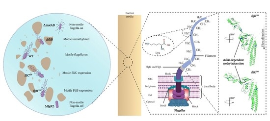
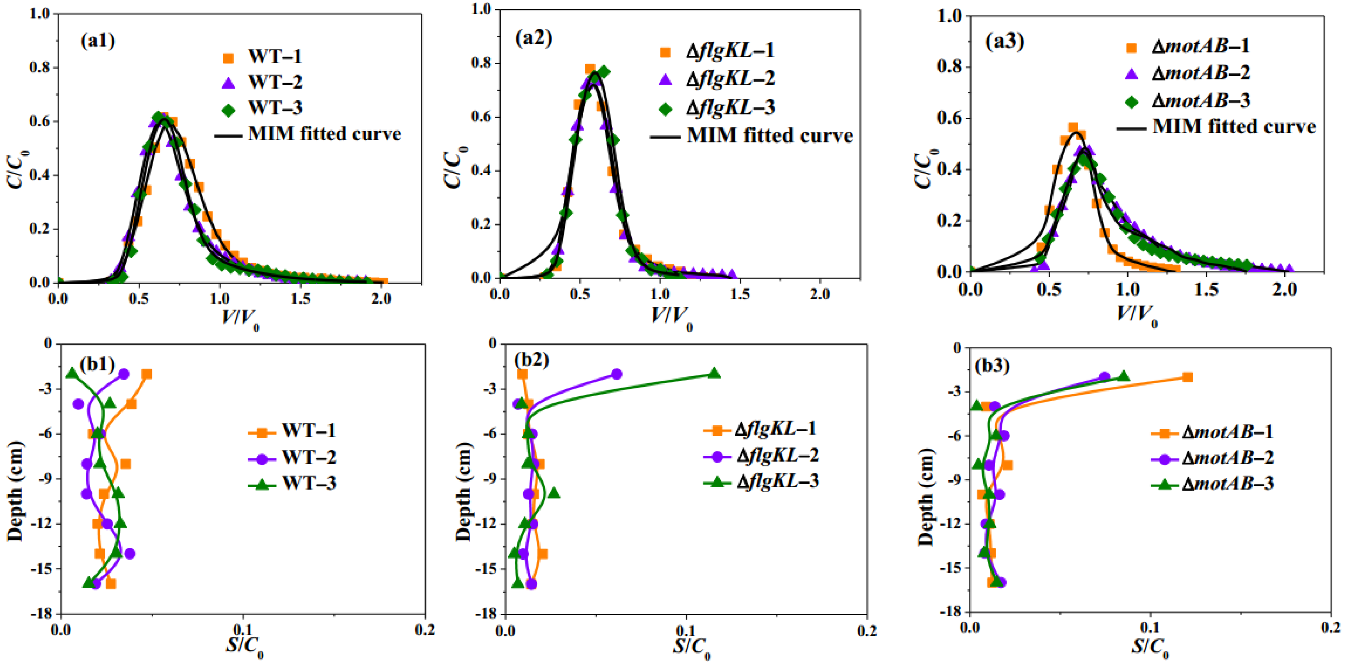
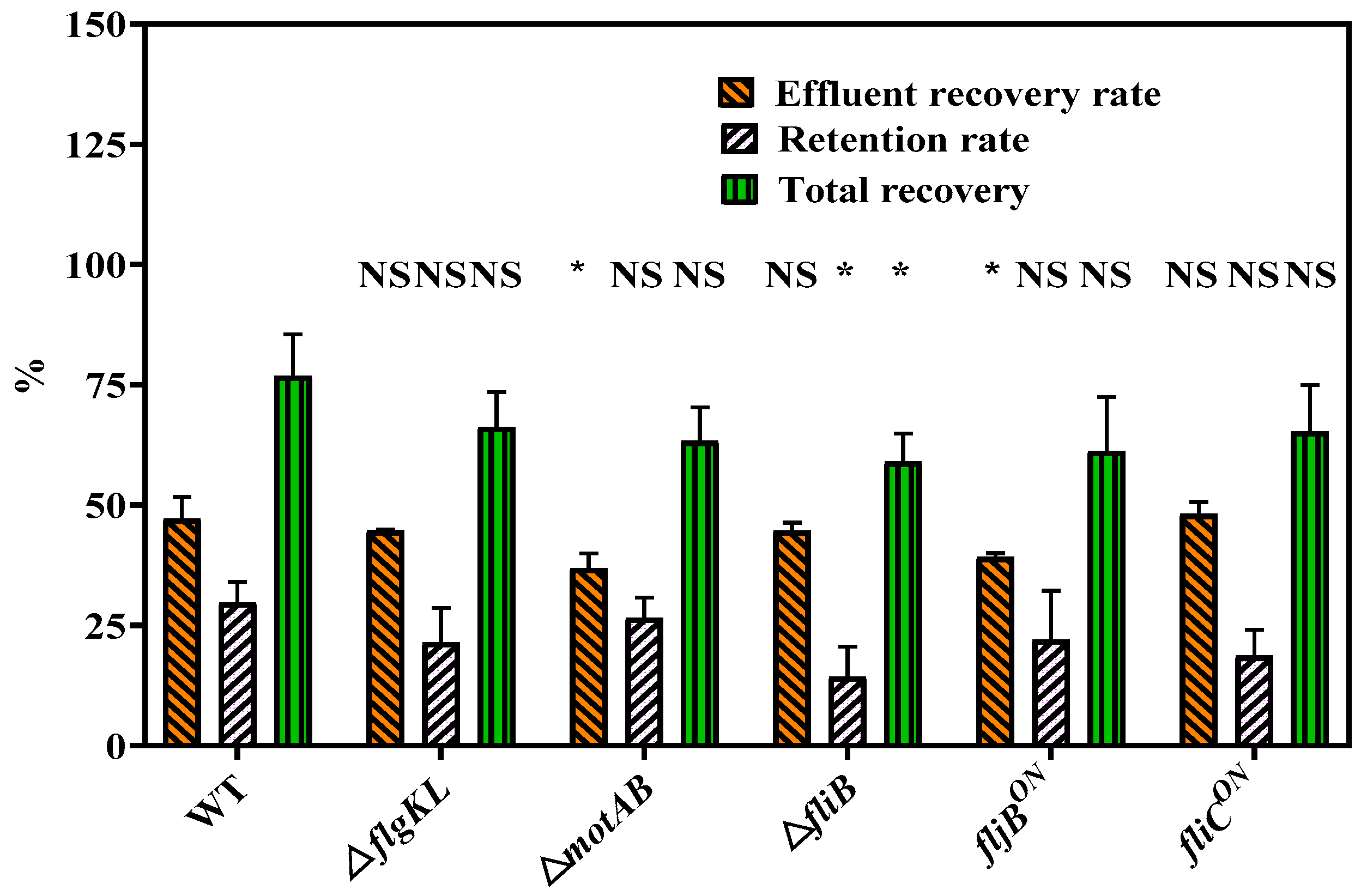
2.1. Flagella and Their Motility Impact Transport and Deposition Behavior of S. Typhimurium Strain in Sandy Porous Media

Several S. Typhimurium strains were used to investigate the importance of motile flagella on bacterial transport and deposition in porous media (Table 1): motile wild-type (WT) strain, non-motile with flagella (ΔmotAB) and non-motile without flagella (ΔflgKL). The ΔmotAB mutant is a non-motile strain without a stator, which is needed for flagella rotation [36]. Bacterial transport and deposition experiments were performed in triplicate, and a good repeatability of breakthrough curves (BTCs), as well as retention profiles (RPs), were obtained for each strain (Figure 1(a1–b3)).
The observed BTCs obtained from WT (Figure 1(a1)) and ΔmotAB (Figure 1(a3)) exhibited an asymmetrical shape with an obvious tailing, conversely to the BTC symmetrical shape of the strain without flagella (ΔflgKL, Figure 1(a2)). However, the flagellated non-motile strain, ΔmotAB, showed a more obvious tailing compared to WT. Meanwhile, some slight differences were observed between WT and ΔmotAB: an average higher peak for the WT (before 0.75 V/V0, 0.6 C/C0, Figure 1(a1)) compared to those of ΔmotAB (around 0.75 V/V0, 0.5 C/C0, Figure 1(a3)). The average mass percentage of ΔmotAB recovered, Meff, was 36.78% (Table 2), lower than WT (47.1%), indicating that bacterial motility could improve bacteria transport through sandy media. This result was consistent with the slightly higher retardation factors observed for ΔmotAB (average retardation factor, 0.91, Table 2) compared to the WT (0.86, Table 2). The overall bacteria mass recovery (Mtotal) was subsequently determined as the sum of Meff and Mretained. However, different bacteria retention rates in the sand, Mretained, were obtained for WT (29.7%) and ΔmotAB (26.5%) strains, and higher Mtotal were obtained for WT (76.8%) in comparison to ΔmotAB (63.2%) (Table 2) due to the higher recovery of WT strain in the effluent. The observed RPs of ΔmotAB showed a different behavior compared to WT, especially the bacteria attached to the column inlet (Figure 1(b1,b3)). All replicates of ΔmotAB showed a non-monotonic distribution profile behavior with a higher number of attached bacteria (S/C0) at the column inlet (layer 0–2 cm). Conversely, the monotonic behavior of WT exhibited no significant difference in retention rate among all layers.
The BTCs of WT and ΔmotAB were well fitted (Figure 1(a1,a3)) with R2 > 0.98 (Table 3) using the MIM model. The dispersivity λ and mobile water fraction θm/θ could reflect the flow patterns in porous media; lower λ and higher θm/θ show a more uniform, thus less preferential flow. The dispersivity of WT was 0.49 cm while for ΔmotAB was 0.38 cm, indicating that WT had more dispersive flow pathways. Slightly lower mobile water fractions θm/θ were obtained for WT (53%) compared to those of ΔmotAB (56%) (Table 3), indicating that lower pore water volumes were required for WT transport compared to those of ΔmotAB. These results showed a different flow pattern between flagellated motile and non-motile strains; the non-motile strain tended to have more homogeneous transport in porous media than the motile strain. The same order of magnitude of the attachment coefficient katt was obtained for WT and ΔmotAB on the sandy columns (Table 3). The same tendency was also observed for the detachment coefficient, kd. The kd values obtained for both mutants were greater than katt values (Table 3), indicating that attachment was approaching linear equilibrium sorption conditions.
To examine the importance of flagella presence in transport behavior, two non-motile bacteria were compared: ΔflgKL (without flagella) and ΔmotAB (with flagella). The observed BTCs obtained from ΔflgKL occurred earlier than for ΔmotAB (Figure 1(a2,a3)). Average earlier breakthrough (0.4 V/V0) for ΔflgKL was obtained compared to ΔmotAB (0.5 V/V0). An average higher peak (around 0.75 C/C0) for ΔflgKL was observed compared to ΔmotAB (around 0.6 C/C0). Earlier breakthroughs and higher peaks for ΔflgKL were consistent with lower average retardation factors of this strain (0.66, Table 2) compared to ΔmotAB (0.91).
Similar overall bacteria mass recovery (Mtotal) was obtained for both mutants with (63.2% for ΔmotAB) or without flagella (68.0% for ΔflgKL). However, the Meff recovery in the effluent of ΔflgKL was higher than for ΔmotAB (44.7% vs. 36.8%), indicating that the flagella presence may reduce bacterial transport. In addition, ΔflgKL exhibited a lower number of bacteria retained in the sand compared to ΔmotAB (Mretained of 23.3% for ΔflgKL and of 26.5% for ΔmotAB, Table 2). The observed RPs of both ΔflgKL and ΔmotAB strains showed similar non-monotonic retention profile behavior (Figure 1b2,b3). Thus, ΔflgKL and ΔmotAB strains showed a higher number of attached bacteria at the column inlet (Figure 1(b2,b3), layer 0–2 cm). This phenomenon indicated that the bacteria were retained mostly in the first layer of the column and displayed the most important step for blocking bacteria contamination.
ΔflgKL had lower mean dispersivity λ (0.28 cm) but higher mobile water fraction θm/θ (62%) compared with ΔmotAB (56%). This phenomenon could be attributed to the difference in flagella presence, meaning that the bacteria with flagella could have a less uniform and more preferential transport in porous media. The mean value of the attachment coefficient katt reached 0.06 min−1 for ΔflgKL, slightly lower than ΔmotAB (0.09 min−1). It showed that ΔflgKL is less adhering to sand than ΔmotAB, a result in agreement with experimental observations. ΔflgKL also had a higher detachment coefficient kd (0.57 min−1) than ΔmotAB (0.40 min−1), indicating a higher possibility of being reversibly detached from the solid grains.

2.2. Flagella Phases Influence Transport and Deposition Behavior of Bacteria in Porous Media

The effect of different flagellins of S. typhimurium, here with fliCON and fljBON strains, was investigated to observe their role in transport and deposition in porous media. We used two mutants that are locked in FliC or FljB, respectively, with different positions and orientations of flagellins. When the flagellum is composed of FliC flagellin, it plays an important role during Salmonella infection in mice and humans [37].
The observed BTCs of fliCON and fljBON strains (Figure 2(a2,a3)) showed similar arrival breakthroughs for both strains. However, slightly higher average C/C0 peaks were observed for fliCON compared to those of fljBON. Thus, the average peak C/C0 value of the fliCON strain was about 0.7, while the fljBON strain was around 0.6. The baseline for fliCON and fljBON strains was achieved in V/V0 1.5 and 2.0, respectively. These results showed that fliCON goes faster through the column than fljBON.
The overall bacteria mass recovery (Mtotal) reached 61.2% for fljBON and 66.6% for the fliCON strain. Even though both strains exhibited slightly similar retardation factors (0.78 for fljBON and 0.74 for fliCON), the fljBON strain had a lower percentage of recovery from the effluent than the fliCON strain (39.3% vs. 48.1%, respectively, Table 2, Figure 3). In addition, the fljBON strain was more retained on the sandy grains (Mretained of 21.9% vs. 18.6%, respectively).
Thus, higher recovery in the effluent and lower retention on the sand for the fliCON compared to the fljBON strain were in agreement with faster transport of the fliCON strain observed from BTCs data. Based on the RPs (Figure 2(b2,b3)), the two mutants also showed that the bacteria were more easily attached to the column inlet but with a greater efficacity for fljBON. For the fliCON and fljBON strains, the fliCON strain had a little lower dispersivity λ but the same mobile water fraction θm/θ of 54% as the fljBON strain. It seemed that there was no obvious difference between these two strains for the transport pattern. The fliCON strain had lower Kstr than the fljBON strain.

2.3. Effects of Flagella Methylation on Transport and Deposition of Bacteria in Porous Media

The posttranslational methylation of flagellin via fliB plays an important role in the invasion of host cells [16]. It should be noted that the BTC behavior of the ΔfliB strain replicates (Figure 2(a1)), and the average peaks, as well as BTCs arrival, were different from those obtained for WT (Figure 1(a1)).
The average retardation factor for the ΔfliB strain was 0.93 and 0.86 for WT (Table 2), indicating a delay for BTCs of ΔfliB. Lower bacteria effluent recovery (Meff) was obtained for ΔfliB (44.7%) compared to WT (47.1%). Meanwhile, ΔfliB presented a much lower retention rate (14.3%) in the sand in comparison to WT (29.7%) (Table 2).
According to the RPs (Figure 1(b1) and Figure 2(b1)), the ΔfliB strain was more easily attached to the first layer of the column inlet than WT. The ΔfliB strain had lower λ (dispersivity of 0.26 cm, Table 3) and higher mobile water fraction θm/θ (58%) compared to WT (53%), fliCON (54%), and fljBON strains (54%). Based on these findings, bacteria without flagellin methylation have more uniform and less preferential transport. ΔfliB had a higher attachment coefficient katt (0.18) than WT (0.09), fliCON (0.13), and fljBON (0.12), which showed that the ΔfliB strain was more likely to be captured by physicochemical mechanisms in the sand than by physical straining. It should be noted that parameters obtained from numerical simulations remained mostly in the same order of magnitude; however, their comparison between various mutants of the same strain allows us to observe some tendencies related to their specific biological properties.

3. Discussion

The prokaryotic flagellum is an extraordinary multi-subunit organelle, complex in its regulation and assembly, best known as a motility organelle responsible for bacterial movement, necessary for chemotaxis, and involved in biofilm formation [31,38,39]. When bacteria are individual cells in liquid, the rotation of their flagella generates a swimming motion. Conversely, bacteria, when immobilized on surfaces, form a biofilm, leading to higher resistance to antimicrobials and long-term colonization [40].
In our results, it should be noted that the total recovery rate of all bacteria was lower than 100%, suggesting that S. Typhimurium was strongly attached to the sand or the column and was starting biofilm formation. Furthermore, bacteria could reach crevices formed by the grains of sand via their flagella, leading to a better attachment [41] and to a lower recovery rate from the column. Similar results were obtained at a low flow rate by others [42]. They found that non-motile E. coli strains had higher retention time than motile E. coli strains in homogeneously packed sand column experiments. It showed that bacterial motility could be a factor in preventing bacterial attachment to solid surfaces. However, the BTCs of non-motile Pseudomonas fluorescens strains were found to be the first to emerge compared to motile bacteria at low water velocity [43]. Unfortunately, no data was available concerning the nature of the mutation present in the non-motile strain of this work.
A lower average retardation factor for the ΔflgKL mutant was found compared to the ΔmotAB mutant (Table 2). This may be due to the absence of flagella for ΔflgKL. It may have reduced the possibility of interaction with sandy grains, thus promoting the preferential transport of this strain through the porous media. The non-motile ΔmotAB mutant has flagella, and they could be attached to the surface of the sand more easily. It is harder for the strain without flagella to be captured by the sandy grains. This result was in agreement with low retardation factors of ΔflgKL, indicating preferential transport and low retention in the porous media. Similar results were observed for Azotobacter vinelandii [44]. Flagellated strain had a higher deposition rate than a non-flagellated one on a quartz surface in a radial stagnation point flow cell setup [44]. Combined with the result of this study, it could be seen that flagella promote bacteria adsorption to different surfaces on different experimental scales, and flagellated bacteria have more probability of sticking on porous media.
WT had higher recovery from effluent than the ΔmotAB mutant. WT had a more preferential transport in porous media than the ΔmotAB mutant, which may explain the high mass recovery in the effluent observed. These results agree with the existing literature, suggesting a linear relationship between preferential transport and mass recovery in the effluent [10]. Due to its motility, WT is not only highly recovered in the effluent but also more retained in the sand compared to ΔmotAB when it is in contact with the sandy grains (Table 2), in agreement with what has been reported in the literature. Thus, Zhang et al. (2021) [21] observed motile and non-motile E. coli ΔfliC strains and found that the motile strain was deposited more easily onto a quartz sand surface, grain-to-grain contacts, and narrow flow channels. It showed that the deposition of non-motile bacteria would be reduced in sand and water environments. The motile bacteria could be transported faster than bacteria without motility by providing a driving force for bacterial transport, which emphasized the importance of motility to bacteria during their movement in porous media. Different conclusions have been reported regarding the impact of cell motility on bacteria transport. Some authors suggested that the ability of the cell to swim is an important factor that enhances transport [45]. Others reported lower mass recovery of the motile strains compared to the non-motile ones. They suggested that small pores of the porous medium increase the possibility of the non-motile bacteria being trapped, causing a low recovery rate [10]. However, it should be noted that these results have been observed for different species: the motile E. coli and the non-flagellated Klebsiella sp., which renders it difficult for comparison, while in this work, different mutants of the same strain were investigated, giving a more accurate comparison between different experiments.
WT and ΔmotAB mutants showed different behaviors in the observed RPs. The difference between the RPs of these two strains might be explained by bacterial near-surface swimming. When bacteria swim next to surfaces, it increases the possibility of being retained onto them. Therefore, S. Typhimurium flagellar motility is needed for reaching the host cell surface but is influenced by physical forces during “near-surface swimming” by increasing local bacterial density [46]. Furthermore, E. coli is known to have near-surface swimming on solid surfaces and to have three swimming modes: landing, near-surface swimming, and swimming away; the three processes require different dynamic conditions [47]. Thus, in this work, S. Typhimurium WT via near-surface swimming on the sand surface could keep an equilibrium among these three modes to maintain stable bacterial retention in each layer, while the non-motile strains are mostly captured by the sand surface of the first layer without flagella dynamic to swim away. Likewise, the ΔmotAB mutant showed a delay in BTCs compared with WT. In this case, the non-motile flagella can add a physical constraint because they are most likely spreading to all directions around the bacterial body, as observed during the tumbling phase. Thus, the bacterial cells are occupying more space, leading to a potential increase in the residence time in the column.
The fliCON mutant had faster transport in BTCs, higher recovery in the effluent, and a lower retention rate compared to the fljBON strain. Furthermore, experiments performed by others have highlighted structural differences between fljBON and fliCON strains [48]. They found that FliC had a higher density in the D3 domain compared to FljB. This allows FljB to be more flexible and motile under high-viscosity conditions. Also, the fljBON mutant has been found to stay longer because of near-surface swimming than the fliCON mutant [24]. This observation can also explain why FliC promotes bacteria transport in porous media. It should be noted that the replicates BTCs of fljBON showed better repeatability than fliCON, probably reflecting the motility advantage of fliCON compared to fljBON described in previous studies. FliC flagella modulate the swimming behavior on the surface to facilitate the search of invasion sites which can be influenced by the column packing [24].
The ΔfliB mutant presented around half of the retention rate in the RPs in comparison to WT. It may probably be because the ΔfliB filament without methylation has lower hydrophobicity, causing lower attachment to sand. Hydrophobicity has been found to be a driving force in bacteria retention in porous media. Thus, a study involving Campylobacter jejuni and E. coli cells has demonstrated that the more hydrophobic C. jejuni had more attachment to porous media than E. coli. However, it should be noted that hydrophobicity’s impact on bacteria transport and deposition has mainly been studied between two very different bacterial species, making it difficult to reach an accurate conclusion [49]. Recently, the flagella methylation of S. Typhimurium has been found to adhere to hydrophobic surfaces of epithelial cells [16]. As observed in this work, when the hydrophobicity is high, the retention is higher.
The findings of this work could be useful to improve the risk assessment process of bacterial contamination in soil and groundwater. The pathogen concentrations and the weather conditions have already been used with hydrodynamic modeling to estimate the risk for public health [50,51,52]. Here, we observe that biological cell motility plays a clear role in the capacity of pathogens to reach groundwater. For that reason, we suggest that microbial motility should be studied on putative soil contaminants and added to risk assessment models.

4. Materials and Methods

4.1. Bacterial Strains

The bacteria used in this study were all in S. Typhimurium SL1344 background and obtained as described here [16,24]. The different flagellar phenotypes used were: wild-type strain (WT), non-motile without flagella (ΔflgKL) [26], non-motile with flagella (ΔmotAB) [36], motile with flagella unmethylated on lysine residues (ΔfliB), motile expressing only the flagellin FliC (fliCON) and motile expressing one flagellin FljB (fljBON) [16] (Table 1) FljB and FliC have different positions and orientations of flagellin in filament and FljB showed higher motility than bacteria expressing FliC under high viscosity [48]. S. Typhimurium have peritrichous flagella distributed all over the cells [36]. The structure of flagella includes the basal body, the hook, and the filament (Figure 4).
Bacteria were inoculated on a lysogeny broth (LB) agar (35 g/L) Petri dish overnight at 37 °C, then bacteria strains were grown at 37 °C, 85 rpm overnight in LB (20 g/L). The growth curves of bacteria were measured by absorbance at OD600. Bacterial suspension harvested from the stationary phase of bacterial culture was used in this work. Batch experiments [53] were conducted to understand whether there was a change in bacterial concentration during column transport experiments in the absence of hydrodynamic flow conditions. Next, 250 mL conical flasks with 30 g Fontainebleau sand, 8 mL 0.1 mmol/L NaCl solution, and 2.5 mL bacteria suspensions were put on a shaker (85 rpm, 21 °C) for 45 min. The time for shaking was the same as the duration of the column transport experiments. The initial and final bacterial concentrations were determined by the number of colony-forming units (CFU) using the plating method [54] on LB agar. The result showed that there was not an obvious change in bacteria amount during the experiment. Accession numbers of FljB and FliC flagellin were PDB: 6 RGV and 1 IO1, respectively.

4.2. Column Transport Experiments

Transport experiments were performed in Plexiglass columns with an inner diameter of 3.4 cm and a length of 16 cm, packed with Fontainebleau sand. The particle size of Fontainebleau sand ranged from 0.16 to 0.79 mm, with a median diameter size of 0.36 mm. Two filter papers were used at the inlet and outlet of the column to make sure the solution came out without fine sand particles during the transport experiment. Before every experiment, the pump, tubes, and other column components were sterilized with bleach and 15 min of UV. Other materials were sterilized using an autoclave. The empty column with pipes, two caps, and two filter papers was weighted, and the mass was noted as M0 (g). The sand was inserted in successive layers into the column to assure homogenous distribution, and both column and sand were weighted and noted as M1 (g). The mass of sand was estimated as:
M s = M 1 M 0
and the sand bulk density (ρb) was calculated by dividing the dry mass of the sand(Ms) by the volume of the column (Vc):
ρ b = M S / V c
The porosity ( ε ) was then estimated by the sand bulk density as follows:
ε = ( 1 ρ b / 2.65 ) × 100 %
The column was flushed upward using a background solution of NaCl (0.1 mmol/L), then the column was rinsed using the background solution reversed, and at the same time, the conductivity of the effluent solution was continuously measured at the column outlet until this parameter reached a stable value. Then, the column fully saturated with the background solution was weighed, and this mass was noted as M2 (g). Assuming that water density is 1 g/cm3, the volume occupied by water (Vw) was estimated as:
V w = M 2 M 1
The saturation degree (S) could then be described as follows:
S = V w / V p
here Vp is the volume of the pores, calculated as
V p = V c × ε
For bacteria injection experiments, 20 mL of bacterial suspension was injected into each column at the initial concentration given in Table 2 for each replicate (Figure 5). The breakthrough curve of each strain was obtained by plotting bacterial concentration as a function of pore volume V/V0. Bacteria concentration was deduced by measuring the absorbance of effluent at OD600 at the column outlet. The bacterial suspension was flushed with a background solution (0.1 mmol/L NaCl solution) until the absorbance of the effluent returned to the baseline level. The ratio of bacteria mass recovered from the effluent (Meff) and retardation factor (R) was then calculated [55,56]. The retardation factor was calculated by the residence time of bacteria to the theoretical residence time of water in the column. Meff corresponds to the ratio of the bacteria mass recovered at the column outlet to their mass injected at the column inlet. The retardation factor represents the ratio of the resident time of bacteria to the theoretical water resident time.
After bacterial transport experiments, the sandy porous media inside each column was divided into 8 layers to explore the spatial distribution of the bacteria. First, a sterilized spoon was used to remove the sand into 8 flasks containing 0.1 mmol/L NaCl solution. The same solution was also used to wash the remaining sand off of the spoon. Then, we shook the flasks filled with sandy porous media for 15 min using an orbital shaker to isolate the retained bacteria from the sand. The Plating method count [54] was employed to determine the bacterial concentration in each layer of sand. The mass of bacteria recovered from all layers was regarded as Mretained, which was calculated by the total number of CFU retained in the sand divided by the CFU of the initial bacterial suspension injected into the column. The Mtotal is the sum of Meff and Mretained.

4.3. Simulation

The same modified mobile-immobile (MIM) model under two kinetic deposition sites, as described by Bai et al. (2016) [27], was used in this work. The two-region MIM model assumes that bacteria transport is limited to the mobile water region and that water in the immobile region is stagnant. Briefly, the one-dimensional bacterial transport model was described as [9]:
θ C t + ρ s 1 t   + ρ s 2 t = x ( θ m D C x ) q C x
where θ is the total water content, C is the bacterial concentration in the liquid phase (CFU/cm3), t is time (min), ρ is the bulk density of the porous media (g/cm3), s1 is the concentration of bacteria in the solid phase accounting for attachment or detachment, s2 is the concentration of bacteria in the solid phase related to irreversible straining, x is the distance (L), θm is the mobile water content, D is the dispersion coefficient (cm2/min), and q is the Darcy velocity (cm/min). It was assumed that there was no bacterial exchange between the mobile and immobile regions. Then the bacterial mass transfer between solution and s1, s2 sites can be described as [57]:
ρ S t = ρ ( s 1 + s 2 ) t = θ m ψ t k a t t C k d ρ s 1 + θ m k s t r ψ x C
where katt, kd, kstr are the first-order attachment, detachment, and straining coefficients, respectively (1/min); ψt and ψx describe the deposition of bacteria in the solid retention under time- and depth-dependent conditions, respectively. Another hydrodynamic parameter is λ (cm) which describes the dispersivity of the medium and can be estimated by [56]:
λ = D m θ m q
where Dm is the dispersion coefficient of the mobile region (cm2/min).
Transport (θm, λ) and deposition parameters (katt, kd, kstr) were obtained by fitting of theoretical MIM model, implemented in Hydrus 1D code (software version: Hydrus-1D_4.17, free in https://www.pc-progress.com/en/Default.aspx?Downloads (accessed on 7 June 2021)) to the experimental breakthrough curves of bacteria.

4.4. Statistical Analysis

To determine whether flagellar phenotypes affect bacterial transport and deposition in porous media, the replicates for each strain were analyzed with WT for significance using Student’s t-test.

5. Conclusions

In this study, the effects of S. Typhimurium flagellar phenotypes on bacterial transport and deposition in sandy porous media were explored.
Flagella affected S. Typhimurium transport and deposition in porous media. S. Typhimurium strain with non-motile flagella reduced bacterial transport and increased bacterial retention in comparison to the strain without flagella. Flagella motility promotes bacterial transport, and this was confirmed by the higher retardation factor of the BTCs and the higher recovery of effluent of motile flagellated strains. As observed during host-pathogen interaction studies, flagella phases of S. Typhimurium have a different influence on bacterial movement in porous media. The fliCON strain showed a faster transport and higher recovery in the effluent than the fljBON strain, but the FljB flagellin promoted bacterial adhesion. Flagella methylation influenced S. Typhimurium transport and deposition in porous media, probably because of the higher hydrophobicity of the flagella filament’s outer surface.
Our results will lead to more robust transport models and could help to develop new environmental strategies through a more accurate risk assessment process.

Author Contributions

Conceptualization, resources, and supervision, E.L. and Y.R.; methodology, E.L., Y.R. and H.B.; software, X.Z. and H.B.; validation and data curation, X.Z., E.L. and Y.R.; formal analysis, X.Z. and Y.T.; investigation, X.Z.; writing—original draft preparation, X.Z.; writing—review and editing, E.L. and Y.R.; visualization, M.A. All authors have read and agreed to the published version of the manuscript.

Funding

This research was funded by China Scholarship Council, National Natural Science Foundation of China (grant number 41807120), Science and Technology Foundation of Henan Province (grant number 212102310026), and Cultivation Plan for Young Core Teachers in Universities of Henan Province (grant number 2021GGJS-063).

Institutional Review Board Statement

Not applicable.

Informed Consent Statement

Not applicable.

Data Availability Statement

All supporting data of the study are available from the corresponding authors upon request.

Acknowledgments

We thank Marc Erhardt and Caroline Kühne for Salmonella enterica serovar Typhimurium strain SL1344 and its isogenic mutants.

Conflicts of Interest

The authors declare no conflict of interest.

References

  1. WWAP; UN-Water. World Water Development Report, Nature-Based Solutions for Water; UNESCO: Paris, France, 2018. [Google Scholar]
  2. Clemens, M.; Khurelbaatar, G.; Merz, R.; Siebert, C.; van Afferden, M.; Rödiger, T. Groundwater protection under water scarcity; from regional risk assessment to local wastewater treatment solutions in Jordan. Sci. Total Environ. 2020, 706, 136066. [Google Scholar] [CrossRef] [PubMed]
  3. Chrysikopoulos, C.V.; Sim, Y. One-dimensional virus transport in homogeneous porous media with time-dependent distribution coefficient. J. Hydrol. 1996, 185, 199–219. [Google Scholar] [CrossRef]
  4. Holden, N.; Pritchard, L.; Toth, I. Colonization outwith the colon: Plants as an alternative environmental reservoir for human pathogenic enterobacteria. FEMS Microbiol. Rev. 2009, 33, 689–703. [Google Scholar] [CrossRef] [PubMed]
  5. Liu, H.; Whitehouse, C.A.; Li, B. Presence and persistence of Salmonella in water: The impact on microbial quality of water and food safety. Front. Public Health 2018, 6, 159. [Google Scholar] [CrossRef] [PubMed]
  6. Abulreesh, H.H. Salmonellae in the environment. In Salmonella-Distribution, Adaptation, Control Measures and Molecular Technologies; Annous, B., Gurtler, J., Eds.; InTech: Rijeka, Croatia, 2012; pp. 19–50. [Google Scholar]
  7. Ma, H.; Bolster, C.; Johnson, W.P.; Li, K.; Pazmino, E.; Camacho, K.M.; Anselmo, A.C.; Mitragotri, S. Coupled influences of particle shape, surface property and flow hydrodynamics on rod-shaped colloid transport in porous media. J. Colloid. Interf. Sci. 2020, 577, 471–480. [Google Scholar] [CrossRef] [PubMed]
  8. Pang, L.; McLeod, M.; Aislabie, J.; Šimůnek, J.; Close, M.; Hector, R. Modeling transport of microbes in ten undisturbed soils under effluent irrigation. Vadose Zone J. 2008, 7, 97–111. [Google Scholar] [CrossRef] [Green Version]
  9. Safadoust, A.; Mahboubi, A.A.; Mosaddeghi, M.R.; Gharabaghi, B.; Unc, A.; Voroney, P.; Heydari, A. Effect of regenerated soil structure on unsaturated transport of Escherichia coli and bromide. J. Hydrol. 2012, 430–431, 80–90. [Google Scholar] [CrossRef]
  10. Bai, H.; Cochet, N.; Drelich, A.; Pauss, A.; Lamy, E. Comparison of transport between two bacteria in saturated porous media with distinct pore size distribution. RSC Adv. 2016, 6, 14602–14614. [Google Scholar] [CrossRef]
  11. Ma, H.; Johnson, W.P. Colloid retention in porous media of various porosities: Predictions by the hemispheres-in-cell model. Langmuir 2010, 26, 1680–1687. [Google Scholar] [CrossRef]
  12. Prédélus, D.; Lassabatere, L.; Louis, C.; Gehan, H.; Brichart, T.; Winiarski, T.; Angulo-Jaramillo, R. Nanoparticle Transport in water-unsaturated porous media: Effects of solution ionic strength and flow rate. J. Nanopart. Res. 2017, 19, 104. [Google Scholar] [CrossRef]
  13. Yang, L.; Kang, J.; Chen, X.; Ripp, S.A.; Johnson, W.P.; Zhuang, J. Real-time bioluminescent imaging of spatiotemporal variation of microbial retention during transport through porous media under variably saturated flow conditions. J. Hydrol. 2021, 601, 126603. [Google Scholar] [CrossRef]
  14. Torkzaban, S.; Tazehkand, S.S.; Walker, S.L.; Bradford, S.A. Transport and fate of bacteria in porous media: Coupled effects of chemical conditions and pore space geometry. Water Resour. Res. 2008, 44, W04403. [Google Scholar] [CrossRef]
  15. Weiss, T.H.; Mills, A.L.; Hornberger, G.M.; Herman, J.S. Effect of bacterial cell shape on transport of bacteria in porous media. Environ. Sci. Technol. 1995, 29, 1737–1740. [Google Scholar] [CrossRef] [PubMed]
  16. Horstmann, J.A.; Lunelli, M.; Cazzola, H.; Heidemann, J.; Kühne, C.; Steffen, P.; Szefs, S.; Rossi, C.; Lokareddy, R.K.; Wang, C.; et al. Methylation of Salmonella Typhimurium flagella promotes bacterial adhesion and host cell invasion. Nat. Commun. 2020, 11, 2013. [Google Scholar] [CrossRef] [Green Version]
  17. Becker, M.W.; Collins, S.A.; Metge, D.W.; Harvey, R.W.; Shapiro, A.M. Effect of cell physicochemical characteristics and motility on bacterial transport in groundwater. J. Contam. Hydrol. 2004, 69, 195–213. [Google Scholar] [CrossRef] [PubMed]
  18. Josenhans, C.; Suerbaum, S. The role of motility as a virulence factor in bacteria. Int. J. Med. Microbiol. 2002, 291, 605–614. [Google Scholar] [CrossRef]
  19. Chen, S.; Beeby, M.; Murphy, G.E.; Leadbetter, J.R.; Hendrixson, D.R.; Briegel, A.; Li, Z.; Shi, J.; Tocheva, E.I.; Müller, A.; et al. Structural diversity of bacterial flagellar motors. EMBO J. 2011, 30, 2972–2981. [Google Scholar] [CrossRef] [Green Version]
  20. Vladimirov, N.; Sourjik, V. Chemotaxis: How bacteria use memory. Biol. Chem. 2009, 390, 1097–1104. [Google Scholar] [CrossRef]
  21. Zhang, M.; He, L.; Jin, X.; Bai, F.; Tong, M.; Ni, J. Flagella and their properties affect the transport and deposition behaviors of Escherichia coli in quartz sand. Environ. Sci. Technol. 2021, 55, 4964–4973. [Google Scholar] [CrossRef]
  22. Haznedaroglu, B.Z.; Zorlu, O.; Hill, J.E.; Walker, S.L. Identifying the role of flagella in the transport of motile and nonmotile Salmonella enterica Serovars. Environ. Sci. Technol. 2010, 44, 4184–4190. [Google Scholar] [CrossRef]
  23. Lederberg, J.; Iino, T. Phase Variation in Salmonella. Genetics 1956, 41, 743–757. [Google Scholar] [CrossRef] [PubMed]
  24. Horstmann, J.A.; Zschieschang, E.; Truschel, T.; de Diego, J.; Lunelli, M.; Rohde, M.; May, T.; Strowig, T.; Stradal, T.; Kolbe, M.; et al. Flagellin phase-dependent swimming on epithelial cell surfaces contributes to productive Salmonella gut colonisation. Cell Microbiol. 2017, 19, e12739. [Google Scholar] [CrossRef] [PubMed] [Green Version]
  25. Ambler, R.P.; Rees, M.W. ε-N-Methyl-Lysine in Bacterial Flagellar Protein. Nature 1959, 184, 56–57. [Google Scholar] [CrossRef]
  26. Laasik, E.; Põllumaa, L.; Pasanen, M.; Mattinen, L.; Pirhonen, M.; Mäe, A. Expression of NipP.w of Pectobacterium wasabiae is dependent on functional FlgKL flagellar genes. Microbiology 2014, 160, 179–186. [Google Scholar] [CrossRef] [PubMed] [Green Version]
  27. Bai, H.; Cochet, N.; Pauss, A.; Lamy, E. Bacteria cell properties and grain size impact on bacteria transport and deposition in porous media. Colloids Surf. B Biointerf. 2016, 139, 148–155. [Google Scholar] [CrossRef] [PubMed]
  28. Bai, H.; Cochet, N.; Pauss, A.; Lamy, E. DLVO, Hydrophobic, capillary and hydrodynamic forces acting on bacteria at solid-air-water interfaces: Their relative impact on bacteria deposition mechanisms in unsaturated porous media. Colloids Surf. B Biointerf. 2017, 150, 41–49. [Google Scholar] [CrossRef]
  29. Friedlander, R.S.; Vogel, N.; Aizenberg, J. Role of flagella in adhesion of Escherichia coli to abiotic surfaces. Langmuir 2015, 31, 6137–6144. [Google Scholar] [CrossRef]
  30. Cazzola, H.; Lemaire, L.; Acket, S.; Prost, E.; Duma, L.; Erhardt, M.; Čechová, P.; Trouillas, P.; Mohareb, F.; Rossi, C.; et al. The impact of plasma membrane lipid composition on flagellum-mediated adhesion of enterohemorrhagic Escherichia coli. mSphere 2020, 5, e00702-20. [Google Scholar] [CrossRef]
  31. Rossez, Y.; Wolfson, E.B.; Holmes, A.; Gally, D.L.; Holden, N.J. Bacterial flagella: Twist and stick, or dodge across the kingdoms. PLoS Pathog. 2015, 11, e1004483. [Google Scholar] [CrossRef]
  32. Rossez, Y.; Holmes, A.; Wolfson, E.B.; Gally, D.L.; Mahajan, A.; Pedersen, H.L.; Willats, W.G.T.; Toth, I.K.; Holden, N.J. Flagella interact with ionic plant lipids to mediate adherence of pathogenic Escherichia coli to fresh produce plants. Environ. Microbiol. 2014, 16, 2181–2195. [Google Scholar] [CrossRef]
  33. Jindai, K.; Nakade, K.; Masuda, K.; Sagawa, T.; Kojima, H.; Shimizu, T.; Shingubara, S.; Ito, T. Adhesion and bactericidal properties of nanostructured surfaces dependent on bacterial motility. RSC Adv. 2020, 10, 5673–5680. [Google Scholar] [CrossRef] [PubMed] [Green Version]
  34. Dewangan, N.K.; Conrad, J.C. Bacterial motility enhances adhesion to oil droplets. Soft Matter 2020, 16, 8237–8244. [Google Scholar] [CrossRef] [PubMed]
  35. Zheng, S.; Bawazir, M.; Dhall, A.; Kim, H.-E.; He, L.; Heo, J.; Hwang, G. Implication of surface properties, bacterial motility, and hydrodynamic conditions on bacterial surface sensing and their initial adhesion. Front. Bioeng. Biotechnol. 2021, 9, 643722. [Google Scholar] [CrossRef]
  36. Chevance, F.F.V.; Hughes, K.T. Coordinating assembly of a bacterial macromolecular machine. Nat. Rev. Microbiol. 2008, 6, 455–465. [Google Scholar] [CrossRef] [PubMed] [Green Version]
  37. Cummings, L.A.; Wilkerson, W.D.; Bergsbaken, T.; Cookson, B.T. In Vivo, FliC expression by Salmonella enterica Serovar Typhimurium is heterogeneous, regulated by ClpX, and anatomically restricted. Mol. Microbiol. 2006, 61, 795–809. [Google Scholar] [CrossRef]
  38. Chaban, B.; Hughes, H.V.; Beeby, M. The flagellum in bacterial pathogens: For motility and a whole lot more. Semin. Cell Dev. Biol. 2015, 46, 91–103. [Google Scholar] [CrossRef] [Green Version]
  39. Barken, K.B.; Pamp, S.J.; Yang, L.; Gjermansen, M.; Bertrand, J.J.; Klausen, M.; Givskov, M.; Whitchurch, C.B.; Engel, J.N.; Tolker-Nielsen, T. Roles of type IV pili, flagellum-mediated motility and extracellular DNA in the formation of mature multicellular structures in Pseudomonas aeruginosa biofilms. Environ. Microbiol. 2008, 10, 2331–2343. [Google Scholar] [CrossRef]
  40. Hall-Stoodley, L.; Costerton, J.W.; Stoodley, P. Bacterial biofilms: From the natural environment to infectious diseases. Nat. Rev. Microbiol. 2004, 2, 95–108. [Google Scholar] [CrossRef]
  41. Friedlander, R.S.; Vlamakis, H.; Kim, P.; Khan, M.; Kolter, R.; Aizenberg, J. Bacterial flagella explore microscale hummocks and hollows to increase adhesion. Proc. Natl. Acad. Sci. USA 2013, 110, 5624–5629. [Google Scholar] [CrossRef] [Green Version]
  42. Liu, J.; Ford, R.M.; Smith, J.A. Idling time of motile bacteria contributes to retardation and dispersion in sand porous medium. Environ. Sci. Technol. 2011, 45, 3945–3951. [Google Scholar] [CrossRef]
  43. Camper, A.K.; Hayes, J.T.; Sturman, P.J.; Jones, W.L.; Cunningham, A.B. Effects of motility and adsorption rate coefficient on transport of bacteria through saturated porous media. Appl. Environ. Microbiol. 1993, 59, 3455–3462. [Google Scholar] [CrossRef] [PubMed] [Green Version]
  44. Lu, N.; Bevard, T.; Massoudieh, A.; Zhang, C.; Dohnalkova, A.C.; Zilles, J.L.; Nguyen, T.H. Flagella-mediated differences in deposition dynamics for Azotobacter vinelandii in porous media. Environ. Sci. Technol. 2013, 47, 5162–5170. [Google Scholar] [CrossRef] [PubMed]
  45. de Kerchove, A.J.; Elimelech, M. Bacterial swimming motility enhances cell deposition and surface coverage. Environ. Sci. Technol. 2008, 42, 4371–4377. [Google Scholar] [CrossRef]
  46. Misselwitz, B.; Barrett, N.; Kreibich, S.; Vonaesch, P.; Andritschke, D.; Rout, S.; Weidner, K.; Sormaz, M.; Songhet, P.; Horvath, P.; et al. Near surface swimming of Salmonella Typhimurium explains target-site selection and cooperative invasion. PLoS Pathog. 2012, 8, 9. [Google Scholar] [CrossRef] [PubMed] [Green Version]
  47. Dunstan, J.; Miño, G.; Clement, E.; Soto, R. A Two-sphere model for bacteria swimming near solid surfaces. Phys. Fluids 2012, 24, 011901. [Google Scholar] [CrossRef] [Green Version]
  48. Yamaguchi, T.; Toma, S.; Terahara, N.; Miyata, T.; Ashihara, M.; Minamino, T.; Namba, K.; Kato, T. Structural and functional comparison of Salmonella flagellar filaments composed of FljB and FliC. Biomolecules 2020, 10, 246. [Google Scholar] [CrossRef] [PubMed] [Green Version]
  49. Bolster, C.H.; Walker, S.L.; Cook, K.L. Comparison of Escherichia coli and Campylobacter jejuni transport in saturated porous media. J. Environ. Qual. 2006, 35, 1018–1025. [Google Scholar] [CrossRef] [Green Version]
  50. Eregno, F.E.; Tryland, I.; Tjomsland, T.; Myrmel, M.; Robertson, L.; Heistad, A. Quantitative microbial risk assessment combined with hydrodynamic modelling to estimate the public health risk associated with bathing after rainfall events. Sci. Total Environ. 2016, 548–549, 270–279. [Google Scholar] [CrossRef]
  51. Tolouei, S.; Dewey, R.; Snodgrass, W.J.; Edge, T.A.; Andrews, R.C.; Taghipour, M.; Prévost, M.; Dorner, S. Assessing microbial risk through event-based pathogen loading and hydrodynamic modelling. Sci. Total Environ. 2019, 693, 133567. [Google Scholar] [CrossRef]
  52. Sokolova, E.; Petterson, S.R.; Dienus, O.; Nyström, F.; Lindgren, P.-E.; Pettersson, T.J.R. Microbial risk assessment of drinking water based on hydrodynamic modelling of pathogen concentrations in source water. Sci. Total Environ. 2015, 526, 177–186. [Google Scholar] [CrossRef]
  53. Bradford, S.A.; Simunek, J.; Walker, S.L. Transport and straining of E. coli O157:H7 in saturated porous media. Water Resour. Res. 2006, 42. [Google Scholar] [CrossRef] [Green Version]
  54. Sanders, E.R. Aseptic laboratory techniques: Plating methods. J. Vis. Exp. 2012, 63, e3064. [Google Scholar] [CrossRef] [PubMed]
  55. Sardin, M.; Schweich, D.; Leij, F.J.; van Genuchten, M.T. Modeling the nonequilibrium transport of linearly interacting solutes in porous media: A review. Water Resour. Res. 1991, 27, 2287–2307. [Google Scholar] [CrossRef]
  56. Lamy, E.; Lassabatere, L.; Bechet, B.; Andrieu, H. Effect of a nonwoven geotextile on solute and colloid transport in porous media under both saturated and unsaturated conditions. Geotext. Geomembr. 2013, 36, 55–65. [Google Scholar] [CrossRef]
  57. Bradford, S.A.; Simunek, J.; Bettahar, M.; van Genuchten, M.T.; Yates, S.R. Modeling colloid attachment, straining, and exclusion in saturated porous media. Environ. Sci. Technol. 2003, 37, 2242–2250. [Google Scholar] [CrossRef]
Figure 1. (a1a3) Observed (symbols) and fitted (lines) breakthrough curves (BTCs); (b1b3) Observed retention profiles (RPs) of S. Typhimurium WT motile flagella-on, ΔflgKL non-motile flagella-off, and ΔmotAB non-motile flagella-on mutants. V is the volume occupied by water in the column, V0 is the total pore volume of the column, and C, C0, and S are the time-dependent, initial, and solid phase concentration of bacteria respectively.
Ijms 23 14460 g001
Figure 2. (a1a3) Observed (symbols) and fitted (lines) breakthrough curves; (b1b3) Observed retention profiles of S. Typhimurium motile ΔfliB, motile fliCON, and motile fljBON mutants.
Ijms 23 14460 g002
Figure 3. The recovery rate of S. Typhimurium different strains. Replicates are shown as mean values, error bars represent standard deviations, and statistical significances were determined by the student’s t-test (* = p < 0.05; NS = not significant).
Ijms 23 14460 g003
Figure 4. Schematic representation of the structure of bacterial flagellum.
Ijms 23 14460 g004
Figure 5. Schematic of column experiments.
Ijms 23 14460 g005
Table 1. Cell properties for S. Typhimurium strains.
Flagellar PhenotypesWild TypeΔflgKLΔmotABfljBONfliCONΔfliB
EM774EM4969EM4939EM1013EM1012EM3734
Cell motilityMotileNon-motileNon-motileMotileMotileMotile
FlagellaOnOffOnOnOnOn
Table 2. Experimental conditions and mass balance information of S. Typhimurium transport experiments under saturated conditions.
Bacterial StrainsReplicateSaturated Degree (%)C Initial (CFU/mL)Porosity (%)Bulk Density (g/cm3)Pulse Time (min)Darcy Velocity (cm/min)Retardation FactorRecovery (%)
MeffMretainedMtotal
WT198.27.60 × 10837.881.652.400.9060.9451.9734.6486.61
298.51.40 × 10936.411.692.330.9360.7946.4926.9973.48
398.16.40 × 10837.981.642.370.9390.8642.8327.3570.18
(0.86) a(47.1)(29.66)(76.76)
ΔflgKL198.41.27 × 10938.921.622.201.0100.6644.4117.6962.10
298.91.23 × 10938.261.641.851.1910.6644.8322.4167.24
399.11.57 × 10937.911.652.230.9910.6544.8729.6974.56
(0.66)(44.7)(23.26)(67.97)
0.42 b0.160.18
ΔmotAB196.41.30 × 10937.681.652.320.9510.9838.7831.2069.98
297.91.73 × 10937.171.662.440.9030.9938.4525.3763.82
396.61.93 × 10937.771.652.670.8260.7533.1222.8155.93
(0.91)(36.78)(26.46)(63.24)
0.030.420.10
ΔfliB198.49.00 × 10837.921.652.360.9341.0144.820.9365.73
297.31.60 × 10935.121.722.111.0451.0446.258.3654.61
399.59.33 × 10838.061.642.220.9940.7542.8913.4856.37
(0.93)(44.65)(14.26)(58.9)
0.440.030.04
fljBON198.61.17 × 10936.841.672.071.0660.7940.4433.5874.02
299.29.00 × 10836.791.672.340.9410.7038.3417.7656.10
398.81.00 × 10936.911.672.360.9350.8438.9814.4653.44
(0.77)(39.25)(21.93)(61.18)
0.040.290.13
fliCON193.51.57 × 10936.461.682.151.0250.7251.0224.5475.56
296.01.03 × 10936.091.692.330.9450.6546.2817.5363.81
3100.01.10 × 10936.381.692.270.9720.8646.9013.7060.60
(0.74)(48.07)(18.59)(66.66)
0.770.050.2
a The values given were the mean value of the corresponding parameters, b the values given parallel were the p-values for analyzing significance with WT using t-test.
Table 3. Fitted parameters (replicates) for S. Typhimurium through saturated columns using the MIM model in HYDRUS-1D code.
Bacterial StrainsReplicateλ (cm)θmKatt (min−1)Kd (min−1)Kstr (min−1)R2
WT10.690.543.12 × 10−23.93 × 10−17.87 × 10−60.9963
(0.06) *(0.004)(1.58 × 10⁻²)(2.10 × 10⁻¹)(6.56 × 10⁻³)
20.480.511.46 × 10−16.07 × 10−18.35 × 10−20.9958
(0.07)(0.008)(1.88 × 10⁻²)(6.09 × 10⁻²)(1.25 × 10⁻¹)
30.290.528.59 × 10−24.29 × 10−11.85 × 10−10.9963
(0.03)(0.002)(1.36 × 10⁻²)(8.16 × 10⁻²)(2.49 × 10⁻²)
Mean values 0.490.530.090.480.090.996
(0.2001) **0.01660.05740.11470.09290.0003
ΔflgKL10.350.623.25 × 10−23.38 × 10−11.76 × 10−10.9987
(0.03)(0.002)(5.00 × 10⁻³)(1.17 × 10⁻¹)(6.23 × 10⁻²)
20.210.581.23 × 10−18.76 × 10−11.71 × 10−10.9968
(0.04)(0.004)(1.82 × 10⁻²)(1.71 × 10⁻¹)(8.46 × 10⁻²)
30.290.652.68 × 10−24.99 × 10−11.71 × 10−10.9948
(0.05)(0.003)(2.52 × 10⁻²)(9.80 × 10⁻¹)(6.95 × 10⁻²)
Mean values 0.280.620.060.570.170.997
0.06990.03750.05390.27610.00290.0020
ΔmotAB10.390.521.35 × 10−13.80 × 10−12.18 × 10−10.9793
(0.14)(0.03)(3.95 × 10⁻²)(7.83 × 10⁻²)(3.14 × 10⁻¹)
20.550.539.22 × 10−22.63 × 10−12.03 × 10−10.9668
(0.20)(0.04)(4.15 × 10⁻²)(1.37 × 10⁻¹)(4.68 × 10⁻¹)
30.210.644.93 × 10−25.72 × 10−14.63 × 10−10.9969
(0.02)(0.002)(5.48 × 10⁻³)(1.37 × 10⁻¹)(5.45 × 10⁻²)
Mean values 0.380.560.090.400.290.981
0.17030.06540.04280.15600.14590.0151
ΔfliB10.140.673.37 × 10−15.63 × 10−11.03 × 10−10.9823
(0.04)(0.02)(1.83 × 10⁻²)(5.74 × 10⁻²)(1.12 × 10⁻¹)
20.300.511.36 × 10−15.74 × 10−15.82 × 10−20.9852
(0.13)(0.02)(5.11 × 10⁻²)(1.33 × 10⁻¹)(3.21 × 10⁻¹)
30.340.567.50 × 10−24.42 × 10−12.00 × 10−10.9989
(0.03)(0.003)(4.58 × 10⁻³)(4.35 × 10⁻²)(5.62 × 10⁻²)
Mean values 0.260.580.180.530.120.989
0.10420.08520.13680.07320.07260.0088
fliCON10.320.652.99 × 10−26.92 × 10−11.69 × 10−40.9945
(0.06)(0.005)(3.91 × 10⁻²)(9.65 × 10⁻¹)(4.69 × 10⁻²)
20.590.402.87 × 10−18.25 × 10−11.57 × 10−10.9958
(0.16)(0.008)(1.09 × 10⁻¹)(1.78 × 10⁻¹)(3.92 × 10⁻²)
30.350.596.01 × 10−25.37 × 10−15.01 × 10−20.9988
(0.02)(0.002)(1.17 × 10⁻²)(1.06 × 10⁻¹)(1.47 × 10⁻²)
Mean values 0.420.540.130.680.070.996
0.14960.12950.14070.14410.08040.0022
fljBON10.220.511.64 × 10−17.61 × 10−13.60 × 10−10.9985
(0.02)(0.002)(2.10 × 10⁻²)(8.43 × 10⁻²)(2.04 × 10⁻²)
20.760.541.14 × 10−15.29 × 10−13.86 × 10−10.9947
(0.24)(0.02)(6.74 × 10⁻²)(2.03 × 10⁻¹)(2.07 × 10⁻¹)
30.330.597.71 × 10−26.00 × 10−12.68 × 10−10.9972
(0.04)(0.005)(6.61 × 10⁻³)(9.98 × 10⁻²)(1.00 × 10⁻¹)
Mean values 0.440.540.120.630.340.997
0.28850.04160.04360.11850.06210.0019
* The values given were the standard error coefficients (S.E.Coeff) obtained from HYDRUS-1D code, ** The values given parallel were the standard deviation.
Publisher’s Note: MDPI stays neutral with regard to jurisdictional claims in published maps and institutional affiliations.

