Article

The Role of the Exonic lncRNA PRKDC-210 in Transcription Regulation

Area of Bioscience and Biotechnology, Japan Advanced Institute of Science and Technology, 1-1 Asahidai, Nomi 923-1292, Ishikawa, Japan
*
Author to whom correspondence should be addressed.
Int. J. Mol. Sci. 2022, 23(22), 13783; https://doi.org/10.3390/ijms232213783
Submission received: 12 October 2022 / Revised: 7 November 2022 / Accepted: 7 November 2022 / Published: 9 November 2022
(This article belongs to the Special Issue RNA Regulatory Networks 2.0)

Abstract

:
In recent years, long noncoding RNAs (lncRNAs) have received increasing attention and have been reported to be associated with various genetic abnormalities. However, the functions of many lncRNAs, including those of long exonic noncoding RNAs (lencRNAs), have not yet been elucidated. Here, we used a novel tethering luciferase assay to analyze the transcriptional regulatory functions of five lencRNAs that are upregulated in cancer. We found that the lencRNA PRKDC-210 interacts with MED12, a component of the CDK8 complex, to regulate the transcription of several genes. The transcriptional activation ability of PRKDC-210 was abolished in siRNA-treated CDK8-depleted cells. We also confirmed the enrichment of PRKDC-210 on RNA polymerase II. RNA-seq analysis of cells in which PRKDC-210 or PRKDC mRNA was knocked down using antisense oligonucleotides revealed that PRKDC-210 can affect the expression levels of genes related to fatty acid metabolism. Finally, we used a ChIRP assay to examine PRKDC-210-enriched sites in the genome. Overall, our findings demonstrate that the lencRNA PRKDC-210 promotes transcription through the CDK8 complex pathway at the transcription initiation site. We propose that PRKDC-210 can affect the transcription of adjacent genes after its transcription and splicing.

Graphical Abstract

1. Introduction

Noncoding RNAs (ncRNAs) are ubiquitous in eukaryotes, and those larger than 200 nt are classified as long noncoding RNAs (lncRNAs) [1,2]. According to their genomic position, lncRNAs are further classified into five types, namely, intronic, exonic, intergenic, antisense, and overlay, although the vast majority of lncRNA research has focused on long intergenic noncoding RNAs (lincRNAs) [3,4,5]. In recent years, lncRNAs have been reported to perform various functions in cells through RNA–protein interactions [6,7] and are also associated with genetic abnormalities in diseases such as cancer [8,9].
Long exonic noncoding RNAs (lencRNAs), which contain retained introns, have not received much attention to date. As they are splice variants of mRNAs, detection and recognition of lencRNAs are difficult; however, advancements in RNA detection technologies have revealed that lencRNAs are indeed retained in cells [10]. Consequently, we assumed that lencRNAs have specific cellular functions [11,12]. Recent studies have shown that several ncRNAs are associated with cancer [13,14]. In addition, a number of lncRNAs interact with transcriptional regulatory proteins to regulate gene transcription [6,7,11,15]. Therefore, we postulated that, after being spliced, lencRNAs may also affect the transcription of adjacent genes, resulting in abnormal gene expression in some genetic disorders.
Here, we designed a tethering luciferase assay system to detect the effects of lencRNAs on gene transcription. This system was used to examine the abilities of five selected lencRNAs (BAP1-206, PRKDC-210, TP53-215, PIK3R1-206, and PIK3R1-209), all of which are expressed at high levels in cancer cells [10]. Mediator complex subunit 12 (MED12) has been reported as a potential biomarker of cancer [16] and is a constituent of the CDK8 complex, which is involved in RNA pol II-mediated transcription. A past report demonstrated that MED12 interacts with ncRNA-A7 to upregulate gene transcription [17].
The results of the tethering luciferase assay and RNA immunoprecipitation (RIP)-qPCR assays revealed that the lencRNA PRKDC-210 interacts with MED12 to drive gene transcription, and this effect was abolished in cells lacking CDK8. The effects of endogenous PRKDC-210 on gene transcription were also examined using RNA-knockdown, RNA-sequencing (RNA-seq), and chromatin isolation by RNA purification (ChIRP) assays.

2. Results

2.1. Tethering Luciferase Assay System

To examine whether lencRNAs can affect gene transcription, we designed a novel screening technique called the tethering luciferase assay system (Figure 1a). This system is divided into three parts: a lencRNA bound to MS2 RNA (MS2-lencRNA), a fusion protein comprising the MS2 coat protein (MCP) and the GAL4 DNA binding domain (MCP-GAL4DBD), and a luciferase reporter gene containing a GAL binding site. The lencRNA is tethered upstream of the luciferase gene by the interaction between MS2 and MCP-GAL4DBD, to construct an enhancer-like structure or cofactor recruiting structure. A TATA box is used as the promoter for the reporter gene because only a few factors bind to this type of element [18,19], which makes it easier to evaluate the role of the lencRNA in transcriptional regulation. The GAL4DBD protein (without MCP), which was unable to tether the lencRNA upstream of the luciferase gene, was used as a nontethering control. Using this system, we anticipated that the tethered lencRNA would be enriched upstream of the luciferase gene, thereby enabling it to regulate transcription of the reporter gene. By contrast, in the nontethering control, the lencRNA would be expected to float in cells and have little effect on transcription of the luciferase gene. We used HEK293T cells for all tethering luciferase assays because HEK293T is highly transfected and easy to culture.

2.2. Identification of Long Exonic Non-Coding RNA with Transcriptional Regulatory Functions

We selected five genes with high intron retention in cancer and selected their lencRNA variant from the gene bank (http://asia.ensembl.org/index.html, accessed on 2 October 2019). The BAP1-206, PRKDC-210, TP53-215, PIK3R1-206, and PIK3R1-209 lencRNAs were inserted into plasmids containing MS2 RNA sequences, and the empty MS2 plasmid was used as a negative control. Plasmids harboring the three components of the tethering luciferase assay system were then co-transfected into HEK293T cells, and relative luciferase activities were examined in cells expressing the tethered or nontethered lencRNAs. The relative luciferase activity in cells expressing tethered PRKDC-210 or PIK3R1-209 was significantly higher than that in cells expressing the nontethered versions of these lencRNAs (Figure 1b). Moreover, compared with that in negative control cells, the luciferase mRNA level was higher in cells expressing BAP1-206, PRKDC-210, PIK3R1-206, or PIK3R1-209 (Figure 1c). The enhancement level of mRNA detection was generally higher than that of the luciferase assay. We guessed that the difference should be caused by different detection objects and methods, as well as uncontrollable factors in the process of transcription to translation. Moreover, the higher transcription levels confirmed what we suspected, namely, that the regulatory function of these lencRNAs occurs at the transcription level.
Next, we performed a RIP-qPCR assay of human breast cancer MCF7 cells using an anti-MED12 antibody, because these lencRNAs are present in relatively high levels in human breast cancer cells [10]. The results indicated that PRKDC-210 can interact with MED12 or CDK8 complex (Figure 1d). Because the TATA promoter is not regulated by MED12 or its complexes, we hypothesized that PRKDC-210 promotes the enrichment of MED12 or its complex, thereby enabling it to affect RNA pol II activity. Therefore, we focused our study on the lencRNA PRKDC-210.

2.3. The long exonic non-coding RNA PRKDC-10 Promotes Activity of the CDK8 Complex to Drive Transcription

As a constituent of the CDK8 complex, MED12 plays a role in activating phosphorylation of RNA pol II at Ser 5 to promote transcription [20]. Given that the RIP-qPCR analysis identified an interaction between PRKDC-210 and MED12, we hypothesized that PRKDC-210 may also affect the activity of the CDK8 complex. To test this hypothesis, we knocked down intracellular CDK8 expression in HEK293T using a predesigned siRNA and performed a tethering luciferase assay in CDK8-depleted HEK293T cells. The ability of PRKDC-210 to upregulate luciferase activity was abolished in CDK8 depleted cells (Figure 2a).
Next, we performed RIP-qPCR assays of MCF7 cells using antibodies targeting total RNA pol II and RNA pol II phosphorylated at Ser 5 (S5P) or Ser 2 (S2P). After immunoprecipitation, RT-qPCR was used to analyze various regions of the gene encoding PRKDC, including the 5 kbp upstream flanking region (used as a negative control), the transcription start site (TSS; used as a positive control), PRKDC-210, and the stop site. The results indicated that PRKDC-210 was enriched on RNA pol II S5P (Figure 2b). At the initiation of transcription, the CDK8 complex activates the phosphorylation of RNA pol II at Ser 5 [20]; therefore, taken together with our previous findings, these results suggest that PRKDC-210 may act as a linker between the MED12–CDK8 complex and/or the CDK8–RNA pol II S5P complex to promote transcription.

2.4. Functions of PRKDC-210 in MCF7 Cells

To further analyze its cellular function, we used an antisense oligo (ASO) to knock down endogenous PRKDC-210 in MCF7 cells. In addition, PRKDC mRNA expression was also knocked down. RT-qPCR analyses performed 24 h after transfection with each ASO confirmed successful knockdown of both PRKDC-210 and the PRKDC mRNA (Figure 3a). Notably, the expression level of the PRKDC mRNA was lower in cells expressing the PRKDC-210 ASO than in those expressing the PRKDC mRNA ASO (Figure 3b).
Next, we performed RNA-seq analysis of total RNA extracts from PRKDC-210 knockdown and PRKDC mRNA knockdown cells. This analysis confirmed the reduction in PRKDC mRNA expression following knockdown of PRKDC-210 (Table S1). There was no significant difference between overall RNA transcription in PRKDC-210 knockdown and PRKDC mRNA knockdown cells, and only a few RNAs were affected by PRKDC-210 knockdown (Figure 3c,d). Compared with those in PRKDC mRNA knockdown cells, the expression levels of 614 and 511 RNAs were downregulated and upregulated, respectively, by knockdown of PRKDC-210 (Figure 3e,f). A gene ontology (GO) analysis of the affected RNAs indicated that PRKDC-210 may affect the expression of fatty acid metabolism-related genes (Figure 3g). Notably, fatty acid metabolism is an additional energy supply pathway in cancer cells [21].

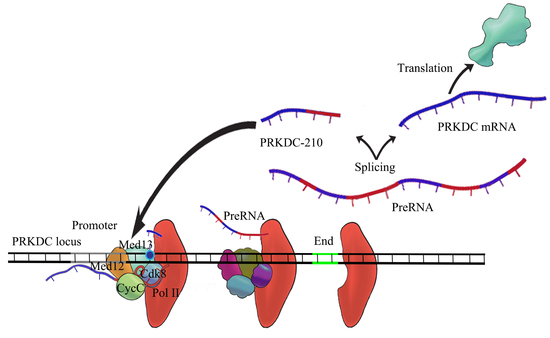
2.5. PRKDC-210 Acts on the Endogenous Genome

Given its interaction with the CKD8 complex and RNA pol II S5P, we hypothesized that PRKDC-210 would be enriched at the promoter or transcription initiation regions of genes. To examine this possibility, we used a ChIRP assay to assess RNA–DNA binding [22]. RT-qPCR analysis confirmed that PRKDC-210 was precipitated by the ChIRP probe set (Figure 4a). We suspected that PRKDC-210 would act on adjacent genes, so we examined enrichment of various regions of the PRKDC locus on the immunoprecipitated PRKDC-210 RNA. We examined the 15 kbp upstream flanking region (used as a negative control), 1 kbp upstream site, promoter region, transcription start site (TSS), Exon region (6, 41 and 64), transcription stop site and the 3’ UTR, respectively. As expected, PRKDC-210 was enriched at the TSS of the PRKDC gene (Figure 4b).
Next, to determine whether PRKDC-210 can act directly on genes other than PRKDC, we performed ChIRP-qPCR analyses of eight randomly selected genes that were detected as differentially expressed in the RNA-seq analyses following knockdown of PRKDC-210. Among them, CEBPD, FANTA, UBE2V2, MCM4 and TCEA1 are located on the same chromosome as PRKDC (chromosome 8), whereas TDRD7, ARMC10, and ETV3 are located on chromosomes 9, 7 and 1, respectively. PRKDC-210 was enriched at the TSSs of the CEBPD, UBE2V2, MCM4, TCEA1 and TDRD7 genes (Figure 4c). We also performed a ChIRP-qPCR analysis of the TSSs of genes included in the GO fatty acid metabolic process, including LYPLA1 (chromosome 8), AASDH (chromosome 4), BAAT (chromosome 9), ACSM2B (chromosome 16), SLC27A1 (chromosome 19) and ACSM2A (chromosome 16). PRKDC-210 was enriched at the LYPLA1 gene only (Figure 4d). Overall, these results suggest that genes on chromosome 8 are more likely to be targeted by PRKDC-210.

3. Discussion

In the past, limitations in detection technologies have meant that the roles of lencRNAs have been overlooked. In particular, as lencRNAs are splice variants of mRNAs, they are often confused with mRNAs during analyses. Here, we designed a novel tethering luciferase assay to detect the regulatory functions of exogenous lencRNAs in transcription and found that the lencRNA PRKDC-210 interacts with MED-12 and/or the CDK8 complex to upregulate gene expression. Since the tethering luciferase assay utilizes a TATA box, which is not associated with MED12 enrichment, we assumed that PRKDC-210 promoted the interaction of MED12 and its complex with RNA polymerase II S5P to drive transcriptional upregulation. In addition to PRKDC-210, the lencRNAs BAP1-206, PIK3R1-206 and PIK3R1-209 also increased transcription of the luciferase gene in our initial screen (Figure 1b,c). We did not analyze the functions of these lencRNAs further because they were unable to interact with MED12 (Figure 1d); however, their potential regulatory roles and interaction with other transcriptional complexes will be examined in the future.
An interesting phenomenon was observed in the ChIRP assay. Most of the genes affected by PRKDC-210 are located on chromosome 8, which is where the PRKDC locus is situated (Figure 4c,d). Among the 14 genes examined in the ChIRP assay (eight randomly selected genes and six fatty acid metabolic process-related genes), CEBPD, FNTA, UBE2V2, MCM4, and LYPLA1 are located on chromosome 8, which has a total length of 146 Mbp. The maximum distance between these genes and the PRKDC locus is about 6.5 Mbp. Notably, the CEBPD and MCM4 loci are located <50 Kbp and 7 bp from the PRKDC locus, respectively. The pairwise genetic distances were calculated according to Rutgers Map v.3, a combined linkage-physical map of the human genome (http://compgen.rutgers.edu/map_interpolator.shtml, accessed on 27 September 2022). The minimal genetic distances of intergenic recombinations were obtained for PRKDC, CEBPD, MCM4, and UBE2V2 (<0.05 cM). The genetic distance between FNTA and PRKDC was about 0.5 cM. In general, linkage disequilibrium is supposedly noticeable at such short distances. PRKDC is located far from LYPLA1, with a genetic distance of about 5.0 cM. Unlike CEBPD, MCM4, and UBE2V2, LYPLA1 does not appear to display linkage disequilibrium with PRKDC. Nonetheless, LYPLA1 was also affected by PRKDC-210. In addition, because chromosomes move randomly in the nucleus, the relative positions of two genes on different chromosomes are uncertain, while the relative positions of two genes on the same chromosome do not change significantly. Of course, this could be due to chromosome folding or another configuration that causes PRKDC-210 to enhance transcription within a certain range of chromatin 8 [23]. However, heterochromatin structure should not be associated with the transcriptional enhancement of PRKDC-210, as heterochromatin structure is thought to be unable to express genes [24]. We propose that genes on the same chromosome as the PRKDC locus may be more susceptible to PRKDC-210 than those on other chromosomes, as after transcription and splicing, PRKDC-210 may be bound rapidly by nearby CDK8 complexes or RNA pol II, which then act on neighboring genes.
Based on the results of the tethering luciferase assay and RIP-qPCR analyses (Figure 1 and Figure 2), we speculate that PRKDC-210 acts directly on the CDK8 complex in the nucleus. Notably, lencRNAs such as PRKDC-210 are not modified by poly(A) tailing; hence, they may act rapidly on nearby proteins and promote gene expression immediately after their generation. The regulatory action of lencRNAs may be a cause of abnormal gene expression in cancer. The lencRNAs examined in our current study are all expressed at high levels in cancer. As a product of alternative splicing, the increased generation of lencRNAs is likely attributable to genetic abnormalities. Indeed, there have been numerous reports of cancers producing abnormal mRNA splicing [10,25,26]. Although these reports have focused mainly on protein-coding RNAs, there are also reports confirming that lncRNAs are spliced from coding genes [27]. More in-depth studies are required to analyze the mechanisms underlying the production and functions of lencRNAs. Such studies may expand our current understanding of diseases caused by splicing abnormalities and lay the foundation for future therapeutic developments.
In conclusion, lencRNA is a type of lncRNA produced by abnormal splicing caused by genetic deviations such as cancer. Here, we found that the lencRNA PRKDC-210 interacts with the CDK8 complex, resulting in transcriptional upregulation, which may be a major cause of abnormal gene expression in cancer. Additional studies are needed to understand the roles of lencRNAs in disease states.

4. Materials and Methods

4.1. Cell Lines

Two cell lines, HEK293T and MCF-7, were used in this study. HEK293T was used for all tethering luciferase assays for detecting exogenous lencRNA. MCF-7 cells were used to detect endogenous lencRNA. It was used for knockdown assay, RNA immunoprecipitation assay, RNA-Seq and chromatin isolation by RNA purification assay, respectively.

4.2. Cell Culture

HEK293T cells and MCF-7 cells were cultured in high-glucose DMEM (Nacalai Tesque, Kyoto, Japan) supplemented with 10% FBS (BioWest, Funakoshi, Japan). The cells were maintained in a 5% CO2 incubator at 37 °C [28].

4.3. Transfection

Twenty-four hours before transfection, cells were seeded into 12-well cell culture plates (TrueLine, New York, NY, USA) at a density of 2–3 × 10⁵ cells/well. The culture medium was replaced with 1 mL of fresh medium before transfection.
For the tethering luciferase assay in HEK293T cells, a mixture containing 400 ng of the Firefly luc plasmid, 10 ng of the Renilla luc plasmid, 50 ng of the MCP-GAL4 plasmid, 50 ng of the MS2-lencRNA plasmid, 2.5 µL of polyethylenimine (1.0 µg/µL; Polysciences, Warrington, PA, USA), and 100 µL of OPTI-MEM (Thermo Fisher Scientific, Waltham, MA, USA) was incubated at room temperature for 20 min. Subsequently, the transfection master mix was added to the cell culture medium, and cells were incubated for 24 h [29,30].
For siRNA transfections, HEK293T cells were seeded into 12-well plates at a density of 1 × 10⁵ cells/well. Following the manufacturer’s protocol, 2 µL of LipofectamineTM RNAiMAX (Thermo Fisher Scientific) and 24 pmol of siRNA were added to 100 µL of OPTI-MEM, and the mixture was incubated at room temperature for 20 min. The transfection master mix was then added to the cell culture medium, and the tethering luciferase assay was performed 24 h later [31]. The MissionTM CDK8 siRNA (SASI_WI_00000016) and control siRNA (Universal Negative Control #1) were obtained from Sigma Aldrich (Burlington, MA, USA).
For knockdown of PRKDC mRNA and PRKDC-210 in MCF-7 cells, following the manufacturer’s protocol, 5 µL of LipofectamineTM 2000 (Thermo Fisher Scientific) was mixed with 100 pmol of ASOs in 100 µL of OPTI-MEM and incubated at room temperature for 5 min. The transfection master mix was then added to the cell culture medium, and the cells were incubated for 24 h [32]. Phosphorothioate-modified ASOs were obtained from Eurofins Genomics (Tokyo, Japan).

4.4. Dual-Luciferase Reporter Assay

The Dual-Luciferase® Reporter Assay System (Promega, Madison, WI, USA) was used to detect luciferase activity in the tethering luciferase assay. After transfection for 24 h, the culture medium was removed, and the cells were washed once with PBS(-) (Nacalai Tesque). Subsequently, 250 µL of 1× passive lysis buffer was added, and the cells were shaken for 15 min at room temperature. To detect the firefly luciferase signal, a 10 µL aliquot of the lysed cells was added to a luminometer tube along with Luciferase Assay Reagent II. Subsequently, Stop & Glo® Reagent was added to the tube to enable detection of the Renilla luciferase signal. The ratio of firefly to Renilla luciferase was then determined [33]. The signals were detected using a Gene Light 55 luminometer (WAKENYAKU, Kyoto, Japan) with the following detection conditions: delay time 3 s, count time 6 s, repeat time 1.

4.5. RNA Extraction

RNA extraction was performed using TRIzol® reagent (Thermo Fisher Scientific), according to the manufacturer’s instructions. Briefly, the culture medium was removed, and the cells were washed once with PBS(-), followed by addition of 400 µL of TRIzol® reagent and RNA extraction using the basal protocol [34].

4.6. RT-qPCR

Total RNA (500 ng) was reverse transcribed into cDNA using ReverTra Ace® reagent (TOYOBO, Osaka, Japan) and random primers (5′-NNNNNNNNN-3′). In the RIP assays, the RNA pellet was dissolved in nuclease-free water, and the entire sample was used for reverse transcription.
The qPCR assays were performed using TB Green® Premix Ex Taq™ II (Tli RNaseH Plus) (TaKaRa, Kusatsu, Japan), according to the manufacturer’s protocol, and primers were obtained from Eurofins Genomics. Analyses were performed using an Mx3000pTM qPCR system (Agilent, Santa Clara, CA, USA) [35].

4.7. RNA Immunoprecipitation

Cells were grown to 80–90% confluency in 10 cm culture dishes. The medium was then changed, and the cells were incubated for 30 min at 37 °C. Subsequently, the medium was removed, and the cells were washed three times with PBS(-); then, 5 mL of 1 × nuclear isolation buffer (3 mL of Milli-Q water, 1 mL of PBS, 1 mL of 5 × NIB (1.28 M sucrose, 40 mM Tris-HCl pH 7.5, 20 mM MgCl2, 4% Triton X-100)) was added. The cells were then scraped from the dish, transferred to a 15 mL centrifuge tube, placed on ice for 20 min, and centrifuged for 15 min at 2000 RCF. Next, the supernatant was removed, and the pellet was resuspended in 500 µL of RNA isolation buffer (150 mM KCl, 25 mM Tris-HCl pH 7.5, 5 mM EDTA, 0.5% NP-40, 0.5 mM DTT, protease inhibitor cocktail, RNase inhibitor, DNase I) using a 25 G syringe tip. After centrifuging at 12,500 RCF for 10 min, the supernatant was collected, and a 50 µL sample (used as input) was stored at 4°C. Another 400 µL sample was mixed with 2 µg of the specific antibody (Abcam, Cambridge, UK) and 10 µL of Dynabeads™ Protein G (Thermo Fisher Scientific), and then incubated overnight at 4°C with gentle rotation. The next morning, the supernatant was separated from the beads using a magnetic device, and the pelleted beads were washed three times with RNA isolation buffer and once with PBS(-) [36]. Finally, 400 µL of TRIzol® reagent was added to the input sample and beads, and RNA extraction was performed as described in Section 4.4.

4.8. RNA Sequencing

Total RNA was extracted from cells transfected with ASOs as described in Section 4.4. Subsequently, the samples were analyzed using the RNA-seq service of Azenta Life Sciences (Tokyo, Japan). The level of gene expression was indicated by the read density and calculated as shown below (where FPKM represents the fragments per kilo bases per million reads) based on read counts from HT-seq (V. 0.6.1) [37].
FPKM = total   exon   fragments mapped   reads ( millions ) ×   exon   length   ( Kbp )
The ratio of total exon fragments to mapped reads is normalized to the gene length (exon length), such that the expression levels of genes with different sequencing depths and lengths are comparable. RNAs with reads > 10 and FPKM > 2 were screened as valid samples.

4.9. Chromatin Isolation by RNA Purification

The experimental method followed the protocol of Ci Chu et al. [22]. To isolate PRKDC-210, a probe set targeting PRKDC-210 needed to be designed. Probes were designed on the PRKDC-210 at about 100 bp intervals, with a total of 6 probes. The ChIRP-probe set was modified with 3’ BiotinTEG and ordered from Eurofins Genomics.
Cells were grown to 80–90% confluency in 10 cm cell culture dishes. The medium was then removed, and the cells were washed three times with PBS(-) before being resuspended in PBS(-), transferred to a 50 mL tube, and centrifuged. Next, the supernatant was removed, and the pellet was resuspended in 10 mL of 1% glutaraldehyde (prepared immediately before use) and incubated for 10 min at room temperature, with gentle shaking to crosslink the cells. To stop the crosslinking, 1 mL of 1.25 M glycine was added, and the sample was incubated for 5 min at room temperature. Subsequently, the tube was centrifuged, the supernatant was removed, the pellet was washed once with PBS(-), and the sample was then centrifuged again. Next, the pellet in PBS(-) was transferred to a 1.5 mL centrifuge tube, the PBS(-) was removed, and 500 µL of cell lysis buffer (50 mM Tris-HCl pH 7.0, 10 mM EDTA, 1% SDS, protease inhibitor cocktail, RNase inhibitor) was added. The sample was then sonicated until it became clear. A 10 µL aliquot of the lysate was reserved as the DNA/RNA input sample, and then 1 mL of hybridization buffer (750 mM NaCl, 1% SDS, 50 mM Tris-HCl pH 7.0, 1 mM EDTA, 15% formamide, protease inhibitor cocktail, RNase inhibitor) and 100 pmol of ChIRP probes (Eurofins Genomics) were added to the remaining sample, followed by incubation for 4 h at 37 °C with rotation. Next, Dynabeads Biotin Binder beads (100 µL; Thermo Fisher Scientific) were washed with cell lysis buffer, added to the sample, and incubated for 30 min at 37 °C with shaking. Subsequently, the beads were washed five times with 1 mL of wash buffer (2× SSC, 0.5% SDS) and then resuspended in 1 mL of wash buffer. A 100 µL aliquot was reserved for RNA isolation. The remaining 900 µL of the bead suspension was removed from the wash buffer and added to 300 µL of elution buffer to elute the DNA from the beads.
For the RNA component, the wash buffer was removed from the beads for treatment with proteinase K. After the addition of TRIzol® reagent (as described in Section 4.4), RT-qPCR analyses were performed using specific primers for RNA products.
For the DNA component, the sample was treated with proteinase K. Subsequently, 300 µL of PCI (phenol: chloroform: isoamyl alcohol; 25:24:1) was added, the sample was shaken vigorously for 10 min, and then the supernatant was removed following centrifugation. Next, 30 µL of NaOAc and 900 µL of 100% EtOH were added, and the sample was incubated overnight at −20°C. The next morning, the supernatant was removed by centrifugation, and the DNA pellet was washed with 70% EtOH, centrifuged again to remove the supernatant, air-dried, and dissolved in Milli-Q water. qPCR analyses were performed using specific primers.

4.10. Primer

qPCR primers, Antisense Oligo DNA and ChIRP probes were all ordered from Eurofins Genomics; sequences can be found in Supplementary Data (Table S2).

Supplementary Materials

The following supporting information can be downloaded at: https://www.mdpi.com/article/10.3390/ijms232213783/s1.

Author Contributions

Conceptualization, J.M. and M.S.; methodology, J.M. and M.S.; software, J.M. and G.F.; validation, J.M.; formal analysis, J.M., G.F. and M.S.; investigation, J.M. and M.S.; resources, T.T.; data curation, J.M.; writing—original draft preparation, J.M.; writing—review and editing, J.M. and T.T.; visualization, J.M. and G.F.; supervision, T.T.; project administration, T.T.; funding acquisition, T.T. All authors have read and agreed to the published version of the manuscript.

Funding

This research was funded by a Grant-in-Aid for Scientific Research from the Japan Society for the Promotion of Science, grant numbers 17H02204, 18K19288 and 21H02067.

Data Availability Statement

The data presented in this study are available on request from the corresponding author. The data are not publicly available due to the data will be used in subsequent studies.

Conflicts of Interest

The authors declare no conflict of interest.

References

  1. Djebali, S.; Davis, C.A.; Merkel, A.; Dobin, A.; Lassmann, T.; Mortazavi, A.; Tanzer, A.; Lagarde, J.; Lin, W.; Schlesinger, F.; et al. Landscape of Transcription in Human Cells. Nature 2012, 489, 101–108. [Google Scholar] [CrossRef] [PubMed] [Green Version]
  2. Montalbano, A.; Canver, M.C.; Sanjana, N.E. High-Throughput Approaches to Pinpoint Function within the Noncoding Genome. Mol. Cell 2017, 68, 44–59. [Google Scholar] [CrossRef] [PubMed] [Green Version]
  3. Chen, S.; Liang, H.; Yang, H.; Zhou, K.; Xu, L.; Liu, J.; Lai, B.; Song, L.; Luo, H.; Peng, J.; et al. LincRNa-P21: Function and Mechanism in Cancer. Med. Oncol. 2017, 34, 98. [Google Scholar] [CrossRef]
  4. Atianand, M.K.; Hu, W.; Satpathy, A.T.; Shen, Y.; Ricci, E.P.; Alvarez-Dominguez, J.R.; Bhatta, A.; Schattgen, S.A.; McGowan, J.D.; Blin, J.; et al. A Long Noncoding RNA LincRNA-EPS Acts as a Transcriptional Brake to Restrain Inflammation. Cell 2016, 165, 1672–1685. [Google Scholar] [CrossRef] [Green Version]
  5. Kuwabara, Y.; Tsuji, S.; Nishiga, M.; Izuhara, M.; Ito, S.; Nagao, K.; Horie, T.; Watanabe, S.; Koyama, S.; Kiryu, H.; et al. Lionheart LincRNA Alleviates Cardiac Systolic Dysfunction under Pressure Overload. Commun. Biol. 2020, 3, 434. [Google Scholar] [CrossRef]
  6. Sun, Q.; Hao, Q.; Prasanth, K.V. Nuclear Long Noncoding RNAs: Key Regulators of Gene Expression. Trends Genet. 2018, 34, 142–157. [Google Scholar] [CrossRef]
  7. Kung, J.T.Y.; Colognori, D.; Lee, J.T. Long Noncoding RNAs: Past, Present, and Future. Genetics 2013, 193, 651–669. [Google Scholar] [CrossRef] [Green Version]
  8. Huarte, M. The Emerging Role of LncRNAs in Cancer. Nat. Med. 2015, 21, 1253–1261. [Google Scholar] [CrossRef]
  9. Wapinski, O.; Chang, H.Y. Long Noncoding RNAs and Human Disease. Trends Cell Biol. 2011, 21, 354–361. [Google Scholar] [CrossRef]
  10. Shiraishi, Y.; Kataoka, K.; Chiba, K.; Okada, A.; Kogure, Y.; Tanaka, H.; Ogawa, S.; Miyano, S. A Comprehensive Characterization of Cis-Acting Splicing-Associated Variants in Human Cancer. Genome Res. 2018, 28, 1111–1125. [Google Scholar] [CrossRef]
  11. Wang, K.C.; Chang, H.Y. Molecular Mechanisms of Long Noncoding RNAs. Mol. Cell 2011, 43, 904–914. [Google Scholar] [CrossRef] [PubMed] [Green Version]
  12. Kopp, F.; Mendell, J.T. Functional Classification and Experimental Dissection of Long Noncoding RNAs. Cell 2018, 172, 393–407. [Google Scholar] [CrossRef] [PubMed] [Green Version]
  13. Wang, L.; Cho, K.B.; Li, Y.; Tao, G.; Xie, Z.; Guo, B. Long Noncoding RNA (LncRNA)-Mediated Competing Endogenous RNA Networks Provide Novel Potential Biomarkers and Therapeutic Targets for Colorectal Cancer. Int. J. Mol. Sci. 2019, 20, 5758. [Google Scholar] [CrossRef] [PubMed] [Green Version]
  14. Bhan, A.; Soleimani, M.; Mandal, S.S. Long Noncoding RNA and Cancer: A New Paradigm. Cancer Res. 2017, 77, 3965–3981. [Google Scholar] [CrossRef] [Green Version]
  15. Engreitz, J.M.; Haines, J.E.; Perez, E.M.; Munson, G.; Chen, J.; Kane, M.; McDonel, P.E.; Guttman, M.; Lander, E.S. Local Regulation of Gene Expression by LncRNA Promoters, Transcription and Splicing. Nature 2016, 539, 452–455. [Google Scholar] [CrossRef] [Green Version]
  16. Broude, E.V.; Győrffy, B.; Chumanevich, A.A.; Chen, M.; Mcdermott, M.S.J.; Shtutman, M.; Catroppo, J.F.; Roninson, I.B. Expression of CDK8 and CDK8-Interacting Genes as Potential Biomarkers in Breast Cancer HHS Public Access. Curr. Cancer Drug Targets 2015, 15, 739–749. [Google Scholar] [CrossRef] [Green Version]
  17. Lai, F.; Orom, U.A.; Cesaroni, M.; Beringer, M.; Taatjes, D.J.; Blobel, G.A.; Shiekhattar, R. Activating RNAs Associate with Mediator to Enhance Chromatin Architecture and Transcription. Nature 2013, 494, 497–501. [Google Scholar] [CrossRef] [Green Version]
  18. Burley, S.K.; Roeder, R.G. TATA Box Mimicry by TFIID. Cell 1998, 94, 551–553. [Google Scholar] [CrossRef] [Green Version]
  19. Savinkova, L.K.; Ponomarenko, M.P.; Ponomarenko, P.M.; Drachkova, I.A.; Lysova, M.v.; Arshinova, T.v.; Kolchanov, N.A. TATA Box Polymorphisms in Human Gene Promoters and Associated Hereditary Pathologies. Biochemistry 2009, 74, 117–129. [Google Scholar] [CrossRef]
  20. Knuesel, M.T.; Meyer, K.D.; Donner, A.J.; Espinosa, J.M.; Taatjes, D.J. The Human CDK8 Subcomplex Is a Histone Kinase That Requires Med12 for Activity and Can Function Independently of Mediator. Mol. Cell Biol. 2009, 29, 650–661. [Google Scholar] [CrossRef]
  21. Currie, E.; Schulze, A.; Zechner, R.; Walther, T.C.; Farese, R.V. Cellular Fatty Acid Metabolism and Cancer. Cell Metab. 2013, 18, 153–161. [Google Scholar] [CrossRef] [PubMed] [Green Version]
  22. Chu, C.; Quinn, J.; Chang, H.Y. Chromatin Isolation by RNA Purification (ChIRP). J. Vis. Exp. 2012, 61, e3912. [Google Scholar] [CrossRef]
  23. Dixon, J.R.; Gorkin, D.U.; Ren, B. Chromatin Domains: The Unit of Chromosome Organization. Mol. Cell 2016, 62, 668–680. [Google Scholar] [CrossRef] [PubMed] [Green Version]
  24. Liu, J.; Ali, M.; Zhou, Q. Establishment and Evolution of Heterochromatin. Ann. N. Y. Acad. Sci. 2020, 1476, 59–77. [Google Scholar] [CrossRef] [PubMed] [Green Version]
  25. Yoshida, K.; Ogawa, S. Splicing Factor Mutations and Cancer. Wiley Interdiscip. Rev. RNA 2014, 5, 445–459. [Google Scholar] [CrossRef] [Green Version]
  26. Srebrow, A.; Kornblihtt, A.R. The Connection between Splicing and Cancer. J. Cell Sci. 2006, 119, 2635–2641. [Google Scholar] [CrossRef] [Green Version]
  27. Huang, M.S.; Zhu, T.; Li, L.; Xie, P.; Li, X.; Zhou, H.H.; Liu, Z.Q. LncRNAs and CircRNAs from the Same Gene: Masterpieces of RNA Splicing. Cancer Lett. 2018, 415, 49–57. [Google Scholar] [CrossRef]
  28. Freshney, R.I. Culture of Animal Cells; John Wiley & Sons, Inc.: Hoboken, NJ, USA, 2010; ISBN 9780470649367. [Google Scholar]
  29. González-Domínguez, I.; Grimaldi, N.; Cervera, L.; Ventosa, N.; Gòdia, F. Impact of Physicochemical Properties of DNA/PEI Complexes on Transient Transfection of Mammalian Cells. N. Biotechnol. 2019, 49, 88–97. [Google Scholar] [CrossRef] [Green Version]
  30. Yue, Y.; Wu, C. Progress and Perspectives in Developing Polymeric Vectors for in Vitro Gene Delivery. Biomater. Sci. 2013, 1, 152–170. [Google Scholar] [CrossRef]
  31. Life Technologies. Lipofectamine® RNAiMAX Reagent Lipofectamine® RNAiMAX Transfection Protocol Scaling Up or Down Transfections Limited Product Warranty and Disclaimer Details Package Contents; Protocol Pub. No. MAN0007825 Rev. 1.0; Life Technologies: Carlsbad, CA, USA, 2013. [Google Scholar]
  32. Life Technologies. Lipofectamine® 2000 Reagent Protocol Outline; Protocol Pub. No. MAN0007824 Rev. 1.0; Life Technologies: Carlsbad, CA, USA, 2013. [Google Scholar]
  33. Promega Corporation. Dual-Luciferase® Reporter Assay System Instructions for Use of Products E1910 and E1960; Rev 6/15; Promega Corporation: Madison, WI, USA, 2015. [Google Scholar]
  34. Thermo Fisher Scientific. TRIzol™ Reagent User Guide; Pub. No. MAN0001271 Rev. 8.0; Thermo Fisher Scientific: Waltham, MA, USA, 2016. [Google Scholar]
  35. Agilent Technologies. Mx3000P & Mx3005P Real-Time QPCR System; Revision# 090530; Agilent Technologies: Santa Clara, CA, USA, 2015. [Google Scholar]
  36. Gagliardi, M.; Matarazzo, M.R. RIP: RNA Immunoprecipitation. In Polycomb Group Proteins; Humana Press: New York, NY, USA, 2016; pp. 73–86. [Google Scholar]
  37. Mortazavi, A.; Williams, B.A.; McCue, K.; Schaeffer, L.; Wold, B. Mapping and Quantifying Mammalian Transcriptomes by RNA-Seq. Nat. Methods 2008, 5, 621–628. [Google Scholar] [CrossRef]
Figure 1. (a) Schematic illustration of the tethering luciferase assay system. (b,c) Tethering luciferase assay using HEK293T cells, relative fluorescence and mRNA levels were detected. We used the empty MS2 plasmid instead of the MS2 lencRNA plasmid as negative control (neg.ctrl). Value = sample/neg.ctrl. (b) We used the Dual-Luciferase® Reporter assay to detect the results of the tethering assay; the luciferase gene expression levels of different lencRNAs in the tethering or nontethering were compared. F/R, Firefly luciferase/Renilla luciferase. (c) Total RNA was extracted from cells transfected with tethering luciferase assay plasmid, and the differences of luciferase mRNA transcript between different lencRNAs were compared. Total RNA was reverse-transcribed into cDNA using random primers and quantified by qPCR using specific primer for luciferase gene. (d) The binding of different lencRNAs to MED12 in MCF7 cells was compared by RIP-qPCR assay. Value = sample/input. Data in all panels are represented as the mean  ±  s.e.m. of three independent experiments. * p  <  0.05 by a two-tailed Student’s t-test (compared with nontethering (a) or IgG (d)).
Ijms 23 13783 g001
Figure 2. (a) This figure shows the expression of luciferase after tethering of PRKDC-210 in control siRNA-treated HEK293T cells and siCDK8-treated HEK293T cells. F/R, firefly luciferase/Renilla luciferase. (b) RIP-qPCR assays of MCF7 cells using antibodies targeting total RNA pol II, RNA pol II S5P, and RNA pol II S2P. The enrichment of PRKDC mRNA at different sites and the enrichment of PRKDC-210 in different phosphorylation of RNA Pol II are shown. Data in all panels are represented as the mean  ±  s.e.m. of three independent experiments. *** p  <  0.001 by a two-tailed Student’s t-test.
Ijms 23 13783 g002
Figure 3. (a,b) Total RNA was extracted from MCF7 cells expressing control (ctrl) ASOs or ASOs targeting the PRKDC mRNA or PRKDC-210, and the RNA transcripts levels were detected via RT-qPCR. Kd means knockdown. Data are represented as the mean  ±  s.e.m. of three independent experiments. (cg) RNA-seq analyses of PRKDC mRNA knockdown and PRKDC-210 knockdown cells. (c,d) FPKM profiles showing the overall gene expression levels. (e) The numbers of RNAs with altered transcript levels in PRKDC-210 knockdown cells versus control cells. (f) Genes that were significantly differentially expressed (fold change >2 and p < 0.05) in PRKDC-210 knockdown cells versus PRKDC mRNA knockdown cells. (g) Gene ontology biological process enrichment analysis.
Ijms 23 13783 g003
Figure 4. MFC7 was used for all ChIRP assays. (a) RT-qPCR analyses of PRKDC-210 and ATP5B (negative control) levels in the RNA precipitated by ChIRP. (bd) The DNAs associated with the immunoprecipitated RNAs in the ChIRP assay were analyzed by qPCR using specific primers. (b) The distribution of PRKDC-210 at the PRKDC locus. (c) Enrichment of PRKDC-210 at the TSSs of various genes. (d) Enrichment of PRKDC-210 at the TSSs of genes related to fatty acid metabolism. Data in all panels are represented as the mean  ±  s.e.m. of three independent experiments. *** p  <  0.001 by a two-tailed Student’s t-test.
Ijms 23 13783 g004
Publisher’s Note: MDPI stays neutral with regard to jurisdictional claims in published maps and institutional affiliations.

