Article

Computer-Aided Screening and Revealing Action Mechanism of Food-Derived Tripeptides Intervention in Acute Colitis

1
Jilin Provincial Key Laboratory of Nutrition and Functional Food, College of Food Science and Engineering, Jilin University, Changchun 130062, China
2
State Key Laboratory of Tea Plant Biology and Utilization, School of Tea and Food Science and Technology, Anhui Agricultural University, Hefei 230036, China
*
Author to whom correspondence should be addressed.
Int. J. Mol. Sci. 2022, 23(21), 13471; https://doi.org/10.3390/ijms232113471
Submission received: 29 September 2022 / Revised: 28 October 2022 / Accepted: 28 October 2022 / Published: 3 November 2022
(This article belongs to the Special Issue Computer-Aided Screening and Action Mechanism of Bioactive Peptides)

Abstract

:
Food-derived tripeptides can relieve colitis symptoms; however, their alleviation mode has not been systematically evaluated as an alternative nutritional compound. This study aimed to reveal the potential mechanism of 8000 food-derived tripeptides against acute colitis using a computer-aided screening strategy. Forty-one potential hub targets related to colitis with a Fit score > 4.0 were screened to construct the protein-protein and protein-tripeptide network based on the PharmMapper database and STRING software (Ver. 11.5). In addition, 30 significant KEGG signaling pathways with p-values < 0.001 that the 41 hub targets mainly participated in were identified using DAVID software (Ver. 6.8), including inflammatory, immunomodulatory, and cell proliferation and differentiation-related signaling pathways, particularly in the Ras- and PI3K-Akt signaling pathways. Furthermore, molecular docking was performed using the Autodock against majorly targeted proteins (AKT1, EGFR, and MMP9) with the selected 52 tripeptides. The interaction model between tripeptides and targets was mainly hydrogen-bonding and hydrophobic interactions, and most of the binding energy of the tripeptide target was less than −7.13 kcal/mol. This work can provide valuable insight for exploring food-derived tripeptide mechanisms and therapeutic indications.

1. Introduction

Inflammatory bowel disease (IBD) has been recognized as a chronic, incurable, and idiopathic disease spreading worldwide. Therefore, alleviating IBD has become an emerging challenge for public health [1,2,3]. Acute colitis is a common intestinal disease; it will develop into IBD without reasonable intervention. Factors, such as oxidative stress, dysregulated immune responses, and nutrition deficiency, have been reported to be negatively related to colon health, which can aggravate colitis symptoms [4,5,6]. Currently, methods of relieving colitis are mainly based on drug intervention, commonly including aminosalicylic acid preparations, steroids, and glucocorticoids [6]; however, the strong side effects severely restrict their more comprehensive application. Thus, an alternative method for alleviating colitis that is efficient and safe is urgently needed.
Small-molecule active peptides extracted from food-derived proteins have nutritional supplementation [5] and physiological regulation functions, such as antioxidant, anti-inflammatory, and antibacterial functions [7,8,9,10,11]. Recent research has also demonstrated that small-molecule peptides could help relieve colitis symptoms [8,12,13]. As a small-molecule active peptide, tripeptides can be wholly absorbed by the body and targeted at a specific site to exert good functional properties. Tripeptides have also been reported to prevent and relieve symptoms of colitis. Lys-Pro-Val (KPV) can be transported into the cytosol in the intestine by binding to the oligopeptide transporter (PepT1), further achieving real-time tracking and visualization of the role of intracellular KPV on ulcerative colitis [14]. Meanwhile, Xiao et al. [15] combined KPV with hyaluronic acid (HA) to obtain polymeric nanoparticles (HA-KPV-NPs), which can be targeted to release in the intestines, effectively inhibiting the production of inflammatory factors and alleviating colitis symptoms. Gly-Pro-Ala (GPA) has also been reported to prevent colitis by increasing AMPK phosphorylation and inhibiting the NLRP3 inflammasome [16]. However, no systematic studies have revealed the underlying mechanisms of the 8000 tripeptides in ameliorating colitis symptoms.
Network pharmacology is an interactive network based on the concept of “disease-gene-target-compounds”, which treats the intervention and effect of active compounds on the disease network from a systematic and comprehensive perspective to reveal the complex mechanisms of active compounds [17,18,19]. This study uses a computer-aided screening strategy to explore tripeptides’ intervention mechanism and molecular targets for acute colitis. Furthermore, the interaction mechanisms between hub targets and tripeptides were revealed through molecular docking. The current study identifies which colitis-related targets tripeptides might interact with. Moreover, the present study can also provide precise target information for research on preventing and alleviating colitis using tripeptides.

2. Results

2.1. Network Interaction Analysis

Based on the pharmacology analysis results, 41 predicted gene targets were screened to construct the set hub targets (Table 1). The PPI network among the target set’s proteins was first analyzed using the STRING database. Figure 1a shows a network with 41 nodes and 225 edges. The results of the topological network analysis showed hub targets with a degree value ≥ 15 played a vital role in the network (Table 2), including AKT1, EGFR, MMP9, CASP3, MMP2, IL2, STAT1, and JAK2.
A tripeptide-target network performed the complex interaction between tripeptides and the screened hub targets. Based on the PharmMapper and GeneCards analysis, tripeptides (related to the most hub targets) were summarized and ranked in the present study. Fifty-two tripeptides were chosen for further tripeptide-target PPI analysis. In Figure 1b, the green nodes represent the hub targets, while the blue nodes denote the 52 tripeptides. The topological analysis of tripeptides and hub targets was also determined (Table S2). Targets GSTM1, BTK, RAF1, G6PD, KIT, ST1A1, C1S, IGF1R, NOS3, AKT2, LCK, CASP3, and ACE showed a high degree value (>28).

2.2. Hub Targets GO Biological Functions Enrichments

GO functional enrichment was performed based on both FunRich (Ver. 3.1.3) and DAVID software (Ver. 6.8) to investigate the function of the selected hub target genes. Figure 2a shows the top ten GO terms based on the analysis of FunRich. Among them, hub targets were enriched in the following cellular component (CC) terms: cytoplasm (30%), plasma membrane, and cytosol (14%). In addition, the hub targets were mainly located in the biological process (BP) pathway terms: signal transduction (25%), cell communication (23%), energy (13%), and metabolism (13%) pathways. For molecular function (MF), the selected hub targets were enriched in terms of protein-tyrosine kinase activity (19%), transmembrane receptor protein tyrosine kinase activity (19%), metallopeptidase activity (19%), metallopeptidase activity (11%), and protein serine/threonine kinase activity (11%).
Furthermore, the DAVID database’s GO enrichment of significant terms for CC, BP, and MF were ranked by p-values and are exhibited as bar charts in Figure 2b. The top three highly enriched BP terms contained the negative regulation of the apoptotic process (13 genes, p-value = 3.81 × 10⁻¹⁰), intracellular signal transduction (10 genes, p-value = 3.42 × 10⁻⁷), and the protein phosphorylation term (10 genes, p-value = 9.60 × 10⁻⁷). The top three CC terms were the cytosol (24 genes, p-value = 3.13 × 10⁻⁸), nucleus (23 genes, p-value = 7.30 × 10⁻⁴), and plasma membrane (20 genes, p-value = 4.56 × 10⁻⁴), respectively. Meanwhile, the top three enrichment MF categories were protein binding (36 genes, p-value = 2.25 × 10⁻⁶), ATP binding (15 genes, p-value = 4.39 × 10⁻⁶), and protein homodimerization activity (10 genes, p-value = 4.12 × 10⁻⁵).

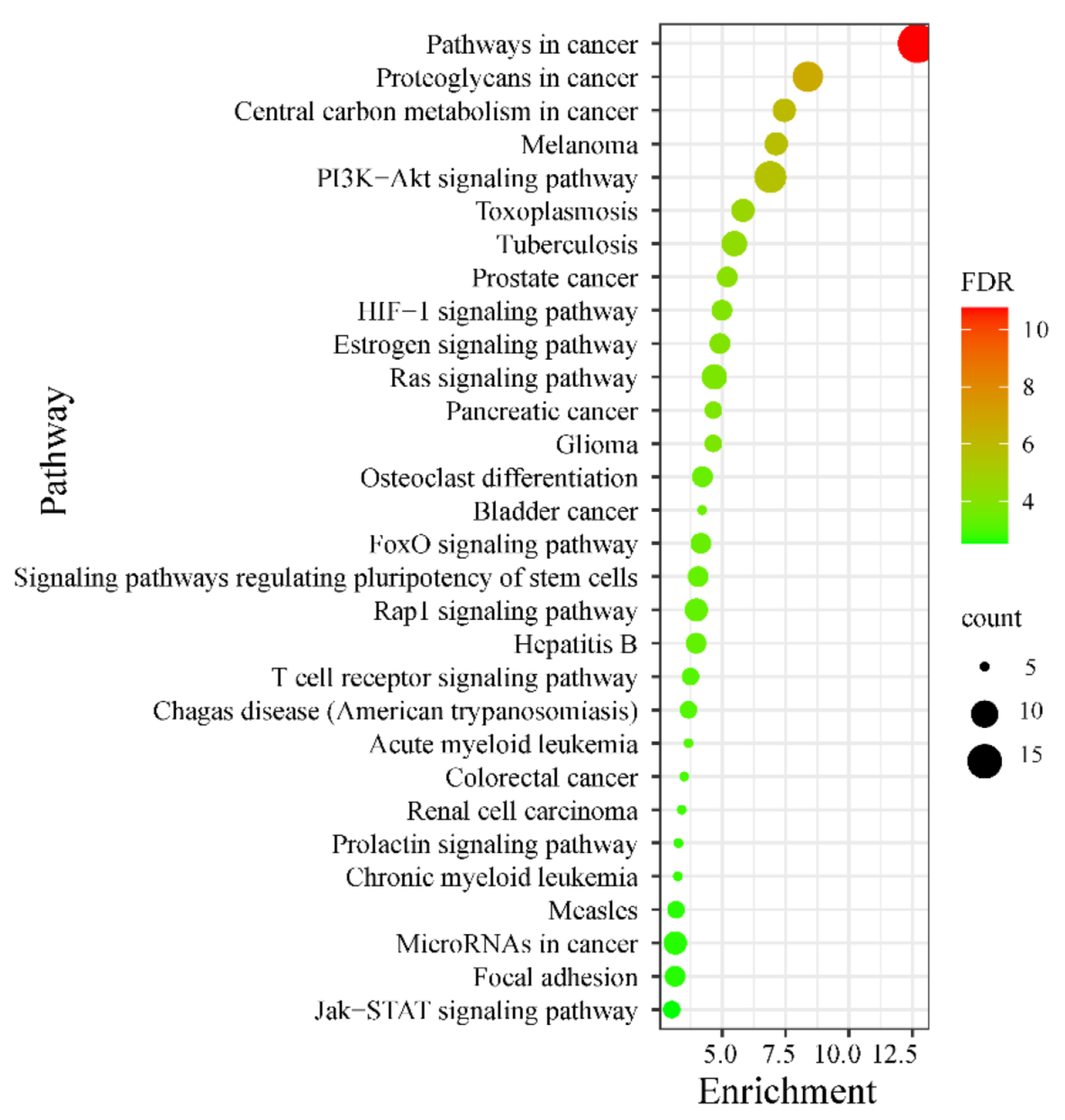
2.3. Hub Targets KEGG Pathway Enrichments

The top 30 significant KEGG signaling pathways that the predicted hub targets participated in (p-value < 0.001) were further investigated and are shown in a bubble map (Figure 3). These 30 involved signaling pathways were mainly enriched in the pathways related to inflammatory, immunomodulatory, and cell proliferation and differentiation (Table S3). These included the HIF-1 signaling pathway (including targets NOS2, NOS3, AKT2, HMOX1, AKT1, EGFR, and IGF1R), the Ras signaling pathway (including targets ZAP70, AKT2, KIT, AKT1, RAF1, MET, EGFR, FGFR1, and IGF1R), the T cell receptor signaling pathway (including targets ZAP70, LCK, AKT2, AKT1, RAF1, and IL2), the Jak-STAT signaling pathway (containing targets STAT1, AKT2, AKT1, JAK2, JAK3, and IL2), and the PI3K-Akt signaling pathway (including targets NOS3, EGFR, IL2, IGF1R, AKT2, KIT, MDM2, AKT1, JAK2, RAF1, JAK3, MET, and FGFR1). Moreover, the hub targets in both the PI3K-Akt and Ras signaling pathways were mapped to those two pathways (in pink symbols) (Figure S1). Furthermore, the significant signaling pathways were also considered to build the tripeptides-target-pathway network in Figure 1b. This complex network diagram makes it easy to find the interaction between those signaling pathways. Thus, tripeptides may intervene in acute colitis by regulating multiple signaling pathways.

2.4. Expression of Hub Targets in Organs Site

The FunRich software (Ver. 3.1.3) revealed the selected 41 hub targets’ organ expression sites (Figure 4). The above 41 hub targets can be expressed in multiple organs and tissues of the human body. Among them, the above targets were significantly related to the organs of the fetal gut, adult colon, and inflammation regulation-connected cells (B cells, CD4 cells, CD8 cells, and NK cells). Hub gene targets (ZAP70, G6PD, MIF, AHCY, LCK, and STAT1) were significantly positively correlated with B cells, CD4 cells, CD8 cells, and NK cells, which showed the opposite trend in both fetal gut and adult colon. Hub gene targets (ACE, KIT, HMOX1, TGFB2, MMP9, MMP2, XIAP, TGM2, MET, RAF1, IGF1R, VDR, MMP13, CIS, CASP3, NOS2, NOS3, IL2, JAK2, JAK3, PARG, EGFR, and RARA) were significantly negatively related to the above inflammation regulation-associated cells. According to the above result, it is reasonable to use the selected hub target as a potential target to regulate acute colitis.

2.5. Molecular Docking Results of Tripeptide and Hub Target

Topological analysis results of the thirty hub targets showed that targets of AKT1, EGFR, and MMP9 had a higher “degree value” and “closeness centrality”, as shown in Table 2; thus, targets AKT1, EGFR, and MMP9 were chosen for molecular docking to further verify the correctness of the above analysis.
Reference ligands obtained from AKT1, EGFR, and MMP9 crystal structures, were first considered to optimize the rationality docking conditions. The above three reference ligand structures had highly overlapped before and after docking with RMSD (root-mean-square deviation) values of 0.83 Å, 1.46 Å, and 3.36 Å, respectively (Figure S2). Meanwhile, the binding energy of the reference ligand to AKT1, EGFR, and MMP9 were −14.30, −14.54, and −9.47 kcal/mol, respectively (Table 3), which means the optimized docking conditions were reasonable for subsequent tripeptide-target docking.
After optimizing the docking conditions, the selected 52 tripeptides were all docked with the above three targets’ protein crystal structures, respectively. Results revealed that these 52 tripeptides showed lower binding energy with the targets of AKT1, EGFR, and MMP9 (all binding energy was less than −6.67 kcal/mol, Table 3). The binding energy of the tripeptides (DDD, DDE, EDD, GDE, EEE, CDD, EED, and EDT) with the target AKT1 were all lower than the reference ligand-1, which ranged from −14.39 to −15.86 kcal/mol (Table 3). The binding energy of the 52 tripeptides with the target EGFR was between −7.53 and −13.60 kcal/mol, especially for the tripeptides EDD, EEE, and IDD. The top three tripeptides with lower binding energy (ranging from −12.31 to −11.63 kcal/mol) with the target MMP9 were tripeptides MEK, VYK, and WIY, respectively.
Moreover, based on the lower binding energy in Table 3, the docking interaction details of the top three tripeptides with hub targets AKT1, EGFR, and MMP9 were further analyzed using Ligplot software (Ver. 2.2), respectively. In Figure S3, tripeptide-target complexes’ binding sites were mainly centralized on the hydrophobic depressions of target crystal structures, and the binding site’s details are shown in Table S4.
In Figure 5a, residues (Lys14, Glu17, Arg23, Arg25, and Arg86) of AKT1 generated hydrogen bond interactions with DDD, with distances of 2.80 Å, 2.83 Å, 2.88 Å, 3.11 Å, and 2.75 Å, respectively, are shown. The hydrogen-bonding interaction details of DDE-AKT1 contained the amino acid residues Lys14 (2.60 Å, 2.77 Å), ARG23 (2.61 Å, 2.97 Å), ARG25 (3.21 Å), Asn53 (3.073 Å). In addition, the residues Arg15, Gly16, Glu17, Leu52, Phe55, and Arg86 of AKT1 generated hydrophobic interactions with tripeptide DDE. Meanwhile, EDD tripeptides also revealed hydrogen bond interactions with the amino acid residues Lys14 (2.68 Å, 2.91 Å), Glu17 (2.80 Å), Arg25 (2.62 Å, 3.32 Å), Leu52 (2.74 Å), Asn53 (2.45 Å, 2.83 Å), and Arg86 (2.61 Å, 2.95 Å) of AKT1 (Figure 5a). The above result indicated the critical amino acid residues of AKT1 were Lys14, Glu17, Arg25, and Arg86. Figure 5b shows the bonding details of tripeptides with target EGFR. Residues Ala722, Arg803, Lys875, and Lys913 of EGFR generated conventional hydrogen bond interactions with EDD, with distances of 2.81 Å, 3.24 Å, 2.65 Å, and 2.98 Å, respectively. Meanwhile, EDD also hydrophobically interacted with the EGFR residues Gly721, Arg841, and Trp880. Tripeptide EEE also revealed a hydrogen bond interaction with the residues Arg803 (3.03 Å, 2.95 Å) and Lys913 (3.09 Å, 2.65 Å) of EGFR, and showed hydrophobia with the residues Ser720, Leu799, Arg841, Trp880, and Lys875 of EGFR. IDD generated hydrogen bond interactions with the residues Ala722 (2.78 Å), Phe723 (3.25 Å, 3.15 Å), Arg841 (3.13 Å, 3.27 Å, 3.06 Å), Aan842 (2.73 Å), and Lys875 (2.84 Å) of EGFR. In addition, IDD displayed hydrophobic interactions with the residues Gly721, Lys745, Asp837, Asp855, and Pro877 of EGFR. Based on the above result, the critical amino acid residues of EGFR were Ala722, Arg803, Arg841, Lys875, and Lys913. The bonding details of MMP9 tripeptides were also analyzed (Figure 5c). The residues Leu188, Ala189, Gln227, Pro246, and Tyr248 of MMP9 generated hydrogen bond interactions with MEK. MEK also displayed hydrophobic interactions with MMP9′s amino acid residues Leu187, His226, His236, Tyr245, and Met247. Meanwhile, the residues Gly186 (2.65 Å), His226 (2.68 Å), Gln227 (3.15 Å), His230 (3.75 Å, 2.89 Å), His236 (3.09 Å, 3.08 Å), and Pro246 (3.31 Å) of MMP9 demonstrated hydrogen-bonding interactions with VYK. Further, VYK also displayed hydrophobic interactions with the residues Leu188, Ala189, His190, Leu243, Tyr245, Met247, and Tyr248 of MMP9. The residues His226 (2.89 Å), Gln227 (2.83 Å, 2.81 Å), His230 (3.10 Å), His236 (2.64 Å), and Tyr245 (2.93 Å) of MMP9 also generated hydrogen bond interactions with WIY. Thus, the critical amino acid residues of MMP9 were Leu188, Ala189, His226, Gln227, His236, Tyr245, Pro246, and Tyr248.

3. Discussion

Acute colitis is a medical emergency and requires prompt recognition, evaluation, and intervention [20]. During the process of colitis, inflammation impairs the colonic mucosal barrier and increases the permeability of the epithelial barrier, further inducing bacterial invasion into underlying tissues and activating the regulation of the body’s immune system [15,21]. Thus, active ingredients with anti-oxidant and anti-inflammatory activities are expected to relieve colitis symptoms.
As an alternative nutritional material, tripeptides have also been reported to have the ability to be anti-oxidants, anti-inflammatory, and alleviate colitis [14,15,16]. Based on the PharmMapper and Genecards databases, 8000 food-derived tripeptides were used as research objects in the present study to explore potential intervention targets for colitis. A total of 41 acute colitis-related hub targets mainly interacting with tripeptides were screened out. The target-target interaction network was further constructed with 41 nodes and 225 edges. Targets AKT1 (RAC-alpha serine/threonine-protein kinase), EGFR (epidermal growth factor receptor), and MMP9 (matrix metalloproteinase 9) were identified as the top three hub targets with a “degree value” > 23 and “closeness centrality” > 0.70.
Bioinformatic analyses focused on the GO and KEGG pathway enrichment of tripeptides intervention with acute colitis-related targets. The GO results showed that the 41 hub targets were mainly enriched in terms of the cytoplasm, plasma membrane, cytosol, signal transduction, cell communication, protein-tyrosine kinase activity, transmembrane receptor protein tyrosine kinase activity, and metallopeptidase activity. KEGG enrichments mainly contained HIF-1, Ras-, T cell receptors, Jak-STAT, and PI3K-Akt signaling pathways. Among them, the PI3K-Akt signaling pathway has been reported to accelerate the progression of ulcerative colitis by activating the inflammatory signaling pathway [22]. Lee et al. found that PI3K-Akt signaling can interfere with cell survival by regulating the expression of epithelial cell receptors [23].
Moreover, Ras is highly expressed in colitis and can also be used as a therapeutic target in inflammatory diseases [24]. Herein, the selected hub targets mainly participated in the PI3K-Akt and Ras-signaling pathways, especially for the hub targets AKT1 and EGFR. EGFR is a type 1 tyrosine kinase receptor with an extracellular ligand-binding domain and an intracellular portion that contains a tyrosine kinase domain [25]. EGFR has been reported to participate in many biological functional processes, such as the proliferation, differentiation, and survival of cells; meanwhile, activating EGFR can help to alleviate inflammation and further limit the progression of colitis lesions [26,27,28]. Matrix metalloproteinases (MMPs) are zinc-dependent neutral endopeptidases with proteolytic activity against extracellular matrix proteins [29]. MMP9 is a unique MMP, which was highly expressed in inflammation, especially in the samples of enteritis and bowel cancer tissues [30,31]. Furthermore, MMP9 can mediate the EGFR-AKT/ERK pathway, further participating in angiogenesis [32].
This paper uses molecular docking to verify the accuracy of the above bioinformatics analysis, and the results confirmed that tripeptides could enter the hydrophobic domain of AKT1, EGFR, and MMP9. Further, the binding mode of the tripeptide-target was mainly hydrogen-bonding and hydrophobic interaction. The above tripeptide-protein binding mode was consistent with the result reported by Kesari et al. [33].
Additionally, the tripeptide-target binding energies mostly ranged from −7.13 to −15.86 kcal/mol, which means tripeptides can bind tightly to the hub targets related to acute colitis. Therefore, we can confirm that tripeptides may alleviate colitis through pleiotropic pathways, including the Ras- and PI3K-Akt signaling pathways.

4. Material and Methods

4.1. Construction of Tripeptide Set and Prediction of Colitis-Related Targets

Firstly, the structures of 8000 food-derived tripeptides were constructed. Then, the UCSF Chimera software (Ver. 1.15) was used to obtain the tripeptide energy minimization structures [2]. Afterward, the 8000 food-derived tripeptides were uploaded into the PharmMapper database (http://www.lilab-ecust.cn/pharmmapper/index.php; accessed on 8 July 2021) to predict the potential colitis-related targets [34]. Based on the Fit score value (>4.0), the predicted targets were summarized and used the U’niProt database (http://www.uniprot.org/; accessed on 16 November 2021) to exclude the same targets and non-Homo sapiens targets. Meanwhile, targets related to colitis obtained from the GeneCards Human database (https://www.genecards.org; accessed on 18 November 2021) were used to improve the objective tripeptide hub targets set.

4.2. Construction of Protein-Protein Interaction (PPI) Network

The PPI network was constructed using the STRING database (http://string-db.org/; accessed on 25 November 2021) to assess the potential interactions between the 41 screened hub targets [35]. The interactions between hub targets or tripeptide-hub targets were then displayed using Cytoscape software (Ver. 3.6.1, https://cytoscape.org/; accessed on 2 December 2021) [36]. Further, the topological characteristics of PPI networks were also analyzed using Cytoscape’s analytical tool [37,38].

4.3. Analysis of GO and KEGG Pathway Enrichment

The DAVID database (http://david.abcc.ncifcrf.gov/home.jsp; accessed on 20 December 2021) has been reported to help integrate functional genomic annotations with intuitive graphical summaries [5,39]. The functional enrichment analysis tool (FunRich), is also widely used to analyze the functional enrichment and interaction network of genes/proteins [40,41]. In this study, the hub tripeptide’s potential targets’ GO (gene ontology) and KEGG (Kyoto encyclopedia of genes and genomes) signaling pathways were systematically analyzed using the DAVID (Ver. 6.8) and FunRich databases. During the analysis process, the significant enriched GO terms (p-value < 0.05) and KEGG pathways (p-value < 0.001) were considered for further evaluation. Finally, the data were visually displayed using the free bioinformatics database (http://www.bioinformatics.com.cn/; accessed on 24 December 2021).

4.4. Hub Target’s Expression Organs Site

This study used FunRich software (Ver. 3.1.3) to research the selected hub target’s main organ site expression based on the “Human proteome” database. The result was displayed in the form of a heatmap. The positively correlated high-expressed targets in the organs are shown in red, while the low-expressed targets are marked in blue. The higher the expression level of the target, the darker the color.

4.5. Molecular Docking

Subsequently, AutoDock 4.2 was used to evaluate the interaction between tripeptides and the above three hub targets [42]. Based on PPI analysis results (the top three hub targets with higher degree values), the crystal structures of AKT1 (1unq, resolution: 0.98 Å) [43], EGFR (3w33, resolution: 1.7 Å) [44], and MMP9 (4h1q, resolution: 1.59 Å) [45] were chosen as the receptor proteins from the RCSB protein data bank (http://www.rcsb.org/pdb; accessed on 3 January 2022) [46] for further molecular docking. The filtering criteria for selecting reporter proteins are as follows: organism “Homo”, “Resolution < 2.0”, and with a unique ligand. Before docking, the unique ligand and water molecules of AKT1, EGFR, and MMP9 crystal structures were removed. Then, hydrogen atoms were added to the three hub targets, respectively. Tripeptide molecular structures were built as the ligand for docking. Before docking, the energy of all tripeptides was minimized by adding the CHARMM force field [47,48].
To verify the rationality-optimized docking conditions, the reference ligands of AKT1, EGFR, and MMP9 crystal structures were first considered. A Lamarckian genetic algorithm as the search engine searched 200 runs. During docking, the grid spacing was set to 0.375 Å [49]. The final docking condition details of each receptor target after completing docking condition optimization are shown in Table S1. The hydrogen bonds, hydrophilic, and coordination interactions between residues of receptor proteins’ active sites and the tripeptide’s poses were identified using Discovery Studio Visualizer (Ver. 4.1). Finally, Ligplot software (Ver. 2.2) was used to display the details of the interactions between tripeptide-targets [50].

5. Conclusions

Based on the computer-aided screening strategy, this study has systematically investigated the potential effects of 8000 tripeptides to alleviate acute colitis, including the possible pleiotropic pathways. Tripeptides play a role in relieving colitis by regulating signaling pathways, such as inflammation, immunomodulation, and cell proliferation and differentiation, especially in the Ras- and PI3K-Akt signaling pathways. This work provides a computer-aided method for screening potential functional targets of active ingredients, which can provide a reference for the functional mining of food-derived peptides.

Supplementary Materials

The following supporting information can be downloaded at: https://www.mdpi.com/article/10.3390/ijms232113471/s1.

Author Contributions

H.G.: writing-original draft, methodology, formal analysis. Y.T. and Y.Z.: resources, software. Y.Y. (Yue Yu) and F.M.: investigation, data curation. J.L.: supervision. T.Z. and Y.Y. (Yiding Yu): conceptualization, supervision. All authors have read and agreed to the published version of the manuscript.

Funding

This research was funded by the National Natural Science Foundation of China (No. 31972096), the Jilin Province Youth Science and Technology Talent Support Project (QT202021), and the Interdisciplinary Integration and Innovation Project of JLU (JLUXKJC2021QZ11).

Institutional Review Board Statement

Not applicable.

Informed Consent Statement

Not applicable.

Data Availability Statement

Not applicable.

Conflicts of Interest

The authors declare no conflict of interest.

References

  1. Hu, B.; Yu, S.; Shi, C.; Gu, J.; Shao, Y.; Chen, Q.; Li, Y.; Mezzenga, R. Amyloid–polyphenol hybrid nanofilaments mitigate colitis and regulate gut microbial dysbiosis. ACS Nano 2020, 14, 2760–2776. [Google Scholar] [CrossRef] [PubMed]
  2. Ge, H.F.; Zhang, B.Y.; Li, T.; Yu, Y.; Men, F.B.; Zhao, S.N.; Liu, J.B.; Zhang, T. Potential targets and the action mechanism of food-derived dipeptides on colitis: Network pharmacology and bioinformatics analysis. Food Funct. 2021, 12, 5989–6000. [Google Scholar] [CrossRef] [PubMed]
  3. Kaplan, G.G. The global burden of IBD: From 2015 to 2025. Nat. Rev. Gastroenterol. Hepatol. 2015, 12, 720–727. [Google Scholar] [CrossRef] [PubMed]
  4. Wang, X.; Gao, Y.; Wang, L.; Yang, D.; Bu, W.; Gou, L.; Huang, J.; Duan, X.; Pan, Y.; Cao, S.; et al. Troxerutin improves dextran sulfate sodium-induced ulcerative colitis in mice. J. Agric. Food Chem. 2021, 69, 2729–2744. [Google Scholar] [CrossRef] [PubMed]
  5. Ge, H.; Zhang, B.; Li, T.; Yang, Q.; Tang, Y.; Liu, J.; Zhang, T. In vivo and in silico studies on the mechanisms of egg white peptides in relieving acute colitis symptoms. Food Funct. 2021, 12, 12774–12787. [Google Scholar] [CrossRef] [PubMed]
  6. Ungaro, R.; Mehandru, S.; Allen, P.B.; Peyrin-Biroulet, L.; Colombel, J.-F. Ulcerative colitis. Lancet 2017, 389, 1756–1770. [Google Scholar] [CrossRef]
  7. Zhang, J.; Fu, X.; Li, W.; Li, H.; Ying, Z.; Liu, X.; Yin, L. Enhancement of nutritional soy protein and peptide supplementation on skin repair in rats. J. Funct. Foods 2020, 75, 104231. [Google Scholar] [CrossRef]
  8. Ge, H.; Cai, Z.; Chai, J.; Liu, J.; Liu, B.; Yu, Y.; Liu, J.; Zhang, T. Egg white peptides ameliorate dextran sulfate sodium-induced acute colitis symptoms by inhibiting the production of pro-inflammatory cytokines and modulation of gut microbiota composition. Food Chem. 2021, 360, 129981. [Google Scholar] [CrossRef]
  9. Shi, W.; Hou, T.; Guo, D.; He, H. Evaluation of hypolipidemic peptide (Val-Phe-Val-Arg-Asn) virtual screened from chickpea peptides by pharmacophore model in high-fat diet-induced obese rat. J. Funct. Foods 2019, 54, 136–145. [Google Scholar] [CrossRef]
  10. Liu, C.; Liu, J.; Wang, M.; Zhang, B.; Wang, E.; Liu, B.; Zhang, T. Construction and application of membrane-bound angiotensin-i converting enzyme system: A new approach for the evaluation of angiotensin-i converting enzyme inhibitory peptides. J. Agric. Food Chem. 2020, 68, 5723–5731. [Google Scholar] [CrossRef]
  11. Wen, H.; Li, Z.; Li, Y.; Hao, Y.; Du, Z.; Liu, X.; Shang, X.; Liu, J.; Zhang, T. Aggregation of egg white peptides (EWP) induced by proanthocyanidins: A promising fabrication strategy for EWP emulsion. Food Chem. 2022, 400, 134019. [Google Scholar] [CrossRef] [PubMed]
  12. Zhang, M.Y.; Zhao, Y.; Wu, N.; Yao, Y.; Xu, M.S.; Du, H.Y.; Tu, Y.G. The anti-inflammatory activity of peptides from simulated gastrointestinal digestion of preserved egg white in DSS-induced mouse colitis. Food Funct. 2018, 9, 6445–6455. [Google Scholar] [CrossRef] [PubMed]
  13. Li, M.; Ge, Q.; Du, H.; Jiang, P.; Bao, Z.; Chen, D.; Lin, S. Potential mechanisms mediating the protective effects of tricholoma matsutake-derived peptides in mitigating dss-induced colitis. J. Agric. Food Chem. 2021, 69, 5536–5546. [Google Scholar] [CrossRef] [PubMed]
  14. Zeng, M.; Shao, A.; Li, H.; Tang, Y.; Li, Q.; Guo, Z.; Wu, C.; Cheng, Y.; Tian, H.; Zhu, W.-H. Peptide receptor-targeted fluorescent probe: Visualization and discrimination between chronic and acute ulcerative colitis. ACS Appl. Mater. 2017, 9, 13029–13036. [Google Scholar] [CrossRef] [PubMed]
  15. Xiao, B.; Xu, Z.; Viennois, E.; Zhang, Y.; Zhang, Z.; Zhang, M.; Han, M.K.; Kang, Y.; Merlin, D. Orally targeted delivery of tripeptide kpv via hyaluronic acid-functionalized nanoparticles efficiently alleviates ulcerative colitis. Mol. Ther. 2017, 25, 1628–1640. [Google Scholar] [CrossRef] [Green Version]
  16. Deng, Z.; Ni, J.; Wu, X.; Wei, H.; Peng, J. GPA peptide inhibits NLRP3 inflammasome activation to ameliorate colitis through AMPK pathway. Aging 2020, 12, 18522–18544. [Google Scholar] [CrossRef]
  17. Wang, J.S.; Gong, X.F.; Feng, J.L.; Li, H.S.; Li, X.; Deng, S.; Ren, P.Z.; Wang, J.M.; Lv, M.S.; Jin, R.F.; et al. Study on the mechanism of Jiawei Shengjiang powder in improving male asthma-induced asthenospermia based on network pharmacology and bioinformatics. Drug. Des. Dev. Ther. 2021, 15, 1245–1259. [Google Scholar] [CrossRef]
  18. Nguyen, N.H.; Fumery, M.; Dulai, P.S.; Prokop, L.J.; Sandborn, W.J.; Murad, M.H.; Singh, S. Comparative efficacy and tolerability of pharmacological agents for management of mild to moderate ulcerative colitis: A systematic review and network meta-analyses. Lancet Gastroenterol. 2018, 3, 742–753. [Google Scholar] [CrossRef]
  19. Duan, H.; Khan, G.J.; Shang, L.-j.; Peng, H.; Hu, W.-C.; Zhang, J.-Y.; Hua, J.; Cassandra, A.; Rashed, M.M.A.; Zhai, K.-F. Computational pharmacology and bioinformatics to explore the potential mechanism of Schisandra against atherosclerosis. Food Chem. Toxicol. 2021, 150, 112058. [Google Scholar] [CrossRef]
  20. Whaley, K.G.; Rosen, M.J. Contemporary medical management of acute severe ulcerative colitis. Inflamm. Bowel. Dis. 2019, 25, 56–66. [Google Scholar] [CrossRef]
  21. Pineton de Chambrun, G.; Peyrin-Biroulet, L.; Lémann, M.; Colombel, J.-F. Clinical implications of mucosal healing for the management of IBD. Nat. Rev. Gastroenterol. Hepatol. 2010, 7, 15–29. [Google Scholar] [CrossRef] [PubMed]
  22. Huang, X.L.; Xu, J.; Zhang, X.H.; Qiu, B.Y.; Peng, L.; Zhang, M.; Gan, H.T. PI3K/Akt signaling pathway is involved in the pathogenesis of ulcerative colitis. Inflamm. Res. 2011, 60, 727–734. [Google Scholar] [CrossRef] [PubMed]
  23. Lee, H.; Son, Y.S.; Lee, M.O.; Ryu, J.W.; Park, K.; Kwon, O.; Jung, K.B.; Kim, K.; Ryu, T.Y.; Baek, A.; et al. Low-dose interleukin-2 alleviates dextran sodium sulfate-induced colitis in mice by recovering intestinal integrity and inhibiting AKT-dependent pathways. Theranostics 2020, 10, 5048–5063. [Google Scholar] [CrossRef] [PubMed]
  24. Yang, W.S.; Kim, H.G.; Kim, E.; Han, S.Y.; Aziz, N.; Yi, Y.S.; Kim, S.; Lee, Y.; Yoo, B.C.; Han, J.W.; et al. Isoprenylcysteine carboxyl methyltransferase and its substrate ras are critical players regulating tlr-mediated inflammatory responses. Cells 2020, 9, 1216. [Google Scholar] [CrossRef]
  25. Lu, N.; Wang, L.; Cao, H.; Liu, L.; Van Kaer, L.; Washington, M.K.; Rosen, M.J.; Dubé, P.E.; Wilson, K.T.; Ren, X.; et al. Activation of the epidermal growth factor receptor in macrophages regulates cytokine production and experimental colitis. J. Immunol. 2014, 192, 1013. [Google Scholar] [CrossRef] [Green Version]
  26. Belle, N.M.; Ji, Y.; Herbine, K.; Wei, Y.; Park, J.; Zullo, K.; Hung, L.-Y.; Srivatsa, S.; Young, T.; Oniskey, T.; et al. TFF3 interacts with LINGO2 to regulate EGFR activation for protection against colitis and gastrointestinal helminths. Nat. Commun. 2019, 10, 4408. [Google Scholar] [CrossRef] [Green Version]
  27. Yarden, Y.; Sliwkowski, M.X. Untangling the ErbB signalling network. Nat. Rev. Mol. Cell Biol. 2001, 2, 127–137. [Google Scholar] [CrossRef]
  28. Dubé, P.E.; Yan, F.; Punit, S.; Girish, N.; McElroy, S.J.; Washington, M.K.; Polk, D.B. Epidermal growth factor receptor inhibits colitis-associated cancer in mice. J. Clin. Investig. 2012, 122, 2780–2792. [Google Scholar] [CrossRef]
  29. Sternlicht, M.D.; Werb, Z. How matrix metalloproteinases regulate cell behavior. Annu. Rev. Cell Dev. Biol. 2001, 17, 463–516. [Google Scholar] [CrossRef] [Green Version]
  30. Walter, L.; Canup, B.; Pujada, A.; Bui, T.A.; Arbasi, B.; Laroui, H.; Merlin, D.; Garg, P. Matrix metalloproteinase 9 (MMP9) limits reactive oxygen species (ROS) accumulation and DNA damage in colitis-associated cancer. Cell Death Dis. 2020, 11, 767. [Google Scholar] [CrossRef]
  31. Walter, L.; Pujada, A.; Bhatnagar, N.; Bialkowska, A.B.; Yang, V.W.; Laroui, H.; Garg, P. Epithelial derived-matrix metalloproteinase (MMP9) exhibits a novel defensive role of tumor suppressor in colitis associated cancer by activating MMP9-Notch1-ARF-p53 axis. Oncotarget 2017, 8, 364–378. [Google Scholar] [CrossRef] [PubMed] [Green Version]
  32. Li, L.; Fan, P.; Chou, H.; Li, J.; Wang, K.; Li, H. Herbacetin suppressed MMP9 mediated angiogenesis of malignant melanoma through blocking EGFR-ERK/AKT signaling pathway. Biochimie 2019, 162, 198–207. [Google Scholar] [CrossRef] [PubMed]
  33. Kesari, P.; Pratap, S.; Dhankhar, P.; Dalal, V.; Mishra, M.; Singh, P.K.; Chauhan, H.; Kumar, P. Structural characterization and in-silico analysis of Momordica charantia 7S globulin for stability and ACE inhibition. Sci. Rep. 2020, 10, 1160. [Google Scholar] [CrossRef] [PubMed] [Green Version]
  34. Liu, X.; Ouyang, S.; Yu, B.; Liu, Y.; Huang, K.; Gong, J.; Zheng, S.; Li, Z.; Li, H.; Jiang, H. PharmMapper server: A web server for potential drug target identification using pharmacophore mapping approach. Nucleic Acids Res. 2010, 38, W609–W614. [Google Scholar] [CrossRef] [PubMed] [Green Version]
  35. Saunders, L.R.; Bankovich, A.J.; Anderson, W.C.; Aujay, M.A.; Bheddah, S.; Black, K.; Desai, R.; Escarpe, P.A.; Hampl, J.; Laysang, A.; et al. A DLL3-targeted antibody-drug conjugate eradicates high-grade pulmonary neuroendocrine tumor-initiating cells in vivo. Sci. Transl. Med. 2015, 7, 302ra136. [Google Scholar] [CrossRef] [PubMed] [Green Version]
  36. Shannon, P.; Markiel, A.; Ozier, O.; Baliga, N.S.; Wang, J.T.; Ramage, D.; Amin, N.; Schwikowski, B.; Ideker, T. Cytoscape: A software environment for integrated models of biomolecular interaction networks. Genome Res. 2003, 13, 2498–2504. [Google Scholar] [CrossRef]
  37. Wu, S.; Tong, L.; Liu, B.; Ai, Z.; Hong, Z.; You, P.; Wu, H.; Yang, Y. Bioactive ingredients obtained from Cortex Fraxini impair interactions between FAS and GPI. Free Radical. Bio. Med. 2020, 152, 504–515. [Google Scholar] [CrossRef]
  38. Xu, W.; Zhang, Y.; Yu, Y.; Li, B.; Liu, J.; Wang, P.; Wu, H.; Liu, Q.; Wei, Z.; Xiao, H.; et al. Dose-dependent target diversion of Danhong injection on the Glu-GLT-1/Gly-GlyRalpha dynamic balance module of cerebral ischemia. Pharmacol. Res. 2018, 135, 80–88. [Google Scholar] [CrossRef]
  39. Dennis, G.; Sherman, B.T.; Hosack, D.A.; Yang, J.; Gao, W.; Lane, H.C.; Lempicki, R.A. DAVID: Database for Annotation, Visualization, and Integrated Discovery. Genome Biol. 2003, 4, R60. [Google Scholar] [CrossRef] [Green Version]
  40. Pathan, M.; Keerthikumar, S.; Ang, C.S.; Gangoda, L.; Quek, C.Y.; Williamson, N.A.; Mouradov, D.; Sieber, O.M.; Simpson, R.J.; Salim, A.; et al. FunRich: An open access standalone functional enrichment and interaction network analysis tool. Proteomics 2015, 15, 2597–2601. [Google Scholar] [CrossRef]
  41. Pathan, M.; Keerthikumar, S.; Chisanga, D.; Alessandro, R.; Ang, C.S.; Askenase, P.; Batagov, A.O.; Benito-Martin, A.; Camussi, G.; Clayton, A.; et al. A novel community driven software for functional enrichment analysis of extracellular vesicles data. J. Extracell. Vesicles 2017, 6, 1321455. [Google Scholar] [CrossRef] [Green Version]
  42. Sanner, M.F. Python: A programming language for software integration and development. J. Mol. Graph. Model. 1999, 17, 57–61. [Google Scholar] [PubMed]
  43. Milburn, C.C.; Deak, M.; Kelly, S.M.; Price, N.C.; Alessi, D.R.; Aalten, D.M.F.V. Binding of phosphatidylinositol 3,4,5-trisphosphate to the pleckstrin homology domain of protein kinase B induces a conformational change. Biochem. J. 2003, 375, 531–538. [Google Scholar] [CrossRef] [PubMed]
  44. Kawakita, Y.; Seto, M.; Ohashi, T.; Tamura, T.; Yusa, T.; Miki, H.; Iwata, H.; Kamiguchi, H.; Tanaka, T.; Sogabe, S.; et al. Design and synthesis of novel pyrimido[4,5-b]azepine derivatives as HER2/EGFR dual inhibitors. Bioorganic Med. Chem. 2013, 21, 2250–2261. [Google Scholar] [CrossRef] [PubMed]
  45. Antoni, C.; Vera, L.; Devel, L.; Catalani, M.P.; Czarny, B.; Cassar-Lajeunesse, E.; Nuti, E.; Rossello, A.; Dive, V.; Stura, E.A. Crystallization of bi-functional ligand protein complexes. J. Struct. Biol. 2013, 182, 246–254. [Google Scholar] [CrossRef] [PubMed]
  46. Berman, H.M.; Westbrook, J.; Feng, Z.; Gilliland, G.; Bhat, T.N.; Weissig, H.; Shindyalov, I.N.; Bourne, P.E. The protein data bank. Nucleic Acids Res. 2000, 28, 235–242. [Google Scholar] [CrossRef] [Green Version]
  47. Zhang, B.; Liu, J.; Liu, C.; Liu, B.; Yu, Y.; Zhang, T. Bifunctional peptides with antioxidant and angiotensin-converting enzyme inhibitory activity in vitro from egg white hydrolysates. J. Food Biochem. 2020, 44, e13347. [Google Scholar] [CrossRef]
  48. Momanyt, F.A.; Rone, R. Validation of the General Purpose QUANTAa3.2/CHARMma Force Field. J. Comput. Chem. 1992, 13, 888–900. [Google Scholar] [CrossRef]
  49. Dalal, V.; Golemi-Kotra, D.; Kumar, P. Quantum mechanics/molecular mechanics studies on the catalytic mechanism of a novel esterase (fmta) of Staphylococcus aureus. J. Chem. Inf. Model. 2022, 62, 2409–2420. [Google Scholar] [CrossRef]
  50. Wallace, A.C.; Laskowski, R.A.; Thornton, J.M. LIGPLOT: A program to generate schematic diagrams of protein-ligand interactions. Protein Eng. 1995, 8, 127–134. [Google Scholar] [CrossRef]
Figure 1. Protein-protein interaction network (a) and tripeptides-targets-pathway interaction network diagram (b). The green nodes in (b) represent the hub targets, the blue nodes represent the tripeptides, and the red nodes stand for the significant signaling pathways.
Ijms 23 13471 g001
Figure 2. Hub targets GO (gene ontology) enrichment. (a) Analysis by the FunRich software (Ver. 3.1.3); (b) analysis by the DAVID database (Ver. 6.8) with a p-value < 0.001.
Ijms 23 13471 g002
Figure 3. Hub targets KEGG (Kyoto Encyclopedia of Genes and Genomes) pathway enrichment, with a p-value < 0.001. The point sizes were positive with the gene numbers in the enrichment pathways, and the color of the point corresponds to the FDR ranges.
Ijms 23 13471 g003
Figure 4. Analysis of the expression of the selected hub targets in the organs based on FunRich software (Ver. 3.1.3). Horizontal coordinates represent organs, while the ordinate coordinates express the hub targets. And the red color represents the high expression of the hub targets in organs, and blue represents the low expression. The green box represents the hub targets involved in the key organs which related to colitis.
Ijms 23 13471 g004
Figure 5. Tripeptides docked into the binding site of the selected target (AKT1 (a), EGFR (b), and MMP9 (c)) crystal structures using the Ligplot software (Ver. 2.2). The peptides are presented as a purple stick model. Hydrogen bonds are indicated by dashed lines between the atoms involved, while an arc represents hydrophobic contacts with spokes radiating towards the ligand atoms they contact.
Ijms 23 13471 g005
Table 1. List of predicted genes of tripeptides intervene in acute colitis.
No.GeneProtein TargetUniProt ID
1A1ATAlpha-1-antitrypsinP01009
2ACEAngiotensin-converting enzymeP12821
3AKT1RAC-alpha serine/threonine-protein kinaseP31749
4AKT2RAC-beta serine/threonine-protein kinaseP31751
5BTKTyrosine-protein kinase BTKQ06187
6C1SComplement C1s subcomponentP09871
7CASP3Caspase-3P42574
8EGFREpidermal growth factor receptorP00533
9ELNENeutrophil elastaseP08246
10FGFR1Fibroblast growth factor receptor 1P11362
11G6PDGlucose-6-phosphate 1-dehydrogenaseP11413
12GALK1GalactokinaseP51570
13GCRGlucocorticoid receptorP04150
14GSTM1Glutathione S-transferase Mu 1P09488
15GSTP1Glutathione S-transferase PP09211
16HMOX1Heme oxygenase 1P09601
17IGF1RInsulin-like growth factor 1 receptorP08069
18IL2Interleukin-2P60568
19JAK2Tyrosine-protein kinase JAK2·O60674
20JAK3Tyrosine-protein kinase JAK3P52333
21KITMast/stem cell growth factor receptor KitP10721
22LCKInterleukin-2P06239
23MDM2E3 ubiquitin-protein ligase Mdm2Q00987
24METHepatocyte growth factor receptorP08581
25MIFMacrophage migration inhibitory factorP14174
26MMP13Collagenase 3P45452
27MMP272 kDa type IV collagenaseP08253
28MMP9Matrix metalloproteinase-9P14780
29NOS2Nitric oxide synthase, inducibleP35228
30NOS3Nitric oxide synthase, endothelialP29474
31PPARGPeroxisome proliferator-activated receptor gammaP37231
32RAF1RAF proto-oncogene serine/threonine-protein kinaseP04049
33RARARetinoic acid receptor alphaP10276
34SAHHAdenosylhomocysteinaseP23526
35STAT1Signal transducer and activator of transcription 1-alpha/betaP42224
36TGFB2Transforming growth factor beta-2 proproteinP61812
37TGFR1TGF-beta receptor type-1P36897
38TGM2Protein-glutamine gamma-glutamyltransferase 2P21980
39VDRVitamin D3 receptorP11473
40XIAPE3 ubiquitin-protein ligase XIAPP98170
41ZAP70Tyrosine-protein kinase ZAP-70P43403
Table 2. Topology analysis of the hub targets protein-protein interactions.
No.NameBetweenness CentralityCloseness CentralityClustering CoefficientDegree
1AKT10.16900.80000.347130
2EGFR0.14770.75470.327629
3MMP90.09490.71430.427524
4CASP30.04230.68970.481924
5MMP20.08540.63490.421120
6IL20.03260.62500.479519
7STAT10.02210.60610.529418
8JAK20.02690.62500.490218
9PPARG0.00930.58820.628615
10HMOX10.03540.57970.505514
11NOS30.00940.57970.615414
12MDM20.01390.57970.516514
13LCK0.00940.56340.626414
14KIT0.00930.56340.589713
15AKT20.00660.57970.679513
16NOS20.00410.54790.697012
17IGF1R0.00600.57140.697012
18XIAP0.06580.55560.563611
19GCR0.00340.54050.672711
20RAF10.00360.55560.727311
21ACE0.00230.54790.800010
22VDR0.00320.52630.61119
23TGM20.00700.54790.71438
24TGFB20.00730.54050.75008
25JAK30.00070.51950.82148
26BTK0.00250.51950.75008
27RARA0.00080.51280.76197
28MMP130.00070.51950.85717
29FGFR10.00090.51950.80957
30ALAT0.09940.51950.38107
31MET0.00040.51950.90487
32GSTP10.01280.52630.46676
33ZAP700.00030.48190.90005
34MIF0.00000.49381.00004
35G6PD0.00010.50000.83334
36GSTM10.00150.43010.33333
37TGFR10.00000.45981.00002
38SAHH0.00000.36040.00001
39C1S0.00000.34480.00001
40ELNE0.00000.39220.00001
41GALK10.00000.34480.00001
Table 3. Binding free energy of ligand-tripeptides (kcal/mol).
LigandsAKT1EGFRMMP9LigandsAKT1EGFRMMP9
Binding Energy (kcal/mol)Binding Energy (kcal/mol)
Ligand-1−14.30--EEC−13.13−10.14−9.27
Ligand-2-−14.54-EED−14.39−12.53−8.09
Ligand-3--−9.47EEE−14.67−12.84−7.77
CDD−14.60−11.81−10.50EEG−13.54−10.05−7.82
DCD−13.68−10.15−9.93EEH−12.97−7.80−9.45
DDD−15.30−11.43−8.13EEV−13.04−10.61−8.26
DDE−15.86−11.77−7.23EFD−12.75−10.85−11.61
DDG−13.63−11.16−9.26EGE−14.03−11.67−7.47
DDQ−13.31−11.52−6.67EMI−11.62−9.64−8.11
DDR−12.74−10.25−11.60EVD−14.25−11.80−8.83
DDS−14.35−12.20−10.54GDE−15.25−12.09−7.79
DEE−13.21−10.56−6.35HDE−12.24−11.46−9.48
DGD−14.34−12.07−9.37IDD−13.81−12.83−8.86
DGE−12.72−11.47−7.13IMI−7.88−7.57−8.09
DIM−10.90−9.63−9.12LYM−8.09−8.43−9.84
DMR−12.48−11.43−9.76MDD−11.99−10.67−10.36
DNN−13.33−11.49−9.91MEF−10.29−9.66−9.23
DTD−14.34−12.42−8.84MEK−11.28−10.59−12.31
EAD−13.78−11.17−9.32MWM−8.68−8.95−9.44
EDA−12.78−10.60−9.20PEE−11.16−10.09−6.71
EDC−14.01−8.53−9.29QEE−13.54−11.57−8.12
EDD−15.52−13.60−8.62QQE−13.05−11.09−9.47
EDE−13.95−12.26−6.95TNE−12.95−11.56−9.33
EDL−12.29−11.09−9.00VRE−11.61−11.35−10.79
EDM−12.23−11.25−9.78VYK−7.71−9.92−11.63
EDQ−12.62−11.01−8.89WIY−11.06−10.27−12.30
EDT−14.38−11.71−9.21WRM−9.16−10.00−10.76
EDV−12.46−10.90−9.51
Publisher’s Note: MDPI stays neutral with regard to jurisdictional claims in published maps and institutional affiliations.

