Article

YM750, an ACAT Inhibitor, Acts on Adrenocortical Cells to Inhibit Aldosterone Secretion Due to Depolarization

1
Division of Pathology, Faculty of Medicine, Tohoku Medical and Pharmaceutical University, 1-15-1 Fukumuro, Miyagino-ku, Sendai 983-8536, Miyagi, Japan
2
Department of Pathology, Tohoku University Graduate School of Medicine, 2-1 Seiryo-machi, Aoba-ku, Sendai 980-8575, Miyagi, Japan
3
Department of Molecular Endocrinology, Tohoku University Graduate School of Medicine, 2-1 Seiryo-machi, Aoba-ku, Sendai 980-8575, Miyagi, Japan
*
Author to whom correspondence should be addressed.
Int. J. Mol. Sci. 2022, 23(21), 12803; https://doi.org/10.3390/ijms232112803
Submission received: 13 September 2022 / Revised: 7 October 2022 / Accepted: 19 October 2022 / Published: 24 October 2022
(This article belongs to the Special Issue Cholesterol and Lipoprotein Metabolism 2.0)

Abstract

:
Primary aldosteronism (PA) is considered the most common form of secondary hypertension, which is associated with excessive aldosterone secretion in the adrenal cortex. The cause of excessive aldosterone secretion is the induction of aldosterone synthase gene (CYP11B2) expression by depolarization of adrenocortical cells. In this study, we found that YM750, an Acyl-coenzyme A: cholesterol acyltransferase (ACAT) inhibitor, acts on adrenocortical cells to suppress CYP11B2 gene expression and aldosterone secretion. YM750 inhibited the induction of CYP11B2 gene expression by KCl stimulation, but not by angiotensin II and forskolin stimulation. Interestingly, YM750 did not inhibit KCl-stimulated depolarization via an increase in intracellular calcium ion concentration. Moreover, ACAT1 expression was relatively abundant in the zona glomerulosa (ZG) including these CYP11B2-positive cells. Thus, YM750 suppresses CYP11B2 gene expression by suppressing intracellular signaling activated by depolarization. In addition, ACAT1 was suggested to play an important role in steroidogenesis in the ZG. YM750 suppresses CYP11B2 gene expression and aldosterone secretion in the adrenal cortex, suggesting that it may be a potential therapeutic agent for PA.

1. Introduction

Aldosterone is synthesized from cholesterol by the action of steroidogenic enzymes in the adrenal cortex. Aldosterone synthase (CYP11B2) is the rate-limiting enzyme in aldosterone synthesis. Primary aldosteronism (PA), a condition characterized by excess production of aldosterone by the adrenal cortex, is a common cause of secondary hypertension [1,2,3]. The causes of aldosterone overproduction in the adrenal cortex include idiopathic hyperaldosteronism (IHA), aldosterone-producing adenomas (APAs), and adrenocortical carcinoma, among others. In 2022, the World Health Organization classified aldosterone-producing lesions in the adrenal cortex based on the area where CYP11B2 was expressed [4]. In particular, aldosterone-producing nodules (APNs), aldosterone-producing micronodules (APMs), multiple APNs, multiple APMs, and aldosterone-producing diffuse hyperplasia (APDH) were reported to be associated with the development of IHA [5]. Mutations in the CACNA1D, ATP1A1, and ATP2B3 genes have been identified in the adrenal CYP11B2-positive regions, such as APNs/APMs (formerly known as aldosterone-producing cell clusters) and APDH (formerly known as diffuse hyperplasia) [6,7,8]. CYP11B2 expression and aldosterone secretion caused by cellular depolarization are linked to PA pathogenesis [9,10,11,12,13]. Therefore, it is essential to develop therapeutic agents that target depolarization in APNs, APMs, and APDH for the treatment of PA.
Drugs that act on the adrenal cortex and suppress aldosterone secretion are currently still under development; however, macrolides that suppress aldosterone secretion in APAs associated with KCNJ5 mutations have been reported [14]. Additionally, CYP11B2 inhibitors have also been developed [15]. The recently elucidated relationship between aldosterone secretion in APAs and the adrenal cortex and the expression of cholesterol-metabolizing enzymes including acyl-coenzyme A: cholesterol acyltransferase 1(ACAT1) [16] is useful in analyzing cholesterol and aldosterone secretion.
ACAT is a cholesterol-metabolizing enzyme, expressed as ACAT1 in macrophages and adrenal glands and as ACAT2 in the liver [17]. ACAT converts cholesterol to cholesteryl esters. In the adrenocortical cells, ACAT1 temporarily converts cholesterol to cholesteryl esters, which are stored as a source of cholesterol for the synthesis of steroid hormones [18].
However, the relationship between ACAT activity and adrenocortical steroid synthesis remains unclear. Additionally, the expression status of ACAT1 in APNs, APMs, and APDH has not been studied in detail.
In this study, we demonstrated that YM750, an ACAT1 inhibitor, suppressed aldosterone synthesis, suggesting its potential as a novel therapeutic agent for PA. In addition, we reported the detailed expression status of ACAT1 in CYP11B2-positive aldosterone-producing cells.

2. Results

2.1. Effect of YM750 on CYP11B2 Expression Induced by Various Stimuli

We first examined the effect of YM750 on CYP11B2 expression. The results showed that YM750 suppressed KCl-stimulated CYP11B2 expression but showed no effect on angiotensin II (Ang II)- and forskolin-stimulated CYP11B2 expression (Figure 1A–C). Cell viability was unaffected by YM750, indicating that suppression of CYP11B2 expression by YM750 was not mediated by cytotoxicity (Figure 1D).

2.2. Effect of YM750 on the Expression of Various Steroidogenic Enzymes and CYP11B2 Related Genes

YM750 suppressed KCl-stimulated induction of CYP11B2 expression. The effect of YM750 on NURR1 and NGFIB genes in the adrenal glands was also examined. NURR1 and NGFIB are orphan nuclear receptors that induce transcription factors essential for CYP11B2 expression [19]. The expression of NURR1, NGFIB, and CYP11B1 was suppressed in the group treated with high concentration of YM750 (Figure 2A,B). In contrast, YM750 showed no significant effect on the expression of genes encoding steroidogenic enzymes (Figure 2C–H).

2.3. Effect of YM750 on Aldosterone Secretion

H295R cells were incubated with YM750 and KCl for 24 h, and aldosterone concentration in the supernatant was measured using the Aldosterone ELISA kit. The results showed that aldosterone secretion was significantly suppressed in the YM750-treated group (Figure 3). These results suggested that YM750 suppresses aldosterone secretion.

2.4. Effect of YM750 on Intracellular Calcium Concentration

As described earlier, YM750 suppressed CYP11B2 expression and aldosterone secretion in KCl-stimulated H295R cells. The effect of YM750 on intracellular calcium concentration was examined. The results showed that YM750 did not alter the intracellular calcium concentration (Figure 4). This suggests that the suppression of CYP11B2 expression by YM750 is not mediated by a decrease in intracellular calcium.

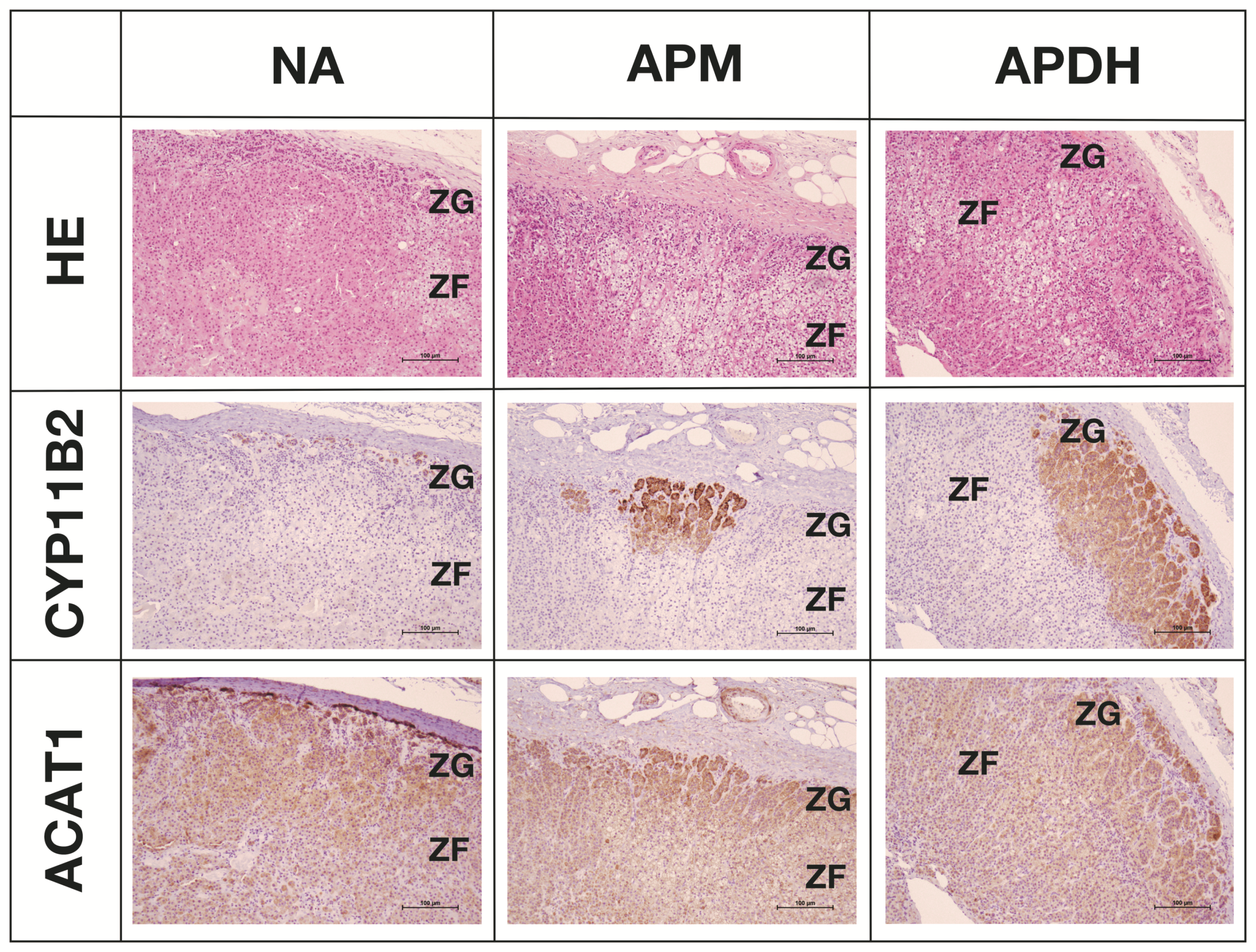
2.5. Expression and Localization of ACAT1 in the Adrenal Cortex

We examined ACAT1 expression in the adrenal cortex. We assessed the localization of CYP11B2 and ACAT1 expression in APNs, APMs, and APDH (Figure 5). The results showed that ACAT1 is expressed in all layers of the adrenal cortex. ACAT1 expression was particularly abundant in the zona glomerulosa (ZG), including CYP11B2-positive cells.

3. Discussion

To the best of our knowledge, this is the first study to demonstrate that YM750 suppresses CYP11B2 expression and aldosterone secretion by inhibiting downstream calcium signaling and inducing expression of NURR1.
YM750 reduces the concentration of intracellular cholesteryl esters by inhibiting ACAT1 [20]. The results from this study showed that YM750 suppressed only KCl-stimulated CYP11B2 gene expression but showed no effect on Ang II- and forskolin-stimulated CYP11B2 expression. Additionally, YM750 did not suppress calcium ion influx into the cells. KCl stimulation depolarizes H295R cells and induces CYP11B2 expression via activation of intracellular calcium signaling [7]. Ang II binds to the angiotensin type 1 receptor and activates the phosphoinositide 3-kinase (PI3K) signaling pathway, MEK-ERK signaling cascade, and intracellular calcium signaling pathway. Forskolin activates adenylate cyclase, which in turn activates the cAMP-PKA signaling pathway. Ang II and forskolin induce CYP11B2 expression via activation of transcription factors such as NURR1 and cyclic AMP response element-binding protein [7]. These results suggest that YM750 suppressed intracellular calcium signaling activated by KCl-stimulated depolarization but showed no effect on the PI3K and cAMP-PKA signaling pathways, thus resulting in suppression of CYP11B2 expression specifically.
It has also been reported that CYP11B2 expression is regulated by transcription factors such as NURR1 and NGFIB [21]. NURR1 and NGFIB bind to the promoter region of CYP11B2 and regulate gene expression [19]. NGFIB binds to the promoter region of HSD3B2 and regulates gene expression [22]. YM750 suppressed NURR1 and NGFIB expression at high concentrations. This suggests that the suppression of NURR1 and NGFIB. This suggests that ACAT1 is associated with suppressed CYP11B2 expression via repression of NURR1 and NGFIB. NURR1 primarily regulates CYP11B2 expression, while NGFIB regulates the expression of other steroidogenic genes [21,23,24]. Therefore, it is hypothesized that YM750 selectively suppresses NURR1 expression as compared to NGFIB expression, thereby suppressing CYP11B2 expression specifically. In addition, TM750 suppressed CYP11B1 expression. It is known that H295R cells share common signal pathway related to the expression of CYP11B1 and CYP11B2 [25]. Therefore, it is postulated that CYP11B1 similarly responded to KCl-stimulated depolarization, compatible with inhibition of CYP11B1 expression by YM750. However, it is speculated that YM750 does not necessarily inhibit CYP11B1 in vivo because CYP11B1 expressed in the ZF but not in the ZG would be mainly regulated by ACTH-stimulated cAMP-PKA signaling pathway rather than potassium-stimulated depolarization. In addition, it remains unclear how YM750 effect on metabolism and cholesterol levels when it is clinically used as an aldosterone secretion inhibitor. Therefore, further studies are required for clarification in the future.
The results from this study also suggest that ACAT1 inhibition suppressed intracellular cholesterol accumulation. Cholesterol accumulation activates oxysterol-binding protein (OSBP) and suppresses the activation of extracellular signal-regulated protein kinase (ERK1/2) and mitogen-activated protein kinase (MAPK) signaling pathways [26]. OSBP is expressed in the adrenal gland (Human Protein Atlas: https://www.proteinatlas.org/ENSG00000110048-OSBP#gene_information, accessed on 5 October 2022 [27]) and may be affected by ACAT inhibitors. ERK1/2 activation induces CYP11B2 expression via NURR1 and NGFIB expression [22]. Although it can be hypothesized that ACAT1 inhibition suppresses CYP11B2 expression via activation of OSBP, confirmatory evidence in support of this hypothesis is yet to be established (Figure 6). Therefore, it awaits future investigation for clarification on this point. ATR-101, an ACAT1 inhibitor, is effective in the treatment of Cushing’s syndrome in dogs [28]. ATR-101 also moderately decreased aldosterone secretion in dogs, suggesting that ACAT1 inhibition may affect steroidogenesis in the adrenal gland.
In this study, we demonstrated that ACAT1 is widely expressed in the adrenal cortex and is abundant in the ZG, including APNs. ACAT1 is also expressed in APAs and cortisol-producing adenomas [16,29], suggesting that ACAT1 participates in steroid production in tumor tissues. NURR1 is also widely expressed in the ZG [30], indicating an association between ACAT1 and NURR1 in steroidogenesis. Additionally, APMs and APDH with a higher concentration of CYP11B2-positive cells were involved in aldosterone secretion to a greater extent as compared to APNs [8]. Therefore, these results suggest that ACAT1 expression plays an important role in excess aldosterone production in APAs, APMs, and APDH.

4. Materials and Methods

4.1. Drugs

Angiotensin II (Sigma, Burlington, MA, USA) was dissolved in phosphate-buffered saline (PBS) containing 0.1% bovine serum albumin (BSA; Sigma, St. Louis, MO, USA). Potassium chloride (KCl; Wako, Osaka, Japan) was dissolved in water. Forskolin was dissolved in ethanol (Wako Pure Chemical Industries, Osaka, Japan). YM750 (Cayman Chemical, Ann Arbor, MI, USA) was dissolved in dimethyl sulfoxide (Wako).

4.2. Cell Culture

H295R cells derived from human adrenocortical carcinoma were purchased from American Type Culture Collection and cultured in Dulbecco’s Modified Eagle Medium (DMEM)/Ham’s F-12 nutrient medium (Wako) supplemented with 10% fetal bovine serum (FBS, Life Technologies, Edina, MN, USA), 1.25 mg/mL BSA (Sigma), 1× insulin-transferrin-selenium solution (Life Technologies), 5.35 mg/mL linoleic acid (Sigma), and 1× penicillin-streptomycin solution (Wako). For angiotensin II-, KCl-, forskolin-, and YM750-induced stimulation, cells were cultured in DMEM (Wako) containing 1% charcoal-treated FBS with penicillin-streptomycin. The culture and stimulation methods were based on previous studies [31,32,33].

4.3. RNA Extraction, cDNA Synthesis, and Quantitative Reverse Trascription Polymerase Chain Reaction (RT-qPCR)

Total RNA was extracted using Sepasol-RNA I Super G (Nacalai Tesque, Kyoto, Japan) and cDNA was synthesized using the Prime Script RT Master Mix (TAKARA, Tokyo, Japan). Real-time RT-qPCR was performed using the Thermal Cycler Dice Real-Time System (TP800, Takara, Kusatsu, Japan) along with the THUNDERBIRD® Probe qPCR Mix (Toyobo, Osaka, Japan) for analyzing CYP11B2 and HSD3B2 and KAPA SYBR FAST qPCR kit (KAPA BIOSYSTEMS, Wilmington, MA, USA) for analyzing other genes. Primer sequences and TaqMan probes are listed in Table 1.

4.4. Cell Viability Assay

Two days after plating 2.5 × 10⁴ H295R cells into 96-well plates, the cells were incubated with and without 10 mM YM750 for 24 h and 20 mM KCl for 6 h. Cell viability was measured using the Cell Counting Kit-8 (Dojindo, Kumamoto, Japan) following the manufacturer’s instructions.

4.5. Aldosterone EIA

Two days after plating 3.0 × 10⁵ H295R cells into 24-well plates, the cells were incubated with and without 10 mM YM750 and 20 mM KCl for 24 h. Aldosterone and cortisol concentrations in the media were measured using the Aldosterone ELISA Kit (Cayman Chemical, Ann Arbor, MI, USA) following the manufacturer’s instructions.

4.6. Intracellular Calcium Ion Concentration Assay

Two days after plating 2.5 × 10⁴ H295R cells into 96-well plates, the cells were incubated with and without 10 mM YM750 for 24 h. The cells were then loaded with Fluo 4-AM (Dojindo, Rockville, MD, USA; 5 mg/mL) in the presence of 1.25 mM probenecid (Dojindo) and 0.04% Pluronic F-127 (Dojindo) for 1 h. Cells were then washed with PBS and the recording medium containing 1.25 mmol/L probenecid and 20 mM KCl was added to the media. Changes in intracellular calcium concentration were determined by measuring the fluorescence intensity (excitation wavelength, 485 nm; emission wavelength, 535 nm).

4.7. Human Adrenal Tissue Samples for Immunohistochemical Analysis

Three non-pathological adrenal glands (normal adrenal (NA) tissues), three multiple APMs (APM tissues), and three APDH (APDH tissues) were selected for immunohistochemical analysis based on information retrieved from the surgical pathology files from Tohoku University Hospital (Sendai, Japan). NA tissues of nephrectomy cases due to renal carcinoma were harvested and subsequently evaluated to confirm the absence of neoplastic invasion, necrosis, and other histopathological abnormalities. The research protocols were approved by the Ethics Committee of Tohoku University Graduate School of Medicine (Sendai, Japan). All patients read and signed informed consent forms that clearly stated the methodologies for the use of tissue samples and clinical data for diagnostic and scientific purposes in the present study. The research protocol was approved by the Institutional Review Board of the Tohoku University School of Medicine (approval number 2020-1-705).

4.8. Immunohistochemical Analysis

Immunohistochemical analysis using hematoxylin and eosin stains was performed on 3 μm thick tissue sections prepared from formalin-fixed, paraffin embedded blocks. The protocols for immunohistochemistry used in this study are summarized in Table 2 [16,34].

4.9. Statistical Analysis

All data are presented as mean ± standard error of the mean. Statistical analyses were performed using Welch’s analysis of variance using GraphPad Prism 9.

5. Conclusions

In this study, we demonstrated that YM750, an ACAT1 inhibitor, suppressed CYP11B2 expression and aldosterone secretion in H295R cells. Additionally, ACAT1 expression was confirmed in the ZG of normal adrenal glands and in non-neoplastic regions of PA. The results from this study suggest that YM750 may be a potential therapeutic agent for PA.

Author Contributions

Conceptualization, H.S. (Hiroki Shimada), A.S. and Y.N.; methodology, all authors.; formal analysis, H.S. (Hiroki Shimada), S.H., Y.O., I.S. and K.I.; investigation, all authors.; data curation, H.S. (Hiroki Shimada); writing—original draft preparation, H.S. and Y.N.; writing—review and editing, all authors.; supervision, Y.Y., A.Y., T.S., H.S. (Hironobu Sasano), A.S. and Y.N.; project administration, A.S. and Y.N.; funding acquisition, A.S. and Y.N. All authors have read and agreed to the published version of the manuscript.

Funding

This research was funded by JSPS KAKENHI Grant Number JP 18J11985.

Institutional Review Board Statement

The research protocols were approved by the Ethics Committee at Tohoku University Graduate School of Medicine (Sendai, Japan). All patients read and signed informed consent documents regarding the diagnostic and scientific use of tissue samples and clinical data. The research protocol of this study was approved by Institutional Review Board of Tohoku University School of Medicine (2020-1-705).

Informed Consent Statement

Informed consent was obtained from all subjects involved in the study.

Data Availability Statement

The data presented in this study are openly available online.

Acknowledgments

This study was conducted in cooperation with the common equipment room of Tohoku Medical and Pharmaceutical University Faculty of Medicine. We thank the members of the Department of Pathology, Tohoku University Graduate School of Medicine for their support. We thank the members of the Department of Molecular endocrinology, Tohoku University Graduate School of Medicine for their support.

Conflicts of Interest

The authors declare no conflict of interest.

References

  1. Funder, J.W. The Genetics of Primary Aldosteronism. Science 2011, 331, 685–686. [Google Scholar] [CrossRef] [PubMed]
  2. Rossi, G.P.; Bernini, G.; Caliumi, C.; Desideri, G.; Fabris, B.; Ferri, C.; Ganzaroli, C.; Giacchetti, G.; Letizia, C.; Maccario, M.; et al. A Prospective Study of the Prevalence of Primary Aldosteronism in 1,125 Hypertensive Patients. J. Am. Coll. Cardiol. 2006, 48, 2293–2300. [Google Scholar] [CrossRef] [PubMed]
  3. Williams, J.S.; Williams, G.H.; Raji, A.; Jeunemaitre, X.; Brown, N.J.; Hopkins, P.N.; Conlin, P.R. Prevalence of Primary Hyperaldosteronism in Mild to Moderate Hypertension without Hypokalaemia. J. Hum. Hypertens. 2005, 20, 129–136. [Google Scholar] [CrossRef] [PubMed] [Green Version]
  4. Mete, O.; Erickson, L.A.; Juhlin, C.C.; de Krijger, R.R.; Sasano, H.; Volante, M.; Papotti, M.G. Overview of the 2022 WHO Classification of Adrenal Cortical Tumors. Endocr. Pathol. 2022, 33, 155–196. [Google Scholar] [CrossRef]
  5. Omata, K.; Satoh, F.; Morimoto, R.; Ito, S.; Yamazaki, Y.; Nakamura, Y.; Anand, S.K.; Guo, Z.; Stowasser, M.; Sasano, H.; et al. Cellular and Genetic Causes of Idiopathic Hyperaldosteronism. Hypertension 2018, 72, 874–880. [Google Scholar] [CrossRef]
  6. Nishimoto, K.; Nakagawa, K.; Li, D.; Kosaka, T.; Oya, M.; Mikami, S.; Shibata, H.; Itoh, H.; Mitani, F.; Yamazaki, T.; et al. Adrenocortical Zonation in Humans under Normal and Pathological Conditions. J. Clin. Endocrinol. Metab. 2010, 95, 2296–2305. [Google Scholar] [CrossRef] [Green Version]
  7. Hattangady, N.G.; Olala, L.O.; Bollag, W.B.; Rainey, W.E. Acute and Chronic Regulation of Aldosterone Production. Mol. Cell. Endocrinol. 2012, 350, 151–162. [Google Scholar] [CrossRef] [Green Version]
  8. Yamazaki, Y.; Nakamura, Y.; Omata, K.; Ise, K.; Tezuka, Y.; Ono, Y.; Morimoto, R.; Nozawa, Y.; Gomez-Sanchez, C.E.; Tomlins, S.A.; et al. Histopathological Classification of Cross-Sectional Image–Negative Hyperaldosteronism. J. Clin. Endocrinol. Metab. 2017, 102, 1182–1192. [Google Scholar] [CrossRef]
  9. Scholl, U.I.; Goh, G.; Stölting, G.; de Oliveira, R.C.; Choi, M.; Overton, J.D.; Fonseca, A.L.; Korah, R.; Starker, L.F.; Kunstman, J.W.; et al. Somatic and Germline CACNA1D Calcium Channel Mutations in Aldosterone-Producing Adenomas and Primary Aldosteronism. Nat. Genet. 2013, 45, 1050–1054. [Google Scholar] [CrossRef]
  10. Beuschlein, F.; Boulkroun, S.; Osswald, A.; Wieland, T.; Nielsen, H.N.; Lichtenauer, U.D.; Penton, D.; Schack, V.R.; Amar, L.; Fischer, E.; et al. Somatic Mutations in ATP1A1 and ATP2B3 Lead to Aldosterone-Producing Adenomas and Secondary Hypertension. Nat. Genet. 2013, 45, 440–444. [Google Scholar] [CrossRef]
  11. Azizan, E.A.B.; Poulsen, H.; Tuluc, P.; Zhou, J.; Clausen, M.V.; Lieb, A.; Maniero, C.; Garg, S.; Bochukova, E.G.; Zhao, W.; et al. Somatic Mutations in ATP1A1 and CACNA1D Underlie a Common Subtype of Adrenal Hypertension. Nat. Genet. 2013, 45, 1055–1060. [Google Scholar] [CrossRef] [PubMed]
  12. Choi, M.; Scholl, U.I.; Yue, P.; Björklund, P.; Zhao, B.; Nelson-Williams, C.; Ji, W.; Cho, Y.; Patel, A.; Men, C.J.; et al. K+ Channel Mutations in Adrenal Aldosterone-Producing Adenomas and Hereditary Hypertension. Science 2011, 331, 768–772. [Google Scholar] [CrossRef] [PubMed] [Green Version]
  13. Nakamura, Y.; Yamazaki, Y.; Tezuka, Y.; Satoh, F.; Sasano, H. Expression of CYP11B2 in Aldosterone-Producing Adrenocortical Adenoma: Regulatory Mechanisms and Clinical Significance. Tohoku J. Exp. Med. 2016, 240, 183–190. [Google Scholar] [CrossRef] [PubMed] [Green Version]
  14. Scholl, U.I.; Abriola, L.; Zhang, C.; Reimer, E.N.; Plummer, M.; Kazmierczak, B.I.; Zhang, J.; Hoyer, D.; Merkel, J.S.; Wang, W.; et al. Macrolides Selectively Inhibit Mutant KCNJ5 Potassium Channels That Cause Aldosterone-Producing Adenoma. J. Clin. Investig. 2017, 127, 2739–2750. [Google Scholar] [CrossRef] [PubMed] [Green Version]
  15. Sparks, S.M.; Danger, D.P.; Hoekstra, W.J.; Leesnitzer, T.; Schotzinger, R.J.; Yates, C.M.; Becherer, J.D. Development of Highly Selective Pyrimidine-Based Aldosterone Synthase (CYP11B2) Inhibitors. ACS Med. Chem. Lett. 2019, 10, 1056–1060. [Google Scholar] [CrossRef] [PubMed]
  16. Harashima, S.; Yamazaki, Y.; Motomura, N.; Ono, Y.; Omata, K.; Tezuka, Y.; Morimoto, R.; Nakamura, Y.; Satoh, F.; Suzuki, H.; et al. Phenotype-Genotype Correlation in Aldosterone-Producing Adenomas Characterized by Intracellular Cholesterol Metabolism. J. Steroid Biochem. Mol. Biol. 2022, 221, 106116. [Google Scholar] [CrossRef]
  17. Lee, R.G.; Willingham, M.C.; Davis, M.A.; Skinner, K.A.; Rudel, L.L. Differential Expression of ACAT1 and ACAT2 among Cells within Liver, Intestine, Kidney, and Adrenal of Nonhuman Primates. J. Lipid Res. 2000, 41, 1991–2001. [Google Scholar] [CrossRef]
  18. Miller, W.L. Steroid Hormone Synthesis in Mitochondria. Mol. Cell. Endocrinol. 2013, 379, 62–73. [Google Scholar] [CrossRef]
  19. Szekeres, M.; Turu, G.; Orient, A.; Szalai, B.; Süpeki, K.; Cserzo, M.; Várnai, P.; Hunyady, L. Mechanisms of Angiotensin II-Mediated Regulation of Aldosterone Synthase Expression in H295R Human Adrenocortical and Rat Adrenal Glomerulosa Cells. Mol. Cell. Endocrinol. 2009, 302, 244–253. [Google Scholar] [CrossRef]
  20. Miike, T.; Shirahase, H.; Jino, H.; Kunishiro, K.; Kanda, M.; Kurahashi, K. Effects of an Anti-Oxidative ACAT Inhibitor on Apoptosis/Necrosis and Cholesterol Accumulation under Oxidative Stress in THP-1 Cell-Derived Foam Cells. Life Sci. 2008, 82, 79–84. [Google Scholar] [CrossRef]
  21. Bassett, M.H.; Suzuki, T.; Sasano, H.; White, P.C.; Rainey, W.E. The Orphan Nuclear Receptors NURR1 and NGFIB Regulate Adrenal Aldosterone Production. Mol. Endocrinol. 2004, 18, 279–290. [Google Scholar] [CrossRef] [PubMed] [Green Version]
  22. Ota, T.; Doi, M.; Yamazaki, F.; Yarimizu, D.; Okada, K.; Murai, I.; Hayashi, H.; Kunisue, S.; Nakagawa, Y.; Okamura, H. Angiotensin II Triggers Expression of the Adrenal Gland Zona Glomerulosa-Specific 3β-Hydroxysteroid Dehydrogenase Isoenzyme through De Novo Protein Synthesis of the Orphan Nuclear Receptors NGFIB and NURR1. Mol. Cell. Biol. 2014, 34, 3880–3894. [Google Scholar] [CrossRef] [PubMed] [Green Version]
  23. Bassett, M.H.; Suzuki, T.; Sasano, H.; de Vries, C.J.M.; Jimenez, P.T.; Carr, B.R.; Rainey, W.E. The Orphan Nuclear Receptor NGFIB Regulates Transcription of 3β-Hydroxysteroid Dehydrogenase: Implications for the control of adrenal functional zonation. J. Biol. Chem. 2004, 279, 37622–37630. [Google Scholar] [CrossRef] [PubMed] [Green Version]
  24. Yarimizu, D.; Doi, M.; Ota, T.; Okamura, H. Stimulus-Selective Induction of the Orphan Nuclear Receptor NGFIB Underlies Different Influences of Angiotensin II and Potassium on the Human Adrenal Gland Zona Glomerulosa-Specific 3β-HSD Isoform Gene Expression in Adrenocortical H295R Cells. Endocr. J. 2015, 62, 765–776. [Google Scholar] [CrossRef] [Green Version]
  25. Denner, K.; Rainey, W.E.; Pezzi, V.; Bird, I.M.; Bernhardt, R.; Mathis, J.M. Differential Regulation of 11β-Hydroxylase and Aldosterone Synthase in Human Adrenocortical H295R Cells. Mol. Cell. Endocrinol. 1996, 121, 87–91. [Google Scholar] [CrossRef]
  26. Wang, P.Y.; Weng, J.; Anderson, R.G.W. OSBP Is a Cholesterol-Regulated Scaffolding Protein in Control of ERK1/2 Activation. Science 2005, 307, 1472–1476. [Google Scholar] [CrossRef]
  27. Uhlén, M.; Fagerberg, L.; Hallström, B.M.; Lindskog, C.; Oksvold, P.; Mardinoglu, A.; Sivertsson, Å.; Kampf, C.; Sjöstedt, E.; Asplund, A.; et al. Tissue-Based Map of the Human Proteome. Science 2015, 347, 1260419. [Google Scholar] [CrossRef]
  28. Langlois, D.K.; Fritz, M.C.; Schall, W.D.; Bari Olivier, N.; Smedley, R.C.; Pearson, P.G.; Bailie, M.B.; Hunt, S.W. ATR-101, a Selective ACAT1 Inhibitor, Decreases ACTH-Stimulated Cortisol Concentrations in Dogs with Naturally Occurring Cushing’s Syndrome. BMC Endocr. Disord. 2018, 18, 24. [Google Scholar] [CrossRef] [Green Version]
  29. Motomura, N.; Yamazaki, Y.; Koga, D.; Harashima, S.; Gao, X.; Tezuka, Y.; Omata, K.; Ono, Y.; Morimoto, R.; Satoh, F.; et al. The Association of Cholesterol Uptake and Synthesis with Histology and Genotype in Cortisol-Producing Adenoma (CPA). Int. J. Mol. Sci. 2022, 23, 2174. [Google Scholar] [CrossRef]
  30. Lu, L.; Suzuki, T.; Yoshikawa, Y.; Murakami, O.; Miki, Y.; Moriya, T.; Bassett, M.H.; Rainey, W.E.; Hayashi, Y.; Sasano, H. Nur-Related Factor 1 and Nerve Growth Factor-Induced Clone B in Human Adrenal Cortex and Its Disorders. J. Clin. Endocrinol. Metab. 2004, 89, 4113–4118. [Google Scholar] [CrossRef]
  31. Suzuki, D.; Saito-Hakoda, A.; Ito, R.; Shimizu, K.; Parvin, R.; Shimada, H.; Noro, E.; Suzuki, S.; Fujiwara, I.; Kagechika, H.; et al. Suppressive Effects of RXR Agonist PA024 on Adrenal CYP11B2 Expression, Aldosterone Secretion and Blood Pressure. PLoS ONE 2017, 12, e0181055. [Google Scholar] [CrossRef] [PubMed] [Green Version]
  32. Shimada, H.; Kogure, N.; Noro, E.; Kudo, M.; Sugawara, K.; Sato, I.; Shimizu, K.; Kobayashi, M.; Suzuki, D.; Parvin, R.; et al. High Glucose Stimulates Expression of Aldosterone Synthase (CYP11B2) and Secretion of Aldosterone in Human Adrenal Cells. FEBS Open Bio 2017, 7, 1410–1421. [Google Scholar] [CrossRef] [PubMed]
  33. Lichtenauer, U.D.; Shapiro, I.; Osswald, A.; Meurer, S.; Kulle, A.; Reincke, M.; Riepe, F.; Beuschlein, F. Characterization of NCI-H295R Cells as an in Vitro Model of Hyperaldosteronism. Horm. Metab. Res. 2013, 45, 124–129. [Google Scholar] [CrossRef] [PubMed] [Green Version]
  34. Gomez-Sanchez, C.E.; Qi, X.; Velarde-Miranda, C.; Plonczynski, M.W.; Parker, C.R.; Rainey, W.; Satoh, F.; Maekawa, T.; Nakamura, Y.; Sasano, H.; et al. Development of Monoclonal Antibodies against Human CYP11B1 and CYP11B2. Mol. Cell. Endocrinol. 2014, 383, 111–117. [Google Scholar] [CrossRef] [PubMed]
Figure 1. Effect of YM750 on CYP11B2 expression induced by various stimuli. (A) Effect of YM750 on CYP11B2 expression induced by KCl (20 mM) stimulation. (B) Effect of YM750 on CYP11B2 expression induced by angiotensin II (100 nM) stimulation. (C) Effect of YM750 on CYP11B2 expression induced by forskolin (10 µM) stimulation. (D) Effect of YM750 (10 µM) on H295R cell proliferation. * p < 0.05, n = 3.
Ijms 23 12803 g001
Figure 2. Effect of YM750 on the expression of various steroidogenic enzymes and CYP11B2 related genes. Effect of YM750 on (A) NURR1, (B) NGFIB, (C) StAR, (D) CYP11A1, (E) CYP17A1, (F) CYP21A2, (G) HSD3B2 and (H) CYP11B1 expression induced by KCl stimulation. * p < 0.05, n = 3.
Ijms 23 12803 g002
Figure 3. Effect of YM750 (10 mM) on aldosterone secretion by KCl (20 mM) stimulation. * p < 0.05, n = 4.
Ijms 23 12803 g003
Figure 4. Effect of YM750 (10 mM) on intracellular calcium concentration by KCl (20 mM) stimulation. n = 4.
Ijms 23 12803 g004
Figure 5. Expression and localization of ACAT1 in the adrenal cortex. HE: hematoxylin and eosin stains, CYP11B2: IHC using anti-CYP11B2 antibody, ACAT1: IHC using anti-ACAT1 antibody, NA: normal adrenal, APM: aldosterone-producing micronodule, APDH: aldosterone-producing diffuse hyperplasia. Scale in the image indicates 100μm.
Ijms 23 12803 g005
Figure 6. Graphical summary based on our current results.
Ijms 23 12803 g006
Table 1. Primer set using RT-PCR.
GeneForward Primer (5′→3′)Reverse Primer (3′→5′)
CYP11B2GGCAGAGGCAGAGATGCTGCTTGAGTTAGTGTCTCCACCAGGA
NURR1AGAGAAGATCCCTGGCTTCGCAAGACCACCCCATTGCAAAA
NGFIBCCTGGAGCTCTTCATCCTCCTGTCAATCCAGTCCCCGAAG
StARGCATCCTTAGCAACCAAGAGTCACTTTGTCCCCATTGTCC
CYP11A1TTCCGCTTTGCCTTTGAGTCTGGCATCAATGAATCGCTGG
CYP17A1CAGAATGTGGGTTTCAGCCGCTCACCGATGCTGGAGTCAA
CYP21A2AGACTACTCCCTGCTCTGGACTCATGCGCTCACAGAACTC
HSD3B2GCGGCTAATGGGTGGAATCTACATTGTTGTTCAGGGCCTCAT
CYP11B1GGCAGAGGCAGAGATGCTGTCTTGGGTTAGTGTCTCCACCTG
GAPDHATCCCATCACCATCTTCCAGATGAGTCCTTCCACGATACC
GeneProbe
CYP11B2[6-FAM]CTGCACCACGTGCTGAAGCACT[TAMPA6-FAM]
HSD3B2[6-FAM]TGATACCTTGTACACTTGTGC[TAMPA6-FAM]
CYP11B1[6-FAM]TGCTGCACCATGTGCTGAAACACCT[TAMRA-6-FAM]
Table 2. Method of immunohistochemical analysis.
Primary AntibodyDilutionSpeciesCloneCompanyAntigen Retrieval Treatment
ACAT11:100RabbitMonoclonal (EPR10359)AbcamAC 121 °C 5 min, pH = 9 Buffer
CYP11B21:500MouseMonoclonalGomez-Sanchez et al., 2014 [34]AC 121 °C 5 min, pH high Buffer
Publisher’s Note: MDPI stays neutral with regard to jurisdictional claims in published maps and institutional affiliations.

