
BODIPYs in PDT: A Journey through the Most Interesting Molecules Produced in the Last 10 Years

Abstract
:1. Introduction
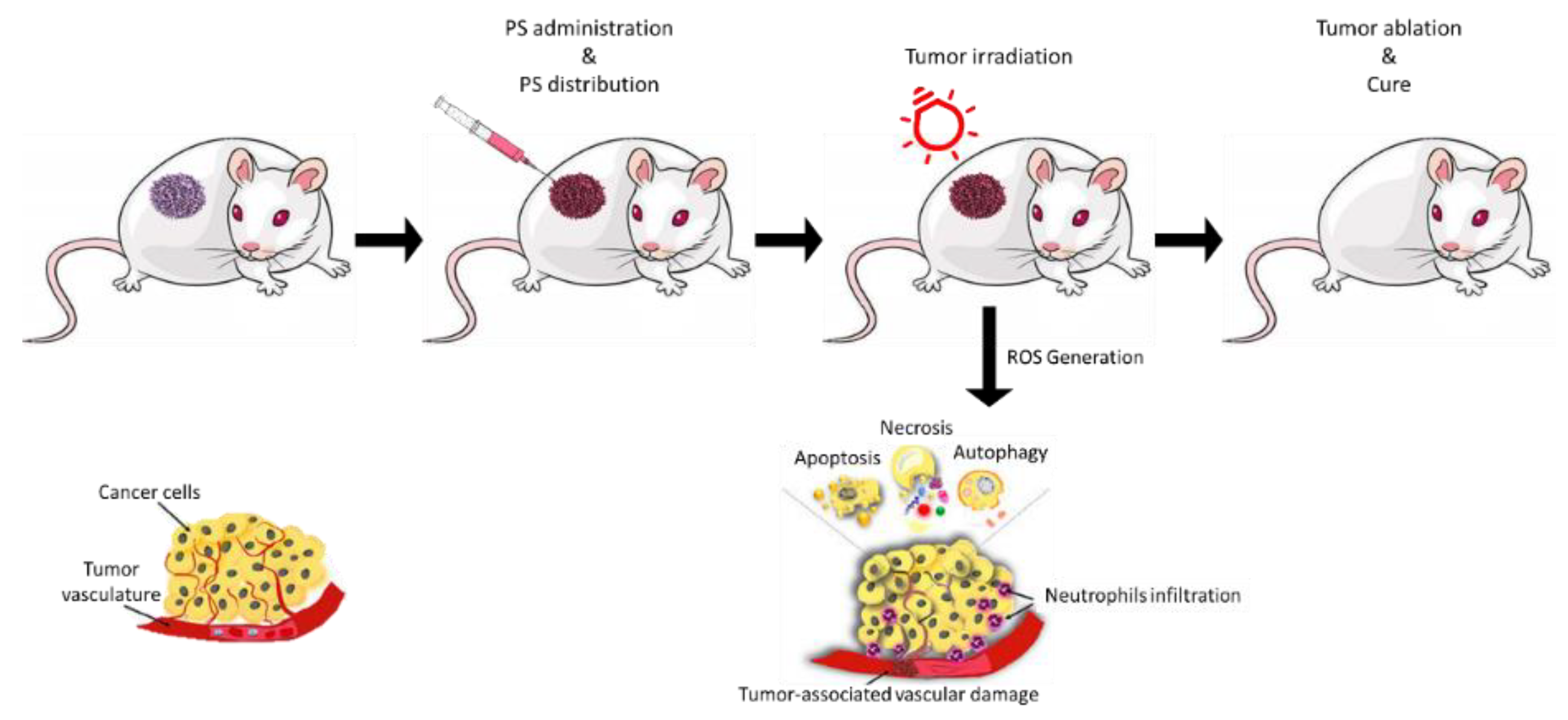
1.1. Photodynamic Therapy (PDT)
1.2. BODIPYs
2. BODIPYs Studies
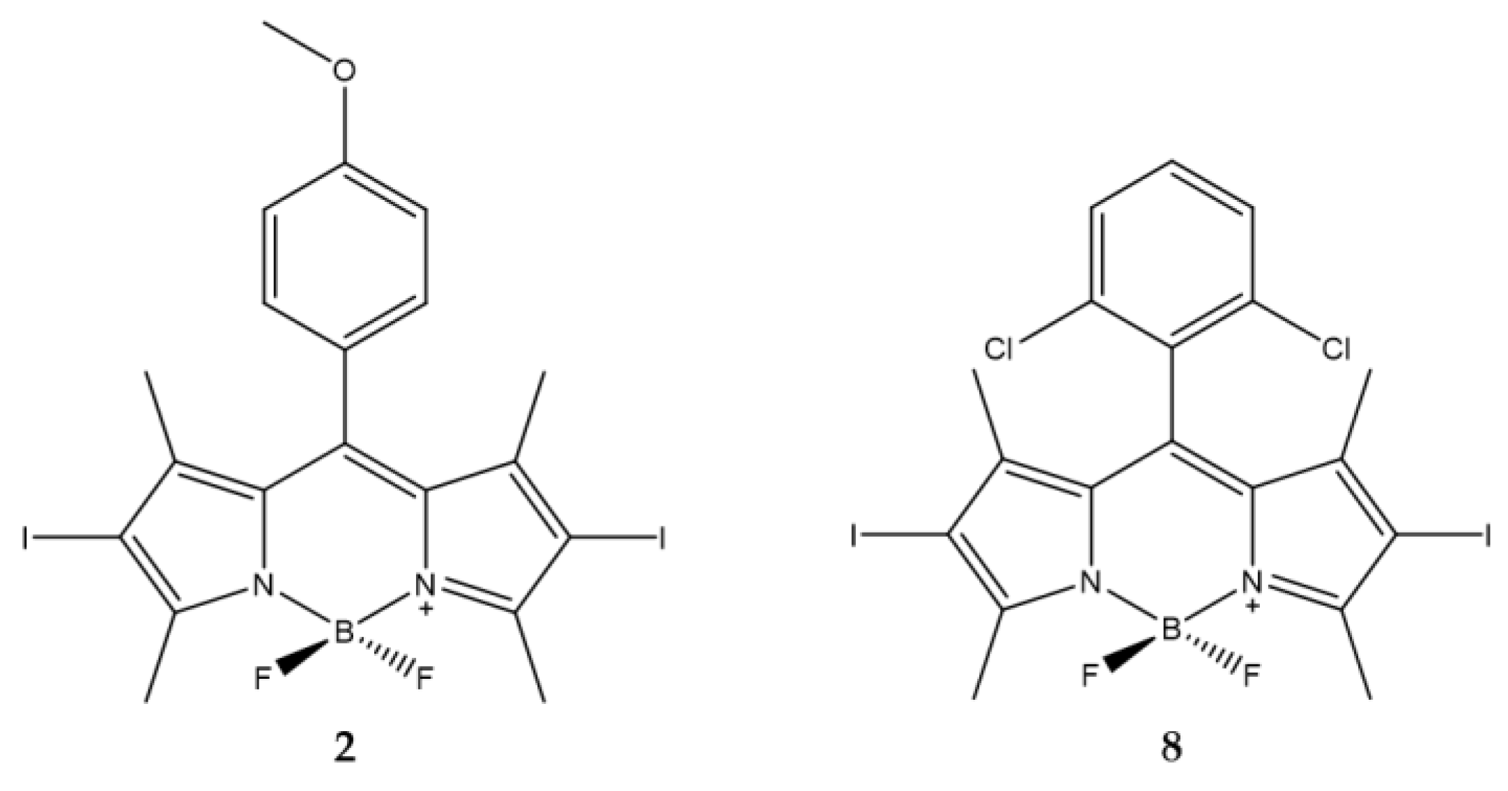
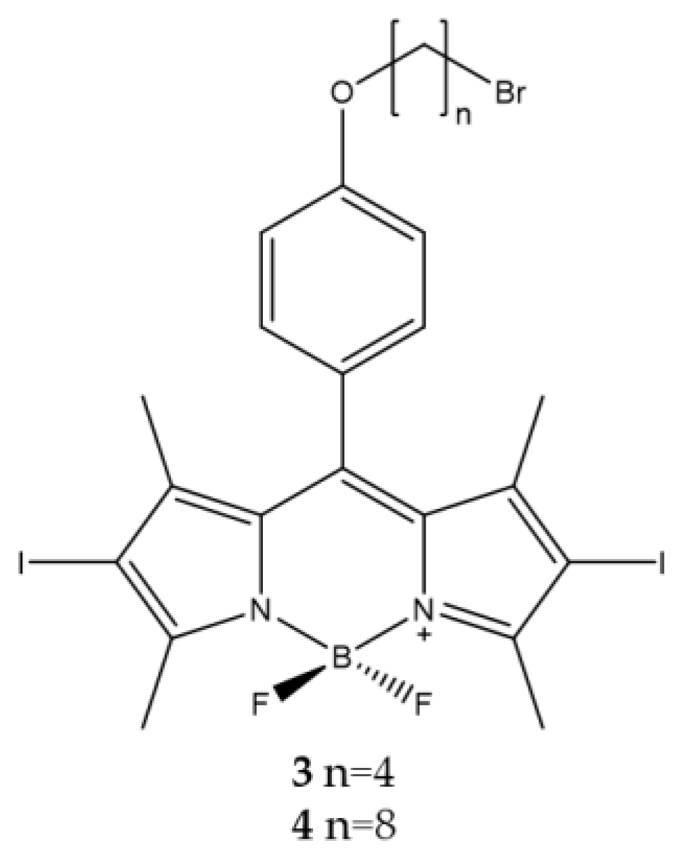
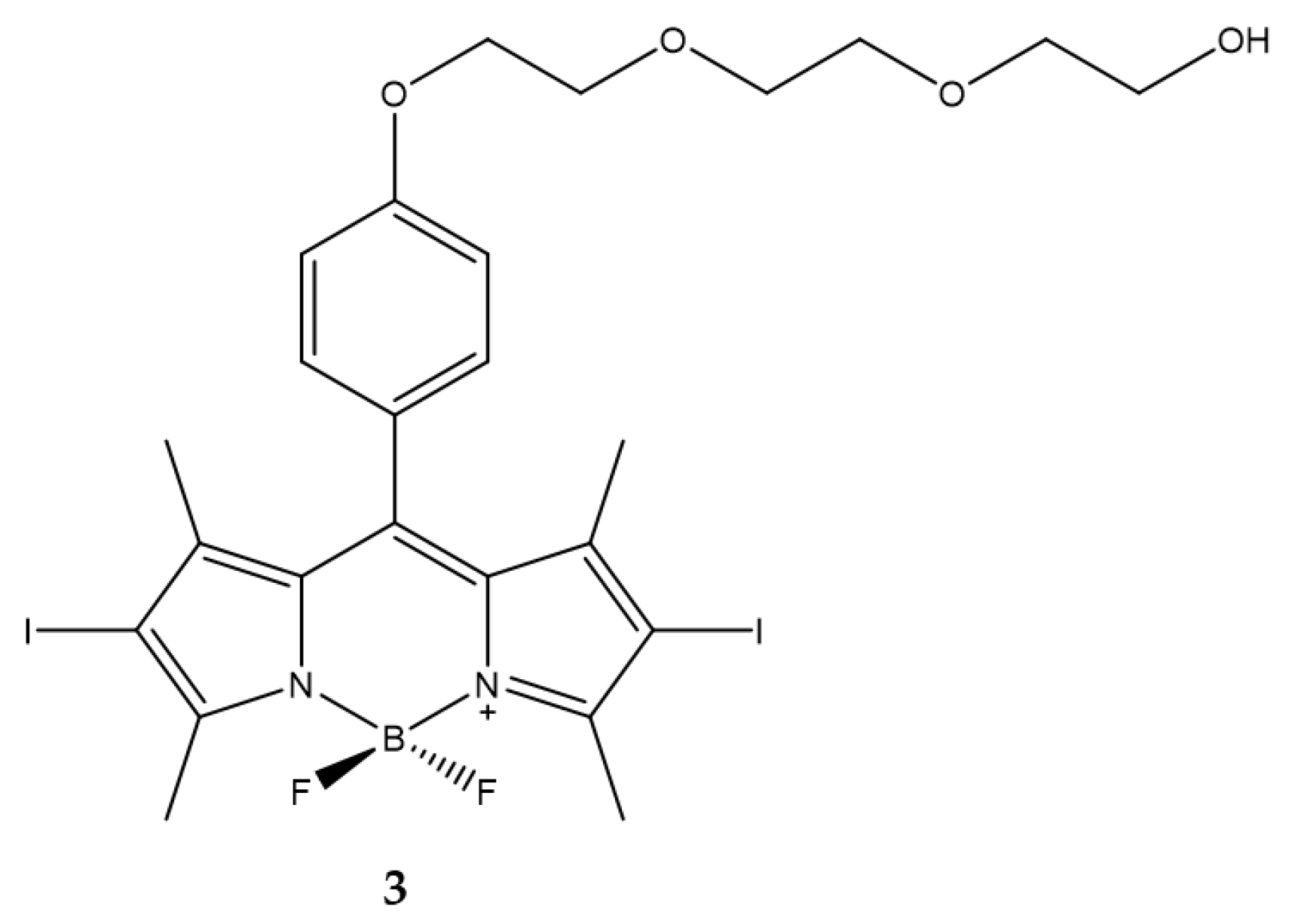
2.1. Structure-Activity
2.2. Singlet Oxygen Generation
2.3. Uptake and Targeting
2.3.1. Uptake
2.3.2. Targeting
Lysosomal Targeting
Reticulum Targeting
Mitochondrial Targeting
Cell Membrane Targeting
2.4. Sensitive Activity
Funding
Conflicts of Interest
References
- Dougherty, T.J.; Gomer, C.J.; Henderson, B.W.; Jori, G.; Kessel, D.; Korbelik, M.; Moan, J.; Peng, Q. Photodynamic therapy. J. Natl. Cancer Inst. 1998, 90, 889–905. [Google Scholar] [CrossRef] [PubMed]
- Chilakamarthi, U.; Giribabu, L. Photodynamic Therapy: Past, Present and Future. Chem. Rec. 2017, 17, 775–802. [Google Scholar] [CrossRef] [PubMed]
- Blum, H.F. Photodynamic action and diseases caused by light. Am. Chem. Soc. Monogr. 1941, 85, 283–295. [Google Scholar] [CrossRef]
- Zhou, Z.; Song, J.; Nie, L.; Chen, X. Reactive oxygen species generating systems meeting challenges of photodynamic cancer therapy. Chem. Soc. Rev. 2016, 45, 6597–6626. [Google Scholar] [CrossRef] [PubMed]
- Allison, R.R.; Downie, G.H.; Cuenca, R.; Hu, X.H.; Childs, C.J.; Sibata, C.H. Photosensitizers in clinical PDT. Photodiagnosis Photodyn. Ther. 2004, 1, 27–42. [Google Scholar] [CrossRef]
- Juzeniene, A.; Peng, Q.; Moan, J. Milestones in the development of photodynamic therapy and fluorescence diagnosis. Photochem. Photobiol. Sci. 2007, 6, 1234–1245. [Google Scholar] [CrossRef]
- Daniell, M.D.; Hill, J.S. A history of photodynamic therapy. Aust. N. Z. J. Surg. 1991, 61, 340–348. [Google Scholar] [CrossRef] [PubMed]
- Castellani, A.; Pace, G.P.; Concioli, M. Photodynamic effect of haematoporphyrin on blood microcirculation. J. Pathol. Bacteriol. 1963, 86, 99–102. [Google Scholar] [CrossRef] [PubMed]
- de Gruijl, F.R.; Van der Leun, J.C. Estimate of the wavelength dependency of ultraviolet carcinogenesis in humans and its relevance to the risk assessment of a stratospheric ozone depletion. Health Phys. 1994, 67, 319–325. [Google Scholar] [CrossRef]
- Dolmans, D.E.; Fukumura, D.; Jain, R.K. Photodynamic therapy for cancer. Nat. Rev. Cancer 2003, 3, 380–387. [Google Scholar] [CrossRef]
- Agostinis, P.; Berg, K.; Cengel, K.A.; Foster, T.H.; Girotti, A.W.; Gollnick, S.O.; Hahn, S.M.; Hamblin, M.R.; Juzeniene, A.; Kessel, D.; et al. Photodynamic therapy of cancer: An update. CA Cancer J. Clin. 2011, 61, 250–281. [Google Scholar] [CrossRef] [PubMed]
- Ledoux-Lebards, C. Action de la lumiere sur la toxicite de l’éosine et de quelques autres substances. Ann. Inst. Pasteur 1902, 16, 587–593. [Google Scholar]
- Foote, C.S. Mechanisms of photosensitized oxidation. There are several different types of photosensitized oxidation which may be important in biological systems. Science 1968, 162, 963–970. [Google Scholar] [CrossRef] [PubMed]
- Moan, J.; Wold, E. Detection of singlet oxygen production by ESR. Nature 1979, 279, 450–451. [Google Scholar] [CrossRef] [PubMed]
- Moan, J.; Sommer, S. Oxygen dependence of the photosensitizing effect of hematoporphyrin derivative in NHIK 3025 cells. Cancer Res. 1985, 45, 1608–1610. [Google Scholar] [PubMed]
- Devasagayam, T.P.A.; Kamat, J.P. Biological significance of singlet oxygen. Indian J. Exp. Biol. 2002, 40, 680–692. [Google Scholar]
- Davies, M.J. Singlet oxygen-mediated damage to proteins and its consequences. Biochem. Biophys. Res. Commun. 2003, 305, 761–770. [Google Scholar] [CrossRef]
- Juzeniene, A.; Nielsen, K.P.; Moan, J. Biophysical aspects of photodynamic therapy. J. Environ. Pathol. Toxicol. Oncol. 2006, 25, 7–28. [Google Scholar] [CrossRef] [PubMed]
- Schmidt, R. Photosensitized Generation of Singlet Oxygen. Photochem. Photobiol. 2007, 82, 1161–1177. [Google Scholar] [CrossRef]
- Sharman, W.M.; Allen, C.M.; van Lier, J.E. Role of activated oxygen species in photodynamic therapy. Methods Enzymol. 2000, 319, 376–400. [Google Scholar] [CrossRef]
- Li, C. Breast Cancer Epidemiology, 1st ed.; Springer: New York, NY, USA, 2010; pp. 1–417. [Google Scholar]
- Castano, A.P.; Demidova, T.N.; Hamblin, M.R. Mechanisms in photodynamic therapy: Part one-photosensitizers, photochemistry and cellular localization. Photodiagnosis Photodyn. Ther. 2004, 1, 279–293. [Google Scholar] [CrossRef]
- Robertson, C.A.; Evans, D.H.; Abrahamse, H. Photodynamic therapy (PDT): A short review on cellular mechanisms and cancer research applications for PDT. J. Photochem. Photobiol. B 2009, 96, 1–8. [Google Scholar] [CrossRef] [PubMed]
- Kwiatkowski, S.; Knap, B.; Przystupski, D.; Saczko, J.; Kedzierska, E.; Knap-Czop, K.; Kotlinska, J.; Michel, O.; Kotowski, K.; Kulbacka, J. Photodynamic therapy-mechanisms, photosensitizers and combinations. Biomed. Pharmacother. 2018, 106, 1098–1107. [Google Scholar] [CrossRef] [PubMed]
- Schenck, G.O. Photosensitized reactions with molecular oxygen. Naturwissenschaften 1984, 71, 28–29. [Google Scholar]
- Foote, C.S.; Wexler, S. Singlet Oxygen. A Probable Intermediate in Photosensitized Autoxidations. J. Am. Chem. Soc. 2002, 86, 3880–3881. [Google Scholar] [CrossRef]
- Niedre, M.J.; Secord, A.J.; Patterson, M.S.; Wilson, B.C. In vitro tests of the validity of singlet oxygen luminescence measurements as a dose metric in photodynamic therapy. Cancer Res. 2003, 63, 7986–7994. [Google Scholar] [PubMed]
- Weishaupt, K.R.; Gomer, C.J.; Dougherty, T.J. Identification of singlet oxygen as the cytotoxic agent in photoinactivation of a murine tumor. Cancer Res. 1976, 36, 2326–2329. [Google Scholar]
- Fisher, A.M.; Murphree, A.L.; Gomer, C.J. Clinical and preclinical photodynamic therapy. Lasers Surg. Med. 1995, 17, 2–31. [Google Scholar] [CrossRef]
- Dos Santos, A.F.; Terra, L.F.; Wailemann, R.A.M.; Oliveira, T.C.; de Morais Gomes, V.; Mineiro, M.F.; Meotti, F.C.; Bruni-Cardoso, A.; Baptista, M.S.; Labriola, L. Methylene blue photodynamic therapy induces selective and massive cell death in human breast cancer cells. BMC Cancer 2017, 17, 194–209. [Google Scholar] [CrossRef]
- Auten, R.L.; Davis, J.M. Oxygen toxicity and reactive oxygen species: The devil is in the details. Pediatr. Res. 2009, 66, 121–127. [Google Scholar] [CrossRef]
- van Duijnhoven, F.H.; Aalbers, R.I.; Rovers, J.P.; Terpstra, O.T.; Kuppen, P.J. The immunological consequences of photodynamic treatment of cancer, a literature review. Immunobiology 2003, 207, 105–113. [Google Scholar] [CrossRef]
- Mroz, P.; Hashmi, J.T.; Huang, Y.Y.; Lange, N.; Hamblin, M.R. Stimulation of anti-tumor immunity by photodynamic therapy. Expert Rev. Clin. Immunol. 2011, 7, 75–91. [Google Scholar] [CrossRef] [PubMed]
- Mroz, P.; Hamblin, M.R. The immunosuppressive side of PDT. Photochem. Photobiol. Sci. 2011, 10, 751–758. [Google Scholar] [CrossRef] [PubMed]
- Reginato, E.; Wolf, P.; Hamblin, M.R. Immune response after photodynamic therapy increases anti-cancer and anti-bacterial effects. World J. Immunol. 2014, 4, 1–11. [Google Scholar] [CrossRef] [PubMed]
- Lobo, A.C.S.; Gomes-da-Silva, L.C.; Rodrigues-Santos, P.; Cabrita, A.; Santos-Rosa, M.; Arnaut, L.G. Immune Responses after Vascular Photodynamic Therapy with Redaporfin. J. Clin. Med. 2019, 9, 104. [Google Scholar] [CrossRef] [PubMed]
- Falk-Mahapatra, R.; Gollnick, S.O. Photodynamic Therapy and Immunity: An Update. Photochem. Photobiol. 2020, 96, 550–559. [Google Scholar] [CrossRef]
- Nkune, N.W.; Simelane, N.W.N.; Montaseri, H.; Abrahamse, H. Photodynamic Therapy-Mediated Immune Responses in Three-Dimensional Tumor Models. Int. J. Mol. Sci. 2021, 22, 12618. [Google Scholar] [CrossRef]
- Gilchrest, B.A. Photodynamic therapy and selected off-label uses. In Winter Clinical Dermatology Conference-Hawaii; Tuleya, S., Ed.; HMP Communications, LLC: Kohala Coast, HI, USA, 2010; pp. 10–12. [Google Scholar]
- Kamkaew, A.; Lim, S.H.; Lee, H.B.; Kiew, L.V.; Chung, L.Y.; Burgess, K. BODIPY dyes in photodynamic therapy. Chem. Soc. Rev. 2013, 42, 77–88. [Google Scholar] [CrossRef]
- Liu, J.; Zheng, L.; Li, Y.; Zhang, Z.; Zhang, L.; Shen, L.; Zhang, X.; Qiao, H. Effect of DTPP-mediated photodynamic therapy on cell morphology, viability, cell cycle, and cytotoxicity in a murine lung adenocarcinoma cell line. Lasers Med. Sci. 2015, 30, 181–191. [Google Scholar] [CrossRef]
- Meyer-Betz, F. Untersuchungen uber die biologische (photodynamische) Wirkung des Hamatoporphyrings und anderer Derivate des Blut- und Gallenfarbstoffs. Dtsch. Arch. Klin. Med. 1913, 112, 476–503. [Google Scholar]
- Ackroyd, R.; Kelty, C.; Brown, N.; Reed, M. The history of photodetection and photodynamic therapy. Photochem. Photobiol. 2001, 74, 656–669. [Google Scholar] [CrossRef]
- Ormond, A.B.; Freeman, H.S. Dye Sensitizers for Photodynamic Therapy. Materials 2013, 6, 817–840. [Google Scholar] [CrossRef] [PubMed]
- Hage, R.; Ferreira, J.; Bagnato, V.S.; Vollet, E.D.; Plapler, H. Pharmacokinetics of Photogem Using Fluorescence Spectroscopy in Dimethylhydrazine-Induced Murine Colorectal Carcinoma. Int. J. Photoenergy 2012, 2012, 1–8. [Google Scholar] [CrossRef]
- Allison, R.R.; Sibata, C.H. Oncologic photodynamic therapy photosensitizers: A clinical review. Photodiagnosis Photodyn. Ther. 2010, 7, 61–75. [Google Scholar] [CrossRef] [PubMed]
- Hammerer, F.; Poyer, F.; Fourmois, L.; Chen, S.; Garcia, G.; Teulade-Fichou, M.P.; Maillard, P.; Mahuteau-Betzer, F. Mitochondria-targeted cationic porphyrin-triphenylamine hybrids for enhanced two-photon photodynamic therapy. Bioorganic Med. Chem. 2018, 26, 107–118. [Google Scholar] [CrossRef] [PubMed]
- Gilson, D.; Ash, D.; Driver, I.; Feather, J.W.; Brown, S. Therapeutic ratio of photodynamic therapy in the treatment of superficial tumours of skin and subcutaneous tissues in man. Br. J. Cancer 1988, 58, 665–667. [Google Scholar] [CrossRef] [PubMed]
- Hausser, K.W.; Vahle, W. Die abhangigkeit des lichterythems und der pigmentbildung von der schwingungszahl (wellenlange der erregenden strahlung). Strahlentherapie 1931, 13, 41–71. [Google Scholar]
- Chevalier, S.; Anidjar, M.; Scarlata, E.; Hamel, L.; Scherz, A.; Ficheux, H.; Borenstein, N.; Fiette, L.; Elhilali, M. Preclinical study of the novel vascular occluding agent, WST11, for photodynamic therapy of the canine prostate. J. Urol. 2011, 186, 302–309. [Google Scholar] [CrossRef]
- Triesscheijn, M.; Ruevekamp, M.; Aalders, M.; Baas, P.; Stewart, F.A. Outcome of mTHPC mediated photodynamic therapy is primarily determined by the vascular response. Photochem. Photobiol. 2005, 81, 1161–1167. [Google Scholar] [CrossRef]
- Detty, M.R.; Gibson, S.L.; Wagner, S.J. Current clinical and preclinical photosensitizers for use in photodynamic therapy. J. Med. Chem. 2004, 47, 3897–3915. [Google Scholar] [CrossRef]
- Dovigo, L.N.; Pavarina, A.C.; Ribeiro, A.P.; Brunetti, I.L.; Costa, C.A.; Jacomassi, D.P.; Bagnato, V.S.; Kurachi, C. Investigation of the photodynamic effects of curcumin against Candida albicans. Photochem. Photobiol. 2011, 87, 895–903. [Google Scholar] [CrossRef]
- Wohrle, D.; Hirth, A.; Bogdahn-Rai, T.; Schnurpfeil, G.; Shopova, M. Photodynamic therapy of cancer: Second and third generations of photosensitizers. Russ. Chem. Bull. 1998, 47, 807–816. [Google Scholar] [CrossRef]
- Li, L.; Nguyen, B.; Burgess, K. Functionalization of the 4,4-difluoro-4-bora-3a,4a-diaza-s-indacene (BODIPY) core. Bioorganic Med. Chem. Lett. 2008, 18, 3112–3116. [Google Scholar] [CrossRef] [PubMed]
- Awuah, S.G.; You, Y. Boron dipyrromethene (BODIPY)-based photosensitizers for photodynamic therapy. RSC Adv. 2012, 2, 11169–11183. [Google Scholar] [CrossRef]
- Yuster, P.; Weissman, S.I. Effects of Perturbations on Phosphorescence: Luminescence of Metal Organic Complexes. J. Chem. Phys. 1949, 17, 1182–1188. [Google Scholar] [CrossRef]
- Treibs, A.; Kreuzer, F.-H. Difluorboryl-Komplexe von Di- und Tripyrrylmethenen. Justus Liebigs Ann. Chem. 1968, 718, 208–223. [Google Scholar] [CrossRef]
- Ulrich, G.; Ziessel, R.; Harriman, A. The chemistry of fluorescent bodipy dyes: Versatility unsurpassed. Angew. Chem. Int. Ed. Engl. 2008, 47, 1184–1201. [Google Scholar] [CrossRef]
- Karolin, J.; Johansson, L.B.A.; Strandberg, L.; Ny, T. Fluorescence and Absorption Spectroscopic Properties of Dipyrrometheneboron Difluoride (BODIPY) Derivatives in Liquids, Lipid Membranes, and Proteins. J. Am. Chem. Soc. 2002, 116, 7801–7806. [Google Scholar] [CrossRef]
- Yogo, T.; Urano, Y.; Ishitsuka, Y.; Maniwa, F.; Nagano, T. Highly efficient and photostable photosensitizer based on BODIPY chromophore. J. Am. Chem. Soc. 2005, 127, 12162–12163. [Google Scholar] [CrossRef]
- Wittmershaus, B.P.; Skibicki, J.J.; McLafferty, J.B.; Zhang, Y.-Z.; Swan, S. Spectral properties of single BODIPY dyes in polystyrene microspheres and in solutions. J. Fluoresc. 2001, 11, 119–128. [Google Scholar] [CrossRef]
- Ventura, B.; Marconi, G.; Bröring, M.; Krüger, R.; Flamigni, L. Bis(BF2)-2,2′-bidipyrrins, a class of BODIPY dyes with new spectroscopic and photophysical properties. New J. Chem. 2009, 33, 428–438. [Google Scholar] [CrossRef]
- Kolemen, S.; Bozdemir, O.A.; Cakmak, Y.; Barin, G.; Erten-Ela, S.; Marszalek, M.; Yum, J.-H.; Zakeeruddin, S.M.; Nazeeruddin, M.K.; Grätzel, M.; et al. Optimization of distyryl-Bodipy chromophores for efficient panchromatic sensitization in dye sensitized solar cells. Chem. Sci. 2011, 2, 949–954. [Google Scholar] [CrossRef]
- Koziar, J.C.; Cowan, D.O. Photochemical heavy-atom effects. Acc. Chem. Res. 2002, 11, 334–341. [Google Scholar] [CrossRef]
- Loudet, A.; Burgess, K. BODIPY dyes and their derivatives: Syntheses and spectroscopic properties. Chem. Rev. 2007, 107, 4891–4932. [Google Scholar] [CrossRef] [PubMed]
- Banfi, S.; Nasini, G.; Zaza, S.; Caruso, E. Synthesis and photo-physical properties of a series of BODIPY dyes. Tetrahedron 2013, 69, 4845–4856. [Google Scholar] [CrossRef]
- Shivran, N.; Tyagi, M.; Mula, S.; Gupta, P.; Saha, B.; Patro, B.S.; Chattopadhyay, S. Syntheses and photodynamic activity of some glucose-conjugated BODIPY dyes. Eur. J. Med. Chem. 2016, 122, 352–365. [Google Scholar] [CrossRef] [PubMed]
- DeRosa, M. Photosensitized singlet oxygen and its applications. Coord. Chem. Rev. 2002, 233–234, 351–371. [Google Scholar] [CrossRef]
- Fagnoni, M. Modern Molecular Photochemistry of Organic Molecules. By Nicholas J. Turro, V. Ramamurthy and Juan C. Scaiano. Angew. Chem. Int. Ed. 2010, 49, 6709–6710. [Google Scholar] [CrossRef]
- Pham, T.C.; Nguyen, V.N.; Choi, Y.; Lee, S.; Yoon, J. Recent Strategies to Develop Innovative Photosensitizers for Enhanced Photodynamic Therapy. Chem. Rev. 2021, 121, 13454–13619. [Google Scholar] [CrossRef]
- Banfi, S.; Caruso, E.; Zaza, S.; Mancini, M.; Gariboldi, M.B.; Monti, E. Synthesis and photodynamic activity of a panel of BODIPY dyes. J. Photochem. Photobiol. B 2012, 114, 52–60. [Google Scholar] [CrossRef]
- Banfi, S.; Caruso, E.; Buccafurni, L.; Murano, R.; Monti, E.; Gariboldi, M.; Papa, E.; Gramatica, P. Comparison between 5,10,15,20-tetraaryl- and 5,15-diarylporphyrins as photosensitizers: Synthesis, photodynamic activity, and quantitative structure-activity relationship modeling. J. Med. Chem. 2006, 49, 3293–3304. [Google Scholar] [CrossRef]
- Frimayanti, N.; Yam, M.L.; Lee, H.B.; Othman, R.; Zain, S.M.; Rahman, N.A. Validation of quantitative structure-activity relationship (QSAR) model for photosensitizer activity prediction. Int. J. Mol. Sci. 2011, 12, 8626–8644. [Google Scholar] [CrossRef] [PubMed]
- Henderson, B.W.; Bellnier, D.A.; Greco, W.R.; Sharma, A.; Pandey, R.K.; Vaughan, L.A.; Weishaupt, K.R.; Dougherty, T.J. An in vivo quantitative structure-activity relationship for a congeneric series of pyropheophorbide derivatives as photosensitizers for photodynamic therapy. Cancer Res. 1997, 57, 4000–4007. [Google Scholar]
- Margaron, P.; Gregoire, M.J.; Scasnar, V.; Ali, H.; van Lier, J.E. Structure-photodynamic activity relationships of a series of 4-substituted zinc phthalocyanines. Photochem. Photobiol. 1996, 63, 217–223. [Google Scholar] [CrossRef] [PubMed]
- Zagami, R.; Sortino, G.; Caruso, E.; Malacarne, M.C.; Banfi, S.; Patane, S.; Monsu Scolaro, L.; Mazzaglia, A. Tailored-BODIPY/Amphiphilic Cyclodextrin Nanoassemblies with PDT Effectiveness. Langmuir 2018, 34, 8639–8651. [Google Scholar] [CrossRef] [PubMed]
- Barut, B.; Yalcin, C.O.; Sari, S.; Coban, O.; Keles, T.; Biyiklioglu, Z.; Abudayyak, M.; Demirbas, U.; Ozel, A. Novel water soluble BODIPY compounds: Synthesis, photochemical, DNA interaction, topoisomerases inhibition and photodynamic activity properties. Eur. J. Med. Chem. 2019, 183, 111685. [Google Scholar] [CrossRef] [PubMed]
- Wang, C.; Qian, Y. A water soluble carbazolyl-BODIPY photosensitizer with an orthogonal D-A structure for photodynamic therapy in living cells and zebrafish. Biomater. Sci. 2020, 8, 830–836. [Google Scholar] [CrossRef]
- Caruso, E.; Orlandi, V.T.; Malacarne, M.C.; Martegani, E.; Scanferla, C.; Pappalardo, D.; Vigliotta, G.; Izzo, L. Bodipy-Loaded Micelles Based on Polylactide as Surface Coating for Photodynamic Control of Staphylococcus aureus. Coatings 2021, 11, 223. [Google Scholar] [CrossRef]
- Turan, I.S.; Cakmak, F.P.; Yildirim, D.C.; Cetin-Atalay, R.; Akkaya, E.U. Near-IR absorbing BODIPY derivatives as glutathione-activated photosensitizers for selective photodynamic action. Chemistry 2014, 20, 16088–16092. [Google Scholar] [CrossRef]
- Malacarne, M.C.; Banfi, S.; Caruso, E. In vitro photodynamic treatment of cancer cells induced by aza-BODIPYs. Photochem. Photobiol. Sci. 2020, 19, 790–799. [Google Scholar] [CrossRef]
- Lincoln, R.; Van Kessel, A.T.M.; Zhang, W.; Cosa, G. A dormant BODIPY-acrolein singlet oxygen photosensitizer intracellularly activated upon adduct formation with cysteine residues. Photochem. Photobiol. Sci. 2019, 18, 2003–2011. [Google Scholar] [CrossRef]
- Prieto-Montero, R.; Prieto-Castaneda, A.; Sola-Llano, R.; Agarrabeitia, A.R.; Garcia-Fresnadillo, D.; Lopez-Arbeloa, I.; Villanueva, A.; Ortiz, M.J.; de la Moya, S.; Martinez-Martinez, V. Exploring BODIPY Derivatives as Singlet Oxygen Photosensitizers for PDT. Photochem. Photobiol. 2020, 96, 458–477. [Google Scholar] [CrossRef] [PubMed]
- Ziessel, R.; Ulrich, G.; Harriman, A. The chemistry of Bodipy: A new El Dorado for fluorescence tools. New J. Chem. 2007, 31, 496–501. [Google Scholar] [CrossRef]
- Caruso, E.; Gariboldi, M.; Sangion, A.; Gramatica, P.; Banfi, S. Synthesis, photodynamic activity, and quantitative structure-activity relationship modelling of a series of BODIPYs. J. Photochem. Photobiol. B 2017, 167, 269–281. [Google Scholar] [CrossRef] [PubMed]
- Caruso, E.; Malacarne, M.C.; Marras, E.; Papa, E.; Bertato, L.; Banfi, S.; Gariboldi, M.B. New BODIPYs for photodynamic therapy (PDT): Synthesis and activity on human cancer cell lines. Bioorganic Med. Chem. 2020, 28, 115737. [Google Scholar] [CrossRef] [PubMed]
- Ballestri, M.; Caruso, E.; Guerrini, A.; Ferroni, C.; Banfi, S.; Gariboldi, M.; Monti, E.; Sotgiu, G.; Varchi, G. Core-shell poly-methyl methacrylate nanoparticles covalently functionalized with a non-symmetric porphyrin for anticancer photodynamic therapy. J. Photochem. Photobiol. B 2018, 186, 169–177. [Google Scholar] [CrossRef] [PubMed]
- Kim, B.; Sui, B.; Yue, X.; Tang, S.; Tichy, M.G.; Belfield, K.D. In Vitro Photodynamic Studies of a BODIPY-Based Photosensitizer. Eur. J. Org. Chem. 2016, 2017, 25–28. [Google Scholar] [CrossRef]
- Lincoln, R.; Durantini, A.M.; Greene, L.E.; Martinez, S.R.; Knox, R.; Becerra, M.C.; Cosa, G. meso-Acetoxymethyl BODIPY dyes for photodynamic therapy: Improved photostability of singlet oxygen photosensitizers. Photochem. Photobiol. Sci. 2017, 16, 178–184. [Google Scholar] [CrossRef]
- Durantini, A.M.; Greene, L.E.; Lincoln, R.; Martinez, S.R.; Cosa, G. Reactive Oxygen Species Mediated Activation of a Dormant Singlet Oxygen Photosensitizer: From Autocatalytic Singlet Oxygen Amplification to Chemicontrolled Photodynamic Therapy. J. Am. Chem. Soc. 2016, 138, 1215–1225. [Google Scholar] [CrossRef]
- Epelde-Elezcano, N.; Martínez-Martínez, V.; Peña-Cabrera, E.; Gómez-Durán, C.F.A.; Arbeloa, I.L.; Lacombe, S. Modulation of singlet oxygen generation in halogenated BODIPY dyes by substitution at their meso position: Towards a solvent-independent standard in the vis region. RSC Adv. 2016, 6, 41991–41998. [Google Scholar] [CrossRef]
- Turan, I.S.; Yildiz, D.; Turksoy, A.; Gunaydin, G.; Akkaya, E.U. A Bifunctional Photosensitizer for Enhanced Fractional Photodynamic Therapy: Singlet Oxygen Generation in the Presence and Absence of Light. Angew. Chem. Int. Ed. Engl. 2016, 55, 2875–2878. [Google Scholar] [CrossRef]
- Poully, J.C.; Schermann, J.P.; Nieuwjaer, N.; Lecomte, F.; Gregoire, G.; Desfrancois, C.; Garcia, G.A.; Nahon, L.; Nandi, D.; Poisson, L.; et al. Photoionization of 2-pyridone and 2-hydroxypyridine. Phys. Chem. Chem. Phys. 2010, 12, 3566–3572. [Google Scholar] [CrossRef] [PubMed]
- Matsumoto, M.; Yamada, M.; Watanabe, N. Reversible 1,4-cycloaddition of singlet oxygen to N-substituted 2-pyridones: 1,4-endoperoxide as a versatile chemical source of singlet oxygen. Chem. Commun. 2005, 28, 483–485. [Google Scholar] [CrossRef] [PubMed]
- Wiegand, C.; Herdtweck, E.; Bach, T. Enantioselectivity in visible light-induced, singlet oxygen [2+4] cycloaddition reactions (type II photooxygenations) of 2-pyridones. Chem. Commun. 2012, 48, 10195–10197. [Google Scholar] [CrossRef] [PubMed]
- Aubry, J.M.; Pierlot, C.; Rigaudy, J.; Schmidt, R. Reversible binding of oxygen to aromatic compounds. Acc. Chem. Res. 2003, 36, 668–675. [Google Scholar] [CrossRef]
- Ayan, S.; Gunaydin, G.; Yesilgul-Mehmetcik, N.; Gedik, M.E.; Akkaya, E.U. Proof-of-principle for two-stage photodynamic therapy: Hypoxia triggered release of singlet oxygen. Chem. Comm. 2020, 56, 14793–14796. [Google Scholar] [CrossRef] [PubMed]
- Zhao, X.; Liu, J.; Fan, J.; Chao, H.; Peng, X. Recent progress in photosensitizers for overcoming the challenges of photodynamic therapy: From molecular design to application. Chem. Soc. Rev. 2021, 50, 4185–4219. [Google Scholar] [CrossRef] [PubMed]
- Huang, L.; Zhao, S.; Wu, J.; Yu, L.; Singh, N.; Yang, K.; Lan, M.; Wang, P.; Kim, J.S. Photodynamic therapy for hypoxic tumors: Advances and perspectives. Coord. Chem. Rev. 2021, 438, 213888. [Google Scholar] [CrossRef]
- Cakmak, Y.; Kolemen, S.; Duman, S.; Dede, Y.; Dolen, Y.; Kilic, B.; Kostereli, Z.; Yildirim, L.T.; Dogan, A.L.; Guc, D.; et al. Designing excited states: Theory-guided access to efficient photosensitizers for photodynamic action. Angew. Chem. Int. Ed. Engl. 2011, 50, 11937–11941. [Google Scholar] [CrossRef]
- Zou, J.; Yin, Z.; Ding, K.; Tang, Q.; Li, J.; Si, W.; Shao, J.; Zhang, Q.; Huang, W.; Dong, X. BODIPY Derivatives for Photodynamic Therapy: Influence of Configuration versus Heavy Atom Effect. ACS Appl. Mater. Interfaces 2017, 9, 32475–32481. [Google Scholar] [CrossRef]
- Pang, W.; Zhang, X.F.; Zhou, J.; Yu, C.; Hao, E.; Jiao, L. Modulating the singlet oxygen generation property of meso-beta directly linked BODIPY dimers. Chem. Commun. 2012, 48, 5437–5439. [Google Scholar] [CrossRef]
- Iyer, A.K.; Greish, K.; Seki, T.; Okazaki, S.; Fang, J.; Takeshita, K.; Maeda, H. Polymeric micelles of zinc protoporphyrin for tumor targeted delivery based on EPR effect and singlet oxygen generation. J. Drug Target. 2007, 15, 496–506. [Google Scholar] [CrossRef] [PubMed]
- Huang, Z.; Xu, H.; Meyers, A.D.; Musani, A.I.; Wang, L.; Tagg, R.; Barqawi, A.B.; Chen, Y.K. Photodynamic therapy for treatment of solid tumors--potential and technical challenges. Technol. Cancer Res. Treat. 2008, 7, 309–320. [Google Scholar] [CrossRef] [PubMed]
- Abels, C. Targeting of the vascular system of solid tumours by photodynamic therapy (PDT). Photochem. Photobiol. Sci. 2004, 3, 765–771. [Google Scholar] [CrossRef] [PubMed]
- Boyle, R.W.; Dolphin, D. Structure and biodistribution relationships of photodynamic sensitizers. Photochem. Photobiol. 1996, 64, 469–485. [Google Scholar] [CrossRef]
- Ricchelli, F. Photophysical properties of porphyrins in biological membranes. J. Photochem. Photobiol. B 1995, 29, 109–118. [Google Scholar] [CrossRef]
- Konan, Y.N.; Gurny, R.; Allemann, E. State of the art in the delivery of photosensitizers for photodynamic therapy. J. Photochem. Photobiol. B 2002, 66, 89–106. [Google Scholar] [CrossRef]
- Taillefer, J.; Jones, M.C.; Brasseur, N.; van Lier, J.E.; Leroux, J.-C. Preparation and characterization of pH-responsive polymeric micelles for the delivery of photosensitizing anticancer drugs. J. Pharm. Sci. 2000, 89, 52–62. [Google Scholar] [CrossRef]
- Lu, Z.T.; Zhang, X.G.; Wu, Z.M.; Zhai, T.T.; Xue, Y.A.; Mei, L.; Li, C.X. BODIPY-based macromolecular photosensitizer with selective recognition and enhanced anticancer efficiency. RSC Adv. 2014, 4, 19495–19501. [Google Scholar] [CrossRef]
- Khuong Mai, D.; Kang, B.; Pegarro Vales, T.; Badon, I.W.; Cho, S.; Lee, J.; Kim, E.; Kim, H.J. Synthesis and Photophysical Properties of Tumor-Targeted Water-Soluble BODIPY Photosensitizers for Photodynamic Therapy. Molecules 2020, 25, 3340. [Google Scholar] [CrossRef]
- Liang, L.Y.; Astruc, D. The copper(I)-catalyzed alkyne-azide cycloaddition (CuAAC) “click” reaction and its applications. An overview. Coord. Chem. Rev. 2011, 255, 2933–2945. [Google Scholar] [CrossRef]
- Kue, C.S.; Kamkaew, A.; Burgess, K.; Kiew, L.V.; Chung, L.Y.; Lee, H.B. Small Molecules for Active Targeting in Cancer. Med. Res. Rev. 2016, 36, 494–575. [Google Scholar] [CrossRef] [PubMed]
- Wang, C.; Qian, Y. A novel BODIPY-based photosensitizer with pH-active singlet oxygen generation for photodynamic therapy in lysosomes. Org. Biomol. Chem. 2019, 17, 8001–8007. [Google Scholar] [CrossRef] [PubMed]
- Kong, X.; Di, L.; Fan, Y.; Zhou, Z.; Feng, X.; Gai, L.; Tian, J.; Lu, H. Lysosome-targeting turn-on red/NIR BODIPY probes for imaging hypoxic cells. Chem. Commun. 2019, 55, 11567–11570. [Google Scholar] [CrossRef] [PubMed]
- Wu, L.; Li, X.; Ling, Y.; Huang, C.; Jia, N. Morpholine Derivative-Functionalized Carbon Dots-Based Fluorescent Probe for Highly Selective Lysosomal Imaging in Living Cells. ACS Appl. Mater. Interfaces 2017, 9, 28222–28232. [Google Scholar] [CrossRef] [PubMed]
- Zhang, Z.Q.; Yao, W.J.; Qiao, L.L.; Yang, X.; Shi, J.; Zhao, M.X. A Lysosome-Targetable Fluorescence Probe Based on L-Cysteine-Polyamine-Morpholine-Modified Quantum Dots for Imaging in Living Cells. Int. J. Nanomed. 2020, 15, 1611–1622. [Google Scholar] [CrossRef] [PubMed]
- Li, M.L.; Tian, R.S.; Fan, J.L.; Du, J.J.; Long, S.; Peng, X.J. A lysosome-targeted BODIPY as potential NIR photosensitizer for photodynamic therapy. Dye. Pigment. 2017, 147, 99–105. [Google Scholar] [CrossRef]
- Dost, Z.; Atilgan, S.; Akkaya, E.U. Distyryl-boradiazaindacenes: Facile synthesis of novel near IR emitting fluorophores. Tetrahedron 2006, 62, 8484–8488. [Google Scholar] [CrossRef]
- Zhou, Y.; Cheung, Y.K.; Ma, C.; Zhao, S.; Gao, D.; Lo, P.C.; Fong, W.P.; Wong, K.S.; Ng, D.K.P. Endoplasmic Reticulum-Localized Two-Photon-Absorbing Boron Dipyrromethenes as Advanced Photosensitizers for Photodynamic Therapy. J. Med. Chem. 2018, 61, 3952–3961. [Google Scholar] [CrossRef] [PubMed]
- Kue, C.S.; Ng, S.Y.; Voon, S.H.; Kamkaew, A.; Chung, L.Y.; Kiew, L.V.; Lee, H.B. Recent strategies to improve boron dipyrromethene (BODIPY) for photodynamic cancer therapy: An updated review. Photochem. Photobiol. Sci. 2018, 17, 1691–1708. [Google Scholar] [CrossRef]
- Bhattacharyya, A.; Jameei, A.; Karande, A.A.; Chakravarty, A.R. BODIPY-attached zinc(II) complexes of curcumin drug for visible light assisted photo-sensitization, cellular imaging and targeted PDT. Eur. J. Med. Chem. 2021, 220, 113438. [Google Scholar] [CrossRef]
- Singh, S.; Aggarwal, B.B. Activation of transcription factor NF-kappa B is suppressed by curcumin (diferuloylmethane) (*). J. Biol. Chem. 1995, 270, 24995–25000. [Google Scholar] [CrossRef] [PubMed]
- Jobin, C.; Bradham, C.A.; Russo, M.P.; Juma, B.; Narula, A.S.; Brenner, D.A.; Sartor, R.B. Curcumin blocks cytokine-mediated NF-kappa B activation and proinflammatory gene expression by inhibiting inhibitory factor I-kappa B kinase activity. J. Immunol. 1999, 163, 3474–3483. [Google Scholar] [PubMed]
- Plummer, S.M.; Holloway, K.A.; Manson, M.M.; Munks, R.J.; Kaptein, A.; Farrow, S.; Howells, L. Inhibition of cyclo-oxygenase 2 expression in colon cells by the chemopreventive agent curcumin involves inhibition of NF-kappaB activation via the NIK/IKK signalling complex. Oncogene 1999, 18, 6013–6020. [Google Scholar] [CrossRef] [PubMed]
- Banerjee, S.; Chakravarty, A.R. Metal complexes of curcumin for cellular imaging, targeting, and photoinduced anticancer activity. Acc. Chem. Res. 2015, 48, 2075–2083. [Google Scholar] [CrossRef] [PubMed]
- Lu, Z.T.; Mei, L.; Zhang, X.G.; Wang, Y.N.; Zhao, Y.; Li, C.X. Water-soluble BODIPY-conjugated glycopolymers as fluorescent probes for live cell imaging. Polym. Chem. 2013, 4, 5743–5750. [Google Scholar] [CrossRef]
- Jung, E.; Shim, I.; An, J.; Ji, M.S.; Jangili, P.; Chi, S.G.; Kim, J.S. Phenylthiourea-Conjugated BODIPY as an Efficient Photosensitizer for Tyrosinase-Positive Melanoma-Targeted Photodynamic Therapy. ACS Appl. Bio Mater. 2021, 4, 2120–2127. [Google Scholar] [CrossRef] [PubMed]
- Hossein-Nejad-Ariani, H.; Althagafi, E.; Kaur, K. Small Peptide Ligands for Targeting EGFR in Triple Negative Breast Cancer Cells. Sci. Rep. 2019, 9, 2723. [Google Scholar] [CrossRef]
- Zhao, N.; Williams, T.M.; Zhou, Z.; Fronczek, F.R.; Sibrian-Vazquez, M.; Jois, S.D.; Vicente, M.G.H. Synthesis of BODIPY-Peptide Conjugates for Fluorescence Labeling of EGFR Overexpressing Cells. Bioconjug. Chem. 2017, 28, 1566–1579. [Google Scholar] [CrossRef]
- Kiesslich, R.; Goetz, M.; Vieth, M.; Galle, P.R.; Neurath, M.F. Technology insight: Confocal laser endoscopy for in vivo diagnosis of colorectal cancer. Nat. Clin. Pract. Oncol. 2007, 4, 480–490. [Google Scholar] [CrossRef]
- Ongarora, B.G.; Fontenot, K.R.; Hu, X.; Sehgal, I.; Satyanarayana-Jois, S.D.; Vicente, M.G. Phthalocyanine-peptide conjugates for epidermal growth factor receptor targeting. J. Med. Chem. 2012, 55, 3725–3738. [Google Scholar] [CrossRef]
- Zhang, S.; Li, Y.; He, X.; Dong, S.; Huang, Y.; Li, X.; Li, Y.; Jin, C.; Zhang, Y.; Wang, Y. Photothermolysis mediated by gold nanorods modified with EGFR monoclonal antibody induces Hep-2 cells apoptosis in vitro and in vivo. Int. J. Nanomed. 2014, 9, 1931–1946. [Google Scholar] [CrossRef]
- Kamkaew, A.; Burgess, K. Double-targeting using a TrkC ligand conjugated to dipyrrometheneboron difluoride (BODIPY) based photodynamic therapy (PDT) agent. J. Med. Chem. 2013, 56, 7608–7614. [Google Scholar] [CrossRef] [PubMed]
- Kue, C.S.; Kamkaew, A.; Lee, H.B.; Chung, L.Y.; Kiew, L.V.; Burgess, K. Targeted PDT agent eradicates TrkC expressing tumors via photodynamic therapy (PDT). Mol. Pharm. 2015, 12, 212–222. [Google Scholar] [CrossRef] [PubMed]
- Kue, C.S.; Kamkaew, A.; Voon, S.H.; Kiew, L.V.; Chung, L.Y.; Burgess, K.; Lee, H.B. Tropomyosin Receptor Kinase C Targeted Delivery of a Peptidomimetic Ligand-Photosensitizer Conjugate Induces Antitumor Immune Responses Following Photodynamic Therapy. Sci. Rep. 2016, 6, 37209. [Google Scholar] [CrossRef] [PubMed]
- Radunz, S.; Wedepohl, S.; Rohr, M.; Calderon, M.; Tschiche, H.R.; Resch-Genger, U. pH-Activatable Singlet Oxygen-Generating Boron-dipyrromethenes (BODIPYs) for Photodynamic Therapy and Bioimaging. J. Med. Chem. 2020, 63, 1699–1708. [Google Scholar] [CrossRef]
- Cao, J.J.; Zhang, M.S.; Li, X.Q.; Yang, D.C.; Xu, G.; Liu, J.Y. A glutathione-responsive photosensitizer with fluorescence resonance energy transfer characteristics for imaging-guided targeting photodynamic therapy. Eur. J. Med. Chem. 2020, 193, 112203. [Google Scholar] [CrossRef]
- Jiang, X.J.; Lau, J.T.; Wang, Q.; Ng, D.K.; Lo, P.C. pH- and Thiol-Responsive BODIPY-Based Photosensitizers for Targeted Photodynamic Therapy. Chemistry 2016, 22, 8273–8281. [Google Scholar] [CrossRef]


| 2 | 8 | |
|---|---|---|
| λabs a | 534 nm | 548 nm |
| ε | 61,500 M−1cm−1 | 76,400 M−1cm−1 |
| Φfluo b | 0.02 | 0.02 |
| 1O2 QY c | 1.04 | 1.53 |
| IC50 (SKOV3) d | 0.65 nM | 0.77 nM |
| 3 | 4 | |
|---|---|---|
| λabs a | 533 nm | 534 nm |
| ε | 66,072 M−1cm−1 | 41,500 M−1cm−1 |
| λem a | 553 nm | 554 nm |
| Φfluo b | 0.01 | 0.01 |
| 1O2 QY c | 1.10 | 1.11 |
| IC50 (HCT116) d | 2.0 nM | 11.7 nM |
| IC50 (SKOV3) d | 3.56 nM | 15.61 nM |
| IC50 (MCF7) d | 6.88 nM | 23.11 nM |
| 3 | |
|---|---|
| λabs a | 528 nm |
| ε | 75,500 M−1cm−1 |
| λem a | 546 nm |
| Φfluo b | 0.02 |
| 1O2 QY c | 0.93 |
| IC50 (LLC) d | 10.0 μM |
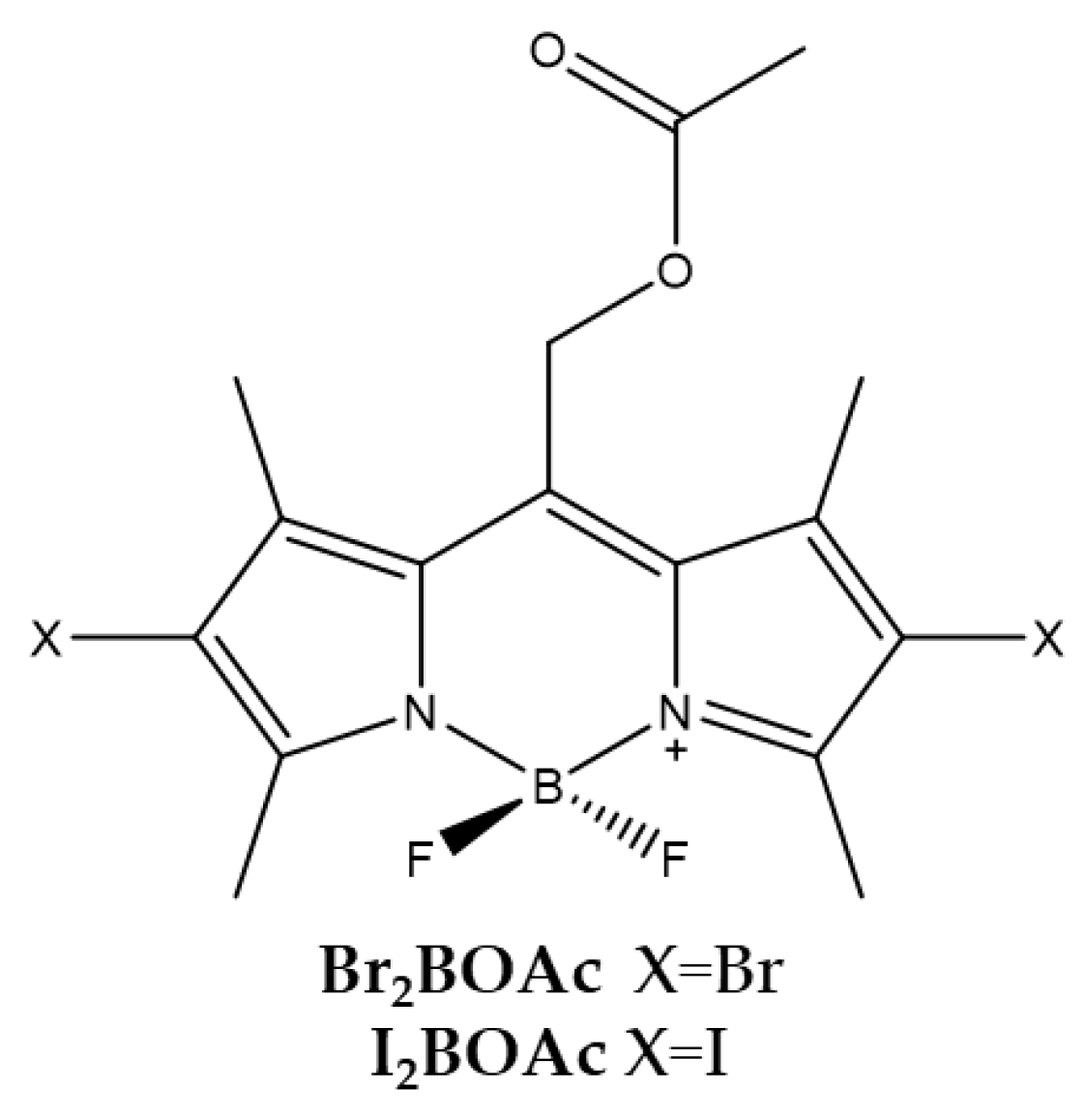
| Br2BOAc | I2BOAc | |
|---|---|---|
| λabs a | 543 nm | 550 nm |
| ε | 81,000 M−1cm−1 | 96,000 M−1cm−1 |
| λem a | 562 nm | 572 nm |
| Φfluo b | 0.14 | 0.02 |
| 1O2 QY c | 0.84 | 0.98 |
| IC50 (HeLa) d | 140.0 nM | 180.0 nM |
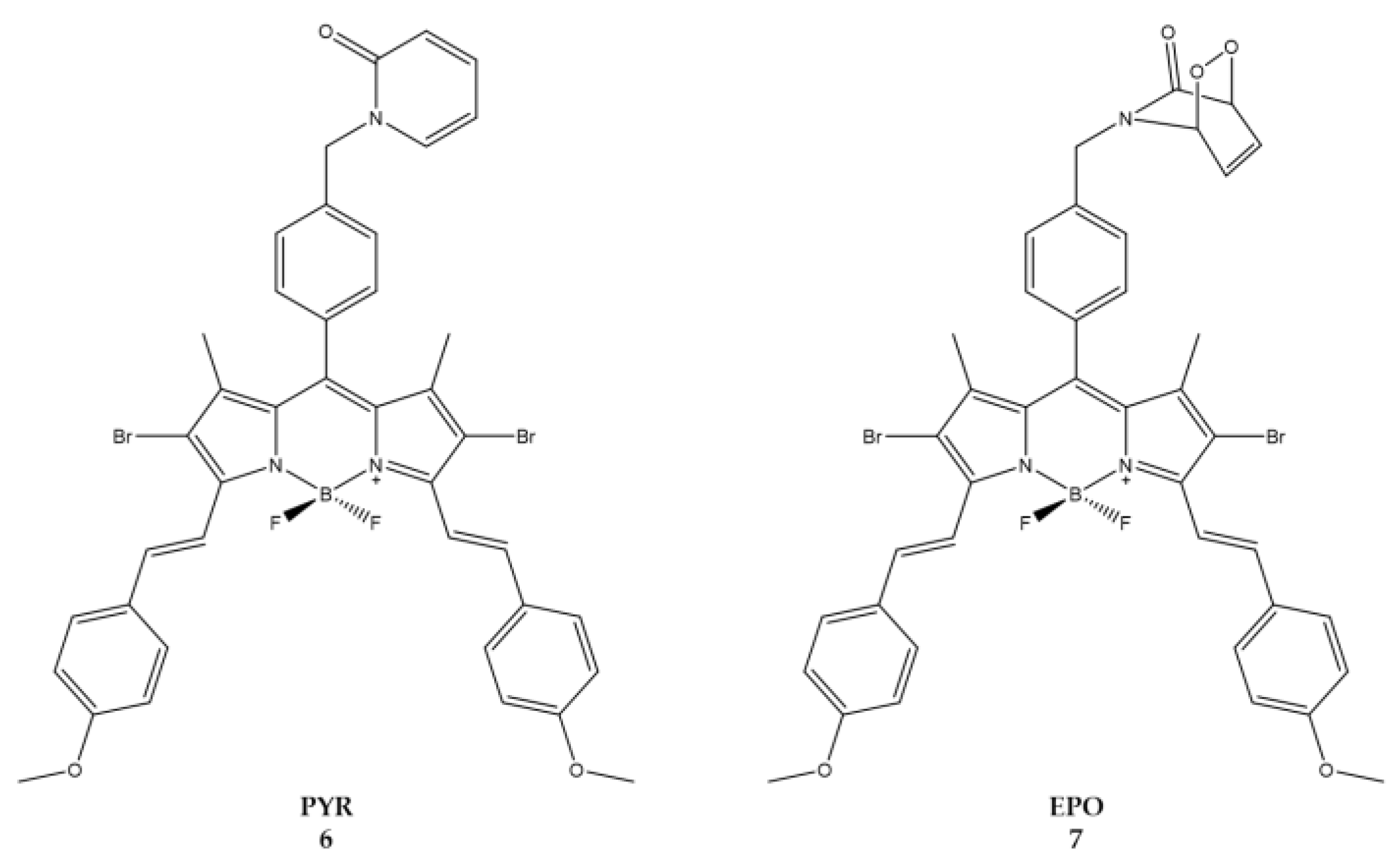
| 6 (PYR) | 7 (EPO) | |
|---|---|---|
| λabs a | 668 nm | 667 nm |
| ε | 22,200 M−1cm−1 | 74,200 M−1cm−1 |
| λem a | 697 nm | 700 nm |
| Φfluo b | 0.14 | 0.10 |
| 1O2 QY c | 0.15 | 0.20 |
| IC50 (HeLa) d | 49.0 nM | 8.6 nM |
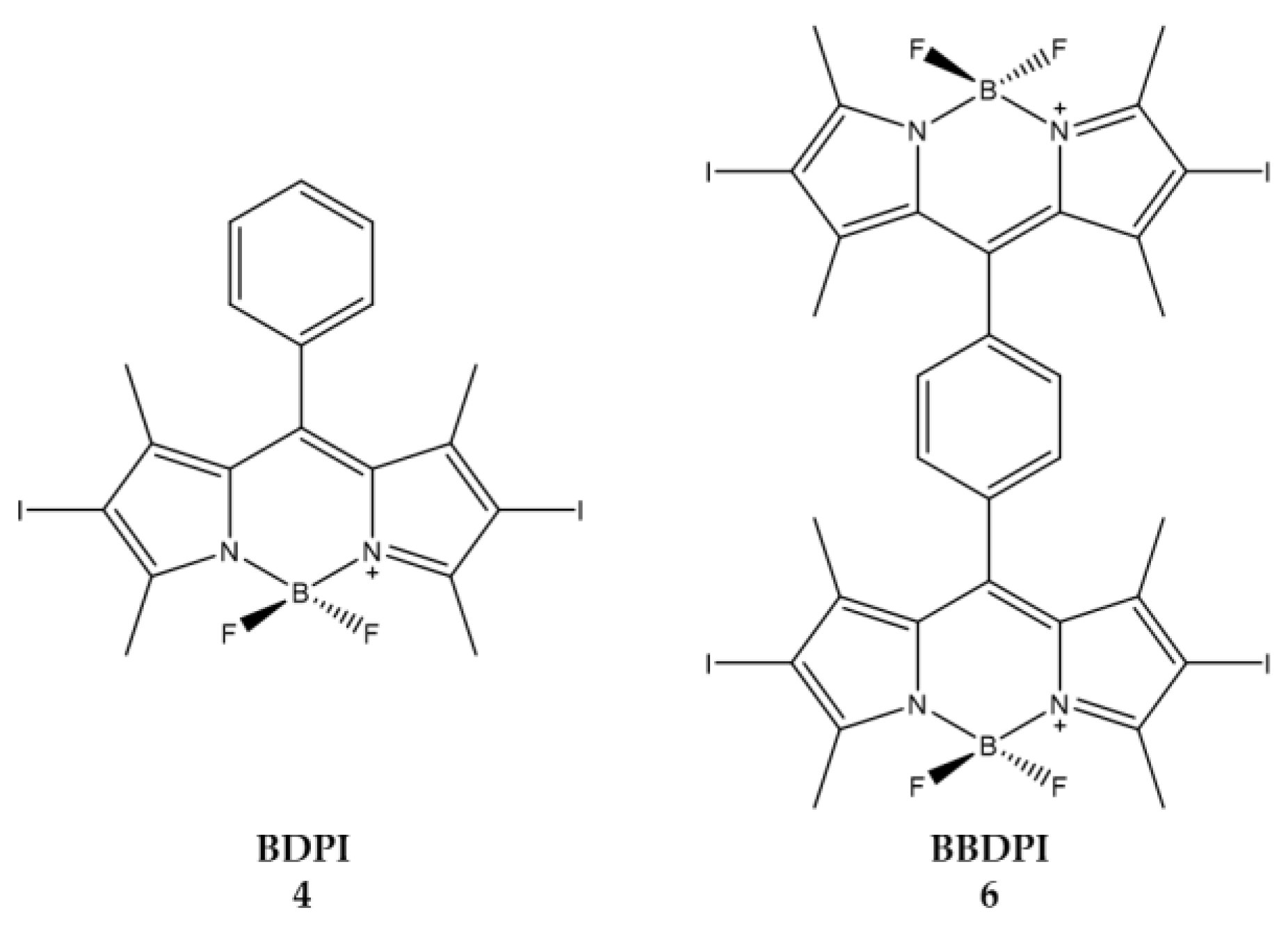
| 4 (BDPI) | 6 (BBDPI) | |
|---|---|---|
| λ abs a | 533 nm | 540 nm |
| 1O2 QY b | 0.73 | 0.68 |
| IC50 (HeLa) c | 1.0 μM | 2.8 μM |
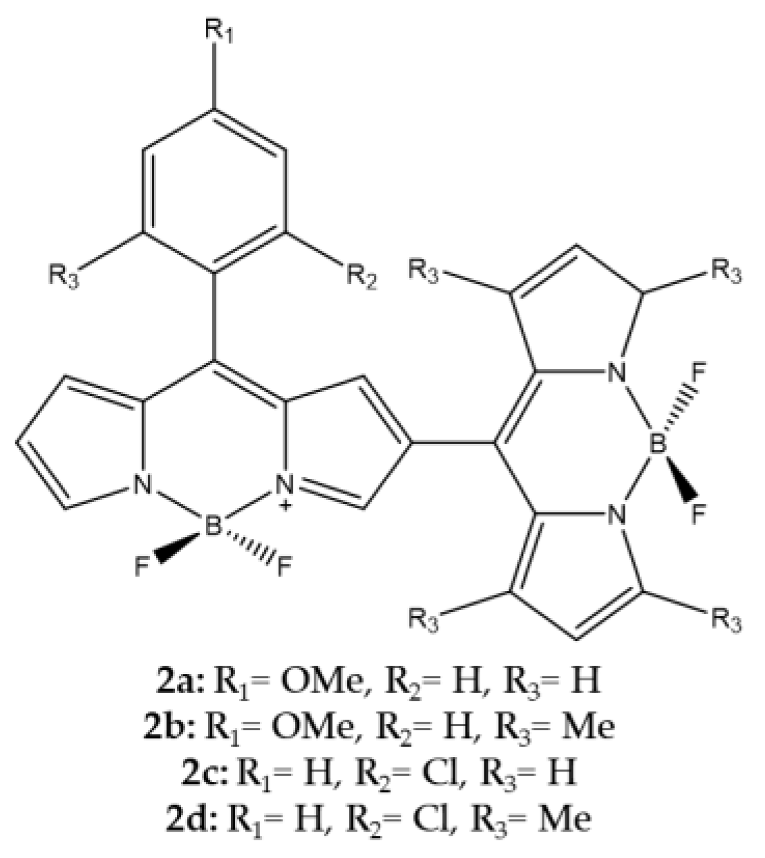
| 2a | 2b | 2c | 2d | |
|---|---|---|---|---|
| λabs a | 521 nm | 507 nm | 538 nm | 507 nm |
| λem a | 563 nm | 540 nm | 568 nm | 542 nm |
| Φfluo b | 0.007 | 0.002 | 0.002 | 0.002 |
| 4 | |
|---|---|
| λabs a | 573.8 nm |
| ε | 8,2000 M−1cm−1 |
| λem a | 590 nm |
| Φfluo b | 0.6 |
| IC50 (A549) c | 2.7 μM |
| BDILa | |
|---|---|
| λabs a | 526 nm |
| ε | 41,800 M−1cm−1 |
| λem a | 542 nm |
| Φfluo b | 0.02 |
| 1O2 QY c | 0.47 |
| IC50 (HeLa) d | 0.55 μM |
| IC50 (MCF7) d | 0.61 μM |
| IC50 (Huh7) d | 0.50 μM |
| BDPI-Lyso | |
|---|---|
| λabs a | 545 nm |
| ε | 41,900 M−1cm−1 |
| λem a | 572 nm |
| Φfluo a | 0.05 |
| 1O2 QY a | 0.95 |
| 1O2 QYpH = 5 b | 0.51 |
| 1O2 QYpH = 7 c | 0.38 |
| IC50 (Bel-7402) d | 0.4 μM |
| MBDP | |
|---|---|
| λabs a | 660 nm |
| ε | 83,226 M−1cm−1 |
| λem a | 694 nm |
| Φfluo b | 0.11 |
| 1O2 QY c | 0.64 |
| IC50 (MCF7) d | 0.2 μM |
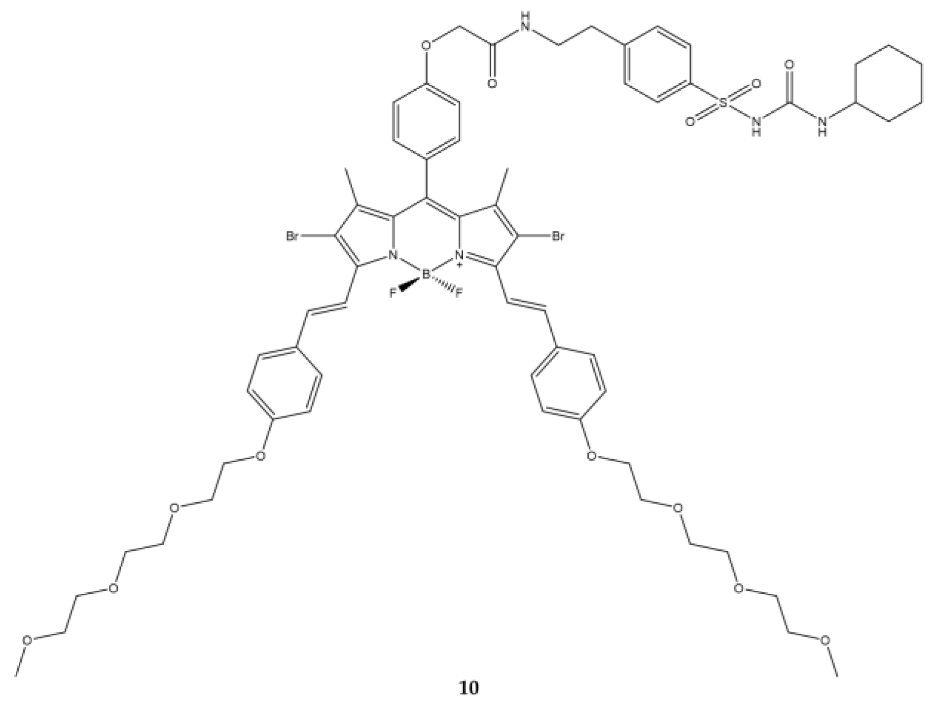
| 10 | |
|---|---|
| λabs a | 669 nm |
| ε | 114,815 M−1cm−1 |
| λem a | 692 nm |
| Φfluo b | 0.32 |
| 1O2 QY c | 0.11 |
| IC50 (Hela) d | 0.09 μM |
| IC50 (HepG2) d | 0.16 μM |
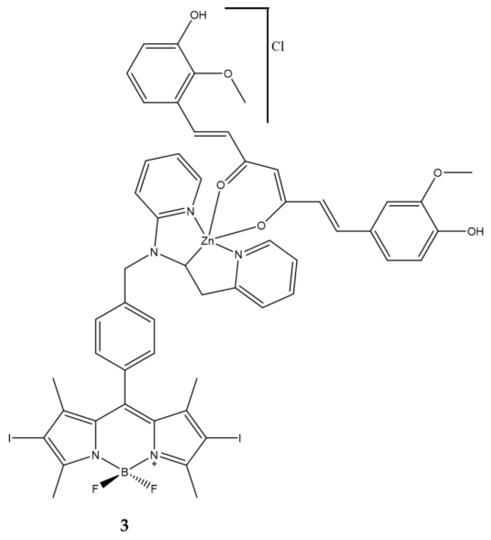
| 3 | |
|---|---|
| λabs a | 543 nm |
| ε | 29,400 M−1cm−1 |
| λem a | 506 nm |
| Φfluo b | 0.02 |
| 1O2 QY c | 0.73 |
| IC50 (HeLa) d | 0.025 μM |
| IC50 (MCF7) d | 0.055 μM |
| IC50 (HPL1D) d | 0.230 μM |
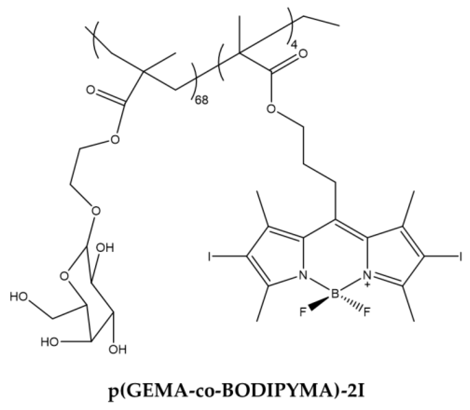
| p(GEMA-co-BODIPYMA)-2I | |
|---|---|
| λabs a | 535 nm |
| 1O2 QY b | 0.79 |
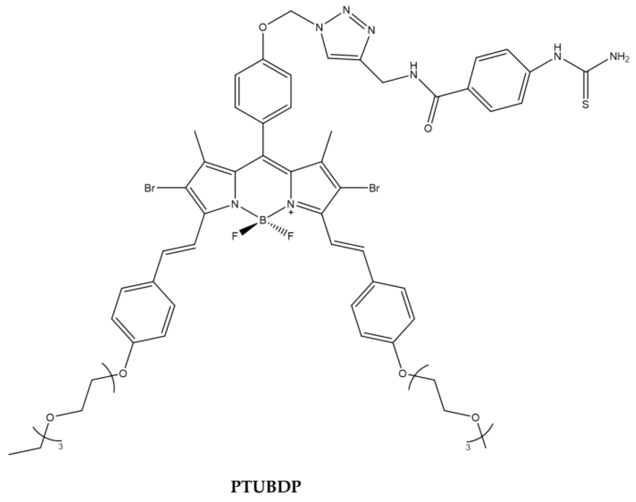
| PTUBDP | |
|---|---|
| λabs a | 667 nm |
| ε | 54,400 M−1cm−1 |
| λem a | 712 nm |
| 1O2 QY b | 0.093 |
| 8 | |
|---|---|
| λabs a | 588 nm |
| ε | 43,000 M−1cm−1 |
| λem a | 634 nm |
| Φfluo b | 0.003 |
| IC50 (HepG2) c | 74 μM |
| IY-IY-PDT | |
|---|---|
| λabs a | 532 nm |
| λem a | 550 nm |
| IC50 (NIH3T3) b | 0.35 μM |
| 3b | |
|---|---|
| λabs a | 534 nm |
| ε | 99,000 M−1cm−1 |
| λem a | 550 nm |
| Φfluo | 0.02 |
| 1O2 QY b | 0.59 |
| IC50 (HeLa) c | 70 nM |
| IC50 (HeLa, pH=5.5) d | 30 nM |
| IC50 (HeLa, pH=7.5) d | 150 nM |
| 8a | |
|---|---|
| λabs a | 664 nm |
| λem a | 768 nm |
| Φfluo b | 0.052 |
| 1O2 QY c | 0.018 |
| IC50 (HeLa) d | 0.67 μM |
| IC50 (A549) d | 0.44 μM |
| IC50 (H22) d | 0.48 μM |
| 7 | |
|---|---|
| λabs a | 376, 441, 662 nm |
| λem a | 686 nm |
| Φfluo a | 0.03 |
| IC50 (MCF7, no DTT b | 146.0 nM |
| IC50 ((MCF7, 2 μM DTT) b | 140.0 nM |
| IC50 (MCF7, 4 mM DTT) b | 81 nM |
Publisher’s Note: MDPI stays neutral with regard to jurisdictional claims in published maps and institutional affiliations. |
© 2022 by the authors. Licensee MDPI, Basel, Switzerland. This article is an open access article distributed under the terms and conditions of the Creative Commons Attribution (CC BY) license (https://creativecommons.org/licenses/by/4.0/).
Share and Cite
Malacarne, M.C.; Gariboldi, M.B.; Caruso, E. BODIPYs in PDT: A Journey through the Most Interesting Molecules Produced in the Last 10 Years. Int. J. Mol. Sci. 2022, 23, 10198. https://doi.org/10.3390/ijms231710198
Malacarne MC, Gariboldi MB, Caruso E. BODIPYs in PDT: A Journey through the Most Interesting Molecules Produced in the Last 10 Years. International Journal of Molecular Sciences. 2022; 23(17):10198. https://doi.org/10.3390/ijms231710198
Chicago/Turabian StyleMalacarne, Miryam Chiara, Marzia Bruna Gariboldi, and Enrico Caruso. 2022. "BODIPYs in PDT: A Journey through the Most Interesting Molecules Produced in the Last 10 Years" International Journal of Molecular Sciences 23, no. 17: 10198. https://doi.org/10.3390/ijms231710198