Review

Methyl Group Metabolism in Differentiation, Aging, and Cancer

1
Institute for Stem Cell Research and Regenerative Medicine, Medical Faculty, Heinrich-Heine University Düsseldorf, 40225 Düsseldorf, Germany
2
Epigenetics Core Laboratory, Institute of Transplantation Diagnostics and Cell Therapeutics, Medical Faculty, Heinrich-Heine University Düsseldorf, Moorenstr. 5, 40225 Düsseldorf, Germany
*
Author to whom correspondence should be addressed.
Int. J. Mol. Sci. 2022, 23(15), 8378; https://doi.org/10.3390/ijms23158378
Submission received: 25 June 2022 / Revised: 21 July 2022 / Accepted: 26 July 2022 / Published: 29 July 2022
(This article belongs to the Collection Anticancer Drug Discovery and Development)

Abstract

:
Methyl group metabolism belongs to a relatively understudied field of research. Its importance lies in the fact that methyl group metabolic pathways are crucial for the successful conversion of dietary nutrients into the basic building blocks to carry out any cellular methylation reaction. Methyl groups play essential roles in numerous cellular functions such as DNA methylation, nucleotide- and protein biosynthesis. Especially, DNA methylation is responsible for organizing the genome into transcriptionally silent and active regions. Ultimately, it is this proper annotation that determines the quality of expression patterns required to ensure and shape the phenotypic integrity and function of a highly specialized cell type. Life is characterized by constantly changing environmental conditions, which are addressed by changes in DNA methylation. This relationship is increasingly coming into focus as it is of fundamental importance for differentiation, aging, and cancer. The stability and permanence of these metabolic processes, fueling the supplementation of methyl groups, seem to be important criteria to prevent deficiencies and erosion of the methylome. Alterations in the metabolic processes can lead to epigenetic and genetic perturbations, causative for diverse disorders, accelerated aging, and various age-related diseases. In recent decades, the intake of methyl group compounds has changed significantly due to, e.g., environmental pollution and food additives. Based on the current knowledge, this review provides a brief overview of the highly interconnected relationship between nutrition, metabolism, changes in epigenetic modifications, cancer, and aging. One goal is to provide an impetus to additionally investigate changes in DNA methylation as a possible consequence of an impaired methyl group metabolism.

1. Introduction

The mammalian life cycle begins with the fertilization of the oocyte, which starts proliferating rapidly and thereby giving rise to the whole organism with hundreds of different cell types. This process is referred to as embryogenesis and required several subsequent steps of differentiation. During embryogenesis, the only totipotent cell is the fertilized oocyte [1], with its unique ability to specialize into all extraembryonic as well as all embryonic tissues and organs [2]. During the following cell divisions, first cell fate decisions are made. At the stage of the blastocyst, two different lineages with divergent differentiation capacity are established. These are the trophoblast, giving rise to the extra embryonal structures, and the inner cell mass (ICM). By subsequent asymmetrical divisions, the ICM is further divided into primitive ectoderm (epiblast) and primitive endoderm (hypoblast) in the late blastocyst stage [1]. The epiblast gives rise to pluripotent stem cells forming the entire embryo which are referred to as embryonic stem cells. With each subsequent division, a gradual loss of differentiation capacity is acquired leading ultimately to a fully differentiated somatic cell. Despite their completely different differentiation capacity, all of the before-mentioned cells in humans have in common, that they all contain the same 3.2-billion base pairs of DNA [3] harboring 20,687 protein-coding genes [4]. So how is the different function and phenotype of all cells within an organism established since all cells share the same genetic information?
Particular mechanisms that are needed for the selective expression of genes are referred to as epigenetics and were first postulated by Conrad Hal Waddington [5]. As already mentioned, each cell of an organism shares the same genetic information but requires a specific regulation of gene expression to attain the respective functional and structural properties. In the 1980s, it was described that the general levels of DNA methylation in the cells of an early developing embryo are relatively low compared to fully differentiated somatic cells [6]. Furthermore, in 1992, Kafri et al. showed that at various genes of an oocyte or in the sperm a “resetting” of the epigenetic code takes place [7]. During the later stages of development, the resetted sequences again get methylated in a process that is referred to as “de novo” methylation, executed by DNA methyltransferases 3a and 3b (DMNT3a/b) [8]. These processes of resetting and “de novo” methylation both contribute to the determination of different cell fates during embryogenesis [9] and are able to repress genes through DNA methylation or activate tissue-specific genes by demethylation [8]. Once these changes are established, they become automatically maintained during each cell division, even in the absence of the originally initiating factors. Responsible for maintaining the DNA methylation pattern during each cell division is DNA methyltransferase 1 (DNMT1).
The aim of this review is to provide a compact overview of the diverse methyl group metabolic pathways operating in each cell to provide the required methyl groups for acquiring and maintaining the appropriate phenotype and function. In addition, we want to highlight that deterioration of these metabolic pathways may be key to an accelerated aging process and the development of aging-related diseases. Finally, we want to discuss some natural compounds and nutrients, which can interfere with the metabolism of methyl groups in humans and thereby have the potential to slowdown the aging process and provide anti-cancer activity.

2. Methyl Group Metabolism

There are two major metabolic pathways regulating the supplementation of methyl groups within a mammalian cell: (1) one-carbon metabolism and (2) polyamine metabolism.

2.1. The One-Carbon Metabolism
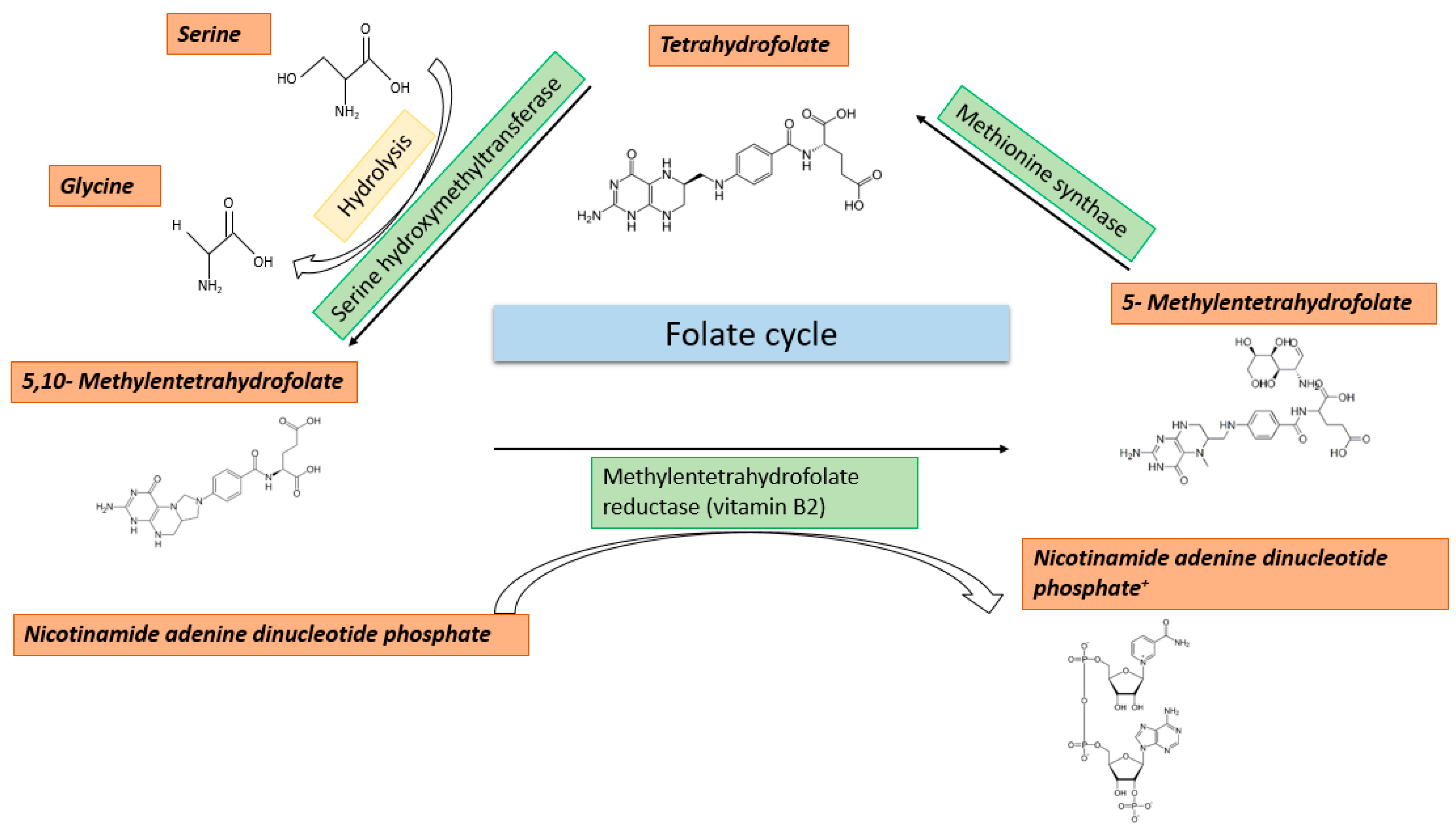
One-carbon metabolism is composed of the folate cycle, methionine cycle (METZ), and the transsulfuration pathway. One-carbon metabolism is characterized by a series of cyclic reactions, in which each time a one-carbon group is transferred. Besides the function of providing methyl groups for all methylation reactions (including DNA, RNA, and proteins), one-carbon metabolism is also essential for the production of phospholipids and nucleotides during proliferation [10]. In addition, one-carbon metabolism can regulate the cellular redox status through the oxidation of NADPH and the generation of glutathione [11].

2.2. Folate Cycle

The folate cycle occurs in both the cytosol and the mitochondria and starts with the uptake of folate with nutrition [12]. Folate is the water-soluble form of vitamin B9 and is brought into the cytosol in its reduced form tetrahydrofolate (THF) by folate receptors. THF is converted to 5,10-methylene-THF by serine hydroxymethyltransferases 1/2 (SHMT1/2), and this happens either in the cytosol or the mitochondrion. For this reaction, mainly serine and to a lesser extent glycine serve as carbon group donors [13,14]. In the next step, 5,10-methylene-THF is converted into 5-methyl-THF by methylenetetrahydrofolate reductase (MTHFR). This reaction only happens in the cytosol and 5-methyl-THF is used in the methionine cycle for the remethylation of homocysteine to methionine. In contrast, the 5,10-methylene-THF in the mitochondrion is converted into 5,10-methenyl-THF and 10-formyl-THF. 10-formyl-THF can only be transferred out of the mitochondrion in this form and can then be used for the majority of carbon-dependent reactions in the cytoplasm and nucleus [15,16]. These include, besides the generation of metabolites used in the folate and methionine cycle, also contributions to purine synthesis [12]. An overview of the folate cycle is given in Figure 1.
The folate cycle is carried out in the cytosol as well as in the mitochondria. It begins with the uptake of folate through the diet. Folate is the water-soluble form of vitamin B9 which is taken up via folate receptors in the reduced form of THF. It can be converted to 5,10-methylene-THF by serine hydroxy methyltransferase in the cytosol and in mitochondria. Primarily, serine and to a lesser extent glycine serve as carbon group donors in this process. Further, the 5,10-methylene-THF can then be converted by methylenetetrahydrofolate reductase into 5-methyl-THF, which is used for the remethylation of homocysteine to methionine. In the cytosol and nucleus, it can then supply carbon groups to the majority of “one-carbon” dependent reactions.

2.3. Methionine Cycle

The methionine cycle starts with the remethylation of homocysteine (HCY) to methionine (MET) by methionine synthase (MTR). For this reaction, the 5-methyl-THF, produced in the folate cycle by MTHFR, and vitamin B12 as a cofactor are required [11]. In some tissues, such as, e.g., the kidneys or the liver, the remethylation reaction can also be carried out by other enzymes such as betaine-homocysteine-S-methyltransferase (BHMT) [17]. In this reaction, 5-methyl-THF is replaced by betaine as a methyl group donor. Subsequently, under ATP consumption, the adenylation of the formed MET leads to the formation of S-adenosyl-L-methionine (SAM), which serves as a central molecule fueling all methylation reactions, including DNA and histone methylation, by donation of a methyl group [10]. This reaction is carried out by the family of methionine-adenosyltransferases (MATs). MET and folate both can enter the human body with our nutrition, with half of the MET being directly converted into SAM [18]. This emphasizes the importance of proper nutrition to establish and maintain a functional epigenome. The process of methylation is performed by histone methyltransferases (HMTs) or DNA methyltransferases (DNMTs). For both reactions, the methyl group of the SAM is either transferred to a lysine or arginine residue of a histone protein or to the 5′-carbon atom of the pyrimidine base cytosine, producing S-adenosyl-homocysteine (SAH) [11]. The re-entry of SAH into the METZ is achieved by the hydrolysis of the molecule into HCY and adenosine. This hydrolysis is carried out by the enzyme adenosylehomocysteinase (AHCY). With the remethylation of HCY to MET, which is carried out by methionine synthase (MTR), the METZ is completed [10]. The main methyl group donor for the remethylation of HCY to MET is serine. An overview of the METZ is given in Figure 2.
The methionine cycle begins with the remethylation of homocysteine to methionine. This reaction requires the 5-methyl-THF produced in the folate cycle by MTHFR and vitamin B12 as a cofactor. The recycling of the B12 cofactor by MTHFR represents a key reaction of remethylation. The subsequent adenylation of methionine leads to the formation of SAM, which serves as a methyl group donor for DNA and histone methylation. This reaction is catalyzed by MAT. MET, just like folate, can be supplied to the body with food, whereby half of the MET is converted directly into SAM.

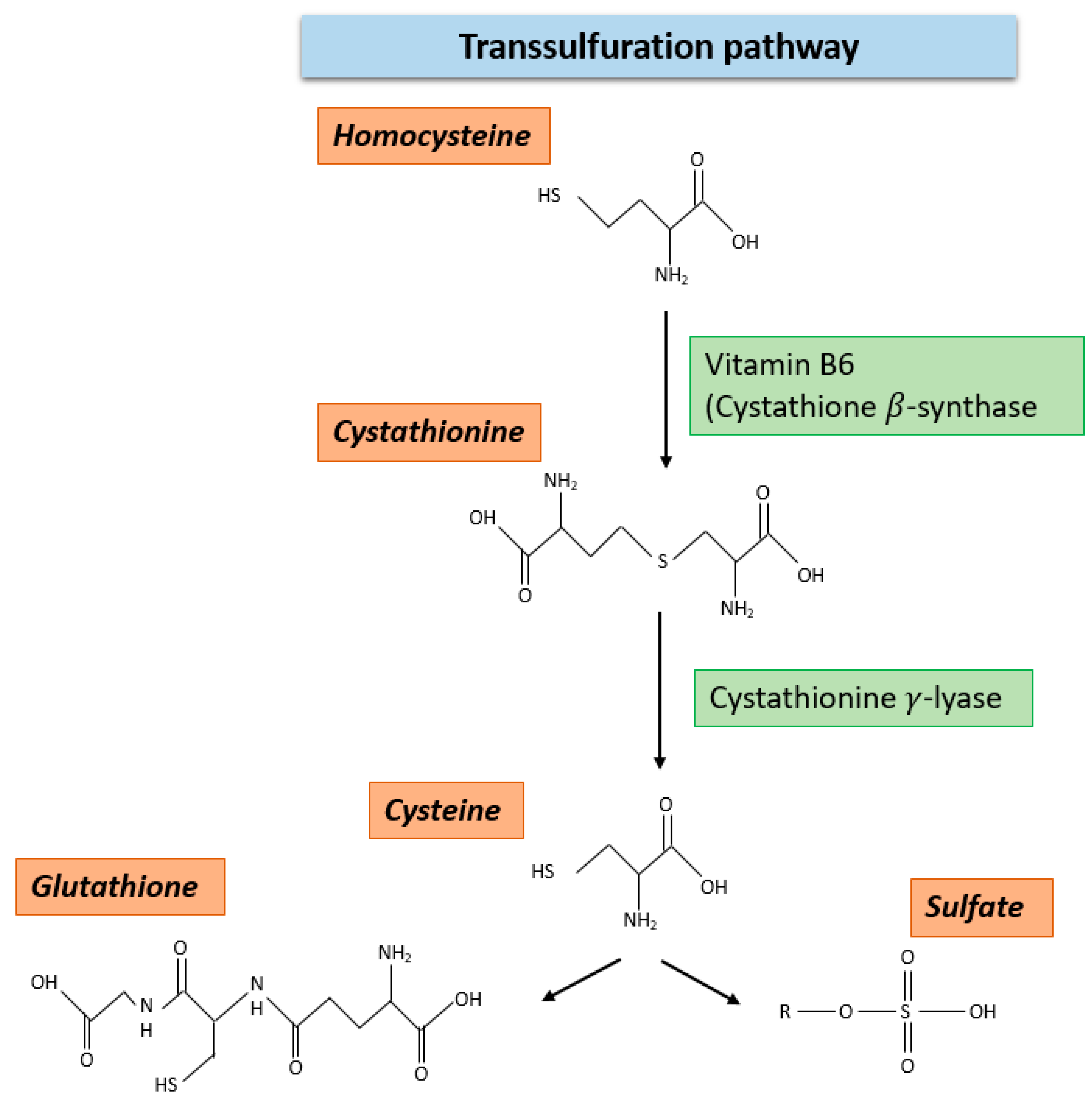
2.4. Transsulfuration Pathway

Another pathway besides the METZ, in which HCY is metabolized, is the transsulfuration pathway. In fact, approximately 60% of the cellular HCY is mined in this cycle [19]. The reactions in this metabolic pathway are irreversible and require vitamin B6 as a cofactor for the enzymatic activity of the initiating enzyme cystathionine ß-synthase (CBS). This enzyme catalyzes the conversion of HCY to cystathione. In the further steps of the cycle, cystathione is converted into cysteine and ultimately to glutathione. In contrast to folate and MET, HCY cannot be taken up by nutrition. It must be synthesized through metabolic processes in the human body itself. An overview of the transsulfuration pathway is given in Figure 3.
Homocysteine can be degraded not only through the METZ but also through the transsulfuration pathway. In this pathway, vitamin B6 is required as a cofactor for the enzymatic activity of CBS. This enzyme catalyzes the conversion of homocysteine to cystathionine. In the further steps of the cycle, cystathionine is converted by cystathionine γ- lyase to cysteine and ultimately to glutathione or to sulphate. Homocysteine cannot be ingested with food but must be synthesized by metabolic processes in the body.

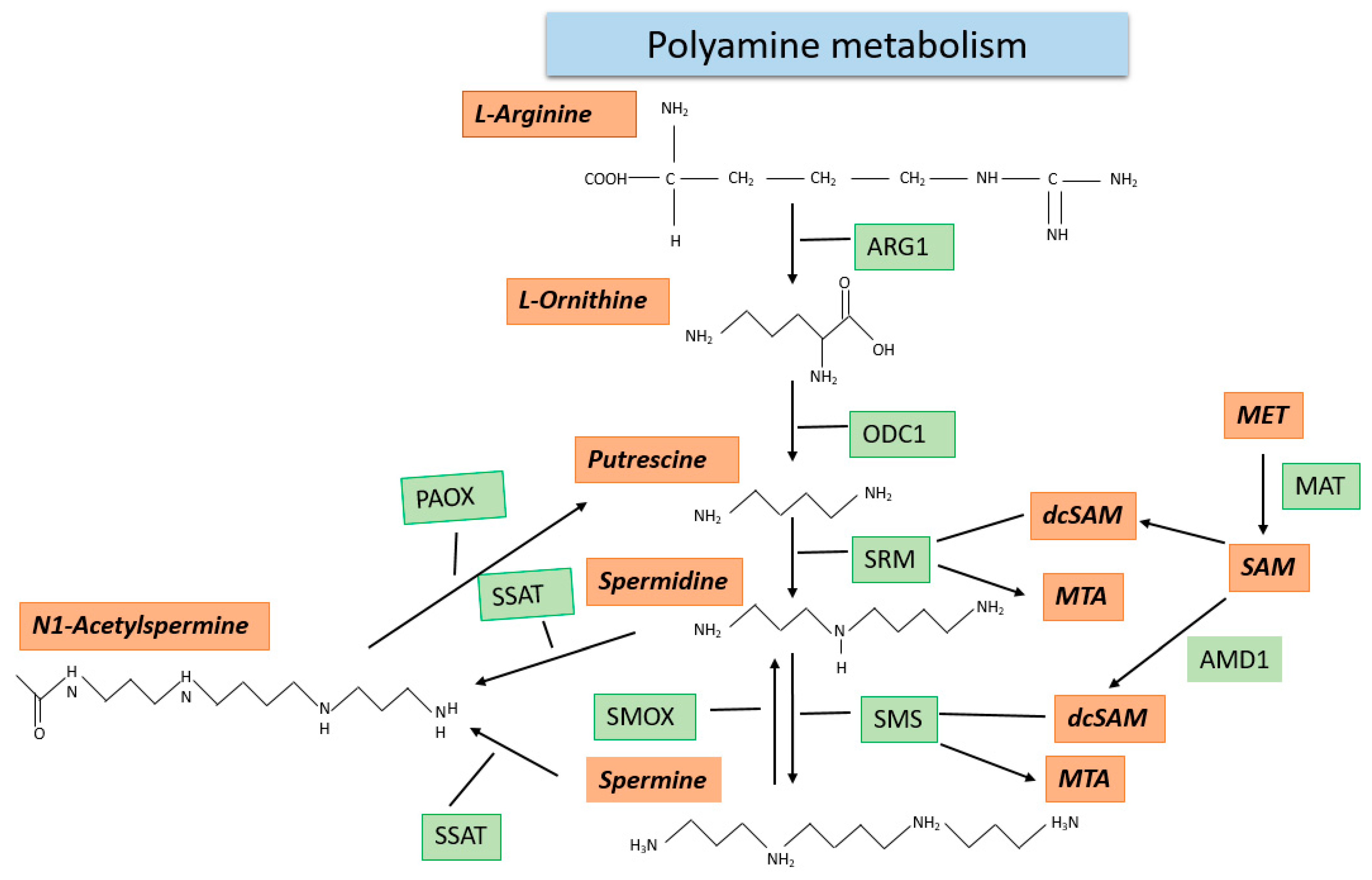
2.5. Polyamine Metabolism

Polyamines, which include putrescine, spermine, and spermidine, are synthesized in every cell type and are essential for cellular growth [20]. So far, three different sources of intake of polyamines in mammals are known: (1) They can be ingested with nutrition;(2) enzymatic synthesis under SAM consumption, hereby the intracellular polyamine concentrations are diet dependent and can be selectively reduced by inhibition of the enzymes required for their biosynthesis [21,22]; (3) production of polyamines by microorganisms [20]. Polyamines can inter alia interact with negatively charges molecules, such as RNA, DNA, phospholipids, and proteins, and thereby regulate a plethora of cellular functions [20]. Polyamine metabolism starts with the basic, non-protein genic α-amino acid L-ornithine. In the first step of the metabolic pathway, L-ornithine is converted to putrescine by ornithine decarboxylase (ODC1). This reaction represents the metabolically limiting step of polyamine metabolism, with ODC1 being the key enzyme [23]. On the cellular level, L-ornithine is synthesized from L-arginine by the enzyme arginase (ARG1). The next steps of polyamine metabolism reveal a connection between this pathway and one-carbon metabolism. Both the transfer of the aminopropyl group on putrescine to create spermidine (this reaction is carried out by the spermidine synthase (SRM)) as well as the transfer of the same chemical group on spermidine to create spermine (this reaction is carried out by the spermine synthase (SMS) require decarboxylated SAM (dcSAM) as the aminopropyl group donor. dcSAM is formed by the decarboxylation of SAM by the adenosylmethionine decarboxylase (AMD1). The intracellular levels of spermidine and spermine are mostly controlled by the export of these metabolites from the cell [20]. As an alternative, both spermidine and spermine can be processed by the enzymes spermine oxidase (SMOX) and polyamine oxidase to be converted back into putrescine [20]. An overview of polyamine metabolism is given in Figure 4.
Polyamine metabolism starts with the basic, non-protein genic α-amino acid L-ornithine. L-ornithine is formed from ARG. In the next step of the metabolic pathway, the diamine putrescine is synthesized by ODC. The next steps reveal the connection to “one-carbon metabolism” because both the transfer of the aminopropyl group by SRMto putrescine, giving rise to spermidine, and by SMS to spermidine, giving rise to spermine, require dcSAM as the aminopropyl group donor. This reaction leads to the formation of 5-methylthioadenosine (MTA). dcSAMis formed by AMD, from SAM synthesized in METZ. Alternatively, to export, spermine and spermidine can also be converted back to putrescine by the enzymes SMOX, SSAT, and PAOX.
Polyamine concentrations are regulated by two major mechanisms. For either the activity of spermidine/spermine N1-acetyltransferase 1 (SSAT), acetylation of spermine or spermidine is required. In this acetylated form, spermine and spermidine are either oxidatively cleaved into lower polyamines, or alternatively, they can be excreted from the cells into animal body fluids, such as urine or blood [20].

3. Influence of the One-Carbon Metabolism and the Polyamine Metabolism on Epigenetics and Cancer

3.1. Metabolic Influence on Epigenetics

The influence of one-carbon and polyamine metabolism on epigenetics is complex and diverse. The nutrients in our daily food intake can regulate gene expression by influencing different epigenetic mechanisms. On the one hand, there are ingredients that directly regulate the availability of SAM or are needed for the de novo synthesis of adenosine, guanosine, and thymine [12]. These nutrients are components of one-carbon metabolism, such as folate, riboflavin, betaine, serine, and methionine. On the other hand, there are ingredients that regulate histone modifications or the transcription of non-coding RNAs, such as vitamin D [24]. Other important metabolites that are not ingested with our daily food intake can also have an impact on epigenetic regulatory mechanisms. These metabolites must be synthesized by the body and include acetyl coenzyme-A (acetyl-CoA), flavine adenine dinucleotide (FAD), and α-ketoglutarate (α-KG). These metabolites can have both enhancing and inhibitory effects on the enzyme activity of histone-modifying enzymes themselves. For instance, the presence of acetyl-CoA is associated with the attachment of acetyl groups to the respective histone proteins, while increased concentrations of its deacetylated version are associated with the inhibition of histone acetyltransferases [25,26,27]. SAM has been recognized as the major player in regulating the cell’s ability to attach methyl groups to DNA as well as histones. Through the accessibility of SAM, both DNA methyltransferases (DNMTs), as well as HMTs, can be regulated [10]. The degradation product of SAM, which is synthesized during the methylation reaction, namely SAH, is responsible for the inhibition of several methyltransferases. Maruti et al. have shown that increased cellular concentrations of SAH lead to global hypomethylation [28]. Further metabolites, whose concentration changes have been linked to global hypomethylation, are HCY [29] and dcSAM [30]. Furthermore, it could be shown that the knockout of the MTHFR gene in mice resulted in significantly lower intracellular SAM and higher SAH concentrations with concomitant global DNA hypomethylation [31]. This finding was also confirmed by two studies, revealing concentration changes in the crucial SAM:SAH concentration in the blood serum by different diets [32,33]. Analogous to DNA methylation, a similar correlation has also been reported for the establishment of trimethylation at histone H4K3. This modification is also directly dependent on the availability of methionine and the intracellular production of SAM [34]. Further insight into the importance of our nutrition, physical activity, and stress was raised by a recently published study [35]. In this study, the participants received a dietary intervention with nutrients that are known to regulate either one-carbon metabolism, DNMTs, modulators of demethylation, or histone modification. Additionally, the participants had to perform physical exercise for 30 minutes per day at least 5 days a week, as well as breathing exercises to reduce stress levels. These lifestyle changes reduced the biological age, as measured by the Horvath DNAm Age clock [36], by 3 years on average compared to the control group. This direct interaction between the availability of metabolites and the activity of the epigenetic modulators has also been proposed in the “nutrient sensing model” [37]. A good example of this is the dependency of the methylation reactions of folate and methionine. Both can be ingested with food and 50% of it will be directly converted into SAM [18]. In this context, our research group was able to link the genome-wide demethylation of long interspersed nuclear elements 1 (LINE1) retrotransposons in cell-free DNA of human blood to the aging process, presenting a new biomarker of aging [38]. Taken together, these findings highlight the importance of proper supplementation of one-carbon metabolites by our daily food intake for the ability of the cell to apply methyl groups and thereby maintain cellular function and prevent or slow down the aging process.
In the case of DNA and histone demethylation, especially α-KG plays a central role as a co-substrate and activator of Jumonji c-domain-containing HDMs and the members of the ten-eleven translocation family (TET). Metabolites that are structurally closely related to α-KG are succinates, fumaric acid, and 2-hydroxyglutarate, and these have been reported to play an inhibitory role for the before-mentioned enzymes [39,40,41]. Another layer of regulation of the epigenetic regulatory mechanisms is added through the local production of the metabolites in the nucleus [37]. Therefore, the enzymes which synthesize the respective metabolites are directly recruited to specific sites of the chromatin where the modification should be added. An example of such a local production is the recruitment of methionine adenosyltransferase isoform type 2 (MAT2A). Through direct protein–protein interactions with the transcription factor MafK, it is linked to a specific sequence in the genome and allows the local synthesis of SAM [42]. Another complex that synthesizes metabolites directly at required sequences of the genome is the so-called “serine-responsive SAM-containing metabolic enzyme complex” (SESAME). It has been shown that this complex interacts with the Set1 methyltransferase complex and supplies it with SAM which is required for the establishment of trimethylation at histone H3K4 [43]. Furthermore, evidence for the involvement of the local synthesis of metabolites and DNA repair mechanisms has been suggested [44].

3.2. Metabolic Influence on Cancer Development and Progression

Since cancer cells are a highly proliferative cell type, an elevated level of exogenous supply of lipids, amino acids, and carbohydrates is needed to maintain the high proliferation rates and thereby cellular survival. Therefore, cancer cells must adjust their metabolism in oxygen and nutrient supply. In the mid-1920s, the initial evidence for altered metabolic processes in cancer cells was provided by Otto Warburg [45]. With the “Warburg effect”, he described the metabolic shift in cancer cells from oxidative phosphorylation to an increased rate of aerobic glycolysis, to produce significantly higher amounts of ATP [46]. In the mid-1940s, researchers started to report the influence and alteration of one-carbon metabolism in cancer cells. It was found that a low folate diet was able to reduce the number of cancer cells in the blood of children suffering from acute leukemia [47]. This discovery led to the introduction of so-called antifolate drugs that are in clinical use to treat various cancer entities [48]. As examples of such drugs, methotrexate and fluorouracil are the most notable, which both act as inhibitors either of THF or thymidine synthesis [49,50]. THF is needed for the remethylation of MET in the METZ and for the synthesis of the purine base thymine. Since cancer cells have an increased need for nucleotides by their high proliferation rate, also the need for metabolites of one-carbon metabolism and especially THF is increased. By the administration of these medications, THF and thymidine synthesis is restricted to such an extent, that uracil instead of thymine is incorporated into the DNA, which prevents proliferation [51,52]. Interestingly, another study reported higher DNAmethylation levels for patients suffering from colorectal cancer who received a diet with more than 400 µg of folate compared to patients receiving less than 200 µg [53].
In addition to this direct influence of the metabolite processes on the availability of the “raw materials” of one-carbon metabolism, the influence of the metabolites can also be regulated by genetic or epigenetic mechanisms. For instance, in different cancer entities that harbor a mutation in either the isocitrate dehydrogenase (NADP (+1)) (IDH1) or NADP(+2) (IDH2) locus, the first oncometabolite (D)-2-hydroxygluterate (D-2HG) was identified. The mutations were associated with the catalyzation of D-2HG instead of α-KG from isocitrate. D-2HG was found to inhibit the function of DNA and histone demethylases and was associated with altered epigenetic regulation, collagen synthesis, and cell signaling [54]. Accordingly, to the inhibition of demethylases, the cells harboring the IDH1 mutations were found to have increased DNA and histone methylation signatures, which were associated with altered expression profiles [55,56].
Another gene of one-carbon metabolism that is transcriptionally inactivated by mutation in human cancers is MTHFR. The contribution of MTHFR to tumorigenesis has been documented in the case of colon [57], pulmonary [58], and gastric carcinoma [59]. For this gene, the transition from cytosine to thymine at position 677 has been reported to decrease the transcription level by 70% in the homozygous and by 40% in the heterozygous case, and both situations were associated with global DNA hypomethylation [60]. Another common mutation of the MTHFR gene is the transversion of adenine to cytosine at position 1298. This mutation was also found to be associated with a decrease in enzyme activity and a 4% decrease in the LINE-1 methylation level [61]. The allele variant A1298C is also associated with a 4.76-fold increased risk of developing bladder cancer [62]. Furthermore, our own study reports a hypermethylation at the 5′-regulatory region of the MTHFR gene in early-stage urothelial carcinoma [63]. We concluded that the hypermethylation within the MTHFR gene could cause transcriptional silencing and therefore alter the crucial SAM:SAH ratio, since the methylation of the MTHFR gene was found to be inversely correlated with gene expression [64].
Our own study also revealed a significant hypermethylation within the AHCY5′-regulatory region in the early stages of urothelial carcinoma [63]. An impairment of this enzyme is associated with an increase in the cellular SAH concentration, which can ultimately result in the inhibition of methyltransferases and is associated with hypomethylation [65]. The downregulation of AHCY was also found to be associated with the promotion of oncogenesis by protecting the affected cells from p53 and p16ink4-induced cell cycle arrest [66]. This study also showed that in 50% of all examined cancer entities, the mRNA levels of AHCY were found to be downregulated. Furthermore, the knock-down of AHCY and depletion of adenosine werelinked to inducing DNAdamage and cell cycle arrest [67].
As already mentioned, dcSAM is a potent inhibitor of methyltransferases and is heavily required for the synthesis of spermidine and spermine. The rate-limiting reaction for this metabolic pathway is the decarboxylation of ornithine to form putrescine whichis carried out by ODC1. This enzyme is expressed in almost all tissues and its high fluctuation in intracellular concentration enables the cells to adapt to changing environmental and metabolic conditions. In 1997, Heljasvaara et al. demonstrated that even mice overexpressing ODC1 were able to maintain their polyamine homeostasis [68]. Increased polyamine synthesis has been linked to improving cancer survival and growth, with overexpression of ODC1 being associated with breast, lung, colon, prostate, pancreatic cancer, and others [69]. For other cancer entities including bladder cancer and oral cavity carcinoma, a downregulation of ODC1 by DNA hypermethylation and subsequently global demethylation has been reported [70,71]. Especially, the transcriptional downregulation of ODC1 by siRNA was shown to lead to hypomethylation of LINE-1 retrotransposons, to induce their transcriptional activity and DNA double-strand breaks in short-term cultivated primary bladder cells [70]. This activation of LINE-1 retrotransposons has been recognized as a hallmark of the early stages of urothelial carcinoma [72] and led to the development of the PrimeEpiHit hypothesis as the initial part of the etiology of urothelial carcinoma as reported [63].

4. Nutrition and Plant-Extracted Compounds Influencing the One-Carbon- and Polyamine Metabolism

4.1. Nutrition

Many key components necessary to supplement the cell with the basic building blocks to carry out all epigenetic modifications can be obtained through daily nutrition. This includes betaine, folate methionine, serine, and most polyamines. Through the digestion of food containing these components, the crucial SAM:SAH ratio within the cells can be modulated and this directly influences, for instance, the cells’ ability to apply methyl groups to the DNA double-strand or synthesize nucleotides necessary for cellular replication. Since the aging process and one of the most common aging [38] associated diseases, namely cancer, have both been linked to the occurrence of global DNAhypomethylation [73], the proper supplementation of methyl groups inour daily nutrition seems to be essential for a healthy and deaccelerated aging process. This association between nutrition and aging has also been observed in a study carried out by Quach et al., in which a reduction in DNA methylation age was observed in individuals consuming a specific diet [74]. Despite nutrients that directly influence the SAM:SAH ratio, there are several other molecules in our food that can affect (a) demethylation by being substrates or cofactors of the ten-eleven translocation demethylases (TET) such as alpha-ketoglutarate, vitamin C and A, (b) modulators of the DNA methyl transferases, such ascurcumin or epigallocatechin gallate (EGCG), rosmarinic acid, quercetin, and luteolin. A full list of nutrients containing key components of one-carbon and polyamine metabolism isgiven in Table 1. Furthermore, there is increasing evidence that the composition of our gut bacteria has an important effect on one-carbon metabolism. For instance, Lactobacillus plantarum has been shown to be able to produce folate if para-aminobenzoic acid (PABA) is available [75] and that this metabolic process is able to influence gene expression [76]. Therefore, probiotic drinks are under consideration for the treatment of several diseases including diabetes [76].
Further complexity into the uptake of metabolites affecting methyl group metabolism is added when considering sex-specific differences in the availability of key metabolites caused by sex hormones. In mammals, it could be shown that pre-menopause females exhibit lower concentrations of HCY [83] and SAM [84], while showing higher levels of betaine [85] in the blood plasma. Moreover, four key enzymes of one-carbon metabolism show sex-specific differences, namely BHMT, MTR, MTHFR, and SHMT [86]. While BHMT and MTHFR show downregulation in females compared to males, MTR and SHMT are upregulated with consequences for the concentration levels of betaine, HCY, and SAM. For instance, an increased activity of MTHFR and BHMT in response to testosterone has been reported [87]. Males have approximately 19 times more testosterone in their blood compared to females and it was shown by Schwahn et al. that the exchange of testosterone for estrogen in females reduced the expression level of BHMT by 40% [88]. Interestingly, several studies concluded that alcohol consumption is able to decrease the levels of testosterone in humans [89,90,91]. Furthermore, testosterone levels can be highly regulated by nutrition. For instance, individuals consuming a diet containing ~20% fat compared with a diet containing ~40% fat showed significantly lower levels of testosterone in the blood [92,93,94]. A similar observation was also reported for people eating a vegetarian diet, since this form of nutrition is also associated to contain fewer fatty acids [95,96,97,98]. Besides nutrition, high-intensity resistance exercises have also been shown to increase testosterone levels in humans [99,100,101]. However, testosterone not only affects enzymes of methionine and folate metabolism but also polyamine metabolism. Jotova and colleagues showed that testosterone can upregulate the enzyme activity of ODC1 up to 4 h and 12 h after hormonal treatment. Thereby, the treatment led to a 1.6-fold increase in ODC1 as well as an increase inthe intracellular concentration of spermidine and spermine after 4 h and putrescine and spermine after 12 h, by 2.2- and 2.6-fold and 1.4- and 1.5-fold, respectively [102]. This crucial interplay between testosterone and polyamine metabolism was also highlighted in the murine kidney. Levillain et al. injected pharmacological and physiological doses of testosterone into female and castrated male mice and found an upregulation of arginase II and ODC, while ornithine aminotransferase was found to be downregulated [103].

4.2. Naturally Existing Plant-Extracted Compounds with Impact on DNA Methylation

DNA methylation alterations successively occur during aging and profound changes persist in various age-related diseases, e.g., cancer. Few bioactive food compounds have been well described to either modulate DNA methylation by impacting the S-adenosylmethionine/S-adenosylhomocysteine ratio, or by directly affecting DNA methyltransferases [104]. Thus, it is assumed that future work may identify the nutritional measures contributing to DNA methylation patterns of healthy aging. A category of compounds naturally found in plant foods such as fruits, vegetables, herbs, spices, tea, dark chocolate, and wine is known as polyphenols. Polyphenols can act as antioxidants. They are thus able to neutralize harmful free radicals that would otherwise damage cells and increase the risk of diseases such as cancer [105]. Currently, 8000 types of polyphenols have been identified. These are divided into four main groups: flavonoids, phenolic acids, polyphenolic amides, and other polyphenols [106,107]. The number of polyphenols in foods depends on their origin, ripeness, transportation, preparation, and farming. Flavonoids are about 60% of all polyphenols, such asquercetin, kaempferol, catechins, and anthocyanins. Apples, onions, dark chocolate, and red cabbage possess these types of flavonoids. Bioactive dietary components can reactivate tumor-suppressor genes by reversing aberrant DNA methylation patterns. Thus, they have a high potential to act against different types of cancer [108]. Other phenolics include for instance resveratrol, which is found in red wine, ellagic acid in berries, curcumin in turmeric, and lignans in flax seeds, sesame seeds, and whole grains. Curcumin (diferuloylmethane), for instance, a component of the golden spice Curcuma longa, commonly known as turmeric, has been reported, beyond possessing many other epigenetic effects [109], to confer DNA hypomethylation, presumably by covalently blocking a catalytic center of DNMT1 [110]. It is thought that this mode of action is involved in curcumin’s capability to act as a powerful, chemoprotective anti-cancer agent, as demonstrated by various studies [111]. This is analogous to resveratrol from grapes, mulberries, apricots, pineapples, and peanuts with a substantial anti-cancer property [24], showing weak inhibition of DNMT activity to inhibit methylation [112], the potential to restore LINE-1 methylation levels [113], and to modulate expression levels of tumor suppressors [114]. Finally, genistein, a phytoestrogen from soybeans, has been shown to have a strong dose-dependent inhibition of DNMT activity [109] and to promote promoter demethylation and reactivation of tumor-suppressor genes in diverse cancer cell types [115,116]. Many other naturally existing plant-extracted compounds with evidence oftheir impact onepigenetic mechanisms and in particular DNA methylation are described. However, future research in this field will determine which application protocols of single or combinations of these naturally existing compounds will optimize the aging process and minimize the likelihood of cancer.

5. Conclusions

More and more it is evident that epigenetic mechanisms, especially DNA methylation, function as distinct genome usage orchestrators which have been shaped by direct interaction with environmental conditions to contribute to the adapted cell types. However, methyl group metabolic pathways acting in every single cell of our organism are required to translate one exogenous noxa, namely nutrition, into an appropriate methylome. Aroundabout 20 million methyl groups are necessary during every cell division. Thus, it should be inferred that deterioration and inefficiency of these key methyl group metabolic pathways may contribute to DNA methylation aberrations as we observed in dedifferentiation processes, aging, and cancer initiation and justifiably assume their causal role. Therefore, more research will be needed in this area to understand how we may be able to counteract this.

Author Contributions

Conceptualization and writing—original draft preparation, L.E.; writing—review and editing, C.T. and S.S.; figures, C.T. All authors have read and agreed to the published version of the manuscript.

Funding

Heinrich-Heine University Duesseldorf.

Institutional Review Board Statement

Not applicable.

Informed Consent Statement

Not applicable.

Data Availability Statement

Not applicable.

Acknowledgments

The authors acknowledge the medical faculty of Heinrich-Heine University for financial support.

Conflicts of Interest

The authors declare no conflict of interest.

References

  1. Hemberger, M.; Dean, W.; Reik, W. Epigenetic dynamics of stem cells and cell lineage commitment: Digging Waddington’s canal. Nat. Rev. Mol. Cell Biol. 2009, 10, 526–537. [Google Scholar] [CrossRef] [PubMed]
  2. Shen, H.; Yang, M.; Li, S.; Zhang, J.; Peng, B.; Wang, C.; Chang, Z.; Ong, J.; Du, P. Mouse totipotent stem cells captured and maintained through spliceosomal repression. Cell 2021, 184, 2843–2859. [Google Scholar] [CrossRef] [PubMed]
  3. Venter, J.C.; Adams, M.D.; Myers, E.W.; Li, P.W.; Mural, R.J.; Sutton, G.G.; Smith, H.O.; Yandell, M.; Evans, C.A.; Holt, R.A.; et al. The sequence of the human genome. Science 2001, 291, 1304–1351. [Google Scholar] [CrossRef] [PubMed] [Green Version]
  4. ENCODE Project Consortium. An integrated encyclopedia of DNA elements in the human genome. Nature 2012, 489, 57–74. [Google Scholar] [CrossRef] [PubMed]
  5. Waddington, C.H. The epigenotype. Endeavour 1942, 1, 18–20. [Google Scholar] [CrossRef] [PubMed] [Green Version]
  6. Monk, M.; Boubelik, M.; Lehnert, S. Temporal and regional changes in DNA methylation in the embryonic, extraembryonic and germ cell lineages during mouse embryo development. Development 1987, 99, 371–382. [Google Scholar] [CrossRef]
  7. Kafri, T.; Ariel, M.; Brandeis, M.; Shemer, R.; Urven, L.; McCarrey, J.; Cedar, H.; Razin, A. Developmental pattern of gene-specific DNA methylation in the mouse embryo and germ line. Genes Dev. 1992, 6, 705–714. [Google Scholar] [CrossRef] [Green Version]
  8. Cedar, H.; Bergman, Y. Programming of DNA methylation patterns. Annu. Rev. Biochem. 2012, 81, 97–117. [Google Scholar] [CrossRef] [Green Version]
  9. Reik, W.; Dean, W.; Walter, J. Epigenetic reprogramming in mammalian development. Science 2001, 293, 1089–1093. [Google Scholar] [CrossRef] [Green Version]
  10. Locasale, J.W. Serine, glycine and one-carbon units: Cancer metabolism in full circle. Nat. Rev. Cancer 2013, 13, 572–583. [Google Scholar] [CrossRef] [Green Version]
  11. Ryall, J.G.; Cliff, T.; Dalton, S.; Sartorelli, V. Metabolic Reprogramming of Stem Cell Epigenetics. Cell Stem Cell 2015, 17, 651–662. [Google Scholar] [CrossRef] [Green Version]
  12. Yang, M.; Vousden, K.H. Serine and one-carbon metabolism in cancer. Nat. Rev. Cancer 2016, 16, 650–662. [Google Scholar] [CrossRef] [PubMed]
  13. Shyh-Chang, N.; Locasale, J.W.; Lyssiotis, C.A.; Zheng, Y.; Teo, R.Y.; Ratanasirintrawoot, S.; Zhang, J.; Onder, T.; Unternaehrer, J.J.; Zhu, H.; et al. Influence of threonine metabolism on S-adenosylmethionine and histone methylation. Science 2013, 339, 222–226. [Google Scholar] [CrossRef] [PubMed] [Green Version]
  14. Zhang, W.C.; Shyh-Chang, N.; Yang, H.; Rai, A.; Umashankar, S.; Ma, S.; Soh, B.S.; Sun, L.L.; Tai, B.C.; Nga, M.E.; et al. Glycine decarboxylase activity drives non-small cell lung cancer tumor-initiating cells and tumorigenesis. Cell 2012, 148, 259–272. [Google Scholar] [CrossRef] [PubMed] [Green Version]
  15. Brosnan, M.E.; MacMillan, L.; Stevens, J.R.; Brosnan, J.T. Division of labour: How does folate metabolism partition between one-carbon metabolism and amino acid oxidation? Biochem. J. 2015, 472, 135–146. [Google Scholar] [CrossRef] [PubMed] [Green Version]
  16. Ducker, G.S.; Chen, L.; Morscher, R.J.; Ghergurovich, J.M.; Esposito, M.; Teng, X.; Kang, Y.; Rabinowitz, J.D. Reversal of Cytosolic One-Carbon Flux Compensates for Loss of the Mitochondrial Folate Pathway. Cell Metab. 2016, 23, 1140–1153. [Google Scholar] [CrossRef] [Green Version]
  17. Sunden, S.L.; Renduchintala, M.S.; Park, E.I.; Miklasz, S.D.; Garrow, T.A. Betaine-homocysteine methyltransferase expression in porcine and human tissues and chromosomal localization of the human gene. Arch. Biochem. Biophys. 1997, 345, 171–174. [Google Scholar] [CrossRef] [PubMed]
  18. Mudd, S.H.; Poole, J.R. Labile methyl balances for normal humans on various dietary regimens. Metab. Clin. Exp. 1975, 24, 721–735. [Google Scholar] [CrossRef]
  19. Storch, K.J.; Wagner, D.A.; Burke, J.F.; Young, V.R. Quantitative study in vivo of methionine cycle in humans using methyl-2H3- and 1-13Cmethionine. Am. J. Physiol. 1988, 255, E322–E331. [Google Scholar] [CrossRef] [PubMed]
  20. Park, M.H.; Igarashi, K. Polyamines and their metabolites as diagnostic markers of human diseases. Biomol. Ther. 2013, 21, 1–9. [Google Scholar] [CrossRef] [PubMed] [Green Version]
  21. Ackermann, J.M.; Pegg, A.E.; McCloskey, D.E. Drugs affecting the cell cycle via actions on the polyamine metabolic pathway. Prog. Cell Cycle Res. 2003, 5, 461–468. [Google Scholar] [PubMed]
  22. Seiler, N.; Raul, F. Polyamines and apoptosis. J. Cell. Mol. Med. 2005, 9, 623–642. [Google Scholar] [CrossRef] [PubMed]
  23. Tabor, C.W.; Tabor, H. Polyamines. Annu. Rev. Biochem. 1984, 53, 749–790. [Google Scholar] [CrossRef] [PubMed]
  24. Stefanska, B.; Karlic, H.; Varga, F.; Fabianowska-Majewska, K.; Haslberger, A. Epigenetic mechanisms in anti-cancer actions of bioactive food components--the implications in cancer prevention. Br. J. Pharmacol. 2012, 167, 279–297. [Google Scholar] [CrossRef] [Green Version]
  25. Albaugh, B.N.; Arnold, K.M.; Denu, J.M. KAT(ching) metabolism by the tail: Insight into the links between lysine acetyltransferases and metabolism. Chembiochem 2011, 12, 290–298. [Google Scholar] [CrossRef] [PubMed] [Green Version]
  26. Meier, J.L. Metabolic mechanisms of epigenetic regulation. ACS Chem. Biol. 2013, 8, 2607–2621. [Google Scholar] [CrossRef] [PubMed] [Green Version]
  27. Lee, J.V.; Carrer, A.; Shah, S.; Snyder, N.W.; Wei, S.; Venneti, S.; Worth, A.J.; Yuan, Z.-F.; Lim, H.-W.; Liu, S.; et al. Akt-dependent metabolic reprogramming regulates tumor cell histone acetylation. Cell Metab. 2014, 20, 306–319. [Google Scholar] [CrossRef] [PubMed] [Green Version]
  28. Maruti, S.S.; Ulrich, C.M.; White, E. Folate and one-carbon metabolism nutrients from supplements and diet in relation to breast cancer risk. Am. J. Clin. Nutr. 2009, 89, 624–633. [Google Scholar] [CrossRef] [Green Version]
  29. Akoglu, B.; Milovic, V.; Caspary, W.F.; Faust, D. Hyperproliferation of homocysteine-treated colon cancer cells is reversed by folate and 5-methyltetrahydrofolate. Eur. J. Nutr. 2004, 43, 93–99. [Google Scholar] [CrossRef]
  30. Pegg, A.E.; Michael, A.J. Spermine synthase. Cell. Mol. Life Sci. 2010, 67, 113–121. [Google Scholar] [CrossRef]
  31. Chen, Z.; Karaplis, A.C.; Ackerman, S.L.; Pogribny, I.P.; Melnyk, S.; Lussier-Cacan, S.; Chen, M.F.; Pai, A.; John, S.W.; Smith, R.S.; et al. Mice deficient in methylenetetrahydrofolate reductase exhibit hyperhomocysteinemia and decreased methylation capacity, with neuropathology and aortic lipid deposition. Hum. Mol. Genet. 2001, 10, 433–443. [Google Scholar] [CrossRef] [PubMed] [Green Version]
  32. Poirier, L.A.; Wise, C.K.; Delongchamp, R.R.; Sinha, R. Blood determinations of S-adenosylmethionine, S-adenosylhomocysteine, and homocysteine: Correlations with diet. Cancer Epidemiol. Biomark. Prev. 2001, 10, 649–655. [Google Scholar]
  33. Lim, U.; Song, M.-A. Dietary and lifestyle factors of DNA methylation. Methods Mol. Biol. 2012, 863, 359–376. [Google Scholar] [CrossRef]
  34. Mentch, S.J.; Mehrmohamadi, M.; Huang, L.; Liu, X.; Gupta, D.; Mattocks, D.; Gómez Padilla, P.; Ables, G.; Bamman, M.M.; Thalacker-Mercer, A.E.; et al. Histone Methylation Dynamics and Gene Regulation Occur through the Sensing of One-Carbon Metabolism. Cell Metab. 2015, 22, 861–873. [Google Scholar] [CrossRef] [PubMed] [Green Version]
  35. Fitzgerald, K.N.; Hodges, R.; Hanes, D.; Stack, E.; Cheishvili, D.; Szyf, M.; Henkel, J.; Twedt, M.W.; Giannopoulou, D.; Herdell, J.; et al. Potential reversal of epigenetic age using a diet and lifestyle intervention: A pilot randomized clinical trial. Aging 2021, 13, 9419–9432. [Google Scholar] [CrossRef] [PubMed]
  36. Horvath, S.; Raj, K. DNA methylation-based biomarkers and the epigenetic clock theory of ageing. Nat. Rev. Genet. 2018, 19, 371–384. [Google Scholar] [CrossRef] [PubMed]
  37. Kinnaird, A.; Zhao, S.; Wellen, K.E.; Michelakis, E.D. Metabolic control of epigenetics in cancer. Nat. Rev. Cancer 2016, 16, 694–707. [Google Scholar] [CrossRef]
  38. Erichsen, L.; Beermann, A.; Arauzo-Bravo, M.J.; Hassan, M.; Dkhil, M.A.; Al-Quraishy, S.; Hafiz, T.A.; Fischer, J.C.; Santourlidis, S. Genome-wide hypomethylation of LINE-1 and Alu retroelements in cell-free DNA of blood is an epigenetic biomarker of human aging. Saudi J. Biol. Sci. 2018, 25, 1220–1226. [Google Scholar] [CrossRef] [PubMed]
  39. Tsukada, Y.-I.; Fang, J.; Erdjument-Bromage, H.; Warren, M.E.; Borchers, C.H.; Tempst, P.; Zhang, Y. Histone demethylation by a family of JmjC domain-containing proteins. Nature 2006, 439, 811–816. [Google Scholar] [CrossRef] [PubMed]
  40. Xiao, M.; Yang, H.; Xu, W.; Ma, S.; Lin, H.; Zhu, H.; Liu, L.; Liu, Y.; Yang, C.; Xu, Y.; et al. Inhibition of α-KG-dependent histone and DNA demethylases by fumarate and succinate that are accumulated in mutations of FH and SDH tumor suppressors. Genes Dev. 2012, 26, 1326–1338. [Google Scholar] [CrossRef] [PubMed] [Green Version]
  41. Killian, J.K.; Kim, S.Y.; Miettinen, M.; Smith, C.; Merino, M.; Tsokos, M.; Quezado, M.; Smith, W.I.; Jahromi, M.S.; Xekouki, P.; et al. Succinate dehydrogenase mutation underlies global epigenomic divergence in gastrointestinal stromal tumor. Cancer Discov. 2013, 3, 648–657. [Google Scholar] [CrossRef] [PubMed] [Green Version]
  42. Katoh, Y.; Ikura, T.; Hoshikawa, Y.; Tashiro, S.; Ito, T.; Ohta, M.; Kera, Y.; Noda, T.; Igarashi, K. Methionine adenosyltransferase II serves as a transcriptional corepressor of Maf oncoprotein. Mol. Cell 2011, 41, 554–566. [Google Scholar] [CrossRef]
  43. Li, S.; Swanson, S.K.; Gogol, M.; Florens, L.; Washburn, M.P.; Workman, J.L.; Suganuma, T. Serine and SAM Responsive Complex SESAME Regulates Histone Modification Crosstalk by Sensing Cellular Metabolism. Mol. Cell 2015, 60, 408–421. [Google Scholar] [CrossRef] [PubMed] [Green Version]
  44. Jiang, Y.; Qian, X.; Shen, J.; Wang, Y.; Li, X.; Liu, R.; Xia, Y.; Chen, Q.; Peng, G.; Lin, S.-Y.; et al. Local generation of fumarate promotes DNA repair through inhibition of histone H3 demethylation. Nat. Cell Biol. 2015, 17, 1158–1168. [Google Scholar] [CrossRef] [PubMed] [Green Version]
  45. Warburg, O. Über den Stoffwechsel der Carcinomzelle. Naturwissenschaften 1924, 12, 1131–1137. [Google Scholar] [CrossRef]
  46. Lu, J.; Tan, M.; Cai, Q. The Warburg effect in tumor progression: Mitochondrial oxidative metabolism as an anti-metastasis mechanism. Cancer Lett. 2015, 356, 156–164. [Google Scholar] [CrossRef] [PubMed] [Green Version]
  47. Farber, S.; Diamond, L.K. Temporary remissions in acute leukemia in children produced by folic acid antagonist, 4-aminopteroyl-glutamic acid. N. Engl. J. Med. 1948, 238, 787–793. [Google Scholar] [CrossRef] [PubMed]
  48. Newman, A.C.; Maddocks, O.D.K. One-carbon metabolism in cancer. Br. J. Cancer 2017, 116, 1499–1504. [Google Scholar] [CrossRef] [Green Version]
  49. Gonen, N.; Assaraf, Y.G. Antifolates in cancer therapy: Structure, activity and mechanisms of drug resistance. Drug Resist. Updat. 2012, 15, 183–210. [Google Scholar] [CrossRef]
  50. Longley, D.B.; Harkin, D.P.; Johnston, P.G. 5-fluorouracil: Mechanisms of action and clinical strategies. Nat. Rev. Cancer 2003, 3, 330–338. [Google Scholar] [CrossRef] [PubMed]
  51. Goulian, M.; Bleile, B.; Tseng, B.Y. Methotrexate-induced misincorporation of uracil into DNA. Proc. Natl. Acad. Sci. USA 1980, 77, 1956–1960. [Google Scholar] [CrossRef] [PubMed] [Green Version]
  52. Blount, B.C.; Mack, M.M.; Wehr, C.M.; MacGregor, J.T.; Hiatt, R.A.; Wang, G.; Wickramasinghe, S.N.; Everson, R.B.; Ames, B.N. Folate deficiency causes uracil misincorporation into human DNA and chromosome breakage: Implications for cancer and neuronal damage. Proc. Natl. Acad. Sci. USA 1997, 94, 3290–3295. [Google Scholar] [CrossRef] [PubMed] [Green Version]
  53. Schernhammer, E.S.; Giovannucci, E.; Kawasaki, T.; Rosner, B.; Fuchs, C.S.; Ogino, S. Dietary folate, alcohol and B vitamins in relation to LINE-1 hypomethylation in colon cancer. Gut 2010, 59, 794–799. [Google Scholar] [CrossRef] [PubMed] [Green Version]
  54. Cairns, R.A.; Mak, T.W. Oncogenic isocitrate dehydrogenase mutations: Mechanisms, models, and clinical opportunities. Cancer Discov. 2013, 3, 730–741. [Google Scholar] [CrossRef] [PubMed] [Green Version]
  55. Figueroa, M.E.; Abdel-Wahab, O.; Lu, C.; Ward, P.S.; Patel, J.; Shih, A.; Li, Y.; Bhagwat, N.; Vasanthakumar, A.; Fernandez, H.F.; et al. Leukemic IDH1 and IDH2 mutations result in a hypermethylation phenotype, disrupt TET2 function, and impair hematopoietic differentiation. Cancer Cell 2010, 18, 553–567. [Google Scholar] [CrossRef] [Green Version]
  56. Losman, J.-A.; Looper, R.E.; Koivunen, P.; Lee, S.; Schneider, R.K.; McMahon, C.; Cowley, G.S.; Root, D.E.; Ebert, B.L.; Kaelin, W.G. (R)-2-hydroxyglutarate is sufficient to promote leukemogenesis and its effects are reversible. Science 2013, 339, 1621–1625. [Google Scholar] [CrossRef]
  57. Yousef, A.-M.; Shomaf, M.; Berger, S.; Ababneh, N.; Bobali, Y.; Ali, D.; Al-Hasan, S.; Diab, O.; Ismail, S. Allele and genotype frequencies of the polymorphic methylenetetrahydrofolate reductase and colorectal cancer among Jordanian population. Asian Pac. J. Cancer Prev. 2013, 14, 4559–4565. [Google Scholar] [CrossRef] [PubMed] [Green Version]
  58. Senses, K.M.; Gonen, M.; Barutcu, A.R.; Kalaylioglu, Z.; Isbilen, M.; Konu, O.; Chen, Y.T.; Altorki, N.K.; Gure, A.O. Cancer-testis gene expression is associated with the methylenetetrahydrofolate reductase 677 CT polymorphism in non-small cell lung carcinoma. BMC Med. Genet. 2013, 14, 97. [Google Scholar] [CrossRef] [PubMed] [Green Version]
  59. Yan, S.; Xu, D.; Wang, P.; Wang, P.; Liu, C.; Hua, C.; Jiang, T.; Zhang, B.; Li, Z.; Lu, L.; et al. MTHFR C677T polymorphism contributes to the risk for gastric cancer. Tumour Biol. 2014, 35, 2123–2132. [Google Scholar] [CrossRef] [PubMed]
  60. Ulrey, C.L.; Liu, L.; Andrews, L.G.; Tollefsbol, T.O. The impact of metabolism on DNA methylation. Hum. Mol. Genet. 2005, 14 (Suppl. S1), R139–R147. [Google Scholar] [CrossRef]
  61. Llanos, A.A.M.; Marian, C.; Brasky, T.M.; Dumitrescu, R.G.; Liu, Z.; Mason, J.B.; Makambi, K.H.; Spear, S.L.; Kallakury, B.V.S.; Freudenheim, J.L.; et al. Associations between genetic variation in one-carbon metabolism and LINE-1 DNA methylation in histologically normal breast tissues. Epigenetics 2015, 10, 727–735. [Google Scholar] [CrossRef]
  62. Ouerhani, S.; Oliveira, E.; Marrakchi, R.; Ben Slama, M.R.; Sfaxi, M.; Ayed, M.; Chebil, M.; Amorim, A.; El Gaaied, A.B.; Prata, M.J. Methylenetetrahydrofolate reductase and methionine synthase polymorphisms and risk of bladder cancer in a Tunisian population. Cancer Genet. Cytogenet. 2007, 176, 48–53. [Google Scholar] [CrossRef] [PubMed]
  63. Erichsen, L.; Ghanjati, F.; Beermann, A.; Poyet, C.; Hermanns, T.; Schulz, W.A.; Seifert, H.-H.; Wild, P.J.; Buser, L.; Kröning, A.; et al. Aberrant methylated key genes of methyl group metabolism within the molecular etiology of urothelial carcinogenesis. Sci. Rep. 2018, 8, 3477. [Google Scholar] [CrossRef] [PubMed]
  64. Vaissière, T.; Hung, R.J.; Zaridze, D.; Moukeria, A.; Cuenin, C.; Fasolo, V.; Ferro, G.; Paliwal, A.; Hainaut, P.; Brennan, P.; et al. Quantitative analysis of DNA methylation profiles in lung cancer identifies aberrant DNA methylation of specific genes and its association with gender and cancer risk factors. Cancer Res. 2009, 69, 243–252. [Google Scholar] [CrossRef] [PubMed] [Green Version]
  65. Medici, V.; Shibata, N.M.; Kharbanda, K.K.; LaSalle, J.M.; Woods, R.; Liu, S.; Engelberg, J.A.; Devaraj, S.; Török, N.J.; Jiang, J.X.; et al. Wilson’s disease: Changes in methionine metabolism and inflammation affect global DNA methylation in early liver disease. Hepatology 2013, 57, 555–565. [Google Scholar] [CrossRef] [PubMed] [Green Version]
  66. Leal, J.F.; Ferrer, I.; Blanco-Aparicio, C.; Hernández-Losa, J.; Ramón, Y.; Cajal, S.; Carnero, A.; Lleonart, M.E. S-adenosylhomocysteine hydrolase downregulation contributes to tumorigenesis. Carcinogenesis 2008, 29, 2089–2095. [Google Scholar] [CrossRef] [PubMed] [Green Version]
  67. Belužić, L.; Grbeša, I.; Belužić, R.; Park, J.H.; Kong, H.K.; Kopjar, N.; Espadas, G.; Sabidó, E.; Lepur, A.; Rokić, F.; et al. Knock-down of AHCY and depletion of adenosine induces DNA damage and cell cycle arrest. Sci. Rep. 2018, 8, 14012. [Google Scholar] [CrossRef] [PubMed] [Green Version]
  68. Heljasvaara, R.; Veress, I.; Halmekytö, M.; Alhonen, L.; Jänne, J.; Laajala, P.; Pajunen, A. Transgenic mice overexpressing ornithine and S-adenosylmethionine decarboxylases maintain a physiological polyamine homoeostasis in their tissues. Biochem. J. 1997, 323 Pt 2, 457–462. [Google Scholar] [CrossRef] [PubMed] [Green Version]
  69. Alexiou, G.A.; Lianos, G.D.; Ragos, V.; Galani, V.; Kyritsis, A.P. Difluoromethylornithine in cancer: New advances. Future Oncol. 2017, 13, 809–819. [Google Scholar] [CrossRef] [PubMed]
  70. Erichsen, L.; Seifert, H.-H.; Schulz, W.A.; Hoffmann, M.J.; Niegisch, G.; Araúzo-Bravo, M.J.; Bendhack, M.L.; Poyet, C.; Hermanns, T.; Beermann, A.; et al. Basic Hallmarks of Urothelial Cancer Unleashed in Primary Uroepithelium by Interference with the Epigenetic Master Regulator ODC1. Sci. Rep. 2020, 10, 3808. [Google Scholar] [CrossRef] [PubMed]
  71. Yamamoto, D.; Shima, K.; Matsuo, K.; Nishioka, T.; Chen, C.Y.; Hu, G.-F.; Sasaki, A.; Tsuji, T. Ornithine decarboxylase antizyme induces hypomethylation of genome DNA and histone H3 lysine 9 dimethylation (H3K9me2) in human oral cancer cell line. PLoS ONE 2010, 5, e12554. [Google Scholar] [CrossRef] [Green Version]
  72. Florl, A.R.; Löwer, R.; Schmitz-Dräger, B.J.; Schulz, W.A. DNA methylation and expression of LINE-1 and HERV-K provirus sequences in urothelial and renal cell carcinomas. Br. J. Cancer 1999, 80, 1312–1321. [Google Scholar] [CrossRef] [Green Version]
  73. Ehrlich, M. DNA hypomethylation in cancer cells. Epigenomics 2009, 1, 239–259. [Google Scholar] [CrossRef] [PubMed] [Green Version]
  74. Quach, A.; Levine, M.E.; Tanaka, T.; Lu, A.T.; Chen, B.H.; Ferrucci, L.; Ritz, B.; Bandinelli, S.; Neuhouser, M.L.; Beasley, J.M.; et al. Epigenetic clock analysis of diet, exercise, education, and lifestyle factors. Aging 2017, 9, 419–446. [Google Scholar] [CrossRef] [PubMed] [Green Version]
  75. Sybesma, W.; Starrenburg, M.; Tijsseling, L.; Hoefnagel, M.H.N.; Hugenholtz, J. Effects of cultivation conditions on folate production by lactic acid bacteria. Appl. Environ. Microbiol. 2003, 69, 4542–4548. [Google Scholar] [CrossRef] [Green Version]
  76. Hariri, M.; Salehi, R.; Feizi, A.; Mirlohi, M.; Ghiasvand, R.; Habibi, N. A randomized, double-blind, placebo-controlled, clinical trial on probiotic soy milk and soy milk: Effects on epigenetics and oxidative stress in patients with type II diabetes. Genes Nutr. 2015, 10, 52. [Google Scholar] [CrossRef] [PubMed]
  77. Institute of Medicine (U.S.). Dietary Reference Intakes for Thiamin, Riboflavin, Niacin, Vitamin B₆, Folate, Vitamin B₁₂, Pantothenic Acid, Biotin, and Choline; National Academy Press: Washington, DC, USA, 1998; ISBN 9780309065542. [Google Scholar]
  78. Shao, A.; Hathcock, J.N. Risk assessment for the amino acids taurine, L-glutamine and L-arginine. Regul. Toxicol. Pharmacol. 2008, 50, 376–399. [Google Scholar] [CrossRef] [PubMed]
  79. Turck, D.; Castenmiller, J.; de Henauw, S.; Hirsch-Ernst, K.I.; Kearney, J.; Maciuk, A.; Mangelsdorf, I.; McArdle, H.J.; Naska, A.; Pelaez, C.; et al. Safety of betaine as a novel food pursuant to Regulation (EU) 2015/2283. EFSA J. 2019, 17, e05658. [Google Scholar] [CrossRef] [PubMed] [Green Version]
  80. Momose, Y.; Maeda-Yamamoto, M.; Nabetani, H. Systematic review of green tea epigallocatechin gallate in reducing low-density lipoprotein cholesterol levels of humans. Int. J. Food Sci. Nutr. 2016, 67, 606–613. [Google Scholar] [CrossRef]
  81. Formica, J.V.; Regelson, W. Review of the biology of quercetin and related bioflavonoids. Food Chem. Toxicol. 1995, 33, 1061–1080. [Google Scholar] [CrossRef]
  82. Institute of Medicine. Dietary Reference Intakes for Energy, Carbohydrate, Fiber, Fat, Fatty Acids, Cholesterol, Protein, and Amino Acids; National Academy Press: Washington, DC, USA, 2005; ISBN 978-0-309-08525-0. [Google Scholar]
  83. Christensen, K.E.; Wu, Q.; Wang, X.; Deng, L.; Caudill, M.A.; Rozen, R. Steatosis in mice is associated with gender, folate intake, and expression of genes of one-carbon metabolism. J. Nutr. 2010, 140, 1736–1741. [Google Scholar] [CrossRef] [PubMed] [Green Version]
  84. Smolders, R.G.V.; de Meer, K.; Kenemans, P.; Jakobs, C.; Kulik, W.; van der Mooren, M.J. Oral estradiol decreases plasma homocysteine, vitamin B6, and albumin in postmenopausal women but does not change the whole-body homocysteine remethylation and transmethylation flux. J. Clin. Endocrinol. Metab. 2005, 90, 2218–2224. [Google Scholar] [CrossRef] [PubMed] [Green Version]
  85. Zeisel, S.H.; Mar, M.H.; Zhou, Z.; da Costa, K.A. Pregnancy and lactation are associated with diminished concentrations of choline and its metabolites in rat liver. J. Nutr. 1995, 125, 3049–3054. [Google Scholar] [CrossRef] [PubMed]
  86. Sadre-Marandi, F.; Dahdoul, T.; Reed, M.C.; Nijhout, H.F. Sex differences in hepatic one-carbon metabolism. BMC Syst. Biol. 2018, 12, 89. [Google Scholar] [CrossRef] [PubMed] [Green Version]
  87. Finkelstein, J.D.; Kyle, W.E.; Harris, B.J. Methionine metabolism in mammals. Regulation of homocysteine methyltransferases in rat tissue. Arch. Biochem. Biophys. 1971, 146, 84–92. [Google Scholar] [CrossRef]
  88. Schwahn, B.C.; Chen, Z.; Laryea, M.D.; Wendel, U.; Lussier-Cacan, S.; Genest, J.; Mar, M.-H.; Zeisel, S.H.; Castro, C.; Garrow, T.; et al. Homocysteine-betaine interactions in a murine model of 5,10-methylenetetrahydrofolate reductase deficiency. FASEB J. 2003, 17, 512–514. [Google Scholar] [CrossRef] [PubMed]
  89. Välimäki, M.J.; Härkönen, M.; Peter Eriksson, C.J.; Ylikahri, R.H. Sex hormones and adrenocortical steroids in men acutely intoxicated with ethanol. Alcohol 1984, 1, 89–93. [Google Scholar] [CrossRef]
  90. Frias, J.; Torres, J.M.; Miranda, M.T.; Ruiz, E.; Ortega, E. Effects of acute alcohol intoxication on pituitary-gonadal axis hormones, pituitary-adrenal axis hormones, beta-endorphin and prolactin in human adults of both sexes. Alcohol Alcohol. 2002, 37, 169–173. [Google Scholar] [CrossRef] [Green Version]
  91. Sierksma, A.; Sarkola, T.; Eriksson, C.J.P.; van der Gaag, M.S.; Grobbee, D.E.; Hendriks, H.F.J. Effect of moderate alcohol consumption on plasma dehydroepiandrosterone sulfate, testosterone, and estradiol levels in middle-aged men and postmenopausal women: A diet-controlled intervention study. Alcohol. Clin. Exp. Res. 2004, 28, 780–785. [Google Scholar] [CrossRef] [Green Version]
  92. Hämäläinen, E.; Adlercreutz, H.; Puska, P.; Pietinen, P. Diet and serum sex hormones in healthy men. J. Steroid Biochem. 1984, 20, 459–464. [Google Scholar] [CrossRef]
  93. Reed, M.J.; Cheng, R.W.; Simmonds, M.; Richmond, W.; James, V.H. Dietary lipids: An additional regulator of plasma levels of sex hormone binding globulin. J. Clin. Endocrinol. Metab. 1987, 64, 1083–1085. [Google Scholar] [CrossRef]
  94. Goldin, B.R.; Woods, M.N.; Spiegelman, D.L.; Longcope, C.; Morrill-LaBrode, A.; Dwyer, J.T.; Gualtieri, L.J.; Hertzmark, E.; Gorbach, S.L. The effect of dietary fat and fiber on serum estrogen concentrations in premenopausal women under controlled dietary conditions. Cancer 1994, 74, 1125–1131. [Google Scholar] [CrossRef]
  95. Hill, P.B.; Wynder, E.L. Effect of a vegetarian diet and dexamethasone on plasma prolactin, testosterone and dehydroepiandrosterone in men and women. Cancer Lett. 1979, 7, 273–282. [Google Scholar] [CrossRef]
  96. Howie, B.J.; Shultz, T.D. Dietary and hormonal interrelationships among vegetarian Seventh-Day Adventists and nonvegetarian men. Am. J. Clin. Nutr. 1985, 42, 127–134. [Google Scholar] [CrossRef] [PubMed] [Green Version]
  97. Bélanger, A.; Locong, A.; Noel, C.; Cusan, L.; Dupont, A.; Prévost, J.; Caron, S.; Sévigny, J. Influence of diet on plasma steroid and sex plasma binding globulin levels in adult men. J. Steroid Biochem. 1989, 32, 829–833. [Google Scholar] [CrossRef]
  98. Key, T.J.; Roe, L.; Thorogood, M.; Moore, J.W.; Clark, G.M.; Wang, D.Y. Testosterone, sex hormone-binding globulin, calculated free testosterone, and oestradiol in male vegans and omnivores. Br. J. Nutr. 1990, 64, 111–119. [Google Scholar] [CrossRef] [PubMed] [Green Version]
  99. Weiss, L.W.; Cureton, K.J.; Thompson, F.N. Comparison of serum testosterone and androstenedione responses to weight lifting in men and women. Europ. J. Appl. Physiol. 1983, 50, 413–419. [Google Scholar] [CrossRef] [PubMed]
  100. Häkkinen, K.; Pakarinen, A. Acute hormonal responses to two different fatiguing heavy-resistance protocols in male athletes. J. Appl. Physiol. 1993, 74, 882–887. [Google Scholar] [CrossRef] [PubMed]
  101. Chandler, R.M.; Byrne, H.K.; Patterson, J.G.; Ivy, J.L. Dietary supplements affect the anabolic hormones after weight-training exercise. J. Appl. Physiol. 1994, 76, 839–845. [Google Scholar] [CrossRef] [PubMed] [Green Version]
  102. Jotova, I.; Wang, C.; Tabib, A.; Dimitrov, O.; Bachrach, U. Effects of testosterone and 17, beta-estradiol on the polyamine metabolism in cultivated normal rat kidney epithelial cells. Amino Acids 2000, 18, 353–361. [Google Scholar] [CrossRef] [PubMed]
  103. Levillain, O.; Diaz, J.-J.; Blanchard, O.; Déchaud, H. Testosterone down-regulates ornithine aminotransferase gene and up-regulates arginase II and ornithine decarboxylase genes for polyamines synthesis in the murine kidney. Endocrinology 2005, 146, 950–959. [Google Scholar] [CrossRef] [PubMed] [Green Version]
  104. Park, L.K.; Friso, S.; Choi, S.-W. Nutritional influences on epigenetics and age-related disease. Proc. Nutr. Soc. 2012, 71, 75–83. [Google Scholar] [CrossRef] [PubMed]
  105. Pham-Huy, L.A.; He, H.; Pham-Huy, C. Free radicals, antioxidants in disease and health. Int. J. Biomed. Sci. 2008, 4, 89–96. [Google Scholar] [PubMed]
  106. Zhou, Y.; Zheng, J.; Li, Y.; Xu, D.-P.; Li, S.; Chen, Y.-M.; Li, H.-B. Natural Polyphenols for Prevention and Treatment of Cancer. Nutrients 2016, 8, 515. [Google Scholar] [CrossRef]
  107. Tsao, R. Chemistry and biochemistry of dietary polyphenols. Nutrients 2010, 2, 1231–1246. [Google Scholar] [CrossRef] [PubMed]
  108. Ghazi, T.; Arumugam, T.; Foolchand, A.; Chuturgoon, A.A. The Impact of Natural Dietary Compounds and Food-Borne Mycotoxins on DNA Methylation and Cancer. Cells 2020, 9, 2004. [Google Scholar] [CrossRef]
  109. Reuter, S.; Gupta, S.C.; Park, B.; Goel, A.; Aggarwal, B.B. Epigenetic changes induced by curcumin and other natural compounds. Genes Nutr. 2011, 6, 93–108. [Google Scholar] [CrossRef] [PubMed] [Green Version]
  110. Liu, Z.; Xie, Z.; Jones, W.; Pavlovicz, R.E.; Liu, S.; Yu, J.; Li, P.-K.; Lin, J.; Fuchs, J.R.; Marcucci, G.; et al. Curcumin is a potent DNA hypomethylation agent. Bioorg. Med. Chem. Lett. 2009, 19, 706–709. [Google Scholar] [CrossRef] [PubMed]
  111. Wargovich, M.J. Experimental evidence for cancer preventive elements in foods. Cancer Lett. 1997, 114, 11–17. [Google Scholar] [CrossRef]
  112. Paluszczak, J.; Krajka-Kuźniak, V.; Baer-Dubowska, W. The effect of dietary polyphenols on the epigenetic regulation of gene expression in MCF7 breast cancer cells. Toxicol. Lett. 2010, 192, 119–125. [Google Scholar] [CrossRef]
  113. Maugeri, A.; Barchitta, M.; Mazzone, M.G.; Giuliano, F.; Basile, G.; Agodi, A. Resveratrol Modulates SIRT1 and DNMT Functions and Restores LINE-1 Methylation Levels in ARPE-19 Cells under Oxidative Stress and Inflammation. Int. J. Mol. Sci. 2018, 19, 2118. [Google Scholar] [CrossRef] [PubMed] [Green Version]
  114. Tili, E.; Michaille, J.-J.; Alder, H.; Volinia, S.; Delmas, D.; Latruffe, N.; Croce, C.M. Resveratrol modulates the levels of microRNAs targeting genes encoding tumor-suppressors and effectors of TGFβ signaling pathway in SW480 cells. Biochem. Pharmacol. 2010, 80, 2057–2065. [Google Scholar] [CrossRef] [PubMed] [Green Version]
  115. Majid, S.; Dar, A.A.; Ahmad, A.E.; Hirata, H.; Kawakami, K.; Shahryari, V.; Saini, S.; Tanaka, Y.; Dahiya, A.V.; Khatri, G.; et al. BTG3 tumor suppressor gene promoter demethylation, histone modification and cell cycle arrest by genistein in renal cancer. Carcinogenesis 2009, 30, 662–670. [Google Scholar] [CrossRef] [PubMed] [Green Version]
  116. Majid, S.; Dar, A.A.; Shahryari, V.; Hirata, H.; Ahmad, A.; Saini, S.; Tanaka, Y.; Dahiya, A.V.; Dahiya, R. Genistein reverses hypermethylation and induces active histone modifications in tumor suppressor gene B-Cell translocation gene 3 in prostate cancer. Cancer 2010, 116, 66–76. [Google Scholar] [CrossRef] [PubMed] [Green Version]
Figure 1. The folate cycle.
Ijms 23 08378 g001
Figure 2. The methionine cycle.
Ijms 23 08378 g002
Figure 3. The transsulfuration pathway.
Ijms 23 08378 g003
Figure 4. Polyamine metabolism.
Ijms 23 08378 g004
Table 1. Food containing nutrients affecting epigenetics.
MoleculeFoodµg/100 gRecommended Dietary Intake
FolateChicken liver578330 μg/day [77]
Calf liver331
Peanuts246
Sunflower seed kernels238
Lentils181
Chickpeas172
Asparagus149
Spinach146
Lettuce136
Peanuts (oil roasted)125
Soybeans111
Broccoli108
Walnuts98
Peanut butter92
Hazelnuts88
Avocados81
Beets80
Kale65
Bread65
Cheese20–60
Cabbage46
Red bell peppers46
Cauliflower44
Chicken eggs44
Salmon35
Tofu29
Potatoes28
Chicken12
Beef12
Yoghurt8–11
Pork8
Milk5
Butter3
mg/100 g
ArgininePumpkin seeds535320 grams per day [78]
Peanuts (roasted)2832
Pine nuts2413
Walnuts2278
Peas (dried)2278
Chicken breast (raw)1436
Pork (raw)1394
Salmon (raw)1221
Buckwheat grains982
Egg820
Wheat flour642
Rice602
Corn flour345
Milk119
BetaineQuinoa6306 mg/kg body weight per day in addition to the intake from the background diet [79]
Wheat germ410
Lamb’s quarters330
Wheat bran320
Canned beetroot260
Dark rye flour150
Spinach110–130
Red wine0.76
Fish (shrimp)0.75
Fish (tuna)0.75
Fish (salmon)0.35
White wine0.12
Grapes0.11
Epigallocatechin gallateGreen tea (brewed)70107 to 856 mg/day [80]
White tea (brewed)42.45
Black tea (brewed)9.36
Green tea3.96
Pecans2.3
Hazelnut1.06
Cranberries0.97
Blackberries0.68
Raspberries0.54
Black tea0.51
Pistachios0.4
Plums0.4
Peaches0.3
Apples0.24
Glutamic acidWheat flour4328
Peas (dried)4196
Chicken breat (raw)3458
Beef (raw)3191
Salmon (raw)2830
Walnuts2816
Egg1676
Rice1618
Corn flour1300
Milk687
Tomato puree685
LuteolinJuniper berries69.05
Paprika (green)4.71
Celery hearts (green)3.5
Artichokes2.3
Chicorée2.08
Lemon1.9
Pumpkin1.63
Grapes (red)1.3
Kohlrabi (raw)1.3
Parsley (fresh)1.09
Paprika (yellow)1.02
Kiwi0.74
Paprika (red)0.61
QuercetinCapers (raw)234Daily consumption of 25–50 mg [81]
Capers (canned)173
Lovage leaves (raw)170
Buckwheat seeds90
Dock-like sorrel86
Radish leaves70
Carob fiber58
Dill55
Cilantro53
Hungarian wax pepper51
Fennel leaves49
Onion (red)32
Radicchio32
Watercress30
Kale23
Chokeberry19
Bog blueberry18
Cranberry15
Lingonberry13
Plums (black)12
SerinePeanuts1862
Cheese (emmentaler)1749
Soybeans1690
Cheese (gouda)1570
Lima beans1520
Lentils1510
Fish (plaice)1210
Fish (tuna)1050
Bacon1020
Walnuts898
g/100g
MethionineEgg (white, dried, powder)3.20419 mg/kg body weight/day [82]
Sesame seed flour1.656
Brazil nuts1.124
Cheese (parmesan)1.114
Hemp seed0.933
Soy protein concentrate0.814
Chicken0.801
Fish (tuna)0.755
Beef0.749
Bacon0.593
Chia seed0.588
Beef0.565
Pork0.564
Soybeans0.547
Wheat germ0.456
Egg (cooked)0.392
Oat0.312
Peanuts0.309
Chickpea0.253
Corn (yellow)0.197
Almonds0.151
Beans (pinto, cooked)0.117
Lentils (cooked)0.077
Rice (brown, cooked)0.052
mg/kg
SpermidineWheat germ243
Soybean (dried)207
Cheese (cheddar)199
Mushroom89
Rice bran50
Chicken liver48
Green peas46
Mango30
Chickpea29
Cauliflower (cooked)25
Broccoli (cooked)25
Publisher’s Note: MDPI stays neutral with regard to jurisdictional claims in published maps and institutional affiliations.

