
Advances in Biologically Applicable Graphene-Based 2D Nanomaterials


Abstract
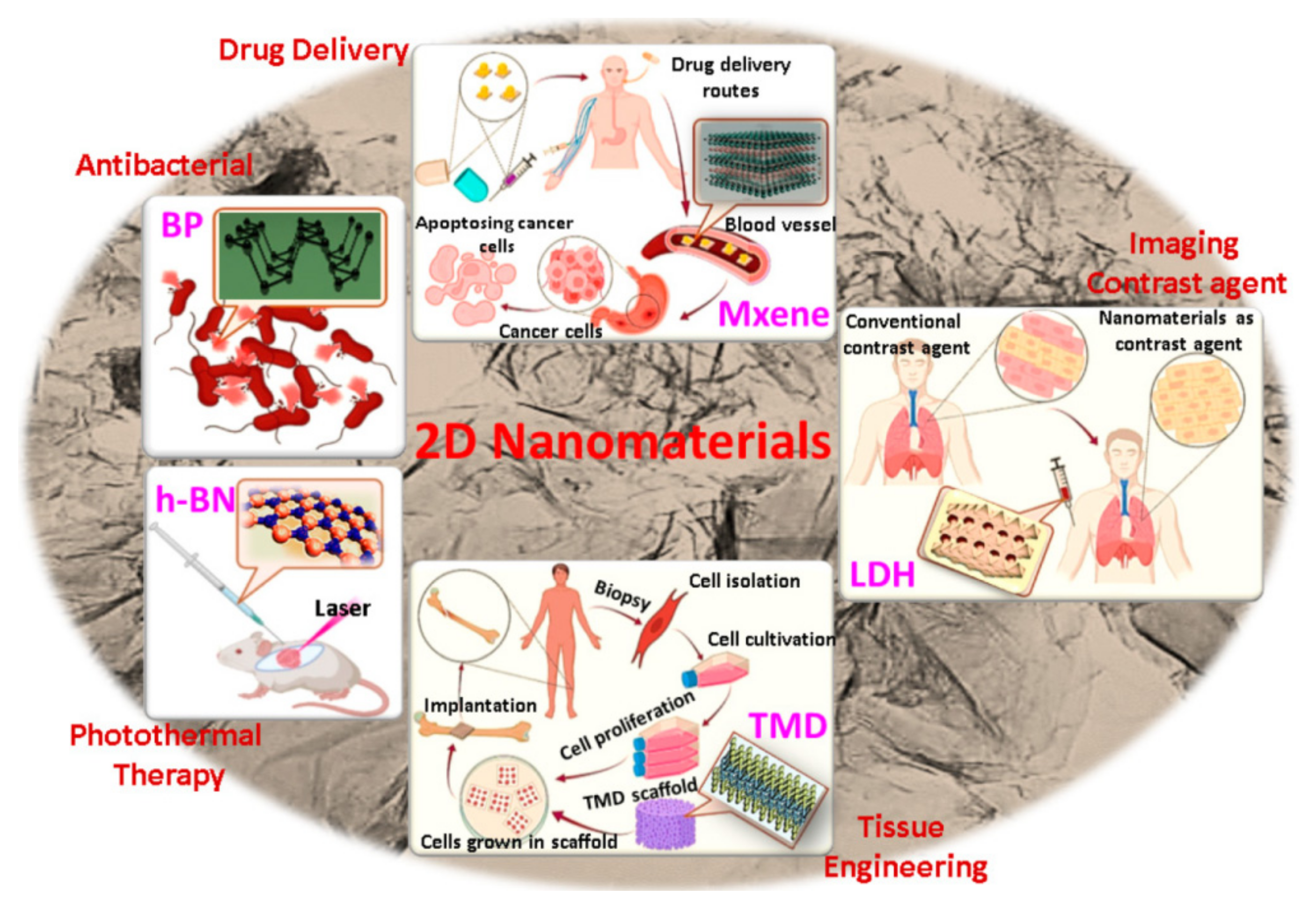
:1. Introduction
2. GBNs as Drugs and Nanocarriers
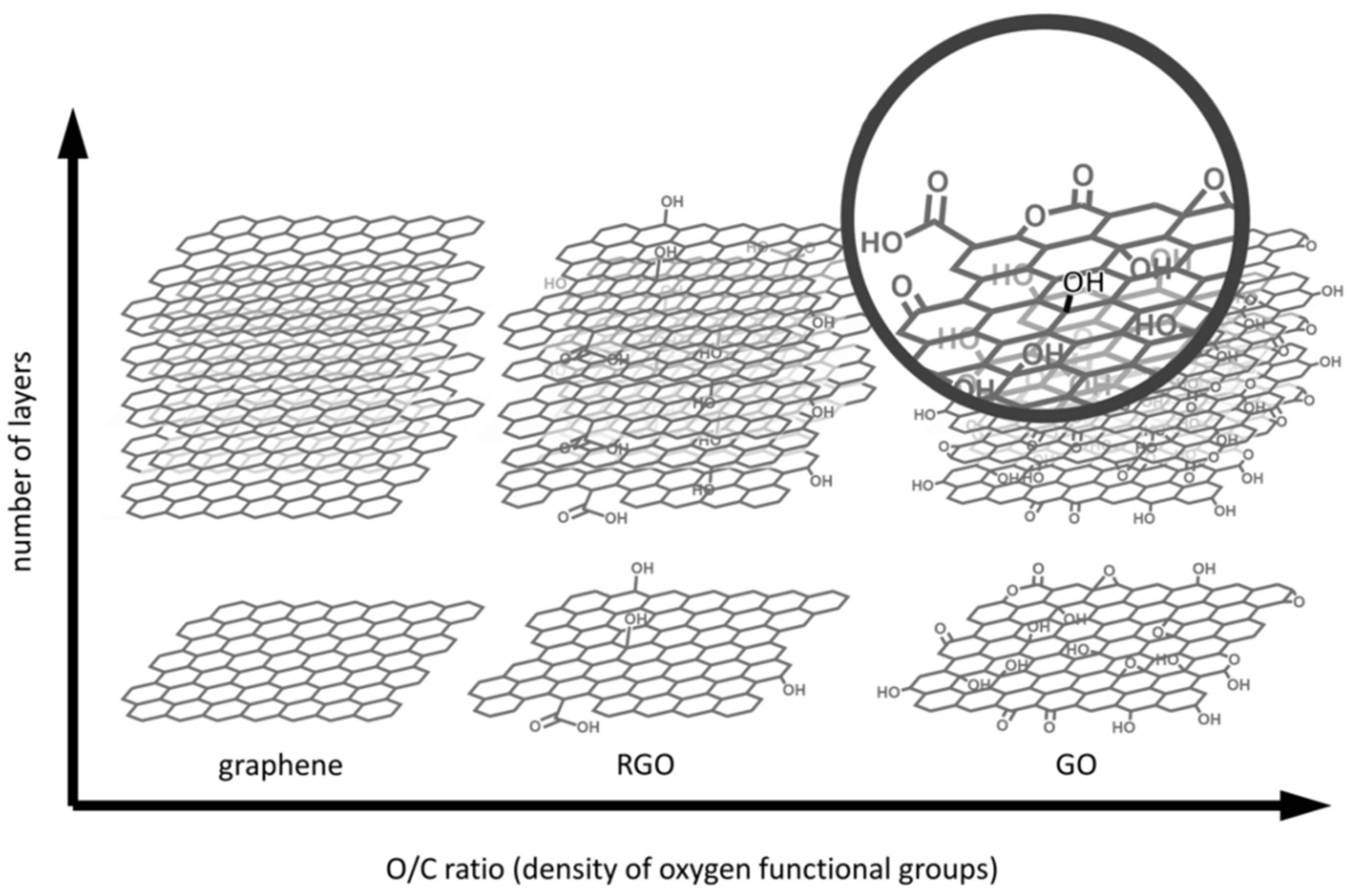
2.1. GRs and GRQDs
2.2. GO and GOQDs
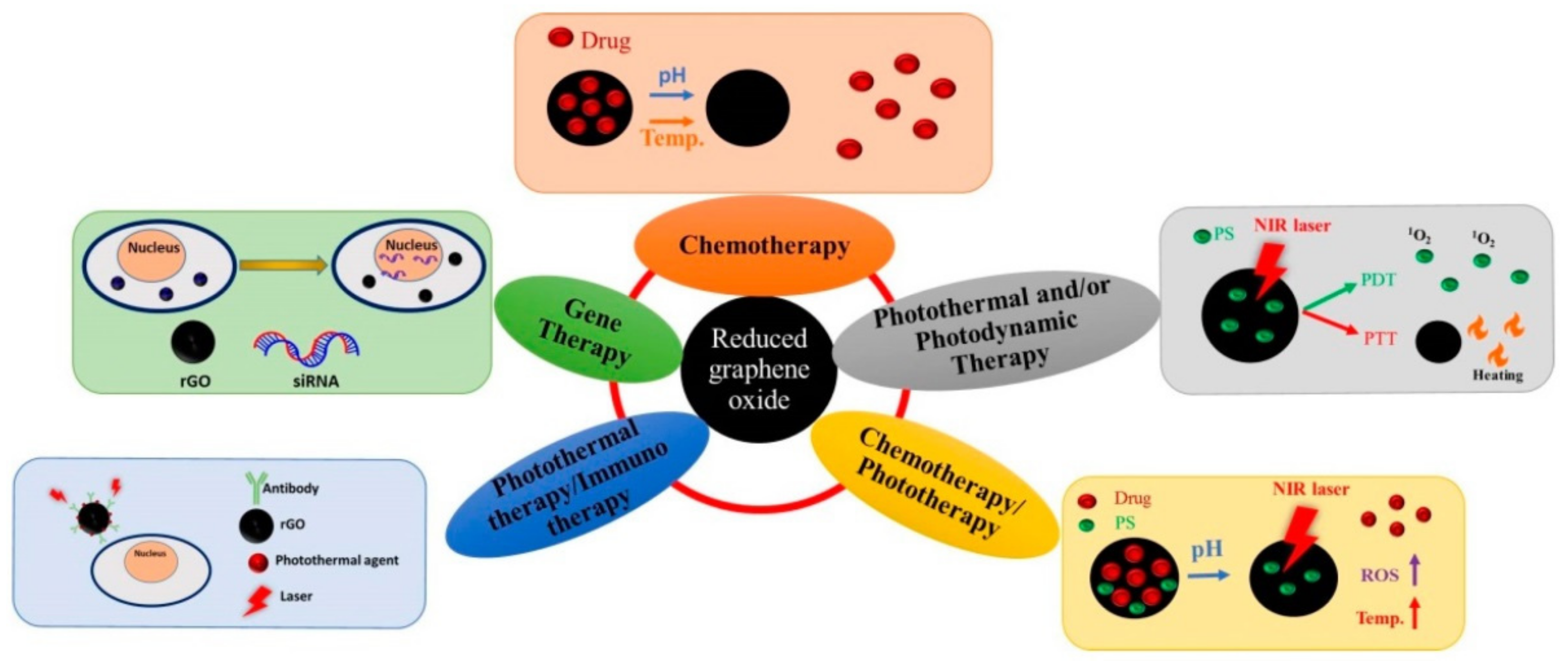
2.3. rGO
3. Impact of GBNs on Harmful Insects
4. Applications against Plant Patogenic Microorganisms
5. Effects of GBNs on Plants
5.1. Phytotoxic Impact
5.2. Beneficial Impact
6. Conclusions
Author Contributions
Funding
Institutional Review Board Statement
Informed Consent Statement
Data Availability Statement
Conflicts of Interest
References
- Rafiei-Sarmazdeh, Z.; Zahedi-Dizaji, S.M.; Kang, A.K. Two-dimensional nanomaterials. In Nanostructures; Ameen, S., Ed.; InTech Open: Rieka, Croatia, 2019; Chapter 3; Available online: https://www.intechopen.com/chapters/67053 (accessed on 25 April 2022).
- Singh, V.; Yadav, P.; Mishra, V. Recent advances on classification, properties, synthesis, and characterization of nanomaterials. In Green Synthesis of Nanomaterials for Bioenergy Applications; Srivastava, N., Srivastava, M., Mishra, P.K., Gupta, V.K., Eds.; John Wiley & Sons: Hoboken, NJ, USA, 2021; pp. 83–97. [Google Scholar]
- Chimene, D.; Alge, D.L.; Gaharwar, A.K. Two-dimensional nanomaterials for biomedical applications: Emerging trends and future prospects. Adv. Mater. 2015, 27, 7261–7284. [Google Scholar] [CrossRef] [PubMed]
- Tan, C.; Cao, X.; Wu, X.J.; He, Q.; Yang, J.; Zhang, X.; Chen, J.; Zhao, W.; Han, S.; Nam, G.H.; et al. Recent advances in ultrathin two-dimensional nanomaterials. Chem. Rev. 2017, 117, 6225–6331. [Google Scholar] [CrossRef] [PubMed]
- Novoselov, K.S.; Geim, A.K.; Morozov, S.V.; Jiang, D.; Zhang, Y.; Dubonos, S.V.; Grigorieva, I.V.; Firsov, A.A. Electric field effect in atomically thin carbon films. Science 2004, 306, 666–669. [Google Scholar] [CrossRef] [Green Version]
- Zhu, Y.; Murali, S.; Cai, W.; Li, X.; Suk, J.W.; Potts, J.R.; Ruoff, R.S. Graphene and graphene oxide: Synthesis, properties, and applications. Adv. Mater. 2010, 22, 3906–3924. [Google Scholar] [CrossRef] [PubMed]
- Smith, A.T.; LaChance, A.M.; Zeng, S.; Liu, B.; Sun, L. Synthesis, properties, and applications of graphene oxide/reduced graphene oxide and their nanocomposites. Nanomat. Sci. 2019, 1, 31–47. [Google Scholar] [CrossRef]
- Weiss, N.O.; Zhou, H.; Liao, L.; Liu, Y.; Jiang, S.; Huang, Y.; Duan, X. Graphene: An emerging electronic material. Adv. Mater. 2012, 24, 5782–5825. [Google Scholar] [CrossRef]
- An, X.; Butler, T.W.; Washington, M.; Nayak, S.K.; Kar, S. Optical and sensing properties of 1-pyrenecarboxylic acid-functionalized graphene films laminated on polydimethylsiloxane membranes. ACS Nano 2011, 5, 1003–10011. [Google Scholar] [CrossRef]
- Speranza, G. Carbon nanomaterials: Synthesis, functionalization and sensing applications. Nanomaterials 2021, 11, 967. [Google Scholar] [CrossRef]
- Balandin, A.A.; Ghosh, S.; Bao, W.; Calizo, I.; Teweldebrhan, D.; Miao, F.; Lau, C.N. Superior thermal conductivity of single-layer graphene. Nano Lett. 2008, 8, 902–907. [Google Scholar] [CrossRef]
- Graphene—What Is It? Graphenea, Inc.: Cambridge, MA, USA; Available online: https://www.graphenea.com/pages/graphene#.YnexYlTP2M8 (accessed on 25 April 2022).
- Graphene—All You Need to Know, NanoWerk. Available online: https://www.nanowerk.com/what_is_graphene.php (accessed on 25 April 2022).
- Rauti, R.; Musto, M.; Bosi, S.; Prato, M.; Ballerini, L. Properties and behavior of carbon nanomaterials when interfacing neuronal cells: How far have we come? Carbon 2019, 143, 430–446. [Google Scholar] [CrossRef]
- Nayak, L.; Rahaman, M.; Giri, R. Surface modification/functionalization of carbon materials by different techniques: An overview. In Carbon-Containing Polymer Composites; Springer Series on Polymer and Composite Materials; Rahaman, M., Khastgir, D., Aldalbahi, A., Eds.; Springer: Singapore, 2019; pp. 65–98. [Google Scholar]
- Barhoum, A.; Barhoum, A.; Shalan, A.E.; El-Hout, S.I.; Ali, G.A.M.; Abdelbasir, S.M.; Abu Serea, E.S.; Ibrahim, A.H.; Pal, K. A broad family of carbon nanomaterials: Classification, properties, synthesis, and emerging applications. In Handbook of Nanofibers; Barhoum, A., Bechelany, M., Makhlouf, A., Eds.; Springer: Cham, Switzerland, 2019; pp. 1–40. [Google Scholar]
- Derakhshi, M.; Daemi, S.; Shahini, P.; Habibzadeh, A.; Mostafavi, E.; Ashkarran, A.A. Two-dimensional nanomaterials beyond graphene for biomedical applications. J. Funct. Biomater. 2022, 13, 27. [Google Scholar] [CrossRef] [PubMed]
- Zhang, H. Ultrathin two-dimensional nanomaterials. ACS Nano 2015, 9, 9451–9469. [Google Scholar] [CrossRef] [PubMed]
- Thomas, S.; Sarathchandran, C.; Ilangovan, S.A.; Moreno-Pirajan, J.C. Handbook of Carbon-Based Nanomaterials: Micro and Nano Technologies; Elsevier: Amsterdam, The Netherlands, 2021. [Google Scholar]
- Wang, J.; Wang, S. A critical review on graphitic carbon nitride (g-C3N4)-based materials: Preparation, modification and environmental application. Coord. Chem. Rev. 2022, 453, 214338. [Google Scholar] [CrossRef]
- Liu, H.; Wang, X.; Wang, H.; Nie, R. Synthesis and biomedical applications of graphitic carbon nitride quantum dots. J. Mater. Chem. B 2019, 7, 5432–5448. [Google Scholar] [CrossRef]
- Abulyazied, D.E.; Ene, A. An investigative study on the progress of nanoclay-reinforced polymers: Preparation, properties, and applications: A review. Polymers 2021, 13, 4401. [Google Scholar] [CrossRef]
- Guo, F.; Aryana, S.; Han, Y.; Jiao, Y. A review of the synthesis and applications of polymer–nanoclay composites. Appl. Sci. 2018, 8, 1696. [Google Scholar] [CrossRef] [Green Version]
- Pizzoferrato, R.; Richetta, M. Layered double hydroxides (LDHs). Crystals 2020, 10, 1121. [Google Scholar] [CrossRef]
- Zhang, Y.; Xua, H.; Lu, S. Preparation and application of layered double hydroxide nanosheets. RSC Adv. 2021, 11, 24254–24281. [Google Scholar] [CrossRef]
- Wijitwongwan, R.; Intasa-ard, S.; Ogawa, M. Preparation of layered double hydroxides toward precisely designed hierarchical organization. ChemEngineering 2019, 3, 68. [Google Scholar] [CrossRef] [Green Version]
- Zhao, B.; Shen, D.; Zhang, Z.; Lu, P.; Hossain, M.; Li, J.; Li, B.; Duan, X. 2D metallic transition-metal dichalcogenides: Structures, synthesis, properties, and applications. Adv. Funct. Mater. 2021, 31, 2105132. [Google Scholar] [CrossRef]
- Song, L.; Li, H.; Zhang, Y.; Shi, J. Recent progress of two-dimensional metallic transition metal dichalcogenides: Syntheses, physical properties, and applications featured. Int. J. Appl. Phys. 2022, 131, 060902. [Google Scholar] [CrossRef]
- Yin, J.; Wang, J.; Ma, Y.; Yu, J.; Zhou, J.; Fan, Z. Recent advances in the controlled synthesis and catalytic applications of two-dimensional rhodium nanomaterials. ACS Mater. Lett. 2021, 3, 121–133. [Google Scholar] [CrossRef]
- Zhao, Y.; Duan, H. Photocatalysis Using 2D Nanomaterials; Royal Society of Chemistry: London, UK, 2022. [Google Scholar]
- Gong, C.; Hu, K.; Wang, X.; Wangyang, P.; Yan, C.; Chu, J.; Liao, M.; Dai, L.; Zhai, T.; Wang, C.; et al. 2D nanomaterial arrays for electronics and optoelectronics. Adv. Funct. Mater. 2018, 28, 1706559. [Google Scholar] [CrossRef]
- Khan, R.; Radoi, A.; Rashid, S.; Hayat, A.; Vasilescu, A.; Andreescu, S. Two-dimensional nanostructures for electrochemical biosensor. Sensors 2021, 21, 3369. [Google Scholar] [CrossRef]
- Shamkhalichenar, H.; Choi, J.W. Review—non-enzymatic hydrogen peroxide electrochemical sensors based on reduced graphene oxide. J. Electrochem. Soc. 2020, 167, 037531. [Google Scholar] [CrossRef]
- Zafeiratos, S. 2D Nanomaterials for Energy Applications: Graphene and Beyond; Elsevier: Amsterdam, The Netherlands, 2019. [Google Scholar]
- Glavin, N.R.; Rao, R.; Varshney, V.; Bianco, E.; Apte, A.; Roy, A.; Ringe, E.; Ajayan, P.M. Emerging applications of elemental 2D materials. Adv. Mat. 2020, 32, 1904302. [Google Scholar] [CrossRef]
- Hu, T.; Mei, X.; Wang, Y.; Weng, X.; Liang, R.; Wei, M. Two-dimensional nanomaterials: Fascinating materials in biomedical field. Sci. Bull. 2019, 64, 1707–1727. [Google Scholar] [CrossRef] [Green Version]
- Placha, D.; Jampilek, J. Graphenic materials for biomedical applications. Nanomaterials 2019, 9, 1758. [Google Scholar] [CrossRef] [Green Version]
- Mukherjee, S.; Bytesnikova, Z.; Ashrafi, A.M.; Adam, V.; Richtera, L. Graphene oxide as a nanocarrier for biochemical molecules: Current understanding and trends. Processes 2020, 8, 1636. [Google Scholar] [CrossRef]
- Alkhouzaam, A.; Qiblawey, H. Synergetic effects of dodecylamine-functionalized graphene oxide nanoparticles on antifouling and antibacterial properties of polysulfone ultrafiltration membranes. J. Water Process. Eng. 2021, 42, 102120. [Google Scholar] [CrossRef]
- Zou, X.; Zhang, L.; Wang, Z.; Luo, Y. Mechanisms of the antimicrobial activities of graphene materials. J. Am. Chem. Soc. 2016, 138, 2064–2077. [Google Scholar] [CrossRef] [PubMed]
- Kozik, V.; Bak, A.; Pentak, D.; Hachula, B.; Pytlakowska, K.; Rojkiewicz, M.; Jampilek, J.; Sieron, K.; Jazowiecka-Rakus, J.; Sochanik, A. Derivatives of graphene oxide as potential drug carriers. J. Nanosci. Nanotechnol. 2019, 19, 2489–2492. [Google Scholar] [CrossRef] [PubMed]
- Mohammed, H.; Kumar, A.; Bekyarova, E.; Al-Hadeethi, Y.; Zhang, X.; Chen, M.; Ansari, M.S.; Cochis, A.; Rimondini, L. Antimicrobial mechanisms and effectiveness of graphene and graphene-functionalized biomaterials. A scope review. Front. Bioeng. Biotechnol. 2020, 8, 465. [Google Scholar] [CrossRef] [PubMed]
- Jampilek, J.; Kralova, K. Advances in drug delivery nanosystems using graphene-based materials and carbon nanotubes. Materials 2021, 14, 1059. [Google Scholar] [CrossRef] [PubMed]
- Placha, D.; Jampilek, J. Chronic Inflammatory diseases, anti-inflammatory agents and their delivery nanosystems. Pharmaceutics 2021, 13, 64. [Google Scholar] [CrossRef]
- Jampilek, J.; Placha, D. Advances in use of nanomaterials for musculoskeletal regeneration. Pharmaceutics 2021, 13, 1994. [Google Scholar] [CrossRef]
- Jampilek, J.; Kralova, K. Advances in nanostructures for antimicrobial therapy. Materials 2022, 15, 2388. [Google Scholar] [CrossRef]
- Barani, M.; Zeeshan, M.; Kalantar-Neyestanaki, D.; Farooq, M.A.; Rahdar, A.; Jha, N.K.; Sargazi, S.; Gupta, P.K.; Thakur, V.K. Nanomaterials in the Management of Gram-Negative Bacterial Infections. Nanomaterials 2021, 11, 2535. [Google Scholar] [CrossRef]
- Cai, S.; Rong, Y. Two-dimensional nanomaterials with enzyme-like properties for biomedical applications. Front. Chem. 2020, 8, 565940. [Google Scholar] [CrossRef]
- Presutti, D.; Agarwal, T.; Zarepour, A.; Celikkin, N.; Hooshmand, S.; Nayak, C.; Ghomi, M.; Zarrabi, A.; Costantini, M.; Behera, B.; et al. Transition metal dichalcogenides (TMDC)-based nanozymes for biosensing and therapeutic applications. Materials 2022, 15, 337. [Google Scholar] [CrossRef]
- Hadji, H.; Bouchemal, K. Effect of micro- and nanoparticle shape on biological processes. J. Control. Release 2022, 342, 93–110. [Google Scholar] [CrossRef] [PubMed]
- Newman, L.; Jasim, D.A.; Prestat, E.; Lozano, N.; de Lazaro, I.; Nam, Y.; Assas, B.M.; Pennock, J.; Haigh, S.J.; Bussy, C.; et al. Splenic capture and in vivo intracellular biodegradation of biological-grade graphene oxide sheets. ACS Nano 2020, 14, 10168–10186. [Google Scholar] [CrossRef] [PubMed]
- Munoz-Wolf, N.; Lavelle, E.C. Promotion of trained innate immunity by nanoparticles. Semin. Immunol. 2021, 56, 101542. [Google Scholar] [CrossRef]
- Arkowski, J.; Obremska, M.; Kedzierski, K.; Slawuta, A.; Wawrzynska, M. Applications for graphene and its derivatives in medical devices: Current knowledge and future applications. Adv. Clin. Exp. Med. 2020, 29, 1497–1504. [Google Scholar] [CrossRef] [PubMed]
- Dziewięcka, M.; Pawlyta, M.; Majchrzycki, L.; Balin, K.; Barteczko, S.; Czerkawska, M.; Augustyniak, M. The structure–properties–cytotoxicity interplay: A crucial pathway to determining graphene oxide biocompatibility. Int. J. Mol. Sci. 2021, 22, 5401. [Google Scholar] [CrossRef]
- Pan, P.; Svirskis, D.; Rees, S.W.P.; Barker, D.; Waterhouse, G.I.N.; Wu, Z. Photosensitive drug delivery systems for cancer therapy: Mechanisms and applications. J. Control. Release 2021, 338, 446–461. [Google Scholar] [CrossRef] [PubMed]
- Li, J.; Zeng, H.; Zeng, Z.; Zeng, Y.; Xie, T. Promising graphene-based nanomaterials and their biomedical applications and potential risks: A comprehensive review. ACS Biomater. Sci. Eng. 2021, 7, 5363–5396. [Google Scholar] [CrossRef] [PubMed]
- Kabiri, S.; Degryse, F.; Tran, D.N.H.; da Silva, R.C.; McLaughlin, M.J.; Losic, D. Graphene oxide: A new carrier for slow release of plant micronutrients. ACS Appl. Mater. Interfaces 2017, 9, 43325–43335. [Google Scholar] [CrossRef] [Green Version]
- May, A.; Coelho, L.F.; da Silva, E.H.F.M.; da Viana, R.S.; Vieira, N.A.; Ferreira, W.P.M. Graphene: A new technology for agriculture. Res. Soc. Dev. 2021, 10, e56610212827. [Google Scholar] [CrossRef]
- Rehan, S. Carbon-Based Nanomaterials in Agriculture. AzoNano. Available online: https://www.azonano.com/article.aspx?ArticleID=6098 (accessed on 26 April 2022).
- Zhang, X.; Cao, H.; Wang, H.; Zhao, J.; Gao, K.; Qiao, J.; Li, J.; Ge, S. The effects of graphene-family nanomaterials on plant growth: A review. Nanomaterials 2022, 12, 936. [Google Scholar] [CrossRef]
- Dasari Shareena, T.P.; McShan, D.; Dasmahapatra, A.K.; Tchounwou, P.B. A review on graphene-based nanomaterials in biomedical applications and risks in environment and health. Nanomicro. Lett. 2018, 10, 53. [Google Scholar] [CrossRef] [PubMed]
- Bellet, P.; Gasparotto, M.; Pressi, S.; Fortunato, A.; Scapin, G.; Mba, M.; Menna, E.; Filippini, F. Graphene-based scaffolds for regenerative medicine. Nanomaterials 2021, 11, 404. [Google Scholar] [CrossRef] [PubMed]
- Song, J.; Cui, N.; Sun, S.; Lu, X.; Wang, Y.; Shi, H.; Lee, E.-S.; Jiang, H.-B. Controllability of graphene oxide doxorubicin loading capacity based on density functional theory. Nanomaterials 2022, 12, 479. [Google Scholar] [CrossRef] [PubMed]
- Chen, S.H.; Bell, D.R.; Luan, B. Understanding interactions between biomolecules and two-dimensional nanomaterials using in silico microscopes. Adv. Drug Deliv. Rev. 2022, 114336. [Google Scholar] [CrossRef]
- Li, Z.; Lei, H.; Kan, A.; Xie, H.; Yu, W. Photothermal applications based on graphene and its derivatives: A state-of-the-art review. Energy 2021, 216, 119262. [Google Scholar] [CrossRef]
- Lagos, K.J.; Buzza, H.H.; Bagnato, V.S.; Romero, M.P. Carbon-based materials in photodynamic and photothermal therapies applied to tumor destruction. Int. J. Mol. Sci. 2022, 23, 22. [Google Scholar] [CrossRef] [PubMed]
- Wang, Y.; Li, J.; Li, X.; Shi, J.; Jiang, Z.; Zhang, C.Y. Graphene-based nanomaterials for cancer therapy and anti-infections. Bioact. Mater. 2022, 14, 335–349. [Google Scholar] [CrossRef]
- Robinson, J.T.; Tabakman, S.M.; Liang, Y.; Wang, H.; Casalongue, H.S.; Vinh, D.; Dai, H. Ultrasmall reduced graphene oxide with high near-infrared absorbance for photothermal therapy. J. Am. Chem. Soc. 2011, 133, 6825–6831. [Google Scholar] [CrossRef]
- Guo, C.; Zhang, J.; Xu, W.; Liu, K.; Yuan, X.; Qin, S.; Zhu, Z. Graphene-based perfect absorption structures in the visible to terahertz band and their optoelectronics applications. Nanomaterials 2018, 8, 1033. [Google Scholar] [CrossRef] [Green Version]
- Liu, B.; Yu, W.; Yan, Z.; Cai, P.; Gao, F.; Tang, C.; Gu, P.; Liu, Z.; Chen, J. The light absorption enhancement in graphene monolayer resulting from the diffraction coupling of surface plasmon polariton resonance. Nanomaterials 2022, 12, 216. [Google Scholar] [CrossRef]
- Kim, T.-H.; Lee, T.; El-Said, W.A.; Choi, J.-W. Graphene-based materials for stem cell applications. Materials 2015, 8, 8674–8690. [Google Scholar] [CrossRef] [PubMed] [Green Version]
- Pena-Paras, L.; Sanchez-Fernandez, J.A.; Vidaltamayo, R. Nanoclays for biomedical applications. In Handbook of Ecomaterials; Martinez, L., Kharissova, O., Kharisov, B., Eds.; Springer: Cham, Switzerland, 2018; pp. 1–19. [Google Scholar]
- Gaharwar, A.K.; Cross, L.M.; Peak, C.W.; Gold, K.; Carrow, J.K.; Brokesh, A.; Singh, K.A. 2D Nanoclay for biomedical applications: Regenerative medicine, therapeutic delivery, and additive manufacturing. Adv. Mater. 2019, 31, e1900332. [Google Scholar] [CrossRef] [PubMed]
- Safder, A. 8 Sustainable Applications of Nanoclays. Nanografi Nano Technology. 2022. Available online: https://nanografi.com/blog/8-sustainable-applications-of-nanoclays/ (accessed on 25 May 2022).
- Rozhina, E.; Batasheva, S.; Miftakhova, R.; Yan, X.; Vikulina, A.; Volodkin, D.; Fakhrullin, R. Comparative cytotoxicity of kaolinite, halloysite, multiwalled carbon nanotubes and graphene oxide. Appl. Clay Sci. 2021, 205, 106041. [Google Scholar] [CrossRef]
- Kryuchkova, M.; Fakhrullin, R. Kaolin alleviates graphene oxide toxicity. Environ. Sci. Technol. Lett. 2018, 5, 295–300. [Google Scholar] [CrossRef]
- Rozhina, E.; Batasheva, S.; Danilushkina, A.; Kryuchkova, M.; Gomzikova, M.; Cherednichenko, Y.; Nigamatzyanova, L.; Akhatova, F.; Fakhrullin, R. Kaolin alleviates the toxicity of graphene oxide for mammalian cells. Med. Chem. Commun. 2019, 10, 1457–1464. [Google Scholar] [CrossRef]
- Mbayachi, V.B.; Ndayiragije, E.; Sammani, T.; Taj, S.; Mbuta, E.R.; Khan, A.U. Graphene synthesis, characterization and its applications: A review. Results Chem. 2021, 3, 100163. [Google Scholar] [CrossRef]
- Tewari, C.; Tatrari, G.; Kumar, S.; Pandey, S.; Rana, A.; Pal, M.; Sahoo, N.G. Green and cost-effective synthesis of 2D and 3D graphene-based nanomaterials from Drepanostachyum falcatum for bio-imaging and water purification applications. Chem. Eng. J. Adv. 2022, 10, 100265. [Google Scholar] [CrossRef]
- Nurunnabi, M.; McCarthy, J. Biomedical Applications of Graphene and 2D Nanomaterials; Elsevier: Amsterdam, The Netherlands, 2019. [Google Scholar]
- Tufano, I.; Vecchione, R.; Netti, P.A. Methods to scale down graphene oxide size and size implication in anti-cancer applications. Front. Bioeng. Biotechnol. 2020, 8, 613280. [Google Scholar] [CrossRef]
- Chen, W.; Pan, W.; Wang, J.; Cheng, L.; Wang, J.; Song, L.; Hu, Y.; Ma, X. Emerging two-dimensional monoelemental materials (Xenes): Fabrication, modification, and applications thereof in the field of bioimaging as nanocarriers. Wiley Interdiscip. Rev. Nanomed. Nanobiotechnol. 2022, 14, e1750. [Google Scholar] [CrossRef]
- Davis, R.; Urbanowski, R.A.; Gaharwar, A.K. 2D layered nanomaterials for therapeutics delivery. Curr. Opin. Biomed. Eng. 2021, 20, 100319. [Google Scholar] [CrossRef]
- Manisekaran, R.; Garcia-Contreras, R.; Chettiar, A.D.R.; Serrano-Diaz, P.; Lopez-Ayuso, C.A.; Arenas-Arrocena, M.C.; Hernandez-Padron, G.; Lopez-Marin, L.M.; Acosta-Torres, L.S. 2D nanosheets-A new class of therapeutic formulations against cancer. Pharmaceutics 2021, 13, 1803. [Google Scholar] [CrossRef] [PubMed]
- Jonoush, Z.A.; Farahani, M.; Bohlouli, M.; Niknam, Z.; Golchin, A.; Hatamie, S.; Rezaei-Tavirani, M.; Omidi, M.; Zali, H. Surface modification of graphene and its derivatives for drug delivery systems. Mini Rev. Org. Chem. 2021, 18, 78–92. [Google Scholar] [CrossRef]
- Jaymand, M.; Taghipour, Y.D.; Rezaei, A.; Derakhshankhah, H.; Abazari, M.F.; Samadian, H.; Hamblin, M.R. Radiolabeled carbon-based nanostructures: New radiopharmaceuticals for cancer therapy? Coord. Chem. Rev. 2021, 440, 213974. [Google Scholar] [CrossRef]
- Tabish, T.A.; Narayan, R.J. Mitochondria-targeted graphene for advanced cancer therapeutics. Acta Biomater. 2021, 129, 43–56. [Google Scholar] [CrossRef] [PubMed]
- Ebrahimi, M.; Asadi, M.; Akhavan, O. Graphene-based nanomaterials in fighting the most challenging viruses and immunogenic disorders. ACS Biomater. Sci. Eng. 2022, 8, 54–81. [Google Scholar] [CrossRef]
- Alkatheeri, M.S. An updated review on the properties of graphene nano filled composites and their applications in dentistry. Biosci. Biotechnol. Res. Commun. 2020, 13, 365–372. [Google Scholar] [CrossRef]
- Begum, S.; Pramanik, A.; Davis, D.; Patibandla, S.; Gates, K.; Gao, Y.; Ray, P.C. 2D and heterostructure nanomaterial based strategies for combating drug-resistant bacteria. ACS Omega 2020, 5, 3116–3130. [Google Scholar] [CrossRef]
- Pandit, S.; De, M. One-pot bottom-up synthesis of a 2D graphene derivative: Application in biomolecular recognition and nanozyme activity. Nanoscale Adv. 2021, 3, 5102–5110. [Google Scholar] [CrossRef]
- Song, Y.J.; Qu, K.G.; Zhao, C.; Ren, J.S.; Qu, X.G. Graphene oxide: Intrinsic peroxidase catalytic activity and its application to glucose detection. Adv. Mater. 2010, 22, 2206–2210. [Google Scholar] [CrossRef]
- Shende, P.; Pathan, N. Graphene nanoribbons: A state-of-the-art in health care. Int. J. Pharm. 2021, 595, 120269. [Google Scholar] [CrossRef]
- Niu, G.F.; Yousefi, B.; Qujeq, D.; Marjani, A.; Asadi, J.; Wang, Z.; Mir, S.M. Melatonin and doxorubicin co-delivered via a functionalized graphene-dendrimeric system enhances apoptosis of osteosarcoma cells. Mater. Sci. Eng. C Mater. Biol. Appl. 2021, 119, 111554. [Google Scholar] [CrossRef] [PubMed]
- Gooneh-Farahani, S.; Naghib, S.M.; Naimi-Jamal, M.R.; Seyfoori, A. A pH-sensitive nanocarrier based on BSA-stabilized graphene-chitosan nanocomposite for sustained and prolonged release of anticancer agents. Sci. Rep. 2021, 11, 17404. [Google Scholar] [CrossRef] [PubMed]
- Stevanovic, M.; Djosic, M.; Jankovic, A.; Kojic, V.; Vukasinovic-Sekulic, M.; Stojanovic, J.; Oclovic, J.; Sakac, M.C.; Yop, R.K.; Miskovic-Stankovic, V. Antibacterial graphene-based hydroxyapatite/chitosan coating with gentamicin for potential applications in bone tissue engineering. J. Biomed. Mater. Res. A 2020, 108, 2175–2189. [Google Scholar] [CrossRef] [PubMed]
- Das Jana, I.; Kumbhakar, P.; Banerjee, S.; Gowda, C.C.; Kedia, N.; Kuila, S.K.; Banerjee, S.; Das, N.C.; Das, A.K.; Manna, I. Copper nanoparticle-graphene composite-based transparent surface coating with antiviral activity against influenza virus. ACS Appl. Nano Mater. 2021, 4, 352–362. [Google Scholar] [CrossRef]
- Kadian, S.; Sethi, S.K.; Manik, G. Recent advancements in synthesis and property control of graphene quantum dots for biomedical and optoelectronic applications. Mater. Chem. Front. 2021, 5, 627–658. [Google Scholar] [CrossRef]
- Cui, Y.X.; Duan, W.; Jin, Y.; Wo, F.J.; Xi, F.N.; Wu, J.M. Graphene quantum dot-decorated luminescent porous silicon dressing for theranostics of diabetic wounds. Acta Biomater. 2021, 131, 544–554. [Google Scholar] [CrossRef]
- Wang, Z.J.; Yang, H.Y.; Bai, Y.X.; Cheng, L.M.; Zhu, R.R. rBMSC osteogenic differentiation enhanced by graphene quantum dots loaded with immunomodulatory layered double hydroxide nanoparticles. Biomed. Mater. 2022, 17, 024101. [Google Scholar] [CrossRef]
- Javadian, S.; Najafi, K.; Sadrpoor, S.M.; Ektefa, F.; Dalir, N.; Nikkhah, M. Graphene quantum dots based magnetic nanoparticles as a promising delivery system for controlled doxorubicin release. J. Mol. Liq. 2021, 331, 115746. [Google Scholar] [CrossRef]
- Vahedi, N.; Tabandeh, F.; Mahmoudifard, M. Hyaluronic acid-graphene quantum dot nanocomposite: Potential target drug delivery and cancer cell imaging. Biotechnol. Appl. Biochem. 2021, 1–12. [Google Scholar] [CrossRef]
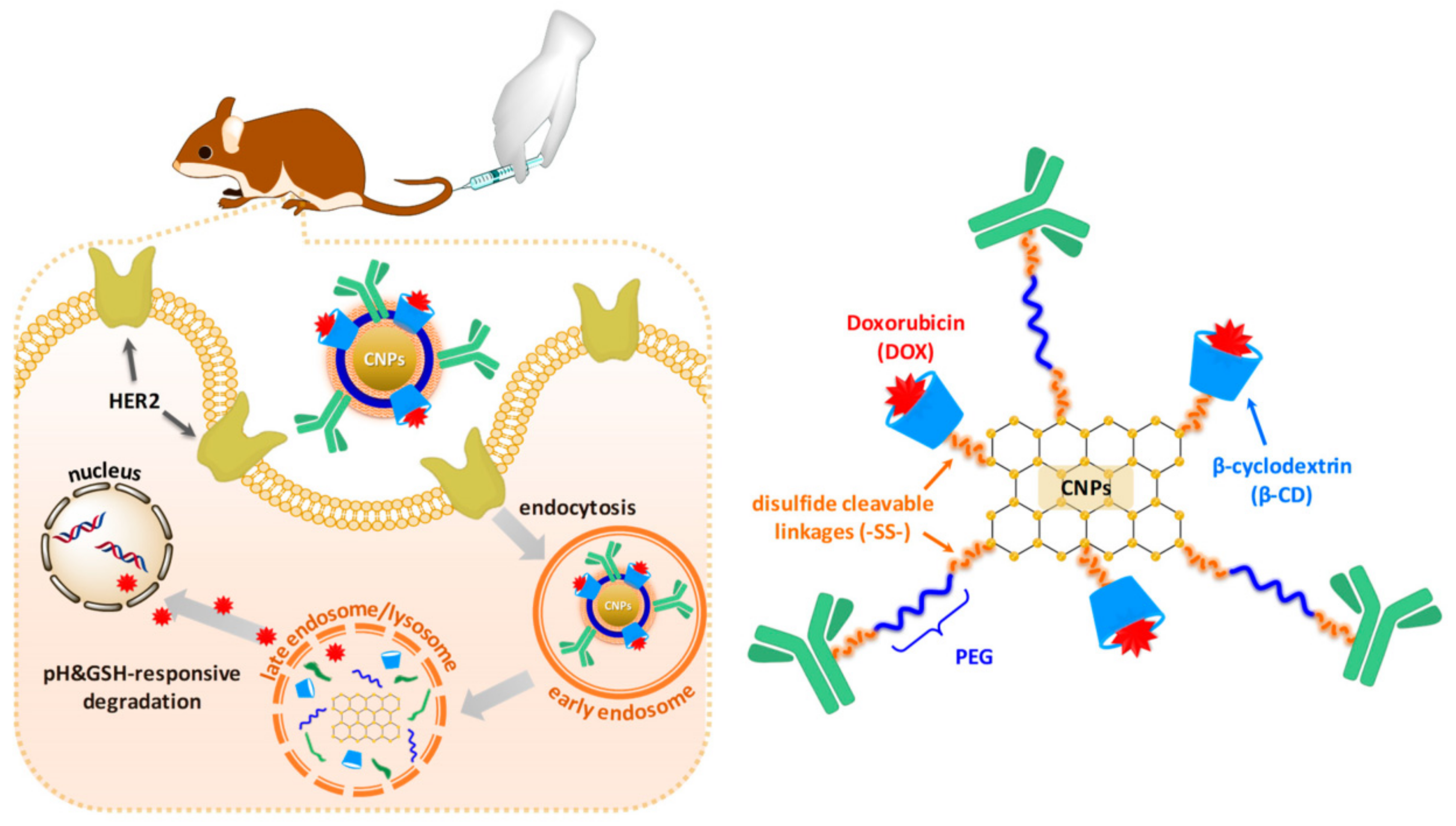
- Ko, N.R.; Van, S.Y.; Hong, S.H.; Kim, S.-Y.; Kim, M.; Lee, J.S.; Lee, S.J.; Lee, Y.-k.; Kwon, I.K.; Oh, S.J. Dual pH- and GSH-responsive degradable PEGylated graphene quantum dot-based nanoparticles for enhanced HER2-positive breast cancer therapy. Nanomaterials 2020, 10, 91. [Google Scholar] [CrossRef] [Green Version]
- Ghanbari, N.; Salehi, Z.; Khodadadi, A.A.; Shokrgozar, M.A.; Saboury, A.A. Glucosamine-conjugated graphene quantum dots as versatile and pH-sensitive nanocarriers for enhanced delivery of curcumin targeting to breast cancer. Mater. Sci. Eng. C Mater. Biol. Appl. 2021, 121, 111809. [Google Scholar] [CrossRef] [PubMed]
- Ghanbari, N.; Salehi, Z.; Khodadadi, A.A.; Shokrgozar, M.A.; Saboury, A.A.; Farzaneh, F. Tryptophan-functionalized graphene quantum dots with enhanced curcumin loading capacity and pH-sensitive release. J. Drug Deliv. Sci. Technol. 2021, 61, 102137. [Google Scholar] [CrossRef]
- Rakhshaei, R.; Namazi, H.; Hamishehkar, H.; Rahimi, M. Graphene quantum dot cross-linked carboxymethyl cellulose nanocomposite hydrogel for pH-sensitive oral anticancer drug delivery with potential bioimaging properties. Int. J. Biol. Macromol. 2020, 150, 1121–1129. [Google Scholar] [CrossRef] [PubMed]
- Mushtaq, S.; Yasin, T.; Saleem, M.; Dai, T.H.; Yameen, M.A. Potentiation of antimicrobial photodynamic therapy by curcumin-loaded graphene quantum dots. Photochem. Photobiol. 2022, 98, 202–210. [Google Scholar] [CrossRef] [PubMed]
- Mangalath, S.; Babu, P.S.S.; Nair, R.R.; Manu, P.M.; Krishna, S.; Nair, S.A.; Joseph, J. Graphene quantum dots decorated with boron dipyrromethene dye derivatives for photodynamic therapy. ACS Appl. Nano Mater. 2021, 4, 4162–4171. [Google Scholar] [CrossRef]
- Hafezi, M.; Rostami, M.; Hosseini, A.; Rahimi-Nasrabadi, M.; Fasihi-Ramandi, M.; Badiei, A.; Ahmadi, F. Cur-loaded ZnFe2O4@mZnO@N-GQDs biocompatible nano-carriers for smart and controlled targeted drug delivery with pH-triggered and ultrasound irradiation. J. Mol. Liq. 2021, 322, 114875. [Google Scholar] [CrossRef]
- Ji, Y.; Zhu, R.Y.; Shen, Y.; Tan, Q.; Chen, J.L. Comparison of loading and unloading of different small drugs on graphene and its oxide. J. Mol. Liq. 2021, 341, 117454. [Google Scholar] [CrossRef]
- Jafarinejad-Farsangi, S.; Hashemi, M.S.; Rouholamini, S.E.Y.; Gharbi, S.; Ansari-Asl, Z.; Jafari, E.; Dezfuli, A.S.; Shahrokhi-Farjah, M. Curcumin loaded on graphene nanosheets induced cell death in mammospheres from MCF-7 and primary breast tumor cells. Biomed. Mater. 2021, 16, 045040. [Google Scholar] [CrossRef]
- Danafar, H.; Salehiabar, M.; Barsbay, M.; Rahimi, H.; Ghaffarlou, M.; Zaboli, K.A.; Faghfoori, M.H.; Kaboli, S.; Nosrati, H.; Faghfoori, Z. Curcumin delivery by modified biosourced carbon-based nanoparticles. Nanomedicine 2022, 17, 95–105. [Google Scholar] [CrossRef]
- Abdelhamid, H.N.; Hussein, K.H. Graphene oxide as a carrier for drug delivery of methotrexate. Biointerface Res. Appl. Chem. 2021, 11, 14726–14735. [Google Scholar]
- Mohanta, Y.K.; Biswas, K.; Rauta, P.R.; Mishra, A.K.; De, D.; Hashem, A.; Al-Arjani, A.F.; Alqarawi, A.A.; Abd-Allah, E.F.; Mahanta, S. Development of graphene oxide nanosheets as potential biomaterials in cancer therapeutics: An in-vitro study against breast cancer cell line. J. Inorg. Organomet. Polym. Mater. 2021, 31, 4236–4249. [Google Scholar] [CrossRef]
- Pelin, M.; Fusco, L.; Martin, C.; Sosa, S.; Frontinan-Rubio, J.; Gonzalez-Dominguez, J.M.; Duran-Prado, M.; Vazquez, E.; Prato, M.; Tubaro, A. Graphene and graphene oxide induce ROS production in human HaCaT skin keratinocytes: The role of xanthine oxidase and NADH dehydrogenase. Nanoscale 2018, 10, 11820–11830. [Google Scholar] [CrossRef] [PubMed] [Green Version]
- Timganova, V.; Zamorina, S.; Bochkova, M.; Nechaev, A.; Khramtsov, P.; Shardina, K.; Uzhviyuk, S.; Rayev, M. The effect of pristine and pegylated graphene oxide nanosheets on the functions of human neutrophils. KnE Life Sci. 2022, 7, 48–57. [Google Scholar] [CrossRef]
- Lee, Y.-M.; Yoon, Y.; Yoon, H.; Song, S.; Park, H.-M.; Lee, Y.Y.; Shin, H.; Hwang, S.W.; Yeum, K.J. Enhanced antioxidant activity of bioactives in colored grains by nano-carriers in human lens epithelial cells. Molecules 2018, 23, 1327. [Google Scholar] [CrossRef] [PubMed] [Green Version]
- Qiu, Y.; Wang, Z.; Owens, A.C.; Kulaots, I.; Chen, Y.; Kane, A.B.; Hurt, R.H. Antioxidant chemistry of graphene-based materials and its role in oxidation protection technology. Nanoscale 2014, 6, 11744–11755. [Google Scholar] [CrossRef] [PubMed] [Green Version]
- Kuropka, P.; Dobrzynski, M.; Bazanow, B.; Stygar, D.; Gebarowski, T.; Leskow, A.; Tarnowska, M.; Szyszka, K.; Malecka, M.; Nowak, N. A study of the impact of graphene oxide on viral infection related to A549 and TC28a2 human cell lines. Materials 2021, 14, 7788. [Google Scholar] [CrossRef] [PubMed]
- Pulingam, T.; Thong, K.L.; Appaturi, J.N.; Lai, C.W.; Leo, B.F. Mechanistic actions and contributing factors affecting the antibacterial property and cytotoxicity of graphene oxide. Chemosphere 2021, 281, 130739. [Google Scholar] [CrossRef]
- Li, X.; Zhao, H.Q.; Wang, S.D.; Zou, W.W.; Yang, P.Y.; Xing, X.D.; Gu, B.S. A synergistic antimicrobial mechanism of GO: Why oxidative stress can inactivate E. coli. Nano 2020, 15, 2050054. [Google Scholar] [CrossRef]
- Cao, W.J.; He, L.; Cao, W.D.; Huang, X.B.; Jia, K.; Dai, J.Y. Recent progress of graphene oxide as a potential vaccine carrier and adjuvant. Acta Biomater. 2020, 112, 14–28. [Google Scholar] [CrossRef]
- Patarroyo, J.L.; Fonseca, E.; Cifuentes, J.; Salcedo, F.; Cruz, J.C.; Reyes, L.H. Gelatin-graphene oxide nanocomposite hydrogels for Kluyveromyces lactis encapsulation: Potential applications in probiotics and bioreactor packings. Biomolecules 2021, 11, 922. [Google Scholar] [CrossRef]
- Jackman, J.A.; Yoon, B.K.; Mokrzecka, N.; Kohli, G.S.; Valle-Gonzalez, E.R.; Zhu, X.; Pumera, M.; Rice, S.A.; Cho, N.J. Graphene oxide mimics biological signaling cue to rescue starving bacteria. Adv. Funct. Mater. 2021, 31, 2102328. [Google Scholar] [CrossRef]
- Basiuk, E.V.; Monroy-Torres, B.; Carrero, J.C.; Basiuk, V.A. Effects of solvent-free amine functionalization of graphene oxide and nanodiamond on bacterial growth. Fuller. Nanotub. Carbon Nanostruct. 2021, 29, 58–66. [Google Scholar] [CrossRef]
- Vaishampayan, A.; Ahmed, R.; Wagner, O.; de Jong, A.; Haag, R.; Kok, J.; Grohmann, E. Transcriptomic analysis of stress response to novel antimicrobial coatings in a clinical MRSA strain. Mater. Sci. Eng. C Mater. Biol. Appl. 2021, 119, 111578. [Google Scholar] [CrossRef] [PubMed]
- Tudose, M.; Anghel, E.M.; Hristea, E.N.; Voicescu, M.; Somacescu, S.; Culita, D.C.; Musuc, A.M.; Dumitrascu, F.; Hanganu, A.; Kuncser, A. Benzofurazan derivatives modified graphene oxide nanocomposite: Physico-chemical characterization and interaction with bacterial and tumoral cells. Mater. Sci. Eng. C Mater. Biol. Appl. 2021, 123, 112028. [Google Scholar] [CrossRef]
- Cibecchini, G.; Veronesi, M.; Catelani, T.; Bandiera, T.; Guarnieri, D.; Pompa, P.P. Antiangiogenic effect of graphene oxide in primary human endothelial cells. ACS Appl. Bio Mater. 2020, 12, 22507–22518. [Google Scholar] [CrossRef]
- Elsabagh, M.F.; Fayez, H.; Motaleb, M.A.; Zaghary, W.A.; Sakr, T.M. Synthesis, modification, characterization, radiolabeling and in vivo behavior of carboxylated nanographene oxide sheets as a tumor imaging agent. Egypt. J. Chem. 2022, 65, 249–258. [Google Scholar] [CrossRef]
- Jovito, B.L.; Paterno, L.G.; Sales, M.J.A.; Gross, M.A.; Silva, L.P.; de Souza, P.; Bao, S.N. Graphene oide/zinc oxide nanocomposite displaying selective toxicity to glioblastoma cell lines. ACS Appl. Bio Mater. 2021, 4, 829–843. [Google Scholar] [CrossRef]
- Kesavan, S.; Meena, K.S.; Sharmili, S.A.; Govindarajan, M.; Alharbi, N.S.; Kadaikunnan, S.; Khaled, J.M.; Alobaidi, A.S.; Alanzi, K.F.; Vaseeharan, B. Ulvan loaded graphene oxide nanoparticle fabricated with chitosan and d-mannose for targeted anticancer drug delivery. J. Drug Deliv. Sci. Technol. 2021, 65, 102760. [Google Scholar] [CrossRef]
- Zhao, P.H.; Wei, Z.; Wang, L.H.; Pang, C.; Zhang, H.R.; Wang, J.; Wang, Y.L. Delivery of temozolomide using PEGylated graphene oxide as a nanocarrier. Mater. Express 2021, 11, 189–195. [Google Scholar]
- Krasteva, N.; Staneva, D.; Vasileva, B.; Miloshev, G.; Georgieva, M. Bioactivity of PEGylated graphene oxide nanoparticles combined with near-infrared laser irradiation studied in colorectal carcinoma cells. Nanomaterials 2021, 11, 3061. [Google Scholar] [CrossRef]
- Georgieva, M.; Gospodinova, Z.; Keremidarska-Markova, M.; Kamenska, T.; Gencheva, G.; Krasteva, N. PEGylated nanographene oxide in combination with near-infrared laser irradiation as a smart nanocarrier in colon cancer targeted therapy. Pharmaceutics 2021, 13, 424. [Google Scholar] [CrossRef] [PubMed]
- Abu Lila, A.S.; Abdallah, M.H.; Wani, S.U.D.; Gangadharappa, H.V.; Younes, K.M.; Khafagy, E.S.; Shehata, T.M.; Soliman, M.S. Folic acid-conjugated raloxifene-loaded graphene-based nanocarrier: Fabrication, characterization and antitumor screening. Colloids Surf. A Physicochem. Eng. Asp. 2021, 625, 126971. [Google Scholar] [CrossRef]
- Wei, L.Y.; Li, G.; Lu, T.C.; Wei, Y.M.; Nong, Z.Z.; Wei, M.; Pan, X.; Qin, Q.X.; Meng, F.Y.; Li, X.H. Functionalized graphene oxide as drug delivery systems for platinum anticancer drugs. J. Pharm. Sci. 2021, 110, 3631–3638. [Google Scholar] [CrossRef] [PubMed]
- Buskaran, K.; Hussein, M.Z.; Moklas, M.A.M.; Masarudin, M.J.; Fakurazi, S. Graphene oxide loaded with protocatechuic acid and chlorogenic acid dual drug nanodelivery system for human hepatocellular carcinoma therapeutic application. Int. J. Mol. Sci. 2021, 22, 5786. [Google Scholar] [CrossRef]
- Asgari, S.; Pourjavadi, A.; Setayeshmehr, M.; Boisen, A.; Ajalloueian, F. Encapsulation of drug-loaded graphene oxide-based nanocarrier into electrospun pullulan nanofibers for potential local chemotherapy of breast cancer. Macromol. Chem. Phys. 2021, 222, 2100096. [Google Scholar] [CrossRef]
- Wang, P.Y.; Wang, X.; Tang, Q.; Chen, H.; Zhang, Q.; Jiang, H.Y.; Wang, Z. Functionalized graphene oxide against U251 glioma cells and its molecular mechanism. Mater. Sci. Eng. C Mater. Biol. Appl. 2020, 116, 111187. [Google Scholar] [CrossRef]
- Bouchareb, S.; Doufnoune, R.; Riahi, F.; Cherif-Silini, H.; Belbahri, L. Non-covalent functionalization of graphene oxide using self-assembly of silver-triphenylphosphine for bactericidal formulations. Mater. Chem. Phys. 2020, 243, 122598. [Google Scholar] [CrossRef]
- Bakhsheshi-Rad, H.R.; Ismail, A.F.; Aziz, M.; Akbari, M.; Hadisi, Z.; Khoshnava, S.M.; Pagan, E.; Chen, X.B. Co-incorporation of graphene oxide/silver nanoparticle into poly-l-lactic acid fibrous: A route toward the development of cytocompatible and antibacterial coating layer on magnesium implants. Mater. Sci. Eng. C Mater. Biol. Appl. 2020, 111, 110812. [Google Scholar] [CrossRef]
- Mao, M.Y.; Zhang, W.J.; Huang, Z.W.; Huang, J.; Wang, J.; Li, W.P.; Gu, S.S. Graphene oxide-copper nanocomposites suppress cariogenic Streptococcus mutans biofilm formation. Int. J. Nanomed. 2021, 16, 7727–7739. [Google Scholar] [CrossRef]
- Chai, M.Z.; An, M.W.; Zhang, X.Y.; Chu, P.K. In vitro and in vivo antibacterial activity of graphene oxide-modified porous TiO2 coatings under 808-nm light irradiation. Rare Met. 2022, 41, 540–545. [Google Scholar] [CrossRef]
- Chata, G.; Nichols, F.; Mercado, R.; Assafa, T.; Millhauser, G.L.; Saltikov, C.; Chen, S.W. Photodynamic activity of graphene oxide/polyaniline/manganese oxide ternary composites toward both Gram-positive and Gram-negative bacteria. ACS Appl. Bio Mater. 2021, 4, 7025–7033. [Google Scholar] [CrossRef] [PubMed]
- Hung, H.S.; Kung, M.L.; Chen, F.C.; Ke, Y.C.; Shen, C.C.; Yang, Y.C.; Tang, C.M.; Yeh, C.A.; Hsieh, H.H.; Hsu, S.H. Nanogold-carried graphene oxide: Anti-inflammation and increased differentiation capacity of mesenchymal stem cells. Nanomaterials 2021, 11, 2046. [Google Scholar] [CrossRef] [PubMed]
- Chen, H.; Xing, L.Q.; Guo, H.R.; Luo, C.X.; Zhang, X.D. Dual-targeting SERS-encoded graphene oxide nanocarrier for intracellular co-delivery of doxorubicin and 9-aminoacridine with enhanced combination therapy. Analyst 2021, 146, 6893–6901. [Google Scholar] [CrossRef] [PubMed]
- Matiyani, M.; Rana, A.; Pal, M.; Rana, S.; Melkani, A.B.; Sahoo, N.G. Polymer grafted magnetic graphene oxide as a potential nanocarrier for pH-responsive delivery of sparingly soluble quercetin against breast cancer cells. RSC Adv. 2022, 12, 2574–2588. [Google Scholar] [CrossRef] [PubMed]
- Wen, C.C.; Cheng, R.N.; Gong, T.; Huang, Y.; Li, D.; Zhao, X.H.; Yu, B.F.; Su, D.; Song, Z.L.; Liang, W.T. β-Cyclodextrin-cholic acid-hyaluronic acid polymer coated Fe3O4-graphene oxide nanohybrids as local chemo-photothermal synergistic agents for enhanced liver tumor therapy. Colloids Surf. B Biointerfaces 2021, 199, 111510. [Google Scholar] [CrossRef]
- Kiamohammadi, L.; Asadi, L.; Shirvalilou, S.; Khoei, S.; Khoee, S.; Soleymani, M.; Minaei, S.E. Physical and biological properties of 5-fluorouracil polymer-coated magnetite nanographene oxide as a new thermosensitizer for alternative magnetic hyperthermia and a magnetic resonance imaging contrast agent: In vitro and in vivo study. ACS Omega 2021, 6, 20192–20204. [Google Scholar] [CrossRef]
- Zhu, H.Q.; Zhang, B.; Zhu, N.L.; Li, M.C.; Yu, Q.L. Mitochondrion targeting peptide-modified magnetic graphene oxide delivering mitoxantrone for impairment of tumor mitochondrial functions. Chin. Chem. Lett. 2021, 32, 1220–1223. [Google Scholar] [CrossRef]
- Karimi, S.; Namazi, H. Fe3O4@PEG-coated dendrimer modified graphene oxide nanocomposite as a pH-sensitive drug carrier for targeted delivery of doxorubicin. J. Alloys Compd. 2021, 879, 160426. [Google Scholar] [CrossRef]
- Qiang, S.R.; Wang, M.Y.; Liang, J.J.; Zhao, X.L.; Fan, Q.H.; Geng, R.Y.; Luo, D.X.; Li, Z.B.; Zhang, L. Effects of morphology regulated by Pb2+ on graphene oxide cytotoxicity: Spectroscopic and in vitro investigations. Mater. Chem. Phys. 2020, 239, 122016. [Google Scholar] [CrossRef]
- Taylor, K.; Tabish, T.A.; Narayan, R.J. Drug release kinetics of DOX-loaded graphene-based nanocarriers for ovarian and breast cancer therapeutics. Appl. Sci. 2021, 11, 11151. [Google Scholar] [CrossRef]
- Biagiotti, G.; Salvatore, A.; Toniolo, G.; Caselli, L.; Di Vito, M.; Cacaci, M.; Contiero, L.; Gori, T.; Maggini, M.; Sanguinetti, M. Metal-free antibacterial additives based on graphene materials and salicylic acid: From the bench to fabric applications. ACS Appl. Mater. Interfaces 2021, 13, 26288–26298. [Google Scholar] [CrossRef] [PubMed]
- Cojocaru, E.; Ghitman, J.; Pircalabioru, G.G.; Stavarache, C.; Serafim, A.; Vasile, E.; Iovu, H. Containing rGO-TEPA with potential application in wound dressings. Polymers 2022, 14, 294. [Google Scholar] [CrossRef] [PubMed]
- Khan, M.U.A.; Haider, S.; Raza, M.A.; Shah, S.A.; Abd Razak, S.I.; Kadir, M.R.A.; Subhan, F.; Haider, A. Smart and pH-sensitive rGO/Arabinoxylan/chitosan composite for wound dressing: In-vitro drug delivery, antibacterial activity, and biological activities. Int. J. Biol. Macromol. 2021, 192, 820–831. [Google Scholar] [CrossRef] [PubMed]
- Zhang, Z.Q.; Wang, X.G.; Li, P.S.; Bai, M.H.; Qi, W.B. Transdermal delivery of buprenorphine from reduced graphene oxide laden hydrogel to treat osteoarthritis. J. Biomater. Sci. Polym. Ed. 2021, 32, 874–885. [Google Scholar] [CrossRef]
- Li, W.F.; Zhang, G.Q.; Wei, X.X. Lidocaine-loaded reduced graphene oxide hydrogel for prolongation of effects of local anesthesia: In vitro and in vivo analyses. J. Biomater. Appl. 2021, 35, 1034–1042. [Google Scholar] [CrossRef]
- Lee, S.; Kim, S.Y. Gold nanorod/reduced graphene oxide composite nanocarriers for near-infrared-induced cancer therapy and photoacoustic imaging. ACS Appl. Nano Mater. 2021, 4, 11849–11860. [Google Scholar] [CrossRef]
- Caires, C.S.A.; Farias, L.A.S.; Gomes, L.E.; Pinto, B.P.; Goncalves, D.A.; Zagonel, L.F.; Nascimento, V.A.; Alves, D.C.B.; Colbeck, I.; Whitby, C.; et al. Effective killing of bacteria under blue-light irradiation promoted by green synthesized silver nanoparticles loaded on reduced graphene oxide sheets. Mater. Sci. Eng. C Mater. Biol. Appl. 2020, 113, 110984. [Google Scholar] [CrossRef]
- Maleki-Ghaleh, H.; Siadati, M.H.; Fallah, A.; Koc, B.; Kavanlouei, M.; Khademi-Azandehi, P.; Moradpur-Tari, E.; Omidi, Y.; Barar, J.; Beygi-Khosrowshahi, Y. Antibacterial and cellular behaviors of novel zinc-doped hydroxyapatite/graphene nanocomposite for bone tissue engineering. Int. J. Mol. Sci. 2021, 22, 9564. [Google Scholar] [CrossRef]
- Prakashkumar, N.; Asik, R.M.; Kavitha, T.; Archunan, G.; Suganthy, N. Unveiling the anticancer and antibiofilm potential of catechin overlaid reduced graphene oxide/zinc oxide nanocomposites. J. Clust. Sci. 2021, 1–18. [Google Scholar] [CrossRef]
- Dash, B.S.; Jose, G.; Lu, Y.-J.; Chen, J.-P. Functionalized reduced graphene oxide as a versatile tool for cancer therapy. Int. J. Mol. Sci. 2021, 22, 2989. [Google Scholar] [CrossRef]
- Wang, X.P.; Zhang, T.T.; Xie, H.C.; Wang, Z.Y.; Jing, D.P.; He, K.L.; Gao, X.D. Phenotypic responses and potential genetic mechanism of lepidopteran insects under exposure to graphene oxide. Ecotoxicol. Environ. Saf. 2021, 228, 113008. [Google Scholar] [CrossRef] [PubMed]
- Flasz, B.; Dziewiecka, M.; Kedziorski, A.; Tarnawska, M.; Augustyniak, M. Multigenerational graphene oxide intoxication results in reproduction disorders at the molecular level of vitellogenin protein expression in Acheta domesticus. Chemosphere 2021, 280, 130772. [Google Scholar] [CrossRef] [PubMed]
- Dziewiecka, M.; Flasz, B.; Rost-Roszkowska, M.; Kedziorski, A.; Kochanowicz, A.; Augustyniak, M. Graphene oxide as a new anthropogenic stress factor—multigenerational study at the molecular, cellular, individual and population level of Acheta domesticus. J. Hazard. Mater. 2020, 396, 122775. [Google Scholar] [CrossRef] [PubMed]
- Alian, R.S.; Dziewiecka, M.; Kedziorski, A.; Majchrzycki, L.; Augustyniak, M. Do nanoparticles cause hormesis? Early physiological compensatory response in house crickets to a dietary admixture of GO, Ag, and GOAg composite. Sci. Total Environ. 2021, 788, 147801. [Google Scholar] [CrossRef]
- Fang, Y.L.; Lu, Z.T.; Li, M.X.; Qu, J.W.; Ye, W.T.; Li, F.C.; Wei, J.; Sun, H.N.; Li, B. An assessment of the reproductive toxicity of GONPs exposure to Bombyx mori. Ecotoxicol. Environ. Saf. 2021, 210, 111888. [Google Scholar] [CrossRef]
- Martins, C.H.Z.; de Sousa, M.; Fonseca, L.C.; Martinez, D.S.T.; Alves, O.L. Biological effects of oxidized carbon nanomaterials (1D versus 2D) on Spodoptera frugiperda: Material dimensionality influences on the insect development, performance and nutritional physiology. Chemosphere 2019, 215, 766–774. [Google Scholar] [CrossRef]
- Batool, M.; Hussain, D.; Akrem, A.; Najam-ul-Haq, M.; Saeed, S.; Zaka, S.M.; Nawaz, M.S.; Buck, F.; Saeed, Q. Graphene quantum dots as cysteine protease nanocarriers against stored grain insect pests. Sci. Rep. 2020, 10, 3444. [Google Scholar] [CrossRef]
- Kaur, K.; Sharma, S.; Gupta, R.; Munikrishnappa, V.K.T.; Chandel, M.; Ahamed, M.; Singhal, N.K.; Bakthavatsalam, N.; Gorantla, M.; Muthusamy, E.; et al. Nanomaze lure: Pheromone sandwich in graphene oxide interlayers for sustainable targeted pest control. ACS Appl. Mater. Interfaces 2021, 13, 48349–48357. [Google Scholar] [CrossRef]
- Song, S.; Wan, M.H.; Feng, W.L.; Zhang, J.; Mo, H.; Jiang, X.F.; Shen, H.; Shen, J. Graphene oide as the ptential vector of hydrophobic pesticides: Ultrahigh pesticide loading capacity and improved antipest activity. ACS Agric. Sci. Technol. 2021, 1, 182–191. [Google Scholar] [CrossRef]
- Muda, M.S.; Kamari, A.; Bakar, S.A.; Yusoff, S.N.M.; Fatimah, I.; Phillip, E.; Din, S.M. Chitosan–graphene oxide nanocomposites as water–solubilising agents for rotenone pesticide. J. Mol. Liq. 2020, 318, 114066. [Google Scholar] [CrossRef]
- Zhou, H.; Liu, S.S.; Wan, F.L.; Jian, Y.F.; Guo, F.Y.; Chen, J.N.; Ning, Y.S.; Ding, W. Graphene oxide-acaricide nanocomposites advance acaricidal activity of acaricides against Tetranychus cinnabarinus by directly inhibiting the transcription of a cuticle protein gene. Environ. Sci. Nano 2021, 8, 3122–3137. [Google Scholar] [CrossRef]
- Wang, X.P.; Xie, H.C.; Wang, Z.Y.; He, K.L. Graphene oxide as a pesticide delivery vector for enhancing acaricidal activity against spider mites. Colloids Surf. B Biointerfaces 2019, 173, 632–638. [Google Scholar] [CrossRef] [PubMed]
- Gao, X.D.; Shi, F.Y.; Peng, F.; Shi, X.J.; Cheng, C.H.; Hou, W.L.; Xie, H.C.; Lin, X.H.; Wang, X.P. Formulation of nanopesticide with graphene oxide as the nanocarrier of pyrethroid pesticide and its application in spider mite control. RSC Adv. 2021, 11, 36089–36097. [Google Scholar] [CrossRef] [PubMed]
- Wang, X.P.; Xie, H.C.; Wang, Z.Y.; He, K.L.; Jing, D.P. Graphene oxide as a multifunctional synergist of insecticides against lepidopteran insect. Environ. Sci. Nano 2019, 6, 75–84. [Google Scholar] [CrossRef]
- Gupta, D.; Samal, R.R.; Gautam, S.; Hooda, S.; Kumar, S. Multifunctional activity of graphene oxide-based nanoformulation against the disease vector, Aedes aegypti. J. Appl. Nat. Sci. 2021, 13, 1265–1273. [Google Scholar] [CrossRef]
- Chen, J.N.; Li, S.L.; Luo, J.X.; Zhang, Y.Q.; Ding, W. Graphene oxide induces toxicity and alters energy metabolism and gene expression in Ralstonia solanacearum. J. Nanosci. Nanotechnol. 2017, 17, 186–195. [Google Scholar] [CrossRef]
- Chong, Y.; Ge, C.C.; Fang, G.; Wu, R.F.; Zhang, H.; Chai, Z.F. Light–enhanced antibacterial activity of graphene oxide, mainly via accelerated electron transfer. Environ. Sci. Technol. 2017, 51, 10154–10161. [Google Scholar] [CrossRef]
- Chen, J.N.; Wang, X.P.; Han, H.Y. A new function of graphene oxide emerges: Inactivating phytopathogenic bacterium Xanthomonas oryzae pv. oryzae. J. Nanopart. Res. 2013, 15, 1658. [Google Scholar] [CrossRef]
- Wang, X.P.; Liu, X.Q.; Han, H.Y. Evaluation of antibacterial effects of carbon nanomaterials against copper-resistant Ralstonia solanacearum. Colloids Surf. B Biointerfaces 2013, 103, 136–142. [Google Scholar] [CrossRef]
- Chen, J.; Peng, H.; Wang, X.; Shao, F.; Yuan, Z.; Han, H. Graphene oxide exhibits broad-spectrum antimicrobial activity against bacterial phytopathogens and fungal conidia by intertwining and membrane perturbation. Nanoscale 2014, 6, 1879–1889. [Google Scholar] [CrossRef]
- Nguyen, H.N.; Chaves-Lopez, C.; Oliveira, R.C.; Paparella, A.; Rodrigues, D.E. Cellular and metabolic approaches to investigate the effects of graphene and graphene oxide in the fungi Aspergillus flavus and Aspergillus niger. Carbon 2019, 143, 419–429. [Google Scholar] [CrossRef]
- Asghar, M.A.; Asghar, M.A.; Rehman, A.A.; Khan, K.; Zehravi, M.; Ali, S.; Ahmed, A. Synthesis and characterization of graphene oxide nanoparticles and their antimicrobial and adsorption activity against Aspergillus and aflatoxins. Lat. Am. J. Pharm. 2019, 38, 1036–1044. [Google Scholar]
- Liu, Y.F.; Yuan, C.F.; Cheng, Y.; Yao, G.X.; Xie, L.L.; Xu, B.B. Graphene oxide affects growth and resistance to Sclerotinia sclerotiorum in Brassica napus L. J. Nanosci. Nanotechnol. 2018, 18, 8345–8351. [Google Scholar] [CrossRef]
- Wang, X.P.; Liu, C.X.; Li, H.Q.; Zhang, H.T.; Ma, R.J.; Zhang, Q.W.; Yang, F.; Liao, Y.C.; Yuan, W.Y.; Chen, F.F. Metabonomics-assisted label-free quantitative proteomic and transcriptomic analysis reveals novel insights into the antifungal effect of graphene oxide for controlling Fusarium graminearum. Environ. Sci. Nano 2019, 6, 3401–3421. [Google Scholar] [CrossRef]
- Wang, X.P.; Liu, X.Q.; Chen, J.N.; Han, H.Y.; Yuan, Z.D. Evaluation and mechanism of antifungal effects of carbon nanomaterials in controlling plant fungal pathogen. Carbon 2014, 68, 798–806. [Google Scholar] [CrossRef]
- Sawangphruk, M.; Srimuk, P.; Chiochan, P.; Sangsri, T.; Siwayaprahm, P. Synthesis and antifungal activity of reduced graphene oxide nanosheets. Carbon 2012, 50, 5156–5161. [Google Scholar] [CrossRef]
- Hao, Y.; Cao, X.Q.; Ma, C.X.; Zhang, Z.T.; Zhao, N.; Ali, A.; Hou, T.Q.; Xiang, Z.Q.; Zhuang, J.; Wu, S.J.; et al. Potential applications and antifungal activities of engineered nanomaterials against gray mold disease agent Botrytis cinerea on rose petals. Front. Plant Sci. 2017, 8, 1332. [Google Scholar] [CrossRef] [Green Version]
- Guroo, J.A.; Khan, M.; Ahmad, A.; Azam, A.; Sidduqui, Z.A. Management of Meloidogyne incognita and Macrophomina phaseolina by graphene oxide on Lens culinaris. Acta Phytopathol. Entomol. Hung. 2016, 51, 43–56. [Google Scholar] [CrossRef] [Green Version]
- Liang, Y.; Yang, D.S.; Cui, J.H. A graphene oxide/silver nanoparticle composite as a novel agricultural antibacterial agent against Xanthomonas oryzae pv. oryzae for crop disease management. New J. Chem. 2017, 41, 13692–13699. [Google Scholar]
- Ocsoy, I.; Paret, M.L.; Ocsoy, M.A.; Kunwar, S.; Chen, T.; You, M.X.; Tan, W.H. Nanotechnology in plant fisease management: DNA-directed silver nanoparticles on graphene oxide as an antibacterial against Xanthomonas perforans. ACS Nano 2013, 7, 8972–8980. [Google Scholar] [CrossRef] [Green Version]
- Strayer, A.; Ocsoy, I.; Tan, W.; Jones, J.B.; Paret, M.L. Low concentrations of a silver-based nanocomposite to manage bacterial spot of tomato in the greenhouse. Plant Dis. 2016, 100, 1460–1465. [Google Scholar] [CrossRef] [PubMed] [Green Version]
- Li, Y.D.; Yang, D.S.; Cui, J.H. Graphene oxide loaded with copper oxide nanoparticles as an antibacterial agent against Pseudomonas syringae pv. tomato. RSC Adv. 2017, 7, 38853–38860. [Google Scholar] [CrossRef] [Green Version]
- Chen, J.N.; Sun, L.; Cheng, Y.; Lu, Z.C.; Shao, K.; Li, T.; Hu, C.; Han, H.Y. Graphene oxide-silver nanocomposite: Novel agricultural antifungal agent against Fusarium graminearum for crop disease prevention. ACS Appl. Mater. Interfaces 2016, 8, 24057–24070. [Google Scholar] [CrossRef] [PubMed]
- Wang, X.P.; Cai, A.J.; Wen, X.L.; Jing, D.P.; Qi, H.H.; Yuan, H. Graphene oxide-Fe3O4 nanocomposites as high-performance antifungal agents against Plasmopara viticola. Sci. China Mater. 2017, 60, 258–268. [Google Scholar] [CrossRef] [Green Version]
- El-Abeid, S.E.; Ahmed, Y.; Daros, J.A.; Mohamed, M.A. Reduced graphene oxide nanosheet-decorated copper oxide nanoparticles: A potent antifungal nanocomposite against Fusarium root rot and wilt diseases of tomato and pepper plants. Nanomaterials 2020, 10, 1001. [Google Scholar] [CrossRef] [PubMed]
- Sharma, S.; Singh, B.; Bindra, P.; Panneerselvam, P.; Dwivedi, N.; Senapati, A.; Adholeya, A.; Shanmugam, V. Triple-smart eco-friendly chili anthracnose control agro-nanocarrier. ACS Appl. Mater. Interfaces 2021, 13, 9143–9155. [Google Scholar] [CrossRef]
- Wang, J.; Li, J.P.; Zhuang, X.W.; Pan, X.; Yu, H.X.; Sun, F.L.; Song, J.G.; Jin, C.D.; Jiang, Y.T. Improved mould resistance and antibacterial activity of bamboo coated with ZnO/graphene. R. Soc. Open Sci. 2018, 5, 180173. [Google Scholar] [CrossRef] [Green Version]
- Wang, X.P.; Peng, F.; Cheng, C.H.; Chen, L.N.; Shi, X.J.; Gao, X.D.; Li, J. Synergistic antifungal activity of graphene oxide and fungicides against Fusarium head blight in vitro and in vivo. Nanomaterials 2021, 11, 2393. [Google Scholar] [CrossRef]
- Hu, P.T.; Zhu, L.; Zheng, F.; Lai, J.Y.; Xu, H.H.; Jia, J.L. Graphene oxide as a pesticide carrier for enhancing fungicide activity against Magnaporthe oryzae. New J. Chem. 2021, 45, 2649–2658. [Google Scholar] [CrossRef]
- Tong, Y.J.; Shao, L.H.; Li, X.L.; Lu, J.Q.; Sun, H.L.; Xiang, S.; Zhang, Z.H.; Wu, Y.; Wu, X.M. Adhesive and stimulus-responsive polydopamine-coated graphene oxide system for pesticide-loss control. J. Agric. Food Chem. 2018, 66, 2616–2622. [Google Scholar] [CrossRef]
- Li, X.K.; Sun, S.; Guo, S.Q.; Hu, X.G. Identifying the phytotoxicity and defense mechanisms associated with graphene-based nanomaterials by integrating multiomics and regular analysis. Environ. Sci. Technol. 2021, 55, 9938–9948. [Google Scholar] [CrossRef] [PubMed]
- Cao, X.S.; Ma, C.X.; Chen, F.R.; Luo, X.; Musante, C.; White, J.C.; Zhao, X.L.; Wang, Z.Y.; Xing, B.S. New insight into the mechanism of graphene oxide-enhanced phytotoxicity of arsenic species. J. Hazard. Mater. 2021, 410, 124959. [Google Scholar] [CrossRef] [PubMed]
- Ganjavi, A.S.; Oraei, M.; Gohari, G.; Akbari, A.; Faramarzi, A. Glycine betaine functionalized graphene oxide as a new engineering nanoparticle lessens salt stress impacts in sweet basil (Ocimum basilicum L.). Plant Physiol. Biochem. 2021, 162, 14–26. [Google Scholar] [CrossRef] [PubMed]
- Zhang, P.; Wu, X.Y.; Guo, Z.L.; Yang, X.N.; Hu, X.G.; Lynch, I. Stress response and nutrient homeostasis in lettuce (Lactuca sativa) exposed to graphene quantum dots are modulated by particle surface functionalization. Adv. Biol. 2021, 5, 2000778. [Google Scholar] [CrossRef]
- Weng, Y.N.; You, Y.; Lu, Q.; Zhong, A.; Liu, S.Y.; Liu, H.J.; Du, S.T. Graphene oxide exposure suppresses nitrate uptake by roots of wheat seedlings. Environ. Pollut. 2020, 262, 114224. [Google Scholar] [CrossRef]
- Zhang, P.; Guo, Z.L.; Luo, W.H.; Monikh, F.A.; Xie, C.J.; Valsami-Jones, E.; Lynch, I.; Zhang, Z.Y. Graphene oxide-induced pH alteration, iron overload, and subsequent oxidative damage in rice (Oryza sativa L.): A new mechanism of nanomaterial phytotoxicity. Environ. Sci. Technol. 2020, 54, 3181–3190. [Google Scholar] [CrossRef]
- Kim, M.J.; Kim, W.; Chung, H. Effects of silver-graphene oxide on seed germination and early growth of crop species. PeerJ 2020, 8, e8387. [Google Scholar] [CrossRef] [Green Version]
- You, Y.; Liu, L.J.; Wang, Y.; Li, J.X.; Ying, Z.N.; Hou, Z.L.; Liu, H.J.; Du, S.T. Graphene oxide decreases Cd concentration in rice seedlings but intensifies growth restriction. J. Hazard. Mater. 2021, 417, 125958. [Google Scholar] [CrossRef]
- Fatehi, S.F.; Oraei, M.; Gohari, G.; Akbari, A.; Faramarzi, A. Proline-functionalized graphene oxide nanoparticles (GO-Pro NPs) mitigate salt-induced adverse effects on morpho-physiological traits and essential oils constituents in Moldavian balm (Dracocephalum moldavica L.). J. Plant Growth Regul. 2021, 1–15. [Google Scholar] [CrossRef]
- Gao, M.L.; Xu, Y.L.; Chang, X.P.; Dong, Y.M.; Song, Z.G. Effects of foliar application of graphene oxide on cadmium uptake by lettuce. J. Hazard. Mater. 2020, 398, 122859. [Google Scholar] [CrossRef]
- Gao, M.L.; Chang, X.P.; Yang, Y.J.; Song, Z.G. Foliar graphene oxide treatment increases photosynthetic capacity and reduces oxidative stress in cadmium-stressed lettuce. Plant Physiol. Biochem. 2020, 154, 287–294. [Google Scholar] [CrossRef] [PubMed]
- Zakharova, O.; Kolesnikova, E.; Muratov, D.; Gusev, A. Stimulating and toxic effects of graphene oxide on Betula pubescens microclones. IOP Conf. Ser. Earth Environ. Sci. 2020, 595, 012010. [Google Scholar] [CrossRef]
- Gonzalez-Garcia, Y.; Lopez-Vargas, E.R.; Cadenas-Pliego, G.; Benavides-Mendoza, A.; Gonzalez-Morales, S.; Robledo-Olivo, A.; Alpuche-Solis, A.G.; Juarez-Maldonado, A. Impact of carbon nanomaterials on the antioxidant system of tomato seedlings. Int. J. Mol. Sci. 2019, 20, 5858. [Google Scholar] [CrossRef] [PubMed] [Green Version]
- Ghorbanpour, M.; Farahani, A.H.K.; Hadian, J. Potential toxicity of nano-graphene oxide on callus cell of Plantago major L. under polyethylene glycol-induced dehydration. Ecotoxicol. Environ. Saf. 2018, 148, 910–922. [Google Scholar] [CrossRef]
- Golkar, P.; Bakhtiari, M.A.; Bazarganipour, M. The effects of nanographene oxide on the morpho-biochemical traits and antioxidant activity of Lepidium sativum L. under in vitro salinity stress. Sci. Hortic. 2021, 288, 110301. [Google Scholar] [CrossRef]
- Yang, L.; Chen, Y.K.; Shi, L.Q.; Yu, J.; Yao, J.; Sun, J.G.; Zhao, L.; Sun, J.S. Enhanced Cd accumulation by graphene oxide (GO) under Cd stress in duckweed. Aquat. Toxicol. 2020, 229, 105579. [Google Scholar] [CrossRef]
- Deng, C.H.; Yang, Z.M.; Cheng, C.H.; Dai, Z.G.; Tang, Q.; Xu, Y.; Chen, X.J.; Su, J.G. Regulating the Cd tolerance of jute (Corchorus olitorius L.) with graphene oxide nanosheets and the toxicity responses. Environ. Eng. Sci. 2021, 38, 1158–1167. [Google Scholar] [CrossRef]
- Nokandeh, S.; Ramezani, M.; Gerami, M. The physiological and biochemical responses to engineered green graphene/metal nanocomposites in Stevia rebaudiana. J. Plant Biochem. Biotechnol. 2021, 30, 579–585. [Google Scholar] [CrossRef]
- Lopez-Vargas, E.R.; Gonzalez-Garcia, Y.; Perez-Alvarez, M.; Cadenas-Pliego, G.; Gonzalez-Morales, S.; Benavides-Mendoza, A.; Cabrera, R.I.; Juarez-Maldonado, A. Seed priming with carbon nanomaterials to modify the germination, growth, and antioxidant status of tomato seedlings. Agronomy 2020, 10, 639. [Google Scholar] [CrossRef]
- Xu, Y.; Lu, Y.H.; Li, J.G.; Liu, R.L.; Zhu, X. Effect of graphene quantum dot size on plant growth. Nanoscale 2020, 12, 15045–15049. [Google Scholar] [CrossRef]
- Guo, X.H.; Zhao, J.G.; Wang, R.M.; Zhang, H.C.; Xing, B.Y.; Naeem, M.; Yao, T.J.; Li, R.Q.; Xu, R.F.; Zhang, Z.F.; et al. Effects of graphene oxide on tomato growth in different stages. Plant Physiol. Biochem. 2021, 162, 447–455. [Google Scholar] [CrossRef] [PubMed]
- Feng, P.; Geng, B.J.; Cheng, Z.; Liao, X.Y.; Pan, D.Y.; Huang, J.Y. Graphene quantum dots-induced physiological and biochemical responses in mung bean and tomato seedlings. Braz. J. Bot. 2019, 42, 29–41. [Google Scholar] [CrossRef]
- Yang, Y.; Zhao, Y.Q.; Wang, M.S.; Meng, H.Q.; Ye, Z.H. Mechanistic analysis of ecological effects of graphene nanomaterials on plant ecosystems. Asia-Pac. J. Chem. Eng. 2020, 15, e2467. [Google Scholar] [CrossRef]
- Chen, Z.; Wang, Q.Z. Graphene ameliorates saline-alkaline stress-induced damage and improves growth and tolerance in alfalfa (Medicago sativa L.). Plant Physiol. Biochem. 2021, 163, 128–138. [Google Scholar] [CrossRef]
- Chen, Z.; Niu, J.P.; Guo, Z.P.; Sui, X.; Xu, N.; Kareem, H.A.; Hassan, M.U.; Yan, M.K.; Zhang, Q.; Cui, J.; et al. Graphene enhances photosynthesis and the antioxidative defense system and alleviates salinity and alkalinity stresses in alfalfa (Medicago sativa L.) by regulating gene expression. Environ. Sci. Nano 2021, 8, 2731–2748. [Google Scholar] [CrossRef]
- Chen, Z.; Niu, J.P.; Guo, Z.P.; Sui, X.; Xu, N.; Kareem, H.A.; Ul Hassan, M.; Zhang, Q.; Cui, J.; Wang, Q.Z. Integrating transcriptome and physiological analyses to elucidate the essential biological mechanisms of graphene phytotoxicity of alfalfa (Medicago sativa L.). Ecotoxicol. Environ. Saf. 2021, 220, 112348. [Google Scholar] [CrossRef]
- Liu, C.Y.; Sun, L.; Sun, Y.X.; You, X.Q.; Wan, Y.; Wu, X.Y.; Tan, M.L.; Wu, Q.; Bai, X.; Ye, X.L.; et al. Integrating transcriptome and physiological analyses to elucidate the molecular responses of buckwheat to graphene oxide. J. Hazard. Mater. 2022, 424, 127443. [Google Scholar] [CrossRef]
- Song, J.F.; Cao, K.; Duan, C.W.; Luo, N.; Cui, X.Y. Effects of graphene on Larix olgensis seedlings and soil properties of Haplic Cambisols in Northeast China. Forests 2020, 11, 258. [Google Scholar] [CrossRef] [Green Version]
- Ouyang, S.H.; Zhou, Q.X.; Zeng, H.; Wang, Y.; Hu, X.G. Natural nanocolloids mediate the phytotoxicity of graphene oxide. Environ. Sci. Technol. 2020, 54, 4865–4875. [Google Scholar] [CrossRef]
- Cruces, E.; Barrios, A.C.; Cahue, Y.P.; Januszewski, B.; Gilbertson, L.M.; Perreault, F. Similar toxicity mechanisms between graphene oxide and oxidized multi-walled carbon nanotubes in Microcystis aeruginosa. Chemosphere 2021, 265, 129137. [Google Scholar] [CrossRef]
- Xie, L.L.; Chen, F.; Du, H.W.; Zhang, X.K.; Wang, X.G.; Yao, G.X.; Xu, B.B. Graphene oxide and indole-3-acetic acid cotreatment regulates the root growth of Brassica napus L. via multiple phytohormone pathways. BMC Plant Biol. 2020, 20, 101. [Google Scholar] [CrossRef] [PubMed] [Green Version]
- Nazari, F.; Jafarirad, S.; Movafeghi, A.; Kosari-Nasab, M.; Kazemi, E.M. Toxicity of microwave-synthesized silver-reduced graphene oxide nanocomposites to the microalga Chlorella vulgaris: Comparison with the hydrothermal method synthesized counterparts. J. Environ. Sci. Health A Tox. Hazard. Subst. Environ. Eng. 2020, 55, 639–649. [Google Scholar] [CrossRef] [PubMed]
- Zhang, X.; Cao, H.F.; Wang, H.Y.; Zhang, R.X.; Jia, H.K.; Huang, J.T.; Zhao, J.G.; Yao, J.Z. Effects of graphene on morphology, microstructure and transcriptomic profiling of Pinus tabuliformis Carr. roots. PLoS ONE 2021, 16, e0253812. [Google Scholar]
- Chen, Z.W.; Zhao, J.G.; Song, J.; Han, S.H.; Du, Y.Q.; Qiao, Y.Y.; Liu, Z.H.; Qiao, J.; Li, W.J.; Li, J.W.; et al. Influence of graphene on the multiple metabolic pathways of Zea mays roots based on transcriptome analysis. PLoS ONE 2021, 16, e0244856. [Google Scholar] [CrossRef]
- Gonzalez-Garcia, Y.; Cadenas-Pliego, G.; Alpuche-Solis, A.G.; Cabrera, R.I.; Juarez-Maldonado, A. Effect of carbon-based nanomaterials on Fusarium wilt in tomato. Sci. Hortic. 2022, 291, 110586. [Google Scholar] [CrossRef]
- Lu, K.; Shen, D.L.; Dong, S.P.; Chen, C.Y.; Lin, S.J.; Lu, S.; Xing, B.S.; Mao, L. Uptake of graphene enhanced the photophosphorylation performed by chloroplasts in rice plants. Nano Res. 2020, 13, 3198–3205. [Google Scholar] [CrossRef]
- Elsheery, N.I.; Sunoj, V.S.J.; Wen, Y.; Zhu, J.J.; Muralidharan, G.; Cao, K.F. Foliar application of nanoparticles mitigates the chilling effect on photosynthesis and photoprotection in sugarcane. Plant Physiol. Biochem. 2020, 149, 50–60. [Google Scholar] [CrossRef]
- Pandey, K.; Anas, M.; Hicks, V.K.; Green, M.J.; Khodakovskaya, M.V. Improvement of commercially valuable traits of industrial crops by application of carbon-based nanomaterials. Sci. Rep. 2019, 9, 19358. [Google Scholar] [CrossRef] [Green Version]
- Park, S.; Choi, K.S.; Kim, S.; Gwon, Y.; Kim, J. Graphene oxide-assisted promotion of plant growth and stability. Nanomaterials 2020, 10, 758. [Google Scholar] [CrossRef] [Green Version]
- He, Y.; Wei, H.M.; Liu, S.J.; Xu, Y.C.; Zhu, Z.Y.; Yan, H.; Li, J.X.; Tian, Z.H. Growth response of Oryza sativa seedlings to graphene oxide and its variability among genotypes. Biol. Plant. 2021, 65, 39–46. [Google Scholar] [CrossRef]
- Zhao, J.; Ning, F.Y.; Cao, X.S.; Yao, H.; Wang, Z.Y.; Xing, B.S. Photo-transformation of graphene oxide in the presence of co-existing metal ions regulated its toxicity to freshwater algae. Water Res. 2020, 176, 115735. [Google Scholar] [CrossRef] [PubMed]
- Mahmoud, N.E.; Abdelhameed, R.M. Superiority of modified graphene oxide for enhancing the growth, yield, and antioxidant potential of pearl millet (Pennisetum glaucum L.) under salt stress. Plant Stress 2021, 2, 100025. [Google Scholar] [CrossRef]
- Lopez-Vargas, E.R.; Perez-Alvarez, M.; Cadenas-Pliego, G.; Hernandez-Fuentes, A.D.; Juarez-Maldonado, A. Seed treatment with carbon nanomaterials impacts growth and nutrient absorption in tomato under saline stress. Rev. Bras. Biocienc. 2021, 8, e1090. [Google Scholar]
- Zhao, D.Q.; Fang, Z.W.; Tang, Y.H.; Tao, J. Graphene oxide as an effective soil water retention agent can confer drought stress tolerance to Paeonia ostii without toxicity. Environ. Sci. Technol. 2020, 54, 8269–8279. [Google Scholar] [CrossRef] [PubMed]
- Lopes, T.; Cruz, C.; Cardoso, P.; Pinto, R.; Marques, P.A.A.P.; Figueira, E. A multifactorial approach to untangle graphene oxide (GO) nanosheets effects on plants: Plant growth-promoting bacteria inoculation, bacterial survival, and drought. Nanomaterials 2021, 11, 771. [Google Scholar] [CrossRef]
- Li, J.; Wu, F.; Fang, Q.; Wu, Z.; Duan, Q.Y.; Li, X.D.; Ye, W.L. The mutual effects of graphene oxide nanosheets and cadmium on the growth, cadmium uptake and accumulation in rice. Plant Physiol. Biochem. 2020, 147, 289–294. [Google Scholar] [CrossRef]
- Gallegos-Perez, R.W.; Reynosa-Martinez, A.C.; Soto-Ortiz, C.; Alvarez-Lemus, M.A.; Barroso-Flores, J.; Montalvo, V.G.; Lopez-Honorato, E. Effect of UV radiation on the structure of graphene oxide in water and its impact on cytotoxicity and As(III) adsorption. Chemospehre 2020, 249, 126160. [Google Scholar] [CrossRef] [Green Version]
- Zhang, Y.; Duan, X.W.; Bai, L.L.; Quan, X. Effects of nanomaterials on metal toxicity: Case study of graphene family on Cd. Ecotox. Environ. Saf. 2020, 194, 110448. [Google Scholar] [CrossRef]
- Yildiztugay, E.; Ozfidan-Konakci, C.; Cavusoglu, H.; Arikan, B.; Alp, F.N.; Elbasan, F.; Kucukoduk, M.; Turkan, I. Nanomaterial sulfonated graphene oxide advances the tolerance against nitrate and ammonium toxicity by regulating chloroplastic redox balance, photochemistry of photosystems and antioxidant capacity in Triticum aestivum. J. Hazard. Mater. 2022, 424, 127310. [Google Scholar] [CrossRef]
- Zhao, L.Q.; Guan, X.; Yu, B.W.; Ding, N.; Liu, X.M.; Ma, Q.; Yang, S.N.; Yilihamu, A.; Yang, S.T. Carboxylated graphene oxide-chitosan spheres immobilize Cu2+ in soil and reduce its bioaccumulation in wheat plants. Environ. Int. 2019, 133, 105208. [Google Scholar] [CrossRef]
- Yu, H.; Wang, L.; Qu, J.H.; Wang, X.; Huang, F.X.; Jiao, Y.Q.; Zhang, Y. Bi2O3/TiO2@reduced graphene oxide with enzyme-like properties efficiently inactivates Pseudomonas syringae pv. tomato DC3000 and enhances abiotic stress tolerance in tomato. Environ. Sci. Nano 2022, 9, 118–132. [Google Scholar]
- Attia, M.S.; El-Sayyad, G.S.; Abd Elkodous, M.; Khalil, W.F.; Nofel, M.M.; Abdelaziz, A.M.; Farghali, A.A.; El-Batal, A.I.; El Rouby, W.M.A. Chitosan and EDTA conjugated graphene oxide antinematodes in Eggplant: Toward improving plant immune response. Int. J. Biol. Macromol. 2021, 179, 333–344. [Google Scholar] [CrossRef] [PubMed]


| Nanocomposites | Effects | Refs. |
|---|---|---|
| GO | toxicity to MDA-MB-231 cells | [114] |
| GO | inhibition of E. coli | [121] |
| GO | inhibition of E. coli and S. aureus | [125] |
| GO | protection of A549 and TC28a2 cells against Rubella virus infection | [119] |
| GO | antiangiogenic effect in primary human endothelial Huvec cells | [128] |
| COOH-CO–99mTc | imaging agent | [129] |
| Pluronic–COOH-GO–ZnO | toxicity to U87MG and U138MG cells | [130] |
| PEG–GO + NIR irradiation | growth inhibition of HT29 cells (wound closure ability) | [134] |
| GO, PCA–CA–GO, PCA–CA–FA–GO | inhibition cell cycle at G2/M phase of HepG2 cells depolarization of mitochondrial membrane potential upregulation of ROS | [137] |
| benzofurazans modified GO | inhibition of biofilm formation (S. aureus, E. coli, P. aeruginosa) cytotoxic to HCT-116 cells | [127] |
| functionalized (poly-l-lysine, CS, alginate, dextran, PEG, PVP, polyetherimide, AlO(OH) loaded with urease B, ovalbumin) GO | activation of cellular and humoral immunity | [122] |
| Ag−GO | kills B. subtilis, E. faecalis, MRSA, S. aureus, E. coli, S. marcescens, Shigella sp., Salmonella sp., S. liquefaciens, Proteus sp., E. cloacae, and P. aeruginosa | [140] |
| Cu−GO | inhibition of S. mutans biofilm formation alterations in biofilm architecture damaged production and distribution of exopolysaccharides dysregulated expression of exopolysaccharide-associated genes | [142] |
| Au–GO | antioxidative activity ↑ cell viability of MSCs ↓ activity of platelets ↑ cell motility and differentiation of various MSCs-derived cell types ↓ induction of fibrotic formation ↓ M1 macrophage polarization ↑ induction of M2 macrophage ↑ endothelialization | [145] |
| TiO2–GO + NIR irradiation | inhibition of S. mutans hyperthermia ROS generation | [143] |
| polyaniline–MnOx–GOQDs + irradiation (365 nm) | antimicrobial activity to E.coli and S. aureus oxidative stress via generation of ·OH radicals and photogenerated holes | [144] |
| PEG–GO loaded with anticancer drugs | controlled release of drugs and bioimaging | [132] |
| FA–GO loaded with Pt anticancer drugs | ↑ cumulative release rate of drugsinhibition SKOV3 cells | [136] |
| FA–GO loaded with raloxifene | pH-dependent drug release cytotoxic to MCF-7 and MDA-MB-231 cells | [135] |
| mannose–CS–GO loaded with ulvan | pH-dependent-controlled release and targeted delivery to U87 cells | [131] |
| Pluronic−GO loaded with DOX | apoptosis of U251 cells impact on MAPK signaling pathway activation of caspase-3 in U251 cells | [139] |
| Au@Ag–Fe3O4–GO loaded with DOX | covalent binding to anti-HER2 antibody active and passive targeting of SKBR3 cells pH-dependent drug release | [146] |
| Fe3O4@PEG–GO loaded with DOX | pH-dependent drug release ↑ apoptotic effects against MCF-7 cells | [151] |
| PVP–Fe3O4–GO loaded with quercetin | pH-dependent drug release ↑ toxicity to MDA MB 231 cells | [147] |
| β-cyclodextrin–cholic acid–hyaluronic acid–Fe3O4–GO loaded with camptothecin | multiple targeting (hepatic, CD44-receptor) ↑ local chemo-photothermal effects apoptosis of hepatocellular carcinoma cells release of drug | [148] |
| SPION–PCL/CS–GO loaded with 5-FU + magnetic field | ↑ tumor site temperature ↓ plating efficiency of the cells ↑ Bax/Bcl-2 ratio↓ growth of CT-26 cells | [149] |
| γ-Fe2O3–GO–MitP loaded with MTX | release of drug ↓ ATP production ↓ mitochondrial membrane potential impairment of mitochondrial functions activation of apoptosis | [150] |
| citric acid–CS–TEPA–rGO | inhibition of biofilm formation (S. aureus, P. aeruginosa) (wound dressings) | [155] |
| Ag–rGO | photoantimicrobial activities inactivation of S. aureus under blue-light irradiation | [160] |
| Zn–hydroxyapatite–rGO | ↑ antibacterial activity ↑ enhanced alkaline phosphatase activity ↑ proliferation of mesenchymal stem | [161] |
| ZnO–rGO | enhanced catechin release at acidic pH antiproliferative effect inhibition of biofilm formation ↑ ROS generation ↑ cell membrane damage ↑ cytochrome C release ↑ apoptosis | [162] |
| FA–Au–rGO +NIR irradiation | improved generation of 1O2 high photothermal conversion efficiency destruction of MCF-7 cells | [159] |
| arabinoxylan–CS–rGO loaded with sulfadiazine | controlled release of drug | [156] |
| Pluronic–rGO loaded with buprenorphine | prolonged release of drug (treatment of chronic pain in osteoarthritis) | [157] |
| Pluronic–rGO loaded with lidocaine | prolonged release of drug (prolonged effects of local anesthesia) | [158] |
| GBNs | Insect | Dose of GBNs | Effects | Refs. |
|---|---|---|---|---|
| GRQDs−cysteine protease from seeds of Albizia procera | Tribolium castaneum, Rhyzopertha dominica | 7 mg/g | ↓ number of eggs and larvae ↑ larval mortality ↓ adult eclosion | [170] |
| GO | Ostrinia furnacalis | 500, 1000, 2000 μg/g | ↑ larval and pupal weights ↑ pupation rate ↓ larval development time ↑ cholesterol, lipids, and triacylglycerides ↑ trypsin-like serine protease, glutathione S-transferase, heat shock protein, and glycosyl-transferase | [164] |
| GO-cyhalothrin GO-bifenthrin GO-fenpropathrin | Tetranychus urticae Koch | 37.5–300 μg/mL 31.25–250 μg/mL | ↑ activity of pesticides (based on IC50 values) temperature-responsive release | [176] |
| GO | Bombyx mori ovary cell line larvae | >25 mg/L 25 mg/L | ↑ oxidative stress, ROS, DNA damage ↓ gonadosomatic index ↑ oxidation levels and antioxidant enzyme activity ovary tissues ↓ number of oogonia and oocytes in ovarian tissues ↓ amount of spawning | [168] |
| GO | Spodoptera frugiperda | 1000 μg/g of diet d.m. | ↓ fecundity and fertility ↓ efficiency of food conversion into biomass ↓ maximal approximate digestibility | [169] |
| GO+β-cyfluthrin GO+monosultap GO+imidacloprid | Ostrinia furnacalis | 12.5–100 μg/mL 62.5–500 μg/mL 125–1000 μg/mL | ↑ mortality synergistic effect on insecticidal activity ↑ dehydratation of the insect shrinking of the cuticle and damage to cement layer structure | [177] |
| GNMs | Plant (Cultivation Medium) | Dose of GNMs | Effects | Refs. |
|---|---|---|---|---|
| GO, rGO, GOQDs | Capsicum annuum; soil | 0.25–25 mg/kg | ↓ intercellular CO2 ↓ Ca content ↓ transpiration rate ↓ stomatal conductance | [204] |
| GO | Triticum aestivum; hydroponics + 10 or 20 mg/L As3+ or As5+ | 10 mg/L | ↑ oxidative stress ↑ damage in root plasma membranes ↓ complexation of As with glutathione ↓ macro-and micro-nutrient content | [205] |
| glycine betaine−GO | Ocimum basilicum; hydroponics+ 50 and 100 mM NaCl | 50, 100 mg/L | ↑ agronomic characteristics ↑ contents of Chl, phenols, and proline ↑ membrane stability ↑ activities of antioxidant enzymes ↓ MDA and H2O2 content | [206] |
| COOH−GQDsOH−GQDs | Lactuca sativa; hydroponics | 50 mg/L | ↓ root and shoot d.w. ↓ photosynthesis ↓ mineral nutrition ↑ ROS modulation of levels of phytohormones | [207] |
| GO | Triticum aestivum; hydroponics | 200–800 mg/L | ↓ net NO3− influx in roots ↓ root length ↓ number of lateral roots ↓ root uptake area ↓ respiration ↑ DNA damage ↓ expression of nitrate transporters in roots | [208] |
| GO | Oryza sativa; hydroponics | 100, 250 mg/L | ↓ shoot growth ↓ shoot biomass ↑ Fe translocation and accumulation ↑ acidification of medium downregulation of coumarins and flavonoids | [209] |
| Ag−GO | Raphanus sativus Cucumis sativus Medicago sativa R. sativus, M. sativa, C. sativus; seed treatment+cultivation on filter paper | 0.2–1.6 g/L 0.2 g/L 0.8 g/L 0.2–1.6 mg/L 0.2–1.6 mg/L | ↑ shoot growth ↑ root growth ↓ root growth ↓ root growth ↑ H2O2 production | [210] |
| GO | Oryza sativa (Cd-stressed); soil | 400 mg/L | ↓ transcript levels of Cd transporters ↓ net Cd influx of rice roots ↓ robustness of plants ↓ plant growth | [211] |
| proline-GO | Dracocephalum moldavica; hydroponics + 50 and 100 mM NaCl | 50,100 mg/L | ↑ morphological parameters ↑ contents of Chl and proline ↑ Chl index (SPAD) ↑ membrane stability index ↑ antioxidant enzymes activities ↑ EO secondary metabolites ↓ MDA and H2O2 content | [212] |
| GO | Lactuca sativa; hydroponics with 2 mg/L Cd; foliar spraying | 30 mg/L | ↑ total length, surface area, average diameter, and hair number of roots ↑ plant growth ↑ soluble sugar, protein, and vitamin C content ↓ Cd accumulation in plant organs ↑ plant tolerance to Cd | [213] |
| GO | Cd-stressed Lactuca sativa; hydroponics | 30 mg/L | ↑ net photosynthetic rates ↑ stomatal conductance ↑ transpiration rates ↑ Chl content ↑ photochemical efficiency of PS II ↑ PET rates ↑ Rubisco levels ↑ plant biomass ↓ contents of O2·−, H2O2 and MDA ↓ activities of antioxidant enzymes | [214] |
| GO | Betula pubescens; Murashige and Skoog nutrient medium | 1.5, 3 μg/L 15 μg/L 1.5–15 μg/L | ↑ shoot length ↑ number of leaves ↑ photosynthetic and CAT activity ↓ height of shoots ↓ number of stomata | [215] |
| GR | Solanum lycopersicum; foliar spraying | 10–1000 mg/L | ↑ contents of phenols, flavonoids, ascorbic acid, and glutathione ↑ contents of photosynthetic pigments and proteins ↑ activities of APX, CAT, glutathione peroxidase and PAL | [216] |
| GO | Plantago major; callus, 1/2 MS medium + drought stress | 800 g/L | ↓ growth rate and osmotic potential ↓ proline content ↑ dry matter and H2O2 ↑ TPC and TFC | [217] |
| GO | Lepidium sativum; callus | 300 mg/L | ↑ TPC and TFC ↑ PAL activity ↑ DPPH activity ↑ callus fresh weight | [218] |
| GO | Cd-stressed Lemna minor (50 μM CdCl2); Datko liquid medium | 100 mg/L | ↑ plant growth ↑ Cd content in fronds and roots up-regulation of 4471 genes down-regulation of 3230 genes | [219] |
| GO | Cd-stressed Corchorus olitorius; hydroponics | 5 mg/L 20 mg/L | ↑ plant growth ↑ activities of antioxidant enzymes ↑ Cd uptake by plants ↑ Cd tolerance of plants ↓ oxidative stress ↓ plant growth ↓ Cd uptake ↑ oxidative stress | [220] |
| AgNPs-GR | Stevia rebaudiana; soil | 40 mM 40, 60 mM | ↑ Chl content and increased ↑ contents of soluble sugars, flavonoids, phenols and proteins ↑ contents of stevioside and rebaudioside | [221] |
| GR | Solanum lycopersicum (priming) | 10–100 mg/L | ↑ contents of Chl, vitamin C, β-carotene, phenols, flavonoids, and H2O2 ↑ activities of PAL), APX, glutathione peroxidase (GPX), SOD, and CAT | [222] |
| GR | Zea mays; soil | 50 mg/L | ↑ root length, root volume, number of root tips, and forks upregulated expression of plant hormone signal transduction, nitrogen and potassium metabolism, and secondary metabolism in roots | [223] |
| GO | Solanum lycopersicum; Murashige and Skoog medium | 50, 100 mg/L | ↑ shoot/stem growth ↑ biomass ↑ surface area of root tips and hairs ↑ expression of root development-related genes ↑ root auxin and number of fruits accelerated fruit ripening | [224] |
| GQDs | Vigna radiata Solanum lycopersicum S. lycopersicum V. radiata; hydroponics | 250–1250 mg/L 250–500 mg/L 1000–1500 mg/L | ↑ Chl content ↑ Chl content ↑ contents of H2O2, MDA, proline, GSH ↑ glutathione reductase and CAT activities | [225] |
Publisher’s Note: MDPI stays neutral with regard to jurisdictional claims in published maps and institutional affiliations. |
© 2022 by the authors. Licensee MDPI, Basel, Switzerland. This article is an open access article distributed under the terms and conditions of the Creative Commons Attribution (CC BY) license (https://creativecommons.org/licenses/by/4.0/).
Share and Cite
Jampilek, J.; Kralova, K. Advances in Biologically Applicable Graphene-Based 2D Nanomaterials. Int. J. Mol. Sci. 2022, 23, 6253. https://doi.org/10.3390/ijms23116253
Jampilek J, Kralova K. Advances in Biologically Applicable Graphene-Based 2D Nanomaterials. International Journal of Molecular Sciences. 2022; 23(11):6253. https://doi.org/10.3390/ijms23116253
Chicago/Turabian StyleJampilek, Josef, and Katarina Kralova. 2022. "Advances in Biologically Applicable Graphene-Based 2D Nanomaterials" International Journal of Molecular Sciences 23, no. 11: 6253. https://doi.org/10.3390/ijms23116253