Article

The Effect of Fullerenol C60(OH)36 on the Antioxidant Defense System in Erythrocytes

by
Jacek Grebowski
1,2,*,
Paulina Kazmierska-Grebowska
3,
Natalia Cichon
4,
Piotr Piotrowski
5 and
Grzegorz Litwinienko
5
1
Department of Molecular Biophysics, Faculty of Biology and Environmental Protection, University of Lodz, Pomorska 141/143, 90-236 Lodz, Poland
2
The Military Medical Training Center, 6-Sierpnia 92, 90-646 Lodz, Poland
3
Department of Neurobiology, Faculty of Biology and Environmental Protection, University of Lodz, Pomorska 141/143, 90-236 Lodz, Poland
4
Biohazard Prevention Centre, Faculty of Biology and Environmental Protection, University of Lodz, Pomorska 141/143, 90-236 Lodz, Poland
5
Faculty of Chemistry, University of Warsaw, Pasteura 1, 02-093 Warsaw, Poland
*
Author to whom correspondence should be addressed.
Int. J. Mol. Sci. 2022, 23(1), 119; https://doi.org/10.3390/ijms23010119
Submission received: 29 November 2021 / Revised: 19 December 2021 / Accepted: 20 December 2021 / Published: 23 December 2021
(This article belongs to the Topic Carbon-Based Nanomaterials and Their Various Applications)

Abstract

:
Background: Fullerenols (water-soluble derivatives of fullerenes), such as C60(OH)36, are biocompatible molecules with a high ability to scavenge reactive oxygen species (ROS), but the mechanism of their antioxidant action and cooperation with endogenous redox machinery remains unrecognized. Fullerenols rapidly distribute through blood cells; therefore, we investigated the effect of C60(OH)36 on the antioxidant defense system in erythrocytes during their prolonged incubation. Methods: Human erythrocytes were treated with fullerenol at concentrations of 50–150 µg/mL, incubated for 3 and 48 h at 37 °C, and then hemolyzed. The level of oxidative stress was determined by examining the level of thiol groups, the activity of antioxidant enzymes (catalase, glutathione peroxidase, glutathione reductase, and glutathione transferase), and by measuring erythrocyte microviscosity. Results: The level of thiol groups in stored erythrocytes decreased; however, in the presence of higher concentrations of C60(OH)36 (100 and 150 µg/mL), the level of -SH groups increased compared to the control. Extending the incubation to 48 h caused a decrease in antioxidant enzyme activity, but the addition of fullerenol, especially at higher concentrations (100–150 µg/mL), increased its activity. We observed that C60(OH)36 had no effect on the microviscosity of the interior of the erythrocytes. Conclusions: In conclusion, our results indicated that water-soluble C60(OH)36 has antioxidant potential and efficiently supports the enzymatic antioxidant system within the cell. These effects are probably related to the direct interaction of C60(OH)36 with the enzyme that causes its structural changes.

1. Introduction

Oxidative stress is a consequence of an increase in the level of reactive oxygen species (ROS) generated by physical or chemical agents, such as ionizing and UV radiation, ultrasound, peroxides, heavy metal ions, herbicides, and several drugs [1]. An additional endogenous source of ROS is intracellular respiration and other metabolic processes [2]. Organisms have developed several protective systems that include antioxidants and enzymes responsible for the redox balance [3,4], with glutathione transferase (GST), glutathione peroxidase (GPx), glutathione reductase (GSR), and catalase (CAT) as examples of the most important enzymes acting in healthy cells [5], and also during carcinogenesis [6]. Glutathione is a key molecule responsible for the deactivation of radicals and regulation of the activity of the above enzymes; therefore, any disruption of redox homeostasis connected with oxidative stress can be evaluated on the basis of the activity of the glutathione-dependent enzymes [7].
Fullerenes and their derivatives are considered effective free radical scavengers, including ROS, based on the large number of conjugated double π bonds [8]. Their water-soluble derivatives C60(OH)n (fullerenols) have recently been the subject of numerous investigations regarding the radioprotective and antioxidant properties in biological and biomimetic systems [9,10,11,12,13,14,15,16,17]. Covalently attached hydroxyl groups play a crucial role for the localization of fullerenol and its accessibility for ROS, whereas the mechanism of antioxidant action of C60(OH)n depends on the nature of ROS [18]. Apart from 1O2, O2•− and OH, and peroxyl radicals, C60(OH)n are potential scavengers of reactive nitrogen species, such as NO (a precursor of highly reactive peroxynitrite), lipid peroxides, and superoxyls [8,13,19,20,21]. Highly hydroxylated fullerenols inhibit the peroxidation of unsaturated lipids, and in combination with pentamethyl hydroxychromane (PMHC, an analogue of α -tocopherol) a hyper-synergy is observed at pH 7 and 10 [17]. A combination of the high polarity (bioavailability) of C60(OH)36 with its ability to effectively trap radicals makes these molecules potentially good inhibitors of oxidative damage. Due to its convenient localization at the lipid/water interface, fullerenol C60(OH)36 should exhibit a high degree of selectivity. We previously demonstrated that fullerenol C60(OH)36 is a strong antioxidant that inhibits the lipid peroxidation process in isolated human erythrocyte membranes (TBARS) and inhibits the peroxidation process in various model systems, including liposomes and micelles [17]. In addition to radical reactions, C60(OH)n can also remove metal ions from the system by forming insoluble cross-linked metal –hydroxyfullerene polymers [22].
Highly hydroxylated fullerenols undergo rapid distribution within blood cells [17,23,24,25], and we decided to use this feature for biological applications of C60(OH)36 [8,26]. We reported that C60(OH)36 interacts with erythrocyte membrane proteins through the available protein -SH groups [27]. Lichota et al. demonstrated the ability of C60(OH)36 to enter cells and the influence of mitochondrial membrane potential [28]. Furthermore, the latest results of Lee et al. suggested that C60(OH)36 acts against particulate matter-induced cytotoxicity via ROS scavenging and anti-inflammatory mechanisms, and the maintenance of the expression of barrier proteins [29].
Mature human erythrocytes constitute a convenient research model to study oxidative damage during prolonged incubation, which triggers ROS-mediated damage [30,31,32,33,34]. The source of ROS in erythrocytes is hemoglobin, which undergoes autoxidation to methemoglobin producing O2•− [30]. Both products are dangerous for cells. Superoxide is easily transformed into the potent oxidant H2O2. ROS can be detoxicated, and MetHb is reduced to ferro-Hb. Reduced coenzymes play an important role in the antioxidant defense of erythrocytes. NADH produced by the glycolytic pathway is the main reductant of MetHb to ferro-Hb. NADPH produced by the hexose monophosphate shunt is a co-substrate for glutathione reductase and plays an important role in maintaining the catalytic activity of catalase [30]. We have previously indicated that, along with the prolonged incubation time, the production of reducing equivalents of NADH and NADPH (coenzymes for enzymatic antioxidant defense) decreases gradually. After a 48 h incubation time in PBS, a depletion of low-molecular energy compounds, such as ATP and NADPH, occurs and structural changes appear. Consequently, the efficiency of the erythrocyte defense system declines, which results in oxidative damage [30]. Erythrocytes are extremely sensitive to oxidative damage due to the lack of cellular organelles that would be responsible for replacing damaged components. Thus, oxidative damage can induce permanent defects that ultimately lead to hemolysis in vitro [30]. Taking into account the above, this study aimed to investigate the effect of fullerenol C60(OH)36 as a potential free radical scavenger on the antioxidant system of erythrocytes in terms of prolonged incubation.

2. Results

2.1. Total Concentration of -SH Groups in Erythrocytes Incubated with C60(OH)36

The effect of C60(OH)36 on the level of antioxidant damage to proteins was analyzed by measuring the concentration of the -SH groups. This was determined on the basis of the P parameter calculated as previously described [35]. The concentration of -SH groups was obtained from the calibration curve. Summarized, after 3 h incubation with C60(OH)36, the level of -SH groups was still the same as in the control sample. After 48 h of incubation, the concentration of -SH groups decreased in both control and analyzed samples; however, in the presence of higher concentrations of C60(OH)36 (100 and 150 µg/mL), -SH groups disappeared slower compared to the control (for details. see Table 1).

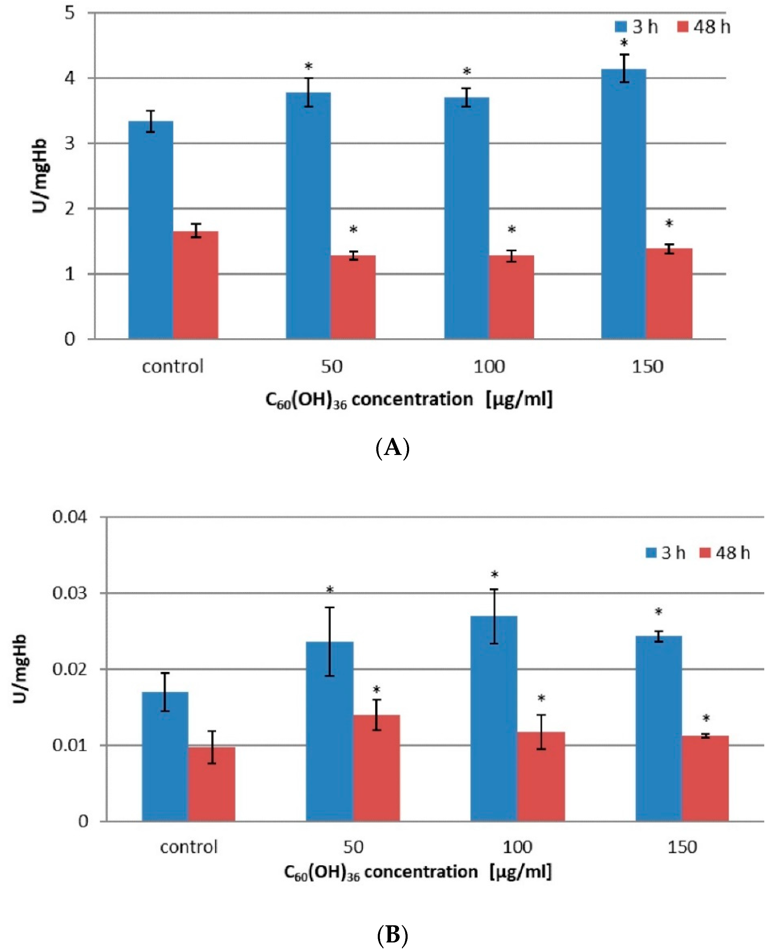
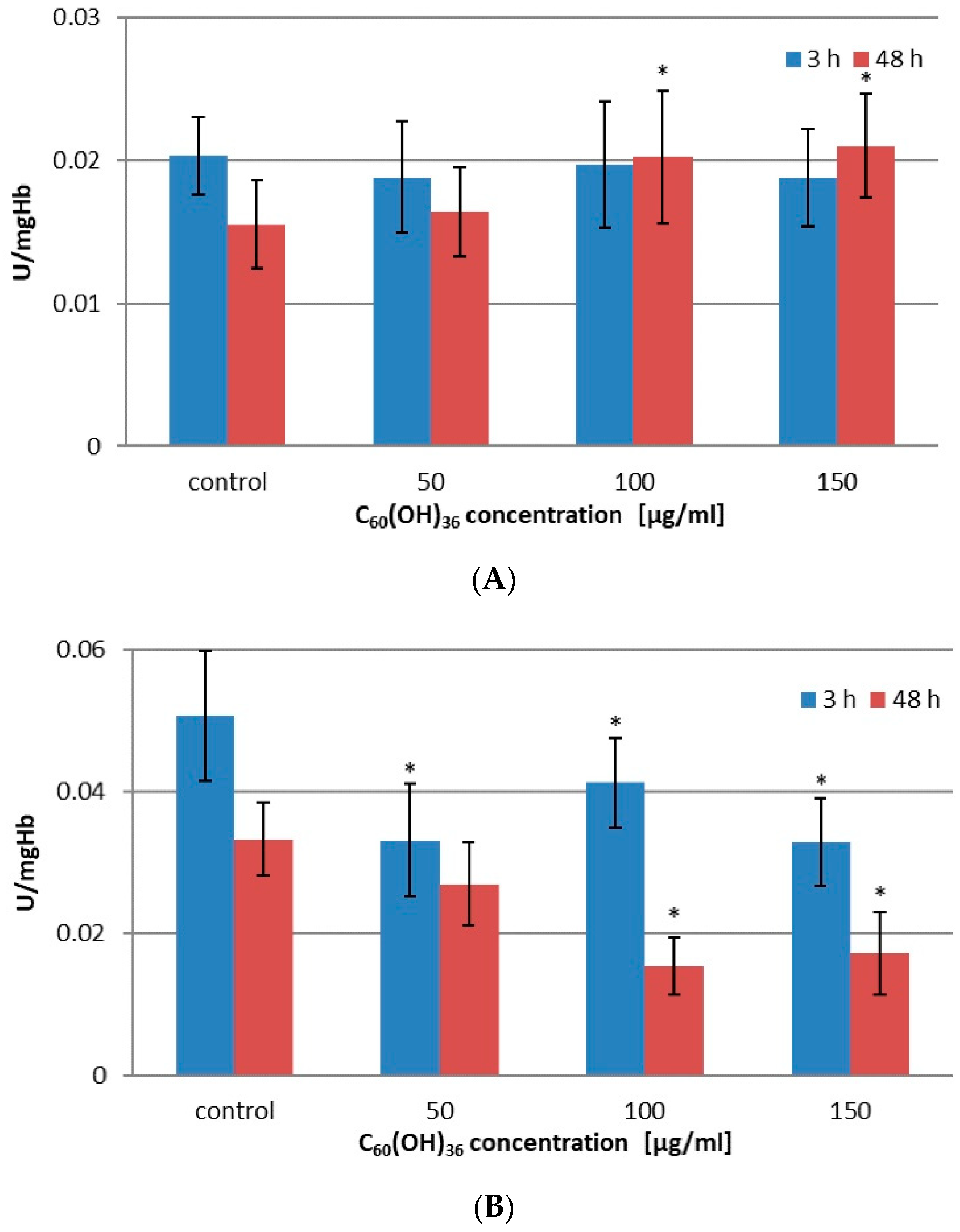
2.2. Activity of Enzymes in Erythrocytes Incubated with C60(OH)36

The effect of C60(OH)36 on catalase activity (CAT) was assessed in 2% hematocrit incubated with C60(OH)36 for 3 and 48 h at 37 °C. The final concentration of C60(OH)36 in suspension ranged from 0 to 150 µg/mL. The results obtained are presented in Figure 1.
For concentrations above 50–150 µg/mL, C60(OH)36 significantly increased CAT activity after 3 h of incubation compared to control (p < 0.05). After extending the incubation time to 48 h, the activity of CAT in the same concentration range was significantly reduced compared to the samples not treated with C60(OH)36 (p < 0.05) (for details, see Table 2 and Figure 1A).
Figure 1B presents changes in glutathione peroxidase (GPx) activity. A statistically significant increase in enzyme activity was observed after 3 h of incubation in 100 µg/mL and 150 µg/mL C60(OH)36 compared to the control (p < 0.05). On the other hand, prolonging the incubation time to 48 h resulted in a statistically significant increase in enzyme activity when erythrocytes were incubated in C60(OH)36 in a concentration range of 50–150 µg/mL in comparison to the control (p < 0.05) (for details, see Table 2).
The changes in activity of two other enzymes belonging to the glutathione family, glutathione reductase (GSR) and glutathione transferase (GST), are presented in Figure 2 and Table 3.
After 3 h of incubation, no statistically significant changes were observed compared to control; however, the extension of the incubation time to 48 h in C60(OH)36 at concentrations of 100 µg/mL and 150 µg/mL (p < 0.05) resulted in an increase in GSR enzymatic activity (for details, see Table 3 and Figure 2A). After 48 h of incubation in C60(OH)36 at concentrations of 100 µg/mL and 150 µg/mL, we saw a significant increase in GSR activity compared to the control (for details see Table 3 and Figure 2A).
The last enzyme, also belonging to the glutathione family, was glutathione transferase (GST) (see Figure 2B and Table 3). The behavior of this enzyme is different from other free enzymes, because after incubation (3 and after 48 h) and hemolysis, we noticed a decrease in activity compared to the control sample (without C60(OH)36). Specifically, a 3 h incubation with C60(OH)36 at concentrations of 50 µg/mL, 100 µg/mL, and 150 µg/mL (p < 0.05) resulted in a decrease of glutathione transferase activity compared to the control, but long incubation caused a decrease in GST activity (with respect to the control, p < 0.05) for 100 µg/mL and 150 µg/mL C60(OH)36 (for details, see Table 3).

2.3. The Effect of C60(OH)36 on Erythrocyte Microviscosity
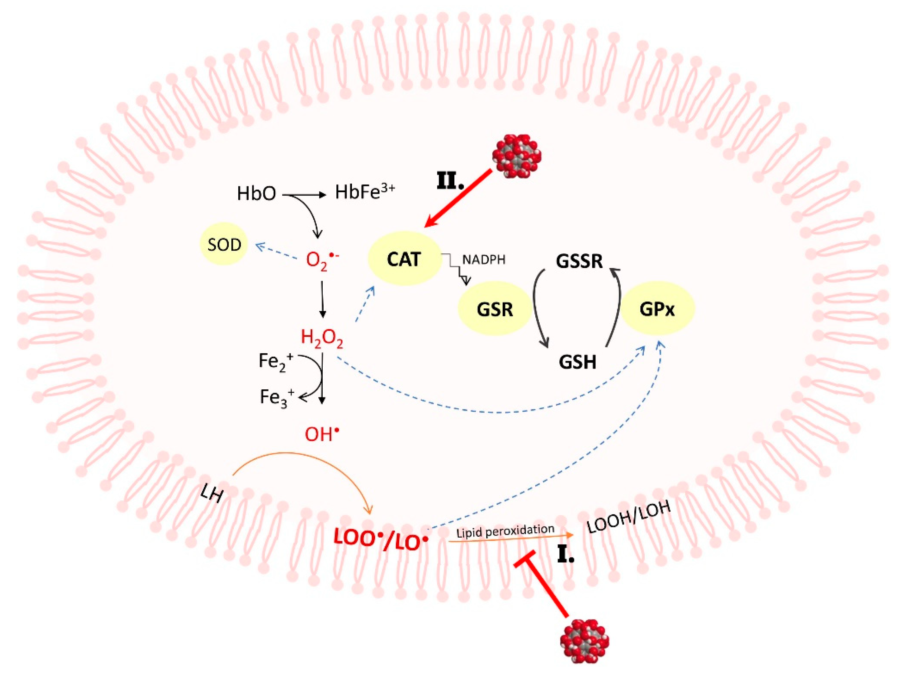
Measurements were made using the Tempamine spin tracer, which penetrates relatively easily through the plasma membrane into the erythrocyte interior [36], and the method compares the value of the rotational correlation time of Tempamine inside the cell, referring to a solution with known viscosity [37]. The effect of C60(OH)36 on the microviscosity of the interior of the erythrocyte was assessed in 2% hematocrit incubated with C60(OH)36 for 3 and 48 h at 37 °C. The final concentrations in suspension were 0, 50, 100, and 150 µg/mL. The results obtained are shown in Table 4, indicating that C60(OH)36 has no effect on the microviscosity of the interior of the erythrocytes. No statistically significant changes in microviscosity were observed after 3 h and after 48 h.

3. Discussion

In our experiments, we tested the enzyme activity in erythrocytes subjected to oxidative stress induced by prolonged incubation, in the presence of C60(OH)36. Fullerenes are an important class of nanomaterials with a wide potential applicability in biomedical sciences [8], and their water-soluble derivatives (fullerenols) are promising candidates for use as antioxidants in biological systems [10,11,12,13,14,15,16,17]. C60(OH)36 prevents ROS cell damage, but also can scavenge NO [13,20,21,38].
Reduced glutathione (GSH) is a primary intracellular source of thiol groups and an important hydrophilic reducing agent that protects cells from electrophiles and free radicals due to the nucleophilicity of -SH groups and reaction with oxidants [39]. In addition to the direct reaction with ROS, glutathione can sacrificially regenerate damaged macromolecules [7]. The results presented in Table 1 clearly demonstrate that prolonged incubation of erythrocytes causes a general reduction in -SH groups (control conditions). This is most likely an effect of oxidative stress, a decrease in ATP levels, and alterations in membrane structure and integrity, resulting in altered ion transport and changed erythrocyte shape [40]. We have observed that incubation in C60(OH)36 for 3 h had no effect on the level of the -SH groups; however, a prolonged 48 h incubation with 100 or 150 µg/mL C60(OH)36 resulted in a significant increase in the levels of the -SH groups in erythrocytes (32% and 41%, respectively). These results, indicating that C60(OH)36 used in relatively high concentrations could protect -SH groups against oxidation, are consistent with our previous findings [35], where we reported that C60(OH)36 significantly protected -SH groups from radiation-induced oxidation, and this protection was due to ROS removal by fullerenol.
In our experiments, the activity of CAT was enhanced (up to 24%) in the presence of 50–150 µg/mL C60(OH)36 after 3 h of incubation, but after 48 h, the activity of catalase decreased by approximately 33% (Figure 1A, Table 2). We propose that C60(OH)36 might act as an allosteric effector for CAT, changing the protein structure. Enzymatic conformational changes induced by C60(OH)36 during short-term incubation could increase CAT activity, but prolonged incubation could cause accelerated oxidation of that enzyme [41]. Our findings partly support previously reported observation reported by Djordjevic et al. [21] that fullerenol protects CAT against the changes induced by NO radicals. The decrease in CAT activity after 48 h can cause the dissociation of NADPH [42], further used by GSR during the reaction to recover GSH from GSSG. An argument in favor of this hypothesis is the increase in the amount of GSH after 48 h of incubation with C60(OH)36 (100–150 µg/mL). Fullerenol C60(OH)36 increases GSR activity, resulting in an increase in the overall pool of GSH, which is a cofactor of GPx (Figure 3).
We noticed that a 3 h treatment with C60(OH)36 (100 and 150 µg/mL) significantly increased GPx activity, whereas a prolonged incubation (48 h) caused a decrease in GPx activity, however, this activity is still higher than in control samples (for details, see Table 2, Figure 1B). Because the effect of protection of GPx was not concentration dependent and after 3 h of incubation with C60(OH)36 the activity of CAT and GPx increased, we suppose that the observed effect can be assigned to the interactions of C60(OH)36 with the enzyme subunits. GPx is a protein that reduces lipid peroxidation, therefore preventing hemolysis, and this explains why in our previous experiments [24] we observed a reduction in hemolysis and potassium leakage after the use of C60(OH)36
The last enzyme studied in our work was GST. Human erythrocytes contain three GST isoenzymes: rho (ρ), sigma (σ), and theta (θ); the ρ isoenzyme belongs to the Pi (π) class, which shows 95% activity against the commonly used CDNB substrate [43]. This enzyme is sensitive to oxidative stress due to the presence of cysteine residues at positions 14, 47, 101, and 169 at the N terminus, which are susceptible to oxidation by ROS, such as H2O2 and HClO. Modification of the cysteine residue at position 47, located near the active center, caused a decrease in GST activity [44,45]. In our experiments, we noticed an inhibition effect of fullerenol. Shorter treatment time resulted in reduced GST activity by approximately 35%, regardless of the concentration of C60(OH)36, whereas for a longer time of incubation, a decrease in enzyme activity was approximately 55% (100 and 150 µg/mL C60(OH)36).
In the present study, we also examined the microviscosity of the interior of the erythrocyte in the presence and absence of C60(OH)36. Aging of erythrocytes is accompanied by an increase in the cell microviscosity of cells [46] because of a decrease in intracellular electrolyte concentration, a lower water content, and a reduction in cell diameter, volume, and surface area [32,47]. The cytoplasmic microviscosity of the interior of erythrocytes is determined mainly by the concentration of hemoglobin, but also depends on the water content of the cell [48]. Viscosity is regulated by the intracellular and extracellular amount of Na+ and K+ ions, which affects the cell volume [49]. The results collected in Table 4 do not show visible changes in erythrocyte microviscosity, and we can explain this lack of measurable changes as the effect of two opposing processes. First, C60(OH)36 causes a decrease in Na+/K+-ATPase activity, which could lead to ion imbalance and hemolysis [50,51,52]. On the other hand, hemolysis induced by prolonged incubation is inhibited by retention of hemoglobin within the erythrocyte [24]. Thus, both processes contribute to the apparent effect of no visible changes in microviscosity.

4. Materials and Methods

4.1. Synthesis of Fullerenol

Fullerenol (polyhydroxyfullerene) C60(OH)36 was synthesized by the solvent-free method proposed by Wang et al. [53], and, in a modified version was described in the previous paper of Grebowski et al. [50]. Fullerenol was prepared by grinding pristine fullerene C60 with NaOH and 30% hydrogen peroxide in a glass mortar in air at room temperature for 25 min (Wang et al. recommended 15 min), then the additional incubation of the reaction mixture at 60 °C (water bath) was carried out for 20 min. The crude mixture was dissolved in 50 mL of deionized water and hydrolysis was performed for 24 h. Fullerenol was precipitated with methanol, resolubilized with water, and traces of NaOH were removed by passing the solution over the ion exchange resin (Amberlit MB-20). The structure of the obtained hydroxyl derivative of fullerene C60 was confirmed by IR spectrophotometry (EXUS FT-IR spectrometer, Thermo Fisher Scientific, Waltham, MA, USA), 1H-NMR (Bruker Avance III 600 MHz, Bruker Corp., Billerica, MA, USA), 13C-NMR (Varian Gemini 200 MHz, Varian Inc., Palo Alto, CA, USA), MALDI-TOF/TOF mass spectrometry (Axima Performance Mass Spectrometer, Shimadzu Biotech, Kyoto, Japan) as described previously [35].

4.2. Erythrocyte Isolation

Blood from the leukocyte-platelet coagulum, taken from the Regional Center for Blood Donation and Transfusion in Lodz, was centrifuged at 4 °C in a horizontal rotor centrifuge for 5 min at 3500 rpm, with the plasma and leukocyte layer removed. Subsequently, the obtained erythrocytes were washed three times with cooled PBS pH 7.4 (145 mM NaCl, 10 mM phosphosodium) and centrifuged under the same conditions. After obtaining erythrocyte suspensions, the hematocrit was determined by the microcapillary method, and for further studies, the suspension was diluted with a PBS solution to a hematocrit of 2%.

4.3. Hemolysate Preparation

Purified erythrocytes were hemolyzed with distilled water. Hemolysate for the determination of CAT activity was prepared by adding 1 mL of water to 0.5 mL of erythrocyte suspension with a hematocrit of 2%, and thoroughly mixed and placed in the freezer for 30 min. The samples were then mixed and centrifuged at 7000 rpm for 5 min. Hemolysate for the determination of GSH-GPx, GR, and GST activities was prepared by centrifuging 0.5 mL of erythrocyte suspension with a hematocrit of 2% at 7000 rpm, collecting 0.48 mL of the solution, and adding 0.5 mL of water. The samples were then thoroughly mixed and placed in the freezer for 15 min, then at room temperature for 10 min, and centrifuged at 7000 rpm for 5 min. The hemolysates obtained were frozen at −70 °C.

4.4. Determination of Hemoglobin Concentration in Hemolysate Using the Drabkin Method

The hemoglobin concentration was determined in 96-well plates. To 280 µL of Drabkin’s reagent (0.03% K3[Fe(CN)6], 0.1% NaHCO3, 0.005% KCN), 20 µL of hemolysate (study sample), and 20 µL of distilled water (blank) were added. The absorbance at λ = 541 nm was measured.

4.5. Fullerenol Treatment of Erythorocytes

A 5 mg/mL solution of C60(OH)36 in PBS was added to a 2% suspension of erythrocytes in PBS at a final concentration of 50, 100, and 150 µg/mL, respectively, and thoroughly mixed. The samples were incubated at 37 °C. The determinations were made after 3 and 48 h of incubation.

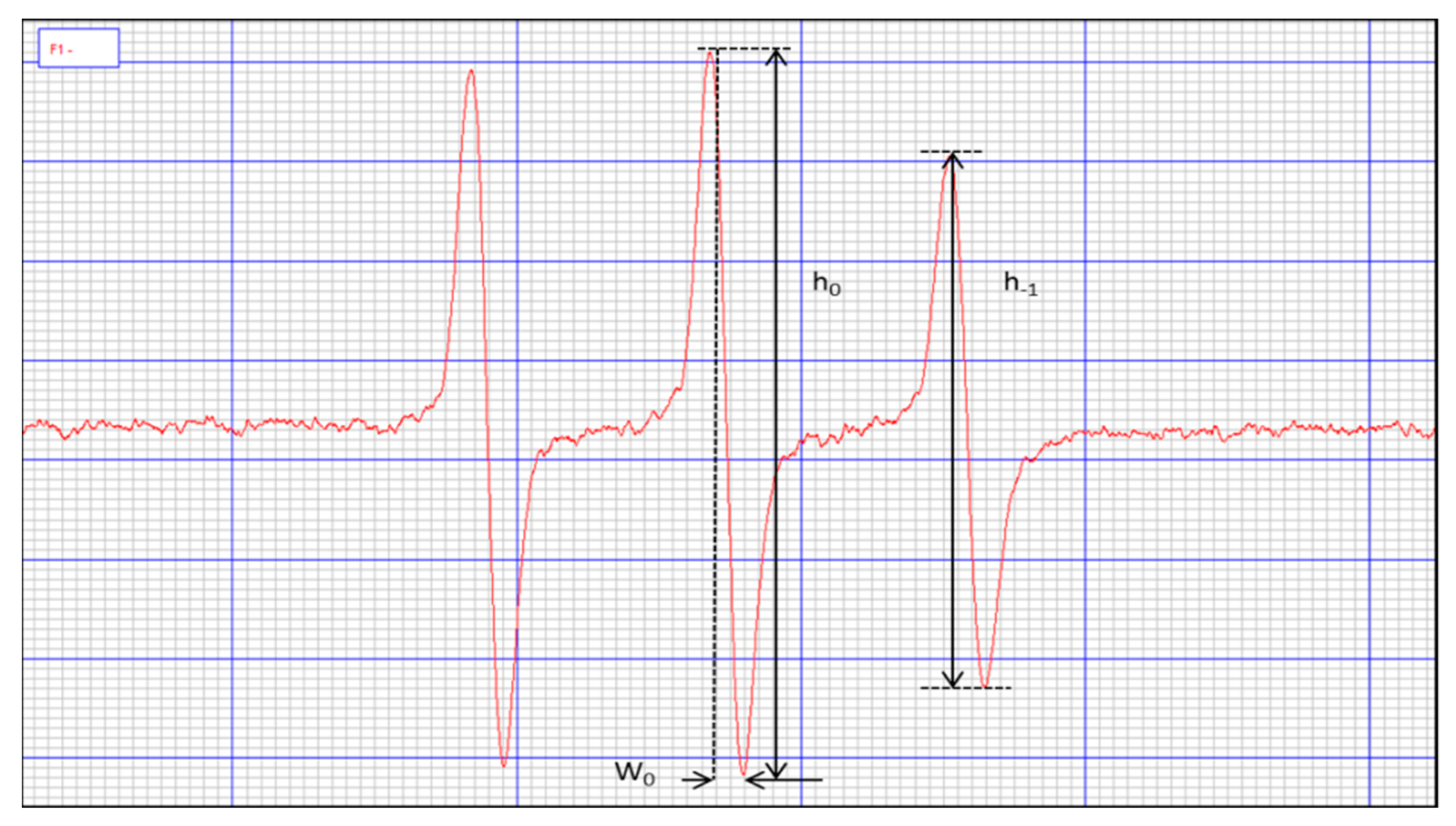
4.6. Identification of Erythrocyte Microviscosity

A 50% hematocrit was prepared by centrifuging 1.6 mL of the incubated erythrocyte suspension and collecting 1.54 mL of the supernatant. To the remaining 60 µL, 0.6 µL Tempamine (spin tracer: 10 mM of Tempamine dissolved in PBS) was added and stirred for 30 min at room temperature. The samples were then washed with 1 mL of 80 mM potassium ferricyanide and centrifuged, and the EPR spectra of the samples (see Figure 4), as well as the tracer spectrum in PBS, were determined. The rotational correlation time of the tracer was calculated from the formula:
τ R = k W 0 ( h 0 h 1 1 )
where:
  • k = 6.5 × 10⁻⁹ s/mT
  • W0 = width of the spectral center line
  • h0 = height of the spectral midfield line
  • h–1 = height of the highfield line of the spectrum
The viscosity of the interior of the erythrocyte was calculated from the following equation:
τ R τ B = η R η B
where:
  • τR = rotational correlation time of the tracer inside erythrocytes
  • τB = rotational correlation time of the tracer dissolved in a solution of known viscosity
  • ηR = erythrocyte inner viscosity
  • ηB = viscosity of known solution

4.7. Total Concentration of -SH Groups in Erythrocytes

Incubated erythrocytes with 2% hematocrit were diluted to a hematocrit of 0.75% with a final volume of 160 µL. Then, 0.45 µL of tracer was added so that the final concentration in the sample was 80 µmol. After 3 min of incubation with the tag, EPR spectra were obtained similarly as described previously [35].

4.8. Determination of Catalase Activity

To a spectrophotometric cuvette, 3 mL of 0.018 M hydrogen peroxide in 0.05 M phosphate buffer (pH 7.0) at 25 °C was added so that the absorbance measured at 240 nm was approximately 1. Then 15 µL of hemolysate was added, and the decrease in absorbance at λ = 240 nm was measured for 1 min. The activity was referred to 1 mg of hemoglobin protein.

4.9. Determination of Glutathione Peroxidase Activity

Glutathione peroxidase activity was determined by an indirect method. In the spectrophotometer cuvette, 250 µL of phosphate buffer (0.1 M pH 7.0 with 0.1 mM EDTA) equilibrated at 37 °C, 50 µL of glutathione reductase (2.4 units/mL), 50 µL of 10 mM GSH, 50 µL of 1.5 mM NADPH in a 0.1% NaHCO3 solution, 50 µL of hemolysate to which 2 µL of 100 mM sodium azide was added to block catalase, and 2 µL of a mixture of 60 mM K3Fe(CN)6 and 48 mM KCN solutions were added to convert hemoglobin to cyanmethemoglobin. The reaction was initiated by adding 50 µL of 12 mM t-butyl hydroperoxide. The decrease in absorbance at λ = 340 nm was measured for 3 min. The activity was referred to 1 mg of hemoglobin protein.

4.10. Determination of Glutathione Transferase Activity

Eight hundred fifty µL of phosphate buffer (0.1 M pH 6.5) equilibrated at 37 °C was added to the spectrophotometer cuvette followed by 50 µL of 0.02 GSH solution, 50 µL of CDNB solution (0.02 M solution of 1-chloro-2,4-dinitrobenzene dissolved in ethanol), and 50 µL of hemolysate. The absorbance at λ = 340 nm was measured for 1 min. The activity was referred to 1 mg of hemoglobin protein.

4.11. Determination of Glutathione Reductase Activity

One thousand µL of 2.2 mmol/L oxidized glutathione solution at 37 °C was added to a spectrophotometer cuvette followed by 40 µL of hemolysate. It was thoroughly mixed and 200 µL of 0.17 mmol/L of NADPH was added. The decrease in absorbance was measured at λ = 340 nm for 2 min. The activity was referred to 1 mg of hemoglobin protein.

4.12. Statistical Analysis

All experiments were carried out 3 to 8 times. Values were expressed as mean ± standard deviation (SD). Data were analyzed by one-way analysis of variance (ANOVA), followed by Tukey’s post-hoc test, all using GraphPad 4.0 software (La Jolla, CA, USA).

5. Conclusions

The results presented here reveal that C60(OH)36 can modulate the redox balance of human erythrocytes subjected to oxidative stress induced during a prolonged incubation in PBS at 37 °C. Apart from direct antioxidant action, fullerenol interacts with the antioxidant enzymes and stimulates them to more efficient and longer activity. A 3 and 48 h treatment with C60(OH)36 (50–150 µg/mL) maintains the proper function of the antioxidant system (CAT and GPx). However, we hypothesize that changes in its activity are not caused directly by the involvement of ROS, but conformational changes induced by C60(OH)36 itself. These effects are related to the concentration of C60(OH)36 and to the kind of parameters examined that characterize cell damage.

Author Contributions

Conceptualization, J.G., G.L. and P.K.-G.; methodology, J.G. and P.P.; software, J.G. and N.C.; validation, J.G. and N.C.; formal analysis, J.G. and G.L.; investigation, J.G.; resources, J.G. and P.P.; data curation, J.G. and N.C.; writing—original draft preparation, J.G., P.K.-G. and G.L.; writing—review and editing, J.G., P.K.-G., N.C., P.P. and G.L.; visualization, N.C.; supervision, J.G.; project administration, J.G.; funding acquisition, J.G. All authors have read and agreed to the published version of the manuscript.

Funding

This work was supported by the National Science Centre, Poland (NCN grant No. 2019/35/D/ST4/02627).

Institutional Review Board Statement

Leukocyte–buffy coat was purchased from the Blood Bank in Lodz, Poland. All procedures related to blood donation were carried out at the Regional Center for Blood, Donation and Blood Treatment in Lodz, Poland. The blood donor recruitment was at the Center, according to national legal procedures and European Union regulations (including the regulation (EU) 2016/679 of the European Parliament and of the Council of 27 April 2016 on the protection of natural persons regarding the processing of personal data and on the free movement of such data).

Informed Consent Statement

Not applicable.

Data Availability Statement

Not applicable.

Acknowledgments

The authors thank Grzegorz Bartosz for valuable comments that improved the manuscript.

Conflicts of Interest

The authors declare that they have no conflict of interest.

References

  1. Meng, Q.; Zaharieva, E.K.; Sasatani, M.; Kobayashi, J. Possible relationship between mitochondrial changes and oxidative stress under low dose-rate irradiation. Redox Rep. 2021, 26, 160–169. [Google Scholar] [CrossRef] [PubMed]
  2. Pizzino, G.; Irrera, N.; Cucinotta, M.; Pallio, G.; Mannino, F.; Arcoraci, V.; Squadrito, F.; Altavilla, D.; Bitto, A. Oxidative Stress: Harms and Benefits for Human Health. Oxid. Med. Cell. Longev. 2017, 2017, 8416763. [Google Scholar] [CrossRef] [PubMed]
  3. Remacle, J.; Michiels, C.; Raes, M. The importance of antioxidant enzymes in cellular aging and degeneration. EXS 1992, 62, 99–108. [Google Scholar] [CrossRef]
  4. Tang, Q.Q.; Su, S.Y.; Fang, M.Y. Zinc supplement modulates oxidative stress and antioxidant values in rats with severe acute pancreatitis. Biol. Trace Elem. Res. 2014, 159, 320–324. [Google Scholar] [CrossRef] [PubMed]
  5. Pannuru, P.; Vaddi, D.R.; Kindinti, R.R.; Varadacharyulu, N. Increased erythrocyte antioxidant status protects against smoking induced hemolysis in moderate smokers. Hum. Exp. Toxicol. 2011, 30, 1475–1481. [Google Scholar] [CrossRef]
  6. Milic, V.D.; Stankov, K.; Injac, R.; Djordjevic, A.; Srdjenovic, B.; Govedarica, B.; Radic, N.; Simic, V.D.; Strukelj, B. Activity of antioxidative enzymes in erythrocytes after a single dose administration of doxorubicin in rats pretreated with fullerenol C60(OH)24. Toxicol. Mech. Methods 2009, 19, 24–28. [Google Scholar] [CrossRef]
  7. Ulrich, K.; Jakob, U. The role of thiols in antioxidant systems. Free Radic. Biol. Med. 2019, 140, 14–27. [Google Scholar] [CrossRef]
  8. Grebowski, J.; Kazmierska, P.; Krokosz, A. Fullerenols as a new therapeutic approach in nanomedicine. Biomed. Res. Int. 2013, 2013, 751913. [Google Scholar] [CrossRef] [Green Version]
  9. Krokosz, A.; Grebowski, J.; Rodacka, A.; Pasternak, B.; Puchala, M. The effect of fullerenol C60(OH)~30 on the alcohol dehydrogenase activity irradiated with X-rays. Radiat. Phys. Chem. 2014, 97, 102–106. [Google Scholar] [CrossRef]
  10. Golomidov, I.; Bolshakova, O.; Komissarov, A.; Sharoyko, V.; Slepneva, E.; Slobodina, A.; Latypova, E.; Zherebyateva, O.; Tennikova, T.; Sarantseva, S. The neuroprotective effect of fullerenols on a model of Parkinson’s disease in Drosophila melanogaster. Biochem. Biophys. Res. Commun. 2020, 523, 446–451. [Google Scholar] [CrossRef]
  11. Kojić, D.; Purać, J.; Čelić, T.V.; Jović, D.; Vukašinović, E.L.; Pihler, I.; Borišev, I.; Djordjevic, A. Effect of fullerenol nanoparticles on oxidative stress induced by paraquat in honey bees. Environ. Sci. Pollut. Res. Int. 2020, 27, 6603–6612. [Google Scholar] [CrossRef] [PubMed]
  12. Shafiq, F.; Iqbal, M.; Ali, M.; Ashraf, M.A. Fullerenol regulates oxidative stress and tissue ionic homeostasis in spring wheat to improve net-primary productivity under salt-stress. Ecotoxicol. Environ. Saf. 2021, 211, 111901. [Google Scholar] [CrossRef]
  13. Djordjevic, A.; Srdjenovic, B.; Seke, M.; Petrovic, D.; Injac, R.; Mrdjanovic, J. Review of Synthesis and Antioxidant Potential of Fullerenol Nanoparticles. J. Nanomater. 2015, 2015, 567073. [Google Scholar] [CrossRef] [Green Version]
  14. Markovic, Z.; Trajkovic, V. Biomedical potential of the reactive oxygen species generation and quenching by fullerenes (C60). Biomaterials 2008, 29, 3561–3573. [Google Scholar] [CrossRef] [PubMed]
  15. Yin, J.J.; Lao, F.; Fu, P.P.; Wamer, W.G.; Zhao, Y.; Wang, P.C.; Qiu, Y.; Sun, B.; Xing, G.; Dong, J.; et al. The scavenging of reactive oxygen species and the potential for cell protection by functionalized fullerene materials. Biomaterials 2009, 30, 611–621. [Google Scholar] [CrossRef]
  16. Kazmierska-Grebowska, P.; Kobrzycka, A.; Bocian, R.; Kowalczyk, T.; Krokosz, A.; Grebowski, J. Fullerenol C60(OH)36 at relatively high concentrations impairs hippocampal theta oscillations (in vivo and in vitro) and triggers epilepsy (in vitro)–A dose response study. Exp. Mol. Pathol. 2018, 105, 98–109. [Google Scholar] [CrossRef]
  17. Grebowski, J.; Konopko, A.; Krokosz, A.; DiLabio, G.A.; Litwinienko, G. Antioxidant activity of highly hydroxylated fullerene C. Free Radic. Biol. Med. 2020, 160, 734–744. [Google Scholar] [CrossRef] [PubMed]
  18. Goodarzi, S.; Da Ros, T.; Conde, J.; Sefat, F.; Mozafari, M. Fullerene: Biomedical engineers get to revisit an old friend. Mater. Today 2017, 20, 460–480. [Google Scholar] [CrossRef] [Green Version]
  19. Dawid, A.; Górny, K.; Gburski, Z. The structural studies of fullerenol C60(OH)24 and nitric oxide mixture in water solvent–MD simulation. Nitric Oxide 2011, 25, 373–380. [Google Scholar] [CrossRef]
  20. Lao, F.; Li, W.; Han, D.; Qu, Y.; Liu, Y.; Zhao, Y.; Chen, C. Fullerene derivatives protect endothelial cells against NO-induced damage. Nanotechnology 2009, 20, 225103. [Google Scholar] [CrossRef]
  21. Mirkov, S.M.; Djordjevic, A.N.; Andric, N.L.; Andric, S.A.; Kostic, T.S.; Bogdanovic, G.M.; Vojinovic-Miloradov, M.B.; Kovacevic, R.Z. Nitric oxide-scavenging activity of polyhydroxylated fullerenol, C60(OH)24. Nitric Oxide 2004, 11, 201–207. [Google Scholar] [CrossRef] [PubMed]
  22. Akhtar, M.J.; Ahamed, M.; Alhadlaq, H.A.; Alshamsan, A. Mechanism of ROS scavenging and antioxidant signalling by redox metallic and fullerene nanomaterials: Potential implications in ROS associated degenerative disorders. Biochim. Biophys. Acta Gen. Subj. 2017, 1861, 802–813. [Google Scholar] [CrossRef] [PubMed]
  23. Srdjenovic, B.; Milic-Torres, V.; Grujic, N.; Stankov, K.; Djordjevic, A.; Vasovic, V. Antioxidant properties of fullerenol C60(OH)24 in rat kidneys, testes, and lungs treated with doxorubicin. Toxicol. Mech. Methods 2010, 20, 298–305. [Google Scholar] [CrossRef] [PubMed]
  24. Grebowski, J.; Krokosz, A.; Puchala, M. Fullerenol C60(OH)36 could associate to band 3 protein of human erythrocyte membranes. Biochim. Biophys. Acta 2013, 1828, 2007–2014. [Google Scholar] [CrossRef] [PubMed] [Green Version]
  25. Sharoyko, V.V.; Iamalova, N.R.; Ageev, S.V.; Meshcheriakov, A.A.; Iurev, G.O.; Petrov, A.V.; Nerukh, D.A.; Farafonov, V.S.; Vasina, L.V.; Penkova, A.V.; et al. In Vitro and In Silico Investigation of Water-Soluble Fullerenol C60(OH)24: Bioactivity and Biocompatibility. J. Phys. Chem. B 2021, 125, 9197–9212. [Google Scholar] [CrossRef] [PubMed]
  26. Grebowski, J.; Kaźmierska, P.; Krokosz, A. Fullerenol–Properties and applications in biomedical sciences. Postȩpy Hig. I Med. Doświadczalnej 2013, 67, 859–872. [Google Scholar] [CrossRef] [PubMed]
  27. Grebowski, J.; Krokosz, A. The Effect of Highly Hydroxylated Fullerenol C60(OH)36 on Human Erythrocyte Membrane Organization. J. Spectrosc. 2015, 2015, 825914. [Google Scholar] [CrossRef] [Green Version]
  28. Lichota, A.; Piwoński, I.; Michlewska, S.; Krokosz, A. A Multiparametric Study of Internalization of Fullerenol C60(OH)36 Nanoparticles into Peripheral Blood Mononuclear Cells: Cytotoxicity in Oxidative Stress Induced by Ionizing Radiation. Int. J. Mol. Sci. 2020, 21, 2281. [Google Scholar] [CrossRef] [Green Version]
  29. Lee, C.W.; Chi, M.C.; Peng, K.T.; Chiang, Y.C.; Hsu, L.F.; Yan, Y.L.; Li, H.Y.; Chen, M.C.; Lee, I.T.; Lai, C.H. Water-Soluble Fullerenol C60(OH)36 toward Effective Anti-Air Pollution Induced by Urban Particulate Matter in HaCaT Cell. Int. J. Mol. Sci. 2019, 20, 4259. [Google Scholar] [CrossRef] [Green Version]
  30. Krokosz, A.; Grebowski, J.; Szweda-Lewandowska, Z.; Rodacka, A.; Puchala, M. Can melatonin delay oxidative damage of human erythrocytes during prolonged incubation? Adv. Med. Sci. 2013, 58, 134–142. [Google Scholar] [CrossRef] [Green Version]
  31. Qasim, N.; Mahmood, R. Diminution of Oxidative Damage to Human Erythrocytes and Lymphocytes by Creatine: Possible Role of Creatine in Blood. PLoS ONE 2015, 10, e0141975. [Google Scholar] [CrossRef] [Green Version]
  32. Yao, Q.; Yang, G.; Wang, H.; Liu, J.; Zheng, J.; Lv, B.; Yang, M.; Yang, Y.; Gao, C.; Guo, Y. Aging erythrocyte membranes as biomimetic nanometer carriers of liver-targeting chromium poisoning treatment. Drug Deliv. 2021, 28, 1455–1465. [Google Scholar] [CrossRef] [PubMed]
  33. Remigante, A.; Morabito, R.; Marino, A. Natural Antioxidants Beneficial Effects on Anion Exchange through Band 3 Protein in Human Erythrocytes. Antioxidants 2019, 9, 25. [Google Scholar] [CrossRef] [PubMed] [Green Version]
  34. Remigante, A.; Morabito, R.; Marino, A. Band 3 protein function and oxidative stress in erythrocytes. J. Cell. Physiol. 2021, 236, 6225–6234. [Google Scholar] [CrossRef]
  35. Grebowski, J.; Kazmierska, P.; Litwinienko, G.; Lankoff, A.; Wolszczak, M.; Krokosz, A. Fullerenol C60(OH)36 protects human erythrocyte membrane against high-energy electrons. Biochim. Biophys. Acta (BBA)–Biomembr. 2018, 1860, 1528–1536. [Google Scholar] [CrossRef] [PubMed]
  36. Morse, P.D., II. Use of the spin label tempamine for measuring the internal viscosity of red blood cells. Biochem. Biophys. Res. Commun. 1977, 77, 1486–1491. [Google Scholar] [CrossRef]
  37. Bartosz, G.; Gaczyńska, M.; Grzelińska, E.; Judkiewicz, L. A spin-label study of membrane proteins and internal microviscosity of erythrocytes in hereditary spherocytosis. Life Sci. 1987, 41, 2285–2288. [Google Scholar] [CrossRef]
  38. Injac, R.; Prijatelj, M.; Strukelj, B. Fullerenol nanoparticles: Toxicity and antioxidant activity. Methods Mol. Biol. 2013, 1028, 75–100. [Google Scholar] [CrossRef]
  39. Gaucher, C.; Boudier, A.; Bonetti, J.; Clarot, I.; Leroy, P.; Parent, M. Glutathione: Antioxidant Properties Dedicated to Nanotechnologies. Antioxidants 2018, 7, 62. [Google Scholar] [CrossRef] [Green Version]
  40. Pandey, K.B.; Rizvi, S.I. Markers of oxidative stress in erythrocytes and plasma during aging in humans. Oxid. Med. Cell. Longev. 2010, 3, 2–12. [Google Scholar] [CrossRef]
  41. Yamawaki, H.; Iwai, N. Cytotoxicity of water-soluble fullerene in vascular endothelial cells. Am. J. Physiol. Cell. Physiol. 2006, 290, C1495–C1502. [Google Scholar] [CrossRef] [PubMed] [Green Version]
  42. Kirkman, H.N.; Galiano, S.; Gaetani, G.F. The function of catalase-bound NADPH. J. Biol. Chem. 1987, 262, 660–666. [Google Scholar] [CrossRef]
  43. Bocedi, A.; Fabrini, R.; Lai, O.; Alfieri, L.; Roncoroni, C.; Noce, A.; Pedersen, J.Z.; Ricci, G. Erythrocyte glutathione transferase: A general probe for chemical contaminations in mammals. Cell. Death. Discov. 2016, 2, 16029. [Google Scholar] [CrossRef] [Green Version]
  44. Bocedi, A.; Noce, A.; Marrone, G.; Noce, G.; Cattani, G.; Gambardella, G.; Di Lauro, M.; Di Daniele, N.; Ricci, G. Glutathione Transferase P1-1 an Enzyme Useful in Biomedicine and as Biomarker in Clinical Practice and in Environmental Pollution. Nutrients 2019, 11, 1741. [Google Scholar] [CrossRef] [PubMed] [Green Version]
  45. Marchewka, Z.; Piwowar, A.; Ruzik, S.; Długosz, A. Glutathione S–transferases class Pi and Mi and their significance in oncology. Postepy. Hig. Med. Dosw. 2017, 71, 541–550. [Google Scholar] [CrossRef]
  46. Rebrova, T.Y.; Afanas’ev, S.A.; Putrova, O.D.; Popov, S.V. Age-related characteristics of erythrocyte membrane microviscosity in experimental cardiosclerosis. Adv. Gerontol. 2013, 3, 211–214. [Google Scholar] [CrossRef]
  47. Blat, A.; Stepanenko, T.; Bulat, K.; Wajda, A.; Dybas, J.; Mohaissen, T.; Alcicek, F.C.; Szczesny-Malysiak, E.; Malek, K.; Fedorowicz, A.; et al. Spectroscopic Signature of Red Blood Cells in a D-Galactose-Induced Accelerated Aging Model. Int. J. Mol. Sci. 2021, 22, 2660. [Google Scholar] [CrossRef]
  48. Himbert, S.; Alsop, R.J.; Rose, M.; Hertz, L.; Dhaliwal, A.; Moran-Mirabal, J.M.; Verschoor, C.P.; Bowdish, D.M.; Kaestner, L.; Wagner, C.; et al. The Molecular Structure of Human Red Blood Cell Membranes from Highly Oriented, Solid Supported Multi-Lamellar Membranes. Sci. Rep. 2017, 7, 39661. [Google Scholar] [CrossRef] [Green Version]
  49. Huisjes, R.; Bogdanova, A.; van Solinge, W.W.; Schiffelers, R.M.; Kaestner, L.; van Wijk, R. Squeezing for Life–Properties of Red Blood Cell Deformability. Front. Physi. 2018, 9, 656. [Google Scholar] [CrossRef]
  50. Grebowski, J.; Krokosz, A.; Puchala, M. Membrane fluidity and activity of membrane ATPases in human erythrocytes under the influence of polyhydroxylated fullerene. Biochim. Biophys. Acta 2013, 1828, 241–248. [Google Scholar] [CrossRef] [Green Version]
  51. Sousa, L.; Garcia, I.J.; Costa, T.G.; Silva, L.N.; Renó, C.O.; Oliveira, E.S.; Tilelli, C.Q.; Santos, L.L.; Cortes, V.F.; Santos, H.L.; et al. Effects of Iron Overload on the Activity of Na,K-ATPase and Lipid Profile of the Human Erythrocyte Membrane. PLoS ONE 2015, 10, e0132852. [Google Scholar] [CrossRef] [PubMed] [Green Version]
  52. Radosinska, J.; Vrbjar, N. Erythrocyte Deformability and Na,K-ATPase Activity in Various Pathophysiological Situations and Their Protection by Selected Nutritional Antioxidants in Humans. Int. J. Mol. Sci. 2021, 22, 11924. [Google Scholar] [CrossRef] [PubMed]
  53. Wang, S.; He, P.; Zhang, J.M.; Jiang, H.; Zhu, S.Z. Novel and Efficient Synthesis of Water-Soluble [60]Fullerenol by Solvent-Free Reaction. Synth. Commun. 2005, 35, 1803–1808. [Google Scholar] [CrossRef]
Figure 1. The effect of C60(OH)36 on the activity of catalase (A) and glutathione peroxidase (B) converted to 1 mg of hemoglobin protein. Measurements were made after 3 and 48 h of incubation of erythrocytes with C60(OH)36 at concentrations of 0, 50, 100, and 150 µg/mL at 37 °C. The bars represent the mean values ± SD for 3 independent measurements. * Statistically significant values in relation to controls after 3 h and 48 h incubation at p < 0.05.
Ijms 23 00119 g001
Figure 2. (A) Effect of C60(OH)36 on the activity of glutathione reductase. Measurements were made after 3 and 48 h of incubation with C60(OH)36 at a concentration of 0, 50, 100, and 150 µg/mL at 37 °C. The bars represent the mean values ± SD for 3 independent measurements. * Statistically significant values relative to controls after 48 h of incubation at p < 0.05. (B) Effect of C60(OH)36 on the activity of glutathione transferase. Measurements were made after 3 and 48 h of incubation with C60(OH)36 at a concentration of 0, 50, 100, and 150 µg/mL at 37 °C. The bars represent the mean values ± SD for 3 independent measurements. * Statistically significant values relative to controls after 3 and 48 h of incubation at p < 0.05.
Ijms 23 00119 g002
Figure 3. (I) Direct interaction with the membrane and radical scavenging of radicals by C60(OH)36 [17,24,35] and (II) increasing the activity of antioxidant enzymes such as GSR activity, resulting in an increase in the total pool of GSH, which is a cofactor of GPx, which reduced the formation of lipid peroxides. Therefore, the reduction in hemolysis observed in our previous work could be the result of both processes [35].
Ijms 23 00119 g003
Figure 4. View of the Tempamine marker.
Ijms 23 00119 g004
Table 1. Total concentration of -SH groups in erythrocytes incubated at 37 °C with C60(OH)36.
C60(OH)36
Concentration [µg/mL]
Incubation Time
3 h48 h
Parameter
P
SDThiol Group
Concentration [µM]
Parameter
P
SDThiol Group
Concentration [µM]
00.930.0422.930.370.059.22
500.890.0622.120.390.049.56
1000.920.1322.680.49 *0.0510.54
1500.920.0722.710.52 *0.0710.72
* statistically significant values in relation to controls after 48 h of incubation at p < 0.05.
Table 2. Catalase (CAT) and peroxidase (GPx) activity converted into an mg of hemoglobin protein in the suspension of erythrocytes incubated with C60(OH)36 in the concentration range of 0–150 µg/mL for 3 and 48 h at 37 °C. After incubation, the erythrocytes were subjected to hemolysis.
Concentration C60(OH)36 [µg/mL]CAT Activity ± SD [U/mgHb]GPx Activity ± SD [U/mgHb]
3 h48 h3 h48 h
03.337 ± 0.1661.662 ± 0.1050.0169 ± 0.00250.0097 ± 0.0021
503.781 * ± 0.2201.280 * ± 0.0660.0236 * ± 0.00450.0139 * ± 0.0020
1003.701 * ± 0.1381.277 * ± 0.0880.0269 * ± 0.00360.0127 * ± 0.0022
1504.146 * ± 0.2061.384 * ± 0.0740.0243 * ± 0.00070.0124 * ± 0.0003
* statistically significant values in relation to controls after 3 h and 48 h of incubation at p <0.05.
Table 3. Glutathione reductase and glutathione transferase activity converted to 1 mg of hemoglobin protein in the suspension of erythrocytes incubated with C60(OH)36 in the concentration range of 0–150 µg/mL for 3 and 48 h at 37 °C. After incubation, the erythrocytes were subjected to hemolysis.
Concentration C60(OH)36 [µg/mL]GSR Activity ± SD [U/mgHb]GST Activity ± SD [U/mgHb]
3 h48 h3 h48 h
00.0203 ± 0.00270.0155 ± 0.00310.0506 ± 0.00920.0333 ± 0.0052
500.0188 ± 0.00390.0164 ± 0.00310.0331 * ± 0.00800.0269 ± 0.0059
1000.0197 ± 0.00440.0202 * ± 0.00460.0412 * ± 0.00630.0153 * ± 0.0040
1500.0188 ± 0.00340.0210 * ± 0.00360.0329 * ± 0.00620.0172 * ± 0.0058
* statistically significant values relative to controls after 3 and 48 h incubation at p < 0.05.
Table 4. Values of η determined using Tempamine in a suspension of erythrocytes incubated with C60(OH)36 in the concentration range of 0–150 µg/mL for 3 and 48 h at 37 °C.
Concentration C60(OH)36 [µg/mL]η ± SD * [Pa × s]
3 h48 h
04.64 ± 0.575.17 ± 0.59
505.14 ± 0.385.72 ± 0.56
1004.47 ± 0.295.32 ± 0.73
1504.72 ± 0.995.15 ± 0.51
* Results are presented as mean values ± SD of 3–5 independent measurements.
Publisher’s Note: MDPI stays neutral with regard to jurisdictional claims in published maps and institutional affiliations.

