
Antibacterial Titanium Implants Biofunctionalized by Plasma Electrolytic Oxidation with Silver, Zinc, and Copper: A Systematic Review

 , ,
, ,
Abstract
:1. Introduction
2. Methods
2.1. Literature Search
2.2. Inclusion and Exclusion Criteria
2.3. Study Selection
2.4. Risk of Bias
2.5. Data Extraction
2.6. Search Results
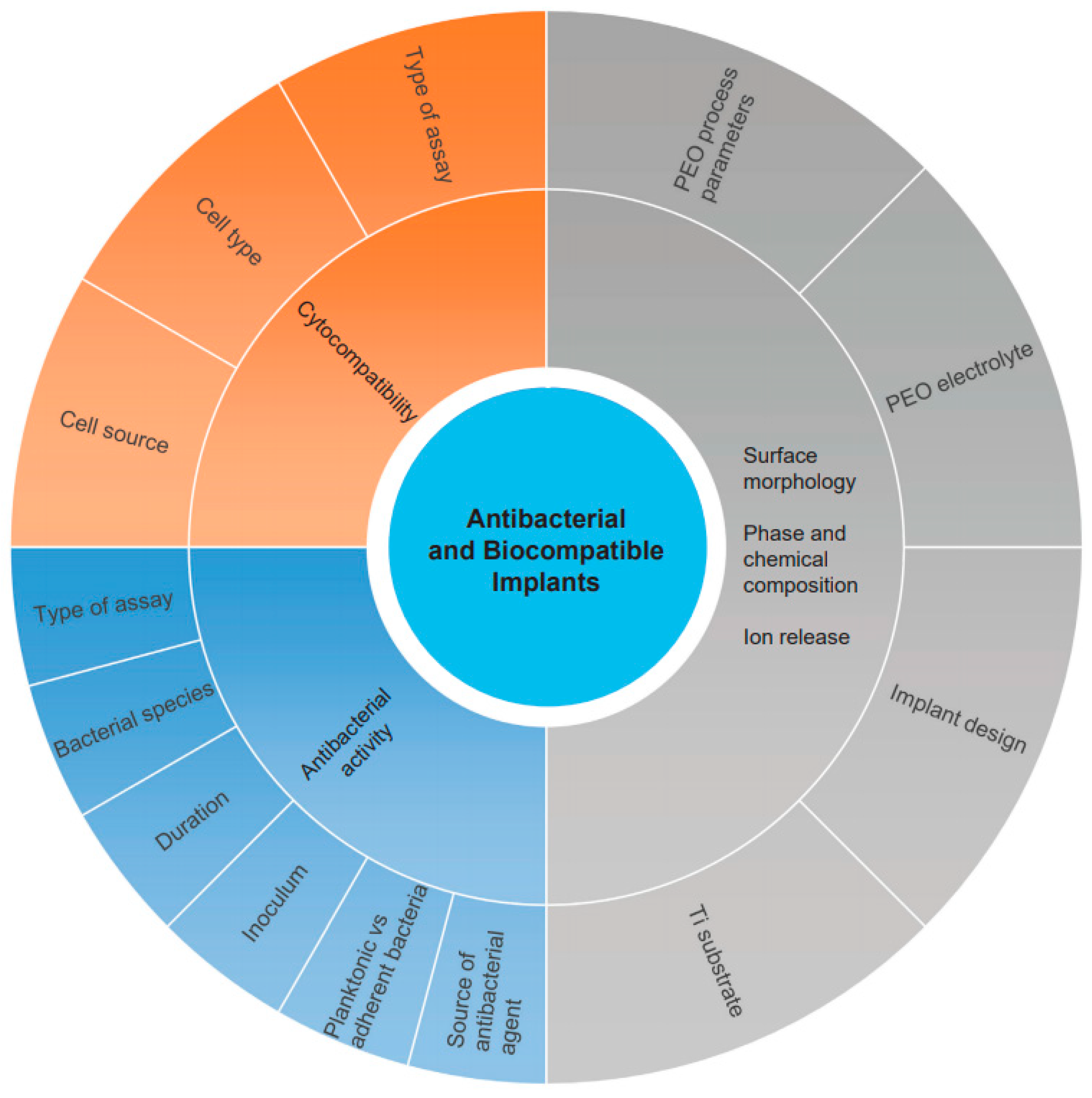
3. Summary of Study Characteristics
4. Synthesis and Characterization of PEO Biofunctionalized Surfaces
4.1. Titanium Substrate
4.2. PEO Electrolyte
4.3. PEO Processing Parameters
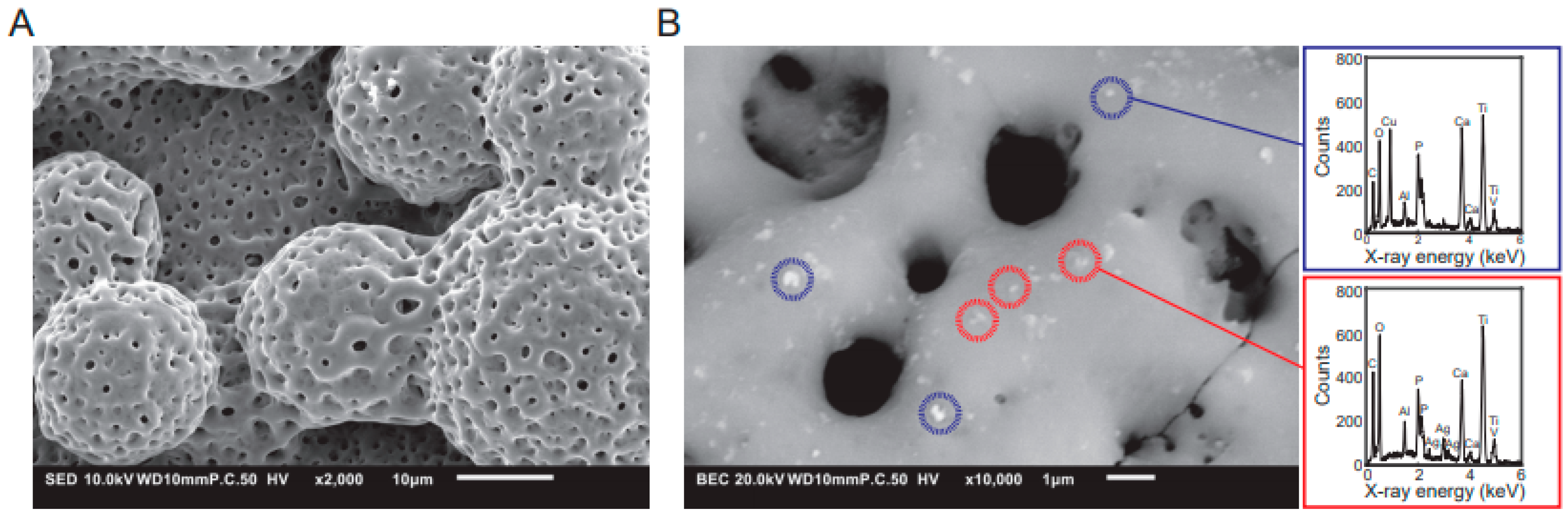
4.4. Surface Morphology
4.5. Phase Composition by XRD
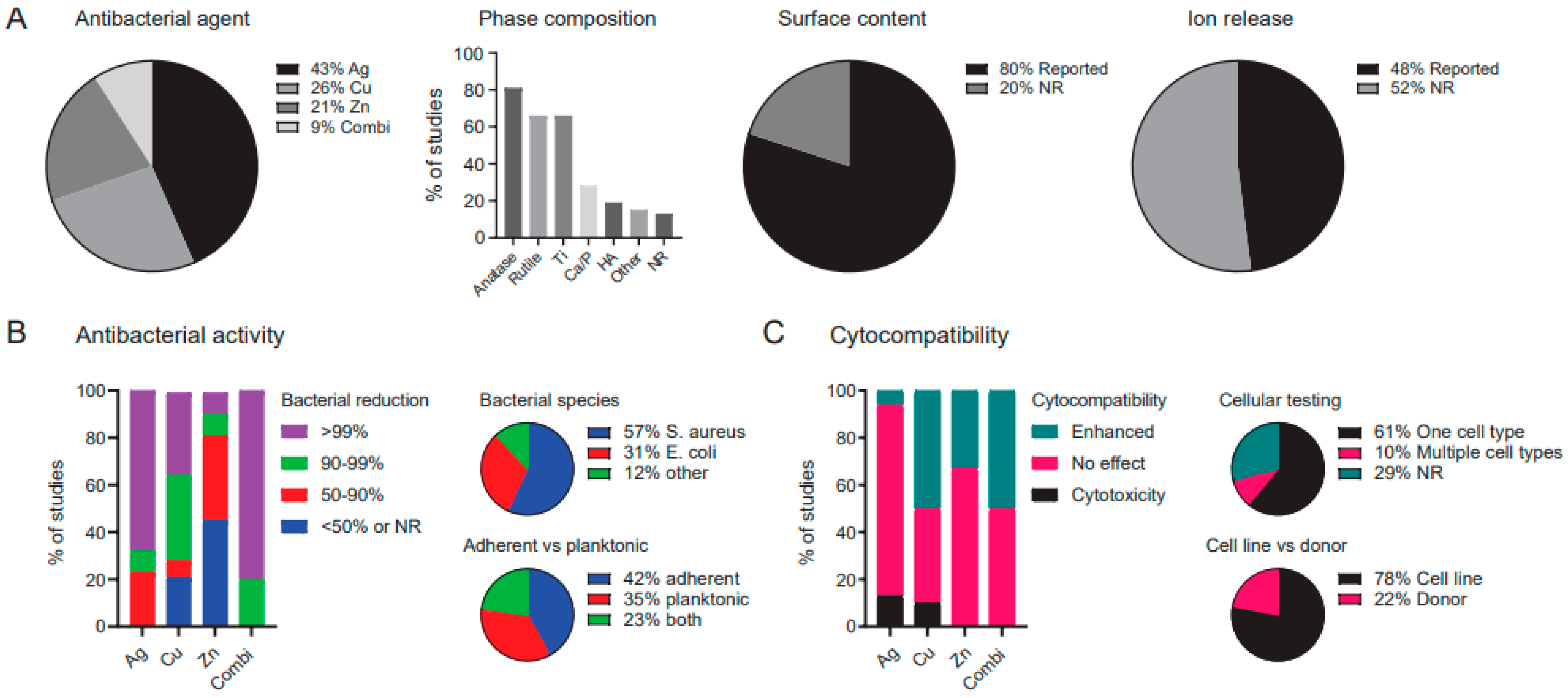
4.6. Content of the Antibacterial Elements Incorporated in the PEO Layers
4.7. Ion Release
5. Antibacterial Properties
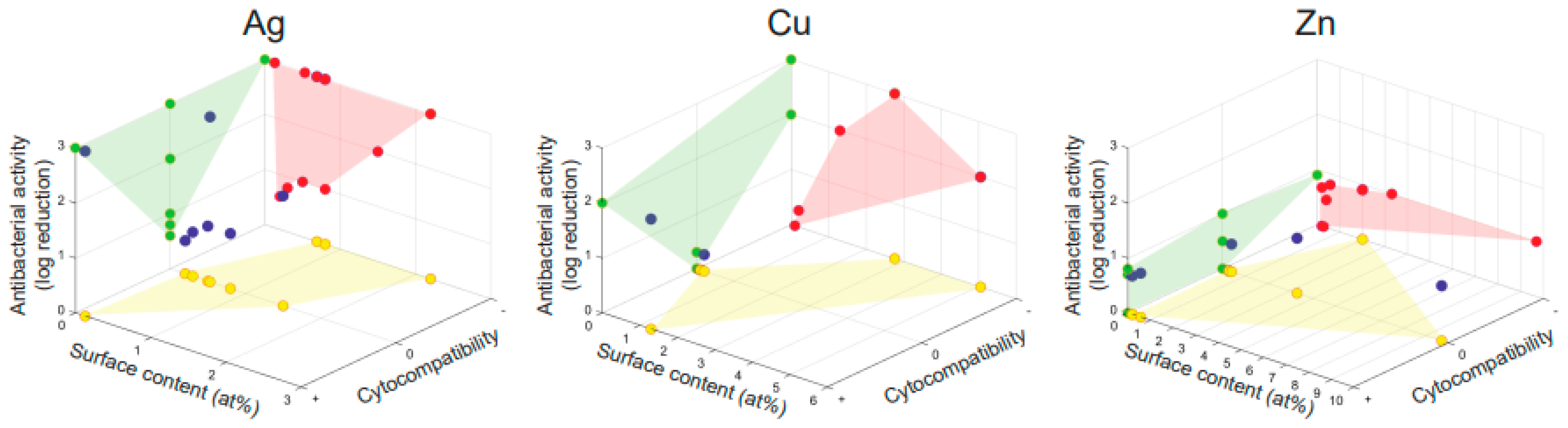
5.1. Comparing Antibacterial Activities of Ag, Cu, and Zn
5.2. Bacterial Species and Strains
5.3. Source of Antibacterial Agent
5.4. Analysis Method
5.5. Duration and Inoculum of Antibacterial Assay
5.6. Planktonic vs. Adherent Bacteria
6. Biocompatibility
6.1. Cytocompatibility of Ag, Cu, and Zn Surfaces
6.2. Type of Assay
6.3. Cell Type
6.4. Cell Source
7. Discussion
7.1. Antibacterial Results
7.2. Biocompatibility
7.3. Towards Clinically Relevant Implants
8. Conclusions
Supplementary Materials
Funding
Conflicts of Interest
References
- Zimmerli, W. Clinical presentation and treatment of orthopaedic implant-associated infection. J. Intern. Med. 2014, 276, 111–119. [Google Scholar] [CrossRef]
- Govaert, G.A.M.; Kuehl, R.; Atkins, B.L.; Trampuz, A.; Morgenstern, M.; Obremskey, W.T.; Verhofstad, M.H.J.; McNally, M.A.; Metsemakers, W.J.; Fracture-Related Infection (FRI) Consensus Group. Fracture-related infection consensus, diagnosing fracture-related infection: Current concepts and recommendations. J. Orthop. Trauma 2020, 34, 8–17. [Google Scholar] [CrossRef]
- Kapoor, S.K.; Thiyam, R. Management of infection following reconstruction in bone tumors. J. Clin. Orthop. Trauma 2015, 6, 244–251. [Google Scholar] [CrossRef] [Green Version]
- Singh, J.A. Epidemiology of knee and hip arthroplasty: A systematic review. Open Orthop. J. 2011, 5, 80–85. [Google Scholar] [CrossRef] [Green Version]
- Kremers, H.M.; Larson, D.R.; Crowson, C.S.; Kremers, W.K.; Washington, R.E.; Steiner, C.A.; Jiranek, W.A.; Berry, D.J. Prevalence of total hip and knee replacement in the United States. J. Bone Jt. Surg. Am. 2015, 97, 1386–1397. [Google Scholar] [CrossRef] [Green Version]
- Singh, J.A.; Yu, S.; Chen, L.; Cleveland, J.D. Rates of total joint replacement in the United States: Future projections to 2020–2040 using the national inpatient sample. J. Rheumatol. 2019, 46, 1134–1140. [Google Scholar] [CrossRef]
- Chambers, H.F.; Deleo, F.R. Waves of resistance: Staphylococcus aureus in the antibiotic era. Nat. Rev. Microbiol. 2009, 7, 629–641. [Google Scholar] [CrossRef]
- Guo, Y.; Song, G.; Sun, M.; Wang, J.; Wang, Y. Prevalence and therapies of antibiotic-resistance in Staphylococcus aureus. Front. Cell. Infect. Microbiol. 2020, 10, 107. [Google Scholar] [CrossRef] [Green Version]
- Teterycz, D.; Ferry, T.; Lew, D.; Stern, R.; Assal, M.; Hoffmeyer, P.; Bernard, L.; Uckay, I. Outcome of orthopedic implant infections due to different staphylococci. Int. J. Infect. Dis. 2010, 14, e913–e918. [Google Scholar] [CrossRef] [Green Version]
- Cho, O.H.; Bae, I.G.; Moon, S.M.; Park, S.Y.; Kwak, Y.G.; Kim, B.N.; Yu, S.N.; Jeon, M.H.; Kim, T.; Choo, E.J.; et al. Therapeutic outcome of spinal implant infections caused by Staphylococcus aureus: A retrospective observational study. Medicine 2018, 97, e12629. [Google Scholar] [CrossRef]
- Gross, T.M.; Lahiri, J.; Golas, A.; Luo, J.; Verrier, F.; Kurzejewski, J.L.; Baker, D.E.; Wang, J.; Novak, P.F.; Snyder, M.J. Copper-containing glass ceramic with high antimicrobial efficacy. Nat. Commun. 2019, 10, 1979. [Google Scholar] [CrossRef] [Green Version]
- Lara, H.H.; Ayala-Núñez, N.V.; Ixtepan Turrent, L.d.C.; Rodríguez Padilla, C. Bactericidal effect of silver nanoparticles against multidrug-resistant bacteria. World J. Microbiol. Biotechnol. 2009, 26, 615–621. [Google Scholar] [CrossRef]
- Nanda, A.; Saravanan, M. Biosynthesis of silver nanoparticles from Staphylococcus aureus and its antimicrobial activity against MRSA and MRSE. Nanomedicine 2009, 5, 452–456. [Google Scholar] [CrossRef] [PubMed]
- Siddiqi, K.S.; Rahman, A.U.; Tajuddin; Husen, A. Properties of Zinc Oxide nanoparticles and their activity against microbes. Nanoscale Res. Lett. 2018, 13, 141. [Google Scholar] [CrossRef]
- Necula, B.S.; Van Leeuwen, J.P.; Fratila-Apachitei, L.E.; Zaat, S.A.; Apachitei, I.; Duszczyk, J. In vitro cytotoxicity evaluation of porous TiO(2)-Ag antibacterial coatings for human fetal osteoblasts. Acta Biomater. 2012, 8, 4191–4197. [Google Scholar] [CrossRef]
- Croes, M.; Bakhshandeh, S.; Van Hengel, I.A.J.; Lietaert, K.; Van Kessel, K.P.M.; Pouran, B.; Van der Wal, B.C.H.; Vogely, H.C.; Van Hecke, W.; Fluit, A.C.; et al. Antibacterial and immunogenic behavior of silver coatings on additively manufactured porous titanium. Acta Biomater. 2018, 81, 315–327. [Google Scholar] [CrossRef]
- Bergemann, C.; Zaatreh, S.; Wegner, K.; Arndt, K.; Podbielski, A.; Bader, R.; Prinz, C.; Lembke, U.; Nebe, J.B. Copper as an alternative antimicrobial coating for implants–An in vitro study. World J. Transpl. 2017, 7, 193–202. [Google Scholar] [CrossRef] [PubMed]
- Ding, Q.; Zhang, X.; Huang, Y.; Yan, Y.; Pang, X. In vitro cytocompatibility and corrosion resistance of zinc-doped hydroxyapatite coatings on a titanium substrate. J. Mater. Sci. 2014, 50, 189–202. [Google Scholar] [CrossRef]
- Van Hengel, I.A.J.; Tierolf, M.; Valerio, V.P.M.; Minneboo, M.; Fluit, A.C.; Fratila-Apachitei, L.E.; Apachitei, I.; Zadpoor, A.A. Self-defending additively manufactured bone implants bearing silver and copper nanoparticles. J. Mater. Chem. B 2020, 8, 1589–1602. [Google Scholar] [CrossRef] [Green Version]
- Van Hengel, I.A.J.; Putra, N.E.; Tierolf, M.; Minneboo, M.; Fluit, A.C.; Fratila-Apachitei, L.E.; Apachitei, I.; Zadpoor, A.A. Biofunctionalization of selective laser melted porous titanium using silver and zinc nanoparticles to prevent infections by antibiotic-resistant bacteria. Acta Biomater. 2020, 107, 325–337. [Google Scholar] [CrossRef]
- Yoshitani, J.; Kabata, T.; Arakawa, H.; Kato, Y.; Nojima, T.; Hayashi, K.; Tokoro, M.; Sugimoto, N.; Kajino, Y.; Inoue, D.; et al. Combinational therapy with antibiotics and antibiotic-loaded adipose-derived stem cells reduce abscess formation in implant-related infection in rats. Sci. Rep. 2020, 10, 11182. [Google Scholar] [CrossRef] [PubMed]
- Cavanaugh, D.L.; Berry, J.; Yarboro, S.R.; Dahners, L.E. Better prophylaxis against surgical site infection with local as well as systemic antibiotics. An in vivo study. J. Bone Jt. Surg. Am. 2009, 91, 1907–1912. [Google Scholar] [CrossRef] [Green Version]
- Metsemakers, W.J.; Fragomen, A.T.; Moriarty, T.F.; Morgenstern, M.; Egol, K.A.; Zalavras, C.; Obremskey, W.T.; Raschke, M.; McNally, M.A.; Fracture-Related Infection (FRI) Consensus Group. Evidence-based recommendations for local antimicrobial strategies and dead space management in fracture-related infection. J. Orthop. Trauma 2020, 34, 18–29. [Google Scholar] [CrossRef]
- Celis, J.P.; Drees, D.; Huq, M.Z.; Wu, P.Q.; De Bonte, M. Hybrid processes—A versatile technique to match process requirements and coating needs. Surf. Coat. Technol. 1999, 113, 165–181. [Google Scholar] [CrossRef]
- Kumar, R.; Munstedt, H. Silver ion release from antimicrobial polyamide/silver composites. Biomaterials 2004, 26, 2081–2088. [Google Scholar] [CrossRef]
- Zaporojtchenko, V.; Podschun, R.; Schurmann, U.; Kulkarni, A.; Faupel, F. Physico-chemical and antimicrobial properties of co-sputtered Ag–Au/PTFE nanocomposite coatings. Nanotechnology 2006, 17, 4904–4908. [Google Scholar] [CrossRef]
- Gollwitzer, H.; Haenle, M.; Mittelmeier, W.; Heidenau, F.; Harasser, N. A biocompatible sol–gel derived titania coating for medical implants with antibacterial modification by copper integration. AMB Express 2018, 8, 1–9. [Google Scholar] [CrossRef] [PubMed] [Green Version]
- Su, Y.; Wang, K.; Gao, J.; Yang, Y.; Qin, Y.; Zheng, Y.; Zhu, D. Enhanced cytocompatibility and antibacterial property of zinc phosphate coating on biodegradable zinc materials. Acta Biomater. 2019, 98, 174–185. [Google Scholar] [CrossRef]
- Cao, H.; Liu, X.; Meng, F.; Chu, P.K. Biological actions of silver nanoparticles embedded in titanium controlled by micro-galvanic effects. Biomaterials 2011, 32, 693–705. [Google Scholar] [CrossRef]
- Piwonski, I.; Kadzioła, K.; Kisielewska, A.; Soliwoda, K.; Wolszczak, M.; Lisowska, K.; Wronska, N.; Felczak, A. The effect of the deposition parameters on size, distribution and antimicrobial properties of photoinduced silver nanoparticles on titania coatings. Appl. Surf. Sci. 2011, 257, 7076–7082. [Google Scholar] [CrossRef]
- Van Hengel, I.A.J.; Riool, M.; Fratila-Apachitei, L.E.; Witte-Bouma, J.; Farrell, E.; Zadpoor, A.A.; Zaat, S.A.J.; Apachitei, I. Selective laser melting porous metallic implants with immobilized silver nanoparticles kill and prevent biofilm formation by methicillin-resistant Staphylococcus aureus. Biomaterials 2017, 140, 1–15. [Google Scholar] [CrossRef] [PubMed]
- Necula, B.S.; Fratila-Apachitei, L.E.; Zaat, S.A.; Apachitei, I.; Duszczyk, J. In vitro antibacterial activity of porous TiO2-Ag composite layers against methicillin-resistant Staphylococcus aureus. Acta Biomater. 2009, 5, 3573–3580. [Google Scholar] [CrossRef]
- Rizwan, M.; Alias, R.; Zaidi, U.Z.; Mahmoodian, R.; Hamdi, M. Surface modification of valve metals using plasma electrolytic oxidation for antibacterial applications: A review. J. Biomed. Mater. Res. A 2018, 106, 590–605. [Google Scholar] [CrossRef]
- Asharani, P.V.; Low Kah Mun, G.; Prakash Hande, M.; Valiyaveettil, S. Cytotoxicity and genotoxicity of silver nanoparticles in human cells. ACS Nano 2009, 3, 279–290. [Google Scholar] [CrossRef]
- Song, W.H.; Ryu, H.S.; Hong, S.H. Antibacterial properties of Ag (or Pt)-containing calcium phosphate coatings formed by micro-arc oxidation. J. Biomed. Mater. Res. A 2009, 88, 246–254. [Google Scholar] [CrossRef] [PubMed]
- Zhu, W.; Zhang, Z.; Gu, B.; Sun, J.; Zhu, L. Biological activity and antibacterial property of nano-structured TiO2 coating incorporated with Cu prepared by micro-arc oxidation. J. Mater. Sci. Technol. 2013, 29, 237–244. [Google Scholar] [CrossRef]
- Roknian, M.; Fattah-Alhosseini, A.; Gashti, S.O.; Keshavarz, M.K. Study of the effect of ZnO nanoparticles addition to PEO coatings on pure titanium substrate: Microstructural analysis, antibacterial effect and corrosion behavior of coatings in Ringer’s physiological solution. J. Alloy. Compd. 2018, 740, 330–345. [Google Scholar] [CrossRef]
- Santos-Coquillat, A.; Martínez-Campos, E.; Mohedano, M.; Martínez-Corriá, R.; Ramos, V.; Arrabal, R.; Matykina, E. In vitro and in vivo evaluation of PEO-modified titanium for bone implant applications. Surf. Coat. Technol. 2018, 347, 358–368. [Google Scholar] [CrossRef]
- Chung, C.J.; Su, R.T.; Chu, H.J.; Chen, H.T.; Tsou, H.K.; He, J.L. Plasma electrolytic oxidation of titanium and improvement in osseointegration. J. Biomed. Mater. Res. B Appl. Biomater. 2013, 101, 1023–1030. [Google Scholar] [CrossRef]
- Martin, J.; Melhem, A.; Shchedrina, I.; Duchanoy, T.; Nominé, A.; Henrion, G.; Czerwiec, T.; Belmonte, T. Effects of electrical parameters on plasma electrolytic oxidation of aluminium. Surf. Coat. Technol. 2013, 221, 70–76. [Google Scholar] [CrossRef]
- Van Hengel, I.A.J.; Laçin, M.; Minneboo, M.; Fratila-Apachitei, L.E.; Apachitei, I.; Zadpoor, A.A. The effects of plasma electrolytically oxidized layers containing Sr and Ca on the osteogenic behavior of selective laser melted Ti6Al4V porous implants. J. Mater. Sci. Eng. C 2021, 124. [Google Scholar] [CrossRef]
- Clyne, T.W.; Troughton, S.C. A review of recent work on discharge characteristics during plasma electrolytic oxidation of various metals. Int. Mater. Rev. 2018, 64, 127–162. [Google Scholar] [CrossRef] [Green Version]
- Moher, D.; Liberati, A.; Tetzlaff, J.; Altman, D.G. Preferred reporting items for systematic reviews and meta-analyses: The PRISMA statement. PLoS Med. 2009, 6, e1000097. [Google Scholar] [CrossRef] [PubMed] [Green Version]
- Yerokhin, A.L.; Nie, X.; Leyland, A.; Matthews, A.; Dowey, S.J. Plasma electrolysis for surface engineering. Surf. Coat. Technol. 1999, 122, 73–93. [Google Scholar] [CrossRef]
- Curran, J.A.; Clyne, T.W. Thermo-physical properties of plasma electrolytic oxide coatings on aluminium. Surf. Coat. Technol. 2005, 199, 168–176. [Google Scholar] [CrossRef]
- Wang, Y.; Yu, H.; Chen, C.; Zhao, Z. Review of the biocompatibility of micro-arc oxidation coated titanium alloys. Mater. Des. 2015, 85, 640–652. [Google Scholar] [CrossRef]
- Matykina, E.; Skeldon, P.; Thompson, G.E. Fundamental and practical evaluations of PEO coatings of titanium. Int. Heat Treat. Surf. Eng. 2013, 3, 45–51. [Google Scholar] [CrossRef]
- Zhang, L.; Li, B.; Zhang, X.; Wang, D.; Zhou, L.; Li, H.; Liang, C.; Liu, S.; Wang, H. Biological and antibacterial properties of TiO2 coatings containing Ca/P/Ag by one-step and two-step methods. Biomed. Microdevices 2020, 22, 24. [Google Scholar] [CrossRef]
- Thukkaram, M.; Cools, P.; Nikiforov, A.; Rigole, P.; Coenye, T.; Van Der Voort, P.; Du Laing, G.; Vercruysse, C.; Declercq, H.; Morent, R.; et al. Antibacterial activity of a porous silver doped TiO2 coating on titanium substrates synthesized by plasma electrolytic oxidation. Appl. Surf. Sci. 2020, 500. [Google Scholar] [CrossRef]
- Van Hengel, I.A.J.; Gelderman, F.S.A.; Athanasiadis, S.; Minneboo, M.; Weinans, H.; Fluit, A.C.; Van der Eerden, B.C.J.; Fratila-Apachitei, L.E.; Apachitei, I.; Zadpoor, A.A. Functionality-packed additively manufactured porous titanium implants. Materials Today Bio 2020, 7. [Google Scholar] [CrossRef]
- Shimabukuro, M.; Tsutsumi, Y.; Yamada, R.; Ashida, M.; Chen, P.; Doi, H.; Nozaki, K.; Nagai, A.; Hanawa, T. Investigation of realizing both antibacterial property and osteogenic cell compatibility on titanium surface by simple electrochemical treatment. ACS Biomater. Sci. Eng. 2019, 5, 5623–5630. [Google Scholar] [CrossRef]
- Sedelnikova, M.B.; Komarova, E.G.; Sharkeev, Y.P.; Ugodchikova, A.V.; Mushtovatova, L.S.; Karpova, M.R.; Sheikin, V.V.; Litvinova, L.S.; Khlusov, I.A. Zn-, Cu- or Ag-incorporated micro-arc coatings on titanium alloys: Properties and behavior in synthetic biological media. Surf. Coat. Technol. 2019, 369, 52–68. [Google Scholar] [CrossRef]
- Sedelnikova, M.B.; Komarova, E.G.; Sharkeev, Y.P.; Ugodchikova, A.V.; Tolkacheva, T.V.; Rau, J.V.; Buyko, E.E.; Ivanov, V.V.; Sheikin, V.V. Modification of titanium surface via Ag-, Sr- and Si-containing micro-arc calcium phosphate coating. Bioact. Mater. 2019, 4, 224–235. [Google Scholar] [CrossRef]
- Lv, Y.; Wu, Y.; Lu, X.; Yu, Y.; Fu, S.; Yang, L.; Dong, Z.; Zhang, X. Microstructure, bio-corrosion and biological property of Ag-incorporated TiO2 coatings: Influence of Ag2O contents. Ceram. Int. 2019, 45, 22357–22367. [Google Scholar] [CrossRef]
- Teker, D.; Muhaffel, F.; Menekse, M.; Karaguler, N.G.; Baydogan, M.; Cimenoglu, H. Characteristics of multi-layer coating formed on commercially pure titanium for biomedical applications. Mater. Sci. Eng. C Mater. Biol. Appl. 2015, 48, 579–585. [Google Scholar] [CrossRef] [PubMed]
- Teker Aydogan, D.; Muhaffel, F.; Menekse Kilic, M.; Karabiyik Acar, O.; Cempura, G.; Baydogan, M.; Karaguler, N.G.; Torun Kose, G.; Czyrska-Filemonowicz, A.; Cimenoglu, H. Optimisation of micro-arc oxidation electrolyte for fabrication of antibacterial coating on titanium. Mater. Technol. 2017, 33, 119–126. [Google Scholar] [CrossRef]
- Dilek Aydogan, T.; Muhaffel, F.; Acar, O.K.; Topcuoglu, E.N.; Kulekci, H.G.; Kose, G.T.; Baydogan, M.; Cimenoglu, H. Surface modification of Ti6Al4V by micro-arc oxidation in AgC2H3O2-containing electrolyte. Surf. Innov. 2018, 6, 277–285. [Google Scholar] [CrossRef]
- Muhaffel, F.; Cempura, G.; Menekse, M.; Czyrska-Filemonowicz, A.; Karaguler, N.; Cimenoglu, H. Characteristics of multi-layer coatings synthesized on Ti6Al4V alloy by micro-arc oxidation in silver nitrate added electrolytes. Surf. Coat. Technol. 2016, 307, 308–315. [Google Scholar] [CrossRef]
- Tsutsumi, Y.; Niinomi, M.; Nakai, M.; Shimabukuro, M.; Ashida, M.; Chen, P.; Doi, H.; Hanawa, T. Electrochemical surface treatment of a β-titanium alloy to realize an antibacterial property and bioactivity. Metals 2016, 6, 76. [Google Scholar] [CrossRef] [Green Version]
- Shin, K.R.; Kim, Y.S.; Kim, G.W.; Yang, H.W.; Ko, Y.G.; Shin, D.H. Effects of concentration of Ag nanoparticles on surface structure and in vitro biological responses of oxide layer on pure titanium via plasma electrolytic oxidation. Appl. Surf. Sci. 2015, 347, 574–582. [Google Scholar] [CrossRef]
- Marques, I.D.; Barao, V.A.; Da Cruz, N.C.; Yuan, J.C.; Mesquita, M.F.; Ricomini-Filho, A.P.; Sukotjo, C.; Mathew, M.T. Electrochemical behavior of bioactive coatings on cp-Ti surface for dental application. Corros. Sci. 2015, 100, 133–146. [Google Scholar] [CrossRef] [Green Version]
- Da Silva Viera Marques, I.; Alfaro, M.F.; Saito, M.T.; Da Cruz, N.C.; Takoudis, C.; Landers, R.; Mesquita, M.F.; Nociti, F.H., Jr.; Mathew, M.T.; Sukotjo, C.; et al. Biomimetic coatings enhance tribocorrosion behavior and cell responses of commercially pure titanium surfaces. Biointerphases 2016, 11, 031008. [Google Scholar] [CrossRef]
- Gasquères, C.; Schneider, G.; Nusko, R.; Maier, G.; Dingeldein, E.; Eliezer, A. Innovative antibacterial coating by anodic spark deposition. Surf. Coat. Technol. 2012, 206, 3410–3414. [Google Scholar] [CrossRef]
- Zhou, L.; Lü, G.-H.; Mao, F.-F.; Yang, S.-Z. Preparation of biomedical Ag incorporated hydroxyapatite/titania coatings on Ti6Al4V alloy by plasma electrolytic oxidation. Chin. Phys. B 2014, 23. [Google Scholar] [CrossRef]
- Thukkaram, M.; Coryn, R.; Asadian, M.; Esbah Tabaei, P.S.; Rigole, P.; Rajendhran, N.; Nikiforov, A.; Sukumaran, J.; Coenye, T.; Van Der Voort, P.; et al. Fabrication of microporous coatings on titanium implants with improved mechanical, antibacterial, and cell-interactive properties. ACS Appl. Mater. Interfaces 2020, 12, 30155–30169. [Google Scholar] [CrossRef]
- Oleshko, O.; Liubchak, I.; Husak, Y.; Korniienko, V.; Yusupova, A.; Oleshko, T.; Banasiuk, R.; Szkodo, M.; Matros-Taranets, I.; Kazek-Kesik, A.; et al. In vitro biological characterization of silver-doped anodic oxide coating on titanium. Materials 2020, 13, 4359. [Google Scholar] [CrossRef] [PubMed]
- Zhao, Q.; Yi, L.; Hu, A.; Jiang, L.; Hong, L.; Dong, J. Antibacterial and osteogenic activity of a multifunctional microporous coating codoped with Mg, Cu and F on titanium. J. Mater. Chem. B 2019, 7, 2284–2299. [Google Scholar] [CrossRef] [PubMed]
- Zhang, X.; Peng, Z.; Lu, X.; Lv, Y.; Cai, G.; Yang, L.; Dong, Z. Microstructural evolution and biological performance of Cu-incorporated TiO2 coating fabricated through one-step micro-arc oxidation. Appl. Surf. Sci. 2020, 508. [Google Scholar] [CrossRef]
- Rokosz, K.; Hryniewicz, T.; Kacalak, W.; Tandecka, K.; Raaen, S.; Gaiaschi, S.; Chapon, P.; Malorny, W.; Matysek, D.; Pietrzak, K.; et al. Porous coatings containing copper and phosphorus obtained by plasma electrolytic oxidation of titanium. Materials 2020, 13, 828. [Google Scholar] [CrossRef] [PubMed] [Green Version]
- Liang, T.; Wang, Y.; Zeng, L.; Liu, Y.; Qiao, L.; Zhang, S.; Zhao, R.; Li, G.; Zhang, R.; Xiang, J.; et al. Copper-doped 3D porous coating developed on Ti-6Al-4V alloys and its in vitro long-term antibacterial ability. Appl. Surf. Sci. 2020, 509. [Google Scholar] [CrossRef]
- He, X.; Zhang, G.; Zhang, H.; Hang, R.; Huang, X.; Yao, X.; Zhang, X. Cu and Si co-doped microporous TiO2 coating for osseointegration by the coordinated stimulus action. Appl. Surf. Sci. 2020, 503. [Google Scholar] [CrossRef]
- Zhang, L.; Guo, J.; Huang, X.; Zhang, Y.; Han, Y. The dual function of Cu-doped TiO2 coatings on titanium for application in percutaneous implants. J. Mater. Chem. B 2016, 4, 3788–3800. [Google Scholar] [CrossRef] [PubMed]
- Huang, Q.; Li, X.; Elkhooly, T.A.; Liu, X.; Zhang, R.; Wu, H.; Feng, Q.; Liu, Y. The Cu-containing TiO2 coatings with modulatory effects on macrophage polarization and bactericidal capacity prepared by micro-arc oxidation on titanium substrates. Colloids Surf B Biointerfaces 2018, 170, 242–250. [Google Scholar] [CrossRef]
- Yao, X.; Zhang, X.; Wu, H.; Tian, L.; Ma, Y.; Tang, B. Microstructure and antibacterial properties of Cu-doped TiO2 coating on titanium by micro-arc oxidation. Appl. Surf. Sci. 2014, 292, 944–947. [Google Scholar] [CrossRef]
- Zhang, X.; Li, J.; Wang, X.; Wang, Y.; Hang, R.; Huang, X.; Tang, B.; Chu, P.K. Effects of copper nanoparticles in porous TiO2 coatings on bacterial resistance and cytocompatibility of osteoblasts and endothelial cells. Mater. Sci. Eng. C Mater. Biol. Appl. 2018, 82, 110–120. [Google Scholar] [CrossRef]
- Zhao, D.; Lu, Y.; Zeng, X.; Wang, Z.; Liu, S.; Wang, T. Antifouling property of micro-arc oxidation coating incorporating Cu2O nanoparticles on Ti6Al4V. Surf. Eng. 2017, 33, 796–802. [Google Scholar] [CrossRef]
- Zhang, L.; Guo, J.; Yan, T.; Han, Y. Fibroblast responses and antibacterial activity of Cu and Zn co-doped TiO2 for percutaneous implants. Appl. Surf. Sci. 2018, 434, 633–642. [Google Scholar] [CrossRef]
- Zhao, D.; Lu, Y.; Wang, Z.; Zeng, X.; Liu, S.; Wang, T. Antifouling properties of micro arc oxidation coatings containing Cu2O/ZnO nanoparticles on Ti6Al4V. Int. J. Refract. Met. Hard Mater. 2016, 54, 417–421. [Google Scholar] [CrossRef]
- Zhang, X.; Li, C.; Yu, Y.; Lu, X.; Lv, Y.; Jiang, D.; Peng, Z.; Zhou, J.; Zhang, X.; Sun, S.; et al. Characterization and property of bifunctional Zn-incorporated TiO2 micro-arc oxidation coatings: The influence of different Zn sources. Ceram. Int. 2019, 45, 19747–19756. [Google Scholar] [CrossRef]
- Shimabukuro, M.; Tsutsumi, Y.; Nozaki, K.; Chen, P.; Yamada, R.; Ashida, M.; Doi, H.; Nagai, A.; Hanawa, T. Chemical and biological roles of Zinc in a porous titanium dioxide layer formed by micro-arc oxidation. Coatings 2019, 9, 705. [Google Scholar] [CrossRef] [Green Version]
- Du, Q.; Wei, D.; Wang, Y.; Cheng, S.; Liu, S.; Zhou, Y.; Jia, D. The effect of applied voltages on the structure, apatite-inducing ability and antibacterial ability of micro arc oxidation coating formed on titanium surface. Bioact. Mater. 2018, 3, 426–433. [Google Scholar] [CrossRef] [PubMed]
- Sopchenski, L.; Popat, K.; Soares, P. Bactericidal activity and cytotoxicity of a zinc doped PEO titanium coating. Thin Solid Film. 2018, 660, 477–483. [Google Scholar] [CrossRef]
- Hu, H.; Zhang, W.; Qiao, Y.; Jiang, X.; Liu, X.; Ding, C. Antibacterial activity and increased bone marrow stem cell functions of Zn-incorporated TiO2 coatings on titanium. Acta Biomater. 2012, 8, 904–915. [Google Scholar] [CrossRef]
- Zhang, X.; Wang, H.; Li, J.; He, X.; Hang, R.; Huang, X.; Tian, L.; Tang, B. Corrosion behavior of Zn-incorporated antibacterial TiO2 porous coating on titanium. Ceram. Int. 2016, 42, 17095–17100. [Google Scholar] [CrossRef]
- Zhao, B.H.; Zhang, W.; Wang, D.N.; Feng, W.; Liu, Y.; Lin, Z.; Du, K.Q.; Deng, C.F. Effect of Zn content on cytoactivity and bacteriostasis of micro-arc oxidation coatings on pure titanium. Surf. Coat. Technol. 2013, 228, S428–S432. [Google Scholar] [CrossRef]
- Leśniak-Ziółkowska, K.; Kazek-Kęsik, A.; Rokosz, K.; Raaen, S.; Stolarczyk, A.; Krok-Borkowicz, M.; Pamuła, E.; Gołda-Cępa, M.; Brzychczy-Włoch, M.; Simka, W. Electrochemical modification of the Ti-15Mo alloy surface in solutions containing ZnO and Zn3(PO4)2 particles. Mater. Sci. Eng. C 2020, 115. [Google Scholar] [CrossRef] [PubMed]
- Zhang, L.; Gao, Q.; Han, Y. Zn and Ag Co-doped anti-microbial TiO2 coatings on Ti by micro-arc oxidation. J. Mater. Sci. Technol. 2016, 32, 919–924. [Google Scholar] [CrossRef]
- Wang, Y.; Zhao, S.; Li, G.; Zhang, S.; Zhao, R.; Dong, A.; Zhang, R. Preparation and in vitro antibacterial properties of anodic coatings co-doped with Cu, Zn, and P on a Ti–6Al–4V alloy. Mater. Chem. Phys. 2020, 241. [Google Scholar] [CrossRef]
- Matykina, E.; Arrabal, R.; Mingo, B.; Mohedano, M.; Pardo, A.; Merino, M.C. In vitro corrosion performance of PEO coated Ti and Ti6Al4V used for dental and orthopaedic implants. Surf. Coat. Technol. 2016, 307, 1255–1264. [Google Scholar] [CrossRef]
- Wauthle, R.; Ahmadi, S.M.; Amin Yavari, S.; Mulier, M.; Zadpoor, A.A.; Weinans, H.; Van Humbeeck, J.; Kruth, J.P.; Schrooten, J. Revival of pure titanium for dynamically loaded porous implants using additive manufacturing. Mater. Sci. Eng. C Mater. Biol. Appl. 2015, 54, 94–100. [Google Scholar] [CrossRef]
- Shah, F.A.; Thomsen, P.; Palmquist, A. Osseointegration and current interpretations of the bone-implant interface. Acta Biomater. 2019, 84, 1–15. [Google Scholar] [CrossRef]
- Shah, F.A.; Trobos, M.; Thomsen, P.; Palmquist, A. Commercially pure titanium (cp-Ti) versus titanium alloy (Ti6Al4V) materials as bone anchored implants–Is one truly better than the other? Mater. Sci. Eng. C Mater. Biol. Appl. 2016, 62, 960–966. [Google Scholar] [CrossRef] [PubMed]
- Elias, C.N.; Fernandes, D.J.; Souza, F.M.D.; Monteiro, E.d.S.; Biasi, R.S.D. Mechanical and clinical properties of titanium and titanium-based alloys (Ti G2, Ti G4 cold worked nanostructured and Ti G5) for biomedical applications. J. Mater. Res. Technol. 2019, 8, 1060–1069. [Google Scholar] [CrossRef]
- Challa, V.S.; Mali, S.; Misra, R.D. Reduced toxicity and superior cellular response of preosteoblasts to Ti-6Al-7Nb alloy and comparison with Ti-6Al-4V. J. Biomed. Mater. Res. A 2013, 101, 2083–2089. [Google Scholar] [CrossRef]
- Krząkała, A.; Kazek-Kęsik, A.; Simka, W. Application of plasma electrolytic oxidation to bioactive surface formation on titanium and its alloys. RSC Adv. 2013, 3. [Google Scholar] [CrossRef]
- Rafieerad, A.R.; Ashra, M.R.; Mahmoodian, R.; Bushroa, A.R. Surface characterization and corrosion behavior of calcium phosphate-base composite layer on titanium and its alloys via plasma electrolytic oxidation: A review paper. Mater. Sci. Eng. C Mater. Biol. Appl. 2015, 57, 397–413. [Google Scholar] [CrossRef] [PubMed]
- Lugovskoy, A.; Lugovskoy, S. Production of hydroxyapatite layers on the plasma electrolytically oxidized surface of titanium alloys. Mater. Sci. Eng. C Mater. Biol. Appl. 2014, 43, 527–532. [Google Scholar] [CrossRef] [PubMed]
- Du, Q.; Wei, D.; Liu, S.; Cheng, S.; Hu, N.; Wang, Y.; Li, B.; Jia, D.; Zhou, Y. The hydrothermal treated Zn-incorporated titania based microarc oxidation coating: Surface characteristics, apatite-inducing ability and antibacterial ability. Surf. Coat. Technol. 2018, 352, 489–500. [Google Scholar] [CrossRef]
- Lee, D.K.; Hwang, D.R.; Sohn, Y.-S. Surface properties of plasma electrolytic oxidation coatings on Ti-alloy in phosphate–based electrolytes with the addition of sodium metasilicate. Mol. Cryst. Liq. Cryst. 2019, 687, 7–13. [Google Scholar] [CrossRef]
- Mohedano, M.; Guzman, R.; Arrabal, R.; Lopez Lacomba, J.L.; Matykina, E. Bioactive plasma electrolytic oxidation coatings--the role of the composition, microstructure, and electrochemical stability. J. Biomed. Mater. Res. B Appl. Biomater. 2013, 101, 1524–1537. [Google Scholar] [CrossRef]
- Lu, X.; Mohedano, M.; Blawert, C.; Matykina, E.; Arrabal, R.; Kainer, K.U.; Zheludkevich, M.L. Plasma electrolytic oxidation coatings with particle additions—A review. Surf. Coat. Technol. 2016, 307, 1165–1182. [Google Scholar] [CrossRef]
- Maitz, M.F.; Poon, R.W.; Liu, X.Y.; Pham, M.T.; Chu, P.K. Bioactivity of titanium following sodium plasma immersion ion implantation and deposition. Biomaterials 2005, 26, 5465–5473. [Google Scholar] [CrossRef]
- Cai, K.; Lai, M.; Yang, W.; Hu, R.; Xin, R.; Liu, Q.; Sung, K.L. Surface engineering of titanium with potassium hydroxide and its effects on the growth behavior of mesenchymal stem cells. Acta Biomater. 2010, 6, 2314–2321. [Google Scholar] [CrossRef]
- Zhang, X.; Yao, Z.; Jiang, Z.; Zhang, Y.; Liu, X. Investigation of the plasma electrolytic oxidation of Ti6Al4V under single-pulse power supply. Corros. Sci. 2011, 53, 2253–2262. [Google Scholar] [CrossRef]
- Shokouhfar, M.; Dehghanian, C.; Montazeri, M.; Baradaran, A. Preparation of ceramic coating on Ti substrate by plasma electrolytic oxidation in different electrolytes and evaluation of its corrosion resistance: Part II. Appl. Surf. Sci. 2012, 258, 2416–2423. [Google Scholar] [CrossRef]
- Galvis, O.A.; Quintero, D.; Castaño, J.G.; Liu, H.; Thompson, G.E.; Skeldon, P.; Echeverría, F. Formation of grooved and porous coatings on titanium by plasma electrolytic oxidation in H2SO4/H3PO4 electrolytes and effects of coating morphology on adhesive bonding. Surf. Coat. Technol. 2015, 269, 238–249. [Google Scholar] [CrossRef]
- Durdu, S.; Bayramoğlu, S.; Demirtaş, A.; Usta, M.; Üçışık, A.H. Characterization of AZ31 Mg Alloy coated by plasma electrolytic oxidation. Vacuum 2013, 88, 130–133. [Google Scholar] [CrossRef]
- Li, Z.; Yuan, Y.; Jing, X. Effect of current density on the structure, composition and corrosion resistance of plasma electrolytic oxidation coatings on Mg–Li alloy. J. Alloy. Compd. 2012, 541, 380–391. [Google Scholar] [CrossRef]
- Montazeri, M.; Dehghanian, C.; Shokouhfar, M.; Baradaran, A. Investigation of the voltage and time effects on the formation of hydroxyapatite-containing titania prepared by plasma electrolytic oxidation on Ti–6Al–4V alloy and its corrosion behavior. Appl. Surf. Sci. 2011, 257, 7268–7275. [Google Scholar] [CrossRef]
- Kossenko, A.; Lugovskoy, S.; Astashina, N.; Lugovskoy, A.; Zinigrad, M. Effect of time on the formation of hydroxyapatite in PEO process with hydrothermal treatment of the Ti-6Al-4V alloy. Glass Phys. Chem. 2013, 39, 639–642. [Google Scholar] [CrossRef]
- Necula, B.S.; Apachitei, I.; Tichelaar, F.D.; Fratila-Apachitei, L.E.; Duszczyk, J. An electron microscopical study on the growth of TiO2-Ag antibacterial coatings on Ti6Al7Nb biomedical alloy. Acta Biomater 2011, 7, 2751–2757. [Google Scholar] [CrossRef]
- Hasan, J.; Jain, S.; Padmarajan, R.; Purighalla, S.; Sambandamurthy, V.K.; Chatterjee, K. Multi-scale surface topography to minimize adherence and viability of nosocomial drug-resistant bacteria. Mater. Des. 2018, 140, 332–344. [Google Scholar] [CrossRef]
- Zhang, Y.; Chen, S.E.; Shao, J.; Van den Beucken, J. Combinatorial surface roughness effects on osteoclastogenesis and osteogenesis. ACS Appl. Mater. Interfaces 2018, 10, 36652–36663. [Google Scholar] [CrossRef] [Green Version]
- Sun, L.; Pereira, D.; Wang, Q.; Barata, D.B.; Truckenmuller, R.; Li, Z.; Xu, X.; Habibovic, P. Controlling growth and osteogenic differentiation of osteoblasts on microgrooved polystyrene surfaces. PLoS ONE 2016, 11, e0161466. [Google Scholar]
- Yamada, M.; Ueno, T.; Tsukimura, N.; Ikeda, T.; Nakagawa, K.; Hori, N.; Suzuki, T.; Ogawa, T. Bone integration capability of nanopolymorphic crystalline hydroxyapatite coated on titanium implants. Int. J. Nanomed. 2012, 7, 859–873. [Google Scholar]
- Lin, G.-W.; Chen, J.-S.; Tseng, W.; Lu, F.-H. Formation of anatase TiO2 coatings by plasma electrolytic oxidation for photocatalytic applications. Surf. Coat. Technol. 2019, 357, 28–35. [Google Scholar] [CrossRef]
- Nosaka, Y.; Nosaka, A.Y. Generation and detection of reactive oxygen species in photocatalysis. Chem. Rev. 2017, 117, 11302–11336. [Google Scholar] [CrossRef]
- Memar, M.Y.; Ghotaslou, R.; Samiei, M.; Adibkia, K. Antimicrobial use of reactive oxygen therapy: Current insights. Infect. Drug Resist. 2018, 11, 567–576. [Google Scholar] [CrossRef] [Green Version]
- Li, K.; Xia, C.; Qiao, Y.; Liu, X. Dose-response relationships between copper and its biocompatibility/antibacterial activities. J. Trace Elem. Med. Biol. 2019, 55, 127–135. [Google Scholar] [CrossRef]
- Pazos-Ortiz, E.; Roque-Ruiz, J.H.; Hinojos-Márquez, E.A.; López-Esparza, J.; Donohué-Cornejo, A.; Cuevas-González, J.C.; Espinosa-Cristóbal, L.F.; Reyes-López, S.Y. Dose-dependent antimicrobial activity of silver nanoparticles on polycaprolactone fibers against gram-positive and gram-negative bacteria. J. Nanomater. 2017, 2017, 4752314. [Google Scholar] [CrossRef] [Green Version]
- Horky, P.; Skalickova, S.; Urbankova, L.; Baholet, D.; Kociova, S.; Bytesnikova, Z.; Kabourkova, E.; Lackova, Z.; Cernei, N.; Gagic, M.; et al. Zinc phosphate-based nanoparticles as a novel antibacterial agent: In vivo study on rats after dietary exposure. J. Anim. Sci. Biotechnol. 2019, 10, 17. [Google Scholar] [CrossRef] [Green Version]
- Ferraris, S.; Spriano, S. Antibacterial titanium surfaces for medical implants. Mater. Sci. Eng. C Mater. Biol. Appl. 2016, 61, 965–978. [Google Scholar] [CrossRef]
- Kedziora, A.; Speruda, M.; Krzyzewska, E.; Rybka, J.; Lukowiak, A.; Bugla-Ploskonska, G. Similarities and differences between silver ions and silver in nanoforms as antibacterial agents. Int. J. Mol. Sci. 2018, 19, 444. [Google Scholar] [CrossRef] [Green Version]
- Stabryla, L.M.; Johnston, K.A.; Millstone, J.E.; Gilbertson, L.M. Emerging investigator series: It’s not all about the ion: Support for particle-specific contributions to silver nanoparticle antimicrobial activity. Environ. Sci. Nano 2018, 5, 2047–2068. [Google Scholar] [CrossRef]
- Riool, M.; De Boer, L.; Jaspers, V.; Van der Loos, C.M.; Van Wamel, W.J.B.; Wu, G.; Kwakman, P.H.S.; Zaat, S.A.J. Staphylococcus epidermidis originating from titanium implants infects surrounding tissue and immune cells. Acta Biomater. 2014, 10, 5202–5212. [Google Scholar] [CrossRef] [PubMed]
- Jin, G.; Qin, H.; Cao, H.; Qian, S.; Zhao, Y.; Peng, X.; Zhang, X.; Liu, X.; Chu, P.K. Synergistic effects of dual Zn/Ag ion implantation in osteogenic activity and antibacterial ability of titanium. Biomaterials 2014, 35, 7699–7713. [Google Scholar] [CrossRef]
- Jin, G.; Qin, H.; Cao, H.; Qiao, Y.; Zhao, Y.; Peng, X.; Zhang, X.; Liu, X.; Chu, P.K. Zn/Ag micro-galvanic couples formed on titanium and osseointegration effects in the presence of S. aureus. Biomaterials 2015, 65, 22–31. [Google Scholar] [CrossRef] [PubMed]
- Kokubo, T.; Takadama, H. How useful is SBF in predicting in vivo bone bioactivity? Biomaterials 2006, 27, 2907–2915. [Google Scholar] [CrossRef]
- Uhm, S.H.; Kwon, J.S.; Song, D.H.; Lee, E.J.; Jeong, W.S.; Oh, S.; Kim, K.N.; Choi, E.H.; Kim, K.M. Long-term antibacterial performance and bioactivity of plasma-engineered ag-nps/tio(2). J. Biomed. Nanotechnol. 2016, 12, 1890–1906. [Google Scholar] [CrossRef]
- Otarigho, B.; Falade, B.O. Analysis of antibiotics resistant genes in different strains of Staphylococcus aureus. Bioinformation 2018, 14, 113–122. [Google Scholar] [CrossRef] [PubMed] [Green Version]
- Cremet, L.; Broquet, A.; Brulin, B.; Jacqueline, C.; Dauvergne, S.; Brion, R.; Asehnoune, K.; Corvec, S.; Heymann, D.; Caroff, N. Pathogenic potential of Escherichia coli clinical strains from orthopedic implant infections towards human osteoblastic cells. Pathog. Dis. 2015, 73, ftv065. [Google Scholar] [CrossRef] [Green Version]
- Zimmerli, W.; Moser, C. Pathogenesis and treatment concepts of orthopaedic biofilm infections. FEMS Immunol. Med. Microbiol. 2012, 65, 158–168. [Google Scholar] [CrossRef] [Green Version]
- McConoughey, S.J.; Howlin, R.; Granger, J.F.; Manring, M.M.; Calhoun, J.H.; Shirtliff, M.; Kathju, S.; Stoodley, P. Biofilms in periprosthetic orthopedic infections. Future Microbiol. 2014, 9, 987–1007. [Google Scholar] [CrossRef] [Green Version]
- Barry, A.L.; National Committee for Clinical Laboratory Standards. Methods for Determining Bactericidal Activity of Antimicrobial Agents; Approved Guideline; CLSI: Wayne, PA, USA, 1999. [Google Scholar]
- Jia, Z.; Xiu, P.; Li, M.; Xu, X.; Shi, Y.; Cheng, Y.; Wei, S.; Zheng, Y.; Xi, T.; Cai, H.; et al. Bioinspired anchoring AgNPs onto micro-nanoporous TiO2 orthopedic coatings: Trap-killing of bacteria, surface-regulated osteoblast functions and host responses. Biomaterials 2016, 75, 203–222. [Google Scholar] [CrossRef] [PubMed]
- Applerot, G.; Lellouche, J.; Lipovsky, A.; Nitzan, Y.; Lubart, R.; Gedanken, A.; Banin, E. Understanding the antibacterial mechanism of CuO nanoparticles: Revealing the route of induced oxidative stress. Small 2012, 8, 3326–3337. [Google Scholar] [CrossRef]
- Hans, M.; Erbe, A.; Mathews, S.; Chen, Y.; Solioz, M.; Mücklich, F. Role of copper oxides in contact killing of bacteria. Langmuir 2013, 29, 16160–16166. [Google Scholar] [CrossRef]
- Afzal, M.A.F.; Kalmodia, S.; Kesarwani, P.; Basu, B.; Balani, K. Bactericidal effect of silver-reinforced carbon nanotube and hydroxyapatite composites. J. Biomater. Appl. 2012, 27, 967–978. [Google Scholar] [CrossRef]
- Cao, H.; Qiao, Y.; Liu, X.; Lu, T.; Cui, T.; Meng, F.; Paul, C.K. Electron storage mediated dark antibacterial action of bound silver nanoparticles: Smaller is not always better. Acta Biomater. 2013, 9, 5100–5110. [Google Scholar] [CrossRef] [PubMed]
- Torbert, J.T.; Joshi, M.; Moraff, A.; Matuszewski, P.E.; Holmes, A.; Pollak, A.N.; O’Toole, R.V. Current bacterial speciation and antibiotic resistance in deep infections after operative fixation of fractures. J. Orthop. Trauma 2015, 29, 7–17. [Google Scholar] [CrossRef] [PubMed]
- Chen, A.F.; Schreiber, V.M.; Washington, W.; Rao, N.; Evans, A.R. What is the rate of methicillin-resistant Staphylococcus aureus and Gram-negative infections in open fractures? Clin. Orthop. Relat. Res. 2013, 471, 3135–3140. [Google Scholar] [CrossRef] [PubMed] [Green Version]
- Tande, A.J.; Patel, R. Prosthetic joint infection. Clin. Microbiol. Rev. 2014, 27, 302–345. [Google Scholar] [CrossRef] [PubMed] [Green Version]
- Corvec, S.; Portillo, M.E.; Pasticci, B.M.; Borens, O.; Trampuz, A. Epidemiology and new developments in the diagnosis of prosthetic joint infection. Int. J. Artif. Organs 2012, 35, 923–934. [Google Scholar] [CrossRef]
- Campoccia, D.; Montanaro, L.; Arciola, C.R. The significance of infection related to orthopedic devices and issues of antibiotic resistance. Biomaterials 2006, 27, 2331–2339. [Google Scholar] [CrossRef]
- Montanaro, L.; Speziale, P.; Campoccia, D.; Ravaioli, S.; Cangini, I.; Pietrocola, G.; Giannini, S.; Arciola, C.R. Scenery of Staphylococcus implant infections in orthopedics. Future Microbiol. 2011, 6, 1329–1349. [Google Scholar] [CrossRef] [Green Version]
- Arciola, C.R.; Campoccia, D.; An, Y.H.; Baldassarri, L.; Pirini, V.; Donati, M.E.; Pegreffi, F.; Montanaro, L. Prevalence and antibiotic resistance of 15 minor staphylococcal species colonizing orthopedic implants. Int. J. Artif. Organs 2006, 29, 395–401. [Google Scholar] [CrossRef] [PubMed]
- Moriarty, T.F.; Kuehl, R.; Coenye, T.; Metsemakers, W.J.; Morgenstern, M.; Schwarz, E.M.; Riool, M.; Zaat, S.A.J.; Khana, N.; Kates, S.L.; et al. Orthopaedic device-related infection: Current and future interventions for improved prevention and treatment. EFORT Open Rev. 2016, 1, 89–99. [Google Scholar] [CrossRef]
- Del Pozo, J.L.; Patel, R. Infection associated with prosthetic joints. N. Engl. J. Med. 2009, 361, 787–794. [Google Scholar] [CrossRef] [Green Version]
- Tan, T.L.; Kheir, M.M.; Shohat, N.; Tan, D.D.; Kheir, M.; Chen, C.; Parvizi, J. Culture-negative periprosthetic joint infection: An update on what to expect. J. Bone Jt. Surg. 2018, 3, e0060. [Google Scholar] [CrossRef] [PubMed]
- Yoon, H.K.; Cho, S.H.; Lee, D.Y.; Kang, B.H.; Lee, S.H.; Moon, D.G.; Kim, D.H.; Nam, D.C.; Hwang, S.C. A review of the literature on culture-negative periprosthetic joint infection: Epidemiology. Diagn. Treat. Knee Surg. Relat. Res. 2017, 29, 155–164. [Google Scholar] [CrossRef]
- Van Opijnen, T.; Dedrick, S.; Bento, J. Strain dependent genetic networks for antibiotic-sensitivity in a bacterial pathogen with a large pan-genome. PLoS Pathog. 2016, 12, e1005869. [Google Scholar] [CrossRef] [PubMed]
- Losasso, C.; Belluco, S.; Cibin, V.; Zavagnin, P.; Micetic, I.; Gallocchio, F.; Zanella, M.; Bregoli, L.; Biancotto, G.; Ricci, A. Antibacterial activity of silver nanoparticles: Sensitivity of different Salmonella serovars. Front. Microbiol. 2014, 5, 227. [Google Scholar] [CrossRef] [PubMed] [Green Version]
- Ruparelia, J.P.; Chatterjee, A.K.; Duttagupta, S.P.; Mukherji, S. Strain specificity in antimicrobial activity of silver and copper nanoparticles. Acta Biomater. 2008, 4, 707–716. [Google Scholar] [CrossRef] [PubMed]
- Shivaram, A.; Bose, S.; Bandyopadhyay, A. Understanding long-term silver release from surface modified porous titanium implants. Acta Biomater. 2017, 58, 550–560. [Google Scholar] [CrossRef] [PubMed]
- Zhai, Y.; Hunting, E.R.; Wouters, M.; Peijnenburg, W.J.; Vijver, M.G. Silver nanoparticles, ions, and shape governing soil microbial functional diversity: Nano shapes micro. Front. Microbiol. 2016, 7, 1123. [Google Scholar] [CrossRef]
- Reidy, B.; Haase, A.; Luch, A.; Dawson, K.A.; Lynch, I. Mechanisms of silver nanoparticle release, transformation and toxicity: A critical review of current knowledge and recommendations for future studies and applications. Materials 2013, 6, 2295–2350. [Google Scholar] [CrossRef] [PubMed] [Green Version]
- Balouiri, M.; Sadiki, M.; Ibnsouda, S.K. Methods for in vitro evaluating antimicrobial activity: A review. J. Pharm. Anal. 2016, 6, 71–79. [Google Scholar] [CrossRef] [PubMed] [Green Version]
- Hudson, M.C.; Ramp, W.K.; Frankenburg, K.P. Staphylococcus aureus adhesion to bone matrix and bone-associated biomaterials. FEMS Microbiol. Lett. 1999, 173, 279–284. [Google Scholar] [CrossRef] [PubMed] [Green Version]
- Yang, D.; Wijenayaka, A.R.; Solomon, L.B.; Pederson, S.M.; Findlay, D.M.; Kidd, S.P.; Atkins, G.J. Novel insights into staphylococcus aureus deep bone infections: The involvement of osteocytes. mBio 2018, 9. [Google Scholar] [CrossRef] [Green Version]
- Zimmerli, W.; Trampuz, A.; Ochsner, P.E. Prosthetic-joint infections. N. Engl. J. Med. 2004, 351, 1645–1654. [Google Scholar] [CrossRef] [Green Version]
- Barrett, L.; Atkins, B. The clinical presentation of prosthetic joint infection. J. Antimicrob. Chemother. 2014, 69, i25–i27. [Google Scholar] [CrossRef] [Green Version]
- Jorgensen, N.P.; Meyer, R.L.; Dagnaes-Hansen, F.; Fuursted, K.; Petersen, E. A modified chronic infection model for testing treatment of Staphylococcus aureus biofilms on implants. PLoS ONE 2014, 9, e103688. [Google Scholar]
- Lebeaux, D.; Chauhan, A.; Rendueles, O.; Beloin, C. From in vitro to in vivo models of bacterial biofilm-related infections. Pathogens 2013, 2, 288–356. [Google Scholar] [CrossRef] [PubMed] [Green Version]
- Zimmerli, W.; Waldvogel, F.A.; Vaudaux, P.; Nydegger, U.E. Pathogenesis of foreign body infection: Description and characteristics of an animal model. J. Infect. Dis. 1982, 146, 487–497. [Google Scholar] [CrossRef] [PubMed] [Green Version]
- Christo, S.N.; Diener, K.R.; Bachhuka, A.; Vasilev, K.; Hayball, J.D. Innate immunity and biomaterials at the nexus: Friends or foes. Biomed. Res. Int. 2015, 2015, 342304. [Google Scholar] [CrossRef] [PubMed] [Green Version]
- Smith, A.W. Biofilms and antibiotic therapy: Is there a role for combating bacterial resistance by the use of novel drug delivery systems? Adv. Drug Deliv. Rev. 2005, 57, 1539–1550. [Google Scholar] [CrossRef]
- Chan, K.H.; Zhuo, S.; Ni, M. Priming the surface of orthopedic implants for osteoblast attachment in bone tissue engineering. Int. J. Med. Sci. 2015, 12, 701–707. [Google Scholar] [CrossRef] [PubMed] [Green Version]
- Jayaraman, M.; Meyer, U.; Bühner, M.; Joos, U.; Wiesmann, H.-P. Influence of titanium surfaces on attachment of osteoblast-like cells in vitro. Biomaterials 2004, 25, 625–631. [Google Scholar] [CrossRef]
- Yao, J.J.; Lewallen, E.A.; Trousdale, W.H.; Xu, W.; Thaler, R.; Salib, C.G.; Reina, N.; Abdel, M.P.; Lewallen, D.G.; Van Wijnen, A.J. Local cellular responses to titanium dioxide from orthopedic implants. Biores Open Access 2017, 6, 94–103. [Google Scholar] [CrossRef]
- Stewart, C.; Akhavan, B.; Wise, S.G.; Bilek, M.M.M. A review of biomimetic surface functionalization for bone-integrating orthopedic implants: Mechanisms, current approaches, and future directions. Prog. Mater. Sci. 2019, 106. [Google Scholar] [CrossRef]
- Shekaran, A.; Garcia, A.J. Extracellular matrix-mimetic adhesive biomaterials for bone repair. J. Biomed. Mater. Res. A 2011, 96, 261–272. [Google Scholar] [CrossRef] [Green Version]
- Long, E.G.; Buluk, M.; Gallagher, M.B.; Schneider, J.M.; Brown, J.L. Human mesenchymal stem cell morphology, migration, and differentiation on micro and nano-textured titanium. Bioact. Mater. 2019, 4, 249–255. [Google Scholar] [CrossRef]
- Jiang, H.; Hong, T.; Wang, T.; Wang, X.; Cao, L.; Xu, X.; Zheng, M. Gene expression profiling of human bone marrow mesenchymal stem cells during osteogenic differentiation. J. Cell. Physiol. 2019, 234, 7070–7077. [Google Scholar] [CrossRef] [PubMed]
- Klontzas, M.E.; Vernardis, S.I.; Heliotis, M.; Tsiridis, E.; Mantalaris, A. Metabolomics analysis of the osteogenic differentiation of umbilical cord blood mesenchymal stem cells reveals differential sensitivity to osteogenic agents. Stem Cells Dev. 2017, 26, 723–733. [Google Scholar] [CrossRef] [PubMed]
- Bennett, K.P.; Bergeron, C.; Acar, E.; Klees, R.F.; Vandenberg, S.L.; Yener, B.; Plopper, G.E. Proteomics reveals multiple routes to the osteogenic phenotype in mesenchymal stem cells. BMC Genom. 2007, 8, 380. [Google Scholar] [CrossRef] [PubMed]
- Hosseini, F.S.; Soleimanifar, F.; Ardeshirylajimi, A.; Vakilian, S.; Mossahebi-Mohammadi, M.; Enderami, S.E.; Khojasteh, A.; Zare Karizi, S. In vitro osteogenic differentiation of stem cells with different sources on composite scaffold containing natural bioceramic and polycaprolactone. Artif. Cells Nanomed. Biotechnol. 2019, 47, 300–307. [Google Scholar] [CrossRef] [Green Version]
- Pineiro-Ramil, M.; Sanjurjo-Rodriguez, C.; Castro-Vinuelas, R.; Rodriguez-Fernandez, S.; Fuentes-Boquete, I.M.; Blanco, F.J.; Diaz-Prado, S.M. Usefulness of Mesenchymal cell lines for bone and cartilage regeneration research. Int. J. Mol. Sci. 2019, 20, 6286. [Google Scholar] [CrossRef] [Green Version]
- Florencio-Silva, R.; Sasso, G.R.; Sasso-Cerri, E.; Simoes, M.J.; Cerri, P.S. Biology of bone tissue: Structure, function, and factors that influence bone cells. Biomed. Res. Int. 2015, 2015, 421746. [Google Scholar] [CrossRef] [Green Version]
- Qin, Y.; Guan, J.; Zhang, C. Mesenchymal stem cells: Mechanisms and role in bone regeneration. Postgrad. Med. J. 2014, 90, 643–647. [Google Scholar] [CrossRef]
- Chiba, H.; Sawada, N.; Ono, T.; Ishii, S.; Mori, M. Establishment and characterization of Simian virus-40 immortalized osteoblastic cell line from normal human bone. Jpn. J. Cancer Res. 1993, 84, 290–297. [Google Scholar] [CrossRef] [PubMed]
- Wagner, E.R.; Luther, G.; Zhu, G.; Luo, Q.; Shi, Q.; Kim, S.H.; Gao, J.L.; Huang, E.; Gao, Y.; Yang, K.; et al. Defective osteogenic differentiation in the development of osteosarcoma. Sarcoma 2011, 2011, 325238. [Google Scholar] [CrossRef]
- Hankenson, K.D.; Dishowitz, M.; Gray, C.; Schenker, M. Angiogenesis in bone regeneration. Injury 2011, 42, 556–561. [Google Scholar] [CrossRef] [PubMed] [Green Version]
- Razzi, F.; Fratila-Apachitei, L.E.; Fahy, N.; Bastiaansen-Jenniskens, Y.M.; Apachitei, I.; Farrell, E.; Zadpoor, A.A. Immunomodulation of surface biofunctionalized 3D printed porous titanium implants. Biomed. Mater. 2020, 15, 035017. [Google Scholar] [CrossRef] [PubMed]
- Pirraco, R.P.; Cerqueira, M.T.; Reis, R.L.; Marques, A.P. Fibroblasts regulate osteoblasts through gap junctional communication. Cytotherapy 2012, 14, 1276–1287. [Google Scholar] [CrossRef] [PubMed]
- Pennings, I.; Van Dijk, L.A.; Van Huuksloot, J.; Fledderus, J.O.; Schepers, K.; Braat, A.K.; Hsiao, E.C.; Barruet, E.; Morales, B.M.; Verhaar, M.C.; et al. Effect of donor variation on osteogenesis and vasculogenesis in hydrogel cocultures. J. Tissue Eng. Regen. Med. 2019, 13, 433–445. [Google Scholar] [CrossRef] [PubMed] [Green Version]
- Zhao, L.; Li, G.; Chan, K.M.; Wang, Y.; Tang, P.F. Comparison of multipotent differentiation potentials of murine primary bone marrow stromal cells and mesenchymal stem cell line C3H10T1/2. Calcif. Tissue Int. 2009, 84, 56–64. [Google Scholar] [CrossRef] [PubMed]
- Scuteri, A.; Donzelli, E.; Foudah, D.; Caldara, C.; Redondo, J.; D’Amico, G.; Tredici, G.; Miloso, M. Mesengenic differentiation: Comparison of human and rat bone marrow mesenchymal stem cells. Int. J. Stem Cells 2014, 7, 127–134. [Google Scholar] [CrossRef] [PubMed]
- Levi, B.; Nelson, E.R.; Brown, K.; James, A.W.; Xu, D.; Dunlevie, R.; Wu, J.C.; Lee, M.; Wu, B.; Commons, G.W.; et al. Longaker, Differences in osteogenic differentiation of adipose-derived stromal cells from murine, canine, and human sources in vitro and in vivo. Plast. Reconstr. Surg. 2011, 128, 373–386. [Google Scholar] [CrossRef] [Green Version]
- Zhao, L.; Wang, H.; Huo, K.; Cui, L.; Zhang, W.; Ni, H.; Zhang, Y.; Wu, Z.; Chu, P.K. Antibacterial nano-structured titania coating incorporated with silver nanoparticles. Biomaterials 2011, 32, 5706–5716. [Google Scholar] [CrossRef]
- Drago, L.; De Vecchi, E.; Bortolin, M.; Zagra, L.; Romano, C.L.; Cappelletti, L. Epidemiology and antibiotic resistance of late prosthetic knee and hip infections. J. Arthroplast. 2017, 32, 2496–2500. [Google Scholar] [CrossRef]
- Panacek, A.; Kvitek, L.; Smekalova, M.; Vecerova, R.; Kolar, M.; Roderova, M.; Dycka, F.; Sebela, M.; Prucek, R.; Tomanec, O.; et al. Bacterial resistance to silver nanoparticles and how to overcome it. Nat. Nanotechnol. 2018, 13, 65–71. [Google Scholar] [CrossRef]
- Richard, D.; Ravigne, V.; Rieux, A.; Facon, B.; Boyer, C.; Boyer, K.; Grygiel, P.; Javegny, S.; Terville, M.; Canteros, B.I.; et al. Adaptation of genetically monomorphic bacteria: Evolution of copper resistance through multiple horizontal gene transfers of complex and versatile mobile genetic elements. Mol. Ecol. 2017, 26, 2131–2149. [Google Scholar] [CrossRef]
- Cavaco, L.M.; Hasman, H.; Aarestrup, F.M. Zinc resistance of Staphylococcus aureus of animal origin is strongly associated with methicillin resistance. Vet. Microbiol. 2011, 150, 344–348. [Google Scholar] [CrossRef] [Green Version]
- Percival, S.L.; Bowler, P.G.; Russell, D. Bacterial resistance to silver in wound care. J. Hosp. Infect. 2005, 60, 1–7. [Google Scholar] [CrossRef] [PubMed]
- Saran, U.; Gemini Piperni, S.; Chatterjee, S. Role of angiogenesis in bone repair. Arch. Biochem. Biophys. 2014, 561, 109–117. [Google Scholar] [CrossRef]
- Lee, J.; Byun, H.; Madhurakkat Perikamana, S.K.; Lee, S.; Shin, H. Current advances in immunomodulatory biomaterials for bone regeneration. Adv. Healthc. Mater. 2019, 8, e1801106. [Google Scholar] [CrossRef] [PubMed]
- Geraghty, R.J.; Capes-Davis, A.; Davis, J.M.; Downward, J.; Freshney, R.I.; Knezevic, I.; Lovell-Badge, R.; Masters, J.R.; Meredith, J.; Stacey, G.N.; et al. Guidelines for the use of cell lines in biomedical research. Br. J. Cancer 2014, 111, 1021–1046. [Google Scholar] [CrossRef] [PubMed] [Green Version]
- Bankier, C.; Matharu, R.K.; Cheong, Y.K.; Ren, G.G.; Cloutman-Green, E.; Ciric, L. Synergistic antibacterial effects of metallic nanoparticle combinations. Sci. Rep. 2019, 9, 16074. [Google Scholar] [CrossRef] [PubMed] [Green Version]
- Worthington, R.J.; Melander, C. Combination approaches to combat multidrug-resistant bacteria. Trends Biotechnol. 2013, 31, 177–184. [Google Scholar] [CrossRef] [Green Version]
- Sobolev, A.; Valkov, A.; Kossenko, A.; Wolicki, I.; Zinigrad, M.; Borodianskiy, K. Bioactive coating on Ti alloy with high osseointegration and antibacterial Ag nanoparticles. ACS Appl. Mater. Interfaces 2019, 11, 39534–39544. [Google Scholar] [CrossRef]
- Durdu, S. Characterization, bioactivity and antibacterial properties of copper-based TiO2 bioceramic coatings fabricated on Titanium. Coatings 2018, 9, 1. [Google Scholar] [CrossRef] [Green Version]
- Fazel, M.; Salimijazi, H.R.; Shamanian, M.; Apachitei, I.; Zadpoor, A.A. Influence of hydrothermal treatment on the surface characteristics and electrochemical behavior of Ti-6Al-4V bio-functionalized through plasma electrolytic oxidation. Surf. Coat. Technol. 2019, 374, 222–231. [Google Scholar] [CrossRef]
- Park, T.-E.; Choe, H.-C.; Brantley, W.A. Bioactivity evaluation of porous TiO2 surface formed on titanium in mixed electrolyte by spark anodization. Surf. Coat. Technol. 2013, 235, 706–713. [Google Scholar] [CrossRef]
- He, J.; Feng, W.; Zhao, B.H.; Zhang, W.; Lin, Z. In vivo effect of Titanium implants with porous Zinc-containing coatings prepared by plasma electrolytic oxidation method on osseointegration in rabbits. Int. J. Oral Maxillofac. Implant. 2018, 33, 298–310. [Google Scholar] [CrossRef] [PubMed] [Green Version]
- Croes, M.; De Visser, H.; Meij, B.P.; Lietart, K.; Van der Wal, B.C.H.; Vogely, H.C.; Fluit, A.C.; Boel, C.H.E.; Alblas, J.; Weinans, H.; et al. Data on a rat infection model to assess porous titanium implant coatings. Data Brief. 2018, 21, 1642–1648. [Google Scholar] [CrossRef] [PubMed]


| PEO Processing Parameters | ||||||||||
|---|---|---|---|---|---|---|---|---|---|---|
| Substrate | # of Exp Groups with Ag | Electrolyte | Voltage (V) | Current Density (A/dm2) | Time (min) | Surface Topography | Phase Composition | Surface Content of Ag | Cumulative Ag Ion Release (ppb) | Ref. |
| Ti6Al7Nb | 2 | 0.02 M CA, 0.15 M Ca-GP, and (0.3 and 3.0) g/L Ag NPs | - | 20 | 5 | Porous structures (<5 µm) | - | - | 12—day 7 89—day 7 | [15] |
| Ti6Al4V | 2 | 0.15 M CA, 0.02 M Ca-GP, and 3.0 g/L Ag NPs | - | 20 | 5 | Micro- and nano-porous structures with Ag NPs of 7–25 nm | Ti, anatase, rutile, HA, CaTiO3, and Ca3(PO4)2 | - | 138—day 28 600—day 28 | [31] |
| Ti6Al7Nb | 1 | 0.15 M CA, 0.02 M Ca-GP, and 3.0 g/L Ag NPs | - | 20 | 5 | Porous structures (<3 µm) with Ag NPs of 37 nm | Ti, anatase, and rutile | 0.03 wt% | - | [32] |
| CP-Ti | 3 | 0.4 M CA, 0.04 M β-GP, and (0.00003, 0.00006 and 0.004 M) AgNO3 | 380–420 | - | 180 | Irregular and rough morphology with spherical particles and flakes | Rutile, α-TCP, β-Ca2P2O7, and HA | <0.1 wt% <0.1 wt% 0.21–0.45 wt% | - | [35] |
| CP-Ti | 1 | 0.15 M CA, 0.05 M NaH2PO4, 0.25 mM AgNO3 | 280–320 | - | 6 | Porous surface with 1.5 µm pore size and 8.5% pore density | Anatase, rutile | 0.13 at% | 48—day 18 | [48] |
| CP-Ti | 3 | 0.4 g/L NaOH, 4.0 g/L NaH2PO4, and 0.1–1.0 g/L Ag NPs | 400 | - | 5 | Homogenous porous surface layer | Ti, anatase, rutile | 1.5 at% 3.5 at% 5.8 at% | 40—day 7 200—day 7 240—day 7 | [49] |
| Ti6Al4V | 2 | 0.15 M CA, 0.02 M Ca-GP, 0.3 M SrA, and 3.0 g/L Ag NPs | - | 20 | 5 | Uniform coverage with a micro-/nanopores. Addition of SrA resulted in smaller pore size. | Ti, anatase, rutile, HA, SrTiO3, Sr2Ca(PO4)2 | - | 1500—day 28 1800—day 28 | [50] |
| CP-Ti | 3 | 100 mM Ca-GP, 150 mM CA, 0,5, and 10 mM AgNO3 | - | 2.51 | 10 | Porous oxide layer for 0 and 5 mM Ag, non-porous surface for 10 mM Ag | Anatase, α-Ti | 0.5 at% 1.5 at% 3.0 at% | 300—day 28 3000—day 28 104—day 28 | [51] |
| CP-Ti, Ti-40Nb | 2 | Na2HPO4, NaOH, β-Ca3(PO4)2, and 0.3—1 g/L AgNO3 | 200–450 | - | 5–10 | Uniformly distributed β-TCP particles over a porous surface with 0–8 µm pore sizes | Anatase, α-TCP, β-TCP | 0.2 at% 0.8 at% | - | [52] |
| CP-Ti | 4 | Na2HPO4, NaOH, β-Ca3(PO4)2, and 1 g/L AgNO3 | 200–450 | - | 5–10 | Uniformly distributed β-TCP particles over a porous surface with 0–8 µm pore sizes | Anatase, α-TCP, β-TCP | 0.3 at% 0.5 at% 0.8 at% | - | [53] |
| CP-Ti | 3 | 0.1 M CA, 0.06 M NaH2P, and 0.01—0.05 M Ag2O NPs | - | 10 | 10 | Porous structure with typical micro-sized pores | Anatase, rutile | 1.6 wt% 3.1 wt% 5.8 wt% | 2000—day 28 4000—day 28 104—day 28 | [54] |
| CP-Ti | 1 | CA, Na2HPO4, and 0.0025 M Ag-A | 380 | - | 5 | Flake-like morphology with regional Ag particles of <200 nm | Ti, anatase, rutile, HA, and CaTiO3 | 4.6 wt% | - | [55] |
| CP-Ti | 3 | 20.5 g/L CA, 7.2 G/L Na2HPO4, and (0.0005, 0.001, and 0.002) M Ag-A | 400 | - | 5 | Micro-porous structures with Ag NPs surrounding micro-pores | Ti, anatase, rutile, HA, and CaTiO3 | 1.14 wt% | - | [56] |
| Ti6Al4V | 1 | 20.5 g/L CA, 7.2 g/L Na2HPO4, and 0.001 M Ag-A | 400 | - | 5 | Micro-porous structures with Ag NPs of <100 nm surrounding micro-pores | Ti, anatase, rutile, HA, and CaTiO3 | 0.7 wt% | 1500—day 14 | [57] |
| Ti6Al4V | 2 | CA, β-GP and (0.1 and 0.4) g/L AgNO3 | 400 | - | 5 | Granular and needle-like morphology with Ag NPs of 20–30 nm | Ti, anatase, rutile, HA, and CaTiO3 | 0.6 wt% 2.1 wt% | 2500—day 14 8000—day 14 | [58] |
| Ti-29Nb-13Ta-4.6Zr | 2 | 0.15 M CA, 0.1 M Ca-GP, and (0.0005 and 0.0025) M AgNO3 | - | 2.51 | 10 | Porous structures (<10 µm) | - | 0.01 wt% 0.01 wt% | - | [59] |
| CP-Ti | 3 | 0.1 M KOH, 0.015 M K4P2O7, and (0.1, 0.3 and 0.5) g/L Ag NPs | - | 10 | 5 | Micro-porous structures with Ag NPs of <20 nm (3–7.5 µm) | - | 0.53 at% 0.69 at% 0.80 at% | 12.2—day 1 22.7—day 1 28.8—day 1 | [60] |
| CP-Ti | 1 | 0.3 M CA, 0.02 M GP, and 0.62 g/L Ag NPs | 290 | - | 10 | Porous structures with volcano top-like micro-pores | Ti, anatase, and rutile | 1.07 at% | - | [61] |
| CP-Ti | 1 | 0.3 M CA, 0.02 M GP, and 0.62 g/L Ag NPs | 290 | - | 10 | Porous structures with Ag NPs of <100 nm | Ti, anatase, and rutile | - | - | [62] |
| Ti6Al4V | 1 | Pure water and AgPURETM W10 nanosilver suspension | - | 20 | 0.5 | Flake-like morphology with Ag particles of <200 nm | - | 3.6 at% | - | [63] |
| Ti6Al4V | 2 | 0.2 M CA, 0.02 M β-GP, and (0.005 and 0.05) g/L Ag NPs | 387 ± 3 385 ± 2 | 8 | 3 | Porous structures with volcano top-like micro-pores (<3 µm) | Ti, rutile, and HA | <0.1 wt% <0.1 wt% | - | [64] |
| CP-Ti | 3 | 2.0 g/L NaH2PO4·2H2O, 5.0 g/L CA, and 0.1, 0.5, and 0.8 g/L Ag-A | 500 | - | 5 | Porous structures uniformly covering surface | Ti, anatase, rutile, HA, CaTiO3 | 0.8 at% 1.5 at% 2.2 at% | 264—day 7 813—day 7 1110—day 7 | [65] |
| CP-Ti | 2 | NTA, Ca(OH)2, and 180 mg/L Ag NPs | 250–300 | - | 5 | Rough, thick oxide layer with a highly porous structure | - | 0.3 wt% 0.7 wt% | - | [66] |
| PEO Processing Parameters | ||||||||||
|---|---|---|---|---|---|---|---|---|---|---|
| Substrate | # of Exp Groups with Cu | Electrolyte | Voltage (V) | Current Density (A/dm2) | Time (min) | Surface Topography | Phase Composition | Surface Content of Cu | Cumulative Cu Ion Release (ppb) | Ref. |
| CP-Ti | 1 | 0.1 M CA, 0.05 M GP, and 0.05 M Cu(OAc)2 | - | 16.5 | 4 | Micro-porous or crater structures (3–5 µm) with nano-grains of 30–50 nm | Ti and anatase | 1.4 ± 0.08 wt% | - | [36] |
| CP-Ti, Ti-40Nb | 2 | H3PO4, 50–75 g/L CaCO3, 40–60 g/L Cu-substituted HA | 200–450 | - | 5–10 | Uniformly distributed β-TCP particles over a porous coating surface with 0–8 µm pore sizes. | Anatase, β-TCP, α-TCP, Ca2P2O7 | 0.1 at% 0.2 at% | - | [52] |
| CP-Ti | 1 | 0.02 M C12H22CaO14, 0.01 M (NaPO3)6, 0.02 M C12H22CuO14 | NR | NR | 6 | Porous surface with irregularly shaped and sized pores | - | - | - | [67] |
| CP-Ti | 2 | 0.1 M CA, 0.06 M NaH2P, 5–10 g/L Na2Cu-EDTA | - | 10 | 10 | Highly porous area with micro-sized pores and a rough less porous area | - | 2.3 wt% 4.2 wt% | 3.3/cm2—day 8 8.1/cm2— day 8 | [68] |
| CP-Ti | 3 | H3PO4, 300–600 g/L Cu(NO3)2∙H2O | 450 | - | 5 | With increasing Cu-salt levels sharpening of pores | Ti, anatase | 0.54 at% 0.55 at% 0.72 at% | - | [69] |
| Ti6Al4V | 2 | 11 g/L KOH, 10 g/L EDTA-CuNa2, 5 or 15 g/L phytic acid | - | 10 | 3 | Uniformly distributed three-dimensional porous structure | Anatase, rutile, and TiP2O7 | 1.01 wt% 1.92 wt% | 192—day 8 197—day 8 | [70] |
| CP-Ti | 1 | 0.2 M CA monohydrate, 0.02 M NaH2PO4, 0.01 M CuA monohydrate | - | 3.25 | 5 | Volcanic uniform porous morphology with 1–5 µm pores | Ti, rutile, anatase, Ca3(PO4)2 | 5.05 at% | 32.8—day 14 | [71] |
| CP-Ti | 4 | 0.2 M CA, 0.02 M β-GP, and (0.00125, 0.0025, 0.00375, and 0.005) M Cu(OAc)2 | 450 | - | 1.5 | Micro-porous structures (1–4 µm) | Ti, anatase, and rutile | 0.67 wt% 1.17 wt% 1.51 wt% 1.93 wt% | 6.75—day 21 - - 60.2—day 21 | [72] |
| CP-Ti | 2 | 0.1 M Na2, 0.25 M NaOH, 0.1 M CA, 0.02 M Na2SiO3, and (0.0002 and 0.002) M CuSO4 | 250 | - | 5 | Macro-pores or crater structures (>100 µm) with nano-grains | - | - | 411.3—day 2 27.0—day 2 | [73] |
| CP-Ti | 1 | 15 g/L NaH2PO4, 2 g/L NaOH, and 3.0 g/L Cu NPs | - | 20 | 5 | Porous structures (<5 µm) with Cu NPs of <60 nm | Ti, anatase, and rutile | - | - | [74] |
| CP-Ti | 2 | 15 g∙L-1 NaH2PO4, 2 g/L NaOH, and (0.3 and 3.0) g/L Cu NPs | 470 ± 3 465 ± 3 | 20 | 5 | Micro-porous structures (1–5 µm) | Ti, anatase | 1.30 at% 2.76 at% | 0.117—day 1 0.135—day 1 | [75] |
| Ti6Al4V | 3 | Phosphate electrolyte with (2,6 and 10) g/L Cu2O NPs | 450 | - | 15 | Micro-porous structures (<30 µm) with Cu2O NPs of 20–30 nm | Ti, anatase, rutile, Cu, Cu2O, and CuO | 16.0 wt% 23.2 wt% 24.5 wt% | - | [76] |
| CP-Ti | 1 | 0.002 M CA, 0.02 M β-GP, and 0.0013 M Cu(OAc)2 | 480 | - | 2 | Micro-porous structures (1–4 µm) | Ti, anatase, and rutile | 0.77 wt% | 4.5—day 7 | [77] |
| Ti6Al4V | 1 | 50 g/L Na2SiO3 and 4 g/L Cu2O NPs | 350 | - | 15 | Porous structures (<3 µm) with Cu2O NPs of 20–50 nm | Ti, anatase, rutile, Cu, Cu2O, and CuO | 27.27 wt% | - | [78] |
| PEO Processing Parameters | ||||||||||
|---|---|---|---|---|---|---|---|---|---|---|
| Substrate | # of Exp Groups with Zn | Electrolyte | Voltage (V) | Current Density (A/dm2) | Time (min) | Surface Topography | Phase Composition | Surface Content of Zn | Cumulative Zn Ion Release (ppb) | Ref. |
| CP-Ti | 3 | 20 g/L Na3PO4, 4 g/L NaOH, and (5, 10, and 15) g/L NPs | 301 304 310 | 1000 | 7 | Porous structures with ZnO NPs of 25 nm (<1.51–0.98 µm) | Ti, anatase, and rutile | 20 wt% 25 wt% 35 wt% | - | [37] |
| CP-Ti, Ti-40Nb | 2 | H3PO4, 50–75 g/L CaCO3, 40–60 g/L Zn-substituted HA | 200–450 | - | 5–10 | Uniformly distributed β-TCP particles over a porous coating surface with 0–8 µm pore sizes | Anatase, β-TCP, α-TCP, Ca2P2O7 | 0.28 at% 0.4 at% | - | [52] |
| Ti6Al4V | 1 | 50 g/L Na2SiO3 and 4 g/L ZnO NPs | 350 | - | 15 | Porous structures (<3 µm) with ZnO NPs of 20–50 nm | Ti, anatase, rutile, and ZnO | 35.54 wt% | - | [78] |
| CP-Ti | 2 | 0.1 M CA, 0.06 M NaH2P, 0.02 M Na2Zn-EDTA, or 0.02 M ZnO NPs | - | 10 | 10 | Porous surface at micrometer scale | Anatase, rutile, ZnO | - | - | [79] |
| CP-Ti | 3 | 0.15 M CA, 0.1 M Ca-GP, 0.5–2.5 mM ZnCl2 | - | 2.51 | 10 | Continuous porous surface with circular pores of 5.3 µm in size | α-Ti, anatase | 3.3 at% | 250—day 7 | [80] |
| CP-Ti | 1 | 15 g EDTA-2Na, 8.8 g Ca(CH3COO)2·H2O, 6.3 g Ca(H2PO4)·H2O, 7.1 g Na2SiO3·9H2O, 5 g NaOH, 6 mL H2O2, 8.5 g Zn(CH3COO)2 in 1 L | 350–500 | - | 7 | Porous and rough surface with 1–3 µm pore sizes increasing voltages resulting in decreasing pore density and increased pore sizes | Ti, anatase, rutile | 2 at% | 250—day 15 | [81] |
| CP-Ti | 1 | 0.15 M CA, 0.15 M Ca-GP, and 0.02 M ZnA | 350 | - | 1 | Porous structures with volcano-shaped structures | Ti, anatase, and rutile | 9.7 at% | 300—day 1 <1000—day 28 | [82] |
| CP-Ti | 3 | 0.1 M CA, 0.05 GP, and (0.02, 0.04, and 0.06) M ZnA | - | 16.5 | 4 | Porous (<5 µm) with nano-grains of 20–100 nm | Ti, anatase, and rutile | 4.6 ± 0.7 wt% 7.1 ± 0.6 wt% 9.3 ± 0.8 wt% | 1180—day 14 2235—day 14 3620—day 14 | [83] |
| CP-Ti | 1 | 0.02 M CA, 0.15 M Ca-GP, and 0.06 M ZnA | - | 30 | 5 | Porous structures (<5 µm) | Ti, anatase, and rutile | 8.7 at% | - | [84] |
| CP-Ti | 3 | 0.1 M CA, 0.025 M Na5P3O10, and (0.01, 0.03, and 0.05) M ZnA | 380 | - | 20 | Micro-porous structures | - | 0.199 at% 0.574 at% 1.995 at% | - | [85] |
| Ti-15Mo | 3 | 0.1 M Ca(H2PO2)2, 10 g/L ZnO, or 25 g/L Zn3(PO4)2 or 10 g/L Ca3(PO4)2 and 10 g/L Zn3(PO4)2 particles | 300 | 15 | 5 | Porous oxide layer with micropores | - | 1.5 at% 1.1 at% 0.2 at% | 115—week 16 64—week 16 60—week 16 | [86] |
| PEO Processing Parameters | ||||||||||
|---|---|---|---|---|---|---|---|---|---|---|
| Substrate | # of Exp groups | Electrolyte | Voltage (V) | Current Density (A/dm2) | Time (min) | Surface Topography | Phase Composition | Surface Content of Zn | Cumulative Ion Release (ppb) | Ref. |
| Ag and Cu | ||||||||||
| Ti6Al4V | 6 | 0.15 M CA, 0.02 M Ca-GP, 0.75–3.0 g/L Ag, and/or Cu NPs in ratios 0–100% | - | 20 | 5 | Homogeneous porous surface with circular pores. Ag and/or Cu NPs scattered on surface. | - | - | Day 28: 1491 (Ag)/- 1906 (Ag)/- 1573 (Ag)/1527 (Cu) 1425 (Ag)/1392 (Cu) 1291 (Ag)/1225 (Cu) -/1981 (Cu) | [19] |
| Ag and Zn | ||||||||||
| Ti6Al4V | 6 | 0.15 M CA, 0.02 M Ca-GP, 0.75–3.0 g/L Ag, and/or Zn NPs in ratios 0–100% | - | 20 | 5 | Homogeneous porous surface with circular pores. Ag and/or Zn NPs scattered on surface. | - | - | Day 28: 1491 (Ag)/- 1906 (Ag)/- 1573 (Ag)/1467 (Zn) 1682 (Ag)/1697 (Zn) 1749 (Ag)/1678 (Zn) -/2281 (Zn) | [20] |
| CP-Ti | 3 | 0.1 M CA, 0.02 M β-GP, 0.25 g∙L-1 SDBS, 0.1 M ZnA, and 6 g/L Ag NPs | 390 | - | 0.5 1.5 2 | Micro-porous structures with nano-grains of 5–40 nm and Ag NPs of <20 nm (1–4 µm) | Ti, anatase, rutile, and ZnO | 1.06 (Ag)/22.19 (Zn) 1.42 (Ag)/26.93 (Zn) 1.56 (Ag)/29.38 (Zn) | Week 36 - - 684 (Ag)/6880 (Zn) | [87] |
| Cu and Zn | ||||||||||
| CP-Ti | 5 | 0.002 M CA, 0.02 M β-GP, (0, 0.005, 0.01, 0.02, and 0.04) M ZnA and 0.0013 M Cu(OAc)2 | 480 | - | 2 | Micro-porous structures (1–4 µm) | Ti, anatase, and rutile | 0.77 (Cu) 0.62 (Cu)/1.79 (Zn) 0.55 (Cu)/2.53 (Zn) 0.39 (Cu)/6.47 (Zn) 0.33 (Cu)/8.92 (Zn) | Day 20: 4.5 (Cu) 3.2 (Cu)/7.8 (Zn) 2.7 (Cu)/23.2 (Zn) 2.3 (Cu)/64.5 (Zn) 1.9 (Cu)/94.9 (Zn) | [77] |
| Ti6Al4V | 9 | 3–9 g/L KOH, 5–11 g/L phytic acid, 2–10 g/L EDTA-CuNa2, 2–10 g/L EDTA-ZnNa2 | - | 11 | 3 | Porous surface with increasing pore sizes for increased levels of Cu and/or Zn in surface | Ti, anatase | -/3.47 (Zn) -/9.84 (Zn) -/7.90 (Zn) 0.61 (Cu)/11.41 (Zn) 0.98 (Cu)/4.42 (Zn) 2.15 (Cu)/5.42 (Zn) -/5.64 (Zn) 1.25 (Cu)/6.71 (Zn) 4.18 (Cu)/2.89 (Zn) | - | [88] |
| Bacterial Species | Bacterial Strain | Source | Analysis Method | Duration (h) | Test Inoculum | Planktonic/ Adherent | Main Outcomes | Ref. |
|---|---|---|---|---|---|---|---|---|
| Ag | ||||||||
| MRSA | AMC201 | Ag NPs | Modified version of JIS Z 2801:2000 | 24 | 107 CFU/mL | Adherent | After 24 h: 98 and 99.75% reduction by incorporation of 0.3 and 3 g/L Ag NPs | [15] |
| MRSA | AMC201 | Ag NPs | PetrifilmTM assay Zone of inhibition CFU count SEM Ex vivo | 48 | 103–108 CFU/mL | Adherent | Significantly reduced numbers of viable bacterial colonies by incorporation of Ag NPs in the surface after 15 min. Four-logs reduction in the numbers of viable bacterial colonies in the ex vivo infection model by incorporation of Ag, compared with a 2-logs reduction in absence of Ag after 24 h. Prevention biofilm formation for at least 48 h | [31] |
| MRSA | AMC201 | Ag NPs | Modified version of JIS Z 2801:2000 | 24 | 107 CFU/mL | Adherent | 100% killed by incorporation of 0.03wt% Ag at 24 h | [32] |
| S. aureus E. coli | ATCC6538 ATCC25922 | AgNO3 | Spread plate analysis | 24 | 1.6∙105 CFU/mL | Planktonic | After 24 h: >99.8 reduction by incorporation of >0.1 wt% Ag, compared with a reduction of 20% in absence of Ag | [35] |
| E. coli | ATCC25933 | AgNO3 | Spread plate analysis | 12 | 106 CFU/mL | Adherent | After 12 h: >99.9% eradication of E. coli | [48] |
| S. aureus E. coli | ATCC6538 ATCC25922 | Ag NPs | CFU count Fluorescence measurement | 24 | 0.0001 OD590 | Adherent | After 24 h: complete eradication for E. coli and 6-log reduction for S.aureus with 5.8 at% Ag Stronger antibacterial effect on E. coli compared to S. aureus | [49] |
| MRSA | USA300 | Ag NPs | Zone of inhibition CFU count SEM Ex vivo | 48 | 104–107 CFU/mL | Adherent Planktonic | After 24 h: enhanced zone of inhibition for PT-AgSr samples compared to PT-Ag samples. Complete eradiation of adherent and planktonic bacteria in vitro and ex vivo. After 48 h: prevention of biofilm formation in Ag-containing surfaces. | [50] |
| S.aureus E.coli | NBRC122135 NBRC3972 | AgNO3 | ISO 22196:2007 | 24 | 0.4–3.0∙106 CFU/mL | Adherent | After 24 h: >0.05 mM Ag in PEO electrolyte reduced bacteria >90%. Inhibitory effect was stronger for E. coli compared to S. aureus | [51] |
| S. aureus | 209P | AgNO3 | Spread plate analysis | 2 | 500 CFU/mL | Planktonic | After 2 h: 53% reduction in CFU after incubation in supernatant | [52] |
| S. aureus | ATCC6538-P | AgNO3 | Spread plate analysis | 2 | 250 CFU/mL | Planktonic | After 2 h: 70% reduction in CFU and 45% antibacterial rate for >0.3%at Ag | [53] |
| S. aureus | NR | Ag2O | Spread plate analysis | 24 | 105 CFU/mL | Adherent | After 24 h: antibacterial rate >1 with 5.8wt% Ag | [54] |
| S. aureus E. coli | ATCC6538 ATCC25822 | Ag-A | Spread plate analysis | 24 | 2.5∙105 CFU/mL | Planktonic | At 24 h: 99.9 and 58.3% reduction of E. coli for 4.6 wt% Ag and Ag-free. At 24 h: 99.8 and 47.8% reduction of S. aureus for 4.6 wt% Ag and Ag-free | [55] |
| S. aureus | ATCC6538 | Ag-A | Modified version of JIS Z 2801:2000 | 24 | 2.5∙105 CFU/mL | Planktonic | After 24 h: 99.98% reduction by incorporation of 1.14 wt% Ag | [56] |
| S. mutans | ATCC25175 | Ag-A | Spread plate analysis SEM | 16.5 | 1.5∙108 CFU/mL | Adherent | After 16.5 h: 67% reduction by incorporation of 0.7 wt% Ag | [57] |
| E. coli | ATCC25822 | AgNO3 | Spread plate analysis | 24 | 109 CFU/mL | Planktonic | After 24 h: 97.4 and 99.2% reduction by incorporation of 0.6 and 2.1 wt% Ag, compared with a reduction of 22.7% in absence of Ag: Ag-free PEO-modified surface | [58] |
| E. coli | NBRC3972 | AgNO3 | ISO 22196:2011 | 24 | 5∙106 CFU/mL | Planktonic | 100% killed in presence of 0.01 wt% Ag at 24 h | [59] |
| E. coli | ATCC25922 | Ag NPs | Spread plate analysis | 24 | 106 CFU/mL | Planktonic | 100% killed by incorporation of 0.53 wt% Ag within 12 h | [60] |
| S. sanguinis | IAL1832 | Ag NPs | Spread plate analysis | 24 | 107 CFU/mL | Planktonic | At 24 h: 62 and 53% reduction by incorporation of 1.9wt% Ag, compared to pure Ti and the Ag-free PEO-modified surface, respectively | [61] |
| S. epidermidis | ATCC35984 | Ag NPs | Spread plate analysis SEM | 18 | 106 CFU/mL | Adherent Planktonic | 100% killed by incorporation of 3.6at% Ag within 12 h | [63] |
| P. gingivalis | NR | Ag NPs | Microbial Viability Assay SEM | 24 | 107 CFU/mL | Adherent | Reduction of the bacterial viability to 21–31% by incorporation of <0.1wt% Ag at 8 h, compared with a mean viability of 96.6% in absence of Ag in the PEO-modified surface | [64] |
| E. coli S. aureus MRSA | ATCC25922 ATCC6538 Mu50 | Ag-A | CFU count SEM | 24 | 0.0005 OD590 | Adherent | 4–6 log inhibition of E. coli, 3–5 log inhibition of S. aureus, and 2–5 log inhibition of MRSA after 24 h for 0.1 and 0.5 and 0.8 g/L Ag respectively | [65] |
| S. aureus | B 918 | Ag NPs | Spread plate analysis | 24 | 106 CFU/mL | Adherent | Lower amounts of adherent bacteria after 2 h. No inhibition at later time points | [66] |
| Cu | ||||||||
| S. aureus | NR | Cu(OAc)2 | Spread plate analysis | 4 | 106 CFU/mL | Planktonic | Significantly reduced numbers of bacterial colonies by incorporation of 1.4 wt% Cu in the surface after 4 h | [36] |
| S. aureus | 209P | Cu-substituted HA | Spread plate analysis | 2 | 500 CFU/mL | Planktonic | After 2 h: 27% reduction in optical density after incubation in supernatant | [52] |
| S. aureus | NR | C12H22-CuO14 | Spread plate analysis SEM | 24 | 104 CFU/mL | Adherent | After 24 h: 100% antibacterial rate on Cu surfaces Morphological changes and disrupted membrane of bacterial cells. | [67] |
| S. aureus | ATCC6538 | EDTA-CuNa2 | Live/dead staining SEM | 24 | 105 CFU/mL | Adherent | After 24 h: more dead bacteria on Cu surface compared to Ti control. Shape changes and membrane disruption of bacteria under SEM | [68] |
| E. coli | ATCC25922 | Cu(NO3)2∙H2O | Zone of inhibition Adhesion test | 24 | 108 CFU/mL | Adherent Planktonic | After 24 h: zone of inhibition around 0.54–0.72 wt% Cu. No bacterial cells adhering after 24 h | [69] |
| S. aureus E. Coli | ATCC43300 ATCC25922 | EDTA-CuNA2 | Spread plate analysis | 24 | 5∙105 CFU/mL | Adherent | After 24 h: complete eradication of S. aureus and E. coli for 1.92 wt% Cu. After 14 days no antibacterial activity. | [70] |
| S. aureus | ATCC6538 | CuA monohydrate | Spread plate analysis | 24 | 105 CFU/mL | Adherent | After 24 h: >99% growth reduction with 5.05 at% Cu in the surface. | [71] |
| S. aureus | ATCC25923 | Cu(OAc)2 | Spread plate analysis Live/dead staining SEM | 96 | 105 CFU/mL | Adherent Planktonic | At 6 h: 0.6 × 10⁵ CFU/cm2 on 1.93 wt% Cu-PEO and 1.5 × 10⁵ CFU/cm2 on Cu-free. At 24 h: 0.6 × 10⁵ CFU/cm2 on 1.93 wt% Cu-PEO and 9.7 × 10⁵ CFU/cm2 on Cu-free. At 6 h: 1.0 × 10⁵ CFU/mL for 1.93 wt% Cu- PEO and 3.8 × 10⁵ CFU/mL on Cu-free. At 24 h: 5.2 × 10⁵ CFU/mL for 1.93 wt% Cu-PEO and 200 × 105 CFU/mL on Cu-free. | [72] |
| S. aureus | NR | CuSO4 | Macrophage bactericidal assay SEM | 2 | 107 CFU/mL | Planktonic | Significantly enhanced macrophage-bactericidal capacity on 2 mM Cu-incorporated PEO-modified surface | [73] |
| S. aureus E. coli | NR | Cu NPs | Live/dead staining | 24 | 105 CFU/mL | Adherent | Majority of bacteria killed after 24 h | [74] |
| S. aureus | NR | Cu NPs | Spread plate analysis Live/dead staining SEM | 24 | 107 CFU/mL | Adherent Planktonic | 100% killed by incorporation of 2.76 at% Cu at 24 h | [75] |
| E. coli | CMCC44102 | Cu2O NPs | ASTM G21-13 | 24 | NR | Adherent | At 24 h: 99.74% killed by incorporation of 10 g∙L-1 Cu2O NPs, compared to 95.25% killed in absence of Cu in the PEO-modified surface | [76] |
| Zn | ||||||||
| S. aureus E. coli | ATCC25923 ATCC25922 | ZnO NPs | ASTM G21-1996 | 24 | 106 CFU/mL | Planktonic | After 24 h: reduced numbers of viable colonies by incorporation of Zn compared with Zn-free surfaces | [37] |
| S. aureus | 209P | Zn-substituted HA | Spread plate analysis | 2 | 500 CFU/mL | Planktonic | After 2 h: 40% reduction in optical density after incubation in supernatant | [52] |
| E. coli | NR | ZnO NPs Zn-EDTA | Measurement of OD600 | 24 | NR | Planktonic | After 24 h: 50% reduction in OD600 values of culture medium | [79] |
| E. coli | NBRC3972 | ZnCl2 | Spread plate analysis | 24 | 4.9∙106 CFU/mL | Adherent | After 24 h: less than 1 log reduction | [80] |
| S. aureus E. coli | ATCC25923 ATCC25922 | ZnA | Spread plate analysis SEM | 24 | 107 CFU/mL | Planktonic | After 24 h: 40% enhanced antibacterial rate on E.coli. No effect on S. aureus | [81] |
| S. aureus P. aeruginosa | NR | ZnA | Live/dead staining SEM | 24 | OD600~0.35 | Adherent | Significantly reduced numbers of viable colonies by incorporation of 9.7at% Zn at 6 and 24 h | [82] |
| S. aureus E. coli | ATCC25923 ATCC25922 | ZnA | Spread plate analysis SEM | 24 | 107 CFU/mL | Adherent | At 24 h: 40.2, 99.2 and 100% reduction of E. coli for 4.6, 7.1, and 9.3 wt% Zn. At 24 h: 96.3, 99.5, and 99.8% reduction of S. aureus for 4.6, 7.1, and 9.3 wt% Zn | [83] |
| S. aureus E. coli | NR | ZnA | Spread plate analysis Live/dead staining SEM | 24 | 105 CFU/mL | Adherent Planktonic | >90% killed at 24 h | [84] |
| S. mutans | ATCC 25175 | ZnA | Spread plate analysis SEM | 48 | 109 CFU/mL | Adherent | At 24 h: 62.54, 69.84 and 79.19% reduction for 0.199, 0.574 and 1.995at% Zn | [85] |
| S. aureus MRSA S. epidermidis | ATCC25923 MRSA1030 ATCC700296 S. epidermidis 15560 | ZnO and Zn3(PO4)2 particles | Spread plate analysis | 4 | 106 CFU/mL | Adherent | After 4 h: no growth inhibition for S. aureus and MRSA, and 90% eradication of S. epidermidis on Zn-bearing surfaces. | [86] |
| Ag and Cu | ||||||||
| MRSA | USA300 | Ag and Cu NPs | Zone of inhibition CFU count SEM Ex vivo | 24 | 104–107 CFU/mL | Adherent Planktonic | After 24 h: zone of inhibition and eradication of adhering and planktonic bacteria in vitro and ex vivo for surface containing >50% Ag and Cu NPs. No antibacterial properties for solely Cu NP-bearing surfaces and controls. | [19] |
| Ag and Zn | ||||||||
| MRSA | USA300 | Ag and Zn NPs | Zone of inhibition CFU count SEM Ex vivo | 24 | 104–107 CFU/mL | Adherent Planktonic | After 24 h: zone of inhibition and eradication of adhering and planktonic bacteria in vitro and ex vivo for surface containing >50% Ag and Zn NPs. No antibacterial properties for solely Zn NP bearing surfaces and controls. | [20] |
| S. aureus | ATCC25923 | Ag NPs and ZnA | Spread plate analysis SEM | 24 | 105 CFU/mL | Adherent Planktonic | At 24 h: 4.1, 2.5, and 2.4∙103 CFU/cm2 on Ag and Zn co-doped surfaces compared with 2.3∙106 CFU/cm2 on polished Ti, respectively. Significantly reduced numbers of viable colonies by incorporation of Ag NPs and Zn compared to polished Ti. | [87] |
| Cu and Zn | ||||||||
| S. aureus | ATCC25923 | Cu(OAc)2 ZnA | Spread plate analysis Live/dead staining SEM | 24 | 105 CFU/mL | Adherent Planktonic | At 6 h: 2.63, 1.47, and 0.84∙105 CFU/cm2 on Cu and Zn co-doped surfaces compared with 1.8, and 8.5∙105 CFU/cm−2 on Cu-single doped and Cu-free surfaces, respectively. At 24 h: 3.72, 2.89, and 1.32∙105 CFU/cm2 on Cu and Zn co-doped surfaces compared to 2.89 and 16∙105 CFU/cm2 on Cu-single doped and Cu-free surfaces, respectively. Significantly reduced number of viable colonies by incorporation of >2.53 wt% Zn and <0.55 wt% Cu, compared to 0.77 wt% Cu | [77] |
| E. coli | CMCC44102 | Cu2O and ZnO NPs | ASTM G21-13 | 24 | 106 CFU/mL | Planktonic | PEO-modified surfaces bearing Cu2O NPs demonstrated a superior antibacterial activity~100% killed, compared with PEO-modified surfaces bearing ZnO NPs | [78] |
| MRSA S. aureus E. coli | ATCC43300 CGMCC12465 CGMCC13373 | EDTA-CuNa2 EDTA-ZnNa2 | Spread plate analysis | 24 | 106 CFU/mL | Adherent | After 24 h: complete prevention of growth with >6 g/L Cu or Zn in PEO electrolyte against MRSA, S. aureus and E. coli. | [88] |
Publisher’s Note: MDPI stays neutral with regard to jurisdictional claims in published maps and institutional affiliations. |
© 2021 by the authors. Licensee MDPI, Basel, Switzerland. This article is an open access article distributed under the terms and conditions of the Creative Commons Attribution (CC BY) license (https://creativecommons.org/licenses/by/4.0/).
Share and Cite
van Hengel, I.A.J.; Tierolf, M.W.A.M.; Fratila-Apachitei, L.E.; Apachitei, I.; Zadpoor, A.A. Antibacterial Titanium Implants Biofunctionalized by Plasma Electrolytic Oxidation with Silver, Zinc, and Copper: A Systematic Review. Int. J. Mol. Sci. 2021, 22, 3800. https://doi.org/10.3390/ijms22073800
van Hengel IAJ, Tierolf MWAM, Fratila-Apachitei LE, Apachitei I, Zadpoor AA. Antibacterial Titanium Implants Biofunctionalized by Plasma Electrolytic Oxidation with Silver, Zinc, and Copper: A Systematic Review. International Journal of Molecular Sciences. 2021; 22(7):3800. https://doi.org/10.3390/ijms22073800
Chicago/Turabian Stylevan Hengel, Ingmar A. J., Melissa W. A. M. Tierolf, Lidy E. Fratila-Apachitei, Iulian Apachitei, and Amir A. Zadpoor. 2021. "Antibacterial Titanium Implants Biofunctionalized by Plasma Electrolytic Oxidation with Silver, Zinc, and Copper: A Systematic Review" International Journal of Molecular Sciences 22, no. 7: 3800. https://doi.org/10.3390/ijms22073800