
Anti-Platelet Properties of Phenolic and Nonpolar Fractions Isolated from Various Organs of Elaeagnus rhamnoides (L.) A. Nelson in Whole Blood

 , , and
Abstract
:1. Introduction
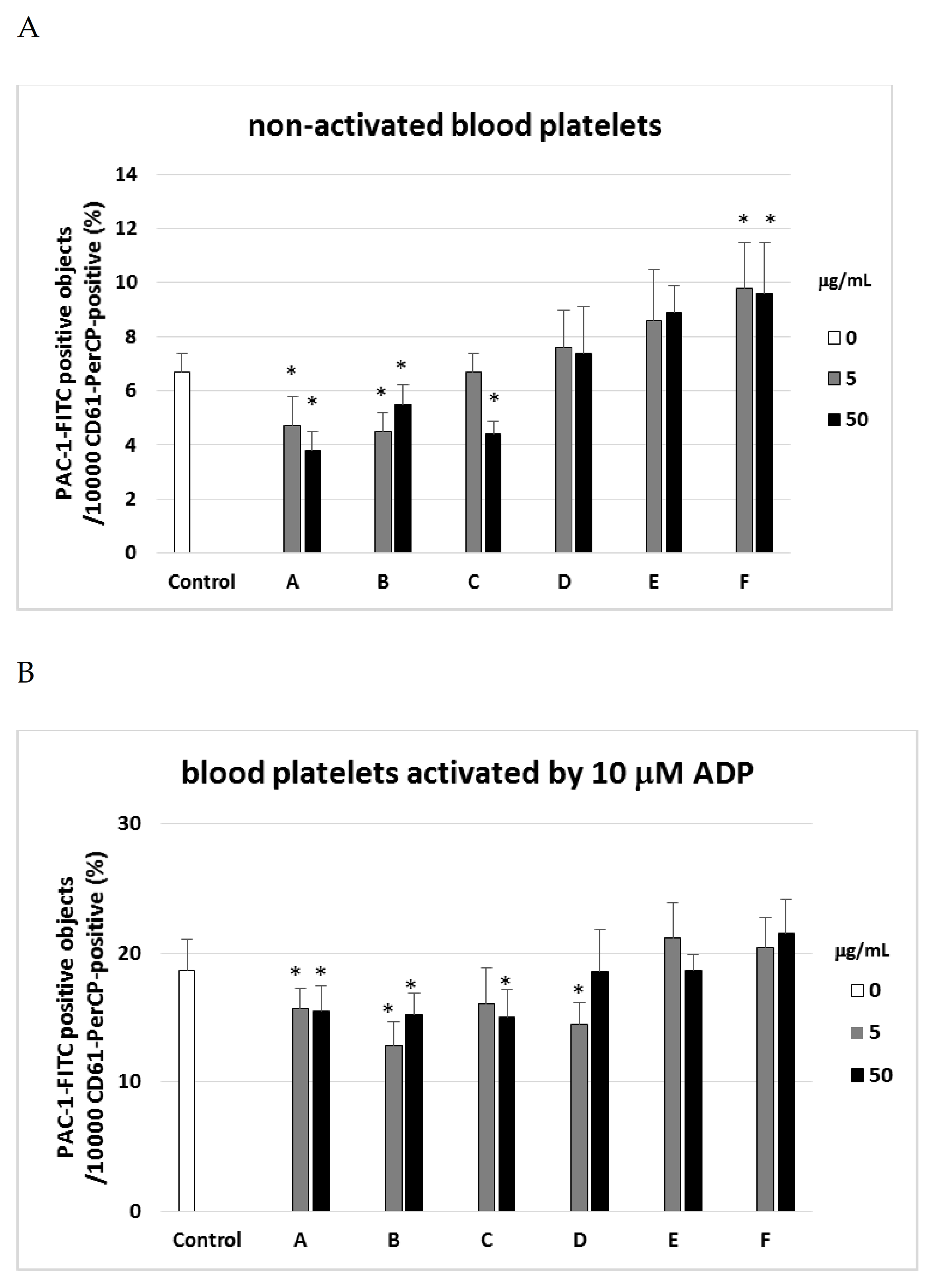
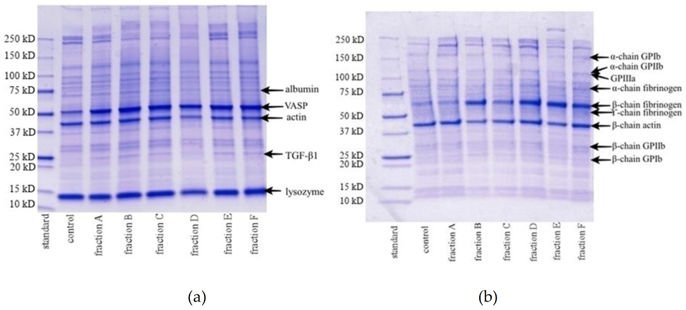
2. Results
3. Discussion
4. Materials and Methods
4.1. Chemicals
4.2. Plant Material
4.3. Chemical Characteristics of Fractions from Sea Buckthorn Fruits, Twigs, and Leaves
4.4. Blood and Blood Platelets Samples
4.5. Flow Cytometry Analysis
4.6. Platelet VASP Phosphorylation
4.7. Polyacrylamide gel Electrophoresis Analysis
4.8. Total Thrombus-Formation Analysis System (T-TAS)
4.9. Data Analysis
Author Contributions
Funding
Institutional Review Board Statement
Informed Consent Statement
Data Availability Statement
Conflicts of Interest
Abbreviations
| ADP | adenosine diphosphate |
| AUC | Area Under the Curve |
| CPDA1 | citrate/phosphate/dextrose/adenine |
| CVDs | cardiovascular diseases |
| DMSO | dimethylsulfoxide |
| PRI | platelet reactivity indicator |
| T-TAS | Total Thrombus-formation Analysis System |
| VASP | vasodilator-stimulated phosphoprotein |
References
- Agarwal, N.; Mahmoud, A.N.; Patel, N.K.; Jain, A.; Garg, J.; Mojadidi, M.K.; Agrawal, S.; Qamar, A.; Golwala, H.; Gupta, T.; et al. Meta-analysis of aspirin versus dual antiplatelet therapy following coronary artery bypass grafting. Am. J. Cardiol. 2018, 121, 32–40. [Google Scholar] [CrossRef]
- Chong, M.F.F.; Macdonal, R.; Lovegrove, J.A. Fruit Polyphenols and CDV risk: A review of human intervention studies. Br. J. Nutr. 2010, 104, 28–39. [Google Scholar] [CrossRef] [PubMed] [Green Version]
- Hirsch, G.E.; Viecili, P.R.N.; de Almeida, A.S.; Nascimento, S.; Porto, F.G.; Otero, J.; Schmidt, A.; da Silva, B.; Parisi, M.M.; Klafke, J.Z. Natural products with antiplatelet action. Curr. Pharm. Des. 2017, 23, 1228–1246. [Google Scholar] [CrossRef]
- Olas, B.; Żuchowski, J.; Lis, B.; Skalski, B.; Kontek, B.; Grabarczyk, Ł.; Stochmal, A. Comparative chemical composition, antioxidant and anticoagulant properties of phenolic fraction (a rich in non-acylated and acylated flavonoids and non-polar compounds) and non-polar fraction from Elaeagnus rhamnoides (L.) A.Nelson fruits”. Food Chem. 2018, 247, 39–45. [Google Scholar] [CrossRef]
- Eccleston, C.; Baoru, Y.; Tahvonen, R.; Kallio, H.; Rimbach, G.H.; Minihane, A.M. Effects of an antioxidant-rich juice (sea buckthorn) on risk factors for coronary heart disease in humans. J. Nutr. Biochem. 2002, 13, 346–354. [Google Scholar] [CrossRef]
- Basu, M.; Prasad, R.; Jayamurthy, P.; Pal, K.; Arumughan, C.; Sawhney, R.C. Antiatherogenic effects of seabuckthorn (Hippophaea rhamnoides) seed oil. Phytomedicine 2007, 14, 770–777. [Google Scholar] [CrossRef] [PubMed]
- Olas, B.; Kontek, B.; Szczęsna, M.; Grabarczyk, Ł.; Stochmal, A.; Żuchowski, J. Inhibition of blood platelet adhesion by the phenolics’ rich fraction of Hippophae rhamnoides L. fruits. J. Physiol. Pharm. 2017, 2, 23–29. [Google Scholar]
- Olas, B. The beneficial health aspects of sea buckthorn (Elaeagnus rhamnoides (L.) A. Nelson) oil. J. Ethnopharmacol. 2018, 213, 183–190. [Google Scholar] [CrossRef]
- Skalski, B.; Kontek, B.; Olas, B.; Żuchowski, J.; Stochmal, A. Phenolic fraction and nonpolar fraction from sea buckthorn leaves and twigs: Chemical profile and biological activity. Future Med. Chem. 2018, 10, 2381–2394. [Google Scholar] [CrossRef] [PubMed]
- Skalski, B.; Stochmal, A.; Żuchowski, J.; Grabarczyk, Ł.; Olas, B. Response of blood platelets to phenolic fraction and non-polar fraction from the leaves and twigs of Elaeagnus rhamnoides (L.) A. Nelson in vitro. Biomed. Pharm. 2020, 124, 1–11. [Google Scholar] [CrossRef] [PubMed]
- Pignone, M.; Williams, C.D. Aspirin for primary prevention of cardiovascular disease in diabetes mellitus. Nat. Rev. Endocrinol. 2010, 6, 619–628. [Google Scholar] [CrossRef] [Green Version]
- Kaikita, K.; Hosokawa, K.; Dahlen, J.R.; Tsujita, K. Total thrombus-formation analysis system (T-TAS): Clinical application of quantitative analysis of thrombus formation in cardiovascular disease. Thromb. Haemost. 2019, 119, 1554–1562. [Google Scholar] [CrossRef] [Green Version]
- Arias-Salgado, E.G.; Larrucea, S.; Butta, N.; Fernández, D.; García-Muñoz, S.; Parrilla, R.; Ayuso, M.S. Variations in platelet protein associated with arterial thrombosis. Thromb. Res. 2008, 122, 640–647. [Google Scholar] [CrossRef] [Green Version]
- Bagoly, Z.; Sarkady, F.; Magyar, T.; Kappelmayer, J.; Pongrácz, E.; Csiba, L.; Muszbek, L. Comparison of a new P2Y12 receptor specific platelet aggregation test with other laboratory methods in stroke patients on clopidogrel monotherapy. PLoS ONE 2013, 8, e69417. [Google Scholar] [CrossRef] [Green Version]
- Aleil, B.; Rvanat, C.; Cazenave, J.P.; Rochoux, G.; Heitz, A.; Gachet, C. Flow cytmetric analysis of intrpltelet VASP phosphorylation for the detection of clopidogrel resistance in patients with ischemic cardiovascular diseases. J. Thromb. Haemost. 2005, 3, 85–92. [Google Scholar] [CrossRef] [PubMed]
- Skalski, B.; Kontek, B.; Rolnik, A.; Olas, B.; Stochmal, A.; Żuchowski, J. Anti-Platelet Properties of Phenolic Extracts from the Leaves and Twigs of Elaeagnus rhamnoides (L.) A. Nelson. Molecules 2019, 24, 3620. [Google Scholar] [CrossRef] [Green Version]
- Olas, B.; Kędzierska, M.; Wachowicz, B.; Stochmal, A.; Oleszek, W. Effects of polyphenol-rich extract from berries of Aronia melanocarpa on the markers of oxidative stress and blood platelet activation. Platelets 2010, 21, 274–281. [Google Scholar] [CrossRef]
- Sędek, Ł.; Sonsala, A.; Szczepański, T.; Mazur, B. Techniczne aspekty cytometrii przepływowej. Diagn. Lab. 2010, 46, 415–420. [Google Scholar]
- Morel, A.; Rywaniak, J.; Bijak, M.; Miller, E.; Niwald, M.; Saluk, J. Flow cytometric analysis reveals the high levels of platelet activation parameters in circulation of multiple sclerosis patients. Mol. Cell. Biochem. 2017, 430, 69–80. [Google Scholar] [CrossRef] [PubMed] [Green Version]
- Rywaniak, J.; Luzak, B.; Podsędek, A.; Dudzińska, D.; Różalski, M.; Watała, C. Comparison of cytotoxic and anti-platelet activities of polyphenolic extracts from Arnica montana flowers and Juglans regia husks. Platelets 2015, 26, 168–176. [Google Scholar] [CrossRef]
- Laemli, U.K. Cleavage of Structural Proteins during the Assembly of the Head of Bacteriophage T4. Nature 1970, 227, 680–685. [Google Scholar] [CrossRef] [PubMed]
- Hosokawa, K.; Ohnishi, T.; Kondo, T.; Fukasawa, M.; Koide, T.; Maruyama, I.; Tanaka, K.A. A novel automated microchip flow-chamber system to quantitatively evaluate thrombus formation and antithrombotic agents under blood flow conditions. J. Thromb. Haemost. 2018, 9, 2019–2037. [Google Scholar] [CrossRef] [PubMed]


| Fraction | CD62P Expression | PAC-1 Expression | T-TAS | VASP Phosphorylation | ||||||
|---|---|---|---|---|---|---|---|---|---|---|
| Non-Activated Platelets | Platelets Activated by 10 µM ADP | Platelets Activated by 20 µM ADP | Platelets Activated by 10 µg/mL Collagen | Non-Activated Platelets | Platelets Activated by 10 µM ADP | Platelets Activated by 20 µM ADP | Platelets Activated by 10 µg/mL Collagen | |||
| A | No effect | No effect | No effect | No effect | Decrease (anti-platelet potential) | Decrease (anti-platelet potential) | No effect | Decrease (anti-platelet potential) | Anti-coagulant poteintial | No effect |
| B | No effect | No effect | No effect | No effect | Decrease (anti-platelet potential) | Decrease (anti-platelet potential) | No effect | No effect | No effect | No effect |
| C | Decrease (anti-platelet potential) | No effect | No effect | No effect | No effect | No effect | No effect | Decrease (anti-platelet potential) | Anti-coagulant poteintial | No effect |
| D | No effect | No effect | No effect | No effect | No effect | No effect | No effect | No effect | Anti-coagulant poteintial | No effect |
| E | No effect | No effect | No effect | No effect | No effect | No effect | No effect | Decrease (anti-platelet potential) | Anti-coagulant poteintial | No effect |
| F | No effect | No effect | No effect | No effect | Increase (pro-activation potential) | No effect | Increase (pro-activation potential) | No effect | No effect | No effect |
| Aronia berry extract | No effect | Decrease (anti-platelet potential) | Decrease (anti-platelet potential) | Decrease (anti-platelet potential) | No effect | No effect | Decrease (anti-platelet potential) | Decrease (anti-platelet potential) | No effect | No effect |
| Fractions | Relative Peak Area % |
| Polyphenols rich fraction of sea buckthorn from fruits (fraction A) | |
| Flavonol glycosides, non-acylated and acylated | 67.1 |
| Triterpenoids and acylated triterpenoids | 9.1 |
| Triterpenic acids rich fraction of sea buckthorn fruits (fraction B) | |
| Triterpenoids and acylated triterpenoids | 83.6 |
| Flavonol glycosides, non-acylated and acylated | 0.9 |
| Polyphenols rich fraction of sea buckthorn from leaves (fraction C) | |
| Hydrolysable tannins and ellagic acid | 31.3 |
| Flavonol glycosides, non-acylated and acylated | 24.5 |
| Triterpenic acids rich fraction of sea buckthorn leaves (fraction D) | |
| Triterpenoids and acylated triterpenoids | 50.7 |
| Triterpenoid saponins | 30.5 |
| Polyphenols rich fraction of sea buckthorn from twigs (fraction E) | |
| Proanthocyanidins and catechin | 54.3 |
| Spermidine derivatives | 10.7 |
| Triterpenic acids rich fraction of sea buckthorn from twigs (fraction F) | |
| Triterpenoids and acylated triterpenoids | 89.0 |
| Catechin and proanthocyanidins | 1.3 |
Publisher’s Note: MDPI stays neutral with regard to jurisdictional claims in published maps and institutional affiliations. |
© 2021 by the authors. Licensee MDPI, Basel, Switzerland. This article is an open access article distributed under the terms and conditions of the Creative Commons Attribution (CC BY) license (http://creativecommons.org/licenses/by/4.0/).
Share and Cite
Skalski, B.; Rywaniak, J.; Szustka, A.; Żuchowski, J.; Stochmal, A.; Olas, B. Anti-Platelet Properties of Phenolic and Nonpolar Fractions Isolated from Various Organs of Elaeagnus rhamnoides (L.) A. Nelson in Whole Blood. Int. J. Mol. Sci. 2021, 22, 3282. https://doi.org/10.3390/ijms22063282
Skalski B, Rywaniak J, Szustka A, Żuchowski J, Stochmal A, Olas B. Anti-Platelet Properties of Phenolic and Nonpolar Fractions Isolated from Various Organs of Elaeagnus rhamnoides (L.) A. Nelson in Whole Blood. International Journal of Molecular Sciences. 2021; 22(6):3282. https://doi.org/10.3390/ijms22063282
Chicago/Turabian StyleSkalski, Bartosz, Joanna Rywaniak, Aleksandra Szustka, Jerzy Żuchowski, Anna Stochmal, and Beata Olas. 2021. "Anti-Platelet Properties of Phenolic and Nonpolar Fractions Isolated from Various Organs of Elaeagnus rhamnoides (L.) A. Nelson in Whole Blood" International Journal of Molecular Sciences 22, no. 6: 3282. https://doi.org/10.3390/ijms22063282