Review

Lynch Syndrome: Its Impact on Urothelial Carcinoma

1
Department of Urology, Medical University Innsbruck, 6020 Innsbruck, Austria
2
Immunotherapy Unit, Department of Urology, Medical University Innsbruck, 6020 Innsbruck, Austria
3
Department of Internal Medicine V, Medical University Innsbruck, 6020 Innsbruck, Austria
4
Experimental Oncogenomic Group, Tyrolean Cancer Research Institute, 6020 Innsbruck, Austria
5
Pathology Laboratory Obrist and Brunhuber, 6511 Zams, Austria
6
Clinical Pathology and Cytodiagnostics, tirol-kliniken, 6020 Innsbruck, Austria
7
Department of Radiology, Medical University Innsbruck, 6020 Innsbruck, Austria
*
Author to whom correspondence should be addressed.
Int. J. Mol. Sci. 2021, 22(2), 531; https://doi.org/10.3390/ijms22020531
Submission received: 18 December 2020 / Revised: 30 December 2020 / Accepted: 3 January 2021 / Published: 7 January 2021
(This article belongs to the Special Issue Germ Cells and Genitourinary Cancers)

Abstract

:
Lynch syndrome, known as hereditary nonpolyposis colorectal cancer (HNPCC), is an autosomal-dominant familial cancer syndrome with an increased risk for urothelial cancer (UC). Mismatch repair (MMR) deficiency, due to pathogenic variants in MLH1, MSH2, MSH6, and PMS2, and microsatellite instability, are known for development of Lynch syndrome (LS) associated carcinogenesis. UC is the third most common cancer type in LS-associated tumors. The diversity of germline variants in the affected MMR genes and their following subsequent function loss might be responsible for the variation in cancer risk, suggesting an increased risk of developing UC in MSH2 mutation carriers. In this review, we will focus on LS-associated UC of the upper urinary tract (UUT) and bladder, their germline profiles, and outcomes compared to sporadic UC, the impact of genetic testing, as well as urological follow-up strategies in LS. In addition, we present a case of metastatic LS-associated UC of the UUT and bladder, achieving complete response during checkpoint inhibition since more than 2 years.

Graphical Abstract

1. Introduction

Lynch syndrome (LS), also known as hereditary nonpolyposis colorectal cancer (HNPCC), is the most common hereditary type of colon cancer, accounting for 3% of new colon cancer diagnoses [1]. Apart from early onset colon cancer with proximal predominance and an excess of synchronous and/or metachronous colon cancer [2], the cancer types most frequently associated with LS are endometrial and ovarian cancer [3], and malignancies affecting the stomach, small bowel, prostate, breast, brain, and hepato-biliary tract [4,5,6,7]. Due to the increased recognition of tumor heterogeneity in LS, LS-associated cancer types include urothelial cancer (UC) as the third most common cancer in subsets of these families as first described by Lynch et al. in 1990 [8]. Therefore, the aim of this review article is to focus on LS-associated UC of the upper urinary tract (UUT) and bladder, highlighting genetics, molecular subtype classifications, and germline variant profiles compared to sporadic UC, the impact of genetic testing, incidence, outcomes, and potential future treatment strategies such as immune checkpoint inhibitors (ICI), which might help to optimize treatment in LS-associated cancer.

1.1. Materials and Methods

Literature research was performed by search of the commonly used databases PubMed® (National Library of Medicine, National Center for Biotechnology Information, Bethesda, MD, USA) and MedLine® (Medical Literature Analysis and Retrieval System Online, National Center for Biotechnology Information, Bethesda, MD, USA), which were accessed until July 2020. The study language was limited to English language. The following medical subject heading terms were used to identify the results suitable to our review topic: Lynch syndrome, urothelial cancer, upper urinary tract, mismatch repair genes, microsatellite instability, immunotherapy, checkpoint inhibitor. A workflow, shown in Figure 1, gives on overview of our literature search structure.
Thereafter, suitable findings that we presumed to be of clinical interest were included in the detailed analysis in our review article.

1.2. Lynch Syndrome and Genetics

Sporadic colon cancer is often linked to point mutations in tumor suppressor genes such as p53 [9] and APC [1], which are less represented in LS [2]. After the onset of MMR in LS, mutations in the KRAS gene commonly occur, followed by APC mutations [10,11]. The syndrome causes a wide variety of oncological cancer types. Its underlying mechanism is a germline variant of DNA mismatch repair (MMR) genes, which are found in 88% to 95% of patients diagnosed with this disorder [3,4]. Additionally, EPCAM, which is a gene adjacent to MSH2 that when mutated can cause the MSH2 to be inactivated, constitutes for about 3% of LS cases [12]. MMR is one of the essential factors in preventing cancer development in a biological system, as it corrects miss-paired DNA insertions and replication errors and serves as a checkpoint to maintain vital genomic stability by restoring improperly assembled single-base matches during replication [5]. In addition, oxidation [6] or alkylation damage can be recognized and either be repaired, or cell apoptosis can be induced. Germline variants in one of two MMR genes is a precondition in individuals with LS. Inactivation of the second healthy allele usually arises due to small pathogenic variants and gene loss, leading to a defect in the MMR system [13].
Four main MMR genes have been found to be responsible for LS associated cancer development to date. These predisposing genes are mutL homolog 1 (MLH1) [14] and mutS homolog 2 (MSH2) [15], followed by mutS homolog 6 (MSH6) [16] and postmeiotic segregation increased 2 (PMS2) [17]. MSH2 has been shown to form the MSH2-MSH6 heterodimer, identifying base-base mismatches. The MSH2 and MLH1 proteins are the mandatory partners of their particular dimer [18]. MutLα then promotes the excision of the mismatched locus, which is performed by proteins such as exonuclease-1 and DNA polymerase, and DNA ligase resynthesize and ligate the DNA strand [19,20]. The sequence of synthesis is illustrated in Figure 2.
Any defect in these proteins results in an inactive DNA repair process, which increases pathogenic alteration rates in genes of the cell growth cycle, leading to defects in tumor suppressor genes and oncogenes, followed by an elevated cancer risk [22]. Insufficient MMR and subsequent variation in the number of nucleotides in a microsatellite region, defined as small repetitive DNA sequences, is referred to as microsatellite instability (MSI), which is present in approximately 95% for of all tumors associated with LS [15]. In contrast, MSI is not common in sporadic colorectal cancer (CRC). Tumor mutational burden (TMB) was found to be consistently high across colorectal cancer with MMR gene inactivation [23]. Fabrizio et al., additionally reported that TMB assessment can accurately classify MSI tumors as TMB-high [24], as TMB is known to be a predictive response marker in ICI therapy [25,26], and it underlines the use of targeted therapy in patients with LS.

1.3. Lynch Syndrome and Cancer Susceptibility

Interestingly, current findings from the prospective LS database confirmed distinct gene and gender-specific patterns of cancer risk depending on the affected MMR pathogenic variant carrier in LS patients. In detail, MSH2 pathogenic variant carriers confirmed a higher risk of UC of the UUT. UC of the bladder is most related to MSH2 pathogenic alterations, yet not to the extent of UC of the UUT. Interestingly, neither urinary tract cancers, nor colorectal or endometrial cancer (EC), were observed before the age of 50 in carriers of a PMS2 variant. At older age, it has been shown that germline pathological variants of MSH2 and MLH1 are even more associated with urinary tract cancer. In EC, MMR deficiency has already been shown in 20–40% of cases, with still controversial data on its prognostic value [27]. Additionally, the MSH6 pathogenic variant seems to be a gender-specific factor in cancer susceptibility with higher EC risk, but only a low risk of colon cancer in both sexes [7].
The findings result in cumulative incidences at an age of 75 years for urinary tract cancers with MLH1, MSH2, and MSH6 variants in 8%, 25%, and 11%, respectively [28], suggesting that MSH2 pathogenic variant carriers were at significant increased risk of developing UC compared with individuals with pathogenic germline variants in MLH1 or MSH6. Loss of co-expression of MLH1 and PMS2, as described above, was shown to be associated with lower mutational burden (TMB) in contrast to loss of the MSH2/MSH6 dimer. This variability may be responsible for tissue-specific differences and the variation of cancer risk in LS [29]. Additionally, it has been demonstrated that tumors with MSI due to defects in MMR proteins often present with high Th1 type T-cell infiltration [30,31,32]. Additionally, MMR protein deficient tumors characteristically have T cell infiltration that could indicate an ongoing antitumor immune response and may counteract the immunosuppressive effects of standard chemotherapy [33]. A detailed overview of incidence rates of LS-associated cancers at 75 years depending on the involved MMR gene mutation is presented in Figure 3.

1.4. Diagnosis of Lynch Syndrome

Several clinical models and strategies have been developed for identification of patients at high risk for LS. The first guideline criteria were published in 1990 as the Amsterdam criteria I, in which all criteria listed in Table 1 had to be fulfilled by affected families [34]:
As mentioned, LS includes various extra-colonic malignancies, which were not considered in these first diagnostic criteria. In 1999, the revised Amsterdam criteria II stipulated at least three relatives with LS-associated cancer instead of only CRC, defined as CRC, cancer of the endometrium, small bowel, ureter, or renal pelvis [35]. Because LS is a hereditary disease, anamnesis and an accurate medical family record concerning malignancies are essential for early screening and diagnosis, but in clinical practice this can be difficult [36]. Histopathology alone is not usually able to distinguish LS associated CRC from sporadic CRC cases [37], but specific histopathological characteristics have been described as being representative for LS. These include peritumoral Crohn’s like lymphatic reaction, medullary histology with poor differentiation, and intratumoral lymphocytic infiltration [38,39]. Mucinous histopathologic character is often present [40,41]. Identifying individuals who do not meet the Amsterdam criteria, yet have a potential risk of LS, is still a challenge. For this reason the Bethesda Guidelines were defined in 1997 which, when fulfilled, propose testing of replication errors and MSI to detect mutation carriers [42]. The Bethesda guidelines were revised in 2004 and are listed in Table 2 [43].
The MIPA criteria, published in 2005 and listed in Table 3, propose a simplified version of the Bethesda Guidelines by excluding details of family history [44], which are often not applicable in clinical practice [36].
The molecular diagnostic approach combines MSI analysis and MMR protein immunostaining and seems to be a productive way of pre-selecting those patients at high risk for LS for further germline variant analysis [45]. Ninety-five percent of LS-associated cancers are MSI-positive [46]. Tumors are characterized on the basis of frequency of instability: Low-frequency MSI (MSI-L) and high-frequency MSI (MSI-H), which is suspicious for LS. MSI-H status has been associated with better survival due to accumulation of somatic mutations [47,48,49]. In CRC, MSI is a well-known prognostic marker [50]. Furthermore, in patients with stage II disease of CRC, MSI is also of predictive importance for the response to immunotherapy in metastatic disease [51]. Association between MSI and IHC in extra-colonic LS cancers has been shown to become more and more weaker, suggesting that microsatellites are organ-specific [50]. Different staining patterns in immunohistochemistry (IHC) allow an assessment of MMR protein loss and have been proposed as an alternative screening method to MSI testing, both being valid and effective diagnostic tools for selecting patients for further genetic germline variant analysis [52,53]. However, IHC cannot differentiate between dysfunctional proteins deriving from either missense mutations or polypeptides. It has been shown that MSH2 and MLH1 proteins are the obligatory partners of their heterodimers MSH6 and PMS2, respectively (Figure 1). Furthermore, MSH2 and MLH1 are stable without their dimer partners, but not reversely. To conclude, tumors with an MLH1 pathogenic germline variant will show loss of both partners MLH1 and PMS2. Pathogenic variants in MSH2 will result in disintegration to missense of both MSH2 and MSH6. On the other hand, pathogenic variants in the secondary genes MSH6 and PMS2 result in selective loss of only these genes, as additionally summarized in Table 4. IHC should therefore include antibodies of all four proteins in order to detect as many as MLH1 and MSH2 abnormalities as possible. Thus, various IHC expression patterns associated with MMR-gene pathogenic variants are possibly based on the heterodimeric nature of the MMR proteins as already described by Van Lier et al. [45].
In comparison, IHC may be slightly superior in predicting pathogenic germline variants to MSI analysis. Additionally, it is cheaper and more easily applicable [54] as a fast and simple procedure. IHC for MMR proteins should not replace MSI testing to detect LS, encouraging a so called ‘combined diagnostic molecular concept’, as IHC interpretation may be difficult due to inter- and intraobserver variability, missense mutations, or low or absent intensity of nuclear staining in tumors and normal tissue [55,56]. Nevertheless, both molecular screening methods, IHC staining and MSI analysis, confirm the absence of one the four MMR proteins or a high MSI count, which assigns patients to further germline analysis. On the other hand, if the clinical suspicion for LS is very high, even in the case of no tumoral MMR deficiency or MSS tumors, genetic counselling and testing are still indicated, although the false-negative rate of MSI analysis is very low (<5%). Due to the fact that germline analysis is time consuming and expensive, the current standard practice is to select patients for genetic testing by a molecular diagnostic work-up, guided by clinical and pathological criteria, such as MSI testing and IHC staining of MMR proteins [45].
Genetic testing is performed on DNA isolated from peripheral blood mononuclear cells obtained by blood draw or from an oral rinse. Currently, in addition to MLH1 and MSH2, whose testing was introduced in 1990s, mutations in the genes MSH6, PMS2, and EPCAM are now also included in testing of individuals suspected to have LS [57,58,59]. Germline testing confirms LS in only 24 to 67% of MMR-deficient CRC and in 16 to 80% of MMR-deficient EC [60,61], depending on the IHC expression patterns. Accordingly, germline analysis by next-generation sequencing has improved, generating a more comprehensive genetic profile of both germline and somatic mutations, thereby analyzing DNA isolated from both blood and tumor samples in parallel [62]. Subsequently, ‘paired tumor and germline testing’ seems to be an attractive diagnostic tool in the LS testing algorithm, as it identifies an underlying cause for MMR-deficient tumors such as CRC and EC in 76% and 61% of patients without and with prior LS germline testing. Combined testing allows the simultaneous diagnosis of LS and describes atypical IHC patterns in patients where germline pathogenic variants were not concordant with specific protein expression, e.g., double somatic mutations, which explain IHC staining in addition to a germline mutation in a different LS gene. Moreover, solely tumor sequencing for germline screening is limited in detecting exon-level somatic copy number variants in regions with significant pseudogene homology such as the PMS2 pseudogene region [63]. These findings may corroborate the importance of performing a comprehensive germline testing regardless of IHC staining patterns, reducing the risk of germline rearrangements.

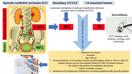
1.5. Lynch Syndrome and UC of the UUT and Bladder

UC of the UUT accounts for only 5 to 10% of UC [64], but about 60% of primary diagnosed UUT cancers are invasive, and 7% have already metastasized at first diagnosis [65,66]. Tumors of the renal pelvis are 3 to 4 times more common than malignancies of the ureteric tract [67]. Hereditary UC of the UUT is linked to LS and accounts for approximately 20% of all UC of the UUT [68,69], and it ranks third (5%) after colon cancer (63%) and EC (9%) within the group of HNPCC-associated tumors [70]. Patients diagnosed with LS have a 22-fold higher risk and a cumulative lifetime risk of 3% to develop UC of the UUT, as compared to the general population [70,71]. The relative risk of UUT cancer in patients with LS therefore is 14% [72]. Compared to sporadic UC, LS-associated UC is known to have a female predominance and to occur at a younger age [73]. Hubosky et al. first described that patients with LS who develop UC of the UUT in their cohort appear to be more likely to have bilateral UC of the UUT over their lifetimes compared to sporadic UUT cancer patients [25].
Whereas UC of the UUT and bladder are biologically and histomorphologically very similar [74], differences in germline variant profiles have been noticed, especially in high-grade urothelial tumors [75]. Comparison of next-generation sequencing of high-grade UUT cancer with high-grade UC of the bladder identified similar pathogenic germline variants in both cancer types, but at different frequencies [76], confirming a higher prevalence of fibroblast growth factor receptor 3 (FGFR3), HRAS and CDKN2B mutated genes in high-grade UUT cancers, and TP53 and RB1 in high-grade UC of the bladder [76,77]. FGFR3 is a member of the tyrosine kinase family and its signaling plays a role toward cell differentiation and proliferation [78] and is known to contribute to an early and essential stage in the molecular pathogenesis of papillary bladder cancer [79,80]. Moss et al. confirmed four unique molecular and clinical subtypes of UC of the UUT by whole-exome sequencing of DNA, RNA sequencing, and protein analysis, as described in Table 5.
A systemic evaluation of current genomic sequencing and proteomic data in UC of the UUT confirmed molecular differences in FGFR3 (fibroblast growth receptor 3), TP53, and MSI between sporadic (80–90%) and hereditary (10–20%) UUT cancers. Whereas sporadic low-grade UC of the UUT is FGFR3 mutated in over 90%, high-grade UC of the UUT is associated with TP53/MDM2 mutations. Nevertheless, UC in patients with LS share molecular similarities of subtype classification with sporadic UC, identifying a predominance of the ‘urothelial-like’ molecular subtype in LS with only the remaining 20% being genomically unstable, basal-like, or related to other subtypes [73]. MMR protein loss has been shown to be present in 7% of all UUT cancer cases and 30% in LS-related UC of the UUT, in this cohort of patients with UUT urothelial cancers and verified loss of mismatch repair protein expression, up to 86% were affected in loss of MSH2 and MSH6, with the remaining 14% showing isolated loss of MSH6 [82]. Cases with MSH2 variants were shown to have the highest risk of developing UC [73,83,84], with an odds ratio of 4.6 (p = 0.001) [85]. Development of UC has been shown to be strongly associated with MSH2 variants in up to 73%, leaving patients with this specific protein-loss at higher risk for UC than individuals with loss of MLH1 or MSH6. [15]. MSI is described to be rare in bladder cancer and to be present in up to 40% of cases of UC of the UUT cases [86,87]. Certain clinical and pathological criteria are more common in UC of the UUT with MSI, such as location in the lower ureter, female sex, younger age, and an inverted growth pattern [88].
Huang et al. have reviewed the association between LS and bladder cancer. It has to be said that it is difficult to assess whether bladder cancer is a LS-associated malignancy, as data regarding the risk of bladder cancer in LS are sparse, yet penetrance was shown to be much less than for LS-associated UC of the UUT [89]. Generally, it has been shown that bladder cancer generally was more common in MSH2 variant families than in the general population. Additionally, these patients had evidence of UC of the UUT as well, which leads the authors to assume that individuals with bladder cancer had an increased risk for UC of the UUT in the first place [90]. Especially amongst patients with MSH2 pathogenic variants, the incidence not only for UC of the UUT, but also for bladder cancer, has shown to be increased [91]. A Swedish cohort of patients fulfilling the Bethesda criteria showed an increased risk of malignancies of UC of the UUT, but interestingly not of UC of the bladder itself [92]. In contrast, van der Post et al. confirmed an increased risk of UC of both the UUT and the bladder in patients with LS carrying a germline MSH2 variant. The cumulative risk of bladder cancer alone until the age of 70 years in MSH2 pathogenic variant carriers and first-degree relatives was 12.3% for men and 2.6% for women. The overall cumulative risk for urinary tract cancer, including bladder and UUT, in MSH2 germline variant carriers and first-degree relatives was 18.2% in men and 8.4% in women [84]. However, there are several factors that may point to the link between bladder cancer and LS [89]. First of all, patients with prior or concurrent UC of the UUT can confound results. Moreover, the low rate of MSI in UC of the bladder may be due to the association with LS [93]. Most importantly, patients with LS-associated UC have similar overall survival rates compared to stage-matched sporadic UC, but are diagnosed at younger age in female patients. In addition, in MMR-deficient LS patients the overall survival was not significantly influenced by MSI [73]. In patients who underwent nephroureterectomy due to invasive UC of the UUT, a high MSI incidence (17%) was an independent positive prognostic factor for survival, especially in patients younger than 71 years with tumor stage T2-T3N0M0 [94]. Five-year and 10-year survival rates for LS-associated bladder cancer with germline pathogenic variants in any of the four MMR genes were very promising with 93% and 81%, respectively [28]. These data suggest that bladder cancer is part of the LS tumors spectrum and consequently, surveillance should be considered, especially in MSH2 pathological germline variant carriers.

1.6. Recommendations of Screening in LS

Early diagnosis of LS enables precise screening for potential LS-associated tumors and inclusion into follow-up programs, reducing the lifetime cancer risk [95,96]. Universal molecular testing can miss up to 28% of LS cases after applying the revised Bethesda Guidelines [95]. Specific screening of patients presenting with UC of the UUT using IHC of MMR proteins and the revised Amsterdam Criteria (ACII) [35] showed that in patients with no known cancer predisposition, up to 21% were estimated to have underlying LS [97]. The sensitivity and specificity of immunohistochemistry are approximately 83% and 89% [95,98]. Furthermore, specific histopathological features, such as pleomorphism, inverted growth, and intratumoral lymphocytes, were associated with the presence of LS [99]. MSI occurred significantly more often when the pathological feature of inverted growth was present, leading to the consideration that this histological parameter could serve as a marker lesion for MSI and help identify patients with LS with UC of the UUT. Using positive ACI/II, MSI, or IHC, Metcalfe et al. identified 13.9% of UC of the UUT as potential LS-tumors. Subsequent genetic testing confirmed LS in 5.2% [97], which is still highly frequented and highlights the importance of this genetic syndrome in UC. Screening for EC and CRC for loss of MMR proteins has already become a standard of care [100,101]. Usage of IHC and PCR was implemented for screening of all new CRC cases, resulting in 94% concordance [102,103]. Similar screening concepts, which are clinically easy applicable and cost-effective, are yet to be established to identify patients with LS-associated UTUC. Currently, the only recommendation for testing of LS is by the European Association of Urology (EAU) Guidelines. Patients at high risk for HNPCC syndrome, which means age <60 years, a personal history of HNPCC-related cancers or first-degree relative <50 years with HNPCC-related cancers, or two first-degree relatives with HNPCC-related cancers [68] should undergo germ-line DNA sequencing with family counselling [64]. In a cohort of 117 patients, it was demonstrated that previously suggested demographic and histologic factors were found to be ineffective at flagging patients with UC of the UUT for further screening, proposing reflexive MMR screening by IHC followed by MSI testing for all UC of the UUT patients [104]. Implementation of a universal screening method remains challenging. European guidelines for managing patients with MMR mutations recommend sonography and urine analysis every one to two years from the age of 30 only in family constellations of two or more UC of the UUT cases [105].

1.7. Recommendations of Urological Surveillance in LS

Mork et al. suggested surveillance of patients with diagnosed LS, yet without development of UC of the UUT. The recommendations include (1) frequent urinalysis with a threshold of three red blood cells per high power field for further investigation, (2) CT scan including a urographic phase when follow-up of CRC is performed, and (3) cystoscopy with a retrograde pyelography. Urine cytology is not recommended [106]. There is general agreement that regular urinalysis should be the leading diagnostic examination when it comes to surveillance in patients diagnosed or suspicious for LS [70,107,108]. In cases of UC of the UUT, MSI testing therefore should be sought, and hereditary predisposition should be investigated, pursuing patients with MSH2 protein loss in IHC to undergo further testing for germline mutations. Special attention should be put on surveillance of UC of the bladder in patients with a verified MSH2 variant, as the association between MSH2 and UC has evolved in the past [15,109,110]. In literature, recommendations for UC surveillance in LS are defined as ultrasound of the bladder and UUT with urine cytology and sediment in every MSH2 mutation carrier starting at age 40 and above, performed every one to two years [84].

1.8. Lynch Syndrome and Immunotherapy

Immune checkpoint inhibitors (ICI) have had a profound impact on the oncological treatment landscape and are known to act as regulators of T cell activation [111]. The core concept is the ability of ICI to enhance the anti-tumor activity of infiltrating T cells. The immune checkpoints (IC) targeted by ICI are programmed cell death 1 (PD-1), programmed cell death ligand 1 (PD-L1), and cytotoxic T lymphocyte antigen 4 (CTLA-4). IC prevent infiltrating T cells from attacking tumor cells. Conversely, blockade of these IC by ICI abrogates the immunosuppressive effects and facilitates antitumor T cell responses. MMR protein loss causes an upregulation of mutation-associated neoantigens, which then trigger more T cells to infiltrate the tumor [112]. However, PD-1 and PD-L1 are often upregulated in infiltrating T cells, preventing antitumor responses. These tumors are reported to express high PD-(L)1 levels on their cell surface [113,114]. Upregulated expression of immune checkpoints, e.g., PD-L1, then leads to inhibition of cytotoxic T cell activity in the tumor environment and thereby promotes immunosuppression, associated with poorer overall survival [115]. Administration of PD-1 and PD-L1 inhibitors in MSI tumors can therefore be held accountable for good response rates. Cancers with proven MSI produce large amounts of neo-antigens that tumor infiltrating lymphocytes (TILs) subsequently target; these tumors are reported to express high PD-(L)1 levels on their cell surface [113,114]. In the advanced therapy-refractory treatment setting, it has been shown that MSI tumors are suitable for immunotherapy [116]. In the Checkmate 142 phase 2 trial, nivolumab achieved further durable responses, consisting of an objective response rate (ORR) of 31%, with almost 69% of patients having disease control for longer than 12 months [117]. Previous case reports have also shown a better response in both CRC and UTUC in patients who had received pembrolizumab after failure of chemotherapy [118,119]. Based on the results of the Checkmate 142 trail, nivolumab received approval for metastatic colorectal cancers with MMR-deficiency and MSI-H that has progressed following prior treatment with fluoropyrimidine, oxaliplatin, and irinotecan [117]. Le et al. found that patients with MMR deficiency showed a significantly higher ORR (40%) compared to patients with MMR proficiency (11%) in colon cancer in response to pembrolizumab treatment. In this cohort, MSI was a significant predictor of ORR [32]. This was also verified for tumors with MMR deficiency regardless of their origin [120], which finally resulted in FDA approval of pembrolizumab in MMR deficient tumors [121]. In epithelial ovarian cancer, the common histologic subtype of high grade serous cancer showed higher PD-L1 positivity and a higher rate of TILs, supporting the consideration of immunotherapy being a promising treatment strategy for this type of ovarian cancer [122]. Recently, pembrolizumab has been approved as first line therapy of metastatic MMR/MSI colorectal cancer [123]. Currently, trials that use ICI in LS-associated tumors are restricted to CRC with no ongoing trials in LS-associated UC. Yet, they point out the upcoming importance of immunotherapy in all MMR deficient tumors. A detailed list of studies concerning LS is shown in Table 6.

2. Case Presentation

In the following, we present the case of a 53-year-old patient who developed UC of the right renal pelvis who, at the age of 48, has been diagnosed with an adenocarcinoma of the stomach, managed by gastrectomy and perioperative chemotherapy with 5-fluorouracil, folic acid, oxaliplatin, and docetaxel. Gastrectomy specimens showed histology of adenocarcinoma of the stomach, described as intestinal type according to Lauren’s criteria, staged ypT3 with resection boundaries free of cancer cells (R0). Histopathology also showed evidence of MSI and loss of expression in the mismatch repair gene MSH2, as shown in Figure 4.
The patient was a non-smoker, and the family history revealed that his aunt had developed CRC at the age of 84, his brother was diagnosed with an oncological disease of unknown entity, and there was a cousin marriage between his parents. At follow-up, nine months after gastrectomy, a computed tomography (CT) scan showed a suspicious contrast defect in the right renal pelvis (Figure 5A). After undergoing ureterorenoscopy, urothelial cancer staged pT1, grade 2 was confirmed in the biopsy material. The patient then had laparoscopic nephroureterectomy at our institution with the pathological finding of high-grade UC of the UUT, staged pT3 with resection boundaries free of cancer cells (R0). Six months later, the patient developed intermittent microhematuria and mucosal alterations at the ostium of the right ureter at cystoscopy on follow-up. A CT scan additionally showed the presence of a contrast medium enhancing mass in the right ostial region of the bladder (Figure 5B) and enlarged retroperitoneal lymph nodes. Transurethral resection of the bladder showed the finding of current urothelial cancer, staged pT2a, where after two cycles of neoadjuvant cisplatin-based chemotherapy (gemcitabine 1000 mg/m2 on days 1, 8, and 15, and cisplatin 70 mg/m2 on day 2; 1 cycle = 28 days) were administered [124] followed by radical cystoprostatectomy (RC) with ileal conduit and extended pelvic lymphadenectomy. Final histology at RC confirmed pathological complete response, staged ypT0R0L0V0N0.
Three months later, enlarged retroperitoneal lymph nodes were found during oncologic follow-up (Figure 5C). These were biopsied under CT guidance (Figure 5D) and revealed a metastasis of the known UC of the bladder. After meeting the clinical criteria with the presence of metachronous LS-associated tumors, MSI and loss of expression of the MMR protein MSH2 was confirmed. With the knowledge that MSH6 expression can be retained in absence of MSH2 staining [125] and given the clinical suspicion for LS, this patient was assigned to germline testing—confirming a germline MSH2 mutation, and therefore LS [95]. IHC of both gastrectomy and cystectomy specimens showed loss of MSH2 expression. (Figure 4 and Figure 6).
Therapy with pembrolizumab was started at a dose of 200 mg every three weeks. Three months later, after five cycles, CT imaging showed partial remission, with a significant decrease of the retroperitoneal mass (Figure 5E). After an additional 30 cycles of pembrolizumab, the patient showed complete remission (Figure 5F). Therapy was very well tolerated, and the patient is in good general health since more than 2 years. In accordance with previous observations, our case report shows immunotherapy to be suitable and efficient in patients with LS or MMR pathological germline variants.

3. Conclusions

Hereditary urological cancers, especially of the UUT, can often be misclassified as sporadic, resulting in an underestimation of their incidence. Hereditary UC of the UUT accounts to approximately 20% of all UC of the UUT cases and ranks third (5%) after colon cancer (63%) and EC (9%) within all reported LS-associated tumors. Compared to sporadic UC, LS-associated UC, especially in the UUT, is associated with a female predominance, bilateral occurrence, and younger age at first diagnosis. Specific histopathological features, such as intratumoral lymphocytes and inverted papilloma-like growth pattern, may be useful in combination with IHC in identifying those at risk for LS. Urological surveillance in LS should include imaging of the UUT, urine analysis, cytology, and cystoscopy, especially in patients with MSH2 germline variants. MSI was detected more frequently when the pathological feature of inverted growth was present, leading to the consideration that this pathological feature could serve as a surrogate marker for MSI and help identify patients with LS and UC of the UUT at first oncological diagnosis. In cases of UC of the UUT, MSI testing should be sought, and hereditary predisposition should be investigated by genetic testing. In this context, IHC serves as a clinically more applicable strategy to identify patients with MMR protein loss for subsequent genetic testing. Since MSI, which occurs in up to 40% of UC of the UUT cases, is associated with a better survival, and given that tumors with a high MSI show good responses to immunotherapy, it is tempting to predict that ICI will have a central role in the therapeutic regimes of LS.
As presented in our case, ICI show promising results in the therapeutic landscape of LS-associated tumors, which is supported by our case presentation. Future research is dedicated to the development of guidelines that provide a framework for screening, surveillance, and therapy regimes of patients diagnosed with LS-associated UC.

Author Contributions

A.K.L., G.S., and R.P. conceived and designed research. R.P., M.T., and Z.C. were responsible for validation. G.U., P.O., and I.P. adjudged the histological specimens, performed IHC and provided histological imaging. F.S. provided radiological imaging to our case report. A.K.L. and G.S. drafted the original manuscript. R.P., G.T., M.T., G.U., I.P., W.H. and Z.C. critically revised and edited the manuscript draft. R.P., M.T., W.H., and Z.C. were responsible for supervision. All authors made substantial contributions to the manuscript draft and critical revised it. All authors have read and agreed to the published version of the manuscript.

Funding

This research received no external funding.

Conflicts of Interest

The authors declare no conflict of interest.

Abbreviations

ACIIAmsterdam Criteria II
AUAAmerican Urological Association
CTcomputed tomography
CRCcolorectal cancer
CTLA-4cytotoxic T lymphocyte antigen 4
DNAdeoxyribonucleic acid
ECendometrial cancer
FAPfamilial adenomatous polyposis
FGFR3fibroblast growth factor receptor 3
HPFhigh-power field
ICIimmune checkpoint inhibitors
IHCimmunohistochemistry
LSLynch syndrome
MLH1mutL homolog 1
MMRmismatch repair
MSH2mutS homolog 2
MSH6mutS homolog 6
MSImicrosatellite instability
MSI-Hhigh-frequency MSI
MSI-L low-frequency MSI
MSSmicrosatellite stable
ORRobjective response rate
PD-1programmed cell death 1 protein
PD-L1programmed cell death ligand 1 protein
PMS2postmeiotic segregation increased 2
RBCred blood cells
Th1 Type 1 T helper cells
TILtumor infiltrating lymphocytes
TMB tumor mutational burden
UCurothelial cancer
UUTupper urothelial tract

References

  1. Fodde, R. The APC gene in colorectal cancer. Eur. J. Cancer 2002, 38, 867–871. [Google Scholar] [CrossRef]
  2. Kloth, M.; Ruesseler, V.; Engel, C.; Koenig, K.; Peifer, M.; Mariotti, E.; Kuenstlinger, H.; Florin, A.; Rommerscheidt-Fuss, U.; Koitzsch, U.; et al. Activating ERBB2/HER2 mutations indicate susceptibility to pan-HER inhibitors in Lynch and Lynch-like colorectal cancer. Gut 2016, 65, 1296–1305. [Google Scholar] [CrossRef] [PubMed]
  3. Mangold, E.; Pagenstecher, C.; Friedl, W.; Mathiak, M.; Buettner, R.; Engel, C.; Loeffler, M.; Holinski-Feder, E.; Müller-Koch, Y.; Keller, G.; et al. Spectrum and frequencies of mutations in MSH2 and MLH1 identified in 1,721 German families suspected of hereditary nonpolyposis colorectal cancer. Int. J. Cancer 2005, 116, 692–702. [Google Scholar] [CrossRef] [PubMed]
  4. Boland, C.R. Recent discoveries in the molecular genetics of Lynch syndrome. Fam. Cancer 2016, 15, 395–403. [Google Scholar] [CrossRef]
  5. Modrich, P. Mechanisms in eukaryotic mismatch repair. J. Biol. Chem. 2006, 281, 30305–30309. [Google Scholar] [CrossRef] [Green Version]
  6. Bridge, G.; Rashid, S.; Martin, S.A. DNA mismatch repair and oxidative DNA damage: Implications for cancer biology and treatment. Cancers 2014, 6, 1597–1614. [Google Scholar] [CrossRef] [Green Version]
  7. Dominguez-Valentin, M.; Sampson, J.R.; Seppälä, T.T.; Ten Broeke, S.W.; Plazzer, J.-P.; Nakken, S.; Engel, C.; Aretz, S.; Jenkins, M.A.; Sunde, L.; et al. Cancer risks by gene, age, and gender in 6350 carriers of pathogenic mismatch repair variants: Findings from the Prospective Lynch Syndrome Database. Genet. Med. 2020, 22, 15–25. [Google Scholar] [CrossRef] [Green Version]
  8. Lynch, H.T.; Ens, J.A.; Lynch, J.F. The Lynch syndrome II and urological malignancies. J. Urol. 1990, 143, 24–28. [Google Scholar] [CrossRef]
  9. Li, X.-L.; Zhou, J.; Chen, Z.-R.; Chng, W.-J. P53 mutations in colorectal cancer—Molecular pathogenesis and pharmacological reactivation. World J. Gastroenterol. 2015, 21, 84–93. [Google Scholar] [CrossRef]
  10. Ahadova, A.; Gallon, R.; Gebert, J.; Ballhausen, A.; Endris, V.; Kirchner, M.; Stenzinger, A.; Burn, J.; von Knebel Doeberitz, M.; Bläker, H.; et al. Three molecular pathways model colorectal carcinogenesis in Lynch syndrome. Int. J. Cancer 2018, 143, 139–150. [Google Scholar] [CrossRef] [Green Version]
  11. Ten Broeke, S.W.; van Bavel, T.C.; Jansen, A.M.L.; Gómez-García, E.; Hes, F.J.; van Hest, L.P.; Letteboer, T.G.W.; Olderode-Berends, M.J.W.; Ruano, D.; Spruijt, L.; et al. Molecular Background of Colorectal Tumors from Patients with Lynch Syndrome Associated with Germline Variants in PMS2. Gastroenterology 2018, 155, 844–851. [Google Scholar] [CrossRef] [PubMed]
  12. Tiwari, A.K.; Roy, H.K.; Lynch, H.T. Lynch syndrome in the 21st century: Clinical perspectives. QJM 2016, 109, 151–158. [Google Scholar] [CrossRef] [PubMed] [Green Version]
  13. Chung, D.C.; Rustgi, A.K. DNA mismatch repair and cancer. Gastroenterology 1995, 109, 1685–1699. [Google Scholar] [CrossRef]
  14. Pinto, D.; Pinto, C.; Guerra, J.; Pinheiro, M.; Santos, R.; Vedeld, H.M.; Yohannes, Z.; Peixoto, A.; Santos, C.; Pinto, P.; et al. Contribution of MLH1 constitutional methylation for Lynch syndrome diagnosis in patients with tumor MLH1 downregulation. Cancer Med. 2018, 7, 433–444. [Google Scholar] [CrossRef] [Green Version]
  15. Joost, P.; Therkildsen, C.; Dominguez-Valentin, M.; Jönsson, M.; Nilbert, M. Urinary Tract Cancer in Lynch Syndrome; Increased Risk in Carriers of MSH2 Mutations. Urology 2015, 86, 1212–1217. [Google Scholar] [CrossRef]
  16. Bonadona, V.; Bonaïti, B.; Olschwang, S.; Grandjouan, S.; Huiart, L.; Longy, M.; Guimbaud, R.; Buecher, B.; Bignon, Y.-J.; Caron, O.; et al. Cancer risks associated with germline mutations in MLH1, MSH2, and MSH6 genes in Lynch syndrome. JAMA 2011, 305, 2304–2310. [Google Scholar] [CrossRef]
  17. Ten Broeke, S.W.; van der Klift, H.M.; Tops, C.M.J.; Aretz, S.; Bernstein, I.; Buchanan, D.D.; de la Chapelle, A.; Capella, G.; Clendenning, M.; Engel, C.; et al. Cancer Risks for PMS2-Associated Lynch Syndrome. J. Clin. Oncol. 2018, 36, 2961–2968. [Google Scholar] [CrossRef] [Green Version]
  18. de Jong, A.E.; van Puijenbroek, M.; Hendriks, Y.; Tops, C.; Wijnen, J.; Ausems, M.G.E.M.; Meijers-Heijboer, H.; Wagner, A.; van Os, T.A.M.; Bröcker-Vriends, A.H.J.T.; et al. Microsatellite instability, immunohistochemistry, and additional PMS2 staining in suspected hereditary nonpolyposis colorectal cancer. Clin. Cancer Res. 2004, 10, 972–980. [Google Scholar] [CrossRef] [Green Version]
  19. Vilar, E.; Gruber, S.B. Microsatellite instability in colorectal cancer-the stable evidence. Nat. Rev. Clin. Oncol. 2010, 7, 153–162. [Google Scholar] [CrossRef] [Green Version]
  20. Hoeijmakers, J.H. Genome maintenance mechanisms for preventing cancer. Nature 2001, 411, 366–374. [Google Scholar] [CrossRef]
  21. Peltomäki, P. Update on Lynch syndrome genomics. Fam. Cancer 2016, 15, 385–393. [Google Scholar] [CrossRef] [PubMed] [Green Version]
  22. Sobocińska, J.; Kolenda, T.; Teresiak, A.; Badziąg-Leśniak, N.; Kopczyńska, M.; Guglas, K.; Przybyła, A.; Filas, V.; Bogajewska-Ryłko, E.; Lamperska, K.; et al. Diagnostics of Mutations in MMR/EPCAM Genes and Their Role in the Treatment and Care of Patients with Lynch Syndrome. Diagnostics 2020, 10, 786. [Google Scholar] [CrossRef] [PubMed]
  23. Wang, T.; Lee, L.H.; Vyas, M.; Zhang, L.; Ganesh, K.; Firat, C.; Segal, N.H.; Desai, A.; Hechtman, J.F.; Ntiamoah, P.; et al. Colorectal carcinoma with double somatic mismatch repair gene inactivation: Clinical and pathological characteristics and response to immune checkpoint blockade. Mod. Pathol. 2019, 32, 1551–1562. [Google Scholar] [CrossRef] [PubMed]
  24. Fabrizio, D.A.; George, T.J.; Dunne, R.F.; Frampton, G.; Sun, J.; Gowen, K.; Kennedy, M.; Greenbowe, J.; Schrock, A.B.; Hezel, A.F.; et al. Beyond microsatellite testing: Assessment of tumor mutational burden identifies subsets of colorectal cancer who may respond to immune checkpoint inhibition. J. Gastrointest. Oncol. 2018, 9, 610–617. [Google Scholar] [CrossRef]
  25. Rosenberg, J.E.; Hoffman-Censits, J.; Powles, T.; van der Heijden, M.S.; Balar, A.V.; Necchi, A.; Dawson, N.; O’Donnell, P.H.; Balmanoukian, A.; Loriot, Y.; et al. Atezolizumab in patients with locally advanced and metastatic urothelial carcinoma who have progressed following treatment with platinum-based chemotherapy: A single-arm, multicentre, phase 2 trial. Lancet 2016, 387, 1909–1920. [Google Scholar] [CrossRef] [Green Version]
  26. Johnson, D.B.; Frampton, G.M.; Rioth, M.J.; Yusko, E.; Xu, Y.; Guo, X.; Ennis, R.C.; Fabrizio, D.; Chalmers, Z.R.; Greenbowe, J.; et al. Targeted Next Generation Sequencing Identifies Markers of Response to PD-1 Blockade. Cancer Immunol. Res. 2016, 4, 959–967. [Google Scholar] [CrossRef] [Green Version]
  27. Boussios, S.; Mikropoulos, C.; Samartzis, E.; Karihtala, P.; Moschetta, M.; Sheriff, M.; Karathanasi, A.; Sadauskaite, A.; Rassy, E.; Pavlidis, N. Wise Management of Ovarian Cancer: On the Cutting Edge. J. Pers. Med. 2020, 10, 41. [Google Scholar] [CrossRef]
  28. Møller, P.; Seppälä, T.T.; Bernstein, I.; Holinski-Feder, E.; Sala, P.; Gareth Evans, D.; Lindblom, A.; Macrae, F.; Blanco, I.; Sijmons, R.H.; et al. Cancer risk and survival in path_MMR carriers by gene and gender up to 75 years of age: A report from the Prospective Lynch Syndrome Database. Gut 2018, 67, 1306–1316. [Google Scholar] [CrossRef] [Green Version]
  29. Salem, M.E.; Bodor, J.N.; Puccini, A.; Xiu, J.; Goldberg, R.M.; Grothey, A.; Korn, W.M.; Shields, A.F.; Worrilow, W.M.; Kim, E.S.; et al. Relationship between MLH1, PMS2, MSH2 and MSH6 gene-specific alterations and tumor mutational burden in 1057 microsatellite instability-high solid tumors. Int. J. Cancer 2020, 147, 2948–2956. [Google Scholar] [CrossRef]
  30. Smyrk, T.C.; Watson, P.; Kaul, K.; Lynch, H.T. Tumor-infiltrating lymphocytes are a marker for microsatellite instability in colorectal carcinoma. Cancer 2001, 91, 2417–2422. [Google Scholar] [CrossRef]
  31. Kim, H.; Jen, J.; Vogelstein, B.; Hamilton, S.R. Clinical and pathological characteristics of sporadic colorectal carcinomas with DNA replication errors in microsatellite sequences. Am. J. Pathol. 1994, 145, 148–156. [Google Scholar] [PubMed]
  32. Le, D.T.; Uram, J.N.; Wang, H.; Bartlett, B.R.; Kemberling, H.; Eyring, A.D.; Skora, A.D.; Luber, B.S.; Azad, N.S.; Laheru, D.; et al. PD-1 Blockade in Tumors with Mismatch-Repair Deficiency. N. Engl. J. Med. 2015, 372, 2509–2520. [Google Scholar] [CrossRef] [PubMed]
  33. Li, L.S.; Morales, J.C.; Veigl, M.; Sedwick, D.; Greer, S.; Meyers, M.; Wagner, M.; Fishel, R.; Boothman, D.A. DNA mismatch repair (MMR)-dependent 5-fluorouracil cytotoxicity and the potential for new therapeutic targets. Br. J. Pharmacol. 2009, 158, 679–692. [Google Scholar] [CrossRef] [PubMed] [Green Version]
  34. Vasen, H.F.; Mecklin, J.P.; Khan, P.M.; Lynch, H.T. The International Collaborative Group on Hereditary Non-Polyposis Colorectal Cancer (ICG-HNPCC). Dis. Colon Rectum 1991, 34, 424–425. [Google Scholar] [CrossRef]
  35. Vasen, H.; Watson, P.; Mecklin, J.; Lynch, H. New clinical criteria for hereditary nonpolyposis colorectal cancer (HNPCC, Lynch syndrome) proposed by the International Collaborative Group on HNPCC. Gastroenterology 1999, 116, 1453–1456. [Google Scholar] [CrossRef]
  36. Church, J.; McGannon, E. Family history of colorectal cancer: How often and how accurately is it recorded? Dis. Colon Rectum 2000, 43, 1540–1544. [Google Scholar] [CrossRef]
  37. Sanchez, J.A.; Vogel, J.D.; Kalady, M.F.; Bronner, M.P.; Skacel, M.; Church, J.M. Identifying Lynch syndrome: We are all responsible. Dis. Colon Rectum 2008, 51, 1750–1756. [Google Scholar] [CrossRef]
  38. Gaf, R.; Maestri, I.; Matteuzzi, M.; Santini, A.; Ferretti, S.; Cavazzini, L.; Lanza, G. Sporadic colorectal adenocarcinomas with high-frequency microsatellite instability. Cancer 2000, 89, 2025–2037. [Google Scholar] [CrossRef]
  39. Truta, B.; Chen, Y.-Y.; Blanco, A.M.; Deng, G.; Conrad, P.G.; Kim, Y.H.; Park, E.T.; Kakar, S.; Kim, Y.S.; Velayos, F.; et al. Tumor histology helps to identify Lynch syndrome among colorectal cancer patients. Fam. Cancer 2008, 7, 267–274. [Google Scholar] [CrossRef]
  40. Hugen, N.; van Beek, J.J.P.; de Wilt, J.H.W.; Nagtegaal, I.D. Insight into mucinous colorectal carcinoma: Clues from etiology. Ann. Surg. Oncol. 2014, 21, 2963–2970. [Google Scholar] [CrossRef]
  41. Ott, C.; Gerken, M.; Hirsch, D.; Fest, P.; Fichtner-Feigl, S.; Munker, S.; Schnoy, E.; Stroszczynski, C.; Vogelhuber, M.; Herr, W.; et al. Advanced Mucinous Colorectal Cancer: Epidemiology, Prognosis and Efficacy of Chemotherapeutic Treatment. Digestion 2018, 98, 143–152. [Google Scholar] [CrossRef] [PubMed]
  42. Rodriguez-Bigas, M.A.; Boland, C.R.; Hamilton, S.R.; Henson, D.E.; Jass, J.R.; Khan, P.M.; Lynch, H.; Perucho, M.; Smyrk, T.; Sobin, L.; et al. A National Cancer Institute Workshop on Hereditary Nonpolyposis Colorectal Cancer Syndrome: Meeting highlights and Bethesda guidelines. J. Natl. Cancer Inst. 1997, 89, 1758–1762. [Google Scholar] [CrossRef] [PubMed] [Green Version]
  43. Umar, A.; Boland, C.R.; Terdiman, J.P.; Syngal, S.; de La Chapelle, A.; Rüschoff, J.; Fishel, R.; Lindor, N.M.; Burgart, L.J.; Hamelin, R.; et al. Revised Bethesda Guidelines for hereditary nonpolyposis colorectal cancer (Lynch syndrome) and microsatellite instability. J. Natl. Cancer Inst. 2004, 96, 261–268. [Google Scholar] [CrossRef] [PubMed]
  44. Kievit, W.; de Bruin, J.H.F.M.; Adang, E.M.M.; Severens, J.L.; Kleibeuker, J.H.; Sijmons, R.H.; Ruers, T.J.; Nagengast, F.M.; Vasen, H.F.A.; van Krieken, J.H.J.M.; et al. Cost effectiveness of a new strategy to identify HNPCC patients. Gut 2005, 54, 97–102. [Google Scholar] [CrossRef] [PubMed]
  45. van Lier, M.G.F.; Wagner, A.; van Leerdam, M.E.; Biermann, K.; Kuipers, E.J.; Steyerberg, E.W.; Dubbink, H.J.; Dinjens, W.N.M. A review on the molecular diagnostics of Lynch syndrome: A central role for the pathology laboratory. J. Cell Mol. Med. 2010, 14, 181–197. [Google Scholar] [CrossRef] [Green Version]
  46. Lynch, H.T.; de La Chapelle, A. Hereditary colorectal cancer. N. Engl. J. Med. 2003, 348, 919–932. [Google Scholar] [CrossRef]
  47. Gryfe, R.; Kim, H.; Hsieh, E.T.; Aronson, M.D.; Holowaty, E.J.; Bull, S.B.; Redston, M.; Gallinger, S. Tumor microsatellite instability and clinical outcome in young patients with colorectal cancer. N. Engl. J. Med. 2000, 342, 69–77. [Google Scholar] [CrossRef]
  48. Germano, G.; Lamba, S.; Rospo, G.; Barault, L.; Magrì, A.; Maione, F.; Russo, M.; Crisafulli, G.; Bartolini, A.; Lerda, G.; et al. Inactivation of DNA repair triggers neoantigen generation and impairs tumour growth. Nature 2017, 552, 116–120. [Google Scholar] [CrossRef]
  49. Kang, S.; Na, Y.; Joung, S.Y.; Lee, S.I.; Oh, S.C.; Min, B.W. The significance of microsatellite instability in colorectal cancer after controlling for clinicopathological factors. Medicine (Baltimore) 2018, 97, e0019. [Google Scholar] [CrossRef]
  50. Kuismanen, S.A.; Moisio, A.-L.; Schweizer, P.; Truninger, K.; Salovaara, R.; Arola, J.; Butzow, R.; Jiricny, J.; Nyström-Lahti, M.; Peltomäki, P. Endometrial and Colorectal Tumors from Patients with Hereditary Nonpolyposis Colon Cancer Display Different Patterns of Microsatellite Instability. Am. J. Pathol. 2002, 160, 1953–1958. [Google Scholar] [CrossRef] [Green Version]
  51. Boussios, S.; Ozturk, M.A.; Moschetta, M.; Karathanasi, A.; Zakynthinakis-Kyriakou, N.; Katsanos, K.H.; Christodoulou, D.K.; Pavlidis, N. The Developing Story of Predictive Biomarkers in Colorectal Cancer. J. Pers. Med. 2019, 9, 12. [Google Scholar] [CrossRef] [PubMed] [Green Version]
  52. Hampel, H.; Frankel, W.L.; Martin, E.; Arnold, M.; Khanduja, K.; Kuebler, P.; Nakagawa, H.; Sotamaa, K.; Prior, T.W.; Westman, J. Screening for the Lynch syndrome (hereditary nonpolyposis colorectal cancer). N. Engl. J. Med. 2005, 352, 1851–1860. [Google Scholar] [CrossRef] [PubMed] [Green Version]
  53. Piñol, V.; Castells, A.; Andreu, M.; Castellví-Bel, S.; Alenda, C.; Llor, X.; Xicola, R.M.; Rodríguez-Moranta, F.; Payá, A.; Jover, R.; et al. Accuracy of revised Bethesda guidelines, microsatellite instability, and immunohistochemistry for the identification of patients with hereditary nonpolyposis colorectal cancer. JAMA 2005, 293, 1986–1994. [Google Scholar] [CrossRef] [PubMed]
  54. Shia, J. Immunohistochemistry versus microsatellite instability testing for screening colorectal cancer patients at risk for hereditary nonpolyposis colorectal cancer syndrome. Part, I. The utility of immunohistochemistry. J. Mol. Diagn. 2008, 10, 293–300. [Google Scholar] [CrossRef] [PubMed] [Green Version]
  55. Müller, W.; Burgart, L.J.; Krause-Paulus, R.; Thibodeau, S.N.; Almeida, M.; Edmonston, T.B.; Boland, C.R.; Sutter, C.; Jass, J.R.; Lindblom, A.; et al. The reliability of immunohistochemistry as a prescreening method for the diagnosis of hereditary nonpolyposis colorectal cancer (HNPCC)--results of an international collaborative study. Fam. Cancer 2001, 1, 87–92. [Google Scholar] [CrossRef]
  56. Overbeek, L.I.H.; Ligtenberg, M.J.L.; Willems, R.W.; Hermens, R.P.M.G.; Blokx, W.A.M.; Dubois, S.V.; van der Linden, H.; Meijer, J.W.R.; Mlynek-Kersjes, M.L.; Hoogerbrugge, N.; et al. Interpretation of immunohistochemistry for mismatch repair proteins is only reliable in a specialized setting. Am. J. Surg. Pathol. 2008, 32, 1246–1251. [Google Scholar] [CrossRef]
  57. Dinh, T.A.; Rosner, B.I.; Atwood, J.C.; Boland, C.R.; Syngal, S.; Vasen, H.F.A.; Gruber, S.B.; Burt, R.W. Health benefits and cost-effectiveness of primary genetic screening for Lynch syndrome in the general population. Cancer Prev. Res. 2011, 4, 9–22. [Google Scholar] [CrossRef] [Green Version]
  58. Strafford, J.C. Genetic testing for lynch syndrome, an inherited cancer of the bowel, endometrium, and ovary. Rev. Obstet. Gynecol. 2012, 5, 42–49. [Google Scholar]
  59. Xicola, R.M.; Llor, X. Cancer risk assessment in Lynch syndrome: Does the gene matter? JAMA 2011, 305, 2351–2352. [Google Scholar] [CrossRef]
  60. Hampel, H. Population Screening for Hereditary Colorectal Cancer. Surg. Oncol. Clin. N. Am. 2018, 27, 319–325. [Google Scholar] [CrossRef]
  61. Buchanan, D.D.; Rosty, C.; Clendenning, M.; Spurdle, A.B.; Win, A.K. Clinical problems of colorectal cancer and endometrial cancer cases with unknown cause of tumor mismatch repair deficiency (suspected Lynch syndrome). Appl. Clin. Genet. 2014, 7, 183–193. [Google Scholar] [PubMed] [Green Version]
  62. Gray, P.N.; Tsai, P.; Chen, D.; Wu, S.; Hoo, J.; Mu, W.; Li, B.; Vuong, H.; Lu, H.-M.; Batth, N.; et al. TumorNext-Lynch-MMR: A comprehensive next generation sequencing assay for the detection of germline and somatic mutations in genes associated with mismatch repair deficiency and Lynch syndrome. Oncotarget 2018, 9, 20304–20322. [Google Scholar] [CrossRef] [PubMed]
  63. Salvador, M.U.; Truelson, M.R.F.; Mason, C.; Souders, B.; LaDuca, H.; Dougall, B.; Black, M.H.; Fulk, K.; Profato, J.; Gutierrez, S.; et al. Comprehensive Paired Tumor/Germline Testing for Lynch Syndrome: Bringing Resolution to the Diagnostic Process. J. Clin. Oncol. 2019, 37, 647–657. [Google Scholar] [CrossRef] [PubMed]
  64. Rouprêt, M.; Babjuk, M.; Compérat, E.; Zigeuner, R.; Sylvester, R.J.; Burger, M.; Cowan, N.C.; Gontero, P.; van Rhijn, B.W.G.; Mostafid, A.H.; et al. European Association of Urology Guidelines on Upper Urinary Tract Urothelial Carcinoma: 2017 Update. Eur. Urol. 2018, 73, 111–122. [Google Scholar] [CrossRef] [PubMed]
  65. Soria, F.; Shariat, S.F.; Lerner, S.P.; Fritsche, H.-M.; Rink, M.; Kassouf, W.; Spiess, P.E.; Lotan, Y.; Ye, D.; Fernández, M.I.; et al. Epidemiology, diagnosis, preoperative evaluation and prognostic assessment of upper-tract urothelial carcinoma (UTUC). World J. Urol. 2017, 35, 379–387. [Google Scholar] [CrossRef]
  66. Margulis, V.; Shariat, S.F.; Matin, S.F.; Kamat, A.M.; Zigeuner, R.; Kikuchi, E.; Lotan, Y.; Weizer, A.; Raman, J.D.; Wood, C.G. Outcomes of radical nephroureterectomy: A series from the Upper Tract Urothelial Carcinoma Collaboration. Cancer 2009, 115, 1224–1233. [Google Scholar] [CrossRef]
  67. Park, S.; Hong, B.; Kim, C.-S.; Ahn, H. The impact of tumor location on prognosis of transitional cell carcinoma of the upper urinary tract. J. Urol. 2004, 171, 621–625. [Google Scholar] [CrossRef]
  68. Audenet, F.; Colin, P.; Yates, D.R.; Ouzzane, A.; Pignot, G.; Long, J.-A.; Soulie, M.; Phé, V.; Bensadoun, H.; Guy, L.; et al. A proportion of hereditary upper urinary tract urothelial carcinomas are misclassified as sporadic according to a multi-institutional database analysis: Proposal of patient-specific risk identification tool. BJU Int. 2012, 110, E583–E589. [Google Scholar] [CrossRef]
  69. Hassler, M.R.; Bray, F.; Catto, J.W.F.; Grollman, A.P.; Hartmann, A.; Margulis, V.; Matin, S.F.; Roupret, M.; Sfakianos, J.P.; Shariat, S.F.; et al. Molecular Characterization of Upper Tract Urothelial Carcinoma in the Era of Next-generation Sequencing: A Systematic Review of the Current Literature. Eur. Urol. 2020, 78, 209–220. [Google Scholar] [CrossRef]
  70. Watson, P.; Vasen, H.F.A.; Mecklin, J.-P.; Bernstein, I.; Aarnio, M.; Järvinen, H.J.; Myrhøj, T.; Sunde, L.; Wijnen, J.T.; Lynch, H.T. The risk of extra-colonic, extra-endometrial cancer in the Lynch syndrome. Int. J. Cancer 2008, 123, 444–449. [Google Scholar] [CrossRef] [Green Version]
  71. Crockett, D.G.; Wagner, D.G.; Holmäng, S.; Johansson, S.L.; Lynch, H.T. Upper urinary tract carcinoma in Lynch syndrome cases. J. Urol. 2011, 185, 1627–1630. [Google Scholar] [CrossRef] [PubMed]
  72. Sijmons, R.H.; Kiemeney, L.A.; Witjes, J.A.; Vasen, H.F. Urinary tract cancer and hereditary nonpolyposis colorectal cancer: Risks and screening options. J. Urol. 1998, 160, 466–470. [Google Scholar] [CrossRef]
  73. Therkildsen, C.; Eriksson, P.; Höglund, M.; Jönsson, M.; Sjödahl, G.; Nilbert, M.; Liedberg, F. Molecular subtype classification of urothelial carcinoma in Lynch syndrome. Mol. Oncol. 2018, 12, 1286–1295. [Google Scholar] [CrossRef] [PubMed] [Green Version]
  74. Krabbe, L.-M.; Lotan, Y.; Bagrodia, A.; Gayed, B.A.; Darwish, O.M.; Youssef, R.F.; Bolenz, C.; Sagalowsky, A.I.; Raj, G.V.; Shariat, S.F.; et al. Prospective comparison of molecular signatures in urothelial cancer of the bladder and the upper urinary tract—Is there evidence for discordant biology? J. Urol. 2014, 191, 926–931. [Google Scholar] [CrossRef]
  75. Sanford, T.; Porten, S.; Meng, M.V. Molecular Analysis of Upper Tract and Bladder Urothelial Carcinoma: Results from a Microarray Comparison. PLoS ONE 2015, 10, e0137141. [Google Scholar] [CrossRef]
  76. Sfakianos, J.P.; Cha, E.K.; Iyer, G.; Scott, S.N.; Zabor, E.C.; Shah, R.H.; Ren, Q.; Bagrodia, A.; Kim, P.H.; Hakimi, A.A.; et al. Genomic Characterization of Upper Tract Urothelial Carcinoma. Eur. Urol. 2015, 68, 970–977. [Google Scholar] [CrossRef] [Green Version]
  77. Mork, M.; Hubosky, S.G.; Rouprêt, M.; Margulis, V.; Raman, J.; Lotan, Y.; O’Brien, T.; You, N.; Shariat, S.F.; Matin, S.F.; et al. Lynch Syndrome: A Primer for Urologists and Panel Recommendations. J. Urol. 2015, 194, 21–29. [Google Scholar] [CrossRef]
  78. Dailey, L.; Ambrosetti, D.; Mansukhani, A.; Basilico, C. Mechanisms underlying differential responses to FGF signaling. Cytokine Growth Factor Rev. 2005, 16, 233–247. [Google Scholar] [CrossRef]
  79. van Rhijn, B.W.G.; Montironi, R.; Zwarthoff, E.C.; Jöbsis, A.C.; van der Kwast, T.H. Frequent FGFR3 mutations in urothelial papilloma. J. Pathol. 2002, 198, 245–251. [Google Scholar] [CrossRef]
  80. Knowles, M.A. Role of FGFR3 in urothelial cell carcinoma: Biomarker and potential therapeutic target. World J. Urol. 2007, 25, 581–593. [Google Scholar] [CrossRef] [Green Version]
  81. Moss, T.J.; Qi, Y.; Xi, L.; Peng, B.; Kim, T.-B.; Ezzedine, N.E.; Mosqueda, M.E.; Guo, C.C.; Czerniak, B.A.; Ittmann, M.; et al. Comprehensive Genomic Characterization of Upper Tract Urothelial Carcinoma. Eur. Urol. 2017, 72, 641–649. [Google Scholar] [CrossRef] [PubMed]
  82. Harper, H.L.; McKenney, J.K.; Heald, B.; Stephenson, A.; Campbell, S.C.; Plesec, T.; Magi-Galluzzi, C. Upper tract urothelial carcinomas: Frequency of association with mismatch repair protein loss and lynch syndrome. Mod. Pathol. 2017, 30, 146–156. [Google Scholar] [CrossRef] [PubMed]
  83. Vasen, H.F.; Stormorken, A.; Menko, F.H.; Nagengast, F.M.; Kleibeuker, J.H.; Griffioen, G.; Taal, B.G.; Moller, P.; Wijnen, J.T. MSH2 mutation carriers are at higher risk of cancer than MLH1 mutation carriers: A study of hereditary nonpolyposis colorectal cancer families. J. Clin. Oncol. 2001, 19, 4074–4080. [Google Scholar] [CrossRef] [PubMed] [Green Version]
  84. van der Post, R.S.; Kiemeney, L.A.; Ligtenberg, M.J.L.; Witjes, J.A.; Hulsbergen-van de Kaa, C.A.; Bodmer, D.; Schaap, L.; Kets, C.M.; van Krieken, J.H.J.M.; Hoogerbrugge, N. Risk of urothelial bladder cancer in Lynch syndrome is increased, in particular among MSH2 mutation carriers. J. Med. Genet. 2010, 47, 464–470. [Google Scholar] [CrossRef] [PubMed]
  85. Carlo, M.I.; Ravichandran, V.; Srinavasan, P.; Bandlamudi, C.; Kemel, Y.; Ceyhan-Birsoy, O.; Mukherjee, S.; Mandelker, D.; Chaim, J.; Knezevic, A.; et al. Cancer Susceptibility Mutations in Patients with Urothelial Malignancies. J. Clin. Oncol. 2020, 38, 406–414. [Google Scholar] [CrossRef] [PubMed]
  86. Amira, N.; Rivet, J.; Soliman, H.; Cancel-Tassin, G.; Le Duc, A.; Janin, A.; Cussenot, O. Microsatellite instability in urothelial carcinoma of the upper urinary tract. J. Urol. 2003, 170, 1151–1154. [Google Scholar] [CrossRef]
  87. Hartmann, A.; Zanardo, L.; Bocker-Edmonston, T.; Blaszyk, H.; Dietmaier, W.; Stoehr, R.; Cheville, J.C.; Junker, K.; Wieland, W.; Knuechel, R.; et al. Frequent microsatellite instability in sporadic tumors of the upper urinary tract. Cancer Res. 2002, 62, 6796–6802. [Google Scholar]
  88. Hartmann, A.; Dietmaier, W.; Hofstädter, F.; Burgart, L.J.; Cheville, J.C.; Blaszyk, H. Urothelial carcinoma of the upper urinary tract: Inverted growth pattern is predictive of microsatellite instability. Hum. Pathol. 2003, 34, 222–227. [Google Scholar] [CrossRef]
  89. Huang, D.; Matin, S.F.; Lawrentschuk, N.; Roupret, M. Systematic Review: An Update on the Spectrum of Urological Malignancies in Lynch Syndrome. Bladder Cancer 2018, 4, 261–268. [Google Scholar] [CrossRef] [Green Version]
  90. Geary, J.; Sasieni, P.; Houlston, R.; Izatt, L.; Eeles, R.; Payne, S.J.; Fisher, S.; Hodgson, S.V. Gene-related cancer spectrum in families with hereditary non-polyposis colorectal cancer (HNPCC). Fam. Cancer 2008, 7, 163–172. [Google Scholar] [CrossRef]
  91. Skeldon, S.C.; Semotiuk, K.; Aronson, M.; Holter, S.; Gallinger, S.; Pollett, A.; Kuk, C.; van Rhijn, B.; Bostrom, P.; Cohen, Z.; et al. Patients with Lynch syndrome mismatch repair gene mutations are at higher risk for not only upper tract urothelial cancer but also bladder cancer. Eur. Urol. 2013, 63, 379–385. [Google Scholar] [CrossRef]
  92. Bermejo, J.L.; Eng, C.; Hemminki, K. Cancer characteristics in Swedish families fulfilling criteria for hereditary nonpolyposis colorectal cancer. Gastroenterology 2005, 129, 1889–1899. [Google Scholar] [CrossRef] [PubMed]
  93. Rouprêt, M.; Yates, D.R.; Comperat, E.; Cussenot, O. Upper urinary tract urothelial cell carcinomas and other urological malignancies involved in the hereditary nonpolyposis colorectal cancer (lynch syndrome) tumor spectrum. Eur. Urol. 2008, 54, 1226–1236. [Google Scholar] [CrossRef] [PubMed]
  94. Rouprêt, M.; Fromont, G.; Azzouzi, A.-R.; Catto, J.W.; Vallancien, G.; Hamdy, F.C.; Cussenot, O. Microsatellite instability as predictor of survival in patients with invasive upper urinary tract transitional cell carcinoma. Urology 2005, 65, 1233–1237. [Google Scholar] [CrossRef] [PubMed]
  95. Giardiello, F.M.; Allen, J.I.; Axilbund, J.E.; Boland, C.R.; Burke, C.A.; Burt, R.W.; Church, J.M.; Dominitz, J.A.; Johnson, D.A.; Kaltenbach, T.; et al. Guidelines on genetic evaluation and management of Lynch syndrome: A consensus statement by the US Multi-Society Task Force on colorectal cancer. Gastroenterology 2014, 147, 502–526. [Google Scholar] [CrossRef] [Green Version]
  96. Yurgelun, M.B.; Hampel, H. Recent Advances in Lynch Syndrome: Diagnosis, Treatment, and Cancer Prevention. Am. Soc. Clin. Oncol. Educ. Book 2018, 38, 101–109. [Google Scholar] [CrossRef]
  97. Metcalfe, M.J.; Petros, F.G.; Rao, P.; Mork, M.E.; Xiao, L.; Broaddus, R.R.; Matin, S.F. Universal Point of Care Testing for Lynch Syndrome in Patients with Upper Tract Urothelial Carcinoma. J. Urol. 2018, 199, 60–65. [Google Scholar] [CrossRef]
  98. Hegde, M.; Ferber, M.; Mao, R.; Samowitz, W.; Ganguly, A. ACMG technical standards and guidelines for genetic testing for inherited colorectal cancer (Lynch syndrome, familial adenomatous polyposis, and MYH-associated polyposis). Genet. Med. 2014, 16, 101–116. [Google Scholar] [CrossRef] [Green Version]
  99. Urakami, S.; Inoshita, N.; Oka, S.; Miyama, Y.; Nomura, S.; Arai, M.; Sakaguchi, K.; Kurosawa, K.; Okaneya, T. Clinicopathological characteristics of patients with upper urinary tract urothelial cancer with loss of immunohistochemical expression of the DNA mismatch repair proteins in universal screening. Int. J. Urol. 2018, 25, 151–156. [Google Scholar] [CrossRef] [Green Version]
  100. Watkins, J.C.; Yang, E.J.; Muto, M.G.; Feltmate, C.M.; Berkowitz, R.S.; Horowitz, N.S.; Syngal, S.; Yurgelun, M.B.; Chittenden, A.; Hornick, J.L.; et al. Universal Screening for Mismatch-Repair Deficiency in Endometrial Cancers to Identify Patients With Lynch Syndrome and Lynch-like Syndrome. Int. J. Gynecol. Pathol. 2017, 36, 115–127. [Google Scholar] [CrossRef]
  101. O’Kane, G.M.; Ryan, É.; McVeigh, T.P.; Creavin, B.; Hyland, J.M.; O’Donoghue, D.P.; Keegan, D.; Geraghty, R.; Flannery, D.; Nolan, C.; et al. Screening for mismatch repair deficiency in colorectal cancer: Data from three academic medical centers. Cancer Med. 2017, 6, 1465–1472. [Google Scholar] [CrossRef] [PubMed]
  102. Mills, A.M.; Longacre, T.A. Lynch Syndrome Screening in the Gynecologic Tract: Current State of the Art. Am. J. Surg. Pathol. 2016, 40, e35–e44. [Google Scholar] [CrossRef] [PubMed]
  103. Pasche, B.; Pennison, M.J.; DeYoung, B. Lynch Syndrome Testing: A Missed Opportunity in the Era of Precision Medicine. JAMA 2016, 316, 38–39. [Google Scholar] [CrossRef] [PubMed]
  104. Ju, J.Y.; Mills, A.M.; Mahadevan, M.S.; Fan, J.; Culp, S.H.; Thomas, M.H.; Cathro, H.P. Universal Lynch Syndrome Screening should be Performed in All Upper Tract Urothelial Carcinomas. Am. J. Surg. Pathol. 2018, 42, 1549–1555. [Google Scholar] [CrossRef]
  105. Vasen, H.F.A.; Möslein, G.; Alonso, A.; Bernstein, I.; Bertario, L.; Blanco, I.; Burn, J.; Capella, G.; Engel, C.; Frayling, I.; et al. Guidelines for the clinical management of Lynch syndrome (hereditary non-polyposis cancer). J. Med. Genet. 2007, 44, 353–362. [Google Scholar] [CrossRef]
  106. Myrhøj, T.; Andersen, M.-B.; Bernstein, I. Screening for urinary tract cancer with urine cytology in Lynch syndrome and familial colorectal cancer. Fam. Cancer 2008, 7, 303–307. [Google Scholar] [CrossRef]
  107. Lindor, N.M.; Petersen, G.M.; Hadley, D.W.; Kinney, A.Y.; Miesfeldt, S.; Lu, K.H.; Lynch, P.; Burke, W.; Press, N. Recommendations for the care of individuals with an inherited predisposition to Lynch syndrome: A systematic review. JAMA 2006, 296, 1507–1517. [Google Scholar] [CrossRef]
  108. Koornstra, J.J.; Mourits, M.J.; Sijmons, R.H.; Leliveld, A.M.; Hollema, H.; Kleibeuker, J.H. Management of extracolonic tumours in patients with Lynch syndrome. Lancet Oncol. 2009, 10, 400–408. [Google Scholar] [CrossRef]
  109. Lim, A.; Rao, P.; Matin, S.F. Lynch syndrome and urologic malignancies: A contemporary review. Curr. Opin. Urol. 2019, 29, 357–363. [Google Scholar] [CrossRef]
  110. Saita, C.; Yamaguchi, T.; Horiguchi, S.-I.; Yamada, R.; Takao, M.; Iijima, T.; Wakaume, R.; Aruga, T.; Tabata, T.; Koizumi, K. Tumor development in Japanese patients with Lynch syndrome. PLoS ONE 2018, 13, e0195572. [Google Scholar] [CrossRef]
  111. Boussiotis, V.A. Molecular and Biochemical Aspects of the PD-1 Checkpoint Pathway. N. Engl. J. Med. 2016, 375, 1767–1778. [Google Scholar] [CrossRef] [PubMed] [Green Version]
  112. Raj, N.; Zheng, Y.; Kelly, V.; Katz, S.S.; Chou, J.; Do, R.K.G.; Capanu, M.; Zamarin, D.; Saltz, L.B.; Ariyan, C.E. PD-1 Blockade in Advanced Adrenocortical Carcinoma. J. Clin. Oncol. 2020, 38, 71–80. [Google Scholar] [CrossRef] [PubMed]
  113. Salem, M.E.; Puccini, A.; Grothey, A.; Raghavan, D.; Goldberg, R.M.; Xiu, J.; Korn, W.M.; Weinberg, B.A.; Hwang, J.J.; Shields, A.F.; et al. Landscape of Tumor Mutation Load, Mismatch Repair Deficiency, and PD-L1 Expression in a Large Patient Cohort of Gastrointestinal Cancers. Mol. Cancer Res. 2018, 16, 805–812. [Google Scholar] [CrossRef] [PubMed] [Green Version]
  114. Korehisa, S.; Oki, E.; Iimori, M.; Nakaji, Y.; Shimokawa, M.; Saeki, H.; Okano, S.; Oda, Y.; Maehara, Y. Clinical significance of programmed cell death-ligand 1 expression and the immune microenvironment at the invasive front of colorectal cancers with high microsatellite instability. Int. J. Cancer 2018, 142, 822–832. [Google Scholar] [CrossRef] [Green Version]
  115. Hamanishi, J.; Mandai, M.; Matsumura, N.; Abiko, K.; Baba, T.; Konishi, I. PD-1/PD-L1 blockade in cancer treatment: Perspectives and issues. Int. J. Clin. Oncol. 2016, 21, 462–473. [Google Scholar] [CrossRef] [Green Version]
  116. Sclafani, F. PD-1 inhibition in metastatic dMMR/MSI-H colorectal cancer. Lancet Oncol. 2017, 18, 1141–1142. [Google Scholar] [CrossRef]
  117. Overman, M.J.; Lonardi, S.; Wong, K.Y.M.; Lenz, H.-J.; Gelsomino, F.; Aglietta, M.; Morse, M.A.; van Cutsem, E.; McDermott, R.; Hill, A.; et al. Durable Clinical Benefit with Nivolumab Plus Ipilimumab in DNA Mismatch Repair-Deficient/Microsatellite Instability-High Metastatic Colorectal Cancer. J. Clin. Oncol. 2018, 36, 773–779. [Google Scholar] [CrossRef]
  118. Feng, Y.; Cao, Y.; Yuan, M.; Chen, R.; Ji, X.; Hu, X. Different responses to anti-programmed cell death protein 1 (PD-1) immunotherapy in a patient with Lynch syndrome and metachronous urothelial and colon cancer: A case report. Oncol. Lett. 2019, 18, 5085–5090. [Google Scholar] [CrossRef] [Green Version]
  119. Keating, M.; Giscombe, L.; Tannous, T.; Hartshorn, K. Prolonged Treatment Response to Pembrolizumab in a Patient with Pretreated Metastatic Colon Cancer and Lynch Syndrome. Case Rep. Oncol. Med. 2019, 2019, 3847672. [Google Scholar] [CrossRef]
  120. Le, D.T.; Durham, J.N.; Smith, K.N.; Wang, H.; Bartlett, B.R.; Aulakh, L.K.; Lu, S.; Kemberling, H.; Wilt, C.; Luber, B.S.; et al. Mismatch repair deficiency predicts response of solid tumors to PD-1 blockade. Science 2017, 357, 409–413. [Google Scholar] [CrossRef] [Green Version]
  121. Lemery, S.; Keegan, P.; Pazdur, R. First FDA Approval Agnostic of Cancer Site—When a Biomarker Defines the Indication. N. Engl. J. Med. 2017, 377, 1409–1412. [Google Scholar] [CrossRef] [PubMed]
  122. Demircan, N.C.; Boussios, S.; Tasci, T.; Öztürk, M.A. Current and future immunotherapy approaches in ovarian cancer. Ann. Transl. Med. 2020. [Google Scholar] [CrossRef]
  123. PD-1 Inhibitor Bests Chemo for Colorectal Cancer. Cancer Discov. 2020, 10. [CrossRef]
  124. Alfred Witjes, J.; Lebret, T.; Compérat, E.M.; Cowan, N.C.; de Santis, M.; Bruins, H.M.; Hernández, V.; Espinós, E.L.; Dunn, J.; Rouanne, M.; et al. Updated 2016 EAU Guidelines on Muscle-invasive and Metastatic Bladder Cancer. Eur. Urol. 2017, 71, 462–475. [Google Scholar] [CrossRef]
  125. Pearlman, R.; Markow, M.; Knight, D.; Chen, W.; Arnold, C.A.; Pritchard, C.C.; Hampel, H.; Frankel, W.L. Two-stain immunohistochemical screening for Lynch syndrome in colorectal cancer may fail to detect mismatch repair deficiency. Mod. Pathol. 2018, 31, 1891–1900. [Google Scholar] [CrossRef] [PubMed]
Figure 1. Workflow giving an overview of our literature search structure.
Ijms 22 00531 g001
Figure 2. Sequence of protein synthesis. Four DNA mismatch repair genes are responsible for Lynch syndrome-related cancer development. MSH2 could bind mismatched nucleotides together with MSH6. MLH1 complexes with PMS2, thus forming the MutLα complex, which is responsible for excision of the mismatched locus. Defects in these proteins may increase the percentage of mutations and diminish effectiveness of tumor suppressors. Adapted from Peltomäk et al. [21].
Ijms 22 00531 g002
Figure 3. Incidence rate of Lynch syndrome (LS)-associated cancers depending on the involved mismatch repair (MMR) gene mutation up to the age of 75 years [28].
Ijms 22 00531 g003
Figure 4. Immunohistochemical analysis of mismatch repair protein complexes in gastric biopsy. Gastric mucosa staining positive for MLH1 (A), PMS2 (B), and MSH6 (C), and lost expression of MSH2 (D). Scale bar indicates 100 µm.
Ijms 22 00531 g004
Figure 5. Frequent computed tomography (CT) scans of a patient diagnosed with LS during intravenously administered therapy with pembrolizumab. (A) Axial CT scan with an enlarged mass of the right renal pelvis (red arrow) in first diagnosis. (B) Axial CT scan prior to electroresection of the bladder, showing an enhancing mass (red arrow) at the region of the right ureter ostium. (C) Retroperitoneal lymph node bulk (red arrow) occurring three months after cystoprostatectomy. (D) CT-guided biopsy of the interaortocaval lymph nodes (red arrow). (E) Normal sized lymph nodes (red arrow) after five cycles of pembrolizumab 200 mg. (F) Remaining normal sized lymph nodes (red arrow) after 28 cycles pembrolizumab 200 mg with no other signs of regional or local metastasis.
Ijms 22 00531 g005
Figure 6. Immunohistochemical analysis of the mismatch repair protein complexes in the radical cystectomy specimen: Urothelial mucosa staining positive for MLH1, PMS2, MSH6, and MSH2 (arrow in (AD)) versus lost expression of MSH6 (C) and MSH2 (D) in the urothelial carcinoma (asterisk).
Ijms 22 00531 g006
Table 1. Amsterdam criteria I for LS patient identification [34].
Amsterdam Criteria I
There should be at least three relatives with a CRC (colorectal cancer).
One should be a first-degree relative of the other two.
At least two successive generations should be affected.
At least one should be diagnosed before the age of 50 years.
Familial adenomatous polyposis (FAP) should be excluded.
Tumors should be verified by pathological examination.
Table 2. Bethesda Guidelines as current recommendation for LS identification.
Revised Bethesda Guidelines
Patients meeting any one of the following should undergo microsatellite instability (MSI) testing:
CRC diagnosed in an individual under age 50 years.
Presence of synchronous, metachronous colorectal, or other LS-associated tumors *, regardless of age.
CRC with the MSI-H (high-frequency MSI) histology , in a patient <60 years of age.
CRC diagnosed in 2 or more first- or second-degree relatives with LS-related tumors *, regardless of age.
CRC in 1 or more first-degree relatives with a LS-related tumor *, with 1 of the cancers being diagnosed under age 50 years.
* Endometrial, ovarian, gastric, small bowel, pancreas, hepatobiliary tract, renal pelvis, or ureter and brain tumors, sebaceous gland adenomas, and keratoacanthomas. Presence of tumor-infiltrating lymphocytes, Crohn’s like lymphocytic reaction, mucinous differentiation, or medullary growth pattern [25,26].
Table 3. MIPA criteria. Identifying LS in patients without known family history.
MIPA Criteria
Patients meeting any one of the following should undergo MSI analysis:
CRC before the age of 50 years.
Two LS-associated tumors, including synchronous or metachronous CRCs or LS-associated tumors.
Adenoma before the age of 40 years.
Table 4. Protein expression in MMR-gene mutations.
GeneMLH1MSH2MSH6PMS2
MLH − *+++
MSH2+++
MSH6++
PMS2++
* MLH1 gene mutation or promoter hypermethylation. + Positive nuclear protein expression in tumor and normal cells. − Negative nuclear protein expression in tumor cells and positive staining in normal cells.
Table 5. Proposal of four clinical and molecular subtypes of urothelial cancer (UC) of the upper urinary tract (UUT) [81].
Cluster 1No PIK3CA mutation, non-smokers, high-grade < pT2 tumors, high recurrence
Cluster 2100% FGFR3 mutation, tobacco use, low-grade tumors, non-invasive disease, no bladder recurrences
Cluster 3100% FGFR3 mutations, 71% PIK3CA, no TP53 mutations, tobacco use, tumors all <pT2, five bladder recurrences
Cluster 4KMT2D (62.5%), FGFR3 (50%), TP53 (50%) mutations, no PIK3CA mutations, tobacco use, high-grade pT2+ disease, carcinoma in situ, shorter survival
Table 6. Trials with immunotherapy in Lynch syndrome.
AgentsTargetsComparatorStudyStudy PhaseStatusPatient EnrollmentStudy NumberPrimary Outcome MeasuresSecondary Outcome Measures
PembrolizumabPD-1-MK-3475-016IIcompleted113NCT01876511irPFS 20 w
irORR
PFS 20 w
OS
irPFS 28 w
ORR
AE
PFS 28 w
DCR
MSI as marker
Nivolumab/
Nivolumab + Ipilimumab/
Nivolumab + Ipilimumab + Cobimetinib/
Nivolumab + Daratumumab
PD-1
CTLA-4
MEK
CD38
-Checkmate 142IIactive, not recruiting340NCT02060188ORRORR
Combination
Chemotherapy + Atezolizumab
PD-L1Combination ChemotherapyNCI-2016-01417IIIrecruiting700NCT02912559DFSOS
AE
PembrolizumabPD-1Standard of CareKeynote-177IIIactive, not recruiting308NCT02563002PFS
OS
ORR
NivolumabPD-1-NCI-2018-01491IIactive, not recruiting3NCT03631641Adenoma incidence-
irPFS—immune related progression free survival, irORR—immune related objective response rate, ORR—objective response rate, PFS—progression free survival, OS—overall survival, AE—adverse events, DCR—disease control rate, DFS—disease free survival.
Publisher’s Note: MDPI stays neutral with regard to jurisdictional claims in published maps and institutional affiliations.

