Article

Global Proteotoxicity Caused by Human β2 Microglobulin Variants Impairs the Unfolded Protein Response in C. elegans

by
Sarah C. Good
,
Katherine M. Dewison
,
Sheena E. Radford
and
Patricija van Oosten-Hawle
*
Faculty of Biological Sciences, School of Molecular and Cell Biology & Astbury Centre for Structural Molecular Biology, University of Leeds, Leeds LS2 9JT, UK
*
Author to whom correspondence should be addressed.
Int. J. Mol. Sci. 2021, 22(19), 10752; https://doi.org/10.3390/ijms221910752
Submission received: 31 July 2021 / Revised: 27 September 2021 / Accepted: 1 October 2021 / Published: 4 October 2021

Abstract

:
Aggregation of β2 microglobulin (β2m) into amyloid fibrils is associated with systemic amyloidosis, caused by the deposition of amyloid fibrils containing the wild-type protein and its truncated variant, ΔN6 β2m, in haemo-dialysed patients. A second form of familial systemic amyloidosis caused by the β2m variant, D76N, results in amyloid deposits in the viscera, without renal dysfunction. Although the folding and misfolding mechanisms of β2 microglobulin have been widely studied in vitro and in vivo, we lack a comparable understanding of the molecular mechanisms underlying toxicity in a cellular and organismal environment. Here, we established transgenic C. elegans lines expressing wild-type (WT) human β2m, or the two highly amyloidogenic naturally occurring variants, D76N β2m and ΔN6 β2m, in the C. elegans bodywall muscle. Nematodes expressing the D76N β2m and ΔN6 β2m variants exhibit increased age-dependent and cell nonautonomous proteotoxicity associated with reduced motility, delayed development and shortened lifespan. Both β2m variants cause widespread endogenous protein aggregation contributing to the increased toxicity in aged animals. We show that expression of β2m reduces the capacity of C. elegans to cope with heat and endoplasmic reticulum (ER) stress, correlating with a deficiency to upregulate BiP/hsp-4 transcripts in response to ER stress in young adult animals. Interestingly, protein secretion in all β2m variants is reduced, despite the presence of the natural signal sequence, suggesting a possible link between organismal β2m toxicity and a disrupted ER secretory metabolism.

1. Introduction

Amyloid diseases such as Alzheimer’s Disease, Parkinson’s Disease and Dialysis-Related Amyloidosis are characterised by the self-assembly of proteins into insoluble amyloid fibrils containing a cross-β fold that leads to age-dependent cellular toxicity [1,2,3,4]. β2 Microglobulin (β2m), is a 99 amino acid protein with a β-sandwich immunoglobulin fold in its native state [5,6], that comprises the non-covalently bound light chain of the major histocompatibility complex class I (MHC I) [7]. In patients undergoing long-term haemodialysis, β2m dissociates from MHC I and evades dialysis due to its small size, leading to a > 60-fold increase in concentration in the plasma compared with healthy individuals [7]. As a consequence, WT β2m self-associates, forming amyloid fibrils that deposit in osteoarticular tissues, causing a pathology known as dialysis-related amyloidosis (DRA) [8]. Amyloid deposits of β2m are mainly composed of WT β2m (~70%), but also contain truncation products formed by proteolysis, of which 30% represent a six amino acid N-terminal truncation, known as ΔN6 β2m [9]. In contrast with WT β2m, the ΔN6 β2m variant is highly amyloidogenic, and can readily aggregate into amyloid fibrils in vitro at the physiological pH of 6–7, in the absence of additives or fibril seeds [10,11,12]. ΔN6 β2m has also been shown to induce amyloid formation of WT β2m in vitro [10] and co-assembles with WT β2m into amyloid fibrils [13]. In addition, point mutations in β2m can increase its aggregation propensity and cause disease, such as the Asp76Asn (D76N) point mutation that underlies a rare hereditary systemic amyloidosis [14]. Patients with D76N β2m develop extensive amyloid deposits in visceral tissues that are not a result of dialysis complications, thus showing an aggressive aggregation rate and toxicity of the variant protein.
Although β2m is the major component of the amyloid deposits found in DRA, the mechanism by which β2m exerts its toxic effects both in vitro and in vivo is less well understood. In vitro, β2m fibrils disrupt artificial lipid membranes, [15], whereby fibril-lipid interactions at the fibril ends can result in membrane distortion [16,17]. Moreover, fragmented fibrils are more readily internalised by cells and can accumulate in lysosomes, thereby inhibiting protein degradation by lysosomes and perturbing trafficking via the endo-lysosomal pathway [18,19].
In order to counteract the detrimental consequences of amyloid protein misfolding, cells use an intricate repertoire of defence mechanisms to restore cellular proteostasis and increase organismal survival and healthspan [20]. These mechanisms include cellular stress responses that maintain proteostasis by upregulating a protective chaperone response, such as the HSF-1 mediated heat shock response (HSR) in the cytosol [21] and the unfolded protein response (UPR) of the endoplasmic reticulum (ER) [22]. The accumulation of misfolded proteins in the ER lumen can activate the UPR to initiate processes that adjust its capacity to deal with the increased load of protein aggregates. ER stress is sensed by the inositol-requiring enzyme 1 (IRE-1) that then transduces the activation of the XBP-1s transcription factor to activate UPR genes such as ER-resident chaperones including Grp78/BiP, and ER-associated degradation proteins (ERAD) [23] to restore the challenged protein folding environment in the ER [22]. Importantly, the IRE-1 branch of the UPR is required for protein secretion as well as degradation of misfolded proteins passing through the secretory pathways and into the extracellular environment [24]. Many amyloid pathologies are associated with the accumulation of extracellular amyloid deposits [25,26,27] and the UPRER is often affected by the accumulation of amyloid proteins destined for secretion, such as transthyretin and amyloid beta (Aβ) peptides [28,29,30]. Via secretion of non-native proteins, the ER can even influence homeostasis in the extracellular environment [25].
Several animal models of amyloidosis have been created, including mice, Drosophila and C. elegans [31,32,33,34]. Transgenic C. elegans strains expressing either WT β2m, the six amino acid truncated β2m variant ΔN6, the familial variant of β2m, D76N, or the non-naturally occurring variant of β2m, P32G, in the body wall muscle, has been shown to affect larval growth, to shorten lifespan, and to reduce motility [31,32,33]. However, the mechanisms of the toxicity observed in these C. elegans amyloidosis models were not explored, nor is it understood how β2m affects the cellular proteostasis network in an age-dependent manner. Thus, there is a pressing need for the development of animal models of β2m-associated amyloidosis in order to understand the cellular and molecular basis of the disease pathology in vivo to help facilitate the development of potential therapies.
Here we generated and characterized a C. elegans model of amyloidosis that expresses WT β2m, the ΔN6 β2m variant or the familial variant, D76N β2m. We find that expression of both β2m variants in the C. elegans bodywall muscle results in cell non-autonomous toxicity in an age-dependent manner, leading to reduced lifespan, fecundity and motility. Underlying these effects is the increased accumulation of endogenous protein aggregation in both variants and the inability of β2m-expressing worms to mount an effective UPRER and cytosolic heat shock response (HSR). The expression of all β2m variants decreases the amount of the secreted lipid binding protein LBP-2 into the C. elegans pseudocoelom, supporting a model whereby a disrupted UPRER is linked with a compromised secretory metabolism.

2. Results

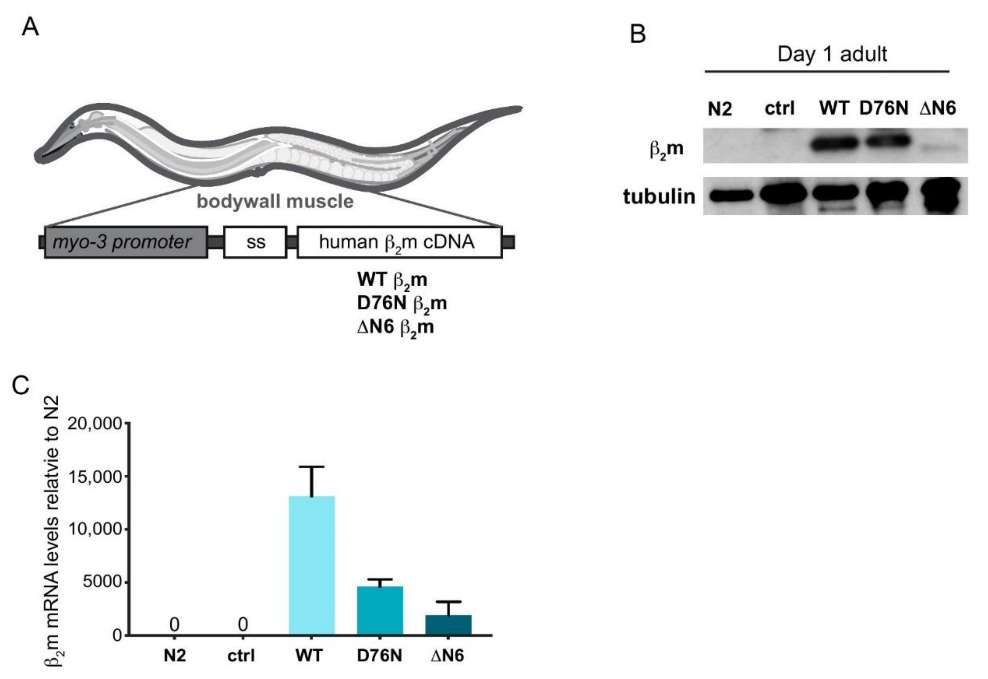
2.1. Generation of C. elegans Models Expressing Human β2m Variants

To model β2m proteotoxicity in vivo we expressed human WT β2m, and its variants ΔN6 β2m and D76N β2m in the C. elegans bodywall muscle under control of the myo-3p promoter (Figure 1A). We generated untagged versions of the three β2m transgene variants to avoid known solubility issues of a fluorescent chromophore tag such as GFP [35], as well as potential issues with secretion of the amyloid protein. In each transgenic strain, the human 20 amino acid β2m signal sequence (ss) was included to enable β2m insertion into the ER and to target it for secretion, to mimic the natural extracellular location of the protein. (Figure 1A).
Expression of β2m protein was analysed by Western blot in age-synchronised Day 1 adult nematodes. As shown in Figure 1B, ΔN6 β2m protein levels were lower compared with protein levels of the WT and the D76N β2m variant (Figure 1B). To investigate whether this is reflected by comparable transcriptional expression levels, we measured β2m transcripts by quantitative real time polymerase chain reaction (qRT PCR) (Figure 1C). mRNA levels of the ΔN6 β2m variant was 6-fold lower relative to WT β2m, (Figure 1C), suggesting the lower ΔN6 β2m protein level correlates with a lower level of transcription (Figure 1B).
Human β2m WT and the D76N β2m variant expressed in C. elegans show the same molecular weight as recombinant WT β2m which lacks the signal sequence (Figure 1B and Supplementary Figure S1A,B), indicating that the ~2.5 kDa β2m signal sequence is cleaved inside the ER, resulting in the expected ~11 kDa molecular weight of the mature β2m sequence. For ΔN6, the loss of the N-terminal 6 residues causes a decrease in mass of ~0.72 kDa, which results in a higher mobility on SDS-PAGE Supplementary (Figure S1A,B). Importantly, this protein also migrates with a mobility equivalent to that of the recombinant protein lacking the signal sequence, suggesting complete cleavage of this protein also in vivo to the mature protein sequence.

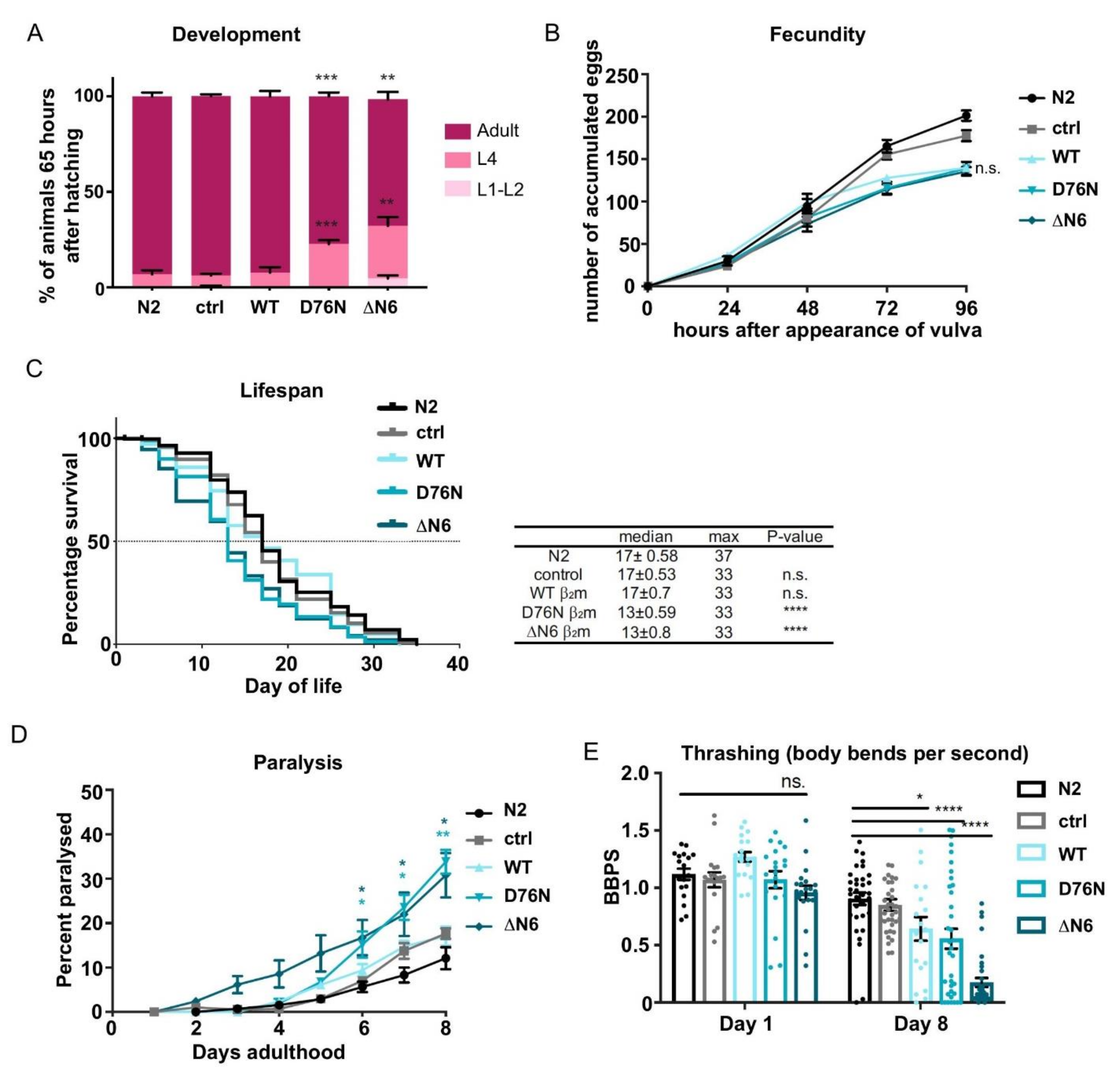
2.2. Expression of D76N β2m and ΔN6 β2m Variants Delays Development and Reduces Lifespan

To investigate whether the expression of the amyloidogenic β2m variants, D76N β2m and ΔN6 β2m, results in toxic behavioural phenotypes we evaluated characteristic measures representing C. elegans health, such as development, brood size and lifespan.
The majority of animals expressing WT β2m or the control strains, N2 Bristol and animals expressing the co-injection marker myo-2p::mCherry (named “ctrl”), reach reproductive adulthood 65 h after hatching (Figure 2A). Only a small percentage of N2 and myo-2p::mCherry animals (6.8 ± 2.0%) as well as WT β2m expressing nematodes (8.4 ± 2.3%) remained at the L4 larval stage at this timepoint. By contrast, the expression of the amyloidogenic β2m variants D76N and ΔN6 resulted in a developmental delay with 22.7% ± 2.0 and 32.3% ± 4.6 of animals remaining at the L4 stage, respectively coherent with previous studies [32]. However, we did not observe embryonic lethality of the D76N β2m mutant strain as previously reported, which could be due to differences in the signal sequence being used [33] (Figure 2A).
Another measure of animal health is fecundity and we explored whether expression of the amyloidogenic β2m variants affected C. elegans brood size. For this, we quantified the number of eggs laid from a single hermaphrodite during the first 96 h of adult life at 20 °C. Control animals accumulated an average of 200 eggs within the 96-h time frame (Figure 2B). However, animals expressing WT β2m, D76N β2m or ΔN6 β2m produced 30% less eggs compared to the control, albeit not statistically significant (Figure 2B). Thus, expression of WT β2m as well as D76N β2m or ΔN6 β2m impair C. elegans fecundity, while development is only affected by expression of the highly amyloidogenic variants D76N β2m and ΔN6 β2m.
Finally, we explored how expression of the β2m variants influences aging by performing life-span assays (Figure 2C). Wild-type (N2) animals have a median lifespan of 17 ± 0.58 days, and this was unaffected in control strains (17 ± 0.53 days) and C. elegans expressing WT β2m (17 ± 0.70 days) (Figure 2C). By contrast, a decrease in median lifespan was observed in transgenic animals expressing the amyloidogenic β2m variant D76N (13 ± 0.59 days) or the ΔN6 β2m variant (13 ± 0.80 days).
Taken together our results show that the highly amyloidogenic β2m variants (D76N and ΔN6) increase organismal toxicity by impairing development, fecundity and lifespan, whereas expression of WT β2m leads to less severe toxic effects.

2.3. Expression of β2m Variants in the C. elegans Body Wall Muscle Reduces Motility

We next investigated whether the reduced lifespan of animals expressing D76N β2m and ΔN6 β2m correlate with reduced healthspan by assessing C. elegans motility as a function of age. To address this, the paralysis of transgenic animals was measured for 8 days. At day 8 of adulthood, 17% of WT β2m animals were paralysed, comparable with control animals (N2 Bristol) or ctrl animals expressing the myo-2p::mCherry co-injection marker only (Figure 2D). Nematodes expressing the D76N β2m or the ΔN6 β2m variant showed a significant increase in paralysis, which was already prevalent at Day 6 of adulthood (15%; p < 0.05) and progressed to 34% in D76N β2m expressing worms and 31% in ΔN6 β2m expressing Day-8 adults (Figure 2D; p < 0.01). Interestingly, animals expressing ΔN6 β2m, started to display paralysis already at day 3 of adulthood: a much earlier display of a phenotype compared with the D76N β2m variant. Thus, the expression of D76N β2m and ΔN6 β2m is proteotoxic to muscle cells, leading to increased paralysis in C. elegans in an age-dependent manner.
To assess the functionality of muscle cells in more detail, we measured the frequency of body bends (thrashing) throughout aging. Control animals N2 Bristol and myo-2p::mCherry expressing nematodes (ctrl) displayed an average of 1.12 ± 0.05 body bends per second (BBPS) and 1.07 ± 0.07 BBPS, respectively, at Day 1 of adulthood, and this decreased to 0.90 ± 0.05 BBPS and 0.85 ± 0.06 BBPS, respectively, in Day 8 adults (Figure 2E). WT β2m or D76N β2m expressing strains performed on average 1.27 ± 0.04 BBPS and 1.07 ± 0.08 BBPS at Day 1 of adulthood, respectively, which was not significantly different to control animals. This thrashing rate then significantly decreased in day 8 adults expressing WT β2m (0.65 ± 0.04; p < 0.05) or D76N β2m (0.56 ± 0.08 BBPS; p < 0.0001) (Figure 2E). Expression of ΔN6 β2m resulted in slightly (but not statistically significant) decreased thrashing rates in Day-1 adults (0.93 ± 0.10) compared with the control, and this then further decreased to the lowest thrashing rate of all animals (0.18 ± 0.04 BBPS; p < 0.0001) at Day 8 of adulthood (Figure 2E). These results indicate that the expression of the β2m variants ΔN6 β2m and D76N β2m, and to a lesser extent, WT β2m, in the body wall muscle of C. elegans is highly proteotoxic, reflecting the higher aggregation rate of both variants in vitro [36].

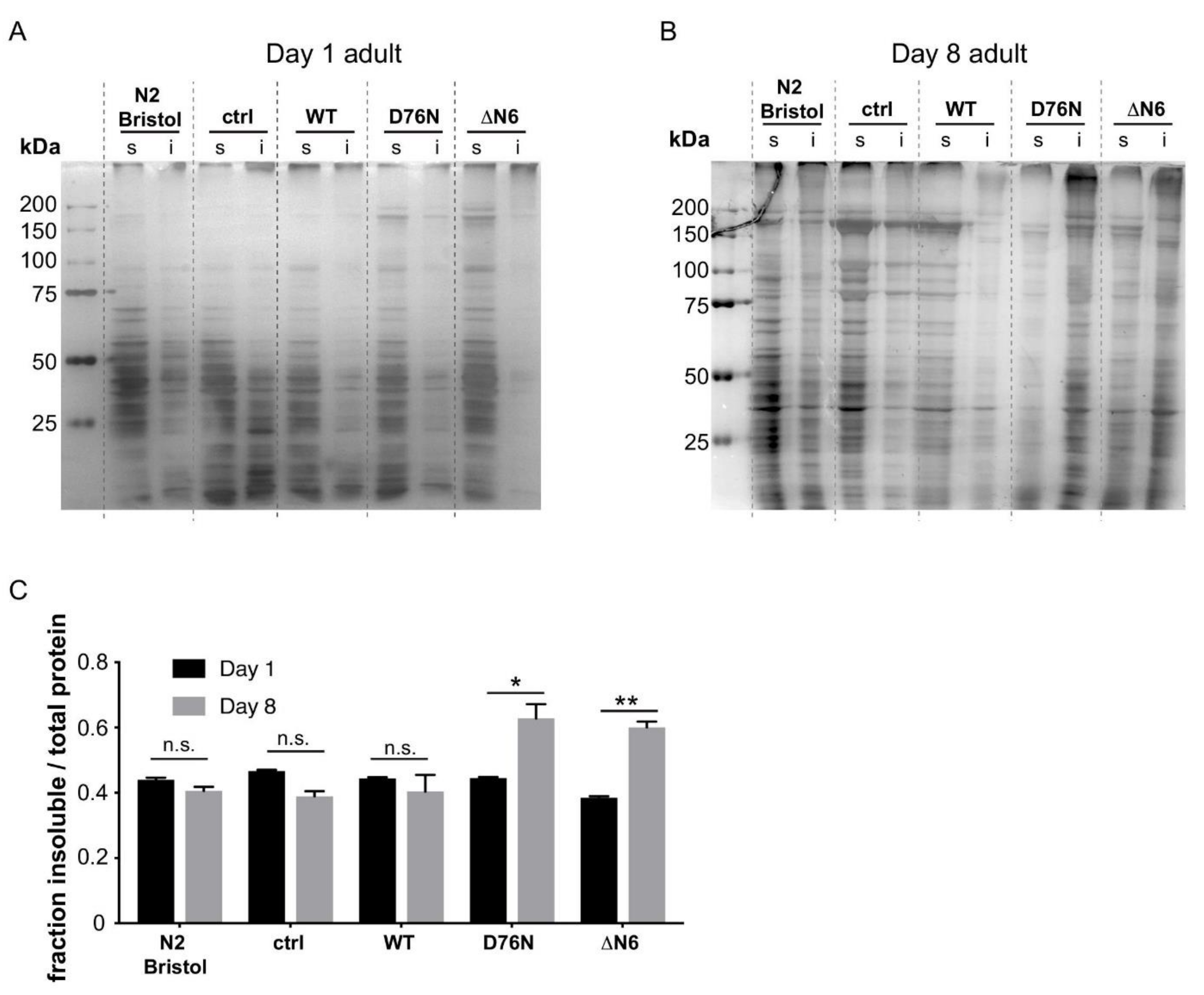
2.4. Expression of the D76N β2m and ΔN6 β2m Variants Leads to Wide-Spread Endogenous Protein Aggregation

To investigate whether the increased organismal toxicity observed for transgenic C. elegans expressing the D76N β2m and ΔN6 β2m variants could be caused by an increased load of misfolded proteins, we assessed the soluble and insoluble fractions of total protein extracts of young (Day 1) and aged (Day 8) adults (Figure 3A,B). Silver staining was used to visualise the total amount of protein present in each sample. In lysates of Day 1 adults, a low proportion of total protein was found in the insoluble fraction in all samples (Figure 3A). By contrast, lysates prepared from Day-8 adults displayed an ~30% higher fraction of insoluble protein in D76N β2m and ΔN6 β2m samples compared with WT β2m (Figure 3B,C). The results demonstrate that the expression of the amyloidogenic β2m variants D76N β2m and ΔN6 β2m cause widespread aggregation of endogenous C. elegans proteins in an age-dependent manner. This suggests that the increased aggregation rate of both β2m variants disrupts the protein folding environment in vivo, leading to increased global protein aggregation and toxic behavioural phenotypes.

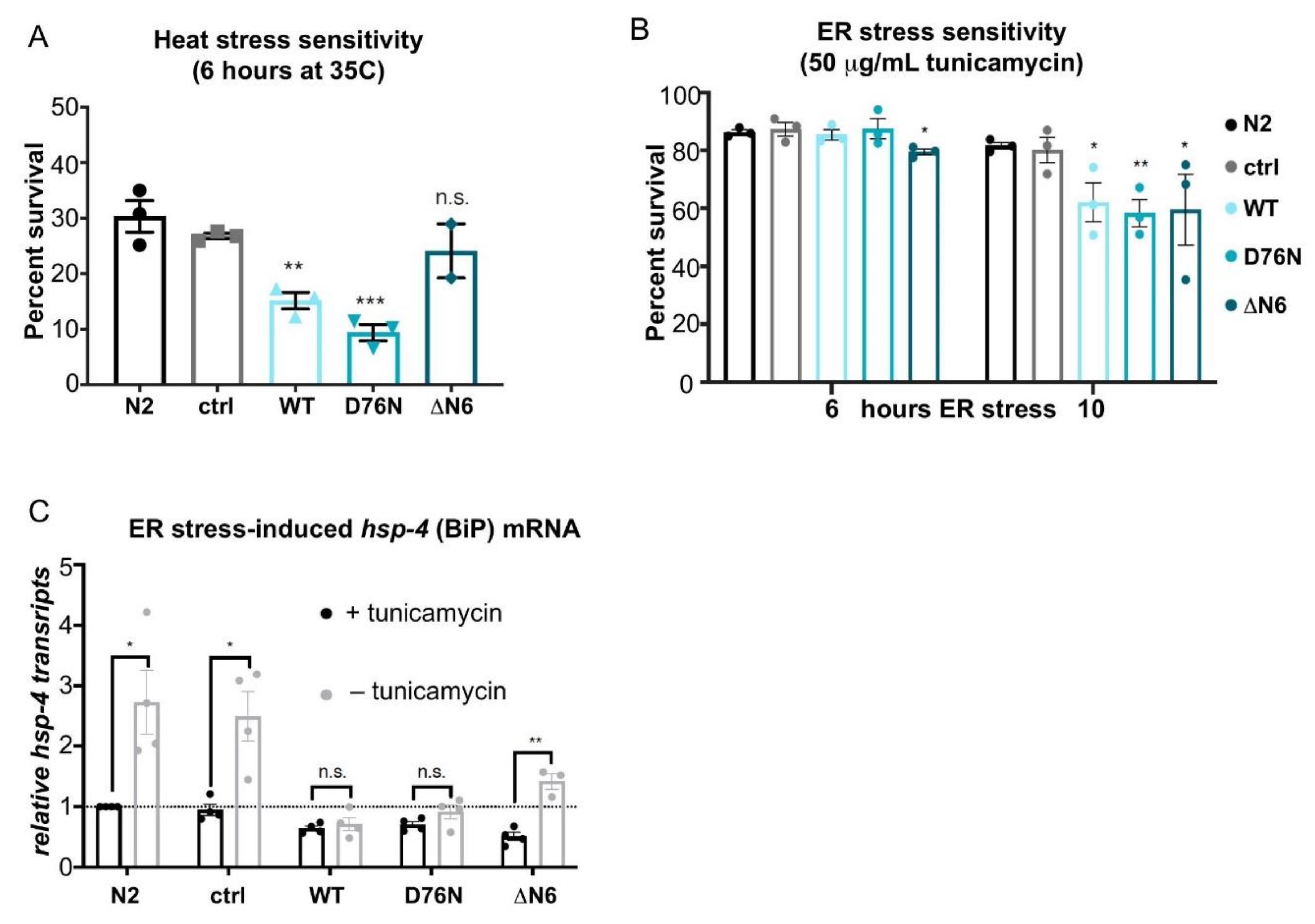
2.5. Expression of Amyloidogenic β2m Variants Impair Cellular Stress Responses

The proteostasis network (PN) has several stress response pathways to cope with the increased burden of misfolded and aggregated proteins throughout aging and acute environmental stresses [3,21,37]. Because expression of the highly amyloidogenic β2m variants D76N β2m and ΔN6 β2m, increases proteotoxicity, we questioned whether potential failure of the HSR or the UPR could underlie the cytotoxicity caused by expression of the amyloidogenic β2m variants.
The ability of animals to cope with heat stress was first investigated. After a 6-h heat shock at 35 °C, C. elegans control strains (N2) and myo-2p::mCherry expressing animals displayed a survival rate of 30 ± 2.9% and 27 ± 0.5%, respectively (Figure 4A). By comparison, nematodes expressing WT β2m or D76N β2m were severely affected, with WT β2m animals showing a survival rate of only 15 ± 1.5% (p < 0.01) and D76N β2m expressing animals surviving at a rate of 9 ± 1.5% (p < 0.001) (Figure 4A). The expression of ΔN6 β2m only slightly reduced C. elegans resistance to heat shock, with 24 ± 4.9% of animals surviving the 6-h HS treatment, albeit not statistically significant compared with controls (Figure 4A).
The β2m variants expressed in the transgenic C. elegans strains are processed in the ER before secretion via the Golgi apparatus. We therefore investigated whether ER stress impairs C. elegans’ ability to cope with an increased burden of misfolded proteins. To induce ER stress, we treated C. elegans with tunicamycin, an inhibitor of N-linked glycosylation that disrupts protein maturation and induces the UPR in the ER [38]. Day 1 adults of control nematodes, N2 and myo-2p::mCherry treated with tunicamycin for a 10-h period showed a survival rate of 81.6% (±1.2) and 80.2% (±4.4) respectively (Figure 4B). By contrast, WT β2m animals showed a significantly reduced survival rate of 62.1% ± 6.8 (p < 0.05) and the D76N β2m and ΔN6 β2m variants a slightly lower survival rate of 58.4% (±4.7) (p < 0.01) and 59.5% (±12.3) (p < 0.05), respectively (Figure 4B). Thus, the expression of β2m disrupts the organism’s ability to deal with ER stress.
Finally, in order to gain further understanding of this observation, induction of the ER-resident Hsp70 chaperone BiP (C. elegans hsp-4) that is upregulated upon ER stress in an IRE-1- and XBP-1- dependent manner was explored [23,39]. Using quantitative RT-PCR, mRNA levels of hsp-4 were measured during basal conditions and ER stress (Figure 4C). Basal hsp-4 transcript levels were reduced in all three β2m expressing strains (Figure 4C). While ER stress resulted in a 2.7-fold (±0.5) upregulation of hsp-4 transcript levels in wild-type (N2) animals and a 2.5-fold (±0.4) induction in the myo-2p::mCherry control strain, no hsp-4 induction was measured in WT β2m and D76N β2m expressing strains (Figure 4C). Interestingly, hsp-4 transcripts were induced 1.4-fold (±0.1) following ER stress in the ΔN6 β2m strain (p < 0.01) (Figure 4C) but remained low compared with control strains. Thus, the impaired ability of the β2m expressing strains to cope with ER stress is reflected in the reduced induction of hsp-4 during basal conditions and in response to ER stress. Although animals expressing the ΔN6 β2m variant retain the ability to mount an UPR, the magnitude of hsp-4 induction is significantly lower when compared with control strains (Figure 4C). In summary, these results demonstrate that cellular stress responses such as the UPRER and the HSR are impaired in young adults, with β2m expression likely disrupting ER homeostasis already at physiological conditions.

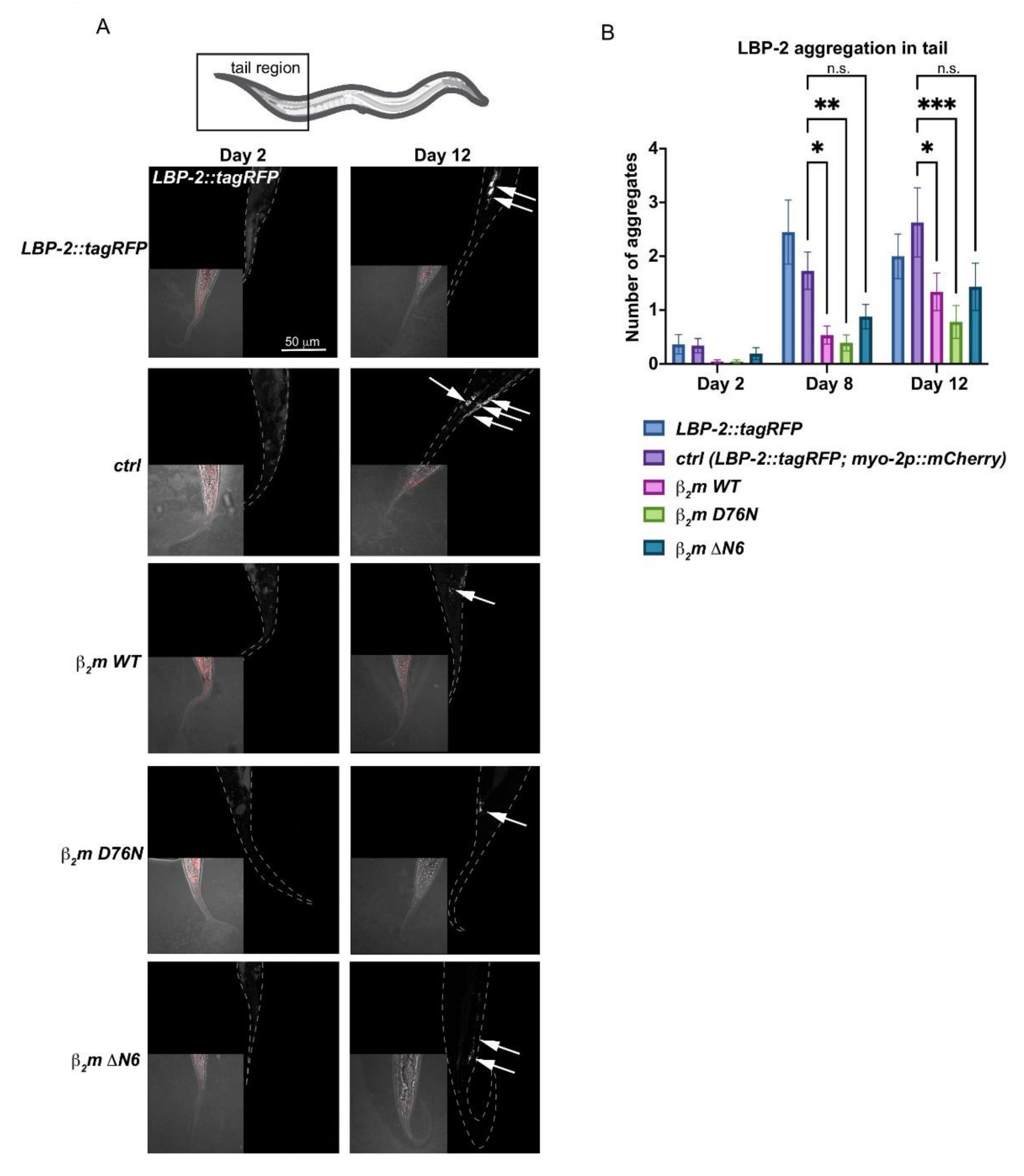
2.6. Expression of β2m Variants Impacts Protein Secretion

One of the ER main functions is to serve as an entry point into the secretory pathway. To investigate whether β2m is still able to be secreted into the extracellular space with disrupted ER homeostasis, we took advantage of the extracellular proteostasis reporter lipid binding protein-2 (LBP-2) fused to tagRFP. LBP-2::tagRFP is secreted from bodywall muscle cells and aggregates in an age-dependent manner in the pseudocoelom [27]. We hypothesised that if the highly amyloidogenic β2m variants indeed are secreted, then this would increase the aggregation of LBP-2::tagRFP in the pseudocoelom.
In wild-type and control animals expressing the pharyngeal co-injection marker, the number of LBP-2::tagRFP foci in the pseudocoelom increases from an average of ~0.3 foci in Day 2 young adults to an average of ~2.5 aggregates in Day 12 adults (Figure 5A,B), coherent with previous data [27]. Surprisingly, the number of extracellular LBP-2 foci is reduced by 50% in aged WT β2m, D76N β2m and ΔN6 β2m variants compared with controls (Figure 5B). This indicates that protein secretion is less efficient in animals expressing WT β2m, D76N β2m and ΔN6 β2m. As both amyloidogenic variants (D76N and ΔN6) show increased endogenous protein aggregation and all β2m-expressing animals have an impaired ER stress response, a possible explanation is that β2m protein misfolds in the ER lumen in an age-dependent manner, before it is secreted. This disrupts basic ER functions including degradation of misfolded proteins and impairs protein secretion of LBP-2, similar to ire-1 and xbp-1 variants of C. elegans that show a defective secretory protein metabolism [24].

3. Discussion

In this study we provide a comparison of transgenic C. elegans expressing human WT β2m with the familial variants D76N β2m and ΔN6 β2m, all of which are involved in β2m-associated amyloid pathology [4,7,8,40]. Consistent with previously generated C. elegans β2m models [32,33], our strains (which each contain the natural human signal sequence) show increased proteotoxicity at an organismal level in an age-dependent manner. The novelty of our work is that it provides molecular insight into the factors contributing to increased proteotoxicity, which was largely unstudied previously. We show that the highly amyloidogenic β2m variants D76N β2m and ΔN6 β2m cause global aggregation of endogenous C. elegans proteins at an advanced age, correlating with the increased cytotoxicity of these variants. Albeit ΔN6 β2m expression levels are low in young adults, the insoluble protein fraction of ΔN6 β2m expressing nematodes at old age (Day 8) is comparable to that in D76N β2m mutants. This suggests that despite the lower expression levels of ΔN6 β2m this variant may cause increased aggregation of endogenous proteins in vivo, leading to the observed age-associated proteotoxicity (reduced lifespan, paralysis) that is comparable with the D76N mutant. Thus, even though D76N shows a faster aggregation rate in vitro its proteotoxicity may be superseded by the ΔN6 variant in vivo. Interestingly, all β2m variants impair the capacity of the proteostasis network by affecting the animals’ ability to cope with stress conditions. The disrupted ER homeostasis in all β2m-expressing nematodes may be linked to reduced secretion of LBP-2, suggesting that both highly aggregation-prone β2m variants misfold intracellularly, possibly even inside the ER lumen, thereby impairing ER secretory-protein metabolism in C. elegans.
Similar to other C. elegans β2m models previously established, our models recapitulate reduced lifespan, motility and developmental delays. A previously generated C. elegans model expressing D76N β2m by Faravelli et al. displayed embryonic lethality that was overcome by the inducible smg-1 system [33]. While expression of the D76N β2m and ΔN6 β2m variants in our study caused developmental delays, we did not observe increased embryonic lethality associated with either variant. A potential reason for this could be differences in the expression levels of D76N β2m, as the here generated strain expresses D76N β2m from an extrachromosomal rather than an integrated array in a smg-1 mutant strain that requires increased temperature conditions to induce expression of the transgene. Moreover, Faravelli et al., used the endogenous sel-1 peptide as a secretion signal, whereas our generated version contains the natural human β2m signal peptide, which could lead to different secretion ratios.
The accumulation of insoluble endogenous protein deposits combined with a debilitated UPRER already in young animals, indicates an early impairment of the proteostasis network by the D76N β2m and ΔN6 β2m variants. While all β2m expressing nematodes (WT, D76N and ΔN6) are sensitive to ER stress, it is interesting to note that the heat stress resistance of ΔN6 β2m mutants is comparable to control animals, whereas both WT β2m and D76N β2m are sensitive to heat stress. A possible reason for this discrepancy could be that all three β2m variants are targeted to the ER, where they misfold, thereby more severely affecting ER proteostasis. While ΔN6 in particular may be more severely affecting ER proteostasis, it should be noted that expression levels of this variant are also very low. Hence a much lower fraction of misfolded ΔN6 may reach the cytosol where it could impact on cytosolic proteostasis that is challenged by heat stress.
Interestingly, all β2m variants are impaired in their ability to induce hsp-4 in response to ER stress. Although the ΔN6 mutant shows a slight induction of hsp-4, the magnitude of induction is severely reduced and does not reach the level necessary to protect against ER stress as it does in the control animals. This inability to induce a protective ER stress response by upregulation of hsp-4/BiP, could be due to an increased oligomeric fraction as was previously reported for ΔN6 β2m [32] and D76N β2m [33].
In future studies, it will be interesting to investigate whether β2m expression in these previously established models exhibit a defective UPRER as well. Based on our observations suggesting reduced secretion in all β2m variants, it is possible that β2m oligomers already form in the ER lumen, thereby affecting both the UPRER and ER-associated degradation (ERAD) before they are secreted. Further work will be required to obtain direct evidence for secretion of β2m oligomers/aggregates and/or accumulation of β2m in the extracellular environment, as staining of amyloid deposits in the animal by antibody staining or the use of amyloid-reactive dyes such as X-34 or ThT proved inconclusive (data not shown). However, we provide clear evidence that accumulation of LBP-2 in the pseudocoelom decreases with age in all variants consistent with reduced protein secretion, which is a known characteristic for UPRER pathway variants of the IRE-1 branch [24]. In this respect, it is however important to note differences in the secretion of β2m in C. elegans compared to human cells. In human cells β2m is secreted as part of MHC I before it dissociates to form amyloid species in the extracellular space [41,42]. C. elegans does not possess MHC I [43], and directly expresses amyloidogenic forms of β2m in these strains. Thus, it is likely that in C. elegans proteostasis is first affected intracellularly, before β2m oligomeric species are secreted. Such a scenario would explain the reduced capability of β2m-expressing animals to induce the UPRER even at basal conditions. Moreover, our data shows that the signal sequence of ΔN6 β2m is successfully removed in the ER lumen in our C. elegans model, as the MW of the protein on Western blot is identical to that of recombinant protein (Figure 1B and Supplementary Figure S1A,B). Our data suggests that ΔN6 already misfolds in the ER, as indicated by the increased toxicity and sensitivity to ER stress and reduced lifespan of the mutant strain.
Despite the above limitations of C. elegans as a model for β2m-associated amyloidosis since it is devoid of MHC I, it is nevertheless a good system to study molecular and organismal aspects of β2m amyloidosis. For example, C. elegans harbours organs and tissues affected by D76N β2m amyloidosis pathology, such as the intestinal tract and muscle tissue of the pharynx, the pumping function of which is often compared to the human heart. This can allow comparisons to the human pathology where the wide-spread concentration of D76N β2m amyloid deposits into heart or the gastro-intestinal tracts leads to life-threatening complication [14,44]. Moreover, collagen as well as glycosaminoglycans which are both important drivers of β2m amyloid aggregation [12,45,46,47] are highly present in C. elegans muscle tissues [48].
In summary, the transgenic C. elegans expressing β2m variants generated here and in previous studies [32,33] will prove important model systems to gain a robust molecular insight into oligomer formation and understanding of the molecular causes of proteotoxicity in vivo. Our work highlighted the importance of the proteostasis network and a disrupted ER homeostasis combined with increased endogenous protein aggregation as drivers of β2m associated toxicity in vivo. A focus in future studies on proteostasis network mechanisms and secretory protein metabolism will further increase our understanding of these diseases in vivo and in the quest of finding potential therapeutic avenues.

4. Material and Methods

4.1. C. elegans Strains and Maintenance

Wild type (N2 Bristol) and transgenic animals were handled using standard methods [49]. Unless otherwise stated, strains were maintained at 20 °C on 60 mm NGM agar plates seeded with OP50-1 Escherichia coli as a food source, according to [49].

4.1.1. Strains Generated in This Study:

  • PVH69 pccEx007[myo3::WT hβ2m::GFP::unc-54 3′UTR + myo2::RFP];
  • PVH177 pccEx009[myo-3p::WT hβ2m::unc-54 3′UTR + myo-2p::RFP];
  • PVH178 pccEx010[myo-3p::D76N β2m::unc-54 3′UTR + myo-2p::RFP];
  • PVH179 pccEx011[myo-3p::ΔN6 β2m::unc-54 3′UTR + myo-2p::RFP];
  • PVH182 pccEx024[myo-2p::RFP].

4.1.2. Cloning and Generation of Transgenic C. elegans Strains

C. elegans strains expressing β2m in the bodywall muscle under control of the myo-3p promoter were constructed in the following manner: The expression vector pPD95_75 (Fire lab, Addgene, Watertown, MA, USA) was digested with AgeI and EcoRI, blunt-ended and re-ligated to generate an expression vector without the GFP cDNA. The 1500 bp myo-3p body-wall muscle promoter (synthesized by Eurofins, Wolverhampton, United Kingdom) was ligated into the pPD95_75 vector using HindII and SalI sites. The signal sequence for human WT β2m was generated by oligonucleotide annealing of SalI-signalpeptideB2m-for and BamHI-signalpeptideB2m-rev. Oligos were mixed in a 1:1 ratio in 10 μL, then placed at 94 °C and allowed to cool until 30 °C. Serial dilutions of the oligos were made (1:5, 1:10, 1:50, 1:100). 5 μL of the undiluted oligos and the serial dilutions were ligated into the successfully sequenced myo-3p-pPD95_75 vector. Ligations were transformed into DH5α cells. Colony restriction digestion was performed using BamHI and SalI restriction enzymes and analysed for a 100 bp band, successful constructs were sent for sequencing. Finally, human WT, D76N β2m and ΔN6 β2m cDNA (Radford lab) were PCR amplified using primers BamHI-B2m-for and MscI-B2m-rev to contain BamHI and MscI restriction sites and ligated into the myo-3p-ss-pPD95_75 vector. Successful colonies containing a 367 bp β2m band were sent for sequencing.

4.2. Primers Used

  • BamHI-B2m-for
  • 5′ AAAGGATCCATTCAAAGAACTCCAAAAATTC 3′
  • MscI-B2m-rev
  • 5′ AAATGGCCATTACATGTCTCGATCCCACTTA 3′
  • SalI-signalpeptide B2m-for
  • 5′ TCGACAAAAATGTCTCGCTCCGTGGCCTTAGCTGTGGCCTTAGCTGTGCTC GCGCTACTCTCTCTTTCTGGCCTGGAGGCTG 3′
  • BamHI-signalpeptide B2m-rev
  • 5′ GATCCAGCCTCCAGGCCAGAAAGAGAGAGTAGCGCGAGCACAGCTAAG GCCACGGAGCGAGACATTTTTG 3′
The transgenes were microinjected into the gonads of the N2 Bristol C. elegans strain, using a DNA solution containing either the co-injection marker only (5 ng/µL; myo-2p::mCherry (pCFJ90; Addgene)), or in combination with 25 ng/µL of the β2m construct. Multiple extrachromosomal lines were generated, based on the red fluorescent pharyngeal co-injection marker. The established transmitting lines used in this study have ~70% meiotic stability.

4.3. Western Blot Analysis and SDS-PAGE Silver Staining

For Western blot analysis, nematodes were collected and lysed on ice using a motorised pestle in lysis buffer (150 mM NaCl, 50 mM Tris pH 7.4, 1 mM EDTA, 0.1% (v/v) NP-40). Extracts were centrifuged at 13,000 rpm for 1 min to pellet the carcasses and the supernatant collected. The protein extract was run on a 10% (w/v) Tris-Tricine gel and proteins were electro-transferred to 0.22 μM nitrocellulose membranes. The blots were blocked using 5% (w/v) milk overnight, washed four times in PBS/Tween (0.1%) for 5 min, then probed with primary anti-human β2m rabbit polyclonal antibody (Dako, Santa Clara, CA, USA) at a 1:1000 (v/v) dilution in PBS/Tween (0.1%) and 1% (v/v) milk for 2 h. Blots were washed then probed with 1:10,000 (v/v) dilution HRP-conjugated goat anti-rabbit secondary antibody for 1 h. Blots were developed using ECL (ThermoFisher Scientific, Waltham, MA, USA).
For silver staining of SDS-PAGE, nematode extracts were loaded on an 4–20% (w/v) SDS-PAGE gel (BioRad, Hercules, CA, USA). The gel was fixed in solution A (50% (v/v) ethanol, 10% (v/v) acetic acid) for 30 min, then incubated in solution B (5% (v/v) ethanol, 1% (v/v) acetic acid) for 15 min on a gentle rocker. The gel was washed in dH2O three times for 5 min, and then incubated in solution D (0.02 g sodium thiosulphate in 100 mL dH2O) for 2 min while gently rocking. The gel was washed again in dH2O three times for 30 s, then incubated in solution E (0.2 g silver nitrate, 75 μL formaldehyde in 100 mL ddH2O) for 20 min. After three washes in dH2O for 20 s, the gel was developed in solution F (6 g sodium carbonate, 50 μL formaldehyde, 0.2 mL solution C in 100 mL dH2O) until protein bands could be visualised and the reaction was stopped using solution G (5% (v/v) acetic acid). The gel was then stored in dH2O and imaged.

4.4. Soluble/Insoluble Protein Fractionation

Animals were aged to day 1 or day 8 adults using NGM containing 50 µg/uL FUdR plates. Nematodes were collected using M9 buffer, and resuspended in lysis buffer (20 mM Tris, pH 7.5, 10 mM β-mercaptoethanol, 0.5% (v/v) Triton X-100, complete protease inhibitor (Roche, Basel, Switzerland)). Extracts were shock frozen in liquid nitrogen, and the frozen nematode pellet was ground using a motorized pestle, then lysed on ice in the presence of 0.0025 U/mL benzonase (Merck SA, Darmstadt, Germany). Lysates were centrifuged at 1000 rpm for 1 min in a tabletop centrifuge and the supernatant was extracted. 2% (v/v) N-Lauroylsarcosine was added to the supernatant and was ultracentrifuged at 100,000 g for 1 h at 4 °C. The supernatant was removed, and the resulting pellet was resuspended in the same volume of dH2O as the supernatant.

4.5. Developmental Delay Assay

Nematodes for the assay were synchronised by placing ten gravid adult nematodes on an NGM plate with OP50-1 as a food source and allowed to lay eggs for 2 h before removing. Eggs were left to develop for 65 h and the developmental stage was monitored and recorded. Three independent experiments were performed for each strain (n > 200 worms per experiment

4.6. Fecundity Assay

Thirty L4-stage nematodes were transferred onto separate individual NGM plates smented with OP50-1. At time-points of 24-, 48-, 72- and 96- hours, individual animals were transferred onto a new NGM plate using a sterilised platinum wire, and the number of eggs laid over the previous 24 h was counted and recorded. Three independent experiments were performed for each strain (n = 30 worms per experiment).

4.7. Lifespan Assay

Animals were synchronised by egg-laying, and 80–100 L4-stage animals were selected per strain and transferred onto five NGM plates with 20 animals per plate. Every other day each nematode was assessed for survival, death or censorship and numbers were recorded. Animals were assessed as dead if they did not display movement when gently touched on the nose using a platinum wire, and no pharyngeal pumping could be observed. Animals were censored if they crawled up the edge of the plate and became desiccated, burrowed into the agar, displayed the ‘bag of worms’ phenotype where eggs hatched internally, or if they could not be found. Any dead or censored animals were recorded and left on plates, while alive animals were transferred onto a new NGM plate. Data was analysed using the OASIS 2 online software [50]

4.8. Paralysis Assay

Animals were synchronised using egg-laying methods, and 100 L4-stage nematodes were transferred onto five NGM plates with OP50-1 (20 animals per plate). Every 24 h for 8 days, each nematode was assessed by the touch-nose response for paralysis and the numbers of mobile, paralysed or censored animals were recorded. Mobile nematodes were determined if they were able to move freely, either when touched by a platinum wire, or when unprompted. Paralysed animals were determined if little movement was seen when touched by a platinum wire, but still displayed minimal movement or pharyngeal pumping to indicate they were still alive. Censored worms included worms that had died, displayed the ‘bag of worms’ phenotype, crawled up the side of the plate, or could not be detected. Mobile worms were moved onto a new plate each day to ensure any progeny did not influence the results. Three independent experiments were performed for each strain (n = 100 worms per experiment).

4.9. Thrashing Assay

Animals were synchronised by egg-laying and aged to either day 1 or day 8 of adulthood by picking. At the appropriate time point, 15–20 nematodes of each strain were transferred into a 24-well plate containing 1 mL M9 buffer. Animals were left to recover for 1 min, then a video was recorded for 30 s using the ToupCam digital camera (GT vision, Sudbury, United Kingdom). At least three videos of each strain were recorded. Videos were analysed in ImageJ using the WrmTrck plugin [51] to quantify the number of body bends per second (BBPS). Each experiment consisted of 15–20 worms being recorded three times and average speed of recorded movies was considered one biological replicate. Three biological replicates were collected for each strain.

4.10. Thermotolerance

Progeny were synchronised by egg-laying and 50 L4-stage animals were selected and placed onto an NGM plate. The next day (day 1 adults), plates were incubated at 35 °C for 6 h then moved immediately into a 20 °C incubator. Animals were left to recover for 16 h at 20 °C, then survival of animals was scored. Animals were scored as alive if movement or pharyngeal pumping was observed. The percentage of alive and dead animals were scored and mean survival rates were determined using Student’s t test. Three independent experiments were performed for each strain (n = 50 worms) and error bars indicate S.E.M.

4.11. ER Stress Assay

Nematodes were synchronised by egg-laying and L4-stage animals were selected and placed onto new NGM plates. Nematodes were left overnight to grow and 50 day-1 adult worms were transferred using a sterilised platinum wire into M9 buffer containing 50 μg/mL tunicamycin. Animals were incubated in tunicamycin for 6 h at 20 °C on a rotator. After incubation, nematodes were washed three times in M9 buffer and plated onto NGM plates. After 16 h of recovery, animals were scored for survival. Alive animals were determined by their ability to move spontaneously or when stimulated by touch, or pharyngeal pumping was observed. Dead animals were determined through lack of movement or pharyngeal pumping. Percentages of alive and dead animals were calculated and plotted. Three independent experiments were performed for each strain (n = 50 worms per experiment); error bars indicate S.E.M.

4.12. Quantitative RT-PCR

RNA was extracted using TriZOL reagent and grinding the frozen worm pellet using a pellet grinder on ice 5 × for 30 s. RNA was purified using the Zymo-prep RNA Mini Isolation kit (Zymo Research, Cambridge Biosciences, Cambridge, United Kingdom), and qRT-PCR was performed as described previously [52,53].

4.13. Quantification of Extracellular Aggregation Using LBP-2::tagRFP as a Reporter

To quantify LBP-2::tagRFP aggregation in the tail region of C. elegans, age-synchronised nematodes at indicated time points were immobilised in 10 mM sodium azide on 2% agarose (w/v) pads on a microscope slide. Images were captured on a LSM880 upright laser scanning confocal microscope (Zeiss, Oberkochen, Germany) with a 40 × oil objective, using the Zen software. tagRFP was detected using a 561 nm excitation laser and an emission range from 565–650 nm. Representative confocal images are displayed as maximum z-stack projections.
For aggregation quantification, the tail region was defined as the region from the tip of the tail to the anal sphincter. We chose the tail region because of interference of the myo-2p::mCherry co-injection marker in the head region of β2m and ctrl animals. Aggregates were quantified in a semi-automated process. ImageJ was used to subtract the background and apply a threshold to a maximum intensity projection images. Aggregates were defined as fluorescent puncta when larger than 1 µm2. Two independent experiments were performed for each strain (n = 10–21 worms per condition per experiment) and error bars indicate S.E.M. For statistical significance, a two-way ANOVA followed by a post-hoc Tukey’s test was performed.

Supplementary Materials

The following are available online at https://www.mdpi.com/article/10.3390/ijms221910752/s1.

Author Contributions

Conceptualization, P.v.O.-H. and S.E.R., performance of methodology and analysis, S.C.G. and K.M.D., Supervision, P.v.O.-H. and S.E.R., Writing, Review and Editing of manuscript, P.v.O.-H., S.E.R., S.C.G. and K.M.D. Funding Acquisition, P.v.O.-H. and S.E.R. All authors have read and agreed to the published version of the manuscript.

Funding

We are grateful to funding from the NC3Rs (NC/P001203/1) to P.v.O-H., the Wellcome Trust (204963) to S.E.R., the BBSRC-Whiterose doctoral training partnership (BB/M011151/1) to S.C.G. and a MRC DiMen doctoral training partnership (MR/N013840/1) to K.M.D. We would like to acknowledge funding from the Wellcome Trust (104918MA) to the Leeds BioImaging Facility.

Institutional Review Board Statement

Not applicable.

Informed Consent Statement

Not applicable.

Acknowledgments

The N2 Bristol C. elegans strain used in this research was provided by the Caenorhabditis Genetics Center, which is funded by the NIH Office of Research Infrastructure Programs (P40 OD010440). We would like to thank Della David (Babraham Institute, Cambridge, UK) for sharing the LBP-2::tagRFP strain. We are grateful for access to and technical support at the BioImaging Facility at the Faculty of Biological Sciences.

Conflicts of Interest

The authors declare no conflict of interest.

References

  1. Knowles, T.P.J.; Vendruscolo, M.; Dobson, C.M. The amyloid state and its association with protein misfolding diseases. Nat. Rev. Mol. Cell Biol. 2014, 15, 384–396. [Google Scholar] [CrossRef] [PubMed]
  2. Hipp, M.S.; Park, S.-H.; Hartl, F.U. Proteostasis impairment in protein-misfolding and -aggregation diseases. Trends Cell Biol. 2014, 24, 506–514. [Google Scholar] [CrossRef]
  3. Hipp, M.S.; Kasturi, P.; Hartl, F.U. The proteostasis network and its decline in ageing. Nat. Rev. Mol. Cell Biol. 2019, 20, 421–435. [Google Scholar] [CrossRef] [PubMed]
  4. Iadanza, M.G.; Jackson, M.P.; Hewitt, E.W.; Ranson, N.A.; Radford, S.E. A new era for understanding amyloid structures and disease. Nat. Rev. Mol. Cell Biol. 2018, 19, 755–773. [Google Scholar] [CrossRef]
  5. Smith, D.P.; Radford, S.E. Role of the single disulphide bond of beta(2)-microglobulin in amyloidosis in vitro. Protein Sci. 2001, 10, 1775–1784. [Google Scholar] [CrossRef]
  6. Isenman, D.E.; Painter, R.H.; Dorrington, K.J. The structure and function of immunoglobulin domains: Studies with beta-2-microglobulin on the role of the intrachain disulfide bond. Proc. Natl. Acad. Sci. USA 1975, 72, 548–552. [Google Scholar] [CrossRef] [PubMed] [Green Version]
  7. Ohashi, K. Pathogenesis of beta2-microglobulin amyloidosis. Pathol Int. 2001, 51, 1–10. [Google Scholar] [CrossRef] [PubMed]
  8. Gejyo, F.; Yamada, T.; Odani, S.; Nakagawa, Y.; Arakawa, M.; Kunitomo, T.; Kataoka, H.; Suzuki, M.; Hirasawa, Y.; Shirahama, T.; et al. A new form of amyloid protein associated with chronic hemodialysis was identified as β2-microglobulin. Biochem. Biophys. Res. Commun. 1985, 129, 701–706. [Google Scholar] [CrossRef]
  9. Esposito, G.; Michelutti, R.; Verdone, G.; Viglino, P.; Ández, H.H.; Robinson, C.; Amoresano, A.; Piaz, F.D.; Monti, M.; Pucci, P.; et al. Removal of the N-terminal hexapeptide from human β2-microglobulin facilitates protein aggregation and fibril formation. Protein Sci. 2000, 9, 831–845. [Google Scholar] [CrossRef]
  10. Eichner, T.; Kalverda, A.P.; Thompson, G.S.; Homans, S.W.; Radford, S.E. Conformational conversion during amyloid formation at atomic resolution. Mol. Cell 2011, 41, 161–172. [Google Scholar] [CrossRef]
  11. Karamanos, T.; Pashley, C.L.; Kalverda, A.P.; Thompson, G.S.; Mayzel, M.; Orekhov, V.Y.; Radford, S.E. A population shift between sparsely populated folding intermediates determines amyloidogenicity. J. Am. Chem. Soc. 2016, 138, 6271–6280. [Google Scholar] [CrossRef] [Green Version]
  12. Myers, S.L.; Jones, S.; Jahn, T.; Morten, I.J.; Tennent, G.A.; Hewitt, E.; Radford, S. A systematic study of the effect of physiological factors on β2-microglobulin amyloid formation at neutral pH. Biochemistry 2006, 45, 2311–2321. [Google Scholar] [CrossRef]
  13. Sarell, C.J.; Woods, L.A.; Su, Y.; Debelouchina, G.T.; Ashcroft, A.E.; Griffin, R.G.; Stockley, P.; Radford, S.E. Expanding the repertoire of amyloid polymorphs by Co-polymerization of related protein precursors. J. Biol. Chem. 2013, 288, 7327–7337. [Google Scholar] [CrossRef] [PubMed] [Green Version]
  14. Valleix, S.; Gillmore, J.D.; Bridoux, F.; Mangione, P.P.; Dogan, A.; Nedelec, B.; Boimard, M.; Touchard, G.; Goujon, J.-M.; Lacombe, C.; et al. Hereditary systemic amyloidosis due to Asp76Asn variant β2-microglobulin. N. Engl. J. Med. 2012, 366, 2276–2283. [Google Scholar] [CrossRef] [PubMed]
  15. Xue, W.-F.; Hellewell, A.; Gosal, W.S.; Homans, S.W.; Hewitt, E.; Radford, S.E. Fibril fragmentation enhances amyloid cytotoxicity. J. Biol. Chem. 2009, 284, 34272–34282. [Google Scholar] [CrossRef] [Green Version]
  16. Milanesi, L.; Sheynis, T.; Xue, W.-F.; Orlova, E.; Hellewell, A.; Jelinek, R.; Hewitt, E.; Radford, S.; Saibil, H.R. Direct three-dimensional visualization of membrane disruption by amyloid fibrils. Proc. Natl. Acad. Sci. USA 2012, 109, 20455–20460. [Google Scholar] [CrossRef] [PubMed] [Green Version]
  17. Goodchild, S.C.; Sheynis, T.; Thompson, R.; Tipping, K.W.; Xue, W.-F.; Ranson, N.A.; Beales, P.A.; Hewitt, E.W.; Radford, S.E. β2-microglobulin amyloid fibril-induced membrane disruption is enhanced by endosomal lipids and acidic pH. PLoS ONE 2014, 9, e104492. [Google Scholar] [CrossRef] [Green Version]
  18. Jakhria, T.; Hellewell, A.L.; Porter, M.Y.; Jackson, M.P.; Tipping, K.W.; Xue, W.-F.; Radford, S.; Hewitt, E.W. β2-microglobulin amyloid fibrils are nanoparticles that disrupt lysosomal membrane protein trafficking and inhibit protein degradation by lysosomes. J. Biol. Chem. 2014, 289, 35781–35794. [Google Scholar] [CrossRef] [Green Version]
  19. Okoshi, T.; Yamaguchi, I.; Ozawa, D.; Hasegawa, K.; Naiki, H. Endocytosed β2-microglobulin amyloid fibrils induce necrosis and apoptosis of rabbit synovial fibroblasts by disrupting endosomal/Lysosomal membranes: A Novel mechanism on the cytotoxicity of amyloid fibrils. PLoS ONE 2015, 10, e0139330. [Google Scholar] [CrossRef] [Green Version]
  20. Balch, W.E.; Morimoto, R.I.; Dillin, A.; Kelly, J.W. Adapting proteostasis for disease intervention. Science 2008, 319, 916–919. [Google Scholar] [CrossRef] [PubMed] [Green Version]
  21. Åkerfelt, M.; Morimoto, R.I.; Sistonen, L. Heat shock factors: Integrators of cell stress, development and lifespan. Nat. Rev. Mol. Cell Biol. 2010, 11, 545–555. [Google Scholar] [CrossRef] [PubMed]
  22. Walter, P.; Ron, D. The unfolded protein response: From stress pathway to homeostatic regulation. Science 2011, 334, 1081–1086. [Google Scholar] [CrossRef] [PubMed] [Green Version]
  23. Calfon, M.; Zeng, H.; Urano, F.; Till, J.H.; Hubbard, S.R.; Harding, H.P.; Clark, S.G.; Ron, D. IRE1 couples endoplasmic reticulum load to secretory capacity by processing the XBP-1 mRNA. Nature 2002, 415, 92–96. [Google Scholar] [CrossRef]
  24. Safra, M.; Ben-Hamo, S.; Kenyon, C.; Henis-Korenblit, S. The ire-1 ER Stress-response pathway is required for normal secretory- protein metabolism in C. elegans. J. Cell Sci. 2013, 126, 4136–4146. [Google Scholar] [CrossRef] [PubMed] [Green Version]
  25. Genereux, J.C.; Qu, S.; Zhou, M.; Ryno, L.M.; Wang, S.; Shoulders, M.D.; Kaufman, R.J.; Lasmezas, C.I.; Kelly, J.W.; Wiseman, R.L. Unfolded protein response-induced ERdj3 secretion links ER stress to extracellular proteostasis. EMBO J. 2015, 34, 4–19. [Google Scholar] [CrossRef]
  26. Chen, J.J.; Genereux, J.C.; Suh, E.H.; Vartabedian, V.F.; Rius, B.; Qu, S.; Dendle, M.T.; Kelly, J.W.; Wiseman, R.L. Endoplasmic reticulum proteostasis influences the oligomeric state of an amyloidogenic protein secreted from mammalian cells. Cell Chem. Biol. 2016, 23, 1282–1293. [Google Scholar] [CrossRef] [Green Version]
  27. Gallotta, I.; Sandhu, A.; Peters, M.; Haslbeck, M.; Jung, R.; Agilkaya, S.; Blersch, J.L.; Rödelsperger, C.; Röseler, W.; Huang, C.; et al. Extracellular proteostasis prevents aggregation during pathogenic attack. Nature 2020, 584, 410–414. [Google Scholar] [CrossRef]
  28. Romine, I.C.; Wiseman, R.L. PERK signaling regulates extracellular proteostasis of an amyloidogenic protein during endoplasmic reticulum stress. Sci. Rep. 2019, 9, 410. [Google Scholar] [CrossRef] [Green Version]
  29. Cornejo, V.H.; Hetz, C. The unfolded protein response in Alzheimer’s disease. Semin. Immunopathol. 2013, 35, 277–292. [Google Scholar] [CrossRef]
  30. Marcora, M.S.; Belfiori-Carrasco, L.F.; Bocai, N.I.; Morelli, L.; Castaño, E.M. Amyloid-β42 clearance and neuroprotection mediated by X-box binding protein 1 signaling decline with aging in the Drosophila brain. Neurobiol. Aging 2017, 60, 57–70. [Google Scholar] [CrossRef]
  31. Zhang, P.; Fu, X.; Sawashita, J.; Yao, J.; Zhang, B.; Qian, J.; Tomozawa, H.; Mori, M.; Ando, Y.; Naiki, H.; et al. Mouse model to study human A β2M amyloidosis: Generation of a transgenic mouse with excessive expression of human β2-microglobulin. Amyloid 2010, 17, 50–62. [Google Scholar] [CrossRef] [PubMed]
  32. Diomede, L.; Soria, C.; Romeo, M.; Giorgetti, S.; Marchese, L.; Mangione, P.P.; Porcari, R.; Zorzoli, I.; Salmona, M.; Bellotti, V.; et al. elegans expressing human β2-microglobulin: A novel model for studying the relationship between the molecular assembly and the toxic phenotype. PLoS ONE 2012, 7, e52314. [Google Scholar] [CrossRef] [PubMed]
  33. Faravelli, G.; Raimondi, S.; Marchese, L.; Partridge, F.A.; Soria, C.; Mangione, P.P.; Canetti, D.; Perni, M.; Aprile, F.A.; Zorzoli, I.; et al. elegans expressing D76N β2-microglobulin: A model for in vivo screening of drug candidates targeting amyloidosis. Sci. Rep. 2019, 9, 19960. [Google Scholar] [CrossRef] [PubMed]
  34. Pokrzywa, M.; Dacklin, I.; Hultmark, D.; Lundgren, E. Misfolded transthyretin causes behavioral changes in a Drosophila model for transthyretin-associated amyloidosis. Eur. J. Neurosci. 2007, 26, 913–924. [Google Scholar] [CrossRef] [PubMed]
  35. Ochiishi, T.; Doi, M.; Yamasaki, K.; Hirose, K.; Kitamura, A.; Urabe, T.; Hattori, N.; Kinjo, M.; Ebihara, T.; Shimura, H. Development of new fusion proteins for visualizing amyloid-β oligomers in vivo. Sci. Rep. 2016, 6, 22712. [Google Scholar] [CrossRef] [PubMed]
  36. Mangione, P.P.; Esposito, G.; Relini, A.; Raimondi, S.; Porcari, R.; Giorgetti, S.; Corazza, A.; Fogolari, F.; Penco, A.; Goto, Y.; et al. Structure, folding dynamics, and amyloidogenesis of D76N β2-microglobulin: Roles of shear flow, hydrophobic surfaces, and α-crystallin. J. Biol. Chem. 2013, 288, 30917–30930. [Google Scholar] [CrossRef] [PubMed] [Green Version]
  37. Tyedmers, J.; Mogk, A.; Bukau, B. Cellular strategies for controlling protein aggregation. Nat. Rev. Mol. Cell Biol. 2010, 11, 777–788. [Google Scholar] [CrossRef] [PubMed]
  38. Oslowski, C.M.; Urano, F. Measuring ER stress and the unfolded protein response using mammalian tissue culture system. Methods Enzymol. 2011, 490, 71–92. [Google Scholar] [CrossRef] [Green Version]
  39. Taylor, R.; Dillin, A. XBP-1 is a cell-nonautonomous regulator of stress resistance and longevity. Cell 2013, 153, 1435–1447. [Google Scholar] [CrossRef] [Green Version]
  40. Bellotti, V.; Mangione, P.; Stoppini, M. Biological activity and pathological implications of misfolded proteins. Cell. Mol. Life Sci. 1999, 55, 977–991. [Google Scholar] [CrossRef]
  41. Gao, B.; Adhikari, R.; Howarth, M.; Nakamura, K.; Gold, M.C.; Hill, A.B.; Knee, R.; Michalak, M.; Elliott, T. Assembly and antigen-presenting function of mhc class i molecules in cells lacking the ER chaperone calreticulin. Immunity 2002, 16, 99–109. [Google Scholar] [CrossRef] [Green Version]
  42. Hodkinson, J.P.; Ashcroft, A.E.; Radford, S.E. Protein misfolding and toxicity in dialysis-related amyloidosis. In Non-fibrillar Amyloidogenic Protein Assemblies—Common Cytotoxins Underlying Degenerative Diseases; Rahimi, F., Bitan, G., Eds.; Springer: Dordrecht, The Netherlands, 2021; pp. 377–405. [Google Scholar]
  43. Pukkila-Worley, R.; Ausubel, F.M. Immune defense mechanisms in the Caenorhabditis elegans intestinal epithelium. Curr. Opin. Immunol. 2012, 24, 3–9. [Google Scholar] [CrossRef] [Green Version]
  44. Blancas-Mejía, L.M.; Ramirez-Alvarado, M. Systemic amyloidoses. Annu. Rev. Biochem. 2013, 82, 745–774. [Google Scholar] [CrossRef] [Green Version]
  45. Borysik, A.J.; Morten, I.J.; Radford, S.E.; Hewitt, E.W. Specific glycosaminoglycans promote unseeded amyloid formation from beta2-microglobulin under physiological conditions. Kidney Int. 2007, 72, 174–181. [Google Scholar] [CrossRef] [PubMed] [Green Version]
  46. Benseny-Cases, N.; Karamanos, T.; Hoop, C.; Baum, J.; Radford, S.E. Extracellular matrix components modulate different stages in β2-microglobulin amyloid formation. J. Biol. Chem. 2019, 294, 9392–9401. [Google Scholar] [CrossRef] [Green Version]
  47. Hoop, C.L.; Zhu, J.; Bhattacharya, S.; Tobita, C.A.; Radford, S.E.; Baum, J. Collagen I weakly interacts with the β-Sheets of β2-Microglobulin and enhances conformational exchange to induce amyloid formation. J. Am. Chem. Soc. 2019, 142, 1321–1331. [Google Scholar] [CrossRef] [PubMed]
  48. Yamada, S.; Van Die, I.; Eijnden, D.H.V.D.; Yokota, A.; Kitagawa, H.; Sugahara, K. Demonstration of glycosaminoglycans in Caenorhabditis elegans. FEBS Lett. 1999, 459, 327–331. [Google Scholar] [CrossRef] [Green Version]
  49. Brenner, S. The genetics of Caenorhabditis elegans. Genetics 1974, 77, 71–94. [Google Scholar] [CrossRef]
  50. Han, S.K.; Lee, D.; Lee, H.; Kim, D.; Son, H.; Yang, J.-S.; Lee, S.-J.V.; Kim, S. OASIS 2: Online application for survival analysis 2 with features for the analysis of maximal lifespan and healthspan in aging research. Oncotarget 2016, 7, 56147–56152. [Google Scholar] [CrossRef] [PubMed] [Green Version]
  51. Nussbaum-Krammer, C.I.; Neto, M.; Brielmann, R.M.; Pedersen, J.S.; Morimoto, R.I. Investigating the spreading and toxicity of prion-like proteins using the metazoan model organism C. elegans. J. Vis. Exp. 2015, 95, e52321. [Google Scholar] [CrossRef]
  52. Van Oosten-Hawle, P.; Porter, R.S.; Morimoto, R.I. Regulation of organismal proteostasis by transcellular chaperone signaling. Cell 2013, 153, 1366–1378. [Google Scholar] [CrossRef] [PubMed] [Green Version]
  53. O’Brien, D.; Jones, L.M.; Good, S.; Miles, J.; Vijayabaskar, M.; Aston, R.; Smith, C.E.; Westhead, D.; van Oosten-Hawle, P. A PQM-1-mediated response triggers transcellular chaperone signaling and regulates organismal proteostasis. Cell Rep. 2018, 23, 3905–3919. [Google Scholar] [CrossRef] [PubMed]
Figure 1. Generation of C. elegans β2m models. (A) β2M C. elegans transgenes generated in this study. Human β2m variants WT, D76N β2m and ΔN6 β2m with the natural human β2m N-terminal signal sequence (SS) were expressed under control of the muscle-specific myo-3 promoter. (B) Representative Western blot analysis of whole nematode extracts of wild-type (N2 Bristol) or transgenic C. elegans Day 1 adults expressing myo-2p::mCherry (ctrl), WT β2m, D76N β2m or ΔN6 β2m. Immunoblots were probed with anti-β2m antibody or anti-tubulin antibody as a loading control. The full (uncropped) version of the Western Blot is shown in Supplementary Figure S1A. (C) β2m transcript levels in Day 1 adults of N2, myo-2p::mCherry (ctrl) and β2m expressing transgenic nematodes WT β2m, D76N β2m or ΔN6 β2m. Three independent experiments were performed (n = 50 animals per experiment); error bars represent S.E.M.
Ijms 22 10752 g001
Figure 2. Physiological effects of β2m expression on the health of C. elegans. (A) Development analysis of β2m expressing nematodes compared with N2 and control animals (myo-2p::mCherry) 65 h after hatching. Data are expressed as the percentage of nematodes on the plate at each developmental stage (L2, L4 and adult stage). Three independent experiments were performed (n = 100 animals per experiment). ** p < 0.01; *** p < 0.001. (B) Fecundity assay analysing the number of eggs laid after 24, 48, 72 and 96 h after the appearance of the vulva. Three independent experiments were performed (n = 30 animals per experiment). (C) Lifespan analysis of WT β2m, D76N β2m and ΔN6 β2m animals compared with control and N2 animals. The plots are representative of three independent experiments with 80-100 nematodes. A log-rank test was performed to calculate statistical significance (**** p < 0.0001; n.s. = not significant.). The median and maximum lifespan for each strain is listed in the table right to the graph. (D) Paralysis assay of C. elegans expressing WT β2m, D76N β2m and ΔN6 β2m compared with wild-type (N2) at 20 °C. The data represent the S.E.M. of three independent experiments (n = 100 animals per experiment). * p < 0.01; ** p < 0.005. (E) Thrashing rates of control, WT β2m, D76N β2m and ΔN6 β2m animals compared with N2 at Day 1 and Day 8 of adulthood. Data represent the SEM of the number of full body bends per second (BBPS) in M9. Three independent experiments were performed (n = 20 animals). A student’s t test was performed to test significance: ** p < 0.01, **** p < 0.005; n.s. = not significant.
Ijms 22 10752 g002
Figure 3. Soluble and insoluble fractions of total protein lysates of Day 1 and Day 8 adult nematodes. Representative silver-stained SDS PAGE of soluble (s) and insoluble (i) fractions of total protein lysates of (A) day-1 and (B) day-8 adult animals expressing the indicated β2m variant. (C) Densitometry analysis of the ratio of insoluble protein relative to the total protein (sum of soluble and insoluble) present in Day 1 and Day 8 animals, as shown in SDS PAGE in (A) and (B). Density of lanes were analysed using ImageJ, and the fraction of insoluble protein was calculated using [density of insoluble proteins]/[density of soluble + density of insoluble proteins]. Western blot images of three independent experiments were analyzed and Student’t t test was used to calculate statistical significance between Day 1 and Day 8. * p < 0.05; ** p <0.01; n.s. not significant.
Ijms 22 10752 g003
Figure 4. Heat stress and ER stress sensitivity and response. (A) Survival rates of N2, control, WT β2m, D76N β2m and ΔN6 β2m Day 1 adults after heat stress. Animals were placed onto seeded NGM plates, incubated in a 35 °C water bath for 6 h, and scored for survival after a 16-h recovery at 20 °C. Three independent experiments were performed (n = 50 per experiment). Error bars indicate S.E.M. A Student’s t test was performed to assess significance: ** p < 0.005, *** p < 0.001, n.s. = not significant. (B) ER stress survival rates of N2, control, WT β2m, D76N β2m and ΔN6 β2m Day 1 adults exposed to 50 µg/mL tunicamycin for 6 and 10 h. Survival was scored after 16 h of recovery. Three independent experiments were performed (n = 40 animals per experiment). Error bars represent SEM; a Student’s t test was performed to calculate significance compared to N2: * p < 0.05; ** p < 0.01. (C) hsp-4 transcript levels with or without tunicamycin treatment in Day 1 adult control, β2m WT β2m, D76N β2m and ΔN6 β2m animals compared to N2. Animals were incubated in 50 µg/mL tunicamycin for 2 h before RNA was extracted. Three independent experiments were performed (n = 50 animals per experiment). Error bars represent S.E.M.; a Student’s t test was performed to calculate significance relative to N2: * p < 0.05; ** p < 0.01; n.s. = not significant.
Ijms 22 10752 g004
Figure 5. LBP-2 aggregation is reduced in β2m expressing nematodes. (A) LBP-2::tagRFP puncta in the tail region of day 2 and day 12 adults. Maximum projection is shown. Scale bar, 50 µm. Aggregates are indicated by white arrows. (B) Quantification of LBP-2::tagRFP aggregation with age (day 2, day 8, day 12) in β2m expressing nematodes compared to control strains. Two independent experiments were performed for each strain (n = 10–21 worms per condition per experiment); error bars indicate S.E.M. P-values were determined by a two-way ANOVA followed by a post-hoc Tukey’s test for day 8 and day 12 relative to the ctrl strain (expressing the myo-2p::mCherry co-injection marker). * p < 0.05; ** p < 0.01; *** p < 0.001; n.s. = not significant.
Ijms 22 10752 g005
Publisher’s Note: MDPI stays neutral with regard to jurisdictional claims in published maps and institutional affiliations.

