Article

Pro-Apoptotic and Immunotherapeutic Effects of Carbon Nanotubes Functionalized with Recombinant Human Surfactant Protein D on Leukemic Cells

1
Department of Biochemistry, College of Science, King Saud University, Riyadh 11451, Saudi Arabia
2
Biosciences, College of Health, Medicine and Life Sciences, Brunel University London, Uxbridge UB8 3PH, UK
*
Author to whom correspondence should be addressed.
Int. J. Mol. Sci. 2021, 22(19), 10445; https://doi.org/10.3390/ijms221910445
Submission received: 31 July 2021 / Revised: 19 September 2021 / Accepted: 21 September 2021 / Published: 28 September 2021
(This article belongs to the Special Issue Functional Nanomaterials for Healthcare)

Abstract

:
Nanoparticles are efficient drug delivery vehicles for targeting specific organs as well as systemic therapy for a range of diseases, including cancer. However, their interaction with the immune system offers an intriguing challenge. Due to the unique physico-chemical properties, carbon nanotubes (CNTs) are considered as nanocarriers of considerable interest in cancer diagnosis and therapy. CNTs, as a promising nanomaterial, are capable of both detecting as well as delivering drugs or small therapeutic molecules to tumour cells. In this study, we coupled a recombinant fragment of human surfactant protein D (rfhSP-D) with carboxymethyl-cellulose (CMC) CNTs (CMC-CNT, 10–20 nm diameter) for augmenting their apoptotic and immunotherapeutic properties using two leukemic cell lines. The cell viability of AML14.3D10 or K562 cancer cell lines was reduced when cultured with CMC-mwCNT-coupled-rfhSP-D (CNT + rfhSP-D) at 24 h. Increased levels of caspase 3, 7 and cleaved caspase 9 in CNT + rfhSP-D treated AML14.3D10 and K562 cells suggested an involvement of an intrinsic pathway of apoptosis. CNT + rfhSP-D treated leukemic cells also showed higher mRNA expression of p53 and cell cycle inhibitors (p21 and p27). This suggested a likely reduction in cdc2-cyclin B1, causing G2/M cell cycle arrest and p53-dependent apoptosis in AML14.3D10 cells, while p53-independent mechanisms appeared to be in operation in K562 cells. We suggest that CNT + rfhSP-D has therapeutic potential in targeting leukemic cells, irrespective of their p53 status, and thus, it is worth setting up pre-clinical trials in animal models.

1. Introduction

The innate immune system plays a key role in the clearance of pathogens and synthetic compounds including nanoparticles [1,2]. Nanoparticles have numerous biomedical applications [3,4,5,6], which can serve as drug delivery carriers or vaccine adjuvants [7]. Among nanoparticles, carbon nanotubes (CNTs) have unique physico-chemical properties, and hence, they are amenable as therapeutic nanocarriers [8,9,10]. CNTs can be single-walled (SWCNTs) and multiple-walled (MWCNTs), depending on length, diameter, and structure, and the layers of single CNT the wall is composed of [11].
Human surfactant protein D (SP-D) is a humoral, pathogen pattern recognition molecule, which is found to be associated with pulmonary surfactant, as well as mucosal surfaces outside the lungs [12,13]. SP-D belongs to the collectin family, a collagen containing C-type (calcium-dependent) lectin [14]. The primary structure of SP-D comprises a cross-linking amino-terminal region, a triple-helical collagen region, a coiled-coil neck region, and a C-type lectin domain or carbohydrate recognition domain (CRD) as a trimeric unit [15,16]. SP-D can bind to various carbohydrate and/or charge patterns on the surface of pathogens and become involved in clearing them by recruiting phagocytic cells such as neutrophils and macrophages [15,16]. SP-D can also interact with a range of cancer cell lines (leukemic, lung, pancreatic, prostate, ovarian and breast). For example, a truncated form of recombinant human SP-D (rfhSP-D), composed of trimeric neck and C-type lectin domain, has been shown to interfere with tumour progression via apoptosis induction, invasion, and epithelial-to-mesenchymal transition [17,18,19,20,21,22]. These studies have thus suggested that SP-D has an immune surveillance role against tumors.
SP-D can associate with nanoparticles and modulate their uptake by macrophages [23,24]. SP-D can bind efficiently with oxidized (Ox) DWCNTs via their C-type lectin domain [2,25]. SP-D mediated enhancement of nanoparticle uptake by alveolar macrophages and dendritic cells in mice has been examined using polystyrene, carbon black and silica nanocarriers [23].
CNTs, when opsonized with rfhSP-D, can provoke a differential pro-inflammatory immune response [26]. Surface modifications of hydrophobic CNTs are used for their good dispersion via covalent or non-covalent surface coatings [27]. For instance, the dispersion of MWNTs via oxidation (Ox-CNT), or with carboxymethyl-cellulose (CMC-CNT), has been reported [27]. Soluble complement components, such as factor H and C1q, opsonize functionalized CNTs, suggesting that key innate immune molecules can bind CNTs and alter inflammatory response [27].
This study was aimed at examining the ability of CNT + rfhSP-D to induce apoptosis using an eosinophilic cell line, AML14.3D10 [28], and a chronic myelogenous leukemia cell line, K562, to assess if CNT + rfhSP-D nanomaterials are worth testing in animal models.

2. Results

2.1. CNT + rfhSP-D Treatment Reduces Cell Viability of AML14.3D10 and K562 Leukemic Cell Lines

First, we analysed and confirmed the stable binding of purified rfhSP-D with CMC-MWCNTs, as evident from the SDS-PAGE (Figure 1). Supernatant after centrifugation was also loaded, which did not show presence of rfhSP-D. rfhSP-D (10 µg/mL), without the addition of CNT, was used as a positive control. The quantitative analysis of viability in treated (cells + CNT + rfhSP-D; 5, 10, and 20 μg/mL in serum-free RPMI medium; cells + CNT as control) leukemic cells was carried out using trypan blue (Figure 2) and MTT (Figure 3) assays at 24 h time point. Trypan blue exclusion assay revealed a significant reduction in the cell viability in CNT + rfhSP-D treated cell lines (AML14.3D10: ~48%; K562: ~56%) at 24 h in a dose-dependent manner (Figure 2). This was confirmed by the MTT assay: AML14.3D10 (~37%) and K562 (~55%) (Figure 3). As evident by the MTT assay, rfhSP-D (20 µg/mL) alone was also able to reduce cell viability in both AML14.3D10 (~51%) and K562 (~69%) cell lines.

2.2. Proliferation of AML14.3D10 and K562 Cell Lines Is Reduced following CNT + rfhSP-D Treatment

Experiments were carried out to determine whether CNT + rfhSP-D (20 µg/mL) affected AML14.3D10 and K562 cell proliferation (Figure 4). Mouse anti-Ki-67 antibody staining was used to measure the percentage proliferation. CNT + rfhSP-D treated AML14.3D10 cells showed ~29% cell proliferation compared to rfhSP-D alone (20 µg/mL) (~57%) (Figure 4). However, a higher percentage of cell proliferation was noted in CNT-treated AML14.3D10 cells (~88%). In the case of K562 cell line (Figure 4B), approximately ~34% cell proliferation was noticed following CNT + rfhSP-D treatment (compared to CNT alone; ~107% proliferative cells stained with Ki-67 antibody), suggesting that cells treated only with CNT continued to proliferate and grow unhindered. rfhSP-D alone (20 µg/mL) treatment was also capable of reducing proliferation of AML14.3D10 (~57%) and K562 (~63%) cells when compared to CNT alone. These data suggested that CNT + rfhSP-D was more effective in reducing cell proliferation of both AML14.3D10 and K562 cell lines, indicating its therapeutic potential against acute and chronic leukemic cell lines.

2.3. Apoptosis Induction by CNT + rfhSP-D in AML14.3D10 and K562 Cell Lines

The quantitative analysis of apoptosis induction by CNT + rfhSP-D was performed using flow cytometry. A significant proportion of AML14.3D10 or K562 (Figure 5) cells treated with CNT + rfhSP-D (20 µg/mL), or rfhSP-D (20 µg/mL) alone, resulted in increased apoptosis induction at 24 h, compared to CNT alone (untreated control). CNT + rfhSP-D was effective in inducing the maximum apoptosis at 24 h; AML14.3D10 (~71%) and K562 (~66%), when compared to CNT alone [AML14.3D10 (~12%) and K562 (~7%)]. rfhSP-D (20 µg/mL) alone was also able to reduce cell viability in both AML14.3D10 (~43%) (Figure 5) and K562 (~37%) cell lines (Figure 5; Supplementary Materials). This assay is based on the ability of annexin V/FITC to bind to phosphatidylserine (PS) on apoptosing cells. A higher percentage of PI positive AML14.3D10 compared to K562 cells appeared to suggest that these cells were late apoptotic/necrotic. Staurosporine (1 μM/mL), used as a positive control for triggering apoptosis, brought about ~72% apoptosis at 24 h.

2.4. Up-Regulation of Cell-Cycle Inhibitors by CNT + rfhSP-D Treatment

To further understand the mechanism of apoptosis induced by CNT + rfhSP-D in AML14.3D10 or K562 cells, we analysed the expression of cell cycle inhibitors by qRT-PCR. p21 was upregulated in CNT + rfhSP-D treated AML14.3D10 (log10 5.7-fold) and K562 (log10 2.7-fold) (Figure 6) [compared to CNT alone: AML14.3D10 (log10 1.2-fold) and K562 (log10 1-fold)]. p27 transcripts were also upregulated in CNT + rfhSP-D challenged AML14.3D10 (log10 2.5-fold) and K562 (log10 2-fold) cells. The level of upregulation was considerably higher compared to CNT or rfhSP-D alone that were negative and positive controls, respectively, suggesting that coating rfhSP-D on CNTs enhanced rfhSP-D potency for targeting tumors.

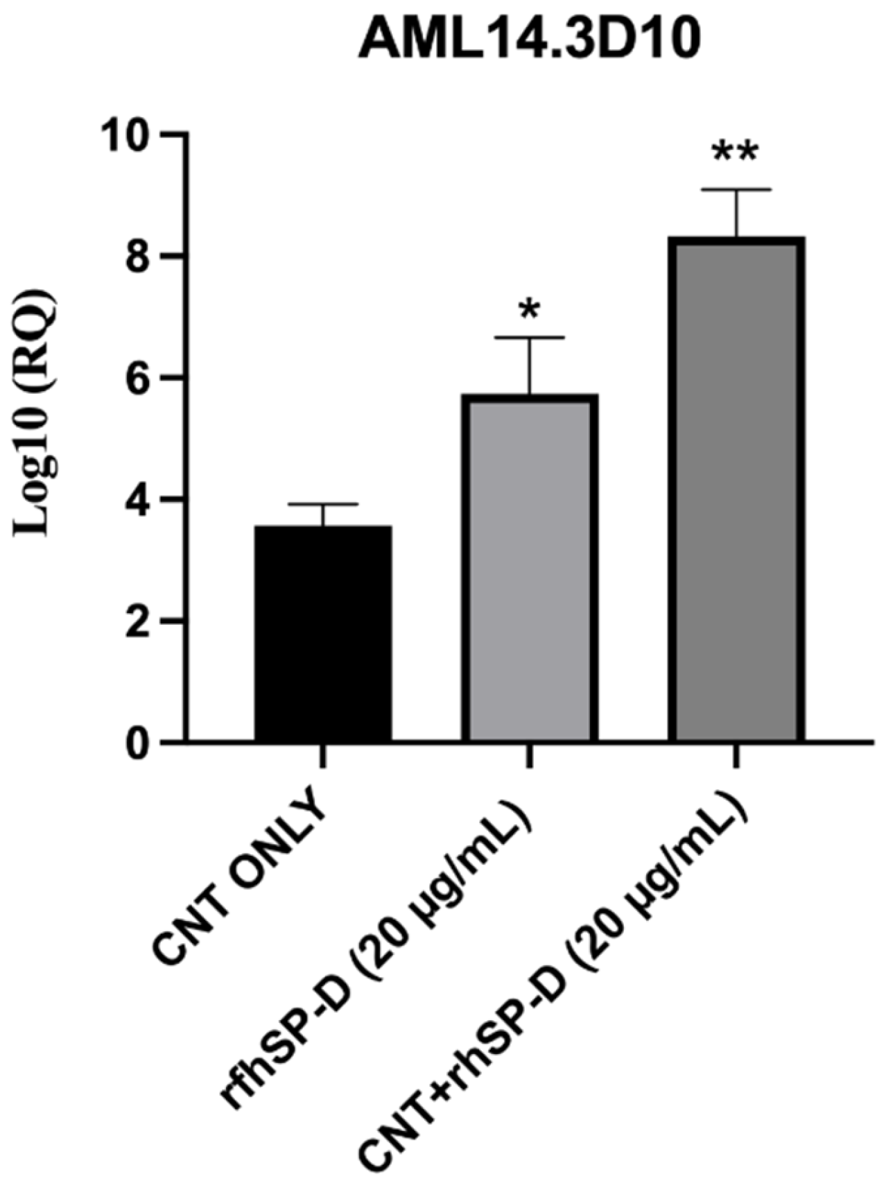
2.5. rfhSP-D Upregulates p53 Expression in AML14.3D10 Cell Line

p53, a transcription factor, regulates oncogenic responses including DNA damage, cell cycle arrest, and apoptosis. CNT + rfhSP-D or rfhSP-D alone treated AML14.3D10 cells showed increased transcript levels of p53 when compared to untreated cells. CNT + rfhSP-D treated cells showed log10 8.2-fold increased mRNA levels, compared to rfhSP-D treated cells (approximately log10 5.2-fold) (Figure 7). p53 transcripts were not measured in K562 cells as these cells do not express wild type p53. These data suggest that CNT + rfhSP-D treatment can induce apoptosis in these cell lines regardless of their p53 status.

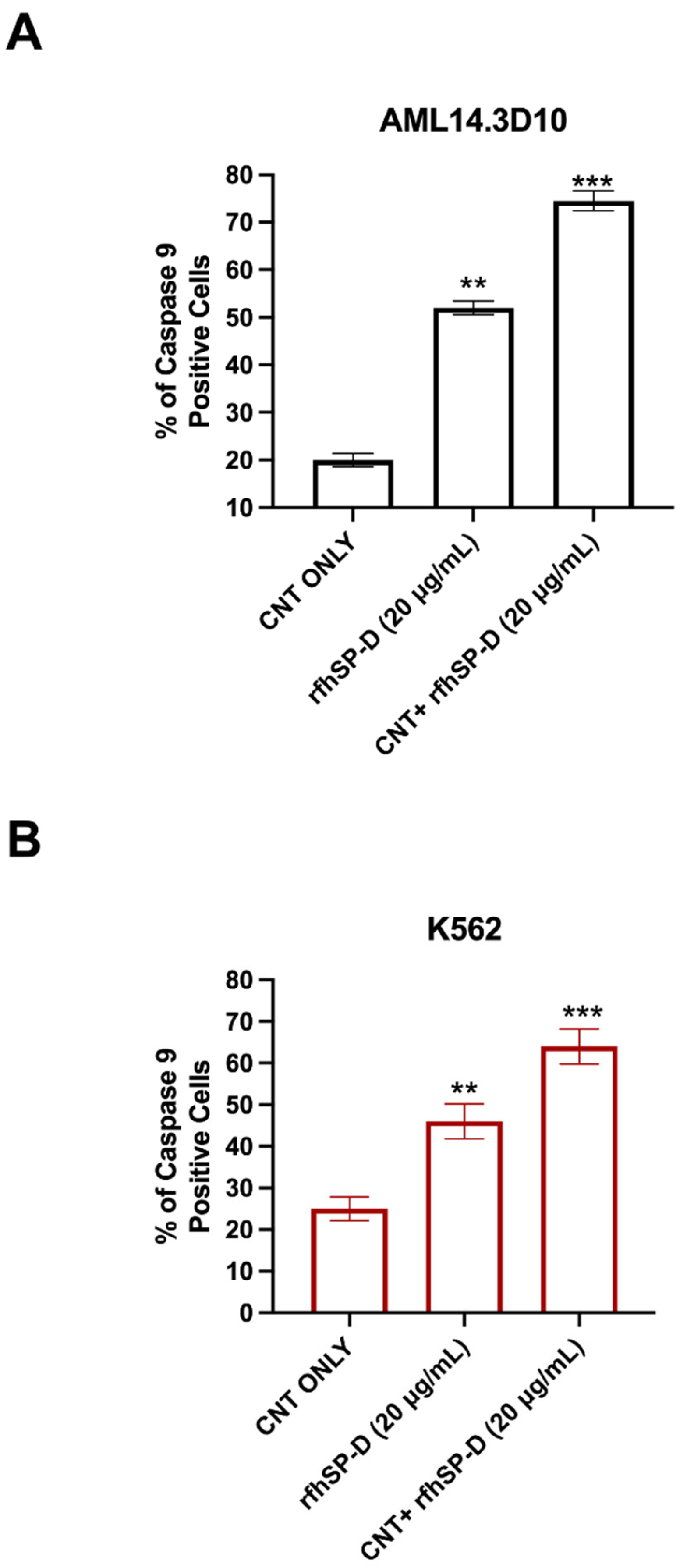
2.6. Apoptosis Induction in AML14.3D10 and K562 Cells by rfhSP-D-CNT via Intrinsic Pathway

Since apoptosis can be initiated via intrinsic or extrinsic pathways, expression of caspases was examined in AML14.3D10 or K562 cell lines treated with CNT + rfhSP-D (20 μg/mL) or rfhSP-D alone (20 μg/mL), using a fluorogenic substrate to detect the activation of caspase 3 and 7 (Figure 8). Higher levels of caspase 3 and 7 were observed in CNT + rfhSP-D treated AML14.3D10 (Figure 8A) and K562 (Figure 8B) cells, when compared to rfhSP-D or CNT alone-treated cells. There was a time-dependent increase in caspase 3 and 7 activation, which peaked at 24 h. Cleaved caspase 9 level was observed in CNT + rfhSP-D (or rfhSP-D-treated) AML14.3D10 or K562 cells at 12h, reflecting an intrinsic pathway (Figure 9).

3. Discussion

The involvement of innate immune mechanisms in cancer progression and resistance has opened up opportunities for using innate immune molecules as a part of anti-tumour therapeutic strategies. Immune system, innate as well as adaptive, is a double-edged sword that can either foster tumour progression via immunosuppression, angiogenesis, and metastasis, or resist oncogenesis [29,30]. SP-D, especially the trimeric CRDs in its recombinant form (rfhSP-D), has recently been shown to be protective against a range of cancer, based on in vitro studies. Coupling rfhSP-D with nanoparticles triggers a differential immune response [26]. rfhSP-D-bound CNTs upregulate the pro-inflammatory response (IL-1β, TNF-α, IL-6 and IL-12) in U937 and THP-1 cells [26]. Here, we examined the ability of CNT + rfhSP-D to act as a potent inducer of apoptosis in leukemic AML14.3D10 or K562 cell lines. CNT + rfhSP-D treatment reduced the cell viability of AML14.3D10 and K562 cells and induced apoptosis at 24 h in a dose- and time-dependent manner, peaking at 24 h and 20 μg/mL. A significant reduction in viability was observed in CNT + rfhSP-D treated AML14.3D10 (~37%) and K562 (~55%) cells compared to untreated control (cells + CNT), based on trypan blue and MTT assays.
FACS analysis revealed a significant increase in the percentage of Annexin V-/PI-positive leukemic cells following CNT + rfhSP-D treatment, characterized by the disruption of the asymmetric arrangement of the membrane, and appearance of PS on the outer side of the cell membrane in the cells undergoing apoptosis [31]. Annexin V, a 36 kDa protein, can bind PS, and also enter the entire plasma membrane in necrotic cells. CNT + rfhSP-D triggered the maximum apoptosis at 24 h [AML14.3D10 (~71%) and K562 (~66%)], when compared to CNT alone [AML14.3D10 (~12%) and K562 (~7%)]. However, no significant difference in terms of cell viability reduction/apoptosis induction was noticed following rfhSP-D treatment at 48 h in AML14.3D10 and K562 cells, suggesting recovery of the cells after 24 h. Apoptosis induction in AML14.3D10 and K562 cell lines by CNT + rfhSP-D may occur through the intrinsic pathway, supported by increased levels of caspase 3, 7 and cleaved caspase 9. This validates earlier studies on AML14.3D10, prostate and breast cancer cells [20,21,32], and the involvement of a mitochondrial pathway [20,21,32].
We also tried to understand the underlying mechanism of apoptosis induction by CNT + rfhSP-D and the associated signaling pathways. CNT + rfhSP-D caused increased transcript level of p53 in AML14.3D10 cell line, probably due to oxidative stress [17,33]. The upregulation of p53 in CNT + rfhSP-D treated AML14.3D10 cells may downregulate pAkt pathway, increasing Bad and Bax, which in turn, causes the release of the cytochrome c, and caspase 9 cleavage. In addition, the increased expression of p53 and cell cycle inhibitors (p21/p27) can cause inactivation of the cyclin B–cdc2 complex, crucial for G2/M cell cycle transition [17]. The existence of a lack of p53 wild type gene in K562 cell line, and its increased susceptibility to CNT + rfhSP-D, the protective effects of rfhSP-D bound to CNTs seem p53 independent. An involvement of cellular receptors expressed by these cancer cell lines is of paramount importance. SP-D interaction with HMGA1, CD14, CD91-calreticulin complex, SIRPα, EGFR, and GRP78 has been reported [20,21,22,33,34]. The presence of rfhSP-D on CNT as an array of therapeutic molecule is likely to have a clustering effect on these putative receptors, enhancing the potency of rfhSP-D.
In conclusion, CNT + rfhSP-D nanomaterial seems to be an attractive and novel therapeutic approach for targeting intracellular signaling cascades. There is a clear therapeutic potential of rfhSP-D against tumour cells. The advantage here is that the enhanced glycosylation of oncogenic targets can evade natural or therapeutic antibodies. Having established the specific nature of interactions between CNT + rfhSP-D and receptors found on leukemic cancer cells, we can hope to investigate host response in the murine models of cancer using wild type and SP-D knock-out mice.

4. Materials and Methods

4.1. Cell Culture

AML14.3D10 and K562 cells (ATCC) were cultured in RMPI media containing 10% v/v fetal calf serum (FCS), 2 mM L-glutamine, and penicillin (100 U/mL)/streptomycin (100 µg/mL) (ThermoFisher Scientific, Oxford, UK). Cells were grown at 37 °C under 5% v/v CO2 until 80–90% confluency was reached.

4.2. Dispersion and Functionalization of CNTs

The CNTs used in this study were characterized and functionalized as previously described [26,27]. Briefly, CNTs (diameters 10–20 nm, length 5–20 µm; Arry Nano) were dispersed using CNT sulfuric acid/nitric acid (3:1 ratio) via sonication and functionalized using CMC (Sigma-Aldrich/Merck, Dorset, UK) in a 1:2 mass ratio [26,27].

4.3. Expression and Purification of rfhSP-D

A recombinant fragment of human SP-D (rfhSP-D) was expressed and purified as described previously [17,32]. Affinity purified rfhSP-D was then subjected to endotoxin level measurement using QCL-1000 Limulus amebocyte lysate system (Lonza, Slough, UK); the endotoxin levels were found to be ~5 pg/μg of rfhSP-D.

4.4. Sodium Dodecyl Sulfate Polyacrylamide Gel Electrophoresis (SDS-PAGE)

The binding of rfhSP-D to CMC-CNTs was assessed via SDS-PAGE (12% v/v). CNT + rfhSP-D samples were boiled in a treatment buffer containing SDS and β-mercaptoethanol at 95 °C for 10 min before loading on to the gel. The SDS-PAGE gel was stained for 2 h using brilliant blue stain containing methanol (50% v/v) and acetic acid (10% v/v). This followed submersion of the stained gel with gentle shaking with de-staining solution (staining solution without brilliant blue).

4.5. Trypan-Blue-Dye Exclusion Assay

AML14.3D10 or K562 cells (0.1 × 10⁶) were seeded in a 12-well plate in complete RPMI complete medium overnight under 5% CO2 at 37 °C. Next, the cells were washed with PBS and treated with CNT + rfhSP-D (5, 10 or 20 µg/mL), or rfhSP-D alone (20 µg/mL), in serum-free RPMI for 24 h. Cells + CNT and Staurosporine (1 µM/mL) were used as an untreated/negative and positive control, respectively. Cells were then washed, detached using 5 mM EDTA, and centrifuged (1200× g). The cell pellet, re-suspended in RPMI, was treated with Trypan blue (10 µL) (60%), and viable cells were counted using hemocytometer in 5 different optical fields with a threshold value of 200 cells per field.

4.6. MTT Assay

MTT [3-(4,5-dimethylthiazol-2-yl)-2,5-diphenyltetrazolium bromide] (Sigma-Aldrich, Dorset, UK) assay was performed to assess the cell metabolic activity (cells + CNT + rfhSP-D; cells + CNT). AML14.3D10 or K562 cells (0.1 × 10⁵) were seeded in 96-well plates in RPMI complete medium until 85% confluency, and treated with CNT + rfhSP-D (5, 10 or 20 µg/mL), or rfhSP-D (20 µg/mL), in serum free RPMI medium for 24 h. MTT (50 µg/µL) per well was added for 4 h at 37 °C. 25 µL medium per well was then mixed with 50 µL DMSO (10′, 37 °C), and the absorbance was read at 570 nm using an ELISA plate reader.

4.7. Flow Cytometry

For apoptosis assays, AML14.3D10 or K562 cells (0.4 × 10⁶) were seeded in culture petri dishes (Nunc) in complete RPMI medium for 24 h and treated with CNT + rfhSP-D (20 µg/mL), or rfhSP-D (20 µg/mL), in serum-free RPMI medium for 24 h. Other controls were used as described above. Detached, centrifuged and PBS washed cells were incubated with Alexa Fluor 488 (1:200) (Sigma-Aldrich/Merck, Dorset, UK) (15°, RT) in dark, and the extent of apoptosis was measured using Novocyte Flow Cytometer. Compensation parameters were acquired using unstained, untreated FITC stained, and untreated PI-stained samples for all the cell lines.
For proliferative studies, AML14.3D10 or K562 cells (0.4 × 10⁶) were washed with PBS, probed with anti-mouse Ki-67 (BioLegend, San Diego, CA, USA) diluted in permeabilization reagent of the FIX&PERM kit (Fisher Scientific), and incubated for 30 min at room temperature (RT). Goat anti-mouse-FITC conjugate (1:200) (Fisher Scientific) was used as a probe at RT in the dark for 30 min. Cells (12,000) were acquired for each experiment and compensated before plotting the acquired data.
For caspase 9 and 8 studies, AML14.3D10 or K562 cells (0.4 × 10⁶) were treated with CNT + rfhSP-D or rfhSP-D (cells + CNT as a control) for 24 h at 37 °C, and probed with rabbit anti-human cleaved caspase 9 or 8 (Cell Signaling Technology, Danvers, MA, USA) (1:200) for 1 h at RT. Cells were washed in PBS, incubated with Alexa Fluor 488 (1:200) (Sigma-Aldrich) at RT in dark for 30 min, acquired and compensated (12,000) prior to plotting the data.

4.8. Caspase-3/7 Analysis

AML14.3D10 or K562 cells (0.1 × 10⁵) were seeded in 96 well plates in RPMI complete medium until 80% confluency. The cells were then treated with CNTs, as described above, in serum-free RPMI medium containing CellEventTM Caspase-3/7 Green Detection Reagent (5 µM; Thermo-Fisher) (0, 10, 20, 30 or 40 h). Cells + CNT was used as an untreated/negative control. CellEventTM Caspase-3/7 Green Detection Reagent is a fluorogenic substrate for activated caspases 3 and 7 in cells undergoing apoptosis. The plates with treated and untreated samples were incubated at 37 °C with 5% CO2 to detect the levels of Caspase 3/7 using a Clariostar plus microplate reader (BMG Labtech, Cary, NC, USA).

4.9. Quantitative RT-PCR

AML14.3D10 or K562 cells (0.5 × 10⁶) were incubated with CNT + rfhSP-D (20 µg/mL) or rfhSP-D (20 µg/m in serum-free RPMI medium for 18 h and RNA was isolated using GenElute Mammalian Total RNA Purification Kit (Sigma-Aldrich) and treated with DNase I. 2 µg of total RNA was used for cDNA synthesis using High Capacity kit (Applied Biosystems/ThermoFisher, Abingdon, UK). Primer sequences were designed using Primer-BLAST software (Table 1). Each PCR reaction, carried out in triplicates, contained SYBR Green (5 µL) MasterMix (Applied Biosystems), primers (75 nM), and cDNA (500 ng) (7900HT; Applied Biosystems). The cycle involved 2′/50 °C and 10′/95 °C, and 40 cycles (15 s/95 °C; 1′/60 °C). Human 18S rRNA was used as a housekeeping gene control. Relative quantification (RQ) value and formula: RQ  =  2 − ΔΔCt were used to calculate the relative expression of each target. Cells + CNT was used as an untreated/negative control.

4.10. Statistical Analysis

The graphs were generated using the GraphPad Prism 6.0 software. A one-way ANOVA test was carried out for statistical significance analysis. values less than 0.05 were considered as statistically significant.

Supplementary Materials

The following are available online at https://www.mdpi.com/article/10.3390/ijms221910445/s1.

Author Contributions

Conceptualization, H.A.K., U.K.; Data curation, U.K. and H.M.A.; Formal analysis, U.K. and S.H.A.; Funding acquisition, H.A.K.; Investigation, H.A.K., U.K. and S.H.A.; Methodology, U.K., H.M.A. and S.H.A.; Project administration, H.A.K.; Resources, H.A.K., U.K.; Software, U.K., H.M.A.; Writing—review & editing, H.A.K. and U.K. All authors have read and agreed to the published version of the manuscript.

Funding

This project was funded by the National Plan for Science, Technology and Innovation (MAARIFAH), King Abdulaziz City for Science and Technology, Kingdom of Saudi Arabia, Award Number (15-NAN-3664-02).

Institutional Review Board Statement

Not applicable.

Informed Consent Statement

Not applicable.

Data Availability Statement

Not applicable.

Acknowledgments

Technical assistance of Valarmathy Murugaiah, Khalid Ibrahim, Ashraf Hatamilah, and Adnan Khan is gratefully acknowledged.

Conflicts of Interest

The authors declare no conflict of interest.

References

  1. Boraschi, D.; Castellano, L.R.C.; Italiani, P. Interaction of Nanomaterials with the Immune System: Role in Nanosafety and Nanomedicine. Front. Immunol. 2017, 8, 1688. [Google Scholar] [CrossRef] [PubMed] [Green Version]
  2. Salvador-Morales, C.; Flahaut, E.; Sim, E.; Sloan, J.; Green, M.L.; Sim, R.B. Complement activation and protein adsorption by carbon nanotubes. Mol. Immunol. 2006, 43, 193–201. [Google Scholar] [CrossRef] [PubMed] [Green Version]
  3. Kang, S.H.; Nafiujjaman, M.; Nurunnabi, M.; Li, L.; Khan, H.A.; Cho, K.J.; Huh, K.M.; Lee, Y. Hybrid photoactive nanomaterial composed of gold nanoparticles, pheophorbide-A and hyaluronic acid as a targeted bimodal phototherapy. Macromol. Res. 2015, 23, 474–484. [Google Scholar] [CrossRef]
  4. Ibrahim, K.E.; Bakhiet, A.O.; Khan, A.; Khan, H.A. Recent trends in biomedical applications of nanomaterials. Biosci. Biotechnol. Res. Asia 2018, 15, 235–243. [Google Scholar] [CrossRef]
  5. Nafiujjaman, M.; Khan, H.A.; Lee, Y.K. Peptide-influenced graphene quantum dots on iron oxide nanoparticles for dual imaging of lung cancer cells. J. Nanosci. Nanotechnol. 2017, 17, 1704–1711. [Google Scholar] [CrossRef]
  6. Khan, H.A.; Sakharkar, M.; Nayak, A.; Kishore, U.; Khan, A. Nanoparticles for biomedical applications. In Nanobiomaterials: Nanostructured Materials for Biomedical Applications; Elsevier: New York, NY, USA, 2018; pp. 357–384. [Google Scholar]
  7. Du, J.; Zhang, Y.S.; Hobson, D.; Hydbring, P. Nanoparticles for immune system targeting. Drug Discov. Today 2017, 22, 1295–1301. [Google Scholar] [CrossRef]
  8. Mishra, V.; Kesharwani, P.; Jain, N.K. Biomedical Applications and Toxicological Aspects of Functionalized Carbon Nanotubes. Crit. Rev. Ther. Drug Carr. Syst. 2018, 35, 293–330. [Google Scholar] [CrossRef] [PubMed]
  9. Vardharajula, S.; Ali, S.Z.; Tiwari, P.M.; Eroglu, E.; Vig, K.; Dennis, V.A.; Singh, S.R. Functionalized carbon nanotubes: Biomedical applications. Int. J. Nanomed. 2012, 7, 5361–5374. [Google Scholar]
  10. Liu, Z.; Tabakman, S.; Welsher, K.; Dai, H. Carbon Nanotubes in Biology and Medicine: In vitro and in vivo Detection, Imaging and Drug Delivery. Nano Res. 2009, 2, 85–120. [Google Scholar] [CrossRef] [Green Version]
  11. Li, W.; Tierce, N.T.; Bekyarova, E.; Bardeen, C.J. Protection of Molecular Microcrystals by Encapsulation under Single-Layer Graphene. ACS Omega 2018, 3, 8129–8134. [Google Scholar] [CrossRef]
  12. Crouch, E.; Wright, J.R. Surfactant proteins A and D and pulmonary host defense. Annu. Rev. Physiol. 2001, 63, 521–554. [Google Scholar] [CrossRef]
  13. Crouch, E.C. Surfactant protein-D and pulmonary host defense. Respir. Res. 2000, 1, 1–16. [Google Scholar] [CrossRef] [Green Version]
  14. Pastva, A.M.; Wright, J.R.; Williams, K.L. Immunomodulatory roles of surfactant proteins A and D: Implications in lung disease. Proc. Am. Thorac. Soc. 2007, 4, 252–257. [Google Scholar] [CrossRef] [PubMed] [Green Version]
  15. Nayak, A.; Dodagatta-Marri, E.; Tsolaki, A.G.; Kishore, U. An insight into the diverse roles of surfactant proteins, SP-A and SP-D in innate and adaptive immunity. Front. Immunol. 2012, 3, 131. [Google Scholar] [CrossRef] [PubMed] [Green Version]
  16. Murugaiah, V.; Tsolaki, A.G.; Kishore, U. Collectins: Innate Immune Pattern Recognition Molecules. Lectin Host Def. Microb. Infect. 2020, 1204, 75–127. [Google Scholar]
  17. Kaur, A.; Riaz, M.S.; Murugaiah, V.; Varghese, P.M.; Singh, S.K.; Kishore, U. A recombinant fragment of human surfactant protein D induces apoptosis in pancreatic cancer cell lines via fas-mediated pathway. Front. Immunol. 2018, 9, 1126. [Google Scholar] [CrossRef]
  18. Kaur, A.; Riaz, M.S.; Singh, S.K.; Kishore, U. Human surfactant protein D suppresses epithelial-to-mesenchymal transition in pancreatic cancer cells by downregulating TGF-β. Front. Immunol. 2018, 9, 1844. [Google Scholar] [CrossRef] [PubMed]
  19. Kumar, J.; Murugaiah, V.; Sotiriadis, G.; Kaur, A.; Jeyaneethi, J.; Sturniolo, I.; Alhamlan, F.; Chatterjee, J.; Hall, M.; Kishore, U. Surfactant protein D as a potential biomarker and therapeutic target in ovarian cancer. Front. Oncol. 2019, 9, 542. [Google Scholar] [CrossRef] [Green Version]
  20. Thakur, G.; Prakash, G.; Murthy, V.; Sable, N.; Menon, S.; Alrokayan, S.; Khan, H.A.; Murugaiah, V.; Bakshi, G.; Kishore, U. Human SP-D acts as an innate immune surveillance molecule against androgen-responsive and androgen-resistant prostate cancer cells. Front. Oncol. 2019, 9, 565. [Google Scholar] [CrossRef] [Green Version]
  21. Mahajan, L.; Pandit, H.; Madan, T.; Gautam, P.; Yadav, A.K.; Warke, H.; Sundaram, C.S.; Sirdeshmukh, R.; Sarma, P.U.; Kishore, U.; et al. Human surfactant protein D alters oxidative stress and HMGA1 expression to induce p53 apoptotic pathway in eosinophil leukemic cell line. PLoS ONE 2013, 8, e85046. [Google Scholar] [CrossRef]
  22. Hasegawa, Y.; Takahashi, M.; Ariki, S.; Asakawa, D.; Tajiri, M.; Wada, Y.; Yamaguchi, Y.; Nishitani, C.; Takamiya, R.; Saito, A. Surfactant protein D suppresses lung cancer progression by downregulation of epidermal growth factor signaling. Oncogene 2015, 34, 838–845. [Google Scholar] [CrossRef] [PubMed]
  23. Kendall, M.; Ding, P.; Mackay, R.M.; Deb, R.; Mckenzie, Z.; Kendall, K.; Madsen, J.; Clark, H. Surfactant protein D (SP-D) alters cellular uptake of particles and nanoparticles. Nanotoxicology 2013, 7, 963–973. [Google Scholar] [CrossRef] [PubMed]
  24. Ruge, C.A.; Schaefer, U.F.; Herrmann, J.; Kirch, J.; Canadas, O.; Echaide, M.; Perez-Gil, J.; Casals, C.; Muller, R.; Lehr, C.M. The interplay of lung surfactant proteins and lipids assimilates the macrophage clearance of nanoparticles. PLoS ONE 2012, 7, e40775. [Google Scholar] [CrossRef]
  25. Cirillo, G.; Hampel, S.; Klingeler, R.; Puoci, F.; Iemma, F.; Curcio, M.; Parisi, O.I.; Spizzirri, U.G.; Picci, N.; Leonhardt, A.; et al. Antioxidant multi-walled carbon nanotubes by free radical grafting of gallic acid: New materials for biomedical applications. J. Pharm. Pharmacol. 2011, 63, 179–188. [Google Scholar] [CrossRef] [PubMed]
  26. Pondman, K.M.; Paudyal, B.; Sim, R.B.; Kaur, A.; Kouser, L.; Tsolaki, A.G.; Jones, L.A.; Salvador-Morales, C.; Khan, H.A.; Ten Haken, B.; et al. Pulmonary surfactant protein SP-D opsonises carbon nanotubes and augments their phagocytosis and subsequent pro-inflammatory immune response. Nanoscale 2017, 9, 1097–1109. [Google Scholar] [CrossRef] [PubMed] [Green Version]
  27. Pondman, K.M.; Tsolaki, A.G.; Paudyal, B.; Shamji, M.H.; Switzer, A.; Pathan, A.A.; Abozaid, S.M.; Ten Haken, B.; Stenbeck, G.; Sim, R.B.; et al. Complement Deposition on Nanoparticles Can Modulate Immune Responses by Macrophage, B and T Cells. J. Biomed. Nanotechnol. 2016, 12, 197–216. [Google Scholar] [CrossRef]
  28. Baumann, M.A.; Paul, C.C. The AML14 and AML14.3D10 cell lines: A long-overdue model for the study of eosinophils and more. Stem Cells 1998, 16, 16–24. [Google Scholar] [CrossRef]
  29. Palucka, A.K.; Coussens, L.M. The Basis of Oncoimmunology. Cell 2016, 164, 1233–1247. [Google Scholar] [CrossRef] [Green Version]
  30. Vesely, M.D.; Kershaw, M.H.; Schreiber, R.D.; Smyth, M.J. Natural innate and adaptive immunity to cancer. Annu. Rev. Immunol. 2011, 29, 235–271. [Google Scholar] [CrossRef] [Green Version]
  31. Porter, A.G.; Janicke, R.U. Emerging roles of caspase-3 in apoptosis. Cell Death Differ. 1999, 6, 99–104. [Google Scholar] [CrossRef]
  32. Murugaiah, V.; Agostinis, C.; Varghese, P.M.; Belmonte, B.; Vieni, S.; Alaql, F.A.; Alrokayan, S.H.; Khan, H.A.; Kaur, A.; Roberts, T.; et al. Hyaluronic Acid Present in the Tumor Microenvironment Can Negate the Pro-apototic Effect of a Recombinant Fragment of Human Surfactant Protein D on Breast Cancer Cells. Front. Immunol. 2020, 11, 1171. [Google Scholar] [CrossRef] [PubMed]
  33. Janssen, W.J.; Mcphillips, K.A.; Dickinson, M.G.; Linderman, D.J.; Morimoto, K.; Xiao, Y.Q.; Oldham, K.M.; Vandivier, R.W.; Henson, P.M.; Gardai, S.J. Surfactant proteins A and D suppress alveolar macrophage phagocytosis via interaction with SIRP alpha. Am. J. Respir. Crit. Care Med. 2008, 178, 158–167. [Google Scholar] [CrossRef] [PubMed]
  34. Thakur, G.; Sathe, G.; Kundu, I.; Biswas, B.; Gautam, P.; Alkahtani, S.; Idicula-Thomas, S.; Sirdeshmukh, R.; Kishore, U.; Madan, T. Membrane Interactome of a Recombinant Fragment of Human Surfactant Protein D Reveals GRP78 as a Novel Binding Partner in PC3, a Metastatic Prostate Cancer Cell Line. Front. Immunol. 2021, 11, 3704. [Google Scholar] [CrossRef]
Figure 1. Purified rfhSP-D (10 µg/mL) or carboxymethyl cellulose-coated carbon nanotubes (rfh-SP-D-CNTs) coupled-rfhSP-D (10 µg/mL) samples were subjected to an SDS-PAGE (15% v/v). Lane 1: Supernatant (10 µL/well) taken after centrifugation of rfhSP-D-CNT. Lane 2: purified rfhSP-D. Lane 3: rfhSP-D-CNT. The original image is available as a Supplementary Materials.
Ijms 22 10445 g001
Figure 2. Cell viability following treatment with CNT + rfhSP-D-CNT in AML14.3D10 (A) and K562 (B) cell lines via trypan-blue-dye exclusion assay. Cells (0.1 × 10⁵) were treated with CNT + rfhSP-D (5, 10, 20 µg/mL), rfhSP-D (20 µg/mL) or CNT alone (20 µg/mL) for 24 h at 37 °C. The data has been normalized with cells only as 100% of the cell viability. * p < 0.05, ** p < 0.01 and *** p < 0.001 compared to CNT only group.
Ijms 22 10445 g002
Figure 3. CNT + rfhSP-D treatment reduced viability of AML14.3D10 (A) and K562 (B) cells, as measured by MTT assay. The data have been normalized with cells only as 100% of the cell viability. Values are means ± SEM (n = 3) * p < 0.05, ** p < 0.01 and *** p < 0.001 compared to CNT only group.
Ijms 22 10445 g003
Figure 4. Anti-proliferative effects of CNT + rfhSP-D on AML14.3D10 (A) and K562 (B) cell lines. Values are means ± SD. ** p < 0.01, and *** p < 0.001 compared to CNT group only. The raw data are available as Supplementary Materials.
Ijms 22 10445 g004
Figure 5. Flow cytometry analysis of apoptosis induction in AML14.3D10 (A) or K562 (B) cell lines treated with CNT + rfhSP-D. For Annexin V/FITC and DNA/PI staining, 12,000 cells were acquired and plotted. Values are means ± SEM (n = 3). ** p < 0.01 and *** p < 0.001 compared to CNT only group. The raw data are available in the Supplementary Materials.
Ijms 22 10445 g005
Figure 6. CNT + rfhSP-D treatment causes the upregulation of p21 and p27 cell cycle inhibitors in AML14.3D10 (A) and K562 (B) cell lines. AML14.3D10 or K562 (0.4 × 10⁶) cells, treated with CNT + rfhSP-D (20 µg/mL) or rfhSP-D (20 µg/mL), plus untreated control (cells + CNT) (20 µg/mL), were used for RNA extraction, cDNA synthesis and RT-qPCR, using 18S as an endogenous control. The RQ value was calculated using the formula: RQ = 2 − ΔΔCt. Values represent means ± SEM (n = 3). * p < 0.05 and ** p < 0.01 compared to CNT only group.
Ijms 22 10445 g006
Figure 7. CNT + rfhSP-D treated AML14.3D10 cells show upregulation of the mRNA transcript levels of p53. AML14.3D10 (0.4 × 10⁶) cells were treated with CNT + rfhSP-D or rfhSP-D alone, along with an untreated control (cells + CNT) (20 µg/mL each). The RQ value was calculated using the formula: RQ = 2 − ΔΔCt. * p < 0.05 and ** p < 0.01 compared to CNT only group.
Ijms 22 10445 g007
Figure 8. Activation of caspase 3/7 in AML14.3D10 (A) or K562 (B) cell lines following CNT + rfhSP-D treatment. AML14.3D10 or K562 cells (0.1 × 10⁵) were seeded and challenged with CNT + rfhSP-D (20 µg/mL) or rfhSP-D (20 µg/mL) Cells + CNT was used as an untreated control. **** p < 0.0001 versus control group (n = 3).
Ijms 22 10445 g008
Figure 9. CNT + rfhSP-D treatment upregulates the levels of cleaved caspase 9 on AML14.3D10 (A) or K562 (B) cell lines at 24 h. AML14.3D10 or K562 cells (0.4 × 10⁶) were treated with rfhSP-CNT or rfhSP-D, along with an untreated control (cells + CNT). Values are expressed as mean ± SD (n = 3). ** p < 0.01, and *** p < 0.001 versus control group.
Ijms 22 10445 g009
Table 1. Target genes and terminal primers used in the RT-qPCR analysis.
Target GeneForward Primer Reverse Primer
18S5′-ATGGCCGTTCTTAGTTGGTG-3′5′-CGCTGAGCCAGTCAGTGTAG-3′
P535′-AGCACTGTCCAACAACACCA-3′5′-CTTCAGGTGGCTGGAGTGAG-3′
p215′-TGGAGACTCTCAGGGTCGAAA-3′5′-CGGCGTTTGGAGTGGTAGAA-3′
p275′-CCGGTGGACCACGAAGAGT-3′5′-GCTCGCCTCTTCCATGTCTC-3′
Publisher’s Note: MDPI stays neutral with regard to jurisdictional claims in published maps and institutional affiliations.

