
Endogenous Enzymatic Activity in Dentin Treated with a Chitosan Primer

 ,
,  , , and
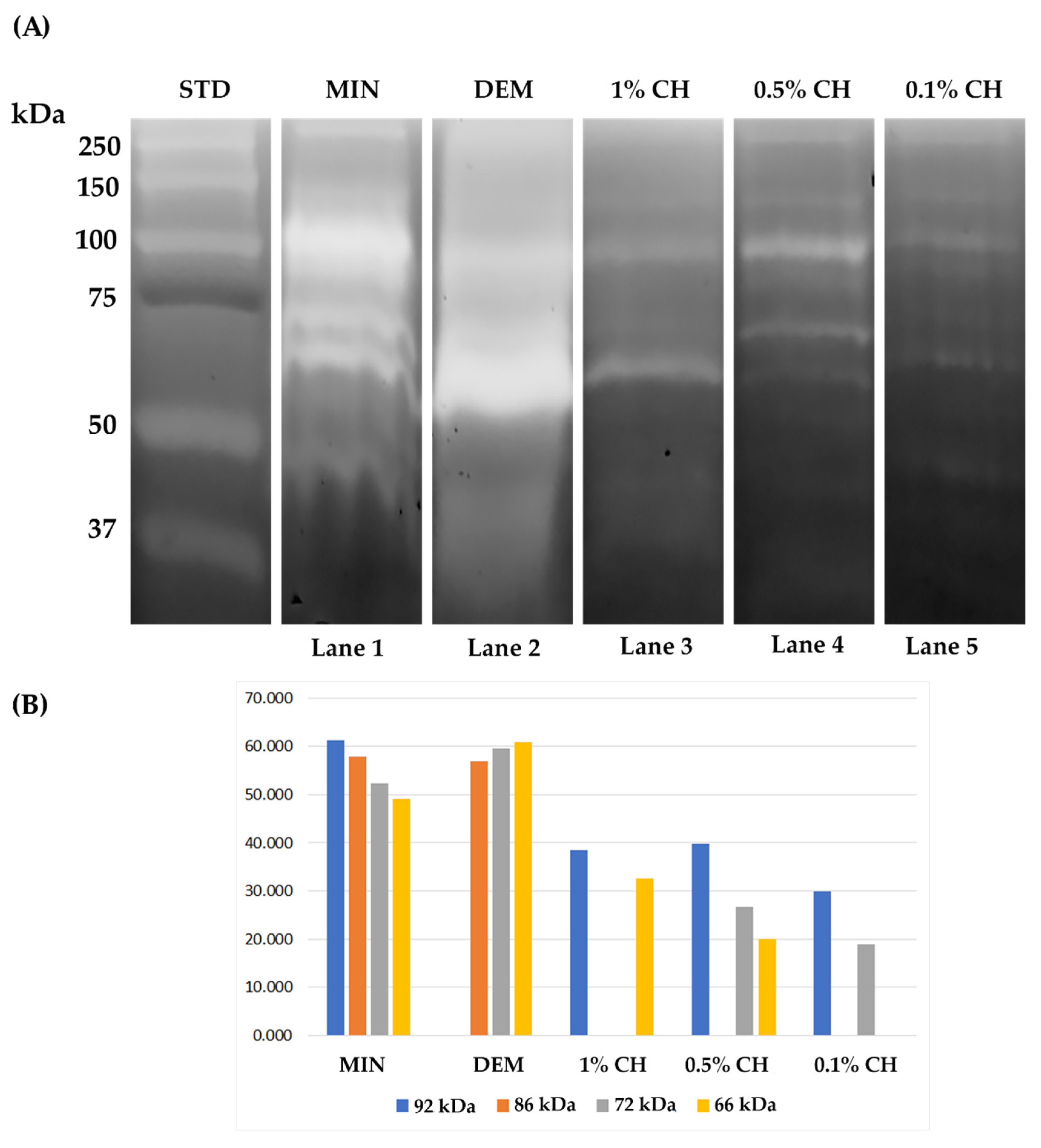
Abstract
:1. Introduction
2. Results
2.1. Gelatin Zymography
2.2. In Situ Zymography
3. Discussion
4. Materials and Methods
4.1. Preparation of Solutions
4.2. Gelatin Zymography
- Group 1 (MIN): MD left untreated;
- Group 2 (DEM): MD was treated with 10 wt% phosphoric acid for 10 min at 4 °C, then the acid was neutralized with 4N NaOH and centrifuged;
- Group 3 (1% CH): MD was etched as in Group 3 and then treated with 1% chitosan water solution for 30 min;
- Group 4 (0.5% CH): MD was etched as in Group 3 and then treated with 0.5% chitosan water solution for 30 min;
- Group 5 (0.1% CH): MD was etched as in Group 3 and then mixed with 0.1% chitosan water solution for 30 min.
4.3. In Situ Zymography
- Group 1: Dentin etched with 37% phosphoric acid for 15 s (Vococid, Voco, Cuxhaven, Germany), rinsed thoroughly, blot dried and then primed with the 1 wt% chitosan water solution for 1 min, after which it was gently air-dried for 5 s;
- Group 2: Dentin etched and rinsed as in Group 1 and then primed with the 0.5 wt% chitosan water solution for 1 min, after which it was gently air-dried for 5 s;
- Group 3: Dentin etched and rinsed as in Group 1 and then primed with the 0.1 wt% chitosan water solution for 1 min, after which it was gently air-dried for 5 s;
- Group 4 (control): Dentin etched and rinsed as in Group 1 and gently air dried for 5 s;
- Group 5: Dentin etched and pretreated as in Group 1, followed by the application of a universal adhesive system (Futurabond M, Voco, Cuxhaven, Germany) according to the manufacturer’s instructions and polymerized for 10 s using a LED curing unit (Demi, Kerr, Germany) after which a 1 mm thick layer of flowable composite was applied to the bonded surface (Grandio Flow, Voco, Cuxhaven, Germany) and polymerized for 20 s;
- Group 6: Dentin etched and pretreated as in Group 2, followed by the adhesive and restorative procedures as in Group 5;
- Group 7: Dentin etched and pretreated as in Group 3, followed by the adhesive and restorative procedures as in Group 5;
- Group 8 (control): Dentin etched as in Group 4, followed by the adhesive and restorative procedures as in Group 5.
5. Conclusions
Author Contributions
Funding
Institutional Review Board Statement
Informed Consent Statement
Data Availability Statement
Acknowledgments
Conflicts of Interest
References
- Breschi, L.; Maravic, T.; Cunha, S.R.; Comba, A.; Cadenaro, M.; Tjäderhane, L.; Pashley, D.H.; Tay, F.R.; Mazzoni, A. Dentin bonding systems: From dentin collagen structure to bond preservation and clinical applications. Dent. Mater. 2018, 34, 78–96. [Google Scholar] [CrossRef] [Green Version]
- Maravic, T.; Mazzoni, A.; Comba, A.; Scotti, N.; Checchi, V.; Breschi, L. How stable is dentin as a substrate for bonding? Curr. Oral Health Rep. 2017, 4, 248–257. [Google Scholar] [CrossRef]
- Turco, G.; Cadenaro, M.; Maravić, T.; Frassetto, A.; Marsich, E.; Mazzoni, A.; Di Lenarda, R.; Tay, F.R.; Pashley, D.H.; Breschi, L. Release of ICTP and CTX telopeptides from demineralized dentin matrices: Effect of time, mass and surface area. Dent. Mater. 2018, 34, 452–459. [Google Scholar] [CrossRef]
- Mazzoni, A.; Breschi, L.; Carrilho, M.; Nascimento, F.D.; Orsini, G.; Ruggeri, A.; Gobbi, P.; Manzoli, L.; Tay, F.R.; Pashley, D.H.; et al. A review of the nature, role, and function of dentin non-collagenous proteins. Part II: Enzymes, serum proteins, and growth factors. Endod. Top. 2009, 21, 19–40. [Google Scholar] [CrossRef]
- Checchi, V.; Maravic, T.; Bellini, P.; Generali, L.; Consolo, U.; Breschi, L.; Mazzoni, A. The role of matrix metalloproteinases in periodontal disease. Int. J. Environ. Res. Public Health 2020, 17, 4923. [Google Scholar] [CrossRef]
- Mazzoni, A.; Nascimento, F.; Carrilho, M.; Tersariol, I.; Papa, V.; Tjaderhane, L.; Di Lenarda, R.; Tay, F.; Pashley, D.; Breschi, L. MMP activity in the hybrid layer detected with in situ zymography. J. Dent. Res. 2012, 91, 467–472. [Google Scholar] [CrossRef] [Green Version]
- Mazzoni, A.; Tjäderhane, L.; Checchi, V.; Di Lenarda, R.; Salo, T.; Tay, F.R.; Pashley, D.H.; Breschi, L. Role of dentin MMPs in caries progression and bond stability. J. Dent. Res. 2015, 94, 241–251. [Google Scholar] [CrossRef] [Green Version]
- Liu, Y.; Tjäderhane, L.; Breschi, L.; Mazzoni, A.; Li, N.; Mao, J.; Pashley, D.H.; Tay, F.R. Limitations in bonding to dentin and experimental strategies to prevent bond degradation. J. Dent. Res. 2011, 90, 953–968. [Google Scholar] [CrossRef]
- Shrestha, A.; Friedman, S.; Kishen, A. Photodynamically crosslinked and chitosan-incorporated dentin collagen. J. Dent. Res. 2011, 90, 1346–1351. [Google Scholar] [CrossRef] [PubMed]
- Bonferoni, M.C.; Sandri, G.; Rossi, S.; Ferrari, F.; Caramella, C. Chitosan and its salts for mucosal and transmucosal delivery. Expert Opin. Drug Deliv. 2009, 6, 923–939. [Google Scholar] [CrossRef]
- Diolosà, M.; Donati, I.; Turco, G.; Cadenaro, M.; Di Lenarda, R.; Breschi, L.; Paoletti, S. Use of methacrylate-modified chitosan to increase the durability of dentine bonding systems. Biomacromolecules 2014, 15, 4606–4613. [Google Scholar] [CrossRef]
- Elsaka, S.; Elnaghy, A. Effect of addition of chitosan to self-etching primer: Antibacterial activity and push-out bond strength to radicular dentin. J. Biomed. Res. 2012, 26, 288–294. [Google Scholar] [CrossRef]
- Fawzy, A.S.; Nitisusanta, L.I.; Iqbal, K.; Daood, U.; Beng, L.T.; Neo, J. Chitosan/Riboflavin-modified demineralized dentin as a potential substrate for bonding. J. Mech. Behav. Biomed. Mater. 2012, 17, 278–289. [Google Scholar] [CrossRef] [PubMed]
- Elsaka, S.E. Antibacterial activity and adhesive properties of a chitosan-containing dental adhesive. Quintessence Int. Berl. 2012, 43, 603–613. [Google Scholar]
- Baena, E.; Cunha, S.R.; Maravić, T.; Comba, A.; Paganelli, F.; Alessandri-Bonetti, G.; Ceballos, L.; Tay, F.R.; Breschi, L.; Mazzoni, A. Effect of chitosan as a cross-linker on matrix metalloproteinase activity and bond stability with different adhesive systems. Mar. Drugs 2020, 18, 263. [Google Scholar] [CrossRef]
- Neves, J.G.; Marcato, P.D.; de Paula e Silva, F.W.G.; Mantovani, C.P.T.; Prado, H.S.; Aires, C.P.; Massaro, T.N.C.; Borsato, M.C. Synthesis and characterization of an experimental primer containing chitosan nanoparticles—Effect on the inactivation of metalloproteinases, antimicrobial activity and adhesive strength. Arch. Oral Biol. 2021, 127, 105148. [Google Scholar] [CrossRef] [PubMed]
- Park, S.Y.; Marsh, K.S.; Rhim, J.W. Characteristics of different molecular weight chitosan films affected by the type of organic solvents. J. Food Sci. 2002, 67, 194–197. [Google Scholar] [CrossRef]
- Shi, C.; Zhu, Y.; Ran, X.; Wang, M.; Su, Y.; Cheng, T. Therapeutic potential of chitosan and its derivatives in regenerative medicine. J. Surg. Res. 2006, 133, 185–192. [Google Scholar] [CrossRef]
- Croisier, F.; Jérôme, C. Chitosan-based biomaterials for tissue engineering. Eur. Polym. J. 2013, 49, 780–792. [Google Scholar] [CrossRef] [Green Version]
- Wieckiewicz, M.; Boening, K.; Grychowska, N.; Paradowska-Stolarz, A. Clinical application of chitosan in dental specialities. Mini Rev. Med. Chem. 2017, 17, 401–409. [Google Scholar] [CrossRef]
- Palma, P.; Ramos, J.; Martins, J.; Diogenes, A.; Figueiredo, M.; Ferreira, P.; Viegas, C.; Santos, J. Histologic evaluation of regenerative endodontic procedures with the use of chitosan scaffolds in immature dog teeth with apical periodontitis. J. Endod. 2017, 43, 1279–1287. [Google Scholar] [CrossRef] [PubMed]
- Dye, B.A. The global burden of oral disease: Research and public health significance. J. Dent. Res. 2017, 96, 361–363. [Google Scholar] [CrossRef] [PubMed]
- Frassetto, A.; Breschi, L.; Turco, G.; Marchesi, G.; Di Lenarda, R.; Tay, F.R.; Pashley, D.H.; Cadenaro, M. Mechanisms of degradation of the hybrid layer in adhesive dentistry and therapeutic agents to improve bond durability—A literature review. Dent. Mater. 2016, 32, e41–e53. [Google Scholar] [CrossRef] [PubMed]
- Tezvergil-Mutluay, A.; Agee, K.A.; Mazzoni, A.; Carvalho, R.M.; Carrilho, M.; Tersariol, I.L.; Nascimento, F.D.; Imazato, S.; Tjaderhane, L.; Breschi, L.; et al. Can quaternary ammonium methacrylates inhibit matrix MMPs and cathepsins? Dent. Mater. 2015, 31, e25–e32. [Google Scholar] [CrossRef] [PubMed] [Green Version]
- Breschi, L.; Maravic, T.; Comba, A.; Cunha, S.R.; Loguercio, A.D.; Reis, A.; Hass, V.; Cadenaro, M.; Mancuso, E.; Mayer-Santos, E.; et al. Chlorhexidine preserves the hybrid layer in vitro after 10-years aging. Dent. Mater. 2020, 36, 672–680. [Google Scholar] [CrossRef] [PubMed]
- Maravić, T.; Comba, A.; Cunha, S.R.; Angeloni, V.; Cadenaro, M.; Visinitini, E.; Navarra, C.O.; Salgarello, S.; Breschi, L.; Mazzoni, A. Long-term bond strength and endogenous enzymatic activity of a chlorhexidine-containing commercially available adhesive. J. Dent. 2019, 84, 60–66. [Google Scholar] [CrossRef]
- Comba, A.; Maravic, T.; Valente, L.; Girlando, M.; Cunha, S.R.; Checchi, V.; Salgarello, S.; Tay, F.R.; Scotti, N.; Breschi, L.; et al. Effect of benzalkonium chloride on dentin bond strength and endogenous enzymatic activity. J. Dent. 2019, 85, 25–32. [Google Scholar] [CrossRef]
- Breschi, L.; Martin, P.; Mazzoni, A.; Nato, F.; Carrilho, M.; Tjäderhane, L.; Visintini, E.; Cadenaro, M.; Tay, F.R.; Dorigo, E.D.S.; et al. Use of a specific MMP-inhibitor (galardin) for preservation of hybrid layer. Dent. Mater. 2010, 26, 571–578. [Google Scholar] [CrossRef] [Green Version]
- Bedran-Russo, A.K.; Pauli, G.F.; Chen, S.N.; McAlpine, J.; Castellan, C.S.; Phansalkar, R.S.; Aguiar, T.R.; Vidal, C.M.P.; Napotilano, J.G.; Nam, J.W.; et al. Dentin biomodification: Strategies, renewable resources and clinical applications. Dent. Mater. 2014, 30, 62–76. [Google Scholar] [CrossRef] [Green Version]
- Seseogullari-Dirihan, R.; Apollonio, F.; Mazzoni, A.; Tjaderhane, L.; Pashley, D.; Breschi, L.; Tezvergil-Mutluay, A. Use of crosslinkers to inactivate dentin MMPs. Dent. Mater. 2016, 32, 423–432. [Google Scholar] [CrossRef] [Green Version]
- Comba, A.; Maravić, T.; Villalta, V.; Tozzola, S.; Mazzitelli, C.; Checchi, V.; Mancuso, E.; Scotti, N.; Tay, F.R.; Breschi, L.; et al. Effect of an ethanol cross-linker on universal adhesive. Dent. Mater. 2020, 36, 1645–1654. [Google Scholar] [CrossRef]
- Maravic, T.; Breschi, L.; Comba, A.; Cunha, S.R.; Angeloni, V.; Nucci, C.; Hebling, J.; Pashley, D.; Tay, F.; Mazzoni, A. Experimental use of an acrolein-based primer as collagen cross-linker for dentine bonding. J. Dent. 2017, 68, 85–90. [Google Scholar] [CrossRef] [Green Version]
- Mazzoni, A.; Angeloni, V.; Sartori, N.; Duarte, S.; Maravic, T.; Tjäderhane, L.; Pashley, D.H.; Tay, F.R.; Breschi, L. Substantivity of carbodiimide inhibition on dentinal enzyme activity over time. J. Dent. Res. 2017, 96, 902–908. [Google Scholar] [CrossRef]
- Mazzoni, A.; Angeloni, V.; Comba, A.; Maravic, T.; Cadenaro, M.; Tezvergil-Mutluay, A.; Pashley, D.H.; Tay, F.R.; Breschi, L. Cross-linking effect on dentin bond strength and MMPs activity. Dent. Mater. 2018, 34, 288–295. [Google Scholar] [CrossRef]
- Busenlehner, L.S.; Armstrong, R.N. Insights into enzyme structure and dynamics elucidated by amide H/D exchange mass spectrometry. Arch. Biochem. Biophys. 2005, 433, 34–46. [Google Scholar] [CrossRef]
- Maravic, T.; Mancuso, E.; Comba, A.; Checchi, V.; Generali, L.; Mazzitelli, C.; Josic, U.; Hass, V.; Reis, A.; Loguercio, A.D.; et al. Dentin cross-linking effect of carbodiimide after 5 years. J. Dent. Res. 2021, in press. [Google Scholar] [CrossRef]
- Vidal, C.M.P.; Zhu, W.; Manohar, S.; Aydin, B.; Keiderling, T.A.; Messersmith, P.B.; Bedran-Russo, A.K. Collagen-collagen interactions mediated by plant-derived proanthocyanidins: A spectroscopic and atomic force microscopy study. Acta Biomater. 2016, 41, 110–118. [Google Scholar] [CrossRef] [Green Version]
- Mazzoni, A.; Scaffa, P.; Carrilho, M.; Tjäderhane, L.; Di Lenarda, R.; Polimeni, A.; Tezvergil-Mutluay, A.; Tay, F.R.; Pashley, D.H.; Breschi, L. Effects of etch-and-rinse and self-etch adhesives on dentin MMP-2 and MMP-9. J. Dent. Res. 2013, 92, 82–86. [Google Scholar] [CrossRef] [Green Version]
- Nishitani, Y.; Yoshiyama, M.; Wadgaonkar, B.; Breschi, L.; Mannello, F.; Mazzoni, A.; Carvalho, R.M.; Tjäderhane, L.; Tay, F.R.; Pashley, D.H. Activation of gelatinolytic/collagenolytic activity in dentin by self-etching adhesives. Eur. J. Oral Sci. 2006, 114, 160–166. [Google Scholar] [CrossRef]
- Mazzoni, A.; Carrilho, M.; Papa, V.; Tjäderhane, L.; Gobbi, P.; Nucci, C.; Di Lenarda, R.; Mazzotti, G.; Tay, F.R.; Pashley, D.H.; et al. MMP-2 assay within the hybrid layer created by a two-step etch-and-rinse adhesive: Biochemical and immunohistochemical analysis. J. Dent. 2011, 39, 470–477. [Google Scholar] [CrossRef]
- Faul, F.; Erdfelder, E.; Lang, A.-G.; Buchner, A. G*Power 3: A flexible statistical power analysis program for the social, behavioral, and biomedical sciences. Behav. Res. Methods 2007, 39, 175–191. [Google Scholar] [CrossRef]
- Mazzoni, A.; Apolonio, F.M.; Saboia, V.P.A.; Santi, S.; Angeloni, V.; Checchi, V.; Curci, R.; Di Lenarda, R.; Tay, F.R.; Pashley, D.H.; et al. Carbodiimide inactivation of MMPs and effect on dentin bonding. J. Dent. Res. 2014, 93, 263–268. [Google Scholar] [CrossRef]


Publisher’s Note: MDPI stays neutral with regard to jurisdictional claims in published maps and institutional affiliations. |
© 2021 by the authors. Licensee MDPI, Basel, Switzerland. This article is an open access article distributed under the terms and conditions of the Creative Commons Attribution (CC BY) license (https://creativecommons.org/licenses/by/4.0/).
Share and Cite
Maravić, T.; Baena, E.; Mazzitelli, C.; Josić, U.; Mancuso, E.; Checchi, V.; Generali, L.; Ceballos, L.; Breschi, L.; Mazzoni, A. Endogenous Enzymatic Activity in Dentin Treated with a Chitosan Primer. Int. J. Mol. Sci. 2021, 22, 8852. https://doi.org/10.3390/ijms22168852
Maravić T, Baena E, Mazzitelli C, Josić U, Mancuso E, Checchi V, Generali L, Ceballos L, Breschi L, Mazzoni A. Endogenous Enzymatic Activity in Dentin Treated with a Chitosan Primer. International Journal of Molecular Sciences. 2021; 22(16):8852. https://doi.org/10.3390/ijms22168852
Chicago/Turabian StyleMaravić, Tatjana, Eugenia Baena, Claudia Mazzitelli, Uroš Josić, Edoardo Mancuso, Vittorio Checchi, Luigi Generali, Laura Ceballos, Lorenzo Breschi, and Annalisa Mazzoni. 2021. "Endogenous Enzymatic Activity in Dentin Treated with a Chitosan Primer" International Journal of Molecular Sciences 22, no. 16: 8852. https://doi.org/10.3390/ijms22168852