
Application of Carbon Nanoparticles in Oncology and Regenerative Medicine


Abstract
:1. Introduction
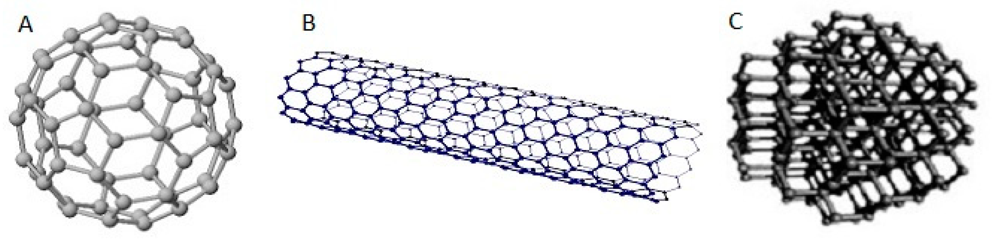
2. Characteristics of Fullerenes, Nanotubes and Nanodiamonds
2.1. Fullerenes
2.2. Nanotubes
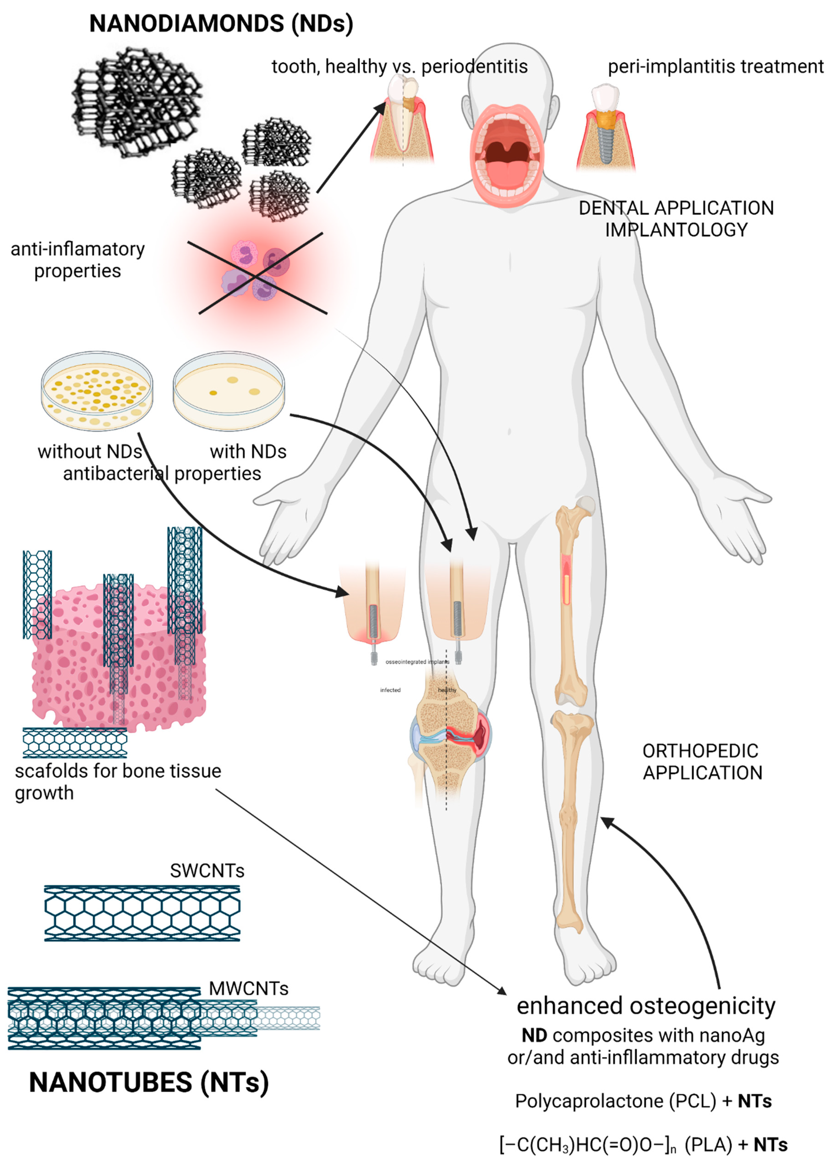
2.3. Nanodiamonds
3. Carbon Nanoparticles in Cancer and Bone Reconstruction
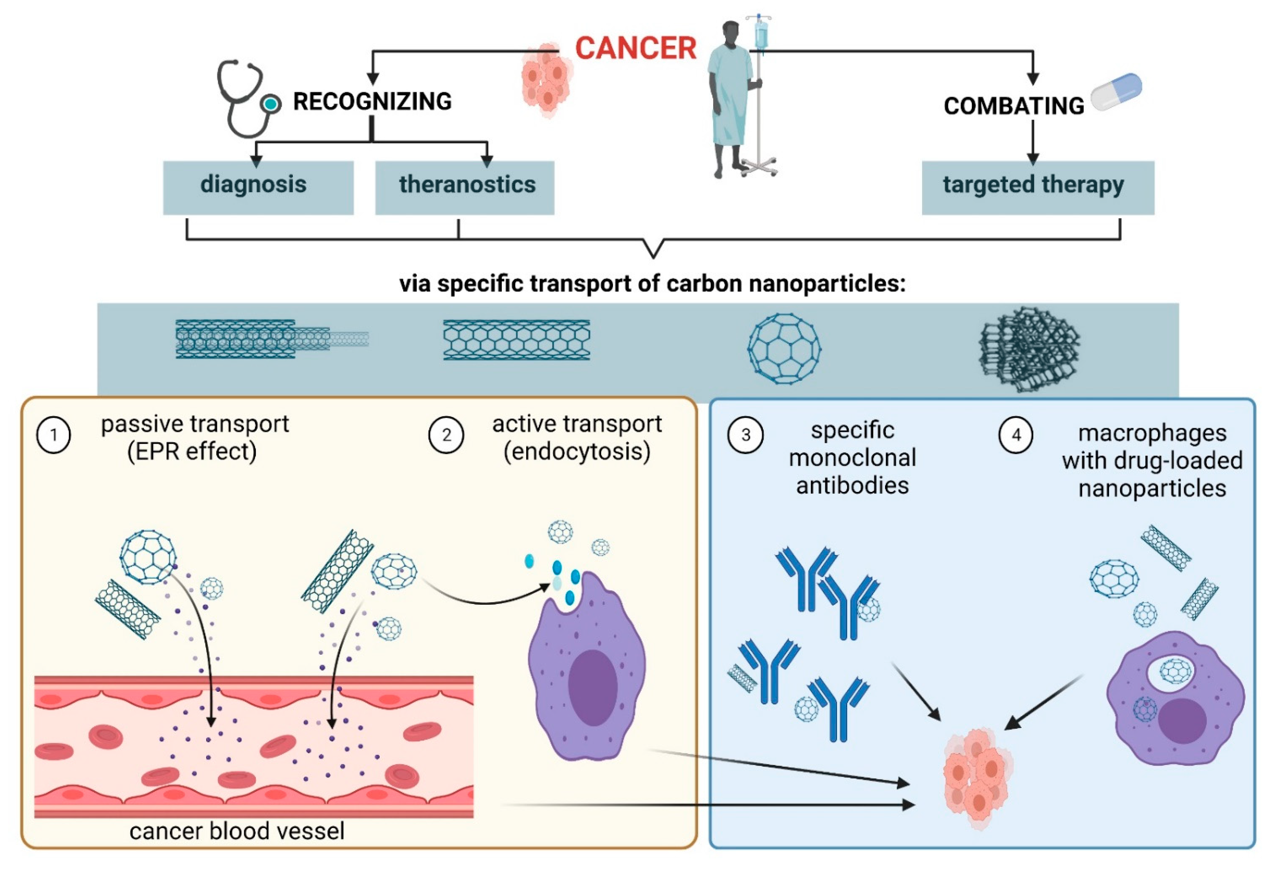
3.1. Targeted Therapy and Theranostics in Cancer
3.2. Reconstruction of Bone and Cartilage
4. Antibacterial and Anti-Inflammatory Properties of Nanoparticles in Implantology
5. Transport of Carbon Nanoparticles into Cells
5.1. Transport of Nanoparticles to Cancer Cells
5.2. Passing the Blood-Brain Barrier
5.3. Macrophages in Transport of Nanoparticles
6. Unfavorable Effects of Carbon Nanoparticles
7. Conclusions
Author Contributions
Funding
Institutional Review Board Statement
Informed Consent Statement
Data Availability Statement
Acknowledgments
Conflicts of Interest
References
- Bera, A.; Belhaj, H. Application of nanotechnology by means of nanoparticles and nanodispersions in oil recovery—A comprehensive review. J. Nat. Gas. Sci. Eng. 2016, 34, 1284–1309. [Google Scholar] [CrossRef]
- Mnyusiwalla, A.; Daar, A.S.; Singer, P.A. Mind the gap: Science and ethics in nanotechnology. Nanotechnology 2003, 14, R9–R13. [Google Scholar] [CrossRef] [Green Version]
- Attota, R.K.; Liu, E.C. Volume determination of irregularly-shaped quasi-spherical nanoparticles. Anal. Bioanal. Chem. 2016, 408, 7897–7903. [Google Scholar] [CrossRef] [Green Version]
- Khan, I.; Saeed, K.; Khan, I. Nanoparticles: Properties, applications and toxicities. Arab. J. Chem. 2019, 12, 908–931. [Google Scholar] [CrossRef]
- Slezakova, K.; Morais, S.; Carmo Pereir, M.D. Atmospheric Nanoparticles and Their Impacts on Public Health; InTech: London, UK, 2013. [Google Scholar]
- Nowack, B.; Bucheli, T.D. Occurrence, behavior and effects of nanoparticles in the environment. Environ. Pollut. 2007, 150, 5–22. [Google Scholar] [CrossRef] [PubMed]
- Jeevanandam, J.; Barhoum, A.; Chan, Y.S.; Dufresne, A.; Danquah, M.K. Review on nanoparticles and nanostructured materials: History, sources, toxicity and regulations. Beilstein. J. Nanotechnol 2018, 9, 1050–1074. [Google Scholar] [CrossRef] [Green Version]
- Zhao, Y.; Shen, X.; Ma, R.; Hou, Y.; Qian, Y.; Fan, C. Biological and biocompatible characteristics of fullerenols nanomaterials for tissue engineering. Histol. Histopathol. 2021, 18316. [Google Scholar]
- Speranza, G. Carbon Nanomaterials: Synthesis, Functionalization and Sensing Applications. Nanomaterials 2021, 11, 967. [Google Scholar] [CrossRef]
- Wang, J.; Hu, Z.; Xu, J.; Zhao, Y. Therapeutic applications of low-toxicity spherical nanocarbon materials. NPG Asia Mater. 2014, 6, e84. [Google Scholar] [CrossRef]
- Auría-Soro, C.; Nesma, T.; Juanes-Velasco, P.; Landeira-Viñuela, A.; Fidalgo-Gomez, H.; Acebes-Fernandez, V.; Gongora, R.; Almendral Parra, M.J.; Manzano-Roman, R.; Fuentes, M. Interactions of Nanoparticles and Biosystems: Microenvironment of Nanoparticles and Biomolecules in Nanomedicine. Nanomaterials 2019, 9, 1365. [Google Scholar] [CrossRef] [PubMed] [Green Version]
- Grebowski, J.; Krokosz, A.; Puchala, M. Fullerenol C60(OH)36 could associate to band 3 protein of human erythrocyte membranes. Biochim. Et Biophys. Acta (BBA) Biomembr. 2013, 1828, 2007–2014. [Google Scholar] [CrossRef] [Green Version]
- Sharoyko, V.V.; Ageev, S.V.; Podolsky, N.E.; Petrov, A.V.; Litasova, E.V.; Vlasov, T.D.; Vasina, L.V.; Murin, I.V.; Piotrovskiy, L.B.; Semenov, K.N. Biologically active water-soluble fullerene adducts: Das Glasperlenspiel (by H. Hesse)? J. Mol. Liq. 2021, 323, 114990. [Google Scholar] [CrossRef]
- Lay, C.L.; Liu, J.; Liu, Y. Functionalized carbon nanotubes for anticancer drug delivery. Expert Rev. Med. Devices 2011, 8, 561–566. [Google Scholar] [CrossRef] [PubMed]
- Garriga, R.; Herrero-Continente, T.; Palos, M.; Cebolla, V.L.; Osada, J.; Muñoz, E.; Rodríguez-Yoldi, M.J. Toxicity of Carbon Nanomaterials and Their Potential Application as Drug Delivery Systems: In Vitro Studies in Caco-2 and MCF-7 Cell Lines. Nanomaterials 2020, 10, 1617. [Google Scholar] [CrossRef] [PubMed]
- Kovel, E.; Sachkova, A.; Vnukova, N.; Churilov, G.; Knyazeva, E.; Kudryasheva, N. Antioxidant Activity and Toxicity of Fullerenols via Bioluminescence Signaling: Role of Oxygen Substituents. Int. J. Mol. Sci. 2019, 20, 2324. [Google Scholar] [CrossRef] [PubMed] [Green Version]
- Zhang, W.; Gong, X.; Liu, C.; Piao, Y.; Sun, Y.; Diao, G. Water-soluble inclusion complex of fullerene with γ-cyclodextrin polymer for photodynamic therapy. J. Mater. Chem. B 2014, 2, 5107–5115. [Google Scholar] [CrossRef]
- Popov, V.N. Carbon nanotubes: Properties and application. Mater. Sci. Eng. R Rep. 2004, 43, 61–102. [Google Scholar] [CrossRef]
- Świdwińska-Gajewska, A.M.; Czerczak, S. Carbon nanotubes—Characteristic of the substance, biological effects and occupational exposure levels. Med. Pr. 2017, 68, 259–276. [Google Scholar] [CrossRef] [Green Version]
- Díez-Pascual, A.M. Chemical Functionalization of Carbon Nanotubes with Polymers: A Brief Overview. Macromol 2021, 1, 64–83. [Google Scholar] [CrossRef]
- Mallakpour, S.; Soltanian, S. Surface functionalization of carbon nanotubes: Fabrication and applications. RSC Adv. 2016, 6, 109916–109935. [Google Scholar] [CrossRef]
- Langauer-Lewowicka, H.; Pawlas, K. Nanoparticles, nanotechnology—Potential environmental and occupational hazards. Med. Srod. 2014, 17, 7–14. [Google Scholar]
- Hirsch, A.; Vostrowsky, O. Functionalization of Carbon Nanotubes. In Functional Molecular Nanostructures; Schlüter, A.D., Ed.; Springer: Berlin/Heidelberg, Germany, 2005; pp. 193–237. [Google Scholar]
- Chen, Z.; Zhang, A.; Wang, X.; Zhu, J.; Fan, Y.; Yu, H.; Yang, Z. The Advances of Carbon Nanotubes in Cancer Diagnostics and Therapeutics. J. Nanomater. 2017, 2017, 1–13. [Google Scholar] [CrossRef] [Green Version]
- Kafa, H.; Wang, J.T.-W.; Rubio, N.; Venner, K.; Anderson, G.; Pach, E.; Ballesteros, B.; Preston, J.E.; Abbott, N.J.; Al-Jamal, K.T. The interaction of carbon nanotubes with an in vitro blood-brain barrier model and mouse brain in vivo. Biomaterials 2015, 53, 437–452. [Google Scholar] [CrossRef] [Green Version]
- Gonzalez-Carter, D.; Goode, A.E.; Kiryushko, D.; Masuda, S.; Hu, S.; Lopes-Rodrigues, R.; Dexter, D.T.; Shaffer, M.S.P.; Porter, A.E. Quantification of blood–brain barrier transport and neuronal toxicity of unlabelled multiwalled carbon nanotubes as a function of surface charge. Nanoscale 2019, 11, 22054–22069. [Google Scholar] [CrossRef] [PubMed]
- Moche, H.; Paget, V.; Chevalier, D.; Lorge, E.; Claude, N.; Girard, H.A.; Arnault, J.C.; Chevillard, S.; Nesslany, F. Carboxylated nanodiamonds can be used as negative reference in in vitro nanogenotoxicity studies. J. Appl. Toxicol. 2017, 37, 954–961. [Google Scholar] [CrossRef]
- Reineck, P.; Lau, D.W.M.; Wilson, E.R.; Fox, K.; Field, M.R.; Deeleepojananan, C.; Mochalin, V.N.; Gibson, B.C. Effect of Surface Chemistry on the Fluorescence of Detonation Nanodiamonds. ACS Nano 2017, 11, 10924–10934. [Google Scholar] [CrossRef] [PubMed]
- Zhao, F.; Liu, R.; Yu, X.; Ding, H.; Qu, X.; Zhang, Q. Carbon Fiber Grafted with Nanodiamond: Preparation and Characterization. J. Nanosci. Nanotechnol. 2015, 15, 5807–5815. [Google Scholar] [CrossRef]
- Ansari, S.A.; Satar, R.; Jafri, M.A.; Rasool, M.; Ahmad, W.; Kashif Zaidi, S. Role of Nanodiamonds in Drug Delivery and Stem Cell Therapy. Iran. J. Biotechnol. 2016, 14, 130–141. [Google Scholar] [CrossRef] [Green Version]
- Ho, D. Nanodiamond-Based Chemotherapy and Imaging. In Nanotechnology-Based Precision Tools for the Detection and Treatment of Cancer; Mirkin, C.A., Meade, T.J., Petrosko, S.H., Stegh, A.H., Eds.; Springer International Publishing: Cham, Switzerland, 2015; pp. 85–102. [Google Scholar]
- Li, Y.; Tong, Y.; Cao, R.; Tian, Z.; Yang, B.; Yang, P. In vivo enhancement of anticancer therapy using bare or chemotherapeutic drug-bearing nanodiamond particles. Int. J. Nanomed. 2014, 9, 1065–1082. [Google Scholar] [CrossRef] [Green Version]
- Ali, M.S.; Metwally, A.A.; Fahmy, R.H.; Osman, R. Nanodiamonds: Minuscule gems that ferry antineoplastic drugs to resistant tumors. Int. J. Pharm. 2019, 558, 165–176. [Google Scholar] [CrossRef] [PubMed]
- Guo, H.; Hu, H.; Yu, X.; Naito, K.; Zhang, Q. Covalent Functionalization of Nanodiamonds with Natural Amino Acids and Ascorbic Acids. J. Nanosci. Nanotechnol. 2019, 19, 7574–7583. [Google Scholar] [CrossRef] [PubMed]
- Kim, S.-W.; Lee, Y.K.; Kim, S.-H.; Park, J.-Y.; Lee, D.U.; Choi, J.; Hong, J.H.; Kim, S.; Khang, D. Covalent, Non-Covalent, Encapsulated Nanodrug Regulate the Fate of Intra- and Extracellular Trafficking: Impact on Cancer and Normal Cells. Sci. Rep. 2017, 7, 6454. [Google Scholar] [CrossRef]
- Pérez-Herrero, E.; Fernández-Medarde, A. Advanced targeted therapies in cancer: Drug nanocarriers, the future of chemotherapy. Eur. J. Pharm. Biopharm. 2015, 93, 52–79. [Google Scholar] [CrossRef] [PubMed] [Green Version]
- Saleem, J.; Wang, L.; Chen, C. Carbon-Based Nanomaterials for Cancer Therapy via Targeting Tumor Microenvironment. Adv. Healthc. Mater. 2018, 7, 1800525. [Google Scholar] [CrossRef] [PubMed]
- Shao, W.; Paul, A.; Rodes, L.; Prakash, S. A New Carbon Nanotube-Based Breast Cancer Drug Delivery System: Preparation and In Vitro Analysis Using Paclitaxel. Cell Biochem. Biophys. 2015, 71, 1405–1414. [Google Scholar] [CrossRef] [PubMed]
- Chipaux, M.; Van der Laan, K.J.; Hemelaar, S.R.; Hasani, M.; Zheng, T.; Schirhagl, R.A.-O. Nanodiamonds and Their Applications in Cells. Small 2018, 14, e1704263. [Google Scholar] [CrossRef]
- Xu, S.; Cui, F.; Huang, D.; Zhang, D.; Zhu, A.; Sun, X.; Cao, Y.; Ding, S.; Wang, Y.; Gao, E.; et al. PD-L1 monoclonal antibody-conjugated nanoparticles enhance drug delivery level and chemotherapy efficacy in gastric cancer cells. Int. J. Nanomed. 2018, 14, 17–32. [Google Scholar] [CrossRef] [Green Version]
- Ji, Z.; Lin, G.; Lu, Q.; Meng, L.; Shen, X.; Dong, L.; Fu, C.; Zhang, X. Targeted therapy of SMMC-7721 liver cancer in vitro and in vivo with carbon nanotubes based drug delivery system. J. Colloid Interface Sci. 2012, 365, 143–149. [Google Scholar] [CrossRef]
- Maeda, H.; Wu, J.; Sawa, T.; Matsumura, Y.; Hori, K. Tumor vascular permeability and the EPR effect in macromolecular therapeutics: A review. J. Control. Release 2000, 65, 271–284. [Google Scholar] [CrossRef]
- Li, D.; Chen, X.; Wang, H.; Liu, J.; Zheng, M.; Fu, Y.; Yu, Y.; Zhi, J. Cetuximab-conjugated nanodiamonds drug delivery system for enhanced targeting therapy and 3D Raman imaging. J. Biophotonics 2017, 10, 1636–1646. [Google Scholar] [CrossRef] [Green Version]
- Welsher, K.; Liu, Z.; Daranciang, D.; Dai, H. Selective probing and imaging of cells with single walled carbon nanotubes as near-infrared fluorescent molecules. Nano Lett. 2008, 8, 586–590. [Google Scholar] [CrossRef]
- Moscariello, P.; Raabe, M.; Liu, W.; Bernhardt, S.; Qi, H.; Kaiser, U.; Wu, Y.; Weil, T.; Luhmann, H.J.; Hedrich, J. Unraveling In Vivo Brain Transport of Protein-Coated Fluorescent Nanodiamonds. Small 2019, 15, e1902992. [Google Scholar] [CrossRef]
- Schrand, A.M.; Lin, J.B.; Hens, S.C.; Hussain, S.M. Temporal and mechanistic tracking of cellular uptake dynamics with novel surface fluorophore-bound nanodiamonds. Nanoscale 2011, 3, 435–445. [Google Scholar] [CrossRef]
- Torelli, M.D.; Nunn, N.A.; Shenderova, O.A. A Perspective on Fluorescent Nanodiamond Bioimaging. Small 2019, 15, e1902151. [Google Scholar] [CrossRef] [PubMed]
- Gupta, C.; Prakash, D.; Gupta, S. Cancer treatment with nano-diamonds. Front. Biosci (Sch. Ed.) 2017, 9, 62–70. [Google Scholar] [CrossRef] [Green Version]
- Wang, X.; Low, X.C.; Hou, W.; Abdullah, L.N.; Toh, T.B.; Mohd Abdul Rashid, M.; Ho, D.; Chow, E.K.-H. Epirubicin-Adsorbed Nanodiamonds Kill Chemoresistant Hepatic Cancer Stem Cells. ACS Nano 2014, 8, 12151–12166. [Google Scholar] [CrossRef] [PubMed] [Green Version]
- Namdar, R.; Nafisi, S. Nanodiamond applications in skin preparations. Drug Discov. Today 2018, 23, 1152–1158. [Google Scholar] [CrossRef]
- Bertrand, J.-R.; Pioche-Durieu, C.; Ayala, J.; Petit, T.; Girard, H.A.; Malvy, C.P.; Le Cam, E.; Treussart, F.; Arnault, J.-C. Plasma hydrogenated cationic detonation nanodiamonds efficiently deliver to human cells in culture functional siRNA targeting the Ewing sarcoma junction oncogene. Biomaterials 2015, 45, 93–98. [Google Scholar] [CrossRef]
- Dahlman, J.E.; Kauffman, K.J.; Langer, R.; Anderson, D.G. Chapter Three—Nanotechnology for In vivo Targeted siRNA Delivery. In Advances in Genetics; Huang, L., Liu, D., Wagner, E., Eds.; Academic Press: Cambridge, MA, USA, 2014; Volume 88, pp. 37–69. [Google Scholar]
- Shahzad, M.M.K.; Mangala, L.S.; Han, H.D.; Lu, C.; Bottsford-Miller, J.; Nishimura, M.; Mora, E.M.; Lee, J.-W.; Stone, R.L.; Pecot, C.V.; et al. Targeted Delivery of Small Interfering RNA Using Reconstituted High-Density Lipoprotein Nanoparticles. Neoplasia 2011, 13, 309–IN8. [Google Scholar] [CrossRef] [PubMed] [Green Version]
- Bi, Y.; Zhang, Y.; Cui, C.; Ren, L.; Jiang, X. Gene-silencing effects of anti-survivin siRNA delivered by RGDV-functionalized nanodiamond carrier in the breast carcinoma cell line MCF-7. Int. J. Nanomed. 2016, 11, 5771–5787. [Google Scholar] [CrossRef] [PubMed] [Green Version]
- Ibrahim, M.; Xue, Y.; Ostermann, M.; Sauter, A.; Steinmueller-Nethl, D.; Schweeberg, S.; Krueger, A.; Cimpan, M.R.; Mustafa, K. In vitro cytotoxicity assessment of nanodiamond particles and their osteogenic potential. J. Biomed. Mater. Res. A 2018, 106, 1690–1707. [Google Scholar] [CrossRef] [PubMed]
- Pan, L.; Pei, X.; He, R.; Wan, Q.; Wang, J. Multiwall carbon nanotubes/polycaprolactone composites for bone tissue engineering application. Colloids Surf. B Biointerfaces 2012, 93, 226–234. [Google Scholar] [CrossRef] [PubMed]
- Vieira, S.A.-O.; Vial, S.; Reis, R.L.; Oliveira, J.M. Nanoparticles for bone tissue engineering. Biotechnol. Prog. 2017, 33, 590–611. [Google Scholar] [CrossRef] [PubMed] [Green Version]
- Ju, Y.M.; Yu, B.; Koob, T.J.; Moussy, Y.; Moussy, F. A novel porous collagen scaffold around an implantable biosensor for improving biocompatibility. I. In vitro/in vivo stability of the scaffold and in vitro sensitivity of the glucose sensor with scaffold. J. Biomed. Mater. Res. A 2008, 87, 136–146. [Google Scholar] [CrossRef]
- Jun Han, Z.; Rider, A.E.; Ishaq, M.; Kumar, S.; Kondyurin, A.; Bilek, M.M.M.; Levchenko, I.; Ostrikov, K. Carbon nanostructures for hard tissue engineering. RSC Adv. 2013, 3, 11058–11072. [Google Scholar] [CrossRef]
- Magiera, A.; Markowski, J.; Menaszek, E.; Pilch, J.; Blazewicz, S. PLA-Based Hybrid and Composite Electrospun Fibrous Scaffolds as Potential Materials for Tissue Engineering. J. Nanomater. 2017, 2017, 9246802. [Google Scholar] [CrossRef]
- Mirmusavi, M.H.; Zadehnajar, P.; Semnani, D.; Karbasi, S.; Fekrat, F.; Heidari, F. Evaluation of physical, mechanical and biological properties of poly 3-hydroxybutyrate-chitosan-multiwalled carbon nanotube/silk nano-micro composite scaffold for cartilage tissue engineering applications. Int. J. Biol. Macromol. 2019, 132, 832–855. [Google Scholar] [CrossRef]
- Wang, L.; Cao, W.; Wang, X.; Li, P.; Zhou, J.; Zhang, G.; Li, X.; Xing, X. Biodegradable silver-loaded polycation modified nanodiamonds/polyurethane scaffold with improved antibacterial and mechanical properties for cartilage tissue repairing. J. Mater. Sci. Mater. Med. 2019, 30, 41. [Google Scholar] [CrossRef] [PubMed]
- Lee, D.-K.; Kim, S.V.; Limansubroto, A.N.; Yen, A.; Soundia, A.; Wang, C.-Y.; Shi, W.; Hong, C.; Tetradis, S.; Kim, Y.; et al. Nanodiamond–Gutta Percha Composite Biomaterials for Root Canal Therapy. ACS Nano 2015, 9, 11490–11501. [Google Scholar] [CrossRef]
- Lee, D.-K.; Kee, T.; Liang, Z.; Hsiou, D.; Miya, D.; Wu, B.; Osawa, E.; Chow, E.K.-H.; Sung, E.C.; Kang, M.K.; et al. Clinical validation of a nanodiamond-embedded thermoplastic biomaterial. Proc. Natl. Acad. Sci. USA 2017, 114, E9445–E9454. [Google Scholar] [CrossRef] [Green Version]
- Prabhakar, N.; Khan, M.H.; Peurla, M.; Chang, H.-C.; Hänninen, P.E.; Rosenholm, J.M. Intracellular Trafficking of Fluorescent Nanodiamonds and Regulation of Their Cellular Toxicity. ACS Omega 2017, 2, 2689–2693. [Google Scholar] [CrossRef] [Green Version]
- Solarska-Ściuk, K.; Gajewska, A.; Glińska, S.; Studzian, M.; Michlewska, S.; Balcerzak, Ł.; Skolimowski, J.; Kolago, B.; Bartosz, G. Intracellular transport of nanodiamond particles in human endothelial and epithelial cells. Chem. Biol. Interact. 2014, 219, 90–100. [Google Scholar] [CrossRef] [PubMed]
- Perevedentseva, E.; Hong, S.F.; Huang, K.J.; Chiang, I.T.; Lee, C.Y.; Tseng, Y.T.; Cheng, C.L. Nanodiamond internalization in cells and the cell uptake mechanism. J. Nanoparticle Res. 2013, 15, 1834. [Google Scholar] [CrossRef]
- Sharma, G.; Sharma, A.R.; Lee, S.-S.; Bhattacharya, M.; Nam, J.-S.; Chakraborty, C. Advances in nanocarriers enabled brain targeted drug delivery across blood brain barrier. Int. J. Pharm. 2019, 559, 360–372. [Google Scholar] [CrossRef]
- Moura, R.P.; Almeida, A.; Sarmento, B. The role of non-endothelial cells on the penetration of nanoparticles through the blood brain barrier. Prog. Neurobiol. 2017, 159, 39–49. [Google Scholar] [CrossRef] [PubMed]
- Cao, Q.; Yan, X.; Chen, K.; Huang, Q.; Melancon, M.P.; Lopez, G.; Cheng, Z.; Li, C. Macrophages as a potential tumor-microenvironment target for noninvasive imaging of early response to anticancer therapy. Biomaterials 2018, 152, 63–76. [Google Scholar] [CrossRef]
- Pang, L.; Qin, J.; Han, L.; Zhao, W.; Liang, J.; Xie, Z.; Yang, P.; Wang, J. Exploiting macrophages as targeted carrier to guide nanoparticles into glioma. Oncotarget 2016, 7, 37081–37091. [Google Scholar] [CrossRef] [PubMed] [Green Version]
- Mantovani, A.; Marchesi, F.; Malesci, A.; Laghi, L.; Allavena, P. Tumour-associated macrophages as treatment targets in oncology. Nat. Rev. Clin. Oncol. 2017, 14, 399–416. [Google Scholar] [CrossRef]
- Fujiwara, Y.; Takeya, M.; Komohara, Y. A novel strategy for inducing the antitumor effects of triterpenoid compounds: Blocking the protumoral functions of tumor-associated macrophages via STAT3 inhibition. Biomed. Res. Int. 2014, 2014, 348539. [Google Scholar] [CrossRef] [Green Version]
- Liu, Y.-C.; Zou, X.-B.; Chai, Y.-F.; Yao, Y.-M. Macrophage polarization in inflammatory diseases. Int. J. Biol. Sci. 2014, 10, 520–529. [Google Scholar] [CrossRef]
- Naota, M.; Shimada, A.; Morita, T.; Inoue, K.; Takano, H. Translocation Pathway of the Intratracheally Instilled C60 Fullerene from the Lung into the Blood Circulation in the Mouse: Possible Association of Diffusion and Caveolae-mediated Pinocytosis. Toxicol. Pathol. 2009, 37, 456–462. [Google Scholar] [CrossRef] [Green Version]
- Lacerda, L.; Bianco, A.; Prato, M.; Kostarelos, K. Carbon nanotubes as nanomedicines: From toxicology to pharmacology. Adv. Drug Deliv. Rev. 2006, 58, 1460–1470. [Google Scholar] [CrossRef]
- Morimoto, Y.; Hirohashi, M.; Ogami, A.; Oyabu, T.; Myojo, T.; Todoroki, M.; Yamamoto, M.; Hashiba, M.; Mizuguchi, Y.; Lee, B.W.; et al. Pulmonary toxicity of well-dispersed multi-wall carbon nanotubes following inhalation and intratracheal instillation. Nanotoxicology 2012, 6, 587–599. [Google Scholar] [CrossRef]
- DeLorme, M.P.; Muro, Y.; Arai, T.; Banas, D.A.; Frame, S.R.; Reed, K.L.; Warheit, D.B. Ninety-Day Inhalation Toxicity Study with A Vapor Grown Carbon Nanofiber in Rats. Toxicol. Sci. 2012, 128, 449–460. [Google Scholar] [CrossRef] [PubMed] [Green Version]
- Kim, J.S.; Song, K.S.; Yu, I.J. Multiwall Carbon Nanotube-Induced DNA Damage and Cytotoxicity in Male Human Peripheral Blood Lymphocytes. Int. J. Toxicol. 2015, 35, 27–37. [Google Scholar] [CrossRef] [Green Version]
- Chernova, T.; Murphy, F.A.; Galavotti, S.; Sun, X.M.; Powley, I.R.; Grosso, S.; Schinwald, A.; Zacarias-Cabeza, J.; Dudek, K.M.; Dinsdale, D.; et al. Long-Fiber Carbon Nanotubes Replicate Asbestos-Induced Mesothelioma with Disruption of the Tumor Suppressor Gene Cdkn2a (Ink4a/Arf). Curr. Biol. 2017, 27, 3302–3314.e6. [Google Scholar] [CrossRef] [PubMed] [Green Version]
- Visalli, G.; Currò, M.; Iannazzo, D.; Pistone, A.; Pruiti Ciarello, M.; Acri, G.; Testagrossa, B.; Bertuccio, M.P.; Squeri, R.; Di Pietro, A. In vitro assessment of neurotoxicity and neuroinflammation of homemade MWCNTs. Environ. Toxicol. Pharmacol. 2017, 56, 121–128. [Google Scholar] [CrossRef] [PubMed]
- Fresta, C.G.; Chakraborty, A.; Wijesinghe, M.B.; Amorini, A.M.; Lazzarino, G.; Lazzarino, G.; Tavazzi, B.; Lunte, S.M.; Caraci, F.; Dhar, P.; et al. Non-toxic engineered carbon nanodiamond concentrations induce oxidative/nitrosative stress, imbalance of energy metabolism, and mitochondrial dysfunction in microglial and alveolar basal epithelial cells. Cell Death Dis. 2018, 9, 245. [Google Scholar] [CrossRef] [PubMed]
- Karpeta-Kaczmarek, J.; Dziewięcka, M.; Augustyniak, M.; Rost-Roszkowska, M.; Pawlyta, M. Oxidative stress and genotoxic effects of diamond nanoparticles. Environ. Res. 2016, 148, 264–272. [Google Scholar] [CrossRef]
- Gaté, L.; Knudsen, K.B.; Seidel, C.; Berthing, T.; Chézeau, L.; Jacobsen, N.R.; Valentino, S.; Wallin, H.; Bau, S.; Wolff, H.; et al. Pulmonary toxicity of two different multi-walled carbon nanotubes in rat: Comparison between intratracheal instillation and inhalation exposure. Toxicol. Appl. Pharmacol. 2019, 375, 17–31. [Google Scholar] [CrossRef]


| Biological System | Administration Route | Type of Nanoparticle | Dose (concentration) and Size of Nanoparticles | Mechanism of Toxicity | Reference |
|---|---|---|---|---|---|
| Mice | Alimentary, respiratory | Fullerene C60 | 25 mg/kg 40 mg/kg | Accumulation in capillaries, lymph nodes, liver, spleen, and lungs | [75] |
| Rats | Intratracheal Inhalation | MWCNT | 12 nm (D), 0.4 µm (L) 67 nm (D), 4.0 µm (L) 0.5 mg/m3 1.5 mg/m3 | Increased levels of DNA damage, inflammation. Fibrosis only at the higher dose of 67nm/4.0 µm NTs | [84] |
| Rats | Intratracheal Inhalation | MWCNT | 63 nm (D), 1.1 μm (L); 0.2 mg/rat 1.0 mg/rat 0.37 mg/m3 | Inflammatory changes in lungs | [77] |
| Rats | Intratracheal | MWCNT | 158 nm (D), 5.8 μm (L) 0.5 mg/m3 2.5 mg/m3 25 mg/m3 | Accumulation of fibers in lungs, at the two highest concentrations, inflammation of the respiratory tract | [78] |
| Mice | Intratracheal | CNT | Long-fiber carbon nanotubes; 5 µg/mouse | Induction of pleural mesothelioma, an asbestos-like hazard | [80] |
| Neuronal cells | Incubation | MWCNT | 15-30 nm (D), 10-20 µm (L) or 0.2-1 µm (L); 15-20 layers; 12.5 µg/mL, 25 µg/mL | DNA damage, ROS formation, neuronal inflammation, elevated levels of cytokines TNFα, IL-1, IL-3, IL-6. | [81] |
| Human peripheral blood lymphocytes | Incubation | SWCNT/Biotin MWCNT/Fe/DOX | 12.5mg/mL 25mg/mL 50mg/mL | Delayed lymphocyte growth and damage to their DNA, increased levels of ROS | [79] |
| Microglial cells BU-2 Alveolar epithelial cells A549 | Incubation | ND | 2 µg/mL | 5% decrease in cell viability Increased production of NO and ROS | [82] |
| Bronchial epithelium cell lines HBE14; Colon carcinoma cells T84 | Incubation | ND | 20nm, 50nm, 100nm (D); 12.5–100 µg/mL | No genotoxic effects were observed in any of the lines | [27] |
| House cricket Acheta Domesticus | Alimentary | ND | 20mg/L in food 200mg/L in food | No toxic effects observed Increased parameters of oxidative stress, catalase, glutathione peroxidase and heat shock proteins, DNA instability | [83] |
Publisher’s Note: MDPI stays neutral with regard to jurisdictional claims in published maps and institutional affiliations. |
© 2021 by the authors. Licensee MDPI, Basel, Switzerland. This article is an open access article distributed under the terms and conditions of the Creative Commons Attribution (CC BY) license (https://creativecommons.org/licenses/by/4.0/).
Share and Cite
Lisik, K.; Krokosz, A. Application of Carbon Nanoparticles in Oncology and Regenerative Medicine. Int. J. Mol. Sci. 2021, 22, 8341. https://doi.org/10.3390/ijms22158341
Lisik K, Krokosz A. Application of Carbon Nanoparticles in Oncology and Regenerative Medicine. International Journal of Molecular Sciences. 2021; 22(15):8341. https://doi.org/10.3390/ijms22158341
Chicago/Turabian StyleLisik, Katarzyna, and Anita Krokosz. 2021. "Application of Carbon Nanoparticles in Oncology and Regenerative Medicine" International Journal of Molecular Sciences 22, no. 15: 8341. https://doi.org/10.3390/ijms22158341