Article

Synthesis of Novel Spiro-Tetrahydroquinoline Derivatives and Evaluation of Their Pharmacological Effects on Wound Healing

1
Department of Chemistry, National Taiwan Normal University, Taipei 116, Taiwan
2
Graduate Institute of Integrated Medicine, China Medical University, Taichung 404, Taiwan
3
Chinese Medicine Research and Development Center, China Medical University Hospital, Taichung 404, Taiwan
4
Department of Medical Laboratory Science and Biotechnology, College of Medical and Health Science, Asia University, Taichung 41354, Taiwan
*
Authors to whom correspondence should be addressed.
These authors contributed equally to this work.
Int. J. Mol. Sci. 2021, 22(12), 6251; https://doi.org/10.3390/ijms22126251
Submission received: 6 May 2021 / Revised: 3 June 2021 / Accepted: 7 June 2021 / Published: 10 June 2021
(This article belongs to the Special Issue Synthesis and Transformations of Bioactive Hydroxyquinolines)

Abstract

:
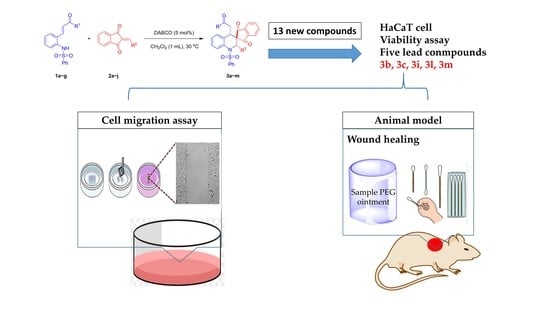
A highly diastereoselective method for the synthesis of novel spiro-tetrahydroquinoline derivatives is reported here, using a one-pot reaction method. All compounds were characterized by 1H-nuclear magnetic resonance (NMR) and mass spectroscopy, and their stereo configurations were confirmed by X-ray analysis. These activities of these derivatives were then tested in human keratocyte cells. The responses of cells to treatment with selected compounds were studied using scratch analysis, and the compounds were tested in a mouse excision wound model. Three of the derivatives demonstrated significant wound-healing activities.

Graphical Abstract

1. Introduction

Skin is the largest organ and the first defense to protect the human body [1,2]. It can be damaged by trauma, burns, skin diseases and so on. Severe skin trauma can impose physical, psychological, and economic burdens on patients. The wound healing process involves the coordination of many distinct but overlapping physiological spaces, comprising hemostasis, inflammation, epithelial cell proliferation, and tissue remodeling [3,4,5]. In elderly or diabetic patients, the risk of wound infection increased due to vascular aging and the lower tissue repair capability, which may eventually lead to chronic wounds [6]. Therefore, wound healing is one of the hot topics in skin surgery. At present, few drugs have been found with substantial abilities of promoting wound healing [7]. Actually, the quality of wound regeneration mainly depends on the efficiency of wound care [8]. In this study, we synthesized a series of novel spiro-tetrahydroquinoline derivatives and compared their effects in wound healing in human epidermal cells and animal models.
Quinoline derivatives have attracted both synthetic and biological chemists because of their diverse chemical and pharmacological properties, such as anticancer, antimycobacterial, anticonvulsant, anti-inflammatory and anti-cardiovascular diseases [9]. Some quinoline derivatives were found to have wound healing activity [10,11,12,13]. Tetrahydroquinoline derivatives, which are the reduced form of quinolines, were used as antibacterials, antitumor and anti-HIV agents [14]. However, there are very few reports about tetrahydroquinoline on wound healing even though their structures are so similar to each other [15]. It is a curious question of whether tetrahydroquinoline will deliver similar effects on wound healing or not.
The natural pharmacophore, spiro-1,3-indandione, has attracted considerable attention due to its diverse biological activities [16,17,18]. Fredericamycin A was isolated from a fermentation broth of the strain Streptomyces griseus and reported to demonstrate antibiotic properties [19]. Synthetic studies were also performed to obtain additional 1,3-indandione derivatives, which demonstrated antitumor and antibiotic activities [20,21]. In recent research, the spiro-1,3-indandione moiety has been considered to represent a valuable functional group for medicinal chemistry [22]. However, the synthesis of carbon spiro-1,3-indandiones remains a challenge [23,24]. Yan et al. previously synthesized spiro-1,3-indandiones from cyclic azomethine imine and 2-arylideneindene-1,3-dione via 1,3-dipolar cycloaddition [25]. Enders et al. used 1,4-dithiane-2,5-diol and 2-arylideneindene-1,3-dione as the starting materials to obtain spiro-1,3-indandione, using a squaramide-catalyzed sulfa-Michael/aldol domino reaction [26]. As a starting material that is commonly used for the generation of spiro-1,3-indandiones, 2-arylidene-1,3-indandiones were the focus of our recent study. In our previous research, 2-arylidene-1,3-indandiones were used to obtain enantioselective spiro-nitrocyclopropanes, which contained the spiro-1,3-indandione skeleton, catalyzed by cinchona alkoloid derivatives [27]. Indanedione-fused 2,6-disubstituted spiro-cyclohexanones have also been studied [28].
Recently, our team has focused on the wound-healing activities of natural products [29]. As tetrahydroquinoline and 1,3-indandione are both widely studied as pharmacophores in medicinal chemistry, we considered whether the synthesis of hybrids of tetrahydroquinoline and spiro-indandione residues could result in any meaningful active compounds. Song and Du have synthesized highly functionalized spirothiazolidinone tetrahydroquinolines via a squaramide-catalyzed cascade reaction [30]. Therefore, we synthesized spiro-tetrahydroquinolines compounds with a similar method for further study, focusing particularly on their wound-healing capabilities.

2. Results

The ortho-N-sulfonated aminophenyl α,β-unsaturated ketone 1a and 2-benzylidene-1,3-indandione 2a were used as model substrates for the synthesis of spiro-tetrahydroqunioline derivative 3a via an aza-Michael/Michael reaction (Table 1). Initially, the reaction of 1a with 2a in the presence of DABCO furnished the desired product 3a in 71% yield at 30 °C in p-xylene (Table 1, entry 1). Examination of different tertiary amines such as DMAP, Et3N and DIPEA as catalysts did not improve the yield of the product 3a (Table 1, entries 2–4). It is worthy to note that all the catalysts furnished the product 3a in excellent diastereomeric ratio (>20:1). Furthermore, various solvents like toluene, CH2Cl2, THF, EtOAc and Et2O were screened to optimize the reaction conditions (Table 1, entries 5–10). The results proved that aprotic solvents are the best for the reaction conditions such as toluene and CH2Cl2 (Table 1, entries 5 and 6). Considering the solubility of the starting materials 1 and 2, we chose to test the CH2Cl2 as optimized solvent for further screening of reaction. When the reaction was carried out at 0 °C, the yield of the desired product 3a was improved to 97%, but the reaction required longer times (38 h) compared to 30 °C (Table 1, entries 6 and 11). The reaction conditions were further evaluated by the catalyst loading and concentration of the solvent, which indicated that 5 mol% of DABCO was sufficient for the completion of the reaction in CH2Cl2 (1 mL) (Table 1, entries 12 and 13). The reaction conditions in Table 1, entry 13 were selected as the optimal conditions for further studies. The products were purified by the recrystallization, and identified by 1H-NMR, 13C-NMR and HRMS. The stereo configuration of the compound 3a was determined by single crystal X-ray diffraction analysis as shown in Figure 1 [31].
With the optimal condition in hand, the substrate scope was further investigated. In general, all the substrates 1 and 2 with different electron-withdrawing and electron-donating R1 and R2 substituents were provided the desired spiro-tetrahydroquinolines in good to excellent yields (Table 2). At first, different indandione derivatives of R2 substituents were tested with 1a. We noticed significant steric and electronic effects of the R2 substituent in the reaction outcome. Substrates with meta- and para-bromo groups as R2 substituents reacted well with 1a to afford the spiro-tetrahydroquinoline products 3c and 3d in up to 83% yields within 3 h, whereas the substrate with R2 as ortho-bromo substituent was less reactive and the desired product 3b was obtained in 71% yield in longer reaction times (12 h). It clearly indicates that the rate of the reaction is reduced by the sterically hindered substituents. We also found that the reaction rate also depends on the electronic properties of the R2 substituents. For example, the substrates bearing electron-withdrawing groups 2d2g subjected with 1a, furnished the corresponding products 3d3g in high yields (81–85%) within 3 h (Table 1, entries 4–7), but the substrate bearing an electron-donating group such as 4-OMePh as R2 substituent 2h resulted in the desired product 3h in only moderate yield (68%), when prolonging the reaction time up to 29 h (Table 1, entry 8). In contrast, when substrates with heteroaryl (furyl or thienyl) groups 2i and 2j were employed, the reaction could not proceed to provide the products even after 24 h (Table 1, entries 9 and 10).
Furthermore, different R1 substituents of 1 were also tested in the reaction conditions to prepare the desired spiro-tetrahydroquinolines 3. Delightfully, the substrate having aldehyde (R1 = H) group reacted well with 2a to afford the corresponding product 3i in 95% yield within 3 h (Table 1, entry 11). As similar as R2, substrates bearing electron-withdrawing R1 groups (1c and 1d) more efficiently furnished the desired products 3j and 3k compared to the electron-donating group such as 1e (Table 1, entries 12–14). When substrate with ester group 1f was employed as the reactant, the corresponding product could not be found in the reaction (Table 1, entry 15). It could be understood that the second Michael addition was not efficient when the electron-rich ester is present rather than an aldehyde or ketone. In addition, 2-naphthyl group of 1g also furnished the product 3m in 85% yield in 6 h (Table 1, entry 16).
The results of the MTT test revealed the cell viability of human keratinocyte cells (HaCaT) treated with the 13 derived compounds as shown in Table 3. The survival rates of cells treated with 3b were approximately 80% at all treatment concentrations (Supplementary Figure S1A). Cells treated with 3c displayed a survival rate of 80% at concentrations of 6.25 and 12.5 µM, but the growth was inhibited at concentrations equal to or greater than 25 µM (Supplementary Figure S1B). Cells treated with 3i displayed low survival rates at concentrations greater than 25 µM (Supplementary Figure S1C). Cells treated with 3l displayed a survival rate of approximately 80% at all concentrations (Supplementary Figure S1D). The survival rates of cells treated with 3m at concentrations of 6.25 and 12.5 µM were 80% and 60%, respectively. Compared with the other tested compounds, the five compounds described above had little effect on HaCaT cell growth (Supplementary Figure S1), whereas the remaining compounds displayed the strong inhibition of HaCaT cell growth. The results of the remaining samples can be found in the supplementary materials. Therefore, we selected the five compounds with limited effects on cell viability for use in subsequent experiments.
Based on the results of the cell viability experiments, the activities of the five selected compounds were evaluated in the scratch assay. The results shown in Figure 2 indicated that the wounds healed gradually healed within 15–18 h when the tested drugs were added to the culture medium. 3b had a significant effect on wound healing at the concentration of 25 µM, but no significant effect was observed when the concentration was increased to 50 µM (Figure 3A). 3c had an effect on wound healing at concentrations of 12.5 and 25 µM (Figure 3B). 3i had a significant effect on wound healing at concentrations of 12.5 and 25 µM. Compared with the control groups, no significant effects on wound healing were observed for low concentration (6.25 µM) or high concentration (50 µM) treatment with either 3c or 3i (Figure 3C). 3l and 3m displayed no significant effects on the promotion of wound healing at any of the tested concentrations and were found to have inhibitory effects (Figure 3D,E). The optimal concentration data based on these results is shown in Figure 3F, and the compounds 3b, 3c, and 3i were selected for further study in an animal model of wound closure.
Based on the results obtained from the scratch assay, wound-healing tests in mice were performed to examine the effects of wound treatment using 3b, 3c, and 3i (Figure 4A). The control group was observed to shed the scab on the 9th day, and the wound repair was completed on the 13th day. In mice treated with 3b at a concentration of 50 µM, the scab fell off on the 12th day, and the wound repair was completed on the 13th day (Figure 4B). Treatment with 6.25 µM of 3c resulted in the shedding of the pupae on the 9th day, but the wound was not completely repaired by the 13th day (Figure 4C). When a 25 µM concentration of 3i was applied, the pupae were shed on the 11th day, and the wound was repaired. The wound healing was completed by the 11th day, and the pupae were shed on the 7th day when the concentration of 3i was increased to 50 µM (Figure 4D). We compared the optimal concentrations of 3b, 3c, and 3i treatment based on the results of the mouse wound healing model. 3i was demonstrated to have the best effect on wound healing at a concentration of 50 µM (Figure 4E).

3. Discussion

The plausible mechanism of the reaction is depicted in Scheme 1. Initially, the chalcone derivative 1 is deprotonated by tertiary amine, yielding a nitrogen-nucleophile I which would attack 2-arylidene-1,3-indandiones 2 to generate aza-Michael adduct II, through a first Michael addition (Scheme 1). The second intramolecular Michael addition upon II to provide III with both R1 and R2 in the same side and subsequent protonation and enolization would result in the spiro-tetrahydroquinolines 3 in high yields.
As shown in Scheme 1, the transition states have two isomers (syn- and anti-isomers) that can interchange with each other. In the piperidine ring, if the green hydrogen atom heads upward (syn-isomer), the steric effect of the sulfonamide and enone moiety was less impact, and compound 3 was more easily formed. On the contrary, green enone moiety heads upward in the anti-isomer which shows more steric effect and makes it more difficult to form 3′.
In previous studies that have examined wound healing, most researchers have focused on natural products, such as extracts and extensions of Chinese herbal medicines or marine natural products. Only a few studies have examined the effects of synthetic chemicals on wound-healing outcomes. In this study, we explored the efficacy of wound healing by studying synthetic compounds and their related skeletal extensions. All compounds were preliminarily studied in human epidermal cells (HaCaT). Compound 3i was selected because, at low concentrations, the survival rate of cells was over 80%. Compounds 3b, 3c, 3k, and 3m were shown to have less effect on the growth of HaCaT cells, based on the results of the MTT assay (Supplementary Figure S1). We performed a scratch analysis and observed that compound 3i has positive effects on wound healing. In the MTT assay, 3i displayed the significant inhibition of cell growth at the 25 µM concentration. Due to the different outcomes observed for these two experiments, we considered whether 3i could be effective in animal models. Compared with 3b and 3c, 3i was demonstrated to represent the best compound for healing wounds on mouse skin. The wound was completely healed by the 11th day in mice treated with 3i. Based on the results of the MTT and wound-healing assay, 3i is thought to promote wound healing by promoting cell proliferation. In summary, we hope that the synthesis of spiro-tetrahydroqunioline derivatives might provide a new method for identifying chemicals for application in future wound-healing research.

4. Materials and Methods

All chemicals were analytical grade, purchased from Sigma-Aldrich (St. Louis, MO, USA), Alfa Aesar (Ward Hill, MA, USA), and Merck (Darmstadt, Germany). The purity of compounds was determined by TLC plates coated with Merck Silica gel 60 F254 (0.2 mm). Spots were observed under UV lamp or stained by dyeing agent. Infrared Spectroscopy (IR) were recorded on JASCOF/IR-5300 (Easton, MD, USA), polystyrene was used as internal standard to mark 1601 cm−1. 1H-NMR and 13C-NMR spectra were recorded on Bruker Avance 400 MHz spectrophotometer (Billerica, MA, USA). In 1H-NMR, CDCl3 was used as d-solvent, TMS as internal standard to mark 0 ppm. The definition of splitting term: singlet (s), doublet (d), triplet (t), qutratet (q), multiplet (m), coupling constant (J). In 13C-NMR, chloroform was used as internal standard to mark 77.0 ppm. Mass Spectroscopy (MS) and High-Resolution Mass Spectroscopy (HRMS) were recorded on JMS-700 (JEOL), (Tokyo, Japan), double focusing mass spectrometer (FAB and EI), Applied Biosystems 4800 Proteomics Analyze (MALDI) (Foster City, CA, USA) or Waters (Milford, MA, USA) LCT Premier XE (ESI). X-ray spectra were recorded on Enraf-Nonius FR590 and Nonious CAD4 Kappa Axis XRD (Bruker). Dulbecco’s Modified Eagle Medium (DMEM) and Penicillin-Streptomycin were purchased from Gibco (Waltham, MA, USA). Fetal Bovine Serum (FBS) was purchased from HyClone (Marlborough, MA, USA). Methythiazolyltetrazolium (MTT), Polyethylene glycol 400 (PEG 400), and Polyethylene glycol 4000 (PEG 4000) were purchased from Sigma-Aldrich. Culture insert for migration assay was purchased from ibidi (Planegg, Germany). Anesthesia for animal test (Zoletil) was purchased from Virbac (Carros, France). Fluorescence interpretation and image analysis were collected using CytationTM 5 Cell Imaging Multi-Mode Reader, BioTek (Winooski, VT, USA).

4.1. Ortho-N-Protected Aminophenyl α,β-Unsaturated Ketones (1)

2-aminobenzyl alcohol (20 mmol, 2.46 g) was dissolved in 100 mL DCM, and benzenesulfonyl chloride (22 mmol, 4.18 g) and 1 mL pyridine were added. The mixture was stirred for 12 h at r.t. The solvent was removed under reduced pressure. The residue 4 was not purified and dissolved in 50 mL DCM. Pyridinium chlorochromate (30 mmol, 6.46 g) was added and the resulting solution was stirred at r.t. for 4 h. The reaction mixture was filtered by Celite 545 and washed by DCM. After solvent was removed under reduced pressure, the residue was purified by flash chromatography (DCM: Hexanes = 2:1) to give 5, yield 97%. 5 (3 mmol, 0.83 g) was dissolved in 15 mL toluene, 6 (3.3 mmol) was added. The mixture was stirred at 80 °C for 12 h. The solvent was removed under reduced pressure, residue was purified by flash chromatography to yield 1 as shown in Scheme 2.
(E)-N-(2-(3-oxo-3-phenylprop-1-en-1-yl)phenyl)benzenesulfonamide (1a)
(mp 147.3–148.2 °C):
1H-NMR (400 MHz, CDCl3, 25 °C) δ/ppm: 7.93 (d, 2H, J = 8.2 Hz), 7.74 (d, 1H, J = 15.8 Hz), 7.71–7.66 (m, 2H),7.65–7.55 (m, 3H), 7.52–7.45 (m, 3H), 7.39 (t, 1H, J = 7.8 Hz), 7.36–7.21 (m, 4H), 7.20 (d, 1H, J = 15.8 Hz).
13C-NMR (100 MHz, CDCl3, 25 °C) δ/ppm: 190.21, 139.1, 138.8, 137.6, 135.2, 133.1, 132.9, 131.1, 131.0, 129.0, 128.63, 128.60, 127.9, 127.3, 127.2, 127.1, 124.2.
HRMS (ESI) for C21H18NO3S, [M + H]+ (364.1002), found 364.1006.
IR (KBr) ῡ (cm−1): 3236, 3063, 2821, 1655, 1602, 1573, 1333, 1218, 1165, 1091, 734.
(E)-N-(2-(3-oxoprop-1-en-1-yl)phenyl)benzenesulfonamide (1b).
mp: 137.0–137.6 °C.
1H-NMR (400 MHz, CDCl3, 25 °C) δ/ppm: 9.50 (d, 1H, J = 7.6 Hz), 7.71 (d, 2H, J = 7.8 Hz), 7.64–7.54 (m, 3H), 7.46 (t, 2H, J = 7.8 Hz), 7.38–7.30 (m, 2H), 7.16–7.07 (m, 2H), 6.54 (dd, 1H, J1 = 15.8 Hz, J2 = 7.8 Hz).
13C-NMR (100 MHz, CDCl3, 25 °C) δ/ppm: 193.8, 147.2, 138.5, 134.5, 133.4, 131.7, 131.4, 129.9, 129.2, 128.3, 128.0, 127.4, 127.3.
IR (KBr) ῡ (cm−1): 3237, 3063, 2828, 1671, 1623, 1448, 1331, 1160, 1132, 1091, 972, 758.
HRMS (ESI) for C15H13NO3SNa, [M + Na] + (310.0508), found 310.0517.
(E)-N-(2-(3-(4-bromophenyl)-3-oxoprop-1-en-1-yl)phenyl)benzenesulfonamide (1c).
mp: 137.0–137.6 °C.
1H-NMR (400 MHz, CDCl3, 25 °C) δ/ppm: 7.82–7.72 (m, 3H), 7.70 (d, 2H, J = 7.8 Hz), 7.63–7.58 (m, 3H), 7.55 (s, 1H), 7.45–7.26 (m, 6H), 7.16 (d, 1H, J = 15.8 Hz).
13C-NMR (100 MHz, CDCl3, 25 °C) δ/ppm: 189.2, 139.8, 138.9, 136.3, 135.2, 133.0, 131.9, 131.3, 131.0, 130.1, 129.0, 128.2, 127.8, 127.3, 127.2, 123.7.
IR (KBr) ῡ (cm−1): 3236, 3072, 2835, 1654, 1598, 1397, 1330, 1216, 1164, 1091, 758.
HRMS (ESI) for C21H17NO3S79Br, [M + H]+ (442.0107), found 442.0111.
HRMS (ESI) for C21H17NO3S81Br, [M + H]+ (444.0087), found 442.0093.
(E)-N-(2-(3-(4-nitrophenyl)-3-oxoprop-1-en-1-yl)phenyl)benzenesulfonamide (1d).
mp: 185.4–185.7 °C.
1H-NMR (400 MHz, CDCl3, 25 °C) δ/ppm: 8.37 (d, 2H, J = 8.8 Hz), 8.13 (d, 2H, J = 8.8 Hz), 7.81 (d, 1H, J = 15.8 Hz), 7.75–7.68 (m, 3H), 7.54–7.34 (m, 6H), 7.27–7.24 (m, 1H), 6.73 (s, 1H).
13C-NMR (100 MHz, CDCl3, 25 °C) δ/ppm: 189.0, 150.2, 142.5, 141.1, 138.7, 135.1, 133.2, 131.7, 131.0, 129.6, 129.2, 127.7, 127.4, 127.3, 123.9.
IR (KBr) ῡ (cm−1): 3253, 3068, 2926, 1664, 1597, 1523, 1329, 1212, 1162, 1090, 734.
HRMS (ESI) for C21H17N2O5S, [M + H]+ (409.0853), found 409.0856.
(E)-N-(2-(3-(4-methoxyphenyl)-3-oxoprop-1-en-1-yl)phenyl)benzenesulfonamide (1e).
mp: 171.8–172.0 °C.
1H-NMR (400 MHz, CDCl3, 25 °C) δ/ppm: 7.95 (d, 2H, J = 7.8 Hz), 7.83 (s, 1H), 7.75 (d, 1H, J = 15.8 Hz), 7.71–7.65 (m, 2H), 7.59–7.50 (m, 2H), 7.39 (t, 1H, J = 7.8 Hz), 7.35–7.23 (m, 4H), 7.18 (d, 1H, J = 15.8 Hz), 6.95 (d, 2H, J = 7.8 Hz), 3.88 (s, 3H).
13C-NMR (100 MHz, CDCl3, 25 °C) δ/ppm: 188.4, 163.6, 138.9, 138.3, 135.1, 132.9, 131.1, 131.0, 130.9, 130.5, 129.0, 127.8, 127.2, 127.1, 124.2, 113.9, 55.5.
IR (KBr) ῡ (cm−1): 3182, 3068, 2840, 1651, 1603, 1335, 1263, 1223, 1167, 1091, 1022, 760.
HRMS (ESI) for C22H20NO4S, [M + H]+ (394.1108), found 394.1114.
Ethyl-(E)-3-(2-(phenylsulfonamido)phenyl)acrylate (1f).
mp: 125.1–125.6 °C.
1H-NMR (400 MHz, CDCl3, 25 °C) δ/ppm: 7.68 (d, 2H, J = 8.0 Hz), 7.56 (d, 1H, J = 15.8 Hz), 7.50 (t, 1H, J = 7.6 Hz), 7.47–7.42 (m, 2H), 7.42–7.33 (m, 3H), 7.27–7.21 (m, 2H), 6.12 (d, 1H, J = 15.8 Hz), 4.23 (q, 2H, J = 7.2 Hz), 1.32 (t, 3H, J = 7.2 Hz).
13C-NMR (100 MHz, CDCl3, 25 °C) δ/ppm: 166.5, 138.8, 134.5, 133.0, 130.9, 130.4, 129.0, 127.5, 127.3, 127.2, 127.0, 120.7, 60.8, 14.2.
IR (KBr) ῡ (cm−1): 3244, 3068, 2983, 1692, 1635, 1448, 1320, 1166, 1091, 979, 762. HRMS (ESI) for C17H17NO4SNa, [M + Na]+ (354.0770), found 354.0775.
(E)-N-(2-(3-(naphthalen-2-yl)-3-oxoprop-1-en-1-yl)phenyl)benzenesulfonamide (1g).
mp: 161.2–161.5 °C.
1H-NMR (400 MHz, CDCl3, 25 °C) δ/ppm: 8.45 (s, 1H), 8.02–7.94 (m, 2H), 7.91–7.84 (m, 2H), 7.77 (d, 1H, J = 15.8 Hz), 7.73–7.67 (m, 2H), 7.67–7.53 (m, 3H), 7.51–7.43 (m, 2H), 7.43–7.25 (m, 6H).
13C-NMR (100 MHz, CDCl3, 25 °C) δ/ppm: 190.0, 139.0, 138.9, 135.6, 135.2, 134.9, 133.0, 132.5, 131.2, 131.1, 130.4, 129.6, 129.1, 128.7, 128.6, 127.8, 127.3, 127.2, 126.9, 124.5, 124.4.
IR (KBr) ῡ (cm−1): 3213, 3063, 2813, 1653, 1626, 1598, 1485, 1327, 1165, 1091, 759.
HRMS (ESI) for C25H20NO3S, [M + H]+ (414.1158), found 414.1164.

4.2. 2-Arylidene-1,3-Indandiones (2)

A solution of the mixture of 1,3-indandione (5 mmol, 0.73 g), aldehyde (5.5 mmol) and L-proline (1.5 mmol, 0.17 g) in methanol (90 mL) was stirred for 12 h at r.t. The solvent was removed under reduced pressure, and the residue was washed by methanol. The solid 2 was purified by flash chromatography as shown in Scheme 3.

4.3. Spiro-Tetrahydroquinoline (3)

Compounds 1 (0.2 mmol) and 2 (0.2 mmol) were dissolved in 1 mL DCM, 1.2 mg DABCO (5 mol%) was added as catalysis. The reaction was sealed and stirred at 30 °C. After the reaction completed, the mixture was quenched with 1 N hydrochloric acid aqueous solution and extracted with DCM. The organic layer was washed with water, dried over MgSO4, filtered, re-crystallized in ethanol and hexane.
3a (110.0 mg, yield 92%, mp 243–244 °C): 1a (72.7 mg, 0.2 mmol) and 2a (46.9 mg, 0.2 mmol) as starting material, reacted for 12 h.
1H-NMR (400 MHz, CDCl3, 25 °C) δ/ppm: 8.03 (d, 1H, J = 7.8 Hz), 7.92 (d, 1H, J = 7.6 Hz), 7.78–7.69 (m, 3H), 7.69–7.60 (m, 3H), 7.59–7.44 (m, 6H), 7.37 (t, 2H, J = 7.8 Hz), 7.20 (t, 1H, J = 7.6 Hz), 7.07–6.94 (m, 5H), 6.68 (d, 1H, J = 8.0 Hz), 5.87 (s, 1H), 3.05 (d, 1H, J = 11 Hz), 2.95 (dd, 1H, J1 = 16.4 Hz, J2 = 11 Hz), 2.34 (dd, 1H, J1 = 16.4 Hz, J2 = 2.0 Hz).
13C-NMR (100 MHz, CDCl3, 25 °C) δ/ppm: 201.2, 197.2, 195.1, 142.7, 141.3, 138.7, 137.6, 137.3, 136.5, 135.9, 135.8, 133.5, 133.4, 133.3, 129.2, 128.7, 128.1, 127.9, 127.8, 127.7, 127.5, 127.1, 126.8, 125.3, 123.03, 122.98, 66.1, 66.0, 37.3, 36.3.
HRMS (ESI) for C37H27NO5S, [M + Na]+ (620.1508), found 620.1509.
IR (KBr) ῡ (cm−1): 3440, 1741, 1705, 1448, 1356, 1250, 1169, 1093, 753, 582.
3b (96.1 mg, yield 71%, mp 229–230 °C): 1a (72.7 mg, 0.2 mmol) and 2b (62.3 mg, 0.2 mmol) as starting material, reacted for 24 h.
1H-NMR (400 MHz, CDCl3, 25 °C) δ/ppm: 7.97 (d, 1H, J = 7.6 Hz), 7.92 (dd, 1H, J1 = 7.6 Hz, J2 = 1.2Hz), 7.84–7.79 (m, 2H), 7.76(td, 1H, J1 = 7.2 Hz, J2 = 0.8 Hz), 7.71–7.67 (m, 2H), 7.64 (td, 1H, J1 = 7.6 Hz, J2 = 0.8 Hz), 7.62–7.55 (m, 3H), 7.52 (t, 1H, J = 7.6 Hz), 7.47 (t, 1H, J = 7.6 Hz), 7.43–7.34 (m, 3H), 7.28–7.20 (m, 2H), 7.14–7.06 (m, 2H), 6.89–6.82 (m, 1H), 6.77 (d, 1H, J = 7.8 Hz), 6.29 (s, 1H), 3.14–3.00 (m, 2H), 2.33 (d, 1H, J = 16 Hz).
13C-NMR (100 MHz, CDCl3, 25 °C) δ/ppm: 199.9, 197.7, 195.2, 142.4, 141.7, 138.6, 137.9, 137.0, 136.3, 135.9, 134.1, 133.4, 132.0, 131.5, 129.4, 129.0, 128.6, 128.04, 127.92, 127.7, 127.4, 127.2, 127.0, 125.6, 123.4, 122.7, 121.7, 66.1, 63.3, 36.3, 36.0.
HRMS (ESI) for C37H26Br79NO5S, [M + Na]+ (698.0607), found 698.0612.
HRMS (ESI) for C37H26Br81NO5S, [M + Na]+ (700.0587), found 700.0599.
IR (KBr) ῡ (cm−1): 3066, 1742, 1707, 1594, 1448, 1358, 1251, 1171, 1090, 949, 754.
3c (109.6 mg, yield 81%, mp 254–255 °C): 1a (72.7 mg, 0.2 mmol) and 2c (62.3 mg, 0.2 mmol) as starting material, reacted for 3 h.
1H-NMR (400 MHz, CDCl3, 25 °C) δ/ppm: 8.03 (d, 1H, J = 7.8 Hz), 7.95 (d, 1H, J = 7.8 Hz), 7.79 (t, 1H, J = 7.4 Hz), 7.76–7.67 (m, 3H), 7.65–7.46 (m, 8H), 7.37 (t, 2H, J = 7.4 Hz), 7.21 (t, 1H, J = 7.4 Hz), 7.17–7.08 (m, 2H), 7.04 (d, 1H, J = 7.4 Hz), 6.92 (t, 1H, J = 7.6 Hz), 6.66 (d, 1H, J = 7.6 Hz), 5.80 (s, 1H), 3.08–2.87 (m, 2H), 2.33 (d, 1H, J = 16.0 Hz).
13C-NMR (100 MHz, CDCl3, 25 °C) δ/ppm: 200.9, 196.8, 194.9, 142.6, 141.2, 141.1, 137.3, 137.1, 136.7, 136.1, 135.8, 133.5, 133.3, 130.8, 129.80, 129.77, 129.30, 128.7, 128.0, 127.9, 127.7, 127.1, 126.9, 125.6, 125.3, 123.2, 123.1, 122.1, 65.8, 65.3, 37.4, 36.3.
HRMS (ESI) for C37H26Br79NO5S, [M + Na]+ (698.0607), found 698.0613.
HRMS (ESI) for C37H26Br81NO5S, [M + Na]+ (700.0587), found 700.0601.
IR (KBr) ῡ (cm−1): 3064, 1741, 1707, 1594, 1448, 1357, 1250, 1169, 1090, 915, 732.
3d (112.3 mg, yield 83%, mp 249–250 °C): 1a (72.7 mg, 0.2 mmol) and 2d (62.3 mg, 0.2 mmol) as starting material, reacted for 3 h.
1H-NMR (400 MHz, CDCl3, 25 °C) δ/ppm: 8.01 (d, 1H, J = 8.0 Hz), 7.92 (d, 1H, J = 7.6 Hz), 7.80 (t, 1H, J = 7.6 Hz), 7.75–7.66 (m, 3H), 7.64–7.44 (m, 8H), 7.36 (t, 2H, J = 7.6 Hz), 7.20 (t, 1H, J = 7.6 Hz), 7.15 (d, 2H, J = 8.4 Hz), 6.95 (d, 2H, J = 8.4 Hz), 6.65 (d, 1H, J = 7.4 Hz), 5.83 (s, 1H), 3.02 (d, 1H, J = 11 Hz), 2.92 (dd, 1H, J1 = 16.4 Hz, J2 = 11 Hz), 2.32 (dd, 1H, J1 = 16.4 Hz, J2 = 1.8 Hz).
13C-NMR (100 MHz, CDCl3, 25 °C) δ/ppm: 200.8, 197.0, 194.9, 142.5, 141.2, 138.0, 137.4, 137.2, 136.8, 136.2, 135.8, 133.45, 133.40, 133.2, 131.3, 129.3, 128.7, 128.6, 127.9, 127.8, 127.6, 127.1, 126.9125.3, 123.3, 123.1, 121.6, 65.7, 65.3, 37.6, 36.2.
HRMS (ESI) for C37H26Br79NO5S, [M + Na]+ (698.0607), found 698.0610.
HRMS (ESI) for C37H26Br81NO5S, [M + Na]+ (700.0587), found 700.0598.
IR (KBr) ῡ (cm−1): 3068, 1741, 1707, 1594, 1488, 1356, 1250, 1169, 1073, 753.
3e (108.7 mg, yield 86%, mp 253–254 °C): 1a (72.7 mg, 0.2 mmol) and 2e (53.7 mg, 0.2 mmol) as starting material, reacted for 3 h.
1H-NMR (400 MHz, CDCl3, 25 °C) δ/ppm: 8.01 (d, 1H, J = 7.8 Hz), 7.92 (d, 1H, J = 7.8 Hz), 7.78 (td, 1H, J1 = 7.6 Hz, J2 = 0.8 Hz), 7.75–7.67 (m, 3H), 7.65–7.44 (m, 8H), 7.36 (t, 2H, J = 7.6 Hz), 7.20 (t, 1H, J = 7.6 Hz), 7.04–6.96 (m, 4H), 6.66 (d, 1H, J = 7.6 Hz), 5.84 (s, 1H), 3.02 (d, 1H, J = 11 Hz), 2.93 (dd, 1H, J1 = 16.4 Hz, J2 = 11 Hz), 2.32 (dd, 1H, J1 = 16.4 Hz, J2 = 1.6 Hz).
13C-NMR (100 MHz, CDCl3, 25 °C) δ/ppm: 200.9, 197.0, 194.9, 142.6, 141.2, 137.5, 137.4, 137.2, 136.7, 136.1, 135.8, 133.5, 133.4, 133.2, 129.3, 128.7, 128.4, 128.3, 127.9, 127.8, 127.6127.1, 126.9, 125.3, 123.3, 123.1, 65.8, 65.3, 37.5, 36.2.
HRMS (ESI) for C37H26Cl35NO5S, [M + Na]+ (654.1112), found 654.1118.
HRMS (ESI) for C37H26Cl37NO5S, [M + Na]+ (655.1146), found 656.1104.
IR (KBr) ῡ (cm−1): 3112, 1739, 1707, 1583, 1355, 1250, 1170, 752.
3f (100.9 mg, yield 81%, mp 239–240 °C): 1a (72.7 mg, 0.2 mmol) and 2f (51.9 mg, 0.2 mmol) as starting material, reacted for 3 h.
1H-NMR (400 MHz, CDCl3, 25 °C) δ/ppm: 8.04 (d, 1H, J = 8.0 Hz), 7.94 (d, 1H, J = 7.8 Hz), 7.81 (t, 1H, J = 7.4Hz), 7.74 (t, 1H, J = 7.6 Hz), 7.70 (d, 2H, J = 7.6 Hz), 7.65–7.58 (m, 3H), 7.58–7.46 (m, 5H), 7.42–7.30 (m, 4H), 7.24–7.16 (m, 3H), 6.65 (d, 1H, J = 7.8 Hz), 5.91 (s, 1H), 3.03 (d, 1H, J = 11 Hz), 2.93 (dd, 1H, J1 = 16.4 Hz, J2 = 11 Hz), 2.32 (dd, 1H, J1 = 16.4 Hz, J2 = 1.6 Hz).
13C-NMR (100 MHz, CDCl3, 25 °C) δ/ppm: 200.4, 196.7, 194.7, 144.3, 142.3, 141.1, 137.1, 136.99, 136.96, 136.4, 135.7, 133.6, 133.5, 132.9, 132.0, 129.4, 128.7, 128.1, 127.8, 127.6, 127.5, 127.1, 125.3, 123.29, 123.23, 118.3, 111.5, 65.6, 65.2,37.7, 36.1.
HRMS (ESI) for C38H26N2O5S, [M + Na]+ (645.1455), found 645.1459.
IR (KBr) ῡ (cm−1): 3485, 2356, 1736, 1707, 1360, 1250, 1170, 749, 581.
3g (105.4 mg, yield 82%, mp 247–248 °C): 1a (72.7 mg, 0.2 mmol) and 2g (55.9 mg, 0.2 mmol) as starting material, reacted for 3 h.
1H-NMR (400 MHz, CDCl3, 25 °C) δ/ppm: 8.06 (d, 1H, J = 8.0 Hz), 7.95 (d, 1H, J = 7.6 Hz), 7.91 (d, 2H, J = 8.6Hz), 7.81 (t, 1H, J = 7.4 Hz), 7.76–7.67 (m, 3H), 7.65–7.58 (m, 3H), 7.58–7.47 (m, 5H), 7.36 (t, 2H, J = 7.8 Hz), 7.31–7.25 (m, 2H), 7.22 (t, 1H, J = 7.6 Hz), 6.66 (d, 1H, J = 7.6 Hz), 5.97 (s, 1H), 3.04 (d, 1H, J = 11 Hz), 2.94 (dd, 1H, J1 = 16.4 Hz, J2 = 11 Hz), 2.33 (dd, 1H, J1 = 16.4 Hz, J2 = 1.8 Hz).
13C-NMR (100 MHz, CDCl3, 25 °C) δ/ppm: 200.3196.5, 194.6, 1478.1, 146.3, 142.2, 141.0, 137.1, 137.0, 136.9, 136.5, 135.7, 133.6, 133.5, 132.8, 129.4, 128.7, 128.1, 127.9, 127.8, 127.5, 127.1, 125.3, 123.4, 123.31, 123.26, 65.6, 64.9, 37.8, 36.1.
HRMS (ESI) for C37H26N2O7S, [M + Na] + (665.1353), found 665.1359.
IR (KBr) ῡ (cm−1): 3068, 1743, 1708, 1598, 1522, 1340, 1250, 1169, 1088, 689.
3h (85.4 mg, yield 68%, mp 241–242 °C): 1a (72.7 mg, 0.2 mmol) and 2h (52.9 mg, 0.2 mmol) as starting material, reacted for 29 h.
1H-NMR (400 MHz, CDCl3, 25 °C) δ/ppm: 8.00 (dd, 1H, J1 = 8.0 Hz, J2 = 1.2 Hz), 7.91 (d, 1H, J = 7.6 Hz), 7.75 (td, 1H, J1 = 7.2 Hz, J2 = 1.2 Hz), 7.72–7.60 (m, 5H), 7.59–7.43 (m, 6H), 7.36 (t, 2H, J = 7.6 Hz), 7.19 (td, 1H, J1 = 7.8 Hz, J2 = 1.2 Hz), 6.96 (d, 2H, J = 8.6 Hz), 6.67 (d, 1H, J = 7.6 Hz), 6.54 (d, 2H, J = 8.6 Hz), 5.83 (s, 1H), 3.61 (s, 3H), 3.04 (d, 1H, J = 11 Hz), 2.93 (dd, 1H, J1 = 16.4 Hz, J2 = 11 Hz), 2.33 (dd, 1H, J1 = 16.4 Hz, J2 = 2.0 Hz).
13C-NMR (100 MHz, CDCl3, 25 °C) δ/ppm: 201.3, 197.3, 195.1, 158.7, 142.8, 141.3, 137.6, 137.4, 136.5, 135.85, 135.8 3, 133.6, 133.4, 133.2, 131.0, 129.2, 128.6, 128.0, 127.9, 127.74, 127.68, 127.1, 126.7, 125.3, 123.1, 123.0, 113.5, 66.1, 65.7, 55.0, 37.3, 36.3.
HRMS (ESI) for C38H29NO6S, [M + Na]+ (650.1608), found 650.1611.
IR (KBr) ῡ (cm−1): 3068, 1739, 1706, 1511, 1353, 1249, 1172, 1086, 720.
3i (99.1 mg, yield 95%, mp 204–205 °C): 1c (54.5 mg, 0.2 mmol) and 2a (46.9 mg, 0.2 mmol) as starting material, reacted for 3 h.
1H-NMR (400 MHz, CDCl3, 25 °C) δ/ppm: 9.13 (d, 1H, J = 2.4 Hz), 8.04 (dd, 1H, J1 = 8.0 Hz, J2 = 0.8 Hz), 7.92 (d, 1H, J = 7.6 Hz), 7.75 (td, 1H, J1 = 7.6 Hz, J2 = 0.8 Hz), 7.73–7.68 (m, 2H), 7.67–7.61 (m, 2H), 7.57–7.51 (m, 3H), 7.41 (d, 1H, J = 7.6 Hz), 7.30 (td, 1H, J1 = 7.6 Hz, J2 = 0.8 Hz), 7.04–6.96 (m, 5H), 6.68 (d, 1H, J = 7.8 Hz), 5.91 (s, 1H), 2.61 (dd, 1H, J1 = 11.8 Hz, J2 = 2.4 Hz), 2.25 (ddd, 1H, J1 = 17.4 Hz, J2 = 11.8 Hz, J3 = 2.4 Hz), 1.83(dd, 1H, J1 = 17.4 Hz, J2 = 2.4 Hz).
13C-NMR (100 MHz, CDCl3, 25 °C) δ/ppm: 200.9, 198.1, 196.5, 124.6, 141.1, 138.4, 137.7, 137.4, 136.6, 136.0, 133.2, 133.0, 129.1, 128.4, 128.3, 128.2, 127.6, 127.3, 127.1, 126.7, 125.0, 123.1, 123.0, 65.9, 65.8, 40.9, 36.2.
HRMS (ESI) for C31H23NO5S, [M + Na]+ (544.1189), found 544.1194.
IR (KBr) ῡ (cm−1): 3059, 2861, 1722, 1710, 1585, 1357, 1246, 1169, 1083, 704.
3j (125.9 mg, yield 93%, mp 265–266 °C): 1c (88.5 mg, 0.2 mmol) and 2a (46.9 mg, 0.2 mmol) as starting material, reacted for 3 h.
1H-NMR (400 MHz, CDCl3, 25 °C) δ/ppm: 8.03 (d, 1H, J = 8.0 Hz), 7.91 (d, 1H, J = 7.4 Hz), 7.74 (t, 1H, J = 7.6 Hz), 7.70–7.62 (m, 3H,), 7.58–7.42 (m, 9H), 7.21 (t, 1H, J = 7.6 Hz), 7.07–6.91(m, 5H), 6.68(d, 1H, J = 7.6 Hz), 5.84 (s, 1H), 3.01 (d, 1H, J = 11.2 Hz), 2.87 (dd, 1H, J1 = 16.8 Hz, J2 = 11.2 Hz), 2.31 (dd, 1H, J1 = 16.8 Hz, J2 = 2.0 Hz).
13C-NMR (100 MHz, CDCl3, 25 °C) δ/ppm: 201.2, 197.1, 194.3, 142.8, 141.2, 138.6, 137.6, 137.3, 136.5, 135.9, 134.4, 133.3, 133.2, 132.0, 129.4, 129.1, 128.7, 128.1, 127.9, 127.8, 127.6, 127.1, 126.8, 125.2, 123.06, 123.00, 66.2, 66.0, 37.3, 36.4.
HRMS (ESI) for C37H26BrNO5S, [M + H]+ (676.0788), found 676.0783.
IR (KBr) ῡ (cm−1): 3064, 1741, 1706, 1585, 1357, 1250, 1170, 1071, 757, 582.
3k (123.4 mg, yield 96%, mp 269–270 °C): 1d (81.7 mg, 0.2 mmol) and 2a (46.9 mg, 0.2 mmol) as starting material, reacted for 3 h.
1H-NMR (400 MHz, CDCl3, 25 °C) δ/ppm: 8.21 (d, 2H, J = 8.8 Hz), 8.04 (d, 1H, J = 7.8 Hz), 7.93 (d, 1H, J = 7.6 Hz), 7.83–7.74 (m, 3H), 7.74–7.64 (m, 3H), 7.61–7.43 (m, 5H), 7.22 (t, 1H, J = 7.4 Hz), 7.03–6.95 (m, 5H), 6.60 (d, 1H, J = 7.6 Hz), 5.84 (s, 1H), 3.08–2.94 (m, 2H), 2.39 (d, 1H, 15.6 Hz).
13C-NMR (100 MHz, CDCl3, 25 °C) δ/ppm: 201.2, 197.0, 193.8, 150.5, 142.8, 141.2, 140.1, 138.5, 137.8, 137.4, 136.7, 136.0, 133.2, 133.0, 129.2, 129.0, 128.2, 128.10, 127.98, 127.7, 127.2, 126.82, 126.79, 124.9, 123.9, 123.2, 123.1, 66.3, 66.0, 37.1, 36.9.
HRMS (ESI) for C37H26N2O7S, [M + H]+ (643.1533), found 643.1588.
IR (KBr) ῡ (cm−1): 3429, 3075, 1706, 1594, 1524, 1346, 1251, 1172, 1089, 735, 558.
3l (111.8 mg, yield 89%, mp 219–220 °C): 1e (78.7 mg, 0.2 mmol) and 2a (46.9 mg, 0.2 mmol) as starting material, reacted for 12 h.
1H-NMR (400 MHz, CDCl3, 25 °C) δ/ppm: 8.02 (d, 1H, J = 8.2 Hz), 7.91 (d, 1H, J = 7.4 Hz), 7.75 (t, 1H, J = 7.4 Hz), 7.71 (d, 2H, J = 7.8 Hz), 7.66 (t, 1H, J = 7.4 Hz), 7.61 (d, 2H, J = 8.8 Hz), 7.58–7.43 (m, 5H), 7.20 (t, 1H, J = 7.6 Hz), 7.06–6.94 (m, 5H), 6.83 (d, 2H, J = 8.8 Hz), 6.70 (d, 1H, J = 7.8 Hz), 5.86 (s, 1H), 3.83 (s, 3H), 3.05 (d, 1H, J = 11.2 Hz), 2.88 (dd, 1H, J1 = 16.6 Hz, J2 = 11.2 Hz), 2.29 (dd, 1H, J1 = 16.6 Hz, J2 = 2.0 Hz).
13C-NMR (100 MHz, CDCl3, 25 °C) δ/ppm: 201.3, 197.3, 193.6, 163.7, 142.7141.3, 138.8, 137.6, 137.3, 136.4, 135.8, 133.7, 133.2, 130.2, 129.2, 128.9, 128.1, 127.8, 127.7, 127.5, 127.1, 126.8, 125.4, 123.0, 123.1, 113.8, 66.1, 66.0, 55.5, 37.5, 36.0.
HRMS (ESI) for C38H29NO6S, [M + H]+ (628.1788), found 628.1800.
IR (KBr) ῡ (cm−1): 3430, 3066, 1706, 1685, 1599, 1354, 1251, 1169, 735, 582.
3m (110.2 mg, yield 85%, mp 250–251 °C): 1g (82.7 mg, 0.2 mmol) and 2a (46.9 mg, 0.2 mmol) as starting material, reacted for 6 h.
1H-NMR (400 MHz, CDCl3, 25 °C) δ/ppm: 8.08 (s, 1H), 8.04 (d, 1H, J = 7.6 Hz), 7.93 (d, 1H, J = 7.6 Hz), 7.89 (d, 1H, J = 7.8 Hz), 7.81 (dd, 2H, J1 = 12.0 Hz, J2 = 8.2 Hz), 7.76–7.67 (m, 2H), 7.64–7.50 (m, 5H), 7.50–7.43 (m, 2H), 7.28–7.19 (m, 3H), 7.14 (t, 1H, J = 7.4 Hz), 7.06 (d, 1H, J = 6.6 Hz), 7.02–6.89 (m, 4H), 5.89 (s, 1H), 3.10 (d, 1H, J = 11.2 Hz), 3.01 (dd, 1H, J1 = 15.6 Hz, J2 = 11.2 Hz), 2.54 (dd, 1H, J1 = 15.6 Hz, J2 = 2.0 Hz).
13C-NMR (100 MHz, CDCl3, 25 °C) δ/ppm: 201.2, 197.0, 195.5, 142.7, 141.2, 138.7, 137.4, 137.2, 136.5, 135.9, 135.5, 133.5, 132.9, 132.8, 132.2, 129.7, 129.5, 128.9, 128.7, 128.5, 128.0, 127.8, 127.6, 127.5, 126.81, 126.77, 126.67, 125.4, 123.5, 122.9, 66.1, 66.0, 37.8, 36.6.
HRMS (ESI) for C41H29NO5S, [M + H]+ (648.1839), found 648.1846.
IR (KBr) ῡ (cm−1): 3430, 3063, 1741, 1706, 1682, 1593, 1356, 1251, 1170, 1091, 755, 733, 582.

4.4. Cell Culture

HaCaT cells which were kindly provided by Prof. Hung-Rong Yen (Integration of Traditional Chinese-Western Medicine Research Institute, China Medical University Hospital, Taiwan) were maintained in a high-glucose Dulbecco’s modified Eagle medium (DMEM), which was supplemented with 10% fetal bovine serum (FBS) and 1% penicillin and streptomycin. Cells were cultured at 37 °C in a humidified environment of 5% CO2. Then, 0.25% Trypsin-EDTA was used to vaccinate when the amount of cultured cells reached 1 × 10⁶ [32].

4.5. Cell Viability Assay

Cytotoxicity was analyzed using the MTT assay, with slight modifications. Briefly, HaCaT cells were seeded in 96-well plates to allow for overnight adhesion. The following day, cells were treated with various concentrations (0, 6.25, 12.5, 25, 50, and 100 µM) of each compound for 24 h. After treatment, MTT (100 µL/mL) was added to each well and incubated for 1 h at 37 °C under 5% CO2, and then DMSO (100 µL) was added to dissolve the formazan in the cells. The absorbance at 575 nm was measured using a microplate spectrophotometer. The results are expressed as a percentage of the corresponding controls [33].

4.6. Scratch Assay

HaCaT cells using a culture-insert (μ-Dish35mm, high, BLOSSOM BIOTECHNOLOGIES INC, Taipei, Taiwan) seeded in the precoated 24-well plates each compartment of the insert filled with 100 µL of cell suspension (about 2 × 10⁵ cells/chamber) for 24 h at 37 °C and 5% CO2. The cell debris was removed by washing with PBS when the cells grow to 80% confluence. Serum medium (control) and medium containing each tested compound (6.25, 12.5, 25, 50, and 100 µM) were added to corresponding wells, and the cells were cultured with serum supplemented DMEM for 36 h at 37 °C with 5% CO2 [34,35]. All scratch assays were conducted in triplicate. The areas of cell migration were analyzed using ImageJ software.
The wound closures were evaluated using the following formula:
Wound closure (%) = 100 − [(sample area/control area) × 100].

4.7. Splints

The outer diameter, inner diameter, and thickness of round silicone splints were 26, 16, and 500–600 mm, respectively. All procedures and instruments were conducted and prepared, respectively, under aseptic conditions, which were maintained using autoclaves, ethylene oxide gas, 70% ethanol, and povidone-iodine. Mice were anesthetized with pentobarbital. An electric razor was used to remove the back hair, and a circular mark (1.0 cm diameter) was placed on the center of the lumbar area; this section of skin was totally excised using scissors. A splint was inserted beneath the skin near the wound defect and attached to the fascia with 6-stitch ligations. The splint was then fixed to the skin with surgical silk thread (6 stitches at regular intervals) [36,37].

4.8. Preparation of Sample-Containing PEG-Based Ointment

PEG 400 Da (100 mg) and PEG 4 kDa (20 mg) were mixed at a ratio of 5:1. The mixture was then heated to above 85 °C until it became a clear solution. The prepared compound (1 mg) was added to the solution before the solution was allowed to cool to room temperature, forming a gel [38].

4.9. Animal Experiments

Compounds were tested in 8-week-old wild-type male C57BL/6 mice (BioLASCO Taiwan Co., Ltd., Taipei, Taiwan). The mice were artificially wounded using splints. The control gel or the compound-containing gel was applied daily and photographed to measure the wound area for 13 days. The mice were sacrificed by CO2 exposure, and the tissues at the location of the silicone ring were removed for subsequent analysis.

4.10. Statistical Analysis

Analyses were performed in triplicate, and the results were expressed as mean ± SD. Analysis of variance (ANOVA) was conducted, followed by Dunnett’s post hoc test, to determine significant (p < 0.05) differences. Statistical analyses were performed using GraphPad Prism v8.0 (GraphPad Software, San Diego, CA, USA).

5. Conclusions

In this article, we developed a one-pot reaction method that requires only a small amount of catalyst to obtain nonselective isomers with high selectivity. In addition, the experimental procedure is simple and only requires extraction and recrystallization. The reaction was performed at room temperature and had a good yield. This reaction will be studied in our further research by using different catalysts. We hope that the synthesis strategy described in this article can be widely applied to various starting materials with diverse substituents. We also hope that the synthesis of spiro-tetrahydroqunioline derivatives could provide a new method for identifying chemicals that can be applied to wound-healing research. Further structural modifications and biological evaluations are ongoing, and the results will be reported in due course.

Supplementary Materials

Supplementary materials can be found at https://www.mdpi.com/article/10.3390/ijms22126251/s1.

Author Contributions

Methodology, conceptualization and design of study Y.-C.L. and Y.-A.L. writing—original draft preparation, K.W.; data curation, W.L. and J.-C.Y.; writing—review and editing, Y.-J.J. and K.W.; project administration and funding acquisition, W.L., Y.-C.W., J.-C.Y. and Y.-J.J. All authors have read and agreed to the published version of the manuscript.

Funding

This work is supported by the Ministry of Science and Technology, Taiwan (MOST 107-2628-M-003-001-MY3, MOST 109-2320-B-039-063-, MOST 107-2320-B-039-017-MY3, and MOST 108-2113-M-039-005-) and China Medical University, Taiwan (CMU108-MF-96) for financial support.

Institutional Review Board Statement

The Institutional Animal Care (IACUC) of China Medical University has approved this specific protocols used in this study (approval number: CMUIACUC-2018-221; date: 19 January 2018).

Data Availability Statement

Not applicable.

Conflicts of Interest

The authors declare no conflict of interest.

Abbreviations

TLCthin layer chromatography
NMRnuclear magnetic resonance
DABCO1:4-diazabicyclo[2.2.2]octane
DMAP4-dimethylaminopyridine
Et3Ntriethylamine
DCMdichloromethane
Et2Oether
THFtetrahydrofuran
EAethyl acetate
MeCNacetonitrile
r.t.room temperature
Mpmelting point
FBSfetal bovine serum
MTTmethythiazolyltetrazolium
PEGpolyethylene glycol

References

  1. Schulz, J.T., III; Tompkins, R.G.; Burke, J.F. Artificial Skin. Annu. Rev. Med. 2000, 51, 231. [Google Scholar] [CrossRef]
  2. Sun, G.; Mao, J.J. Engineering dextran-based scaffolds for drug delivery and tissue repair. Nanomedicine 2012, 1771–1784. [Google Scholar] [CrossRef] [PubMed] [Green Version]
  3. Sabine, W.; Richard, G. Regulation of wound healing by growth factors and cytokines. Physiol. Rev. 2003, 83, 835–870. [Google Scholar]
  4. Eming, S.A.; Martin, P.; Tomic-Canic, M. Wound repair and regeneration: Mechanisms, signaling, and translation. Sci. Transl. Med. 2014, 6, 265. [Google Scholar] [CrossRef] [PubMed] [Green Version]
  5. Sorg, H.; Tilkorn, D.J.; Hager, S.; Hauser, J.; Mirastschijski, U. Skin Wound Healing: An Update on the Current Knowledge and Concepts. Eur. Surg. Res. 2017, 58, 81–94. [Google Scholar] [CrossRef]
  6. Sosne, G.; Qiu, P.; Goldstein, A.L.; Wheater, M. Biological activities of thymosin beta4 defined by active sites in short peptide sequences. FASEB J. 2010, 24, 2144–2151. [Google Scholar] [CrossRef] [Green Version]
  7. Li, X.; Wang, Y.; Zou, Z.; Yang, M.; Wu, C.; Su, Y.; Tang, J.; Yang, X. OM-LV20, a novel peptide from odorous frog skin, accelerates wound healing in vitro and in vivo. Chem. Biol. Drug Des. 2018, 91, 126–136. [Google Scholar] [CrossRef] [PubMed]
  8. Arturson, G. Pathophysiology of the burn wound and pharmacological treatment. The Rudi Hermans Lecture, 1995. Burns 1996, 22, 255. [Google Scholar] [CrossRef]
  9. Kumar, S.; Bawa, S.; Gupta, H. Biological activities of quinoline derivatives. Mini-Rev. Med. Chem. 2009, 9, 1648–1654. [Google Scholar] [CrossRef]
  10. Kubica, K.; Taciak, P.; Czajkowska, A.; Ignasiak, A.S.; Wyrebiak, R.; Podsadni, P.; Bia£y, I.M.; Malejczyk, J.; Mazurek, A.P. Synthesis and anticancer activity evaluation of some new derivatives of 2-(4-benzoyl-1-piperazinyl)-quinoline and 2-(4-cinnamoyl-1-piperazinyl)-quinoline. Acta Pol. Pharm. Drug Res. 2018, 75, 891–901. [Google Scholar] [CrossRef]
  11. Li, W.; Shuai, W.; Sun, H.; Xu, F.; Bi, Y.; Xu, J.; Ma, C.; Yao, H.; Zhu, Z.; Xu, S. Design, synthesis and biological evaluation of quinoline-indole derivatives as anti-tubulin agents targeting the colchicine binding site. Eur. J. Med. Chem. 2019, 163, 428–442. [Google Scholar] [CrossRef] [PubMed]
  12. Prakash Naik, H. Bhojya Naik, H.; Ravikumar Naik, T.; Naika, H.; Gouthamchandra, K.; Mahmood, R.; Khadeer Ahamed, B. Synthesis of novel benzo[h]quinolines: Wound healing, antibacterial, DNA binding and in vitro antioxidant activity. Eur. J. Med. Chem. 2009, 44, 981–989. [Google Scholar] [CrossRef] [PubMed]
  13. Hua, J.; Teng, P.; Zou, Y.; Zhang, C.; Shen, X.; Cai, J.; Hu, Y. Small antimicrobial agents encapsulated in poly(epsilon-caprolactone)-poly(ethylene glycol) nanoparitcles for treatment of S. aureus-infected wounds. J. Nanopart. Res. 2018, 20, 270. [Google Scholar] [CrossRef]
  14. Goli, N.; Mainkar, P.; Kotapalli, S.; Tejaswini, K.; Ummanni, R.; Chandrasekhar, S. Expanding the tetrahydroquinoline pharmacophore. Bioorg. Med. Chem. Lett. 2017, 8, 1714–1720. [Google Scholar] [CrossRef] [PubMed]
  15. Mail kumaran, P.; Sheeja Devi, K.; Manikantha mouli, C.H.; Manjula, M.; Smylin Ajitha Rani, S.; Krishnamalar, G. Synthesis, characterization and wound healing activity of tetrazoloquinoline thiocarbohydrazide derivatives. Int. J. Res. Pharm. Nano Sci. 2012, 1, 70–79. [Google Scholar]
  16. Duan, J.D.; Cheng, J.; Li, P.F. Enantioselective Construction of Spiro-1,3-indandiones with Three Stereocenters via Organocatalytic Michael-Aldol Reaction of 2-Arylideneindane-1,3-diones and Nitro Aldehydes. Org. Chem. Front. 2015, 2, 1048–1052. [Google Scholar] [CrossRef]
  17. Singh, G.S. and Desta, Z.Y. Isatins as privileged molecules in design and synthesis of spiro-fused cyclic frameworks. Chem. Rev. 2012, 112, 6104. [Google Scholar] [CrossRef]
  18. Chai, Z.; Rainey, T.J. Pd(II)/brønsted acid catalyzed enantioselective allylic C–H activation for the synthesis of spiro-cyclic rings. J. Am. Chem. Soc. 2012, 134, 3615. [Google Scholar] [CrossRef]
  19. Pandey, R.C.; Toussaint, M.W.; Stroshane, R.M.; Kahta, C.C.; Aszalos, A.A.; Garretson, A.L.; Wei, T.T.; Byrne, K.M.; Geoghegan, R.F., Jr.; White, R.J.I. Fredericamycin A, a new antitumor antibiotic. I. Production, isolation and physicochemical properties. Antibiotics 1981, 34, 1389. [Google Scholar] [CrossRef]
  20. Clive, D.L.J.; Kong, X.L.; Paul, C.C. Further Model Studies Related to Fredericamycin A: Analogues in which Ring C is Expanded to Six Atoms, and an Examination of the Diastereoselectivity of Radical Spiro-cyclization. Tetrahedron 1996, 52, 6085–6116. [Google Scholar] [CrossRef]
  21. Evans, P.A.; Brandt, T.A. Palladium Catalyzed Cross-Coupling Acylation Approach to the Antitumor Antibiotic Fredericamycin A. Tetrahedron Lett. 1996, 37, 1367–1370. [Google Scholar] [CrossRef]
  22. Pizzirani, D.; Roberti, M.; Grimaudo, S.; Cristina, A.; Pipitone, R.M.; Tolomeo, M.; Recanatini, M. Identification of Biphenyl-Based Hybrid Molecules Able To Decrease the Intracellular Level of Bcl-2 Protein in Bcl-2 Overexpressing Leukemia Cells. J. Med. Chem. 2009, 52, 6936–6940. [Google Scholar] [CrossRef] [PubMed]
  23. Ren, Z.; Cao, W.; Tong, W.; Chen, J.; Deng, H.; Wu, D. Triphenylarsine-catalyzed cyclopropanation: Highly stereoselective synthesis of trans -2,3-dihydro-spiro[cyclopropane-1,2′-indan-1′,3′-dione] from alkene and phenacyl bromide. Synth. Commun. 2008, 38, 2200. [Google Scholar] [CrossRef]
  24. Russo, A.; Meninno, S.; Tedesco, C.; Lattanzi, A. Synthesis of Activated Cyclopropanes by an MIRC Strategy: An Enantioselective Organocatalytic Approach to Spiro-cyclopropanes. Eur. J. Org. Chem. 2011, 5096. [Google Scholar] [CrossRef]
  25. Lu, Y.L.; Sun, J.; Jiang, Y.H.; Yan, C.G. Diastereoselective synthesis of spiro [indene-2,20-pyrazolo [1,2-a] pyrazoles] and spiro [indoline-3,20-pyrazolo [1,2-a] pyrazoles] via 1,3-dipolar cycloaddition. RSC Adv. 2016, 6, 50471–50478. [Google Scholar] [CrossRef]
  26. Mahajan, S.; Chauhan, P.; Blümel, M.; Puttreddy, R.; Rissanen, K.; Raabe, G.; Enders, D. Asymmetric Synthesis of Spiro Tetrahydrothiophene-indan-1,3-diones via a Squaramide-Catalyzed Sulfa-Michael/Aldol Domino Reaction. Synthesis 2016, 48, 41131–41138. [Google Scholar] [CrossRef]
  27. Das, U.; Tsai, Y.L.; Lin, W. An efficient organocatalytic enantioselective synthesis of spiro-nitrocyclopropanes. Org. Biomol. Chem. 2013, 11, 44–47. [Google Scholar] [CrossRef]
  28. Madhusudhan Reddy, G.; Ko, C.T.; Hsieh, K.H.; Lee, C.J.; Das, U.; Lin, W. Expanding the Scope of Primary Amine Catalysis: Stereoselective Synthesis of Indanedione-Fused 2,6-Disubstituted trans-spiro-cyclohexanones. J. Org. Chem. 2016, 81, 2420–2431. [Google Scholar] [CrossRef]
  29. Lin, Y.; Chu, P.; Ma, W.; Cheng, W.; Chan, S.; Yang, J.; Wu, Y. Enzyme-digested peptides derived from Lates calcarifer enhance wound healing after surgical repair. Mar. Drug 2021, 19, 154. [Google Scholar] [CrossRef]
  30. Song, Y.X.; Du, D.M. Asymmetric synthesis of highly functionalized spirothiazolidinone tetrahydroquinolines via a squaramide-catalyzed cascade reaction. Org. Biomol. Chem. 2018, 16, 9390–9401. [Google Scholar] [CrossRef]
  31. X-ray Crystallographic Data of Compound CCDC 1863526. Available online: www.ccdc.cam.ac.uk/data_request/cif (accessed on 1 May 2021).
  32. Liu, S.P.; Shibu, M.A.; Tsai, F.J.; Hsu, Y.M.; Tsai, C.H.; Chung, J.G.; Yang, J.S.; Tang, C.H.; Wang, S.; Li, Q.; et al. Tetramethylpyrazine reverses high-glucose induced hypoxic effects by negatively regulating HIF-1alpha induced BNIP3 expression to ameliorate H9c2 cardiomyoblast apoptosis. Nutr. Metab. 2020, 17, 12. [Google Scholar] [CrossRef] [PubMed] [Green Version]
  33. Lee, H.P.; Wang, S.W.; Wu, Y.C.; Tsai, C.H.; Tsai, F.J.; Chung, J.G.; Huang, C.Y.; Yang, J.S.; Hsu, Y.M.; Yin, M.C.; et al. Glucocerebroside reduces endothelial progenitor cell-induced angiogenesis. Food Agric. Immunol. 2019, 30, 1033–1045. [Google Scholar] [CrossRef] [Green Version]
  34. Koo, Y.M.; Yun, Y.H. Effects of polydeoxyribonucleotides (PDRN) on wound healing: Electric cell-substrate impedance sensing (ECIS). Mater. Sci. Eng. C Mater. Biol. Appl. 2016, 554–560. [Google Scholar] [CrossRef] [PubMed]
  35. Lin, C.C.; Chen, K.B.; Tsai, C.H.; Tsai, F.J.; Huang, C.Y.; Tang, C.H.; Yang, J.S.; Hsu, Y.M.; Peng, S.F.; Chung, J.-G. Casticin inhibits human prostate cancer DU 145 cell migration and invasion via Ras/Akt/NF-κB signaling pathways. J. Food Biochem. 2019, 43, e12902. [Google Scholar] [CrossRef]
  36. Jimi, S.; Francesco, D.F.; Ferraro, G.; Riccio, M.; Hara, S. A Novel Skin Splint for Accurately Mapping Dermal Remodeling and Epithelialization during Wound Healing. J. Cell Physiol. 2016, 232, 1225–1232. [Google Scholar] [CrossRef]
  37. Marshall, C.D.; Hu, M.S.; Leavitt, T.; Barnes, L.A.; Cheung, A.T.; Malhotra, S.; Lorenz, H.P.; Delp, S.L.; Quake, S.R.; Longaker, M.T. Sanativo Wound Healing Product Does Not Accelerate Reepithelialization in a Mouse Cutaneous Wound Healing Model. Plast. Reconstr. Surg. 2017, 139, 343–352. [Google Scholar] [CrossRef]
  38. Kang, Y.K.; Lee, Y.M.; Im, S.; Park, H.; Kim, W.J. Nitric oxide-releasing polymer incorporated ointment for cutaneous wound healing. J. Control. Release 2015, 220, 624–630. [Google Scholar] [CrossRef]
Figure 1. The single crystal X-ray diffraction of 3a.
Ijms 22 06251 g001
Figure 2. Representative images showing wound closures in HaCaT cells 0, 15, 18, and 24 h after wounding. Compounds 3b, 3c, 3i, 3l, and 3m were added to the medium to test their effects on wound healing (magnification: 4× objective). The groups with the strongest effects on cell migration are displayed in this figure. The remaining groups are shown in the supplementary materials.
Ijms 22 06251 g002
Figure 3. The migration of HaCaT cells treated with various doses of the derived compounds. Cells were treated with (A) 3b, (B) 3c, (C) 3i, (D) 3l, and (E) 3m at different concentrations for 24 h and subjected to the scratch assay. (F) Comparisons of the five compounds at their respective best concentrations after 24-h treatment in HaCaT cells subjected to scratch assay, expressed as a percentage of control (set as 100%). * p < 0.01, ** p < 0.001.
Ijms 22 06251 g003
Figure 4. (A) The closure of mouse wounds treated with empty fibrin gel (control) or fibrin gel containing 3b, 3c, and 3i. Representative pictures of the wounds are shown at days 0, 3, 5, 7, 9, 11, and 13. Wound closure progression during treatment with (B) 3b, (C) 3c, and (D) 3i. (E) Comparison of the effects of the optimal concentrations of 3b, 3c, and 3i on wound healing. 3b, 3c, and 3i are arranged in accordance with the specific concentration of each compound that was used in a gel applied to wounds in mice once per day for a total of 13 days.
Ijms 22 06251 g004
Scheme 1. Proposed mechanism of synthesizing spiro-tetrahydroquinolines by one-pot reaction.
Ijms 22 06251 sch001
Scheme 2. The common synthesis of compound 1.
Ijms 22 06251 sch002
Scheme 3. The common synthesis of compound 2.
Ijms 22 06251 sch003
Table 1. Optimization of conditions of the aza-Michael/Michael reaction a.
Ijms 22 06251 i001
EntryCat.SolventTemp. (°C)Time (h)Yield (%) b,c
1DABCOp-Xylene301271
2DMAPp-Xylene301247
3NEt3p-Xylene302457
4DIPEAp-Xylene302417
5DABCOToluene301291
6DABCOCH2Cl2301296
7DABCOTHF301277
8DABCOEtOAc301288
9DABCOMeCN301287
10DABCOEt2O301211
11DABCOCH2Cl203897
12 d,eDABCOCH2Cl2301290
13 d,fDABCOCH2Cl2301292
a Unless noted, all reactions were carried out with 1a (0.1 mmol), 2a (0.1 mmol), and catalyst (20 mol%) in 0.5 mL solvent. b Yield and diastereomeric ratio were determined by 400 MHz NMR with Ph3CH as an internal standard. c Diastereomeric ratio was >20:1. d 1.0 mL solvent was used. e 10 mol% of catalyst was used. f 5 mol% of catalyst was used.
Table 2. Effect of different substituents on reaction rate a.
Ijms 22 06251 i002
EntryR1R2Time (h)Yield (%) b
1Ph (1a)Ph (2a)1292 (3a)
2Ph (1a)2-BrPh (2b)2471 (3b)
3Ph (1a)3-BrPh (2c)381 (3c)
4Ph (1a)4-BrPh (2d)383 (3d)
5Ph (1a)4-ClPh (2e)386 (3e)
6Ph (1a)4-CNPh (2f)381 (3f)
7Ph (1a)4-NO2Ph (2g)382 (3g)
8Ph (1a)4-OMePh (2h)2968 (3h)
9Ph (1a)Furyl (2i)24N.R.
10Ph (1a)Thienyl (2j)24N.R.
11H (1b)Ph (2a)395 (3i)
124-BrPh (1c)Ph (2a)393 (3j)
134-NO2Ph (1d)Ph (2a)396 (3k)
144-OMePh (1e)Ph (2a)1289 (3l)
15OEt (1f)Ph (2a)24N.R.
162-Naph (1g)Ph (2a)685 (3m)
a Unless noted, all reactions were carried out with 1 (0.2 mmol), 2 (0.2 mmol) and CH2Cl2 (1 mL) was used. b Yield of the product 3 was recrystallized from the ethanol and hexane, dr > 20:1 (3a3m).
Table 3. In vitro cytotoxicity data of sample.
Cell Viability after 24 Hours (% Cell Viability) a
Compounds6.2512.52550100
3a51.85 ± 1.5246.53 ± 1.7345.02 ± 2.0739.66 ± 0.9436.15 ± 0.91
3b91.58 ± 3.0089.56 ± 4.5086.63 ± 3.0280.75 ± 2.6175.33 ± 2.27
3c92.63 ± 1.8975.38 ± 3.3666.05 ± 0.5754.61 ± 2.8444.72 ± 4.14
3d71.53 ± 3.0255.22 ± 4.3745.98 ± 0.6341.35 ± 2.3635.24 ± 0.86
3e59.63 ± 3.1051.38 ± 3.1047.06 ± 1.1842.13 ± 1.1837.55 ± 1.78
3f43.08 ± 2.6838.78 ± 3.5439.22 ± 3.2335.45 ± 2.6827.08 ± 6.40
3g49.53 ± 0.6343.68 ± 1.4140.21 ± 1.0438.91 ± 0.6033.18 ± 1.32
3h73.02 ± 3.0465.79 ± 0.7762.66 ± 0.9652.92 ± 0.7643.04 ± 1.19
3i87.90 ± 5.6861.38 ± 6.7818.21 ± 4.2614.23 ± 1.1014.10 ± 1.69
3j40.36 ± 1.7126.33 ± 0.9219.02 ± 0.4015.73 ± 0.4711.45 ± 0.11
3k20.09 ± 0.647.05 ± 0.177.15 ± 0.077.03 ± 0.157.09 ± 0.13
3l87.31 ± 0.9584.57 ± 1.7882.97 ± 1.2277.60 ± 1.4376.15 ± 2.13
3m84.42 ± 1.6970.56 ± 1.2459.69 ± 2.0247.18 ± 1.3435.47 ± 0.28
a Inhibition of cell growth by the listed compounds was determined using the MTT assay. HaCaT are human keratinocyte cell line.
Publisher’s Note: MDPI stays neutral with regard to jurisdictional claims in published maps and institutional affiliations.