Article

Prognostic Role of Tumoral PD-L1 and IDO1 Expression, and Intratumoral CD8+ and FoxP3+ Lymphocyte Infiltrates in 132 Primary Cutaneous Merkel Cell Carcinomas

1
Department of Clinical and Experimental Pathology, Wroclaw Medical University, 50-556 Wroclaw, Poland
2
Department of Pathology, National Cheng Kung University Hospital, College of Medicine, National Cheng Kung University, Tainan 70403, Taiwan
3
Department of Surgical Pathology, Holy Cross Cancer Centre, 25-734 Kielce, Poland
4
Department of Oncology and Division of Surgical Oncology, Wroclaw Medical University, 53-413 Wroclaw, Poland
5
Department of Mathematics and Information Science, Warsaw University of Technology, 00-628 Warsaw, Poland
6
Department of Pathology, Center of Oncology, M. Sklodowska-Curie Memorial Institute, 31-115 Krakow, Poland
7
Department of Pathology, Massachusetts General Hospital and Harvard Medical School, Boston, MA 02114, USA
*
Author to whom correspondence should be addressed.
Int. J. Mol. Sci. 2021, 22(11), 5489; https://doi.org/10.3390/ijms22115489
Submission received: 19 April 2021 / Revised: 17 May 2021 / Accepted: 21 May 2021 / Published: 23 May 2021
(This article belongs to the Special Issue Skin Cancer and Melanoma)

Abstract

:
The association of immune markers and clinicopathologic features and patient outcome has not been extensively studied in Merkel cell carcinoma (MCC). We correlated tumoral PD-L1 and IDO1 expression, and intratumoral CD8+ and FoxP3+ lymphocytes count with clinicopathologic variables, Merkel cell polyomavirus (MCPyV) status, and patient outcomes in a series of 132 MCC. By univariate analyses, tumoral PD-L1 expression >1% and combined tumoral PD-L1 >1% and high intratumoral FoxP3+ lymphocyte count correlated with improved overall survival (OS) (p = 0.016, 0.0072), MCC-specific survival (MSS) (p = 0.019, 0.017), and progression-free survival (PFS) (p = 0.043, 0.004, respectively). High intratumoral CD8+ and FoxP3+ lymphocyte count correlated with longer MSS (p = 0.036) and improved PFS (p = 0.047), respectively. Ulceration correlated with worse OS and worse MSS. Age, male gender, and higher stage (3 and 4) significantly correlated with worse survival. MCPyV positivity correlated with immune response. By multivariate analyses, only ulceration and age remained as independent predictors of worse OS; gender and stage remained for shorter PFS. Tumoral PD-L1 expression and increased density of intratumoral CD8+ lymphocytes and FoxP+ lymphocytes may represent favorable prognosticators in a subset of MCCs. Tumoral PD-L1 expression correlated with intratumoral CD8+ and FoxP3+ lymphocytes, which is supportive of an adaptive immune response.

1. Introduction

Merkel cell carcinoma (MCC) is a rare and aggressive primary cutaneous neuroendocrine carcinoma with frequent recurrences, metastases, high mortality rates, and rapidly increasing reported incidence [1]. Etiology for MCC includes ultraviolet (UV) radiation exposure, Merkel cell polyomavirus (MCPyV) infection, and immunosuppression [1]. Although MCPyV positivity and the presence of intratumoral CD8+ lymphocytes are favorable prognostic factors [2,3,4], there remains a need for additional biomarkers to stratify MCC risk.
Novel immunotherapies targeting the indoleamine 2,3-dioxygenase 1/tryptophan 2,3-dioxygenase/kynurenine/aryl hydrocarbon receptor (IDO1/TDO2-KYN-AhR) signaling cascade are currently under investigation. IDO1 is an intracellular cytoplasmic enzyme that catalyzes the degradation of tryptophan to kynurenine pathway metabolites [5]. The deregulation of tryptophan metabolism (decreasing tryptophan and increasing tryptophan metabolites) is connected with induction of the cell-cycle arrest and effector T-cell apoptosis and promotes Tregs activity, contributing to local immunosuppression [6]. Tumoral IDO1 expression can be variable in various cancers [7,8], and its expression in MCC has been reported in only one prior study where its high expression correlated with unfavorable clinical outcome [9].
Immunotherapies harness the immune system by targeting the programmed death receptor/ligand 1 (PD1/PD-L1) and accelerating adaptive immunologic response. By blocking the interaction between PD1 and PD-L1, inhibitors such as avelumab, nivolumab, and pembrolizumab have demonstrated significant improvements in clinical responses [10,11]. However, prognostic significance of PD-L1 expression in MCC remains still unclear [2,12,13,14]. Although the intratumoral CD8+ lymphocytes have been shown to be a favorable prognostic variable, the prognostic role FoxP3+ infiltrate in MCC has not been widely studied in MCCs.
In our study, we aim to analyze the following: (1) the immunohistochemical expression of PD-L1 and IDO1 in MCC cells and intratumoral CD8+ and FoxP3+ lymphocytes, and (2) the prognostic role of tumoral PD-L1 and IDO1 expression, and intratumoral CD8+ and FoxP3+ counts.

2. Results

One hundred and thirty-two patients were included in the study. The age of the patients ranged from 52 to 94 years (median, 77 years). Seventy-four were males and 58 were females. American Joint Committee on Cancer (AJCC) stages at time of diagnosis were I in 66, II in 53, III in 10, IV in 1, and unknown in 2 patients. Immunosuppression was seen in fifteen patients: chronic lymphocytic leukemia (n = 5), lymphoma (n = 4), renal transplant (n = 3), carcinoma (n = 2), and chronic hepatitis C (n = 1). The range of follow-up for all patients was 0 to 255 months (median, 22 months). Local recurrence and/or metastasis (progression) developed in 53/132 (40%) patients. Death was documented in 73/132 (55%) patients and 35/132 (26.5%) from MCC.
Eight-one (61%) patients received surgical treatment and 42 (32%) had sentinel lymph node biopsies. Fifty-one (39%) patients received adjuvant therapy in addition to surgical excision (18 with combined radiation and chemotherapy, 6 with only chemotherapy, and 27 with only radiation). Ten of these 51 patients received immunotherapy (two with nivolumab, three with pembrolizumab, four with avelumab, and one with both pembrolizumab and avelumab). The tumors of these 10 patients were positive for PD-L1 in 6 and negative in 4.
Sixty-three tumors (48%) were from the head and neck region and 69 (52%) were from other sites. The median tumor size and tumor thickness were 19 mm (range: 2 to 125 mm) and 10 mm (range: 1 to 55 mm), respectively. Mitoses were 1 to over 100 (median, 40). Ulceration, necrosis, perineural invasion, and lymphovascular invasion were present in 44 (33%), 43 (33%), 12 (9%), and 64 (48%) cases, respectively. Although most tumors exhibited immunoprofile characteristic for MCC, three tumors were CK20 negative and one rare tumor was positive for TTF1. Expression of MCPyV T-antigen was seen in 85/132 cases (64%).
Membranous tumoral PD-L1 expression >1% was seen in 48% (64/132) of cases (range, 0–70%). The median H-score of tumoral IDO1 expression was 155 (range, 0–210). There was co-expression of tumoral PD-L1 and IDO1 in 33 cases (33/130, 25%). The median counts of intratumoral CD8+ and FoxP3+ lymphocytes are 20.67 (range, 0–230) and 4.67 (range, 0–89.67), respectively. Cases with level of expression at median or above were considered high (Figure 1).

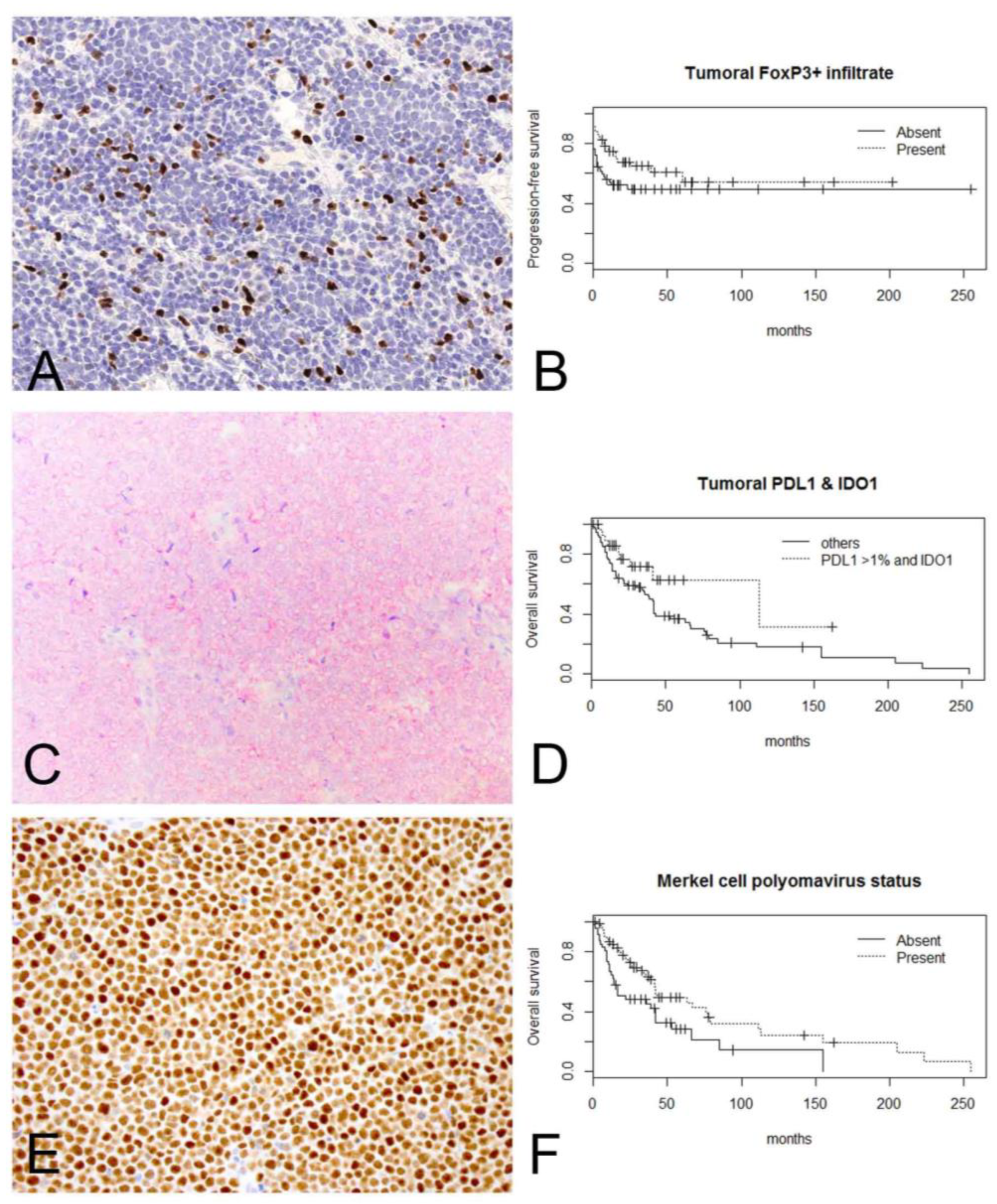
2.1. Positive MCPyV Status Correlated with Tumoral PD-L1 Expression and High Intratumoral CD8+ Cell Count

The clinicopathologic characteristics associated with tumoral PD-L1 expression, tumoral IDO1 expression, intratumoral CD8+ cell count, and intratumoral FoxP3+ cell count evaluated with Fisher’s exact tests are summarized in Table 1. Tumoral PD-L1 expression >1% significantly correlated with tumor thickness >10 mm (p = 0.0048) and positive MCPyV status (p = 0.018). High intratumoral CD8+ cell count significantly correlated with absence of ulceration (p = 0.0092) and positive MCPyV status (p = 0.018).

2.2. Tumoral PD-L1 Expression Correlated with Intratumoral CD8+ and FoxP3+ Lymphocytes, Which Is Supportive of an Adaptive Immune Response

By linear regression analyses, correlations between tumoral PD-L1 expression with intratumoral CD8+ cell count (p < 0.0001) and intratumoral FoxP3+ cell count (p < 0.0001), and intratumoral CD8+ cell count with intratumoral FoxP3+ cell count (p < 0.0001) were observed. There was no correlation between tumoral IDO1 expression and MCPyV status versus tumoral PD-L1 expression, intratumoral CD8+, and FoxP3+ cell counts.

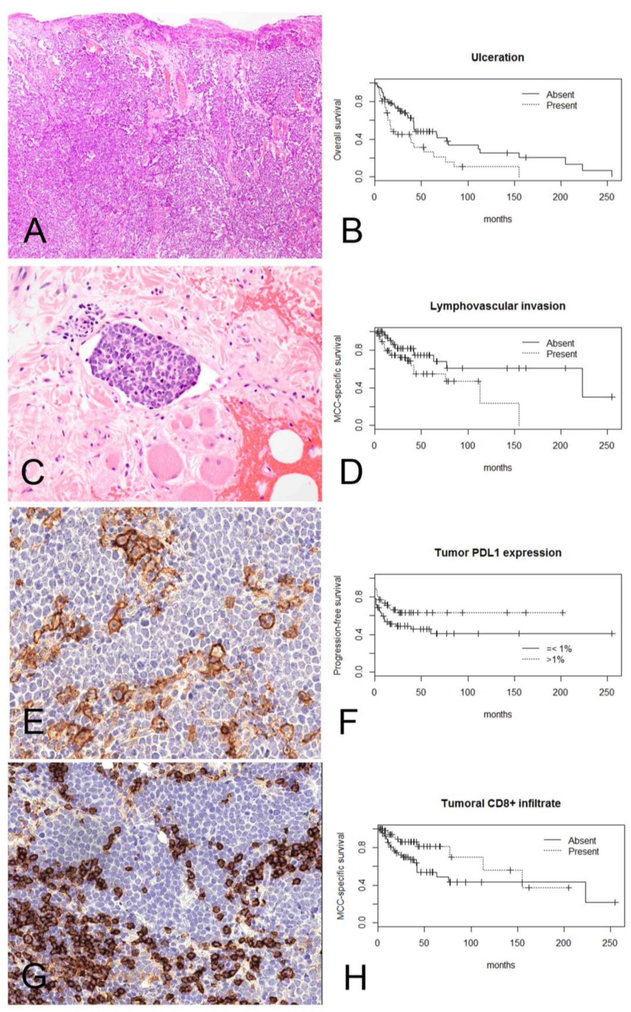
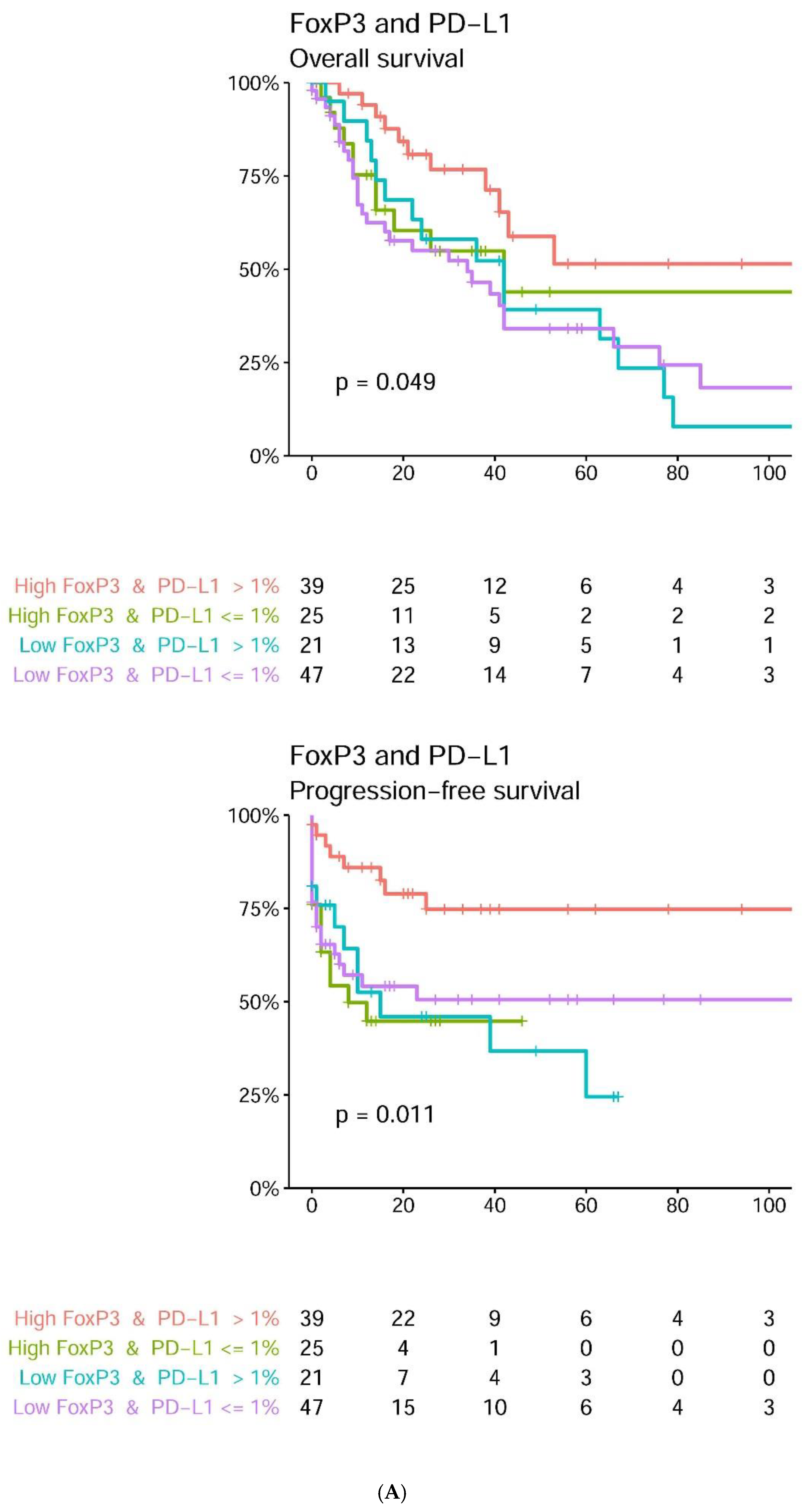
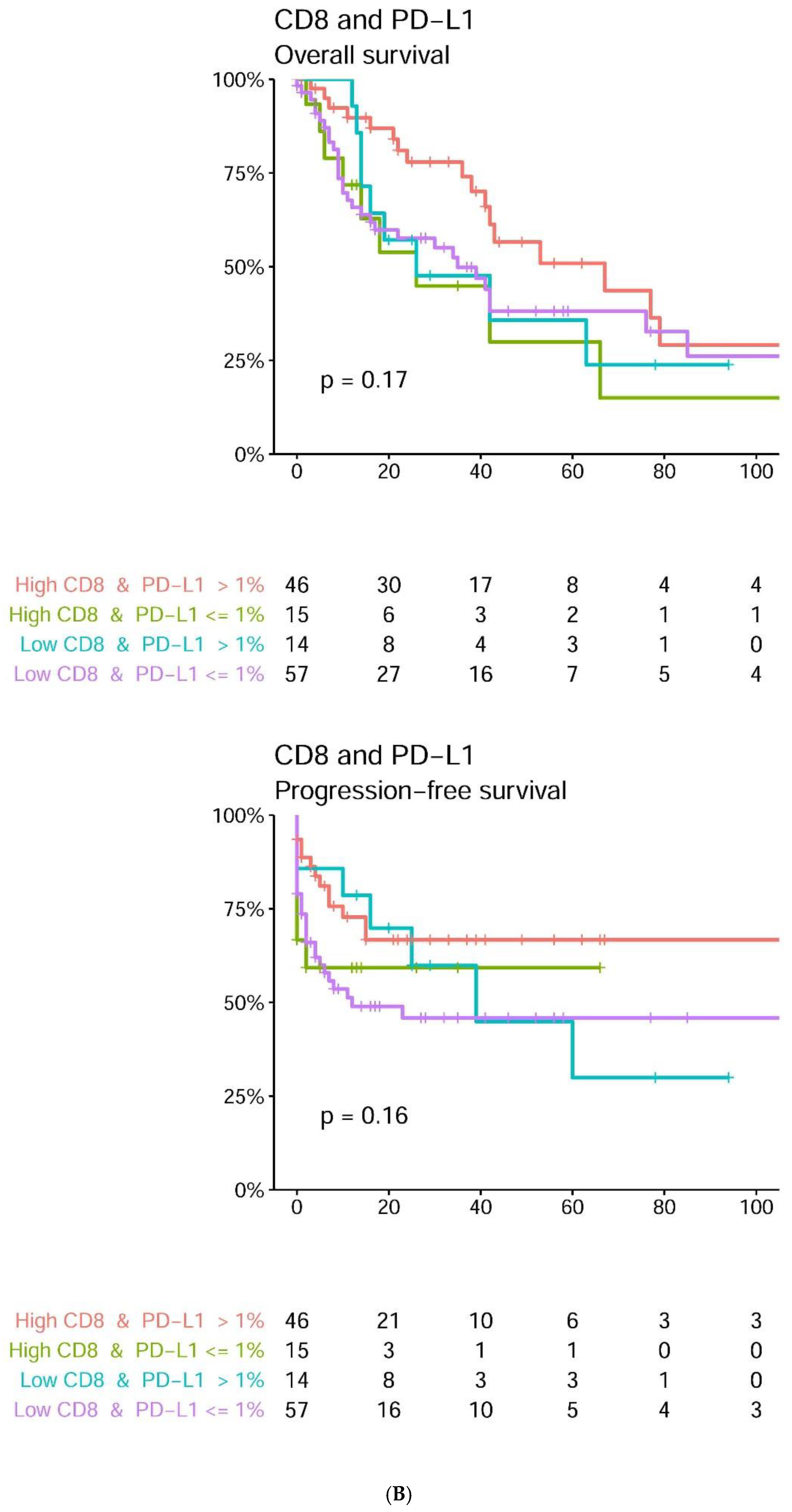
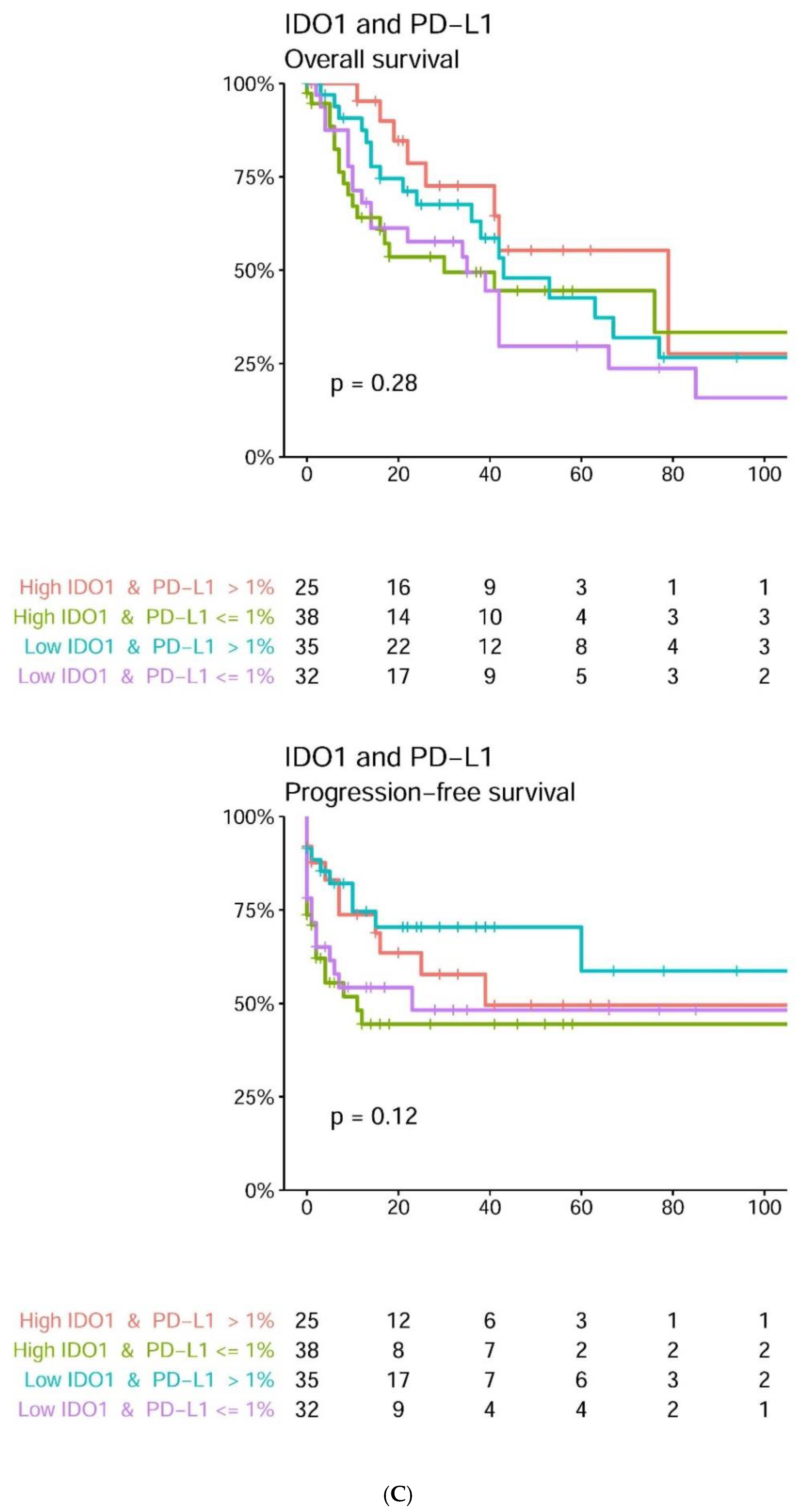
2.3. Tumoral PD-L1 Expression and Increased Density of Intratumoral CD8+ Lymphocytes and FoxP3+ Lymphocytes May Positively Impact Survival in a Subset of MCCs

By univariate analyses, tumoral PD-L1 expression >1% and combined tumoral PD-L1 >1% and high intratumoral FoxP3+ cell count correlated with improved overall survival (OS) (p = 0.016, 0.0072), MCC-specific survival (MSS) (p = 0.019, 0.017), and progression-free survival (PFS) (p = 0.043, 0.004, respectively) (Table 2, Figure 2 and Figure 3). High intratumoral CD8+ and FoxP3+ cell counts correlated with longer MSS (p = 0.036) and improved PFS (p = 0.047), respectively. Combined tumoral PD-L1 >1% and high tumoral IDO1 expression correlated with improved OS (Figure 2), but this result only approached statistical significance (p = 0.057) (Table 2). Expression of MCPyV T-antigen correlated with improved overall survival (OS) (p = 0.0076) and MSS (p = 0.04). Age (p = 0.00067) significantly correlated with worse OS. Male gender and higher stage (3 and 4) significantly correlated with worse MSS (p = 0.029 and 0.038, respectively) and shorter PFS (p = 0.006 and < 0.0001, respectively). Ulceration correlated with worse OS (p = 0.0046) and worse MSS (p = 0.008). Lymphovascular invasion was significantly associated with shorter MSS (p = 0.02) (Table 2).

2.4. By Multivariate Analyses, Ulceration and Age Are Independent Predictors of Worse OS; and Gender and Stage Are Independent Predictors of Shorter PFS

Covariates being significant in univariate analyses (ulceration, lymphovascular invasion, MCPyV large T-antigen, tumoral PD-L1 >1%, tumoral PD-L1 >1% and high intratumoral FoxP3+ cell count, tumoral PD-L1 >1% and high intratumoral CD8 cell count, intratumoral CD8+ and FoxP3+ cell counts, intratumoral CD8+ cell count, intratumoral FoxP3+ cell count, age, gender, site, and stage) were included in the Cox multivariate model. By multivariate analyses, only ulceration (p = 0.03) and age (p = 0.0031) remained as independent predictors of worse OS (p = 0.046); and gender (p = 0.018) and stage (p < 0.0001) remained as independent predictors for shorter PFS (Table 3).

3. Discussion

In the current study tumoral PD-L1 expression >1% significantly correlated with better OS, MSS, and PFS in univariate analysis. Similarly, Lipson et al. [12] reported expression of tumoral PD-L1 to be independently associated with better OS in MCC patients. A possible mechanism of enhanced tumoral PD-L1 expression and better clinical outcome could be connected with the hypomethylation of PD-L1 gene promoter. Findings reported by Micevic et al. [15] suggest that the hypomethylation of PD-L1 (and possible subsequent increased protein expression of PD-L1) is associated with a phenotype of immune activation and expression of interferon signaling pathway genes in cutaneous melanoma. This hypothesis needs further investigations in MCC patients, because some studies did not confirm the significant prognostic impact of PD-L1 on survival in MCC patients [2,13,14].
In our cohort, tumoral IDO1 expression had no prognostic impact on survival in univariate analysis. In the majority of previous studies tumoral IDO1 expression was correlated with a less favorable clinical outcome [7], but others reported a positive prognostic effect on long-term survival [8]. There are two hypotheses that might explain this putative contradiction. First, local tryptophan depletion has been reported to decrease the proliferation of tumor cell lines in an in vitro model [16,17]. Second, the IFN-γ is a crucial cytokine to induce IDO1 expression [17]. During an antitumor immune response, IFN-γ and other pro-inflammatory cytokines are secreted, and IDO1 expression could be a marker of an ongoing antitumoral immune response.
Interestingly, in the current study, there was no correlation between tumoral IDO1 expression and intratumoral CD8+ and FoxP3+ cell counts, supporting a different mechanism of induction of IDO1 expression within the tumor microenvironment of MCC [18,19]. Wardhani et al. [9] reported that higher tumoral IDO1 expression was significantly correlated with worse OS in 43 MCC patients, and enhanced tumoral IDO1 immunoreactivity was correlated with MCPyV-positive status. We did not observe any significant correlation between tumoral IDO1 expression with MCPyV status and clinicopathologic variables.
The association between tumoral IDO1 expression and the efficacy of IDO1 inhibitor is unknown at the current time. However, the co-expression of tumoral PD-L1 and IDO1 raises a possibility regarding therapeutic role of combined IDO1 inhibitor and PD1/PD-L1 inhibitor for MCC. Clinical trials of epacadostat, an IDO1 inhibitor, as single agent for MCC (UCDCC#271) and in combination with pembrolizumab/nivolumab for patients with advanced solid tumors are currently under investigation [20].
Forkhead transcription factor FoxP3 is a master regulator of Treg development. Although FoxP3+ infiltrate in MCC was previously studied by several studies, its prognostic value has not been widely studied in MCCs [21,22,23]. High intratumoral FoxP3+ infiltrate correlated with improved survival in our study. In accordance with our results, Sihto et al. [3] revealed that a high number of intratumoral FoxP3+ lymphocytes was associated with favorable outcome in MCC patients. Interestingly, concomitant intratumoral PD-L1 expression and high intratumoral FoxP3+ lymphocytes exhibited better OS, MSS, and PFS in univariate analyses in our study. The association between PD-L1 and FoxP3 that has been reported with PD-L1 influences the conversion of naïve T cells to Tregs and upregulates FoxP3 expression [24].
A strong association of tumoral PD-L1 expression and high intratumoral CD8+ lymphocytes was observed in our study; however, concomitant tumoral PD-L1 expression and high intratumoral CD8 infiltrate did not correlate with survival. Similar to observations made by Lipson et al. [12] and Mitteldorf et al. [25], tumoral PD-L1 expression in our cases appear to localize to areas with tumor-infiltrating lymphocytes. These results are in line with findings of previous study in which tumoral PD-L1 expression was correlated with CD8+ lymphocyte infiltration [26,27]. These findings suggest that tumoral PD-L1 expression could be a result of an adaptive immune response against viral and/or neoplastic antigens.
Several previous studies suggest that intratumoral CD8+ lymphocytes are strongly correlated with better survival in MCC [2,3,4,28]. The findings of our study confirmed that high intratumoral CD8+ cell count had a positive prognostic impact on survival in MCC patients and highlighted the crucial role of cytotoxic CD8+ lymphocytes in controlling disease progression.
In our cohort, MCPyV T-antigen expression significantly correlated with improved OS, and its expression had a strong association with tumoral PD-L1 expression and high intratumoral CD8+ cell count. In line with our results, Harms et al. [29] revealed that MCPyV-positive tumors were associated with significantly increased CD8+ cells in comparison to MCPyV-negative tumors. Lipson et al. [12] reported a significant association of the presence of MCPyV with tumor PD-L1 expression. Sihto et al. [3] showed significant correlation between MCPyV DNA-positive MCCs and higher numbers of CD8+ and FoxP3+ lymphocytes as compared with MCPyV-negative tumors. In contrast, Feldemeyer et al. [13] observed no significant differences between MCPyV status and PD-L1 expression in tumoral cells.
We observed tumoral PD-L1 expression in MCPyV-positive tumors more frequently than MCPyV-negative MCCs (48 versus 16, p = 0.018) in our series, supporting the hypothesis that virus-positive and virus-negative MCCs have different immune microenvironment. These findings suggest that MCPyV-related immune response upregulates tumoral PD-L1 and tumoral IDO1 expression and intratumoral CD8+ infiltrate.
Although tumoral PD-L1 expression and concomitant tumoral PD-L1 expression and high intratumoral FoxP3+ infiltrate correlated with improved survival in univariate analyses; the presence of ulceration, age, gender, and stage remain as independent predictors of survival.

4. Materials and Methods

One hundred and thirty two patients with a diagnosis of primary cutaneous MCC rendered between 1995 and 2018 were retrieved from the pathology archives of six clinical institutions in Poland, Taiwan, and the United States. The diagnoses were histopathologically confirmed by the contributing pathologists (PD, C-LW, JK and JR) and the corresponding author (MPH). Medical data including clinical notes, imaging results, and histopathology reports were reviewed to confirm that the analyzed tumor is a primary cutaneous MCC and not a metastasis of visceral-located neuroendocrine carcinoma. The study was approved by Institutional Review Boards.

4.1. Clinical Findings and Histologic Features

All tumors have diagnostic features of MCC: compatible histologic appearance, expression of keratin 20 and neuroendocrine markers (chromogranin, synaptophysin, neuron specific enolase, and/or CD56). Neurofilament, EMA, keratin 7, or other keratins (AE1/AE3, CAM5.2, pan or wide spectrum keratin) were performed in a subset of cases. Thyroid transcription factor 1 (TTF1) was performed in the majority of cases. Detailed histopathologic parameters (tumor size, growth pattern (nodular, infiltrative, or mixed), ulceration, tumor thickness, mitotic rate (per squared millimeters), lymphovascular invasion (LVI), perineural invasion (PNI), necrosis, and involvement of the overlying epidermis (epidermotropism) were assessed by the contributing pathologists (PD, C-LW, JK, JR) and confirmed by the corresponding author (MPH). The utilized tumor size cut-offs were those established by the 8th edition American Joint Committee on Cancer (AJCC) staging system for MCC. The following data were extracted from medical records: age of the patients, gender, lesion site, date of biopsy, disease status over time, and at last follow-up (recurrence, metastasis).

4.2. Immunohistochemistry

Tissue microarrays composed of two 2 mm-tissue cores from each tumor were constructed. Immunohistochemical studies were performed on 5-micrometer-thick tissue sections using a Bond 3 automated immunostainer (Leica Microsystems, Bannockburn, IL, USA), with primary antibodies against CD8 (4B11, undiluted, Leica Microsystems), FoxP3 (236A/E7, undiluted, BioCare Medical, Concord, CA, USA), IDO1 (1F8.2, 1:400, Millipore, Burlington, MA, USA), MCPyV large T-antigen (CM2B4, sc-136172, 1:100, Santa Cruz Biotechnology, Santa Cruz, CA, USA), and PD-L1 (E1L3N, 1:200, Cell Signaling Technology, Danvers, MA, USA).
Scoring of the cases was done blinded to clinical outcomes by two independent authors (PD and MPH). The percentage of tumor cells with positive PD-L1 membranous staining of any intensity was evaluated with greater than 1% considered positive. The cytoplasmic IDO1 and nuclear CM2B4 staining of tumor cells was done using the H-score [(percentage at 1+) × 1 + (percentage at 2+) × 2 + (percentage at 3+) × 3]. Any CM2B4 expression was considered positive. Total cell counts of intratumoral CD8+ and FoxP3+ lymphocytes were evaluated in three consecutive images captured at 40× magnification of the hotspot. The median number of positive cells was used as a cut-off value for high and low CD8+ or FoxP+ cases.

4.3. Statistical Analysis

The statistical association between tumoral expression of PD-L1 and IDO1, intratumoral CD8+ and FoxP3+ cell count, and clinicopathologic features (tumor size, tumor thickness, growth pattern, mitotic index, ulceration, necrosis, lymphovascular invasion, and perineural invasion) were evaluated by Fisher’s exact tests. The p-values were corrected for false discovery rate for 4 potential biomarkers. MCC-specific survival (MSS) and overall survival (OS) were defined as the number of months from initial diagnosis of MCC to patient’s death related to MCC and by any cause, respectively. Progression-free survival (PFS) was defined as the number of months from initial diagnosis to identification of local recurrence or metastases in lymph nodes and/or distant organs. Kaplan–Meier curves and log-rank tests were performed to assess the differences in MSS, OS, and PFS between subgroups. The Cox proportional hazards model was performed. All statistically significant parameters from univariate Cox models were included in multivariate analysis. The proportionality assumptions of the Cox models were tested. Linear regression was used for plotting tumoral PD-L1 percent expression against tumoral IDO1 H-score, intratumoral CD8+ and intratumoral FoxP3+ cell count; intratumoral CD8+ against intratumoral FoxP3+ cell count and tumoral IDO1 H-score; and CM2B4 H-score against percent tumoral PD-L1 percent expression, tumoral IDO1 H-score, and intratumoral CD8+ and intratumoral FoxP3+ cell count. All analyses and plots were performed using the R statistical package [30]. A two-tailed p-value of less than 0.05 was considered statistically significant.

5. Conclusions

Tumoral PDL1 expression and increased density of intratumoral CD8+ lymphocytes and FoxP3+ lymphocytes may positively impact on survival in a subset of MCCs. Tumoral PDL1 expression correlated with intratumoral CD8+ and FoxP3+ lymphocytes, which is supportive of an adaptive immune response. There is a correlation between immune response and MCPyV positivity.

Author Contributions

Conceptualization, M.P.H.; Methodology, M.P.H., P.D., P.B., Software, M.P.H., P.B.; Validation, M.P.H., P.B.; Formal Analysis, M.P.H., P.D., P.B., C.-L.W., J.K., M.P., J.R.; Investigation, M.P.H., P.D., P.B., C.-L.W., J.K., M.P., J.R.; Resources, M.P.H., P.D., C.-L.W., J.K., M.P., J.R.; Data Curation, M.P.H., P.D., C.-L.W., J.K., M.P., J.R.; Writing—Original Draft Preparation, M.P.H., P.D.; Writing—Review and Editing, M.P.H., P.D., C.-L.W., P.B., J.K., M.P., J.R.; Visualization, M.P.H., P.D., P.B.; Supervision, M.P.H.; Project Administration, M.P.H., P.D.; Funding Acquisition, M.P.H., P.D. All authors have read and agreed to the published version of the manuscript.

Funding

A statutory subsidy from the Polish Minister of Science and Higher Education as a part of grant SUB.B130.21.062 (record numbers in the Simple system) was awarded to PD.

Institutional Review Board Statement

The study was conducted according to the guidelines of the Declaration of Helsinki, and approved by the Partners Human Research Committee, the institutional review board of Partners HealthCare (protocol code 20018P002057, date of approval: 2 October 2018).

Informed Consent Statement

Patient consent was waived since the study was performed pre-existing archived pathology specimens. No patients were involved.

Data Availability Statement

The data presented in this study are available in this article.

Conflicts of Interest

The authors declare no conflict of interest.

References

  1. Coggshall, K.; Tello, T.L.; North, J.P.; Yu, S.S. Merkel cell carcinoma: An update and review: Pathogenesis, diagnosis, and staging. J. Am. Acad. Dermatol. 2018, 78, 433–442. [Google Scholar] [CrossRef] [Green Version]
  2. Von der Grün, J.; Winkelmann, R.; Meissner, M.; Wieland, U.; Silling, S.; Martin, D.; Fokas, E.; Rödel, C.; Rödel, F.; Balermpas, P. Merkel Cell Polyoma viral load and intratumoral CD8+ lymphocyte infiltration predict overall survival in patients with Merkel cell carcinoma. Front. Oncol. 2019, 9, 20. [Google Scholar] [CrossRef] [Green Version]
  3. Sihto, H.; Böhling, T.; Kavola, H.; Koljonen, V.; Salmi, M.; Jalkanen, S.; Joensuu, H. Tumor infiltrating immune cells and outcome of Merkel cell carcinoma: A population-based study. Clin. Cancer Res. 2012, 18, 2872–2881. [Google Scholar] [CrossRef] [Green Version]
  4. Paulson, K.G.; Iyer, J.G.; Tegeder, A.R.; Thibodeau, R.; Schelter, J.; Koba, S.; Schrama, D.; Simonson, W.T.; Lemos, B.D.; Byrd, D.R.; et al. Transcriptome-wide studies of Merkel cell carcinoma and validation of intratumoral CD8+ lymphocyte invasion as an independent predictor of survival. J. Clin. Oncol. 2011, 29, 1539–1546. [Google Scholar] [CrossRef] [Green Version]
  5. Bilir, C.; Sarisozen, C. Indoleamine 2,3-dioxygenase (IDO): Only an enzyme or a checkpoint controller? J. Oncol. Sci. 2017, 3, 52–56. [Google Scholar] [CrossRef]
  6. Munn, D.H.; Mellor, A.L. Indoleamine 2,3-dioxygenase and tumor-induced tolerance. J. Clin. Investig. 2007, 117, 1147–1154. [Google Scholar] [CrossRef] [PubMed] [Green Version]
  7. Brochez, L.; Chevolet, I.; Kruse, V. The rationale of indoleamine 2,3-dioxygenase inhibition for cancer therapy. Eur. J. Cancer 2017, 76, 167–182. [Google Scholar] [CrossRef] [PubMed]
  8. Jacquemier, J.; Bertucci, F.; Finetti, P.; Esterni, B.; Charafe-Jauffret, E.; Thibult, M.L.; Houvenaeghel, G.; Van den Eynde, B.; Birnbaum, D.; Olive, D.; et al. High expression of indoleamine 2,3-dioxygenase in the tumour is associated with medullary features and favourable outcome in basal-like breast carcinoma. Int. J. Cancer 2012, 130, 96–104. [Google Scholar] [CrossRef] [PubMed]
  9. Wardhani, L.O.; Matsushita, M.; Iwasaki, T.; Kuwamoto, S.; Nonaka, D.; Nagata, K.; Kato, M.; Kitamura, Y.; Hayashi, K. Expression of the IDO1/TDO2-AhR pathway in tumor cells or the tumor microenvironment is associated with Merkel cell polyomavirus status and prognosis in Merkel cell carcinoma. Hum. Pathol. 2019, 84, 52–61. [Google Scholar] [CrossRef] [Green Version]
  10. Barkdull, S.; Brownell, I. PDL1 blockade with avelumab: A new paradigm for treating Merkel cell carcinoma. Cancer Biol. Ther. 2017, 18, 937–939. [Google Scholar] [CrossRef] [PubMed] [Green Version]
  11. Nghiem, P.T.; Bhatia, S.; Lipson, E.J.; Kudchadkar, R.R.; Miller, N.J.; Annamalai, L.; Berry, S.; Chartash, E.K.; Daud, A.; Fling, S.P.; et al. PD1 blockade with Pembrolizumab in advanced Merkel cell carcinoma. N. Engl. J. Med. 2016, 374, 2542–2552. [Google Scholar] [CrossRef] [PubMed]
  12. Lipson, E.J.; Vincent, J.G.; Loyo, M.; Kagohara, L.T.; Luber, B.S.; Wang, H.; Xu, H.; Nayar, S.K.; Wang, T.S.; Sidransky, D.; et al. PD-L1 expression in the Merkel cell carcinoma microenvironment: Association with inflammation, Merkel cell polyomavirus and overall survival. Cancer Immunol. Res. 2013, 1, 54–63. [Google Scholar] [CrossRef] [PubMed] [Green Version]
  13. Feldmeyer, L.; Hudgens, C.W.; Ray-Lyons, G.; Nagarajan, P.; Aung, P.P.; Curry, J.L.; Torres-Cabala, C.A.; Mino, B.; Rodriguez-Canales, J.; Reuben, A.; et al. Density, distribution, and composition of immune infiltrates correlate with survival in Merkel cell carcinoma. Clin. Cancer Res. 2016, 22, 5553–5563. [Google Scholar] [CrossRef] [PubMed] [Green Version]
  14. Wehkamp, U.; Stern, S.; Krüger, S.; Weichenthal, M.; Hauschild, A.; Röcken, C.; Egberts, F. Co-expression of NGF and PD-L1 on tumor-associated immune cells in the microenvironment of Merkel cell carcinoma. J. Cancer Res. Clin. Oncol. 2018, 144, 1301–1308. [Google Scholar] [CrossRef] [PubMed]
  15. Micevic, G.; Thakral, D.; McGeary, M.; Bosenberg, M.W. PD-L1 methylation regulates PD-L1 expression and is associated with melanoma survival. Pigment Cell Melanoma Res. 2019, 32, 435–440. [Google Scholar] [CrossRef]
  16. Ozaki, Y.; Edelstein, M.P.; Duch, D.S. Induction of indoleamine 2,3-dioxygenaseda mechanism of the antitumor-activity of interferon-gamma. Proc. Natl. Acad. Sci. USA 1988, 85, 1242–1246. [Google Scholar] [CrossRef] [Green Version]
  17. Takikawa, O.; Kuroiwa, T.; Yamazaki, F.; Kido, R. Mechanism of interferon-gamma action and characterization of indoleamine 2,3- dioxygenase in cultured human-cells induced by interferon gamma and evaluation of the enzyme-mediated tryptophan degradation in its anticellular activity. J. Biol. Chem. 1988, 263, 2041–2048. [Google Scholar] [CrossRef]
  18. Schalper, K.A.; Carvajal-Hausdorf, D.; McLaughlin, J.; Altan, M.; Velcheti, V.; Gaule, P.; Sanmamed, M.F.; Chen, L.; Herbst, R.S.; Rimm, D.L. Differential expression and significance of PD-L1, IDO-1, and B7-H4 in human lung cancer. Clin. Cancer Res. 2017, 23, 370–378. [Google Scholar] [CrossRef] [Green Version]
  19. Prendergast, G.C.; Malachowski, W.P.; DuHadaway, J.B.; Muller, A.J. Discovery of IDO1 inhibitors: From bench to bedside. Cancer Res. 2017, 77, 6795–6811. [Google Scholar] [CrossRef] [Green Version]
  20. Mitchell, T.C.; Hamid, O.; Smith, D.C.; Bauer, T.M.; Wasser, J.S.; Olszanski, A.J.; Luke, J.J.; Balmanoukian, A.S.; Schmidt, E.V.; Zhao, Y.; et al. Epacadostat plus pembrolizumab in patients with advanced solid tumors: Phase I results from a multicenter, open-label phase I/II trial (ECHO-202/KEYNOTE-037). J. Clin. Oncol. 2018, 36, 3223–3230. [Google Scholar] [CrossRef]
  21. Walsh, N.M.; Fleming, K.E.; Hanly, J.G.; Dakin Hache, K.; Doucette, S.; Ferrara, G.; Cerroni, L. A morphological and immunophenotypic map of the immune response in Merkel cell carcinoma. Hum. Pathol. 2016, 52, 190–196. [Google Scholar] [CrossRef] [PubMed]
  22. Wheat, R.; Roberts, C.; Waterboer, T.; Steele, J.; Marsden, J.; Steven, N.M.; Blackbourn, D.J. Inflammatory cell distribution in primary Merkel cell carcinomas. Cancers 2014, 6, 1047–1064. [Google Scholar] [CrossRef] [PubMed] [Green Version]
  23. Dowlatshahi, M.; Huang, V.; Gehad, A.E.; Jiang, Y.; Calarese, A.; Teague, J.E.; Dorosario, A.A.; Cheng, J.; Nghiem, P.; Schanbacher, C.F.; et al. Tumor-specific T cells in human Merkel cell carcinomas: A possible role for Tregs and T-cell exhaustion in reducing T-cell responses. J. Investig. Dermatol. 2013, 133, 1879–1889. [Google Scholar] [CrossRef] [Green Version]
  24. Jin, H.T.; Ahmed, R.; Okazaki, T. Role of PD-1 in regulating T-cell immunity. Curr. Top. Microbiol. Immunol. 2011, 350, 17–37. [Google Scholar] [PubMed]
  25. Mitteldorf, C.; Berisha, A.; Tronnier, M.; Pfaltz, M.C.; Kempf, W. PD-1 and PD-L1 in neoplastic cells and the tumor microenvironment of Merkel cell carcinoma. J. Cutan. Pathol. 2017, 44, 740–746. [Google Scholar] [CrossRef] [PubMed]
  26. Afanasiev, O.K.; Yelistratova, L.; Miller, N.; Nagase, K.; Paulson, K.; Iyer, J.G.; Ibrani, D.; Koelle, D.M.; Nghiem, P. Merkel polyomavirus-specific T cells fluctuate with Merkel cell carcinoma burden and express therapeutically targetable PD-1 and Tim-3 exhaustion markers. Clin. Cancer Res. 2013, 19, 5351–5360. [Google Scholar] [CrossRef] [PubMed] [Green Version]
  27. Taube, J.M.; Anders, R.A.; Young, G.D.; Xu, H.; Sharma, R.; McMiller, T.L.; Chen, S.; Klein, A.P.; Pardoll, D.M.; Chen, L. Colocalization of inflammatory response with B7-h1 expression in human melanocytic lesions supports an adaptive resistance mechanism of immune escape. Sci. Transl. Med. 2012, 4, 127ra37. [Google Scholar] [CrossRef] [Green Version]
  28. Miller, N.J.; Church, C.D.; Dong, L.; Crispin, D.; Fitzgibbon, M.P.; Lachance, K.; Jing, L.; Shinohara, M.; Gavvovidis, I.; Willimsky, G.; et al. Tumor-infiltrating Merkel cell polyomavirus-specific T cells are diverse and associated with improved patient survival. Cancer Immunol. Res. 2017, 5, 137–147. [Google Scholar] [CrossRef] [Green Version]
  29. Harms, P.W.; Patel, R.M.; Verhaegen, M.E.; Giordano, T.J.; Nash, K.T.; Johnson, C.N.; Daignault, S.; Thomas, D.G.; Gudjonsson, J.E.; Elder, J.T.; et al. Distinct gene expression profiles of viral- and nonviral-associated Merkel cell carcinoma revealed by transcriptome analysis. J. Investig. Dermatol. 2013, 133, 936–945. [Google Scholar] [CrossRef] [PubMed] [Green Version]
  30. R Development Core Team. R: A Language and Environment for Statistical Computing; R Foundation for Statistical Computing: Vienna, Austria, 2019; ISBN 3-900051-07-0. Available online: http://www.R-project.org/ (accessed on 29 October 2019).
Figure 1. Kaplan–Meier curves demonstrate significant correlation between (A) presence of ulceration (10×) and (B) worse overall survival (log-rank p = 0.0039); (C) presence of lymphovascular invasion (40×) and (D) worse MCC-specific survival (log-rank p = 0.02); (E) membranous PD-L1 expression >1% of tumor cells (20×) and (F) improved progression-free survival (log-rank p = 0.041); (G) intratumoral CD8+ lymphocytes and (H) improved Merkel cell carcinoma-specific survival (log-rank p = 0.031).
Ijms 22 05489 g001
Figure 2. Kaplan–Meier curves demonstrate significant correlation between (A) intratumoral FoxP3+ lymphocytes (20×) and (B) improved progression-free survival (log-rank p = 0.044); (C) combined tumoral PD-L1 >1% and high IDO1 expression (40×) and (D) improved overall survival (log-rank p = 0.052); (E) nuclear expression of Merkel cell polyomavirus large T-antigen (40×) and (F) improved overall survival (log-rank p = 0.0066).
Ijms 22 05489 g002
Figure 3. (A) Kaplan–Meier curves illustrate the associations between tumoral PD-L1 expression and intratumoral FoxP3+ cell count for overall survival and progression-free survival. (B) Kaplan–Meier curves illustrate the associations between tumoral PD-L1 expression and intratumoral CD8+ cell count for overall survival and progression-free survival. (C) Kaplan–Meier curves illustrate the associations between tumoral PD-L1 expression and tumoral IDO1 expression for overall survival and progression-free survival.
Ijms 22 05489 g003aIjms 22 05489 g003bIjms 22 05489 g003c
Table 1. Summary of clinical, histologic, and immunohistochemical characteristics.
PD-L1
n = 132
CD8
n = 132
FoxP3
n = 132
IDO1
n = 130
HighLowp-ValueHighLowp-ValueHighLowp-ValueHighLowp-Value
Age
  >77 years27380.4832320.6529360.8633300.65
  <=77 years3730 2939 3136 3037
Gender
  Male37360.7231420.7229440.6434390.72
  Female2732 3029 3128 2928
Site
  Head and neck32300.7330320.7333310.5627350.56
  Other sites3238 3139 2741 3632
Size
  >20 mm36230.07273212634132270.48
  <=20 mm2845 3439 3339 3140
Thickness
  >10 mm39230.019 *3034126370.6434290.58
  <=10 mm2543 3036 3235 2837
Ulceration
  Present20240.7113310.037 *15290.1424200.48
  Absent4444 4840 4543 3947
Mitoses
  >40/mm23133125400.3225400.3230341
  <=40/mm23334 3531 3432 3233
Growth pattern
  Nodular34260.4426340.7331290.4428320.73
  Nodular and infiltrative, infiltrative3042 3537 2943 3535
Necrosis
  Present22210.7117270.4817260.4825180.48
  Absent4247 4444 4346 3849
Lymphovascular invasion
  Present33310.6028360.6025390.3436260.21
  Absent3137 3335 3533 2741
Perineural invasion
  Present661481481661
  Absent5862 5763 5664 5761
MCPyV
  Present48370.036 *46390.036 *42430.2745380.15
  Absent1631 1532 1829 1829
* p < 0.05, statistically significant; p < 0.09, approaching statistical significance. p-values are after correction for multiple hypothesis testing (False Discovery Rate in each row).
Table 2. Univariate Cox proportional hazards model.
Overall SurvivalMerkel Cell Carcinoma-Specific SurvivalProgression-Free Survival
NHazard Ratio p-ValueHazard Ratio p-ValueHazard Ratio p-Value
PD-L1 >1%1320.550.016 *0.410.019 *0.560.043 *
CD8-high1320.760.250.450.036 *0.620.098
FoxP3-high1320.660.0920.620.170.570.047 *
IDO1-high1300.930.751.150.691.290.36
CD8-high FoxP3-high1320.660.130.410.046 *0.580.091
PD-L1 >1% FoxP3-high1320.430.0072 *0.280.017 *0.350.004 *
PD-L1 >1% CD8-high1320.570.0530.280.018 *0.450.024 *
PD-L1 >1% IDO1-high1300.500.0570.870.741.140.66
Size (>20 mm)1321.030.911.310.441.200.50
Thickness (>10 mm)1321.040.891.140.711.060.84
Growth pattern (nodular versus infiltrative/mixed)1320.690.120.580.120.570.051
Ulceration1322.010.0046 *2.510.008 *1.670.067
Mitoses (>40/mm2)1320.970.891.280.471.170.58
Necrosis1321.300.321.190.661.450.19
Lymphovascular invasion1321.500.0952.290.02 *1.510.14
MCPyV large T-antigen1320.520.0076 *0.490.04 *0.710.22
Age (>versus ≤77 years)1322.350.00067 *1.330.410.90.7
Gender (male versus female)1321.290.282.230.029 *2.290.006 *
Site (other versus head & neck)1321.320.260.760.440.480.012 *
Stage (3–4 versus 1–2)1321.270.622.750.038 *27.2<0.0001 *
Immunosuppression1321.180.631.20.711.020.97
Treatment (adjuvant therapy versus surgery1321.130.631.820.0941.520.13
MCPyV: Merkel cell polyomavirus. * p < 0.05, statistically significant; p < 0.09, approaching statistical significance.
Table 3. Multivariate Cox proportional hazards model.
Overall SurvivalMerkel Cell Carcinoma-Specific SurvivalProgression-Free Survival
NHazard Ratio p-ValueHazard Ratio p-ValueHazard Ratio p-Value
PD-L1 >1%1320.920.780.840.761.480.37
Ulceration1321.790.03 *1.730.18--
MCPyV T-antigen expression1320.730.240.720.41--
PD-L1 >1% FoxP3-high1320.540.130.990.990.320.058
PD-L1 >1% CD8-high132--0.270.150.700.48
CD8-high FoxP3-high132--0.440.26--
Lymphovascular invasion132--1.940.1--
CD8132--2.40.24--
FoxP3132----1.350.41
Age1322.130.0031 *----
Gender132--1.780.192.080.018 *
Site (other versus head & neck)132----0.560.064
Stage (3–4 versus 1–2)132--2.370.1220.57<0.0001 *
MCPyV: Merkel cell polyomavirus. * p < 0.05, statistically significant; p < 0.09, approaching statistical significance.
Publisher’s Note: MDPI stays neutral with regard to jurisdictional claims in published maps and institutional affiliations.

