Review

Thermogenesis in Adipose Tissue Activated by Thyroid Hormone

1
Laboratory of Hormonal Regulation, Cardiovascular and Metabolic Disorders Program, Duke NUS Medical School, Singapore 169857, Singapore
2
Duke Molecular Physiology Institute, Duke University, Durham, NC 27708, USA
*
Author to whom correspondence should be addressed.
Int. J. Mol. Sci. 2020, 21(8), 3020; https://doi.org/10.3390/ijms21083020
Submission received: 23 March 2020 / Revised: 19 April 2020 / Accepted: 22 April 2020 / Published: 24 April 2020
(This article belongs to the Special Issue Molecular Biology of Nuclear Receptors 2.0)

Abstract

:
Thermogenesis is the production of heat that occurs in all warm-blooded animals. During cold exposure, there is obligatory thermogenesis derived from body metabolism as well as adaptive thermogenesis through shivering and non-shivering mechanisms. The latter mainly occurs in brown adipose tissue (BAT) and muscle; however, white adipose tissue (WAT) also can undergo browning via adrenergic stimulation to acquire thermogenic potential. Thyroid hormone (TH) also exerts profound effects on thermoregulation, as decreased body temperature and increased body temperature occur during hypothyroidism and hyperthyroidism, respectively. We have termed the TH-mediated thermogenesis under thermoneutral conditions “activated” thermogenesis. TH acts on the brown and/or white adipose tissues to induce uncoupled respiration through the induction of the uncoupling protein (Ucp1) to generate heat. TH acts centrally to activate the BAT and browning through the sympathetic nervous system. However, recent studies also show that TH acts peripherally on the BAT to directly stimulate Ucp1 expression and thermogenesis through an autophagy-dependent mechanism. Additionally, THs can exert Ucp1-independent effects on thermogenesis, most likely through activation of exothermic metabolic pathways. This review summarizes thermogenic effects of THs on adipose tissues.

1. Introduction

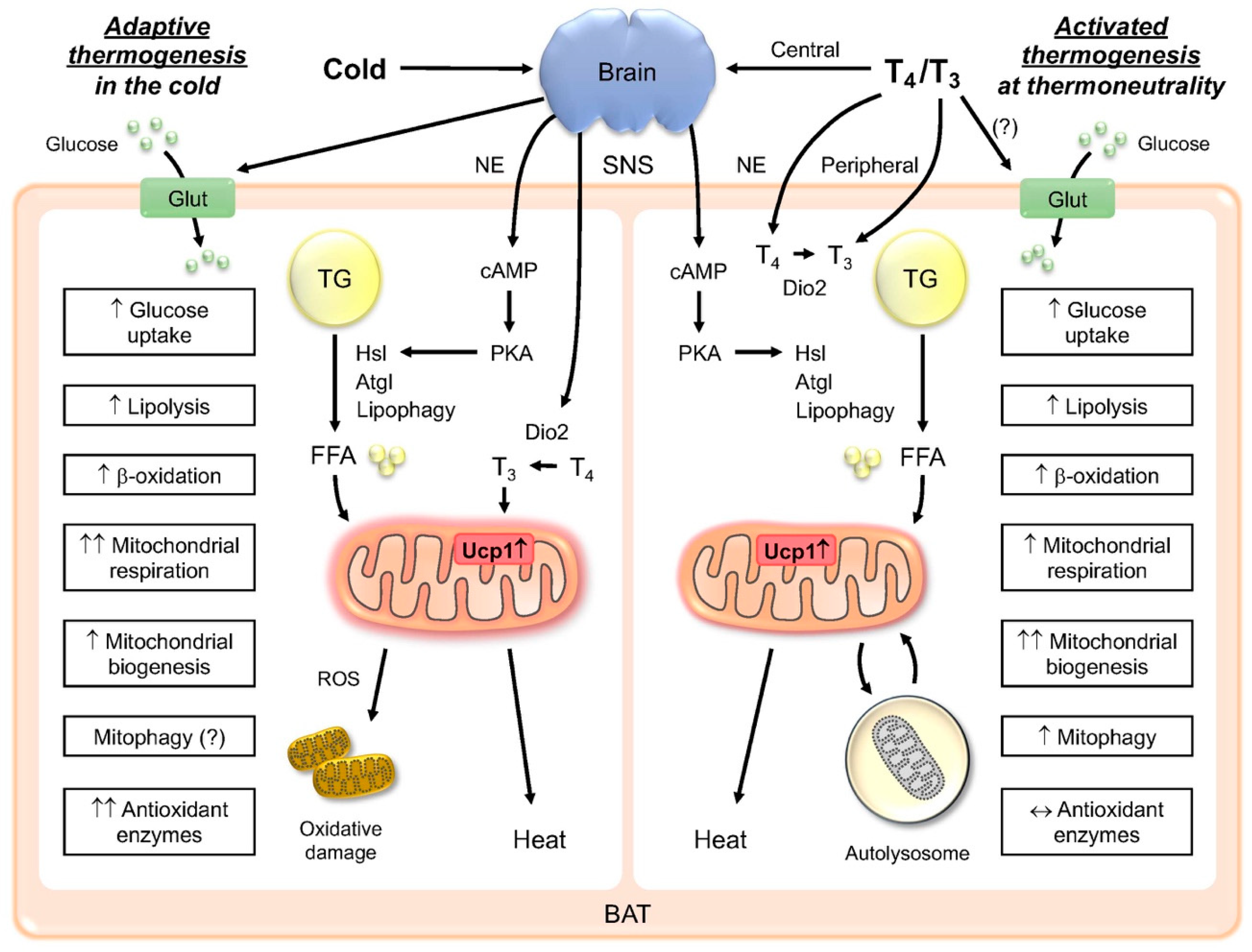
1.1. Thermogenesis and Adipose Tissue

Thermogenesis is an essential survival mechanism for homeotherms. Obligatory thermogenesis is sufficient to maintain body temperature and normal body function when the ambient temperature is at thermoneutrality, which is about 23 °C for an adult man [1]. When ambient temperature falls below thermoneutrality, adaptive (also known as facultative) thermogenic mechanisms that require or do not require shivering are recruited. Shivering thermogenesis increases heat production in response to cold through skeletal muscle contraction. It is able to provoke a 5-fold increase in the resting metabolic rate in humans, and is important for surviving extreme cold conditions [2]. However, it lacks long-term sustainability and may compromise muscle function [3,4]. Non-shivering thermogenesis occurs mainly in brown adipose tissue (BAT) which is able to oxidize lipids and dissipate energy in the form of heat and is important for heat production during sustained cold exposure [5,6]. Shivering may be absent or become minimal when non-shivering thermogenesis is sufficient to generate heat, such as during moderate cold exposure or cold acclimation [7]. In homeothermic species, these processes are critical for maintaining normal body function and temperature.
There are two major types of adipose tissue: white adipose tissue (WAT) and brown adipose tissue (BAT), which stores energy and generates body heat, respectively. WAT is stored subcutaneously and viscerally, where it surrounds intra-abdominal organs such as the liver, pancreas, and intestines. Accumulation of the visceral WAT is highly associated with insulin resistance and diabetes. On the other hand, amount of the active BAT is negatively associated with adiposity and the likelihood of developing non-alcoholic fatty liver disease [8,9]. There are two distinct cell populations with thermogenic potential. The first population, brown adipocytes, has a common embryological origin with myocytes, and is found mainly in the intrascapular, paraspinal, supraclavicular, suprarenal, pericardial, and para-aortic regions in human. Similar to muscle, it has a high concentration of mitochondria, which gives it a characteristic brown color. The second population, known as “beige” or “brite” (“brown in white”) adipocytes, is derived from a subpopulation of white adipocytes interspersed within the WAT [10]. In human, BAT is present at birth and regresses with age, although it remains metabolically active throughout adulthood. BAT is highly innervated and vascularized, allowing it to rapidly respond to external stimulation such as cold and diet [11]. BAT also utilizes available substrates such as the intracellular triacylglycerol (TAG) and blood glucose as fuels [12].
BAT is acutely activated by cold exposure via stimulation of the sympathetic nervous system (SNS) which increases the intracellular cyclic adenosine monophosphate (cAMP) level in BAT. The high level of cAMP then increases protein kinase A (PKA)-mediated lipolysis of intracellular TAG into free fatty acids (FFA) to provide fuel for β-oxidation [6]. FFAs also β-oxidize to generate acetyl coenzyme A (acetyl-CoA), which enters the tricarboxylic acid cycle and is oxidized to generate nicotinamide adenine dinucleotide (NADH) and flavin adenine dinucleotide (FADH2). The NADH and FADH2 are then used by the electron transport system to produce a proton gradient. BAT uniquely expresses the uncoupling protein, Ucp1, which dissipates the proton gradient across the inner mitochondrial membrane, resulting in inefficiency during the formation of ATP in oxidative phosphorylation (OXPHOS). When Ucp1 is induced in the BAT, such as during cold stress or thyroid hormone (TH) stimulation, mitochondrial respiration is stimulated to the maximum amount in order to compensate for the gradient loss, generating heat in the process [6]. During sustained cold exposure, there also is adipose tissue remodeling to increase the thermogenic potential of both BAT and WAT. Chronic cold exposure causes metabolic changes in the BAT to maximize β-oxidation of fatty acids, electron transport activity, and Ucp1 expression to generate heat [6,13,14]. SNS stimulation in response to cold also induces competent white adipocytes to express Ucp1 and various BAT-associated genes such as Cidea and Cox7a1 in a process known as browning/beiging. These beige adipocytes increase their mitochondria levels and, in conjunction with the induction of Ucp1 expression, acquire the thermogenic capability [10]. The recruitment of beige adipocytes contributes substantially to heat generation during chronic cold stress [7,15]. Since they possess the capacity of fat burning, they are also considered therapeutic targets for treating metabolic dysfunction [16]. BAT activation by cold acclimation improves insulin sensitivity in patients with type 2 diabetes [17]. Since cold acclimation is able to recruit the BAT in obese patients, BAT activation may also improve obesity-associated insulin resistance and hyperglycemia [18]. The activity of adult brown and beige fat decreases with aging [19,20,21] and may contribute to the progression of chronic metabolic diseases. Taken together, these findings suggest that activating and recruiting brown and beige fat would be beneficial to improving overall metabolic health.
Interestingly, thyroid hormone (TH) and its analogs can promote tissue remodeling in both BAT and WAT by stimulating both BAT activity and browning at thermoneutral conditions. We have previously named this process “activated” thermogenesis to distinguish it from cold-induced adaptive thermogenesis [22] (Figure 1). We found that although both cold exposure and TH are able to stimulate thermogenesis in the BAT, there are inherent differences in how the metabolic processes are regulated and sustained. First, adaptive thermogenesis occurs during cold exposure, whereas activated thermogenesis induced by T3 occurs at thermoneutral conditions. Second, shivering occurs in conjunction with adaptive thermogenesis, whereas there is no shivering in the activated thermogenesis. Third, during cold exposure, sympathetic stimulation of the BAT plays a predominant role in BAT activation, and leads to the induction of deiodinase 2 (Dio2), the enzyme that converts T4 to T3 in the BAT during cold exposure and increases intracellular T3. Lopez et al. [23] showed that T3 acts centrally to regulate the BAT, and we also showed that local T3 has effects on BAT activation, so it appears that both central and peripheral stimulation are involved in the activated thermogenesis. Fourth, there is an increase in intracellular T3 due to the induction of Dio2 for both types of thermogenesis; however, in the adaptive thermogenesis, the induction of serum T3 concentration can be variable, and may depend upon the temperature and length of cold exposure. On the other hand, in the activated thermogenesis, there is a significant increase in the serum TH concentration, which also has effects on other tissues besides the BAT. In this review, we would like to describe in further detail the key similarities and differences between the TH-induced activated thermogenesis and the conventional cold-induced adaptive thermogenesis.

1.2. TH induction of Thermogenesis in BAT

The two forms of TH, thyroxine (T4) and its active metabolite 3,5,3′-triiodothyronine (T3), regulate obligatory and adaptive thermogenesis by directly increasing metabolic rate in specific tissues [1,24,25]. Normal thyroid status is essential for the adaptive thermogenesis in response to the cold, as hypothyroid patients are prone to hypothermia during prolonged cold exposure [26]. BAT expresses 5-deiodinase type 2 (Dio2) which catalyzes the intracellular conversion of T4 to T3 [1]. During cold exposure, there is noradrenergic induction of Dio2, which leads to an increase in intracellular T3 stimulation of Ucp1 expression, increased mitochondrial respiration with less efficient ATP generation, and heat generation. BAT activation by cold exposure can increase systemic T3 levels [27,28], suggesting that induction of Dio2 in the BAT can have some T3-mediated effects systemically. Cold exposure also strongly induces Dio2 expression and activity in the WAT, although its contribution to the systemic T3 level is not known. This induction of Dio2 by the cold in the BAT and WAT highlights the importance of T4 to T3 conversion on TH actions within adipocytes during the adaptive thermogenesis. In particular, T3 mobilizes triacylglycerides (TAGs) stored in the WAT to generate free fatty acids that serve as a fuel for thermogenesis. Moreover, TH is able to induce Ucp1 expression selectively in the BAT and WAT, which enables sustained induction of thermogenesis by mitochondrial uncoupling. Mice lacking Dio2 cannot maintain their body temperature during cold stress [29,30], further demonstrating that maintenance of intracellular T3 levels in the BAT is necessary for the adaptive thermogenesis.
Thyroid hormone receptors (TRs) belong to the nuclear receptor superfamily and are ligand-inducible transcription factors. There are two major TR isoforms, thyroid hormone receptors a and b (TRα and TRβ). Ligand-bound TRs bind to TH response elements (TREs) located in the promoter region of target genes to induce their expression. The actions of TRα and TRβ are redundant for many genes, and both TRα and TRβ are expressed in the WAT and BAT. TH regulates transcription of many thermogenic genes through its cognate receptors. However, it appears that TRα may play a more predominant role in the obligatory thermogenesis and sympathetic response, whereas TRβ may play a more significant role in stimulating Ucp1 expression in the BAT [31,32,33,34]. TRα knockout (KO), but not TRβ KO mice are hypothermic [31,35]. Administration of a TRβ-selective agonist, sobetirome (GC-1), failed to increase the cAMP level in norepinephrine (NE)-stimulated brown adipocytes from hypothyroid mice, despite normal induction of Ucp1, indicating that adrenergic response in the BAT is mostly TRα-dependent [33].

1.3. Central and Peripheral Regulation of Thermogenesis

During cold stress, there is strong evidence that SNS stimulates thermogenesis in the BAT [6]. However, since Dio2 expression in the BAT is necessary for thermogenesis, the local intracellular T3 concentration is also an important regulator of thermogenesis [29,30]. At thermoneutrality, TH also directly acts on the brain to increase the SNS activity to stimulate thermogenesis. Intracerebroventricular (ICV) administration of T3 decreases activity of the AMP-activated protein kinase (AMPK) and activates the lipogenic pathway in the ventromedial nucleus of the hypothalamus. This increases the sympathetic output to the BAT, as demonstrated by the increased intracellular cAMP level and thermogenic gene expression in the BAT [23]. These effects are accompanied by weight loss and may also be dependent on TH-mediated effects on autophagy in the hypothalamus. ICV administration of T3 increases body temperature, metabolic activity in a Ucp1-dependent manner [36]. To a lesser extent than subcutaneous administration, ICV administration of TH also induces WAT browning [37]. Taken together, these findings suggest the central action of TH plays a critical role in activated thermogenesis. However, we recently showed that T3 directly induces Ucp1 expression and mitochondrial activity in a BAT cell line and primary brown adipocytes suggesting that TH can also have direct effects on the BAT [22]. Additionally, it appears that autophagy (particularly, mitophagy) is critical for the TH-induced thermogenesis in the BAT as the BAT-specific autophagy-related protein 5 (Atg5) knockdown in mice abrogates the activated thermogenesis [22]. Interestingly, Mohácsik et al. used a reporter mouse that detects TH transcriptional activity, and find that TH activation of the BAT is dependent upon noradrenergic stimulation during cold exposure, but is independent of noradrenergic signaling at room temperature [38]. Therefore, both central and peripheral effects of TH are responsible for coordinated activation of thermogenesis in the BAT.
A recent study suggests that some of the thermogenic actions of TH may be Ucp1-independent. Daily subcutaneous injections of high doses of T4 for 3 days increases body temperature and metabolic activity in Ucp1 KO mice at thermoneutrality (23 °C) [39]. However, since BAT denervation was not performed in these studies, it is not possible to determine whether this effect is peripheral and/or central. Nonetheless, these findings suggest that there may be additional, perhaps, compensatory thermogenic effects in the BAT and/or in other tissues when Ucp1 is absent. In fact, besides heat generation, TH may also be responsible for heat conservation. Mice with a mutant TRα are unable to induce vasoconstriction of the tail, an important mechanism to prevent heat loss [40]. Together with the impairment to stimulate thermogenesis in the BAT, a further increase in heat loss could aggravate cold intolerance at the hypothyroid state. Therefore, TH can have thermogenic effects on multiple organs and tissues. Nevertheless, the effects of chronic T4 on metabolic rate, food intake, and thermogenic response are reduced in Ucp1 KO mice, strongly suggesting that metabolic reprogramming of the BAT occurs in these mice. In this connection, TH may facilitate Ucp1-independent thermogenesis by activating futile substrate cycles during glycolysis/gluconeogenesis, lipolysis/lipogenesis, and glycerol-3-phosphate shuttling in brown and beige fat [41,42]. A futile cycle of the arginine/creatine metabolism may also contribute to thermogenesis in the brown and beige adipocytes [43,44]. Furthermore, long-term thermogenic response, especially lipid metabolism in adipose tissues, may not require Ucp1. Grimpo et al. found that Ucp1 KO mice are able to mobilize the intracellular triglyceride (TG) in the BAT during cold adaptation, with the amount of lipid loss unrelated to uncoupled respiration [45]. Keipert el al. also show that Ucp1 KO mice have similar food intake and energy expenditure as wild-type mice when there is a gradual decrease in ambient temperature from 30 °C to 5 °C during a 5-week period [46]. Ucp1 KO mice even have a higher body temperature than wild-type mice at 5 °C, most likely due to a compensatory increase in metabolic activity in the WAT, as evidenced by a morphological browning phenotype, as well as by increased expression of browning markers such as Dio2, Pparα, and Cidea in the WAT. Taken together, these studies strongly suggest that the Ucp1-independent thermogenic response contributes to the remodeling of adipose tissue to a state of thermogenesis by its effects on lipid metabolism and mitochondrial activity.

2. Metabolic Actions of TH in BAT

2.1. TH Increases Glucose Uptake

Glucose uptake in the BAT is markedly stimulated by cold exposure [47,48] and activation of the SNS [49,50] via upregulation and/or translocation of glucose transporters [47,51,52]. In mice, TH directly increases glucose uptake in the BAT, as hyperthyroid mice increase 18F-fluorodeoxyglucose (18F-FDG) uptake in the BAT, and hypothyroid mice demonstrate the opposite effect [53]. Both the β3-adrenergic receptor agonist, BRL37344, and T3 also increase BAT uptake of 18F-FDG in control and obese mice [54]. In human, a positron emission tomography (PET) scan study of hyperthyroid Scandinavian patients shows that they have increases in glucose uptake (GU) and perfusion in BAT, WAT, and skeletal muscle [55]. On the other hand, a study of Chinese hyperthyroid patients shows that 18F-FDG uptake increases in skeletal muscle, but not BAT [56]. The reason(s) for the discrepancies between the two studies is not known, but could be due to differences in ethnicities of the patients and/or sensitivity of the PET scanning methods used in the studies. Finally, Skarulis et al. observed that TH activated the BAT and increased glucose disposal in a patient with extreme insulin resistance [57]. β3-adrenoceptor agonists may stimulate glucose uptake in the BAT by increasing Glut1 transcription in a cAMP-dependent manner and Glut1 translocation to the plasma membrane by mammalian target of rapamycin (mTOR) complex 2 in primary mouse adipocytes [58]. BAT expresses both glucose transporter 1 (Glut1) and the insulin-dependent Glut4. Although the expression of Glut4 is increased by TH in skeletal muscle cells [59], it is currently not known whether TH stimulates expression of glucose transporters and/or their translocation in the BAT.
It appears that adequate intracellular T3 concentration in the BAT is necessary for cold-induced thermogenesis in mice [29,38]. This is also evident in the clinical setting, as severely hypothyroid patients can present with hypothermia and are at increased risk of myxedema coma. Of note, 18F-FDG uptake in the BAT increases during cold exposure when thyroid carcinoma patients are given levothyroxine in order to render them sub-clinically hyperthyroid in order to suppress thyroid stimulating hormone (TSH) production post-surgery [60]. These findings show that exogenous levothyroxine can increase glucose uptake in the BAT during cold exposure [60], and higher levels of thyroid hormones are associated with higher amounts of cold-induced BAT activity.

2.2. TH Increases Fatty Acid β-Oxidation

Epinephrine or TH stimulate lipolysis of the WAT during the adaptive and the activated thermogenesis, respectively. These hormones induce expression of the adipose triglyceride lipase (Atgl) and the hormone-sensitive lipase (Hsl), the key enzymes involved in intracellular degradation of triacylglycerols. This lipolysis generates free fatty acids that can be utilized by the BAT as a fuel for thermogenesis. During the adaptive thermogenesis, induction of Ucp1 uncouples respiration from ATP production. Cold stress also increases expression of the genes involved in β-oxidation of fatty acids to generate maximum thermogenesis [61]. Significantly, pharmacological inhibition of intracellular TG lipolysis suppresses cold-induced BAT metabolism and reciprocally increases shivering in humans [62]. During the activated thermogenesis, TH plays an important role in lipid metabolism by regulating expression of the genes involved in lipid mobilization and fatty acid oxidation, including the master regulator of lipid metabolism, peroxisome proliferator-activated receptor alpha (PPARα) [25,63]. Hyperthyroid patients show increased energy expenditure and use more lipids as energy substrates [55]. Hypothyroid mice show significantly lower 18F-FDG and 14C-acetate uptake in the BAT compared to hyperthyroid mice, demonstrating that TH is indispensable for glucose utilization and lipogenesis/β-oxidation of fatty acids for thermogenesis [53,62]. Moreover, fat-specific Dio2 KO mice show a substantially higher respiratory quotient, indicating a lower contribution of fatty acid oxidation to energy expenditure [64]. Previously, we established a hyperthyroid mouse model by administering intraperitoneal T3 for consecutive 10 days to investigate the chronic effects of T3 on the BAT [22]. We found that T3 induces expression of the genes responsible for fatty acid oxidation (Cpt1b, Acsl1) in the BAT both in vivo and in a cell culture [22]. T3 also increases the levels of short- and long-chain acylcarnitines in the BAT and primary brown adipocytes. Taken together, these findings demonstrate that T3 acts in a direct and cell-autonomous manner to increase fatty acid β-oxidation in the BAT. Finally, TH also stimulates phosphorylation of the Atgl in the BAT, which leads to increased hydrolysis of TG and generation of fatty acids that can undergo β-oxidation within the mitochondria [22].

2.3. TH Increases Lipogenesis

Maintaining a healthy pool of intracellular lipids is important in order to support a high metabolic rate in the BAT. Adrenergic stimulation increases fatty acid β-oxidation and leads to rapid depletion of TG stored in the BAT [29,65]. Although cold stress increases lipolysis in the BAT and WAT, it also increases de novo lipogenesis and fatty acid re-esterification. Cold stress increases uptake of acetates into the BAT, which then is converted to acetyl-CoA to undergo either direct oxidation via the citric acid cycle or fatty acid synthesis [66]. In this regard, lipogenesis is an important anabolic process employed by the BAT to produce free fatty acids that can be used subsequently as a fuel for β-oxidation. In order to prevent detrimental induction of a hypermetabolic state at room temperature, exogenous fatty acids are preferentially packaged into diacylglycerol (DAG) or TG instead of being oxidized immediately [67,68]. Both glycerol-3-phosphate acyltransferase 4 (Gpat4) and diacylglycerol O-acyltransferase 1 (Dgat1) regulate esterification of exogenous fatty acids, while Dgat2 is responsible for esterification of the endogenous free fatty acids generated by de novo adipogenesis in the BAT [67,68]. Gpat4 homozygous knockout mice are hypermetabolic when fed a high-fat diet and oxidize 40% more exogenous fatty acids in their BAT [67]. A similar hypermetabolic phenotype is also observed in Dgat1 KO mice [69]. Dgat2 KO mice are lipopenic and die shortly after birth, apparently due to loss of substrate for energy metabolism [70]. Mice fed MEDICA-16, an inhibitor of lipogenesis, demonstrate lower core and intrascapular BAT temperature after cold exposure, further indicating that BAT lipogenesis is essential for adaptive thermogenesis [29].
TH increases lipogenesis in rats [71], as it directly stimulates transcription of lipogenic enzymes [72,73,74]. Of note, Yeh et al. show that T3 rescues lipogenesis as well as expression of Acc and Fas messenger RNAs (mRNAs) in the denervated BAT in hypothyroid rats [75]. In fact, T3 has been routinely added in the differentiation cocktail for brown adipocytes due to its direct and supportive roles in adipogenesis. TH promotes adipogenesis by regulating expression of the key adipogenic transcription factors, CCAAT/enhancer-binding protein alpha (C/EBPα) and PPARγ [76,77,78]. In this connection, 3T3-L1 cell line variants expressing mutant TRs exhibited impaired adipogenesis and reduced expression of C/EBPα and PPARγ [76]. Moreover, there was more inhibition of adipogenesis in the mutant TRα1 cell line compared to the TRβ1 mutant one, suggesting that TRα1 may play a more important role in TH-mediated adipogenesis. Consistent with this finding, a transgenic mouse model that overexpressed a mutant TRα1 also showed impaired adipogenesis and reduced transcriptional activity of PPARγ [77]. Further supporting the importance of TH in BAT differentiation, Dio2 KO embryos had a lower expression of BAT markers, Fabp4/aP2, Cidea, and Acsl5, consistent with decreased BAT adipogenesis [79]. These defects persisted even in adulthood, as these KO mice exhibited impaired adaptive thermogenesis as adults [30]. Isolated brown adipocytes from the KO mice show reduced lipolysis, Ucp1 mRNA level, oxygen consumption, as well as impaired cAMP generation in response to NE, CL316,243 (β3-adrenergic receptor-selective agonist), or forskolin [30]. These mice also have impaired lipogenesis, as they exhibit reduced induction of Acc mRNA and activity, as well as reduced lipid droplets in the BAT compared to the control mice when subjected to cold stress [29]. Consistent with these findings, an early in vitro study suggested that Dio2 is responsible for NE-stimulated lipogenesis in brown adipocytes [80]. Taken together, these data strongly suggest that T3-mediated lipogenesis is important for both the adaptive and activated thermogenesis.

3. TH effects on Mitochondria in BAT

3.1. TH Increases Mitochondrial Biogenesis

BAT possesses large amounts of mitochondria in order to support the high level of β-oxidation and OXPHOS induced by the SNS. Although the acute thermogenic response does not depend on mitochondrial biogenesis, increases in mitochondria content in a cell generate a higher thermogenic capacity [81]. In this connection, cold adaptation at 10 °C for 4 weeks causes a 145% increase in BAT weight in albino mice and a 185% increase in hairless mice [82]. Moreover, 7–28 days exposure to the cold increases biogenesis of thermogenic mitochondria in Djungarian hamsters [81]. The Cox subunit mRNA level and Cox activity are also increased and suggest that there is an overall increase in mitochondrial biogenesis [81]. Cold exposure or β3-adrenoreceptor stimulation induces expression of PPARγ coactivator 1-α (Pgc1α), which activates a number of transcription factors leading to increased Ucp1 and mitochondrial gene expression [83,84]. Ectopic expression of Pgc1α in adipocytes also increases the Ucp1 and mitochondrial gene expression [84]. Pgc1α KO mice were unable to maintain body temperature upon cold exposure, likely due to an impaired mitochondrial program for fatty acid β-oxidation and electron transport [85,86], further indicating the importance of Pgc1α and mitochondrial biogenesis in the adaptive thermogenesis.
TH plays a critical role in thermogenic response to the cold. TH replacement in hypothyroid patients significantly increases energy expenditure at room temperature and even more during mild cold exposure [87]. However, a study with thyroid cancer patients undergoing thyroidectomy showed that TH replacement did not consistently increase energy expenditure [88], possibly due to suppression of catecholamine synthesis and adrenergic response in the hyperthyroid state [88,89]. T3 and NE synergistically induce Ucp1 induction, as T3 is able to markedly amplify NE-stimulated Ucp1 mRNA induction in rat primary brown adipocytes [90]. Besides Ucp1 induction, TH also regulates mitochondrial protein expression and activity in the cold. Hypothyroid mice have impaired cytochrome c oxidase activity in their BAT upon cold exposure, indicating that cold alone is not sufficient to increase mitochondrial respiration [91]. On the other hand, the activity of cytochrome c oxidase in the BAT is increased by hyperthyroidism and enhanced further upon cold exposure [91]. Therefore, TH and cold exposure may have synergistic effects on mitochondrial biogenesis and activity. TH has been reported to directly induce mitochondrial biogenesis via induction of Pgc1α, Nrf1, and mitochondrial genome expression [92,93,94]. Pgc1α is also a transcriptional coactivator of TRs and is regulated by a feed forward loop that further amplifies T3‘s effects on gene expression [84]. In Dio2 KO mice, Pgc1α and Ucp1 expression is reduced in the BAT and mitochondrial biogenesis is impaired, which leads to a permanent defect in the adaptive thermogenesis. Insufficient intracellular T3 generation and reduced Pgc1α expression likely decrease mitochondrial biogenesis [79]. T3 injection to mice for 10 days increases mRNA and protein expression of Pgc1α as well as mitochondrial DNA and Ucp1, CoxIV, Tom20, and Vdac protein levels, suggesting that T3 directly regulates mitochondrial biogenesis in the BAT via Pgc1α induction [22].

3.2. TH Increases Mitophagy

Since BAT has a high mitochondrial content, it is not only metabolically active, but also prone to oxidative damage. Increases in the level of reduced glutathione, as well as activities of superoxide dismutase, catalase, glutathione peroxidase, and glutathione reductase suggest there is an elevated level of reactive oxidative species (ROS) in rat BAT during cold acclimation [95,96]. This increase in ROS can lead to oxidized protein and mitochondrial damage resulting in decreased mitochondrial function. In response, BAT utilizes autophagy to remove damaged organelles such as mitochondria (mitophagy). Mice with an adipose-specific impairment of autophagy have more mitochondria in their adipose tissue [97] suggesting that autophagy is essential for mitochondrial clearance in the BAT. At present, it is still controversial how autophagy in the BAT is regulated during the adaptive thermogenesis, since both autophagy inhibition and induction have been observed upon cold exposure [98,99,100]. Nevertheless, mitophagy in the BAT is important to maintain mitochondrial quality control during thermogenesis [22,99]. T3 directly stimulates mitophagy in the BAT in order to prevent oxidative damage in the cells. BAT-specific knockdown of Atg5 to inhibit autophagy blocks the increase in body temperature by T3, suggesting that autophagy in the BAT is essential for T3-mediated thermogenesis [22]. In summary, TH regulates coordinated mitochondrial turnover by concomitantly stimulating mitochondrial biogenesis and mitophagy. This coordinated turnover is highly efficient and sufficient to prevent ROS accumulation and protein oxidation in the BAT, since there is no concurrent induction of antioxidant enzymes (e.g., superoxide dismutase) after T3 treatment [22]. Currently, there are no reports on the combined effects of TH and cold on autophagy and anti-oxidant enzyme induction in the euthyroid state. It is possible that TH may ameliorate some of the cold-induced oxidative damage by promoting mitophagy, but more research is needed to determine the additive effects of TH and cold.

4. TH Induces Browning/Beiging in WAT

Prolonged cold exposure induces browning of a subpopulation of white adipocytes interspersed within the subcutaneous WAT [101]. This browning of white fat increases thermogenic capacity in order to maintain body temperature. TH stimulates browning/beiging by increasing mitochondrial biogenesis and Ucp1 expression [102]. Although central administration of T3 can mildly induce browning with increased mRNA expression of Prdm16 and Ucp1 in the WAT [37], TH also has a direct effect on WAT browning. A conjugate of T3 and glucagon, which selectively targets the WAT and liver, was able to induce browning in the WAT [103]. Mice rendered hyperthyroid showed an increased expression of BAT markers in their subcutaneous WAT after 3–4 weeks [53,104]. Moreover, subcutaneous administration of T4 for 3 weeks induced Ucp1, Pgc1α, Cidea, Prdm16 mRNA expression in the gonadal and subcutaneous WAT in rats [37]. T3 treatment also directly increased mitochondrial gene expression in human multipotent adipose-derived stem cells [105] and induced Ucp1 expression in a TR-dependent manner [105].
The specific role(s) of thyroid hormone receptor isoforms, TRα and TRβ, in the regulation of the BAT activity is not fully understood. The mice that lacked TRα had impaired thermogenesis during cold exposure [106] suggesting TRα was essential for temperature regulation. On the other hand, GC-1, a TRβ agonist, caused transformation of the subcutaneous WAT containing multilocular lipid droplets into brown-like adipose tissue [107]. GC-1 induced classical BAT markers Ucp1, Pgc1α, Elovl3, Cidea, Dio2, Cox5a, and CCAAT/enhancer-binding protein beta (Cebpb) [107] in the WAT. Surprisingly, GC-1 decreased Ucp1 expression and BAT glucose uptake, suggesting that browning of the WAT rather than increased BAT activity was responsible for the activated thermogenesis. However, these findings stand in contrast to other studies that showed TH increased BAT activity [22,53,54,55,57]. The discrepancy between GC-1 and TH effects on the BAT suggests that there may be potential differences in BAT activation and browning between TH and some of its analogs. TH also induced TRβ-mediated browning in the inguinal WAT, although the induction of browning did not increase glucose or triglyceride-rich protein uptake at thermoneutrality [104]. These data suggesting TRβ-selective GC-1-activated browning of the WAT and thermogenesis are difficult to reconcile with the TRα KO data showing that lack of functional TRα impaired thermogenesis during cold exposure. It is not clear whether one isoform may play a more important role in central regulation and the other in peripheral regulation of the BAT. Additionally, there may be differential roles for TR isoforms in BAT activation and WAT browning. More studies need to be performed in order to understand the mechanism(s) of browning by TH, determine the features that distinguish browning from BAT activation by TH.

5. Role of TH Metabolites in Thermogenesis

Although most of the studies on TH-activated thermogenesis focused on the effects of T4 and T3, the metabolites of TH can also regulate thermogenesis. 3,5-diiodo-l-thyronine (T2), a TH derivative, is able to activate BAT thermogenesis in hypothyroid rats [108]. Administration of T2 increases sympathetic innervation and vascularization of tissue, and directly increases the BAT oxidative capacity [108]. T2 also induces mitochondrial biogenesis by increasing the protein level of Pgc1α [108] suggesting that some of the thermogenic effects of TH may be mediated by T2. On the other hand, another metabolite, 3-iodothyronamine (3-T1AM), has been found to inhibit thermogenesis [109]. It induces a severe reduction in body temperature when administered to mice, likely due to tail vasodilation and increased heat loss [109]. Since the effects of TH metabolites on thermogenesis may be different from TH, more studies are needed to better understand the relevance of TH metabolites in regulating thermogenesis.

6. Summary

Although the classical pathway for the adaptive thermogenesis in the BAT has been well studied, the role of TH in thermoregulation is only partially understood. Sympathetic activation appears to be the main driver for thermogenic activation; however, the induction of Dio2 in the BAT and WAT during cold exposure strongly implicates an indispensable role of intracellular T3 in adipose tissues during thermogenesis. While some studies have suggested that the central action of TH is sufficient for thermogenic activation of the BAT, peripheral TH may also be involved in cold adaptation by its effects on metabolic remodeling of the BAT and WAT. TH can also induce thermogenesis at thermoneutrality, which we term “activated” thermogenesis (Figure 1). It involves TH-mediated SNS stimulation of the BAT as well as peripheral actions by TH. TH stimulation of the BAT promotes utilization of glucose and fatty acids as fuels, with the latter playing the predominant role during chronic stimulation. TH also promotes autophagy, which facilitates mitochondrial turnover during the activated thermogenesis. In summary, TH and various TH analogs can increase the thermogenic potential of the BAT and WAT by inducing fatty acid β-oxidation, lipogenesis, mitochondrial biogenesis, and autophagy. Careful titration of TH and targeted delivery (e.g., using TH analogs preferentially taken up by the BAT) may reduce the adverse effects of TH on other tissues, such as heart and bone. Given the current obesity epidemic, stimulation of the BAT function and browning of the WAT by TH suggest that TH or its analogs could be promising therapeutic agents to increase energy expenditure and counteract weight gain.

Author Contributions

Conceptualization, P.M.Y. and W.W.Y.; writing—original draft preparation, W.W.Y.; writing—review and editing, P.M.Y.; supervision, P.M.Y.; funding acquisition, P.M.Y. All authors have read and agreed to the published version of the manuscript.

Funding

This research was funded by the National Medical Research Council (NMRC), grant number CSASI19may-0002.

Acknowledgments

The author would like to thank Brijesh Kumar Singh (Assistant Professor, Duke NUS Medical School, Singapore) for his advice on the article.

Conflicts of Interest

The authors declare no conflict of interest.

Abbreviations

Atg5Autophagy-related protein 5
BATBrown adipose tissue
cAMPCyclic adenosine monophosphate
DAGDiacylglycerol
Dgat1Diacylglycerol O-acyltransferase 1
Dio25-Deiodinase type 2
FADH2Flavin adenine dinucleotide
18F-FDG18F-Fluorodeoxyglucose
FFAFree fatty acids
Gpat4Gycerol-3-phosphate acyltransferase 4
GlutGlucose transporter
KOKnockout
mTORMammalian target of rapamycin
NADHNicotinamide adenine dinucleotide (NADH)
OXPHOSOxidative phosphorylation
PETPositron emission tomography
PKAProtein kinase A
THThyroid hormone
TRThyroid hormone receptor
TGTriacylglycerol
Ucp1Uncoupling protein 1
SNSSympathetic nervous system
WATWhite adipose tissue

References

  1. Silva, J.E. The thermogenic effect of thyroid hormone and its clinical implications. Ann. Intern. Med. 2003, 139, 205–213. [Google Scholar] [CrossRef] [PubMed]
  2. Eyolfson, D.A.; Tikuisis, P.; Xu, X.; Weseen, G.; Giesbrecht, G.G. Measurement and prediction of peak shivering intensity in humans. Eur. J. Appl. Physiol. 2001, 84, 100–106. [Google Scholar] [CrossRef] [PubMed]
  3. Periasamy, M.; Herrera, J.L.; Reis, F.C.G. Skeletal Muscle Thermogenesis and Its Role in Whole Body Energy Metabolism. Diabetes Metab. J. 2017, 41, 327–336. [Google Scholar] [CrossRef] [PubMed]
  4. Celi, F.S.; Le, T.N.; Ni, B. Physiology and relevance of human adaptive thermogenesis response. Trends Endocrinol. Metab. 2015, 26, 238–247. [Google Scholar] [CrossRef] [PubMed]
  5. Cannon, B.; Nedergaard, J. Metabolic consequences of the presence or absence of the thermogenic capacity of brown adipose tissue in mice (and probably in humans). Int. J. Obes (Lond.) 2010, 34, S7–S16. [Google Scholar] [CrossRef] [Green Version]
  6. Cannon, B.; Nedergaard, J. Brown adipose tissue: Function and physiological significance. Physiol. Rev. 2004, 84, 277–359. [Google Scholar] [CrossRef]
  7. van der Lans, A.A.; Hoeks, J.; Brans, B.; Vijgen, G.H.; Visser, M.G.; Vosselman, M.J.; Hansen, J.; Jorgensen, J.A.; Wu, J.; Mottaghy, F.M.; et al. Cold acclimation recruits human brown fat and increases nonshivering thermogenesis. J. Clin. Investig. 2013, 123, 3395–3403. [Google Scholar] [CrossRef]
  8. Wang, Q.; Zhang, M.; Xu, M.; Gu, W.; Xi, Y.; Qi, L.; Li, B.; Wang, W. Brown adipose tissue activation is inversely related to central obesity and metabolic parameters in adult human. PLoS ONE 2015, 10, e0123795. [Google Scholar] [CrossRef]
  9. Yilmaz, Y.; Ones, T.; Purnak, T.; Ozguven, S.; Kurt, R.; Atug, O.; Turoglu, H.T.; Imeryuz, N. Association between the presence of brown adipose tissue and non-alcoholic fatty liver disease in adult humans. Aliment. Pharmacol. Ther. 2011, 34, 318–323. [Google Scholar] [CrossRef]
  10. Wu, J.; Bostrom, P.; Sparks, L.M.; Ye, L.; Choi, J.H.; Giang, A.H.; Khandekar, M.; Virtanen, K.A.; Nuutila, P.; Schaart, G.; et al. Beige adipocytes are a distinct type of thermogenic fat cell in mouse and human. Cell 2012, 150, 366–376. [Google Scholar] [CrossRef] [Green Version]
  11. Stock, M.J. Thermogenesis and brown fat: Relevance to human obesity. Infusionstherapie 1989, 16, 282–284. [Google Scholar] [CrossRef] [PubMed]
  12. Townsend, K.L.; Tseng, Y.H. Brown fat fuel utilization and thermogenesis. Trends Endocrinol. Metab. 2014, 25, 168–177. [Google Scholar] [CrossRef] [PubMed] [Green Version]
  13. Blondin, D.P.; Labbe, S.M.; Tingelstad, H.C.; Noll, C.; Kunach, M.; Phoenix, S.; Guerin, B.; Turcotte, E.E.; Carpentier, A.C.; Richard, D.; et al. Increased brown adipose tissue oxidative capacity in cold-acclimated humans. J. Clin. Endocrinol. Metab. 2014, 99, E438–E446. [Google Scholar] [CrossRef]
  14. Blondin, D.P.; Daoud, A.; Taylor, T.; Tingelstad, H.C.; Bezaire, V.; Richard, D.; Carpentier, A.C.; Taylor, A.W.; Harper, M.E.; Aguer, C.; et al. Four-week cold acclimation in adult humans shifts uncoupling thermogenesis from skeletal muscles to brown adipose tissue. J. Physiol. 2017, 595, 2099–2113. [Google Scholar] [CrossRef]
  15. Kajimura, S.; Spiegelman, B.M.; Seale, P. Brown and Beige Fat: Physiological Roles beyond Heat Generation. Cell Metab. 2015, 22, 546–559. [Google Scholar] [CrossRef] [Green Version]
  16. Lizcano, F. The Beige Adipocyte as a Therapy for Metabolic Diseases. Int. J. Mol. Sci. 2019, 20, 5058. [Google Scholar] [CrossRef] [Green Version]
  17. Hanssen, M.J.; Hoeks, J.; Brans, B.; van der Lans, A.A.; Schaart, G.; van den Driessche, J.J.; Jorgensen, J.A.; Boekschoten, M.V.; Hesselink, M.K.; Havekes, B.; et al. Short-term cold acclimation improves insulin sensitivity in patients with type 2 diabetes mellitus. Nat. Med. 2015, 21, 863–865. [Google Scholar] [CrossRef]
  18. Hanssen, M.J.; van der Lans, A.A.; Brans, B.; Hoeks, J.; Jardon, K.M.; Schaart, G.; Mottaghy, F.M.; Schrauwen, P.; van Marken Lichtenbelt, W.D. Short-term Cold Acclimation Recruits Brown Adipose Tissue in Obese Humans. Diabetes 2016, 65, 1179–1189. [Google Scholar] [CrossRef] [Green Version]
  19. Zoico, E.; Rubele, S.; De Caro, A.; Nori, N.; Mazzali, G.; Fantin, F.; Rossi, A.; Zamboni, M. Brown and Beige Adipose Tissue and Aging. Front. Endocrinol. (Lausanne) 2019, 10, 368. [Google Scholar] [CrossRef] [Green Version]
  20. Yoneshiro, T.; Aita, S.; Matsushita, M.; Okamatsu-Ogura, Y.; Kameya, T.; Kawai, Y.; Miyagawa, M.; Tsujisaki, M.; Saito, M. Age-related decrease in cold-activated brown adipose tissue and accumulation of body fat in healthy humans. Obesity (Silver Spring) 2011, 19, 1755–1760. [Google Scholar] [CrossRef]
  21. Matsushita, M.; Yoneshiro, T.; Aita, S.; Kameya, T.; Sugie, H.; Saito, M. Impact of brown adipose tissue on body fatness and glucose metabolism in healthy humans. Int. J. Obes. (Lond.) 2014, 38, 812–817. [Google Scholar] [CrossRef] [PubMed]
  22. Yau, W.W.; Singh, B.K.; Lesmana, R.; Zhou, J.; Sinha, R.A.; Wong, K.A.; Wu, Y.; Bay, B.H.; Sugii, S.; Sun, L.; et al. Thyroid hormone (T3) stimulates brown adipose tissue activation via mitochondrial biogenesis and MTOR-mediated mitophagy. Autophagy 2019, 15, 131–150. [Google Scholar] [CrossRef] [PubMed] [Green Version]
  23. Lopez, M.; Varela, L.; Vazquez, M.J.; Rodriguez-Cuenca, S.; Gonzalez, C.R.; Velagapudi, V.R.; Morgan, D.A.; Schoenmakers, E.; Agassandian, K.; Lage, R.; et al. Hypothalamic AMPK and fatty acid metabolism mediate thyroid regulation of energy balance. Nat. Med. 2010, 16, 1001–1008. [Google Scholar] [CrossRef]
  24. Bianco, A.C.; Maia, A.L.; da Silva, W.S.; Christoffolete, M.A. Adaptive activation of thyroid hormone and energy expenditure. Biosci. Rep. 2005, 25, 191–208. [Google Scholar] [CrossRef]
  25. Obregon, M.J. Adipose tissues and thyroid hormones. Front. Physiol. 2014, 5, 479. [Google Scholar] [CrossRef] [Green Version]
  26. Sellers, E.A.; You, S.S. Role of the thyroid in metabolic responses to a cold environment. Am. J. Physiol. 1950, 163, 81–91. [Google Scholar] [CrossRef] [Green Version]
  27. Silva, J.E.; Larsen, P.R. Adrenergic activation of triiodothyronine production in brown adipose tissue. Nature 1983, 305, 712–713. [Google Scholar] [CrossRef]
  28. Fernandez, J.A.; Mampel, T.; Villarroya, F.; Iglesias, R. Direct assessment of brown adipose tissue as a site of systemic tri-iodothyronine production in the rat. Biochem. J. 1987, 243, 281–284. [Google Scholar] [CrossRef] [Green Version]
  29. Christoffolete, M.A.; Linardi, C.C.; de Jesus, L.; Ebina, K.N.; Carvalho, S.D.; Ribeiro, M.O.; Rabelo, R.; Curcio, C.; Martins, L.; Kimura, E.T.; et al. Mice with targeted disruption of the Dio2 gene have cold-induced overexpression of the uncoupling protein 1 gene but fail to increase brown adipose tissue lipogenesis and adaptive thermogenesis. Diabetes 2004, 53, 577–584. [Google Scholar] [CrossRef] [Green Version]
  30. de Jesus, L.A.; Carvalho, S.D.; Ribeiro, M.O.; Schneider, M.; Kim, S.W.; Harney, J.W.; Larsen, P.R.; Bianco, A.C. The type 2 iodothyronine deiodinase is essential for adaptive thermogenesis in brown adipose tissue. J. Clin. Investig. 2001, 108, 1379–1385. [Google Scholar] [CrossRef]
  31. Wikstrom, L.; Johansson, C.; Salto, C.; Barlow, C.; Campos Barros, A.; Baas, F.; Forrest, D.; Thoren, P.; Vennstrom, B. Abnormal heart rate and body temperature in mice lacking thyroid hormone receptor alpha 1. EMBO J. 1998, 17, 455–461. [Google Scholar] [CrossRef]
  32. Liu, Y.Y.; Schultz, J.J.; Brent, G.A. A thyroid hormone receptor alpha gene mutation (P398H) is associated with visceral adiposity and impaired catecholamine-stimulated lipolysis in mice. J. Biol. Chem. 2003, 278, 38913–38920. [Google Scholar] [CrossRef] [Green Version]
  33. Ribeiro, M.O.; Carvalho, S.D.; Schultz, J.J.; Chiellini, G.; Scanlan, T.S.; Bianco, A.C.; Brent, G.A. Thyroid hormone—Sympathetic interaction and adaptive thermogenesis are thyroid hormone receptor isoform—Specific. J. Clin. Investig. 2001, 108, 97–105. [Google Scholar] [CrossRef] [PubMed]
  34. Ribeiro, M.O.; Bianco, S.D.; Kaneshige, M.; Schultz, J.J.; Cheng, S.Y.; Bianco, A.C.; Brent, G.A. Expression of uncoupling protein 1 in mouse brown adipose tissue is thyroid hormone receptor-beta isoform specific and required for adaptive thermogenesis. Endocrinology 2010, 151, 432–440. [Google Scholar] [CrossRef] [PubMed] [Green Version]
  35. Johansson, C.; Gothe, S.; Forrest, D.; Vennstrom, B.; Thoren, P. Cardiovascular phenotype and temperature control in mice lacking thyroid hormone receptor-beta or both alpha1 and beta. Am. J. Physiol. 1999, 276, H2006–H2012. [Google Scholar] [CrossRef] [PubMed]
  36. Alvarez-Crespo, M.; Csikasz, R.I.; Martinez-Sanchez, N.; Dieguez, C.; Cannon, B.; Nedergaard, J.; Lopez, M. Essential role of UCP1 modulating the central effects of thyroid hormones on energy balance. Mol. Metab. 2016, 5, 271–282. [Google Scholar] [CrossRef]
  37. Martinez-Sanchez, N.; Moreno-Navarrete, J.M.; Contreras, C.; Rial-Pensado, E.; Ferno, J.; Nogueiras, R.; Dieguez, C.; Fernandez-Real, J.M.; Lopez, M. Thyroid hormones induce browning of white fat. J. Endocrinol. 2017, 232, 351–362. [Google Scholar] [CrossRef] [Green Version]
  38. Mohacsik, P.; Erdelyi, F.; Baranyi, M.; Botz, B.; Szabo, G.; Toth, M.; Haltrich, I.; Helyes, Z.; Sperlagh, B.; Toth, Z.; et al. A Transgenic Mouse Model for Detection of Tissue-Specific Thyroid Hormone Action. Endocrinology 2018, 159, 1159–1171. [Google Scholar] [CrossRef] [Green Version]
  39. Dittner, C.; Lindsund, E.; Cannon, B.; Nedergaard, J. At thermoneutrality, acute thyroxine-induced thermogenesis and pyrexia are independent of UCP1. Mol. Metab. 2019, 25, 20–34. [Google Scholar] [CrossRef]
  40. Warner, A.; Rahman, A.; Solsjo, P.; Gottschling, K.; Davis, B.; Vennstrom, B.; Arner, A.; Mittag, J. Inappropriate heat dissipation ignites brown fat thermogenesis in mice with a mutant thyroid hormone receptor alpha1. Proc. Natl. Acad. Sci. USA 2013, 110, 16241–16246. [Google Scholar] [CrossRef] [Green Version]
  41. Vaitkus, J.A.; Farrar, J.S.; Celi, F.S. Thyroid Hormone Mediated Modulation of Energy Expenditure. Int. J. Mol. Sci. 2015, 16, 16158–16175. [Google Scholar] [CrossRef] [Green Version]
  42. Silva, J.E. Thermogenic mechanisms and their hormonal regulation. Physiol. Rev. 2006, 86, 435–464. [Google Scholar] [CrossRef] [PubMed]
  43. Kazak, L.; Rahbani, J.F.; Samborska, B.; Lu, G.Z.; Jedrychowski, M.P.; Lajoie, M.; Zhang, S.; Ramsay, L.C.; Dou, F.Y.; Tenen, D.; et al. Ablation of adipocyte creatine transport impairs thermogenesis and causes diet-induced obesity. Nat. Metab. 2019, 1, 360–370. [Google Scholar] [CrossRef] [PubMed]
  44. Kazak, L.; Chouchani, E.T.; Jedrychowski, M.P.; Erickson, B.K.; Shinoda, K.; Cohen, P.; Vetrivelan, R.; Lu, G.Z.; Laznik-Bogoslavski, D.; Hasenfuss, S.C.; et al. A creatine-driven substrate cycle enhances energy expenditure and thermogenesis in beige fat. Cell 2015, 163, 643–655. [Google Scholar] [CrossRef] [PubMed] [Green Version]
  45. Grimpo, K.; Volker, M.N.; Heppe, E.N.; Braun, S.; Heverhagen, J.T.; Heldmaier, G. Brown adipose tissue dynamics in wild-type and UCP1-knockout mice: In vivo insights with magnetic resonance. J. Lipid Res. 2014, 55, 398–409. [Google Scholar] [CrossRef] [Green Version]
  46. Keipert, S.; Kutschke, M.; Ost, M.; Schwarzmayr, T.; van Schothorst, E.M.; Lamp, D.; Brachthauser, L.; Hamp, I.; Mazibuko, S.E.; Hartwig, S.; et al. Long-Term Cold Adaptation Does Not Require FGF21 or UCP1. Cell Metab. 2017, 26, 437–446. [Google Scholar] [CrossRef]
  47. Greco-Perotto, R.; Zaninetti, D.; Assimacopoulos-Jeannet, F.; Bobbioni, E.; Jeanrenaud, B. Stimulatory effect of cold adaptation on glucose utilization by brown adipose tissue. Relationship with changes in the glucose transporter system. J. Biol. Chem. 1987, 262, 7732–7736. [Google Scholar]
  48. Vallerand, A.L.; Perusse, F.; Bukowiecki, L.J. Stimulatory effects of cold exposure and cold acclimation on glucose uptake in rat peripheral tissues. Am. J. Physiol. 1990, 259, R1043–R1049. [Google Scholar] [CrossRef]
  49. Shimizu, Y.; Nikami, H.; Saito, M. Sympathetic activation of glucose utilization in brown adipose tissue in rats. J. Biochem. 1991, 110, 688–692. [Google Scholar] [CrossRef]
  50. Liu, X.; Perusse, F.; Bukowiecki, L.J. Chronic norepinephrine infusion stimulates glucose uptake in white and brown adipose tissues. Am. J. Physiol. 1994, 266, R914–R920. [Google Scholar] [CrossRef]
  51. Shimizu, Y.; Nikami, H.; Tsukazaki, K.; Machado, U.F.; Yano, H.; Seino, Y.; Saito, M. Increased expression of glucose transporter GLUT-4 in brown adipose tissue of fasted rats after cold exposure. Am. J. Physiol. 1993, 264, E890–E895. [Google Scholar] [CrossRef]
  52. Dallner, O.S.; Chernogubova, E.; Brolinson, K.A.; Bengtsson, T. Beta3-adrenergic receptors stimulate glucose uptake in brown adipocytes by two mechanisms independently of glucose transporter 4 translocation. Endocrinology 2006, 147, 5730–5739. [Google Scholar] [CrossRef] [PubMed] [Green Version]
  53. Weiner, J.; Kranz, M.; Kloting, N.; Kunath, A.; Steinhoff, K.; Rijntjes, E.; Kohrle, J.; Zeisig, V.; Hankir, M.; Gebhardt, C.; et al. Thyroid hormone status defines brown adipose tissue activity and browning of white adipose tissues in mice. Sci. Rep. 2016, 6, 38124. [Google Scholar] [CrossRef] [PubMed] [Green Version]
  54. Wu, C.; Cheng, W.; Sun, Y.; Dang, Y.; Gong, F.; Zhu, H.; Li, N.; Li, F.; Zhu, Z. Activating brown adipose tissue for weight loss and lowering of blood glucose levels: A microPET study using obese and diabetic model mice. PLoS ONE 2014, 9, e113742. [Google Scholar] [CrossRef] [Green Version]
  55. Lahesmaa, M.; Orava, J.; Schalin-Jantti, C.; Soinio, M.; Hannukainen, J.C.; Noponen, T.; Kirjavainen, A.; Iida, H.; Kudomi, N.; Enerback, S.; et al. Hyperthyroidism increases brown fat metabolism in humans. J. Clin. Endocrinol. Metab. 2014, 99, E28–E35. [Google Scholar] [CrossRef] [PubMed]
  56. Zhang, Q.; Miao, Q.; Ye, H.; Zhang, Z.; Zuo, C.; Hua, F.; Guan, Y.; Li, Y. The effects of thyroid hormones on brown adipose tissue in humans: A PET-CT study. Diabetes Metab. Res. Rev. 2014, 30, 513–520. [Google Scholar] [CrossRef] [PubMed]
  57. Skarulis, M.C.; Celi, F.S.; Mueller, E.; Zemskova, M.; Malek, R.; Hugendubler, L.; Cochran, C.; Solomon, J.; Chen, C.; Gorden, P. Thyroid hormone induced brown adipose tissue and amelioration of diabetes in a patient with extreme insulin resistance. J. Clin. Endocrinol. Metab. 2010, 95, 256–262. [Google Scholar] [CrossRef] [Green Version]
  58. Olsen, J.M.; Sato, M.; Dallner, O.S.; Sandstrom, A.L.; Pisani, D.F.; Chambard, J.C.; Amri, E.Z.; Hutchinson, D.S.; Bengtsson, T. Glucose uptake in brown fat cells is dependent on mTOR complex 2-promoted GLUT1 translocation. J. Cell Biol. 2014, 207, 365–374. [Google Scholar] [CrossRef] [Green Version]
  59. Richardson, J.M.; Pessin, J.E. Identification of a skeletal muscle-specific regulatory domain in the rat GLUT4/muscle-fat gene. J. Biol. Chem. 1993, 268, 21021–21027. [Google Scholar]
  60. Broeders, E.P.; Vijgen, G.H.; Havekes, B.; Bouvy, N.D.; Mottaghy, F.M.; Kars, M.; Schaper, N.C.; Schrauwen, P.; Brans, B.; van Marken Lichtenbelt, W.D. Thyroid Hormone Activates Brown Adipose Tissue and Increases Non-Shivering Thermogenesis—A Cohort Study in a Group of Thyroid Carcinoma Patients. PLoS ONE 2016, 11, e0145049. [Google Scholar] [CrossRef] [Green Version]
  61. Yu, X.X.; Lewin, D.A.; Forrest, W.; Adams, S.H. Cold elicits the simultaneous induction of fatty acid synthesis and beta-oxidation in murine brown adipose tissue: Prediction from differential gene expression and confirmation in vivo. FASEB J. 2002, 16, 155–168. [Google Scholar] [CrossRef] [PubMed]
  62. Blondin, D.P.; Frisch, F.; Phoenix, S.; Guerin, B.; Turcotte, E.E.; Haman, F.; Richard, D.; Carpentier, A.C. Inhibition of Intracellular Triglyceride Lipolysis Suppresses Cold-Induced Brown Adipose Tissue Metabolism and Increases Shivering in Humans. Cell Metab. 2017, 25, 438–447. [Google Scholar] [CrossRef] [PubMed] [Green Version]
  63. Flores-Morales, A.; Gullberg, H.; Fernandez, L.; Stahlberg, N.; Lee, N.H.; Vennstrom, B.; Norstedt, G. Patterns of liver gene expression governed by TRbeta. Mol. Endocrinol. 2002, 16, 1257–1268. [Google Scholar] [CrossRef] [PubMed] [Green Version]
  64. Fonseca, T.L.; Werneck-De-Castro, J.P.; Castillo, M.; Bocco, B.M.; Fernandes, G.W.; McAninch, E.A.; Ignacio, D.L.; Moises, C.C.; Ferreira, A.R.; Gereben, B.; et al. Tissue-specific inactivation of type 2 deiodinase reveals multilevel control of fatty acid oxidation by thyroid hormone in the mouse. Diabetes 2014, 63, 1594–1604. [Google Scholar] [CrossRef] [PubMed] [Green Version]
  65. Labbe, S.M.; Caron, A.; Bakan, I.; Laplante, M.; Carpentier, A.C.; Lecomte, R.; Richard, D. In vivo measurement of energy substrate contribution to cold-induced brown adipose tissue thermogenesis. FASEB J. 2015, 29, 2046–2058. [Google Scholar] [CrossRef] [PubMed] [Green Version]
  66. Richard, M.A.; Blondin, D.P.; Noll, C.; Lebel, R.; Lepage, M.; Carpentier, A.C. Determination of a pharmacokinetic model for [(11)C]-acetate in brown adipose tissue. EJNMMI Res. 2019, 9, 31. [Google Scholar] [CrossRef] [Green Version]
  67. Cooper, D.E.; Grevengoed, T.J.; Klett, E.L.; Coleman, R.A. Glycerol-3-phosphate Acyltransferase Isoform-4 (GPAT4) Limits Oxidation of Exogenous Fatty Acids in Brown Adipocytes. J. Biol. Chem. 2015, 290, 15112–15120. [Google Scholar] [CrossRef] [Green Version]
  68. Irshad, Z.; Dimitri, F.; Christian, M.; Zammit, V.A. Diacylglycerol acyltransferase 2 links glucose utilization to fatty acid oxidation in the brown adipocytes. J. Lipid Res. 2017, 58, 15–30. [Google Scholar] [CrossRef] [Green Version]
  69. Chen, H.C.; Smith, S.J.; Ladha, Z.; Jensen, D.R.; Ferreira, L.D.; Pulawa, L.K.; McGuire, J.G.; Pitas, R.E.; Eckel, R.H.; Farese, R.V., Jr. Increased insulin and leptin sensitivity in mice lacking acyl CoA:diacylglycerol acyltransferase 1. J. Clin. Investig. 2002, 109, 1049–1055. [Google Scholar] [CrossRef]
  70. Stone, S.J.; Myers, H.M.; Watkins, S.M.; Brown, B.E.; Feingold, K.R.; Elias, P.M.; Farese, R.V., Jr. Lipopenia and skin barrier abnormalities in DGAT2-deficient mice. J. Biol. Chem. 2004, 279, 11767–11776. [Google Scholar] [CrossRef] [Green Version]
  71. Oppenheimer, J.H.; Schwartz, H.L.; Lane, J.T.; Thompson, M.P. Functional relationship of thyroid hormone-induced lipogenesis, lipolysis, and thermogenesis in the rat. J. Clin. Investig. 1991, 87, 125–132. [Google Scholar] [CrossRef] [PubMed] [Green Version]
  72. Goodridge, A.G. Regulation of malic enzyme synthesis by thyroid hormone and glucagon: Inhibitor and kinetic experiments. Mol. Cell. Endocrinol. 1978, 11, 19–29. [Google Scholar] [CrossRef]
  73. Towle, H.C.; Mariash, C.N.; Oppenheimer, J.H. Changes in the hepatic levels of messenger ribonucleic acid for malic enzyme during induction by thyroid hormone or diet. Biochemistry 1980, 19, 579–585. [Google Scholar] [CrossRef] [PubMed]
  74. Miksicek, R.J.; Towle, H.C. Changes in the rates of synthesis and messenger RNA levels of hepatic glucose-6-phosphate and 6-phosphogluconate dehydrogenases following induction by diet or thyroid hormone. J. Biol. Chem. 1982, 257, 11829–11835. [Google Scholar] [PubMed]
  75. Yeh, W.J.; Leahy, P.; Freake, H.C. Regulation of brown adipose tissue lipogenesis by thyroid hormone and the sympathetic nervous system. Am. J. Physiol. 1993, 265, E252–E258. [Google Scholar] [CrossRef] [PubMed]
  76. Mishra, A.; Zhu, X.G.; Ge, K.; Cheng, S.Y. Adipogenesis is differentially impaired by thyroid hormone receptor mutant isoforms. J. Mol. Endocrinol. 2010, 44, 247–255. [Google Scholar] [CrossRef] [Green Version]
  77. Ying, H.; Araki, O.; Furuya, F.; Kato, Y.; Cheng, S.Y. Impaired adipogenesis caused by a mutated thyroid hormone alpha1 receptor. Mol. Cell. Biol. 2007, 27, 2359–2371. [Google Scholar] [CrossRef] [Green Version]
  78. Lu, C.; Cheng, S.Y. Thyroid hormone receptors regulate adipogenesis and carcinogenesis via crosstalk signaling with peroxisome proliferator-activated receptors. J. Mol. Endocrinol. 2010, 44, 143–154. [Google Scholar] [CrossRef]
  79. Hall, J.A.; Ribich, S.; Christoffolete, M.A.; Simovic, G.; Correa-Medina, M.; Patti, M.E.; Bianco, A.C. Absence of thyroid hormone activation during development underlies a permanent defect in adaptive thermogenesis. Endocrinology 2010, 151, 4573–4582. [Google Scholar] [CrossRef] [Green Version]
  80. Bianco, A.C.; Carvalho, S.D.; Carvalho, C.R.; Rabelo, R.; Moriscot, A.S. Thyroxine 5’-deiodination mediates norepinephrine-induced lipogenesis in dispersed brown adipocytes. Endocrinology 1998, 139, 571–578. [Google Scholar] [CrossRef]
  81. Klingenspor, M.; Ivemeyer, M.; Wiesinger, H.; Haas, K.; Heldmaier, G.; Wiesner, R.J. Biogenesis of thermogenic mitochondria in brown adipose tissue of Djungarian hamsters during cold adaptation. Biochem. J. 1996, 316, 607–613. [Google Scholar] [CrossRef] [PubMed] [Green Version]
  82. Iteldmaier, G. Temperature Adaptation and Brown Adipose Tissue in Hairless and Albino Mice. J. Comp. Physiol. 1974, 92, 281–292. [Google Scholar] [CrossRef]
  83. Wu, Z.; Puigserver, P.; Andersson, U.; Zhang, C.; Adelmant, G.; Mootha, V.; Troy, A.; Cinti, S.; Lowell, B.; Scarpulla, R.C.; et al. Mechanisms controlling mitochondrial biogenesis and respiration through the thermogenic coactivator PGC-1. Cell 1999, 98, 115–124. [Google Scholar] [CrossRef] [Green Version]
  84. Puigserver, P.; Wu, Z.; Park, C.W.; Graves, R.; Wright, M.; Spiegelman, B.M. A cold-inducible coactivator of nuclear receptors linked to adaptive thermogenesis. Cell 1998, 92, 829–839. [Google Scholar] [CrossRef] [Green Version]
  85. Leone, T.C.; Lehman, J.J.; Finck, B.N.; Schaeffer, P.J.; Wende, A.R.; Boudina, S.; Courtois, M.; Wozniak, D.F.; Sambandam, N.; Bernal-Mizrachi, C.; et al. PGC-1alpha deficiency causes multi-system energy metabolic derangements: Muscle dysfunction, abnormal weight control and hepatic steatosis. PLoS Biol. 2005, 3, e101. [Google Scholar] [CrossRef] [Green Version]
  86. Lin, J.; Wu, P.H.; Tarr, P.T.; Lindenberg, K.S.; St-Pierre, J.; Zhang, C.Y.; Mootha, V.K.; Jager, S.; Vianna, C.R.; Reznick, R.M.; et al. Defects in adaptive energy metabolism with CNS-linked hyperactivity in PGC-1alpha null mice. Cell 2004, 119, 121–135. [Google Scholar] [CrossRef] [Green Version]
  87. Maushart, C.I.; Loeliger, R.; Gashi, G.; Christ-Crain, M.; Betz, M.J. Resolution of Hypothyroidism Restores Cold-Induced Thermogenesis in Humans. Thyroid 2019, 29, 493–501. [Google Scholar] [CrossRef] [Green Version]
  88. Gavrila, A.; Hasselgren, P.O.; Glasgow, A.; Doyle, A.N.; Lee, A.J.; Fox, P.; Gautam, S.; Hennessey, J.V.; Kolodny, G.M.; Cypess, A.M. Variable Cold-Induced Brown Adipose Tissue Response to Thyroid Hormone Status. Thyroid 2017, 27, 1–10. [Google Scholar] [CrossRef] [Green Version]
  89. Sato, T.; Imura, E.; Murata, A.; Igarashi, N. Thyroid hormone-catecholamine interrelationship during cold acclimation in rats. Compensatory role of catecholamine for altered thyroid states. Acta Endocrinol. (Copenh.) 1986, 113, 536–542. [Google Scholar] [CrossRef] [Green Version]
  90. Hernandez, A.; Obregon, M.J. Triiodothyronine amplifies the adrenergic stimulation of uncoupling protein expression in rat brown adipocytes. Am. J. Physiol. Endocrinol. Metab. 2000, 278, E769–E777. [Google Scholar] [CrossRef] [Green Version]
  91. Liu, X.T.; Li, Q.F.; Huang, C.X.; Sun, R.Y. Effects of thyroid status on cold-adaptive thermogenesis in Brandt’s vole, Microtus brandti. Physiol. Zool. 1997, 70, 352–361. [Google Scholar] [PubMed]
  92. Weitzel, J.M.; Iwen, K.A.; Seitz, H.J. Regulation of mitochondrial biogenesis by thyroid hormone. Exp. Physiol. 2003, 88, 121–128. [Google Scholar] [CrossRef] [PubMed] [Green Version]
  93. Wrutniak-Cabello, C.; Casas, F.; Cabello, G. Thyroid hormone action in mitochondria. J. Mol. Endocrinol. 2001, 26, 67–77. [Google Scholar] [CrossRef] [PubMed] [Green Version]
  94. Wulf, A.; Harneit, A.; Kroger, M.; Kebenko, M.; Wetzel, M.G.; Weitzel, J.M. T3-mediated expression of PGC-1alpha via a far upstream located thyroid hormone response element. Mol. Cell. Endocrinol. 2008, 287, 90–95. [Google Scholar] [CrossRef] [Green Version]
  95. de Quiroga, G.B.; Lopez-Torres, M.; Perez-Campo, R.; Abelenda, M.; Paz Nava, M.; Puerta, M.L. Effect of cold acclimation on GSH, antioxidant enzymes and lipid peroxidation in brown adipose tissue. Biochem. J. 1991, 277, 289–292. [Google Scholar] [CrossRef] [Green Version]
  96. Buzadzic, B.; Korac, B.; Petrovic, V.M. The effect of adaptation to cold and re-adaptation to room temperature on the level of glutathione in rat tissues. J. Ther. Biol. 1999, 24, 373–377. [Google Scholar] [CrossRef]
  97. Zhang, Y.; Goldman, S.; Baerga, R.; Zhao, Y.; Komatsu, M.; Jin, S. Adipose-specific deletion of autophagy-related gene 7 (atg7) in mice reveals a role in adipogenesis. Proc. Natl. Acad. Sci. USA 2009, 106, 19860–19865. [Google Scholar] [CrossRef] [Green Version]
  98. Mottillo, E.P.; Desjardins, E.M.; Crane, J.D.; Smith, B.K.; Green, A.E.; Ducommun, S.; Henriksen, T.I.; Rebalka, I.A.; Razi, A.; Sakamoto, K.; et al. Lack of Adipocyte AMPK Exacerbates Insulin Resistance and Hepatic Steatosis through Brown and Beige Adipose Tissue Function. Cell Metab. 2016, 24, 118–129. [Google Scholar] [CrossRef] [Green Version]
  99. Lu, Y.; Fujioka, H.; Joshi, D.; Li, Q.; Sangwung, P.; Hsieh, P.; Zhu, J.; Torio, J.; Sweet, D.; Wang, L.; et al. Mitophagy is required for brown adipose tissue mitochondrial homeostasis during cold challenge. Sci. Rep. 2018, 8, 8251. [Google Scholar] [CrossRef]
  100. Cairo, M.; Villarroya, J.; Cereijo, R.; Campderros, L.; Giralt, M.; Villarroya, F. Thermogenic activation represses autophagy in brown adipose tissue. Int. J. Obes. (Lond.) 2016, 40, 1591–1599. [Google Scholar] [CrossRef]
  101. Herz, C.T.; Kiefer, F.W. Adipose tissue browning in mice and humans. J. Endocrinol. 2019, 241, R97–R109. [Google Scholar] [CrossRef] [PubMed]
  102. Krause, K. Novel Aspects of White Adipose Tissue Browning by Thyroid Hormones. Exp. Clin. Endocrinol. Diabetes 2019. [Google Scholar] [CrossRef] [PubMed]
  103. Finan, B.; Clemmensen, C.; Zhu, Z.; Stemmer, K.; Gauthier, K.; Muller, L.; De Angelis, M.; Moreth, K.; Neff, F.; Perez-Tilve, D.; et al. Chemical Hybridization of Glucagon and Thyroid Hormone Optimizes Therapeutic Impact for Metabolic Disease. Cell 2016, 167, 843–857. [Google Scholar] [CrossRef] [PubMed]
  104. Johann, K.; Cremer, A.L.; Fischer, A.W.; Heine, M.; Pensado, E.R.; Resch, J.; Nock, S.; Virtue, S.; Harder, L.; Oelkrug, R.; et al. Thyroid-Hormone-Induced Browning of White Adipose Tissue Does Not Contribute to Thermogenesis and Glucose Consumption. Cell Rep. 2019, 27, 3385–3400. [Google Scholar] [CrossRef] [PubMed] [Green Version]
  105. Lee, J.Y.; Takahashi, N.; Yasubuchi, M.; Kim, Y.I.; Hashizaki, H.; Kim, M.J.; Sakamoto, T.; Goto, T.; Kawada, T. Triiodothyronine induces UCP-1 expression and mitochondrial biogenesis in human adipocytes. Am. J. Physiol. Cell Physiol. 2012, 302, C463–C472. [Google Scholar] [CrossRef] [Green Version]
  106. Marrif, H.; Schifman, A.; Stepanyan, Z.; Gillis, M.A.; Calderone, A.; Weiss, R.E.; Samarut, J.; Silva, J.E. Temperature homeostasis in transgenic mice lacking thyroid hormone receptor-alpha gene products. Endocrinology 2005, 146, 2872–2884. [Google Scholar] [CrossRef] [PubMed] [Green Version]
  107. Lin, J.Z.; Martagon, A.J.; Cimini, S.L.; Gonzalez, D.D.; Tinkey, D.W.; Biter, A.; Baxter, J.D.; Webb, P.; Gustafsson, J.A.; Hartig, S.M.; et al. Pharmacological Activation of Thyroid Hormone Receptors Elicits a Functional Conversion of White to Brown Fat. Cell Rep. 2015, 13, 1528–1537. [Google Scholar] [CrossRef] [Green Version]
  108. Lombardi, A.; Senese, R.; De Matteis, R.; Busiello, R.A.; Cioffi, F.; Goglia, F.; Lanni, A. 3,5-Diiodo-L-thyronine activates brown adipose tissue thermogenesis in hypothyroid rats. PLoS ONE 2015, 10, e0116498. [Google Scholar] [CrossRef] [Green Version]
  109. Gachkar, S.; Oelkrug, R.; Martinez-Sanchez, N.; Rial-Pensado, E.; Warner, A.; Hoefig, C.S.; Lopez, M.; Mittag, J. 3-Iodothyronamine Induces Tail Vasodilation Through Central Action in Male Mice. Endocrinology 2017, 158, 1977–1984. [Google Scholar] [CrossRef]
Figure 1. Schematic diagram of thermogenesis in the cold (adaptive) and at thermoneutrality (activated). In the adaptive thermogenesis, cold exposure stimulates the sympathetic nervous system (SNS) to release norepinephrine (NE) and induce glucose uptake and lipolysis in the brown adipose tissue (BAT). Cold exposure also increases the 5-deiodinase type 2 (Dio2) expression and intracellular conversion from T4 to T3 to facilitate the uncoupling protein 1 (Ucp1) induction and mitochondrial respiration. In the activated thermogenesis, THs (T3 and T4) induce thermogenic response centrally via the sympathetic nervous system (SNS) and peripherally via direct actions on the BAT. Similar to the adaptive thermogenesis, TH increases glucose uptake and lipolysis to provide fuel for β-oxidation. TH also directly induces Ucp1 expression in the BAT. In contrast to the adaptive thermogenesis, TH increases mitochondrial turnover by inducing both mitochondrial biogenesis and mitophagy. This leads to efficient clearance of damaged mitochondria and prevents accumulation of reactive oxidative species (ROS). Antioxidant enzymes are not upregulated in the activated thermogenesis. Glut, glucose transporter; cAMP, cyclic adenosine monophosphate; PKA, protein kinase A; TG, triacylglycerol; FFA, free fatty acids; Atgl, adipose triglyceride lipase; Hsl, hormone-sensitive lipase; ↑, upregulated; ↑↑ strongly upregulated; ↔ no change.
Ijms 21 03020 g001

