Review

Carnosine as a Possible Drug for Zinc-Induced Neurotoxicity and Vascular Dementia

1
Department of Bio-Analytical Chemistry, Faculty of Pharmacy, Musashino University, Tokyo 202-8585, Japan
2
Graduate School of Pharmaceutical Sciences, Suzuka University of Medical Science, Suzuka 513-8670, Japan
3
Department of Forensic Medicine, Faculty of Medicine, Yamagata University, Yamagata 990-9585, Japan
*
Author to whom correspondence should be addressed.
Int. J. Mol. Sci. 2020, 21(7), 2570; https://doi.org/10.3390/ijms21072570
Submission received: 27 February 2020 / Revised: 31 March 2020 / Accepted: 31 March 2020 / Published: 7 April 2020
(This article belongs to the Special Issue CNS Drug Action in Neurodegenerative Diseases)

Abstract

:
Increasing evidence suggests that the metal homeostasis is involved in the pathogenesis of various neurodegenerative diseases including senile type of dementia such as Alzheimer’s disease, dementia with Lewy bodies, and vascular dementia. In particular, synaptic Zn2+ is known to play critical roles in the pathogenesis of vascular dementia. In this article, we review the molecular pathways of Zn2+-induced neurotoxicity based on our and numerous other findings, and demonstrated the implications of the energy production pathway, the disruption of calcium homeostasis, the production of reactive oxygen species (ROS), the endoplasmic reticulum (ER)-stress pathway, and the stress-activated protein kinases/c-Jun amino-terminal kinases (SAPK/JNK) pathway. Furthermore, we have searched for substances that protect neurons from Zn2+-induced neurotoxicity among various agricultural products and determined carnosine (β-alanyl histidine) as a possible therapeutic agent for vascular dementia.

Graphical Abstract

1. Introduction

Senile dementia is a serious problem for our rapidly aging society. It is characterized by profound memory loss and inability to form new memories in older adults, and its prevalence increases with age. Senile dementia is mainly divided into Alzheimer’s disease (AD), vascular dementia (VD), and dementia with Lewy bodies (DLB). Both AD and DLB are characterized by the deposition of abnormally accumulated proteins; β-amyloid protein (AβP) in AD and α-synuclein in DLB [1,2]. However, VD is a degenerative cerebrovascular disease, which is caused by a series of strokes or ischemia [3]. Worldwide, about 50 million people are affected by dementia at 2019, and nearly 10 million cases are annually increasing [4].
Increasing evidence suggests that metal dyshomeostasis is involved in the pathogenesis of AD, VD, and DLB [5,6,7]. It is widely accepted that zinc (Zn) plays critical roles in neurodegeneration after ischemia and in the pathogenesis of VD [8]. Here, we focus on the link between Zn and the pathogenesis of VD and review the molecular mechanism of Zn2+-induced neurotoxicity based on our and numerous other studies. We have already demonstrated the implications of the energy production pathway, the disruption of calcium homeostasis, and the endoplasmic reticulum (ER)-stress pathway in the molecular mechanism of Zn2+-induced neurotoxicity [9]. Based on our recent findings about the involvements of the production of reactive oxygen species (ROS) and the stress-activated protein kinases/c-Jun amino-terminal kinases (SAPK/JNK) pathway [10], we developed a hypothetical scheme about these molecular pathways of Zn2+-induced neurotoxicity.
Furthermore, substances that attenuate Zn2+-induced neurotoxicity may become potential drugs for the treatment or prevention of VD [11]. Based on this idea, we have developed a convenient screening system for such substances, and examined extracts of various agricultural products, e.g., fruits, vegetables, and fish. Among the tested substances, we found that carnosine (β-alanyl histidine) protected neurons from Zn2+-induced neurotoxicity [12]. Carnosine is an endogenous dipeptide, which possesses various advantageous properties such as antioxidant, antiglycation, and anticrosslinking [13,14]. Carnosine is highly accumulated in skeletal muscles and olfactory bulbs in the brain. Since the olfactory bulb is a gateway for external information and substances, carnosine is considered to be an endogenous neuroprotective substance, namely a ‘neuroprotector’ [15]. The supplement therapy of carnosine or its analogues are reportedly effective for the treatments of diabetes [16], cataract [17], and brain-related disorders such as depression and Gulf-war illness [18,19]. We have developed a system for quantitative analysis of carnosine using high-performance liquid chromatography (HPLC) [20] and demonstrated here the developmental changes of the amount of carnosine and its analogues in the brain. We also discussed the perspectives of carnosine supplement therapy for the prevention of VD.

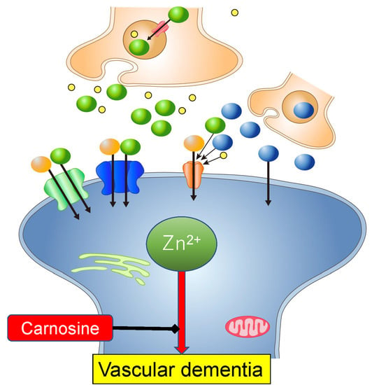
2. Zinc and Vascular Dementia

VD is regarded as the second most common type of senile dementia [21]. Its risk factors are age, male sex, diabetes, and high blood pressure. VD is linked with neurodegeneration that occurs after stroke or ischemia [3,21]. After transient global ischemia, the interruption of blood flow and the resulting deprivation of oxygen and glucose induce abnormal neuronal excitation in most parts of brain, followed by excessive release of glutamate into the synaptic clefts. The successive entry of large quantities of Ca2+ triggers delayed death of vulnerable neurons in the hippocampus or cerebral cortex and causes the development of an infarct, which finally leads to cognitive dysfunction and VD pathogenesis. An epidemiological study has reported the exhibition of dementia symptoms in about 30% of stroke patients after 3 years [22].
Increasing evidence suggests the involvement of Zn in neuronal death after ischemia [7,23,24,25]. Zn is the second most abundant trace element in the brain. High concentrations of Zn accumulate especially in the hippocampus, cerebral cortex and amygdala [26]. Zn acts as a cofactor in more than 300 enzymatic functions and plays essential roles in normal brain functions. Moreover, a considerable amount of Zn forms free Zn ions (Zn2+) that are stored in presynaptic vesicles of glutamatergic neurons. In addition to glutamate, synaptic Zn2+ is secreted from the vesicles to the synaptic clefts during neuronal excitation [27,28]. Then, it binds to several receptors including N-methyl-D-aspartate (NMDA)-type glutamate receptors, γ -aminobutyric acid (GABA) receptors, and glycine receptors to regulate the neuronal excitability. Zn2+ also binds to various channels including Ca2+ channels, K+ channels and modulate their activities [29]. Although the precise role of synaptic Zn2+ is still under investigation, it is widely accepted that Zn2+ plays significant roles in information processing and memory formation [30,31]. Takeda et al. demonstrated that synaptic Zn2+ is involved in the maintenance of memory formation and the AβP neurotoxicity [32]. Indeed, Zn2+ has been reported to influence synaptic plasticity via inhibition of long-term potentiation (LTP). The disorder of ZnT3, the Zn transporter, which involves in the accumulation of Zn at synaptic vesicles, is related to the pathogenesis of AD and other neurodegenerative diseases [33]. Zn deficiency causes retardation in mental and physical development and learning disorders in infants [34]. Low synaptic Zn2+ reportedly enhanced the susceptibility to febrile seizure [35]. Moreover, some neurons have been reported to contain Cu2+ in synaptic vesicles in addition to Zn2+ [36]. Synaptic Cu2+ is also released to the synaptic clefts during neuronal excitation and bind to NMDA-type glutamate receptors, to regulate synaptic functions as well as Zn2+. Therefore, crosstalk between Zn2+ and Cu2+ plays important roles in physiological and pathophysiological functions in the brain [37].
Despite its importance, excess Zn2+ is toxic. In physiological condition, the concentration of free Zn2+ is strictly regulated [29]. In ischemic conditions, a considerable amount of Zn2+ (up to 300 µM) is reportedly co-released with glutamate into synaptic clefts following membrane depolarization [38]. Koh et al. have demonstrated that Zn accumulates in apoptotic neurons in the hippocampus after ischemia [39]. Administration of calcium ethylenediaminetetraacetic acid (Ca-EDTA), a membrane-impermeable Zn2+ chelator, protected hippocampal neurons after transient global ischemia in experimental animals and reduced the infarct volume [40]. Kitamura et al. have revealed an increase in extracellular Zn2+ in transient middle cerebral artery occlusion model (MCAO) rats using a microdialysis method [41]. Furthermore, Zn2+ contributes to the increased permeability of the blood brain barrier after ischemia [42].
The entry of Zn2+ and the increase in intracellular Zn2+ levels ([Zn2+]i), namely “Zn translocation”, may be the primary event in Zn2+-induced neurotoxicity. There are three major routes of Zn2+ entry: voltage-gated Ca2+ channels (VGCCs), NMDA-type glutamate receptors (NMDA-Rs), and AMPA/kainate-type glutamate receptors (A/K-Rs) [8]. Under normal conditions, most hippocampal neurons express AMPA receptors containing the GluR2 subunit, which are poorly permeable to Ca2+ and Zn2+. However, after ischemia, an acute reduction in GluR2 expression occurs and the neurons express a specific type of Ca2+-permeable AMPA receptors (Ca-A/K-R). As the permeabilities of Zn2+ and Ca2+ through Ca-A/K-R channels are greater than through NMDA receptor channels, increased expression of Ca-A/K-R channels enhances the toxicity of Ca2+ and Zn2+. Zn2+ is also implicated in the transcriptional regulation of Ca-A/K-R channels, as Ca-EDTA attenuated the ischemia-induced downregulation of the GluR2 gene [40].
The Zn and Cu levels in the synaptic clefts are estimated to be 1–100 μM and 15 μM, respectively [43,44]. The concentration of Zn in the cerebrospinal fluid (CSF) has been reported to be increased by about 10–20 ppb (0.15–0.31 µM) after ischemia [41]. Considering that the synaptic cleft is a small compartment with 120-nm radius and 20-nm height and that the total volume is estimated to be 1% of the extracellular space [45], the concentration in the synaptic clefts may be much higher compared with that in the CSF.

3. Zn2+-Induced Neurotoxicity

3.1. GT1–7 Cells as a Model System for Investigating Zn2+-Induced Neurotoxicity

The molecular mechanism of Zn2+-induced neuronal death is of great importance for the development of drugs for VD. To this end, primary cultured neurons of rat cerebral cortex or hippocampus, or PC-12 cells, a pheochromocytoma cell line, have been used by many researchers [46,47]. However, the roles of Zn2+ are highly complex in these neuronal cells, which possess glutamate receptors. Since glutamate also causes neurotoxicity and Zn2+ regulates the glutamate-induced excitability, it is difficult to distinguish between the effects of Zn2+ and glutamate. We found that Zn2+ induced marked death of GT1–7 cells (immortalized hypothalamic neurons), as shown in Figure 1A, and that these cells were highly sensitive to Zn2+ compared with other neuronal cells, including primary cultures of the rat cerebral cortex or hippocampus neurons, PC-12 cells, and B-50 cells [48,49]. GT1–7 cells were originally developed by Mellon et al. in 1991 by genetically targeting tumorigenesis in mouse hypothalamic neurons [50]. These cells possess neuronal characteristics such as neurite extension, secretion of gonadotropin-releasing hormone (GnRH), and expression of neuron-specific proteins and receptors including microtubule-associated protein 2 (MAP2), tau protein, neurofilament, synaptophysin, GABAA receptors, dopamine receptors, and L-type Ca2+ channels. However, GT1–7 cells do not exhibit glutamate-induced toxicity, as shown in Figure 1B, because they lack or possess low levels of ionotropic glutamate receptors [51]. Owing to these properties, we considered the GT1–7 cell line an excellent model system for investigating Zn2+-induced neurotoxicity.

3.2. Molecular Pathways Underlying Zn2+-Induced Neurotoxicity

We investigated the molecular mechanism underlying Zn2+-induced neurotoxicity using GT1–7 cells. Zn2+ induced apoptotic death of GT1–7 cells, which were terminal deoxynucleotidyl transferase-mediated biotinylated UTP nick end labeling (TUNEL)-positive and exhibited DNA fragmentation [48,49]. The LD50 was estimated to be 35 µM.
First, we tested the effects of treatment with various pharmacological agents prior to Zn2+ treatment of GT1–7 cells and found that neither antagonists nor agonists of excitatory neurotransmitters (D-APV, glutamate, and CNQX), nor those of inhibitory neurotransmitters (bicuculline, muscimol, baclofen, and GABA) influenced the viability of GT1–7 cells. However, several compounds including energy substrates (pyruvate and citrate), metal chelators (o-phenanthroline and deferoxamine), peptides, and amino acids (carnosine, anserine, and histidine) attenuated the Zn2+-induced death of GT1–7 cells [53,54,55,56,57] (Figure 1). Furthermore, we investigated the viability of GT1–7 cells with or without other metal ions after exposure to Zn2+. We found that co-exposure to sublethal concentrations of Cu2+ and Ni2+ remarkably exacerbated Zn2+-induced death, whereas co-exposure to Al3+, Gd3+, or Ca2+ attenuated the Zn2+-induced death of GT1–7 cells [58,59,60].
Next, we analyzed the Zn2+-induced genetic changes using DNA microarray analysis and real-time PCR (RT-PCR). After 4 h of exposure to Zn2+, various genes including metal-related genes (Zn transporter 1 (ZnT-1), metallothionein 1 (MT1), and metallothionein 2 (MT2)), endoplasmic reticulum (ER) stress-related genes, signal transduction-related genes, and Ca2+ signaling-related genes were upregulated [56,57]. Moreover, the gene expression after co-exposure to Cu2+ and Zn2+ (Cu2+ + Zn2+) was investigated [59]. We then used the substances that attenuated Zn2+-induced neurotoxicity as model compounds and investigated the molecular pathways of Zn2+-induced apoptotic neuronal death. In the following subsections we described the five pathways involved in Zn2+-induced neuronal death.

3.2.1. Energy Production Pathway

Administration of sodium pyruvate, an energy substrate, significantly inhibited the Zn2+-induced death of GT1–7 cells [53]. This result is consistent with findings of other studies using primary cortical neurons [61,62], oligodendrocyte progenitor cells [63], or retinal cells [64]. Shelline and his colleagues have reported that Zn2+ exposure decreased the levels of NAD+ and ATP in cultured cortical neurons, and that treatment with pyruvate restored the NAD+ level [61]. Furthermore, administration of pyruvate to experimental animals reportedly attenuated the neuronal death after ischemia in vivo [65]. We demonstrated that pyruvate and citrate attenuated Cu2+-enhanced Zn2+-induced neurotoxicity of GT1–7 cells [66]. Increasing evidence suggests that Zn2+ is localized within mitochondria and triggers impaired mitochondrial functions [67,68]. Therefore, energy failure and inhibition of glycolysis in mitochondria may be involved in Zn2+-induced neurotoxicity.

3.2.2. Ca2+ Homeostasis

Our pharmacological approach demonstrated that nimodipine, an L-type VGCC blocker, attenuated Zn2+-induced neurotoxicity. Consistently, Kim et al. have reported that Zn2+-induced death in PC-12 cells was attenuated by nimodipine, and enhanced by the L-type VGCC activator, S(−)-Bay K 8644 [69]. Other L-type Ca2+ channel blocker, nifedipine, also attenuated Zn2+-induced neurotoxicity [70]. Additionally, Kim et al. demonstrated that aspirin inhibited N-type VGCC and attenuated Zn2+-induced neurotoxicity by preventing Zn2+ entry [71]. These studies suggest that Ca2+ dyshomeostasis is involved in the mechanism of Zn2+-induced neurotoxicity.
To further address this issue, we used a high-resolution multisite video imaging system with Fura-2 as the cytosolic Ca2+ fluorescent probe to observe temporal changes in the intracellular calcium level ([Ca2+]i) after exposure to Zn2+ [49]. This multisite fluorometry system enables simultaneous long-term observation of temporal changes in [Ca2+]i in more than 50 neurons. After the exposure to Zn, increases in [Ca2+]i were observed in GT1–7 cells. As noted above, addition of Al3+ significantly inhibited Zn2+-induced neurotoxicity in a dose-dependent manner. We found that pretreatment with Al3+ significantly blocked the Zn2+-induced [Ca2+]i elevations, though it did not influence Zn2+ influx. Although Al is neurotoxic, it did not exhibit toxicity in this experimental condition since Al3+ is difficult to enter the cells without membrane permeable chelators [72]. Thus, it is possible that Al3+, a known blocker of various types of Ca2+ channels [73], attenuates Zn2+-induced neurotoxicity by blocking Zn2+-induced elevations in [Ca2+]i.

3.2.3. Endoplasmic Reticulum (ER) Stress Pathway

Our DNA microarray analysis demonstrated that Zn2+ induced a marked upregulation of ER stress-related genes, including CCAAT-enhancer-binding protein homologous protein (CHOP), and growth-arrest and DNA-damage-inducible gene 34 (GADD34) [56,57].
ER stress is associated with the accumulation of unfolded or misfolded proteins and is involved in various neurological disorders, such as cerebral ischemia, AD, and prion diseases [74]. ER stress is distinguished by three signaling proteins (ER stress sensors) termed inositol-requiring enzyme-1α (IRE1α), protein kinase R (PKR)-like ER kinase (PERK) and activating transcription factor 6 (ATF6) [75]. Upon activation, IRE1α, PERK, and ATF6 induce various signal transduction events. The phosphorylation of the α subunit of eukaryotic translation initiation factor 2α (elF2α) is mediated by PERK, and then regulates the translation of activating transcription factor 4 (ATF4). ATF4 is a transcription factor that drives the expression of CHOP and GADD34.
For more detailed analysis, we used RT-PCR to examine the Zn2+-induced expression of these genes and other ER stress-related genes such as immunoglobulin binding protein (Bip), ER degradation-enhancing α-mannosidase-like protein (EDEM), spliced X-box binding protein-1 (sXBP1), glucose-regulated protein 94 (GRP94), and protein disulfide isomerase (PDI) [59]. After 4 h of exposure to Zn2+ alone, enhanced expression levels of activity-regulated cytoskeleton (Arc), CHOP, GADD34, and ATF4 were observed as well as metal-related genes, including ZnT-1, MT1, and MT2. By contrast, other ER stress-related genes including Bip, EDEM, sXBP1, GRP94, and PDI did not exhibit significant changes. Based on our results, it is plausible that the PERK-related pathway is involved in Zn2+-induced ER stress. Furthermore, a synergistic increase in the gene expression levels of Arc, CHOP, and GADD34 was observed in cells co-exposed to Cu2+ and Zn2+, despite the fact that Cu2+ alone did not induce significant changes in these genes. CHOP is responsible for initiating an apoptotic cascade [76] and mediates the activation of GADD34, which reportedly increases after traumatic brain injury [77]. We also demonstrated that the amount of CHOP protein was significantly increased after Cu2++Zn2+ treatment, compared with Zn2+ alone using western blot analysis [59]. Furthermore, we found that dantrolene, an inhibitor of ER stress, attenuated Zn2+-induced neurotoxicity [59]. Based on these findings, it is highly likely that the ER stress pathway is critically involved in Zn2+-induced neurotoxicity, and that low concentrations of Cu2+ promote Zn2+-induced neurotoxicity by potentiating the ER stress pathway. Moreover, we have demonstrated that ER stress is involved in the Ni2+-enhancement of Zn2+-induced neurotoxicity [60].

3.2.4. SAPK/JNK Pathway

Our DNA microarray results demonstrated that several genes downstream of the stress-activated protein kinases/c-Jun amino-terminal kinases (SAPK/JNK) pathway, such as c-Jun and ATF2, were upregulated in cells exposed to Cu2+ + Zn2+. SAPK/JNK are members of the mitogen-activated protein kinase (MAPK) family. The SAPK/JNK signaling pathway plays critical roles in apoptotic cell death, necroptosis, and autophagy [78]. This pathway has been shown to be activated by a variety of environmental stressors, such as oxidative stress, inflammatory cytokines, and metals. Upon activation of this pathway, MAPK kinase 4 (MKK4) or MKK7 phosphorylates and activates the SAPK/JNK. Thereafter, c-Jun and ATF2, major downstream factors of SAPK/JNK, are phosphorylated and activated by SAPK/JNK. Ultimately, the phosphorylated forms of c-Jun and ATF2 induce downstream factors related to cell death and mitochondrial injury, leading to cell death. We examined the expression of these factors and we found that phosphorylated (i.e., active) forms of SAPK/JNK were increased by Cu2+ + Zn2+ treatment in GT1–7 cells [10]. Consistently, phospho-c-Jun and phospho-ATF2 were also induced by Cu2+ + Zn2+ co-treatment. Moreover, SP600125, an inhibitor of the SAPK/JNK signaling pathway, significantly suppressed the activation of the SAPK/JNK signaling pathway by Cu2+ + Zn2+ and the neuronal cell death.

3.2.5. ROS Pathway

Oxidative stress is involved in various degenerative pathways; reactive oxygen species (ROS) induce the SAPK/JNK pathway, the ER stress pathway, and numerous other adverse effects [79,80]. Zn exists only as Zn2+ and is not implicated in the redox pathway, although it is reportedly linked with oxidative stress. In contrast, Cu is a redox-active metal that exists as oxidized Cu2+ and reduced Cu+. We found that the antioxidant, thioredoxin-conjugated human serum albumin (HSA-Trx), attenuated neuronal death induced by Cu2+ + Zn2+ [81]. We also found that the addition of Cu2+ induced ROS production in GT1–7 cells, despite that Zn2+ alone did not produce ROS nor did it influence Cu2+-induced ROS production [10]. Thus, it is possible that Cu2+ triggers ROS production, and then induces the SAPK pathway and/or the ER pathway, thereby enhancing Zn2+-induced neurotoxicity.

3.3. Hypothesis Regarding the Molecular Pathways Underlying Zn2+-Induced Neurotoxicity

Based on these results, we propose a possible scheme of the pathways underlying Zn2+-induced neurotoxicity (Figure 2).
In the case of transient global ischemia, neuronal excitation occurs in a large area of the brain. Thereafter, both Zn2+ and Cu2+ are secreted from synaptic vesicles into the synaptic clefts and translocate into the neurons. Chelators such as Ca-EDTA and o-phenanthroline block this translocation process and attenuate Zn2+-induced neuronal death. Furthermore, the increased [Zn2+]i triggers the inhibition of the energy production machinery in the mitochondria. The energy substrates, pyruvate and citrate, prevent this process. Zn2+ also leads to an increase in [Ca2+]i levels, which is inhibited by Al3+ and other Ca2+ channel blockers. The increase in [Ca2+]i induces ROS production and triggers the ER stress and various apoptotic pathways including the SAPK/JNK pathway. Co-exposure to Cu2+ further induces ROS production and enhances the ER stress pathway and/or the SAPK/JNK pathway. Finally, these processes trigger neurodegenerative pathways, and lead to the neuronal death observed in VD. Carnosine is released from glial cells and enters into neurons by peptide transporters. Carnosine regulates Zn2+ and Cu2+ homeostasis, inhibits Zn2+-induced ER stress and ROS production, and protects neurons from excess Zn2+ (details are discussed in Section 4.4).

4. Carnosine as a Protective Substance Against Zn2+-Induced Neurotoxicity

4.1. Screening System for Protective Substances Against Zn2+-Induced Neurotoxicity

Considering the involvement of Zn in ischemia-induced neuronal death, substances that protect against Zn2+-induced neuronal death may be potential candidates for the prevention or treatment of neurodegeneration following ischemia, and ultimately provide a lead for VD treatments. To explore this idea, we established a convenient and rapid screening system for such substances using GT1–7 cells [10]. We examined the potential inhibitory effects of various agricultural products such as vegetable extracts, fruits extracts, and fish extracts. Among tested, we found that extracts of Japanese eel (Anguilla japonica), mango fruit (Mangifera indica L.), and round herring (Etrumeus teres) protected GT1–7 cells from Zn2+-induced neurotoxicity, then separated the active fraction using HPLC and determined the components’ structures by LC-mass spectrometry (MS). Finally, the active compounds were revealed to be carnosine, citrate, and histidine [12,56,82].

4.2. Carnosine as an Endogenous Neuroprotector

Carnosine is a dipeptide composed of β-alanine and histidine. Figure 3 shows the chemical structures of carnosine and its analogues (anserine (1-methyl carnosine) and homocarnosine). They are naturally occurring dipeptides commonly present in most vertebrate tissues such as birds, fishes, and mammals including humans [13,14]. Carnosine is found in particularly high concentrations in animals and fish that exhibit high levels of exercise, such as horses, chickens, bonitos, and whales. Carnosine is one of the most abundant small-molecule compounds in skeletal muscles with concentrations ranging from 50 to 200 mM, similar to those of creatine and ATP [83]. During high-intensity anaerobic exercise, muscle contractions lead to lactic acid production and a decrease in intracellular pH, which influences various metabolic functions, and the resulting acidosis caused muscle contractile fatigue. Owing to the alkaline property of carnosine (pKa value was 7.01), it is thought to play a significant role in intracellular buffering and in maintaining the pH balance in the muscle [84]. Therefore, the concentration of muscle carnosine is considered to have a positive relationship with exercise performance. Indeed, highly trained athletes have higher carnosine levels than those in untrained individuals [85]. Compared with humans, horses have a 6–10 times higher muscle carnosine concentration. We analyzed the amount of carnosine in five muscles of thoroughbred horses and found that the amount of carnosine is linked to the muscle fiber type [86]. Furthermore, dietary supplementation of carnosine or β-alanine induces an increase in the concentration of muscle carnosine and a delay in fatigue during high-intensity exercise [87].
Additionally, carnosine possesses various beneficial characteristics such as antioxidation, antiglycation, anticrosslinking, and metal chelation [14]. Carnosine scavenges both reactive oxygen and nitrogen, which contain unpaired electrons. Carnosine inhibits lipid oxidation through a combination of free radical scavenging and metal chelation. It inhibits the Maillard reaction that involves reducing sugar and proteins, providing a multitude of end products, most notably advanced glycation end-products (AGEs), which contribute to the pathogenesis of various senile diseases such as AD, vascular stiffening, atherosclerosis, osteoarthritis, inflammatory arthritis, and cataracts. Carnosine and its analogues are reportedly effective for oxidative stress-induced disorders [88]. Furthermore, carnosine has anticrosslinking properties that inhibit the oligomerization of proteins. It is widely accepted that crosslinking of disease-related proteins (e.g., AβP, prion protein, and α-synuclein) and the subsequent conformational changes are central in the pathogenesis of various neurodegenerative diseases termed “conformational diseases” including AD, DLB, and prion diseases [89]. Corona et al. have demonstrated that carnosine supplementation inhibited AβP deposition and improved learning abilities of AD model mice [90]. Carnosine prevents the oxidative stress and inflammation induced by AβP [91]. We showed that carnosine attenuated the neuronal death induced by prion protein fragment peptide (PrP106–126) by changing its conformation [92]. Since carnosine inhibits the fibrillization of lens α-crystalline, N-acetyl carnosine is used as a drug for cataract treatment [17]. Carnosine is a chelator of metal ions and forms complexes with Ca2+, Cu2+, and Zn2+. Thus, it is likely that this dipeptide controls the availability of Zn2+ in neuronal tissue, particularly in the olfactory lobe where both carnosine and Zn are enriched. The Zn-carnosine complex, termed polaprezinc, is effective in the repair of ulcers and other lesions in the alimentary tract [93]. Polaprezinc is also used for Zn supplement therapy and is protective against the cadmium-induced lung injury [94]. Additionally, carnosine reportedly attenuates Mn-induced neurotoxicity [95]. Based on these beneficial characteristics, carnosine is considered to be a “gatekeeper” or “neuroprotector” in the brain [15].

4.3. Carnosine in the Brain

The concentrations of carnosine and related peptides are different among species and vary according to regions [14]. To explore the significance of carnosine and related compounds, we have established a convenient system for quantitative analysis of carnosine, anserine, and homocarnosine using HPLC [20]. Since carnosine and its analogues are highly hydrophilic, it is difficult to separate these compounds using reversed-phase HPLC equipped with a conventional octadecylsilyl (ODS) column, which is generally used in peptide analysis. Thus, we used an HPLC system equipped with a carbon column (HypercarbTM), which contains porous graphite carbon. Using this system, we have analyzed the amount of carnosine and its analogues in various foods using conventional UV spectroscopy at 215 nm [9].
We investigated their presence in the rat brain. Figure 4A shows a typical chromatogram of standard carnosine, anserine, alanyl histidine, and homocarnosine. Figure 4B exhibits a typical chromatogram of a water extract of rat olfactory bulbs dissected after 15 weeks of age, which was heated at 95 °C for 15 min to remove proteins. After this simple pre-treatment, the recovery rates of carnosine and other compounds were determined to be more than 99%. We found that the considerable amount of carnosine in olfactory bulbs. However, only small amounts of carnosine were observed in the cerebral cortex and cerebellum, and anserine was not detected in any of the tested regions. When we examined the changes in carnosine and its analogues in embryos, and rats at 6 and 15 weeks of age, we found that the amount of carnosine in olfactory bulbs increased with age (Figure 4C). Meanwhile, homocarnosine did not exhibit changes with age after birth (Figure 4D). These results are consistent with previous studies. In the mammalian brain, carnosine and homocarnosine, but not anserine, have been detected [96]. It is secreted from oligodendrocytes by the stimulation of glutamate [97]. Boldyrev et al. reviewed that carnosine is mainly present in neurons of olfactory bulbs or in glial cells, and its concentration in the olfactory bulb is more than 1000 µmol/kg [14], similar to our findings in Figure 4C.

4.4. The roles of Carnosine in Protection from Zn2+-Induced Neurotoxicity

We found that carnosine protected neurons against Zn2+-induced neurotoxicity, and then investigated how it affects the pathways. As carnosine can chelate Zn2+, it is plausible that it binds extracellular Zn2+ and inhibits Zn2+ translocation like other chelators. However, our results using a Zn2+-sensitive fluorescent dye and RT-PCR indicated that carnosine did not influence [Zn2+]i nor the expression of ZnT-1 [57]. Meanwhile, we demonstrated that carnosine inhibited Zn2+-induced upregulation of ER stress-related genes such as GADD34 and CHOP, and attenuated neurodegeneration induced by ER-stressors such as thapsigargin and tunicamycin [57]. Thus, it is possible that carnosine protects neurons from Zn2+ by affecting the ER stress pathway, not by inhibiting Zn2+ translocation. Based on the activities of carnosine, we have published two patents for carnosine and related compounds (D-histidine) as drugs or supplements for the prevention and/or treatment of VD [98,99]. Moreover, increasing evidence from experimental animals suggests that carnosine protects against ischemia-induced neurodegeneration in vivo [100,101,102,103].
It is widely believed that orally administered carnosine rapidly degraded to β-alanine and histidine by carnosinase in the blood. However, the supplementation of carnosine or β-alanine reportedly increased the level of carnosine in the brain [104,105]. Therefore, it is possible that dietary carnosine or related amino acids can be synthesized to carnosine in the brain and enters into cells by oligopeptide transporters such as PEPT2, PHT1, and PHT2 [106]. Considering the carnosine level in the body decreases with age [107], the carnosine supplementation therapy may be beneficial for VD, AD, and other diseases. The supplementation of carnosine (40 mg/day) was effective in treatment of patients with major depressive disorders [18]. Recent epidemiological study revealed the inverse correlation between serum β-alanine and pathogenesis of dementia [108]. Furthermore, the carnosine/anserine supplementation (750 mg anserine and 250 mg carnosine per day) reportedly improved episodic memory in elderly people [109] or mild cognitive impairment [110].

5. Conclusions and Future Perspectives

Our hypothesis about the molecular pathways of Zn2+-induced neurotoxicity may aid for the developments of preventive drugs for VD. Carnosine has many beneficial properties such as water solubility, heat-inactivation, and being nontoxic, and therefore, it may become a good neuroprotective drug or supplement that is beneficial for our health. Further research on the molecular mechanism by which carnosine prevents neurotoxicity is required.

Funding

This work was partially supported by a Grant-in Aid for Scientific Research from the Ministry of Education, Culture, Sports, Science, and Technology of Japan. (JSPS Kakennhi Grant numbers. JP26460177 and JP 17H03197).

Acknowledgments

We thank Kobayashi N. for technical assistance. We thank Michal Bell, from Edanz Group https://en-author-services.edanzgroup.com/) for editing a draft of this manuscript.

Conflicts of Interest

Authors declare no conflicts of interest.

Abbreviations

AD Alzheimer’s disease
AβP Alzheimer’s β-amyloid protein
AMPAα-amino-3-hydroxy-5-methylisoxazole-4-propionic acid
Arc activity-regulated cytoskeleton
Ca-EDTA calcium ethylenediaminetetraacetic acid
CHOP CCAAT-enhancer-binding protein homologous protein
D-APV 2-amino-5-phosphonovalerate
ER endoplasmic reticulum
GABA γ-aminobutyric acid
GnRH gonadotropin-releasing hormone
GADD34 growth-arrest and DNA-damage-inducible gene 34
HPLC high performance liquid chromatography
LTP long-term potentiation
[Ca2+]i intracellular calcium levels
NMDA N-methyl-D-aspartate, ROS; reactive oxygen species
SAPK/JNK stress-activated protein kinases/c-Jun amino-terminal kinases
VD vascular dementia
VGCC voltage-dependent Ca2+ channel

References

  1. Selkoe, D.J. The molecular pathology of Alzheimer’s disease. Neuron 1991, 6, 487–498. [Google Scholar] [CrossRef]
  2. Brás, I.C.; Dominguez-Meijide, A.; Gerhardt, E.; Koss, D.; Lázaro, D.F.; Santos, P.I.; Vasili, E.; Xylaki, M.; Outeiro, T.F. Synucleinopathies: Where we are and where we need to go. J. Neurochem. 2020. [Google Scholar] [CrossRef] [Green Version]
  3. Iadecola, C. The pathobiology of vascular dementia. Neuron 2013, 80, 844–866. [Google Scholar] [CrossRef] [PubMed] [Green Version]
  4. World Health Organization. Dementia. Fact sheets of WHO. Available online: https://www.who.int/news-room/fact-sheets/detail/dementia (accessed on 28 March 2020).
  5. Adlard, P.A.; Bush, A.I. Metals and Alzheimer’s disease: how far have we come in the clinic? J. Alzheimers Dis. 2018, 62, 1369–1379. [Google Scholar] [CrossRef] [PubMed] [Green Version]
  6. Brown, D.R. Metalloproteins and neuronal death. Metallomics 2010, 2, 186–194. [Google Scholar] [CrossRef]
  7. Jiang, H.; Song, N.; Jiao, Q.; Shi, L.; Du, X. Iron pathophysiology in parkinson diseases. Adv. Exp. Med. Biol. 2019, 1173, 45–66. [Google Scholar]
  8. Weiss, J.H.; Sensi, S.L.; Koh, J.Y. Zn(2+): a novel ionic mediator of neural injury in brain disease. Trends Pharmacol. Sci. 2000, 21, 395–401. [Google Scholar] [CrossRef]
  9. Kawahara, M.; Tanaka, K.I.; Kato-Negishi, M. Zinc, Carnosine, and Neurodegenerative Diseases. Nutrients 2018, 10, 147. [Google Scholar]
  10. Tanaka, K.I.; Shimoda, M.; Kasai, M.; Ikeda, M.; Ishima, Y.; Kawahara, M. Involvement of SAPK/JNK Signaling Pathway in Copper Enhanced Zinc-Induced Neuronal Cell Death. Toxicol. Sci. 2019, 169, 293–302. [Google Scholar] [CrossRef]
  11. Kawahara, M.; Konoha, K.; Nagata, T.; Sadakane, Y. Protective substances against zinc-induced neuronal death after ischemia: carnosine a target for drug of vascular type of dementia. Recent Pat. CNS Drug Discov. 2007, 2, 145–149. [Google Scholar]
  12. Sadakane, Y.; Konoha, K.; Nagata, T.; Kawahara, M. Protective activity of the extracts from Japanese eel (Anguilla japonica) against zinc-induced neuronal cell death: Carnosine and an unknown substance. Trace Nutrient Res. 2007, 24, 98–105. [Google Scholar]
  13. Hipkiss, A.R. On the relationship between energy metabolism, proteostasis, aging and Parkinson’s disease: possible causative role of methylglyoxal and alleviative potential of carnosine. Aging Dis. 2017, 8, 334–345. [Google Scholar] [CrossRef] [PubMed] [Green Version]
  14. Boldyrev, A.A.; Aldini, G.; Derave, W. Physiology and pathophysiology of carnosine. Physiol Rev. 2013, 93, 1803–1845. [Google Scholar] [CrossRef] [PubMed]
  15. Berezhnoy, D.S.; Stvolinsky, S.L.; Lopachev, A.V.; Devyatov, A.A.; Lopacheva, O.M.; Kulikova, O.I.; Abaimov, D.A.; Fedorova, T.N. Carnosine as an effective neuroprotector in brain pathology and potential neuromodulator in normal conditions. Amino Acids. 2019, 51, 139–150. [Google Scholar] [CrossRef] [PubMed]
  16. Baye, E.; Menon, K.; de Courten, M.P.; Earnest, A.; Cameron, J.; de Courten, B. Does supplementation with carnosine improve cardiometabolic health and cognitive function in patients with pre-diabetes and type 2 diabetes? study protocol for a randomised, double-blind, placebo-controlled trial. BMJ Open 2017, 7, e017691. [Google Scholar] [CrossRef]
  17. Dubois, V.D.; Bastawrous, A. N-acetylcarnosine (NAC) drops for age-related cataract. Cochrane Database Syst. Rev. 2017, 2, CD009493. [Google Scholar] [CrossRef]
  18. Araminia, B.; Shalbafan, M.; Mortezaei, A.; Shirazi, E.; Ghaffari, S.; Sahebolzamani, E.; Mortazavi, S.H.; Shariati, B.; Ardebili, M.E.; Aqamolaei, A.; et al. L-Carnosine combination therapy for major depressive disorder: A randomized, double-blind, placebo-controlled trial. J. Affect. Disord. 2020, 267, 131–136. [Google Scholar] [CrossRef]
  19. Chester, J.E.; Rowneki, M.; Van Doren, W.; Helmer, D.A. Progression of intervention-focused research for Gulf War illness. Mil. Med. Res. 2019, 6, 31. [Google Scholar] [CrossRef]
  20. Mori, M.; Mizuno, D.; Konoha-Mizuno, K.; Sadakane, Y.; Kawahara, M. Quantitative analysis of carnosine and anserine in foods by performing high performance liquid chromatography. Biomed. Res. Trace Elem. 2015, 26, 147–152. [Google Scholar]
  21. Kalaria, R.N. The pathology and pathophysiology of vascular dementia. Neuropharmacology 2018, 134, 226–239. [Google Scholar] [CrossRef]
  22. Pendlebury, S.T.; Rothwell, P.M. Prevalence, incidence, and factors associated with pre-stroke and post-stroke dementia: A systematic review and meta-analysis. Lancet Neurol. 2009, 8, 1006–1018. [Google Scholar] [CrossRef]
  23. Shuttleworth, C.W.; Weiss, J.H. Zinc: New clues to diverse roles in brain ischemia. Trends Pharmacol. Sci. 2011, 32, 480–486. [Google Scholar] [CrossRef] [PubMed] [Green Version]
  24. Kawahara, M.; Mizuno, D.; Koyama, H.; Konoha, K.; Ohkawara, S.; Sadakane, Y. Disruption of zinc homeostasis and the pathogenesis of senile dementia. Metallomics. 2014, 6, 209–219. [Google Scholar] [CrossRef] [PubMed]
  25. Stork, C.J.; Li, Y.V. Elevated cytoplasmic free zinc and increased reactive oxygen species generation in the context of brain injury. Acta Neurochir. Suppl. 2016, 121, 347–353. [Google Scholar] [PubMed]
  26. Becker, J.S.S.; Matusch, A.; Palm, C.; Salber, D.; Morton, K.; Becker, S. Bioimaging of metals in brain tissue by laser ablation inductively coupled plasma mass spectrometry (LA-ICP-MS) and metallomics. Metallomics 2010, 2, 104–111. [Google Scholar] [CrossRef]
  27. Frederickson, C.J.; Suh, S.W.; Silva, D.; Frederickson, C.J.; Thompson, R.B. Importance of zinc in the central nervous system: the zinc-containing neuron. J. Nutr. 2000, 130, 1471S–1483S. [Google Scholar] [CrossRef]
  28. Frederickson, C.J.; Bush, A.I. Synaptically released zinc: physiological functions and pathological effects. Biometals. 2001, 14, 353–366. [Google Scholar] [CrossRef]
  29. Frederickson, C.J.; Koh, J.Y.; Bush, A.I. The neurobiology of zinc in health and disease. Nat Rev Neurosci. 2005, 6, 449–462. [Google Scholar] [CrossRef]
  30. Ueno, S.; Tsukamoto, M.; Hirano, T.; Kikuchi, K.; Yamada, M.K.; Nishiyama, N.; Nagano, T.; Matsuki, N.; Ikegaya, Y. Mossy fiber Zn2+ spillover modulates heterosynaptic N-methyl-D-aspartate receptor activity in hippocampal CA3 circuits. J. Cell Biol. 2002, 158, 215–220. [Google Scholar] [CrossRef] [Green Version]
  31. Takeda, A.; Nakamura, M.; Fujii, H.; Tamano, H. Synaptic Zn(2+) homeostasis and its significance. Metallomics 2013, 5, 417–423. [Google Scholar] [CrossRef]
  32. Takeda, A.; Tamano, H. The impact of synaptic Zn2+ dynamics on cognition and its decline. Int. J. Mol. Sci. 2017, 18, 2411. [Google Scholar] [CrossRef] [Green Version]
  33. McAllister, B.B.; Dyck, R.H. Zinc transporter 3 (ZnT3) and vesicular zinc in central nervous system function. Neurosci. Biobehav. Rev. 2017, 80, 329–350. [Google Scholar] [CrossRef] [PubMed]
  34. Sandstead, H.H. Subclinical zinc deficiency impairs human brain function. J. Trace Elem. Med. Biol. 2012, 26, 70–73. [Google Scholar] [CrossRef] [PubMed]
  35. Reid, C.A.; Hildebrand, M.S.; Mullen, S.A.; Hildebrand, J.M.; Berkovic, S.F.; Petrou, S. Synaptic Zn2+ and febrile seizure susceptibility. Br. J. Pharmacol. 2017, 174, 119–125. [Google Scholar] [CrossRef] [PubMed] [Green Version]
  36. D’Ambrosi, N.; Rossi, L. Copper at synapse: Release, binding and modulation of neurotransmission. Neurochem. Int. 2015, 90, 36–45. [Google Scholar] [CrossRef]
  37. Kawahara, M.; Kato-Negishi, M.; Tanaka, K.I. Amyloids: Regulators of Metal Homeostasis in the Synapse. Molecules 2020, 25, 1441. [Google Scholar] [CrossRef] [Green Version]
  38. Frederickson, C.J.; Klitenick, M.A.; Manton, W.I.; Kirkpatrick, J.B. Cytoarchitectonic distribution of zinc in the hippocampus of man and the rat. Brain Res. 1983, 27, 335–339. [Google Scholar] [CrossRef]
  39. Koh, J.Y.; Suh, S.W.; Gwag, B.J.; He, Y.Y.; Hsu, C.Y.; Choi, D.W. The role of zinc in selective neuronal death after transient global cerebral ischemia. Science 1996, 272, 1013–1016. [Google Scholar] [CrossRef]
  40. Calderone, A.; Jover, T.; Mashiko, T.; Noh, K.M.; Tanaka, H.; Bennett, M.V.; Zukin, R.S. Late calcium EDTA rescues hippocampal CA1 neurons from global ischemia-induced death. J. Neurosci. 2004, 24, 9903–9913. [Google Scholar] [CrossRef] [Green Version]
  41. Kitamura, Y.; Iida, Y.; Abe, J.; Mifune, M.; Kasuya, F.; Ohta, M.; Igarashi, K.; Saito, Y.; Saji, H. Release of vesicular Zn2+ in a rat transient middle cerebral artery occlusion model. Brain Res. Bull. 2006, 69, 622–625. [Google Scholar] [CrossRef]
  42. Qi, Z.; Shi, W.; Zhao, Y.; Ji, X.; Liu, K.J. Zinc accumulation in mitochondria promotes ischemia-induced BBB disruption through Drp1-dependent mitochondria fission. Toxicol. Appl. Pharmacol. 2019, 377, 114601. [Google Scholar] [CrossRef]
  43. Vogt, K.; Mellor, J.; Tong, G.; Nicoll, R. The actions of synaptically released zinc at hippocampal mossy fiber synapses. Neuron 2000, 26, 187–196. [Google Scholar] [CrossRef] [Green Version]
  44. Hopt, A.; Korte, S.; Fink, H.; Panne, U.; Niessner, R.; Jahn, R.; Herms, J. Methods for studying synaptosomal copper release. J. Neurosci. Methods 2003, 128, 159–172. [Google Scholar] [CrossRef] [Green Version]
  45. Schikorski, T.; Stevens, C.F. Quantitative ultrastructural analysis of hippocampal excitatory synapses. J. Neurosci. 1997, 17, 5858–5867. [Google Scholar] [CrossRef] [PubMed]
  46. Chen, C.J.; Liao, S.L. Zinc toxicity on neonatal cortical neurons: involvement of glutathione chelation. J. Neurochem. 2003, 85, 443–453. [Google Scholar] [CrossRef] [PubMed]
  47. Rahman, M.M.; Ukiana, J.; Uson-Lopez, R.; Sikder, M.T.; Saito, T.; Kurasaki, M. Cytotoxic effects of cadmium and zinc co-exposure in PC12 cells and the underlying mechanism. Chem. Biol. Interact. 2017, 269, 41–49. [Google Scholar] [CrossRef]
  48. Kawahara, M.; Kato-Negishi, M.; Kuroda, Y. Pyruvate blocks zinc-induced neurotoxicity in immortalized hypothalamic neurons. Cell. Mol. Neurobiol. 2002, 22, 87–93. [Google Scholar] [CrossRef]
  49. Koyama, H.; Konoha, K.; Sadakane, Y.; Ohkawara, S.; Kawahara, M. Zinc neurotoxicity and the pathogenesis of vascular-type dementia: Involvement of calcium dyshomeostasis and carnosine. J. Clin. Toxicol. 2011, S3, 2161. [Google Scholar]
  50. Mellon, P.L.; Windle, J.J.; Goldsmith, P.C.; Padula, C.A.; Roberts, J.L.; Weiner, R.I. Immortalization of hypothalamic GnRH neurons by genetically targeted tumorigenesis. Neuron 1990, 5, 1–10. [Google Scholar] [CrossRef]
  51. Mahesh, V.B.; Zamorano, P.; De Sevilla, L.; Lewis, D.; Brann, D.W. Characterization of ionotropic glutamate receptors in rat hypothalamus, pituitary and immortalized gonadotropin-releasing hormone (GnRH) neurons (GT1–7 cells). Neuroendocrinology 1999, 69, 397–407. [Google Scholar] [CrossRef]
  52. Nissen, E.; Pauli, G.; Vollenbroich, D. WST-1 assay--a simple colorimetric method for virus titration. In Vitro Cell Dev. Biol. Anim. 1997, 33, 28–29. [Google Scholar] [CrossRef]
  53. Kawahara, M.; Kato-Negishi, M.; Hosoda, R.; Kuroda, Y. Characterization of zinc-induced apoptosis of GT1–7 cells. Biomed. Res. Trace Elements 2002, 13, 280–281. [Google Scholar]
  54. Kawahara, M.; Konoha, K.; Sadakane, Y. Neurotoxicity of zinc: the involvement of calcium homeostasis and carnosine. Biomed. Res. Trace Elements. 2007, 18, 26–34. [Google Scholar]
  55. Konoha, K.; Sadakane, Y.; Kawahara, M. Zinc neurotoxicity and its role in neurodegenerative diseases. J. Health Sci. 2006, 52, 1–8. [Google Scholar] [CrossRef] [Green Version]
  56. Kawahara, M.; Sadakane, Y.; Koyama, H.; Konoha, K.; Ohkawara, S. D-histidine and L-histidine attenuate zinc-induced neuronal death in GT1–7 cells. Metallomics 2013, 5, 453–460. [Google Scholar] [CrossRef]
  57. Mizuno, D.; Konoha-Mizuno, D.; Mori, M.; Sadakane, Y.; Koyama, H.; Ohkawara, S.; Kawahara, M. Protective activity of carnosine and anserine against zinc-induced neurotoxicity: A possible treatment for vascular dementia. Metallomics 2015, 7, 1233–1239. [Google Scholar] [CrossRef]
  58. Konoha, K.; Sadakane, Y.; Kawahara, M. Effects of gadolinium and other metal on the neurotoxicity of immortalized hypothalamic neurons induced by zinc. Biomed. Res. Trace Elements. 2004, 15, 275–277. [Google Scholar]
  59. Tanaka, K.; Kawahara, M. Copper enhances zinc-induced neurotoxicity and the endoplasmic reticulum stress response in a neuronal model of vascular dementia. Front Neurosci. 2017, 11, 58. [Google Scholar] [CrossRef] [Green Version]
  60. Tanaka, K.I.; Kasai, M.; Shimoda, M.; Shimizu, A.; Kubota, M.; Kawahara, M. Nickel enhances zinc-induced neuronal cell death by priming the endoplasmic reticulum stress response. Oxid Med Cell Longev. 2019, 9693726. [Google Scholar] [CrossRef]
  61. Sheline, C.T.; Behrens, M.M.; Choi, D.W. Zinc-induced cortical neuronal death: contribution of energy failure attributable to loss of NAD(+) and inhibition of glycolysis. J. Neurosci. 2000, 20, 3139–3146. [Google Scholar] [CrossRef] [Green Version]
  62. Cai, A.L.; Zipfel, G.J.; Sheline, C.T. Zinc neurotoxicity is dependent on intracellular NAD levels and the sirtuin pathway. Eur. J. Neurosci. 2006, 24, 2169–2176. [Google Scholar] [CrossRef]
  63. Kelland, E.E.; Kelly, M.D.; Toms, N.J. Pyruvate limits zinc-induced rat oligodendrocyte progenitor cell death. Eur. J. Neurosci. 2004, 19, 287–294. [Google Scholar] [CrossRef] [PubMed]
  64. Yoo, M.H.; Lee, J.Y.; Lee, S.E.; Koh, J.Y.; Yoon, Y.H. Protection by pyruvate of rat retinal cells against zinc toxicity in vitro, and pressure-induced ischemia in vivo. Invest Ophthalmol Vis Sci. 2004, 45, 1523–1530. [Google Scholar] [CrossRef] [PubMed] [Green Version]
  65. Lee, J.Y.; Kim, Y.H.; Koh, J.Y. Protection by pyruvate against transient forebrain ischemia in rats. J. Neurosci. 2001, 21, RC171. [Google Scholar] [CrossRef] [PubMed]
  66. Tanaka, K.I.; Shimoda, M.; Kawahara, M. Pyruvic acid prevents Cu2+/Zn2+-induced neurotoxicity by suppressing mitochondrial injury. Biochem Biophys Res Commun. 2018, 495, 1335–1341. [Google Scholar] [CrossRef]
  67. Ji, S.G.; Medvedeva, Y.V.; Weiss, J.H. Zn2+ entry through the mitochondrial calcium uniporter is a critical contributor to mitochondrial dysfunction and neurodegeneration. Exp. Neurol. 2020, 325, 113161. [Google Scholar] [CrossRef]
  68. Olgar, Y.; Tuncay, E.; Turan, B. Mitochondria-targeting antioxidant provides cardioprotection through regulation of cytosolic and mitochondrial Zn2+ levels with re-distribution of Zn2+-transporters in aged rat cardiomyocytes. Int. J. Mol. Sci. 2019, 20, 3783. [Google Scholar] [CrossRef] [Green Version]
  69. Kim, A.H.; Sheline, C.T.; Tian, M.; Higashi, T.; McMahon, R.J.; Cousins, R.J.; Choi, D.W. L-type Ca(2+) channel-mediated Zn(2+) toxicity and modulation by ZnT-1 in PC12 cells. Brain Res. 2000, 886, 99–107. [Google Scholar] [CrossRef]
  70. Zyśk, M.; Gapys, B.; Ronowska, A.; Gul-Hinc, S.; Erlandsson, A.; Iwanicki, A.; Sakowicz-Burkiewicz, M.; Szutowicz, A.; Bielarczyk, H. Protective effects of voltage-gated calcium channel antagonists against zinc toxicity in SN56 neuroblastoma cholinergic cells. PLoS ONE 2018, 13, e0209363. [Google Scholar] [CrossRef] [Green Version]
  71. Kim, E.Y.; Chang, S.Y.; Chung, J.M.; Ryu, B.R.; Joo, C.K.; Moon, H.S.; Kang, K.; Yoon, S.H.; Han, P.L.; Gwag, B.J. Attenuation of Zn2+ neurotoxicity by aspirin: role of N-type Ca2+ channel and the carboxyl acid group. Neurobiol. Dis. 2001, 8, 774–783. [Google Scholar]
  72. Kawahara, M.; Kato-Negishi, M.; Hosoda, R.; Imamura, L.; Tsuda, M.; Kuroda, Y. Brain-derived neurotrophic factor protects cultured rat hippocampal neurons from aluminum maltolate neurotoxicity. J. Inorg. Biochem. 2003, 97, 124–131. [Google Scholar] [CrossRef]
  73. Büsselberg, D.; Platt, B.; Haas, H.L.; Carpenter, D.O. Voltage gated calcium channel currents of rat dorsal root ganglion (DRG) cells are blocked by Al3+. Brain Res. 1993, 622, 163–168. [Google Scholar] [CrossRef]
  74. Kaneko, M.; Imaizumi, K.; Saito, A.; Kanemoto, S.; Asada, R.; Matsuhisa, K.; Ohtake, Y. ER stress and disease: toward prevention and treatment. Biol. Pharm. Bull. 2017, 40, 1337–1343. [Google Scholar] [CrossRef] [PubMed] [Green Version]
  75. Sano, R.; Reed, J.C. ER stress-induced cell death mechanisms. Biochim. Biophys. Acta 2013, 1833, 3460–3470. [Google Scholar] [CrossRef] [PubMed] [Green Version]
  76. Yang, Y.; Liu, L.; Naik, I.; Braunstein, Z.; Zhong, J.; Ren, B. Transcription factor C/EBP homologous protein in health and diseases. Front Immunol. 2017, 8, 1612. [Google Scholar] [CrossRef] [PubMed]
  77. Paschen, W.; Hayashi, T.; Saito, A.; Chan, P.H. GADD34 protein levels increase after transient ischemia in the cortex but not in the CA1 subfield: implications for post-ischemic recovery of protein synthesis in ischemia-resistant cells. J. Neurochem. 2004, 90, 694–701. [Google Scholar] [CrossRef] [PubMed]
  78. Nishina, H.; Wada, T.; Katada, T. Physiological roles of SAPK/JNK signaling pathway. J. Biochem. 2004, 136, 123–126. [Google Scholar] [CrossRef] [PubMed]
  79. Dandekar, A.; Mendez, R.; Zhang, K. Cross talk between ER stress, oxidative stress, and inflammation in health and disease. Methods Mol. Biol. 2015, 1292, 205–214. [Google Scholar]
  80. Papaconstantinou, J. The role of signaling pathways of inflammation and oxidative stress in development of senescence and aging phenotypes in cardiovascular disease. Cells 2019, 8, 1383. [Google Scholar] [CrossRef] [Green Version]
  81. Tanaka, K.I.; Shimoda, M.; Chuang, V.T.G.; Nishida, K.; Kawahara, M.; Ishida, T.; Otagiri, M.; Maruyama, T.; Ishima, Y. Thioredoxin-albumin fusion protein prevents copper enhanced zinc-induced neurotoxicity via its antioxidative activity. Int. J. Pharm. 2018, 535, 140–147. [Google Scholar] [CrossRef]
  82. Sadakane, Y.; Konoha, K.; Kawahara, M. Protective activity of mango (Mangifera indica L.) fruit against a zinc-induced neuronal cell death is independent of its antioxidant activity. Trace Nutr. Res. 2005, 22, 73–79. [Google Scholar]
  83. Harris, R.C.; Wise, J.A.; Price, K.A.; Kim, H.J.; Kim, C.K.; Sale, C. Determinants of muscle carnosine content. Amino Acids. 2012, 43, 5–12. [Google Scholar] [CrossRef] [PubMed] [Green Version]
  84. Abe, H. Role of histidine-related compounds as intracellular proton buffering constituents in vertebrate muscle. Biochemistry (Mosc) 2000, 65, 757–765. [Google Scholar] [PubMed]
  85. Sale, C.; Artioli, G.G.; Gualano, B.; Saunders, B.; Hobson, R.M.; Harris, R.C. Carnosine: From exercise performance to health. Amino Acids. 2013, 44, 1477–1479. [Google Scholar] [CrossRef] [PubMed]
  86. Mori, M.; Mizuno, D.; Konoha-Mizuno, K.; Sadakane, Y.; Kawahara, M. Carnosine concentration in the muscle of thoroughbred horses and its implications in exercise performance. Trace Nutrients Research 2015, 32, 49–53. [Google Scholar]
  87. Quesnele, J.J.; Laframboise, M.A.; Wong, J.J.; Kim, P.; Wells, G.D. The effects of beta-alanine supplementation on performance: A systematic review of the literature. Int. J. Sport Nutr. Exerc. Metab. 2014, 24, 14–27. [Google Scholar] [CrossRef]
  88. Rezzani, R.; Favero, G.; Ferroni, M.; Lonati, C.; Moghadasian, M.H. A carnosine analog with therapeutic potentials in the treatment of disorders related to oxidative stress. PLoS ONE 2019, 14, e0215170. [Google Scholar] [CrossRef] [Green Version]
  89. Kawahara, M.; Kato-Negishi, M.; Tanaka, K. Cross talk between neurometals and amyloidogenic proteins at the synapse and the pathogenesis of neurodegenerative diseases. Metallomics 2017, 9, 619–633. [Google Scholar] [CrossRef]
  90. Corona, C.; Frazzini, V.; Silvestri, E.; Lattanzio, R.; La Sorda, R.; Piantelli, M.; Canzoniero, L.M.; Ciavardelli, D.; Rizzarelli, E.; Sensi, S.L. Effects of dietary supplementation of carnosine on mitochondrial dysfunction, amyloid pathology, and cognitive deficits in 3xTg-AD mice. PLoS ONE 2011, 6, e17971. [Google Scholar] [CrossRef] [Green Version]
  91. Caruso, G.; Fresta, C.G.; Musso, N.; Giambirtone, M.; Grasso, M.; Spampinato, S.F.; Merlo, S.; Drago, F.; Lazzarino, G.; Sortinom, M.A.; et al. Carnosine prevents Aβ-induced oxidative stress and inflammation in microglial cells: A key role of TGF-β. Cells 2019, 8, 64. [Google Scholar] [CrossRef] [Green Version]
  92. Kawahara, M.; Koyama, H.; Nagata, T.; Sadakane, Y. Zinc, copper, and carnosine attenuate neurotoxicity of prion fragment PrP106–126. Metallomics 2011, 3, 726–734. [Google Scholar] [CrossRef]
  93. Matsukura, T.; Tanaka, H. Applicability of zinc complex of L-carnosine for medical use. Biochemistry (Mosc) 2000, 65, 817–823. [Google Scholar] [PubMed]
  94. Kimura, K.; Nakano, Y.; Sugizaki, T.; Shimoda, M.; Kobayashi, N.; Kawahara, M.; Tanaka, K.I. Protective effect of polaprezinc on cadmium-induced injury of lung epithelium. Metallomics 2019, 11, 1310–1320. [Google Scholar] [CrossRef] [PubMed]
  95. Ommati, M.M.; Heidari, R.; Ghanbarinejad, V.; Aminian, A.; Abdoli, N.; Niknahad, H. The neuroprotective properties of carnosine in a mouse model of manganism is mediated via mitochondria regulating and antioxidative mechanisms. Nutr. Neurosci. 2019, 11, 1–13. [Google Scholar]
  96. Baslow, M.H.; Suckow, R.F.; Berg, M.J.; Marks, N.; Saito, M.; Bhakoo, K.K. Differential expression of carnosine, homocarnosine and N-acetyl-L-histidine hydrolytic activities in cultured rat macroglial cells. J. Mol. Neurosci. 2001, 17, 351–359. [Google Scholar] [CrossRef]
  97. Bakardjiev, A. Carnosine and beta-alanine release is stimulated by glutamatergic receptors in cultured rat oligodendrocytes. Glia 1998, 24, 346–351. [Google Scholar] [CrossRef]
  98. Kawahara, M.; Konoha, K. Drugs for prevention or treatment of vascular dementia [Translated from Japanese]. JP5382633, 11 October 2013. [Google Scholar]
  99. Kawahara, M.; Konoha, K. Drugs for prevention or treatment of vascular dementia [Translated from Japanese]. JP5294194, 21 June 2013. [Google Scholar]
  100. Zhang, X.; Song, L.; Cheng, X.; Yang, Y.; Luan, B.; Jia, L.; Xu, F.; Zhang, Z. Carnosine pretreatment protects against hypoxia-ischemia brain damage in the neonatal rat model. Eur. J. Pharmacol. 2011, 667, 202–207. [Google Scholar] [CrossRef]
  101. Park, H.S.; Han, K.H.; Shin, J.A.; Park, J.H.; Song, K.Y.; Kim, D.H. The neuroprotective effects of carnosine in early stage of focal ischemia rodent model. J. Korean Neurosurg. Soc. 2014, 55, 125–130. [Google Scholar] [CrossRef]
  102. Noguchi, K.; Ali, T.F.S.; Miyoshi, J.; Orito, K.; Negoto, T.; Biswas, T.; Taira, N.; Koga, R.; Okamoto, Y.; Fujita, M.; et al. Neuroprotective effects of a novel carnosine-hydrazide derivative on hippocampal CA1 damage after transient cerebral ischemia. Eur. J. Med. Chem. 2019, 163, 207–214. [Google Scholar] [CrossRef]
  103. Davis, C.K.; Laud, P.J.; Bahor, Z.; Rajanikant, G.K.; Majid, A. Systematic review and stratified meta-analysis of the efficacy of carnosine in animal models of ischemic stroke. J. Cereb. Blood Flow Metab. 2016, 36, 1686–1694. [Google Scholar] [CrossRef] [Green Version]
  104. Tomonaga, S.; Hayakawa, T.; Yamane, H.; Maemura, H.; Sato, M.; Takahata, Y.; Morimatsu, F.; Furuse, M. Oral administration of chicken breast extract increases brain carnosine and anserine concentrations in rats. Nutr. Neurosci. 2007, 10, 181–186. [Google Scholar] [CrossRef]
  105. Hoffman, J.R.; Rathmacher, J.A.; Robinson, J.; Gepner, Y.; Cohen, H. Effect of β-alanine supplementation on carnosine and histidine content in the hippocampus of 14-month-old rats. Appl. Physiol. Nutr. Metab. 2019, 44, 1112–1115. [Google Scholar] [CrossRef] [PubMed]
  106. Oppermann, H.; Heinrich, M.; Birkemeyer, C.; Meixensberger, J.; Gaunitz, F. The proton-coupled oligopeptide transporters PEPT2, PHT1 and PHT2 mediate the uptake of carnosine in glioblastoma cells. Amino Acids. 2019, 51, 999–1008. [Google Scholar] [CrossRef]
  107. Stuerenburg, H.J. The roles of carnosine in aging of skeletal muscle and in neuromuscular diseases. Biochemistry 2000, 65, 862–865. [Google Scholar] [PubMed]
  108. Hata, J.; Ohara, T.; Katakura, Y.; Shimizu, K.; Yamashita, S.; Yoshida, D.; Honda, T.; Hirakawa, Y.; Shibata, M.; Sakata, S.; et al. Association between serum β-alanine and risk of dementia. Am. J. Epidemiol. 2019, 188, 1637–1645. [Google Scholar] [PubMed]
  109. Hisatsune, T.; Kaneko, J.; Kurashige, H.; Cao, Y.; Satsu, H.; Totsuka, M.; Katakura, Y.; Imabayashi, E.; Matsuda, H. Effect of anserine/carnosine supplementation on verbal episodic memory in elderly people. J. Alzheimers Dis. 2016, 50, 149–159. [Google Scholar] [CrossRef] [PubMed] [Green Version]
  110. Masuoka, N.; Yoshimine, C.; Hori, M.; Tanaka, M.; Asada, T.; Abe, K.; Hisatsune, T. Effects of anserine/carnosine Supplementation on mild cognitive impairment with APOE4. Nutrients 2019, 11, 1626. [Google Scholar] [CrossRef] [PubMed] [Green Version]
Figure 1. Zn2+-induced neurotoxicity in GT1–7 cells. (A) GT1–7 cells were exposed to with or without 30 µM ZnCl2. After 24 h, live cells were stained with carboxy fluorescein (green) and dead cells were stained with propidium iodide (red). Bar represents 50 µM. (B) Viability of GT1–7 cells after exposure to Zn or glutamate. GT1–7 cells were treated with ZnCl2, or glutamate. After 24 h, cell viability was analyzed using the WST-1 method, which counts the living cell number by measures the cellular mitochondrial dehydrogenase activity [52]. Data are expressed as mean ± SEM, n = 6. * p < 0.05, ** p < 0.01, compared with control. (C) Viability of GT1–7 cells after exposure to Zn with other pharmacological substances. GT1–7 cells were treated with 30 µM Zn with sodium pyruvate (Pyr: 1 mM), o-phenanthroline (o-phe; 20 µM) carnosine (Car: 1 mM), CuCl2 (10 µM), or AlCl3 (100 µM). After 24 h, cell viability was analyzed using the WST-1 method. Data are expressed as mean ± SEM, n = 6. * p < 0.05, ** p < 0.01 compared with Zn alone.
Ijms 21 02570 g001
Figure 2. Schematic of the hypothetical molecular mechanisms underlying the protective effects of carnosine against neuronal death induced by Zn. Zn and Cu are stored in presynaptic vesicles and secreted to the synaptic clefts during ischemia. Excess amounts of secreted Zn can translocate into cells and induce disruption of Ca2+ homeostasis, energy failure in mitochondria, ER stress and oxidative stress, and apoptotic neuronal death. Co-exposure to Cu2+ enhances these effects. These pathways are inhibited by Zn chelators (Ca-EDTA and o-phenanthroline (o-phe)), Ca2+ channel blockers (Al3+ and nimodipine), energy substrates (pyruvate (Pyr) and citrate (Cit)), and the inhibitor of the ER stress pathway (dantrolene (Dant)) or the inhibitor the SAPK/JNK signaling pathway (SP600125), antioxidants (HSA-Trx), or carnosine (CAR). Carnosine is synthesized in glial cells and is secreted in response to stimulation by glutamate and Zn. It protects neurons from Zn neurotoxicity. Carnosine inhibits the ER stress-related apoptotic pathways and the ROS pathway.
Ijms 21 02570 g002
Figure 3. Structures of carnosine and its analogues. Chemical structures of carnosine (Car), anserine (Ans), and homocarnosine (HC) are exhibited.
Ijms 21 02570 g003
Figure 4. Developmental changes in carnosine and related compounds in the brain. Each brain region (cerebrum, cerebellum, and olfactory bulb) was dissected from Wistar rats, homogenized and heated at 95 °C for 15 min. After centrifugation, the supernatant was analyzed by HPLC equipped with a HypercarbTM column (Thermo Fisher Science, Waltham, MA, USA). Carnosine (Car), homocarnosine (HC), alanyl histidine (AH), and anserine (Ans) were analyzed using an isocratic elution of 7% acetonitrile in the presence of 0.1% trifluoroacetic acid at a flow rate of 1 mL/min and monitored with UV at 215 nm. Data are expressed as mean ± S.E.M (n = 4). (A) Typical chromatogram of standard solutions. (B) Typical chromatogram of the olfactory bulb extract at 15 weeks. (C) Developmental changes in carnosine. The amount of carnosine (nmol/g wet tissue) in cerebral cortex, cerebellum, and olfactory bulb at embryo 18 days (E18), and 6 weeks (6 W) and 15 weeks (15 W) postnatal was analyzed by HPLC. Data are expressed as mean ± S.E.M (n = 4). * p<0.05, compared with E18. (D) Developmental changes in homocarnosine. The amount of homocarnosine (nmol/g wet tissue) in cerebral cortex, cerebellum, and olfactory bulb at embryo 18 days (E18), and 6 weeks (6 W) and 15 weeks (15 W) postnatal was analyzed by HPLC. Data are expressed as mean ± S.E.M (n = 4).
Ijms 21 02570 g004

