Conformational Changes of Anoplin, W-MreB1–9, and (KFF)3K Peptides near the Membranes


Abstract
: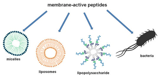
1. Introduction
2. Results
2.1. Synthesis and Characterization of the Peptides
2.2. CD Spectra of Anoplin
2.3. CD Spectra of W-MreB1–9
2.4. CD Spectra of (KFF)3K
2.5. Analysis of the Helicity of Peptides Based on CD Spectra
2.6. Calcein Leakage from LUVs
2.7. Antimicrobial Assays
3. Discussion
4. Materials and Methods
4.1. Peptide Synthesis and Purification
4.2. Preparation of Micelles, POPC:POPE and POPC:POPG SUVs
4.3. Preparation of Cell Culture for CD Experiments
4.4. Recording of CD Spectra
4.5. Secondary Structure Analyses of CD Data
4.6. Preparation of Calcein-Encapsulated LUVs
4.7. Calcein Dye Leakage Assays
4.8. Bacterial Growth Inhibition
Supplementary Materials
Author Contributions
Funding
Acknowledgments
Conflicts of Interest
Abbreviations
| AMP | Antimicrobial peptide |
| CD | Circular dichroism |
| CMC | Critical micelle concentration |
| CPP | Cell-penetrating peptide |
| DPC | Dodecylphosphocholine |
| HT | High tension |
| LB | Lysogeny broth |
| LPS | Lipopolysacharide |
| LUVs | Large unilamellar liposomes |
| MIC | Minimum inhibitory concentration |
| MMW NRMSD | Measured molecular weight Normalized root mean square deviation |
| MS | Mass spectrometry |
| POPC | 2-Oleoyl-1-palmitoyl-sn-glycero-3-phosphocholine |
| POPE | 2-Oleoyl-1-palmitoyl-sn-glycero-3-phosphoethanolamine |
| POPG | 2-Oleoyl-1-palmitoyl-sn-glycero-3-phospho-rac-(1-glycerol) |
| SDS | Sodium dodecyl sulfate |
| SPPS | Solid-phase peptide synthesis |
| SUVs | Small unilamellar vesicles |
| RP-HPLC | Reverse phase-high-performance liquid chromatography |
| RT | Retention time |
| TMW | Theoretical molecular weight |
References
- Antibiotic Resistance. Available online: https://www.who.int/news-room/fact-sheets/detail/antibiotic-resistance (accessed on 13 August 2020).
- Klahn, P.; Brönstrup, M. Bifunctional antimicrobial conjugates and hybrid antimicrobials. Nat. Prod. Rep. 2017, 34, 832–885. [Google Scholar] [CrossRef] [PubMed]
- Corrêa, J.A.F.; Evangelista, A.G.; de Melo Nazareth, T.; Luciano, F.B. Fundamentals on the molecular mechanism of action of antimicrobial peptides. Materialia 2019, 8, 100494. [Google Scholar] [CrossRef]
- Avci, F.G.; Akbulut, B.S.; Ozkirimli, E. Membrane active peptides and their biophysical characterization. Biomolecules 2018, 8, 77. [Google Scholar] [CrossRef] [PubMed] [Green Version]
- Schmidt, N.W.; Deshayes, S.; Hawker, S.; Blacker, A.; Kasko, A.M.; Wong, G.C.L. Engineering persister-specific antibiotics with synergistic antimicrobial functions. ACS Nano 2014, 8, 8786–8793. [Google Scholar] [CrossRef] [PubMed] [Green Version]
- Deshayes, S.; Xian, W.; Schmidt, N.W.; Kordbacheh, S.; Lieng, J.; Wang, J.; Zarmer, S.; Germain, S.S.; Voyen, L.; Thulin, J.; et al. Designing hybrid antibiotic peptide conjugates to cross bacterial membranes. Bioconjug. Chem. 2017, 28, 793–804. [Google Scholar] [CrossRef]
- Brezden, A.; Mohamed, M.F.; Nepal, M.; Harwood, J.S.; Kuriakose, J.; Seleem, M.N.; Chmielewski, J. Dual targeting of intracellular pathogenic bacteria with a cleavable conjugate of kanamycin and an antibacterial cell-penetrating peptide. J. Am. Chem. Soc. 2016, 138, 10945–10949. [Google Scholar] [CrossRef] [PubMed] [Green Version]
- Urushibara, T. Effect of liposome surface charge and peptide side chain charge density on antimicrobial peptide-membrane binding as determined by circular dichroism. J. Membr. Sci. Technol. 2013, 3, 124. [Google Scholar] [CrossRef]
- Wieprecht, T.; Apostolov, O.; Beyermann, M.; Seelig, J. Thermodynamics of the α-helix-coil transition of amphipathic peptides in a membrane environment: Implications for the peptide-membrane binding equilibrium. J. Mol. Biol. 1999, 294, 785–794. [Google Scholar] [CrossRef] [PubMed]
- Miles, A.J.; Wallace, B.A. Circular dichroism spectroscopy of membrane proteins. Chem. Soc. Rev. 2016, 45, 4859–4872. [Google Scholar] [CrossRef] [PubMed] [Green Version]
- The Antimicrobial Peptide Database. Available online: http://aps.unmc.edu/AP/ (accessed on 19 November 2019).
- Gautier, R.; Douguet, D.; Antonny, B.; Drin, G. HELIQUEST: A web server to screen sequences with specific α-helical properties. Bioinformatics 2008, 24, 2101–2102. [Google Scholar] [CrossRef]
- Kulik, M.; Markowska-Zagrajek, A.; Wojciechowska, M.; Grzela, R.; Wituła, T.; Trylska, J. Helix 69 of Escherichia coli 23S ribosomal RNA as a peptide nucleic acid target. Biochimie 2017, 138, 32–42. [Google Scholar] [CrossRef] [PubMed]
- Castillo, J.I.; Równicki, M.; Wojciechowska, M.; Trylska, J. Antimicrobial synergy between mRNA targeted peptide nucleic acid and antibiotics in E. coli. Bioorg. Med. Chem. Lett. 2018, 28, 3094–3098. [Google Scholar] [CrossRef] [PubMed]
- Równicki, M.; Dabrowska, Z.; Wojciechowska, M.; Wierzba, A.J.; Maximova, K.; Gryko, D.; Trylska, J. Inhibition of Escherichia coli growth by vitamin B12-peptide nucleic acid conjugates. ACS Omega 2019, 4, 819–824. [Google Scholar] [CrossRef]
- Równicki, M.; Wojciechowska, M.; Wierzba, A.J.; Czarnecki, J.; Bartosik, D.; Gryko, D.; Trylska, J. Vitamin B12 as a carrier of peptide nucleic acid (PNA) into bacterial cells. Sci. Rep. 2017, 7, 7644. [Google Scholar] [CrossRef] [PubMed]
- Wojciechowska, M.; Równicki, M.; Mieczkowski, A.; Miszkiewicz, J.; Trylska, J. Antibacterial peptide nucleic acids—Facts and perspectives. Molecules 2020, 25, 559. [Google Scholar] [CrossRef] [PubMed] [Green Version]
- Bai, H.; Sang, G.; You, Y.; Xue, X.; Zhou, Y.; Hou, Z.; Meng, J.; Luo, X. Targeting RNA polymerase primary σ 70 as a therapeutic strategy against methicillin-resistant Staphylococcus aureus by antisense peptide nucleic acid. PLoS ONE 2012, 7, e29886. [Google Scholar] [CrossRef]
- Vaara, M.; Porro, M. Group of peptides that act synergistically with hydrophobic antibiotics against gram-negative enteric bacteria. Antimicrob. Agents Chemother. 1996, 40, 1801–1805. [Google Scholar] [CrossRef] [Green Version]
- Salje, J.; van den Ent, F.; de Boer, P.; Löwe, J. Direct membrane binding by bacterial actin MreB. Mol. Cell 2011, 43, 478–487. [Google Scholar] [CrossRef] [PubMed] [Green Version]
- Saikia, K.; Sravani, Y.D.; Ramakrishnan, V.; Chaudhary, N. Highly potent antimicrobial peptides from N-terminal membrane-binding region of E. coli MreB. Sci. Rep. 2017, 7, 1–9. [Google Scholar] [CrossRef] [PubMed] [Green Version]
- Saikia, K.; Chaudhary, N. Interaction of MreB-derived antimicrobial peptides with membranes. Biochem. Biophys. Res. Commun. 2018, 498, 58–63. [Google Scholar] [CrossRef] [PubMed]
- Shang, D.; Zhang, Q.; Dong, W.; Liang, H.; Bi, X. The effects of LPS on the activity of Trp-containing antimicrobial peptides against gram-negative bacteria and endotoxin neutralization. Acta Biomater. 2016, 33, 153–165. [Google Scholar] [CrossRef] [PubMed]
- Konno, K.; Hisada, M.; Fontana, R.; Lorenzi, C.C.B.; Naoki, H.; Itagaki, Y.; Miwa, A.; Kawai, N.; Nakata, Y.; Yasuhara, T.; et al. Anoplin, a novel antimicrobial peptide from the venom of the solitary wasp Anoplius samariensis. Biochim. Biophys. Acta Protein Struct. Mol. Enzymol. 2001, 1550, 70–80. [Google Scholar] [CrossRef]
- Chionis, K.; Krikorian, D.; Koukkou, A.I.; Sakarellos-Daitsiotis, M.; Panou-Pomonis, E. Synthesis and biological activity of lipophilic analogs of the cationic antimicrobial active peptide anoplin. J. Pept. Sci. 2016, 22, 731–736. [Google Scholar] [CrossRef] [PubMed]
- Uggerhøj, L.E.; Poulsen, T.J.; Munk, J.K.; Fredborg, M.; Sondergaard, T.E.; Frimodt-Moller, N.; Hansen, P.R.; Wimmer, R. Rational design of alpha-helical antimicrobial peptides: Do’s and don’ts. ChemBioChem 2015, 16, 242–253. [Google Scholar] [CrossRef] [PubMed]
- Ifrah, D.; Doisy, X.; Ryge, T.S.; Hansen, P.R. Structure-activity relationship study of anoplin. J. Pept. Sci. 2005, 11, 113–121. [Google Scholar] [CrossRef] [PubMed]
- Libardo, M.D.J.; Nagella, S.; Lugo, A.; Pierce, S.; Angeles-Boza, A.M. Copper-binding tripeptide motif increases potency of the antimicrobial peptide anoplin via reactive oxygen species generation. Biochem. Biophys. Res. Commun. 2015, 456, 446–451. [Google Scholar] [CrossRef] [PubMed]
- Salas, R.L.; Garcia, J.K.D.L.; Miranda, A.C.R.; Rivera, W.L.; Nellas, R.B.; Sabido, P.M.G. Effects of truncation of the peptide chain on the secondary structure and bioactivities of palmitoylated anoplin. Peptides 2018, 104, 7–14. [Google Scholar] [CrossRef]
- Chai, H.; Allen, W.E.; Hicks, R.P. Spectroscopic investigations of the binding mechanisms between antimicrobial peptides and membrane models of Pseudomonas aeruginosa and Klebsiella pneumoniae. Bioorg. Med. Chem. 2014, 22, 4210–4222. [Google Scholar] [CrossRef]
- Russell, A.L.; Williams, B.C.; Spuches, A.; Klapper, D.; Srouji, A.H.; Hicks, R.P. The effect of the length and flexibility of the side chain of basic amino acids on the binding of antimicrobial peptides to zwitterionic and anionic membrane model systems. Bioorg. Med. Chem. 2012, 20, 1723–1739. [Google Scholar] [CrossRef]
- Avitabile, C.; D’Andrea, L.D.; Romanelli, A. Circular Dichroism studies on the interactions of antimicrobial peptides with bacterial cells. Sci. Rep. 2014, 4, 1–7. [Google Scholar] [CrossRef]
- Lubecka, E.A.; Sikorska, E.; Sobolewski, D.; Prahl, A.; Slaninová, J.; Ciarkowski, J. Arginine-, d-arginine-vasopressin, and their inverso analogues in micellar and liposomic models of cell membrane: CD, NMR, and molecular dynamics studies. Eur. Biophys. J. 2015, 44, 727–743. [Google Scholar] [CrossRef] [PubMed] [Green Version]
- Spooner, M.J.; Gale, P.A. Anion transport across varying lipid membranes—The effect of lipophilicity. Chem. Commun. 2015, 51, 4883–4886. [Google Scholar] [CrossRef] [PubMed] [Green Version]
- Silhavy, T.J.; Kahne, D.; Walker, S. The bacterial cell envelope. Cold Spring Harb. Perspect. Biol 2010, 2, 1–16. [Google Scholar] [CrossRef]
- Gong, Z.; Ikonomova, S.P.; Karlsson, A.J. Secondary structure of cell-penetrating peptides during interaction with fungal cells. Protein Sci. 2018, 27, 702–713. [Google Scholar] [CrossRef] [PubMed] [Green Version]
- Soundrarajan, N.; Park, S.; Le Van Chanh, Q.; Cho, H.S.; Raghunathan, G.; Ahn, B.; Song, H.; Kim, J.H.; Park, C. Protegrin-1 cytotoxicity towards mammalian cells positively correlates with the magnitude of conformational changes of the unfolded form upon cell interaction. Sci. Rep. 2019, 9, 1–12. [Google Scholar] [CrossRef] [PubMed] [Green Version]
- Yang, C.H.; Chen, Y.C.; Peng, S.Y.; Tsai, A.P.Y.; Lee, T.J.F.; Yen, J.H.; Liou, J.W. An engineered arginine-rich α-helical antimicrobial peptide exhibits broad-spectrum bactericidal activity against pathogenic bacteria and reduces bacterial infections in mice. Sci. Rep. 2018, 8, 1–14. [Google Scholar] [CrossRef] [Green Version]
- Kim, S.; Jeong, H.; Kim, E.Y.; Kim, J.F.; Lee, S.Y.; Yoon, S.H. Genomic and transcriptomic landscape of Escherichia coli BL21(DE3). Nucleic Acids Res. 2017, 45, 5285–5293. [Google Scholar] [CrossRef]
- Ganewatta, M.S.; Chen, Y.P.; Wang, J.; Zhou, J.; Ebalunode, J.; Nagarkatti, M.; Decho, A.W.; Tang, C. Bio-inspired resin acid-derived materials as anti-bacterial resistance agents with unexpected activities. Chem. Sci. 2014, 5, 2011–2016. [Google Scholar] [CrossRef]
- Gusmão, K.A.G.; dos Santos, D.M.; Santos, V.M.; Cortés, M.E.; Reis, P.V.M.; Santos, V.L.; Piló-Veloso, D.; Verly, R.M.; de Lima, M.E.; Resende, J.M. Ocellatin peptides from the skin secretion of the South American frog Leptodactylus labyrinthicus (Leptodactylidae): Characterization, antimicrobial activities and membrane interactions. J. Venom. Anim. Toxins Incl. Trop. Dis. 2017, 23, 1–14. [Google Scholar] [CrossRef] [Green Version]
- Wieprecht, T.; Apostolov, O.; Beyermann, M.; Seelig, J. Membrane binding and pore formation of the antibacterial peptide PGLa: Thermodynamic and mechanistic aspects. Biochemistry 2000, 39, 442–452. [Google Scholar] [CrossRef]
- Russell, A.L.; Kennedy, A.M.; Spuches, A.M.; Venugopal, D.; Bhonsle, J.B.; Hicks, R.P. Spectroscopic and thermodynamic evidence for antimicrobial peptide membrane selectivity. Chem. Phys. Lipids 2010, 163, 488–497. [Google Scholar] [CrossRef] [PubMed]
- Guyer, M.S.; Reed, R.R.; Steitz, J.A.; Low, K.B. Identification of a sex-factor-affinity site in E. coli as gamma delta. Cold Spring Harb. Symp. Quant. Biol. 1981, 45 Pt 1, 135–140. [Google Scholar] [CrossRef]
- Stawikowski, M.; Fields, G.B. Introduction to peptide synthesis. Current protocols in protein science. Curr. Protoc. Protein Sci. 2002, 26, 1–17. [Google Scholar]
- Chen, Y.H. Determination of the helix and β form of proteins in aqueous solution by circular dichroism. Biochemistry 1974, 13, 3350–3359. [Google Scholar] [CrossRef] [PubMed]
- Whitmore, L.; Wallace, B.A. DICHROWEB, an online server for protein secondary structure analyses from circular dichroism spectroscopic data. Nucleic Acids Res. 2004, 32, 668–673. [Google Scholar] [CrossRef] [PubMed] [Green Version]
- Whitmore, L.; Wallace, B.A. Protein secondary structure analyses from circular dichroism spectroscopy: Methods and reference databases. Biopolymers 2008, 89, 392–400. [Google Scholar] [CrossRef]
- Górska, A.; Markowska-Zagrajek, A.; Równicki, M.; Trylska, J. Scanning of 16S ribosomal RNA for peptide nucleic acid targets. J. Phys. Chem. B 2016, 120, 8369–8378. [Google Scholar] [CrossRef]
- Hatamoto, M.; Nakai, K.; Ohashi, A.; Imachi, H. Sequence-specific bacterial growth inhibition by peptide nucleic acid targeted to the mRNA binding site of 16S rRNA. Appl. Microbiol. Biotechnol. 2009, 84, 1161–1168. [Google Scholar] [CrossRef]
- Wallace, B.; Lees, J.; Orry, A. Analyses of circular dichroism spectra of membrane proteins. Protein Sci. 2003, 12, 875–884. [Google Scholar] [CrossRef] [Green Version]
- Abdul-Gader, A.; Miles, A.J.; Wallace, B.A. A reference dataset for the analyses of membrane protein secondary structures and transmembrane residuesusing circular dichroism spectroscopy. Bioinformatics 2011, 27, 1630–1636. [Google Scholar] [CrossRef]
- Tranter, G.E. Protein Structure Analysis by CD, FTIR, and Raman Spectroscopies, 3rd ed.; Elsevier Ltd.: Oxford, UK, 2016; ISBN 9780128032244. [Google Scholar]
- Micsonai, A.; Wien, F.; Kernya, L.; Lee, Y.H.; Goto, Y.; Réfrégiers, M.; Kardos, J. Accurate secondary structure prediction and fold recognition for circular dichroism spectroscopy. Proc. Natl. Acad. Sci. USA 2015, 112, E3095–E3103. [Google Scholar] [CrossRef] [PubMed] [Green Version]
- Shenkarev, Z.O.; Balandin, S.V.; Trunov, K.I.; Paramonov, A.S.; Sukhanov, S.V.; Barsukov, L.I.; Arseniev, A.S.; Ovchinnikova, T.V. Molecular mechanism of action of β-Hairpin antimicrobial peptide arenicin: Oligomeric structure in dodecylphosphocholine micelles and pore formation in planar lipid bilayers. Biochemistry 2011, 50, 6255–6265. [Google Scholar] [CrossRef] [PubMed]
- Zhang, J.; Zhou, X.; Yu, Q.; Yang, L.; Sun, D.; Zhou, Y.; Liu, J. Epigallocatechin-3-gallate (EGCG)-stabilized selenium nanoparticles coated with Tet-1 peptide to reduce amyloid-β aggregation and cytotoxicity. ACS Appl. Mater. Interfaces 2014, 6, 8475–8487. [Google Scholar] [CrossRef] [PubMed]
- Panteleev, P.V.; Bolosov, I.A.; Balandin, S.V.; Ovchinnikova, T.V. Design of antimicrobial peptide arenicin analogs with improved therapeutic indices. J. Pept. Sci. 2015, 21, 105–113. [Google Scholar] [CrossRef] [PubMed]
- Adams, P.G.; Lamoureux, L.; Swingle, K.L.; Mukundan, H.; Montaño, G.A. Lipopolysaccharide-induced dynamic lipid membrane reorganization: Tubules, perforations, and stacks. Biophys. J. 2014, 106, 2395–2407. [Google Scholar] [CrossRef] [Green Version]
- Sun, Y.; Shang, D. Inhibitory Effects of Antimicrobial Peptides on Lipopolysaccharide-Induced Inflammation. Mediators Inflamm. 2015, 2015, 167572. [Google Scholar] [CrossRef] [Green Version]
- Patenge, N.; Pappesch, R.; Krawack, F.; Walda, C.; Mraheil, M.A.; Jacob, A.; Hain, T.; Kreikemeyer, B. Inhibition of growth and gene expression by PNA-peptide conjugates in Streptococcus pyogenes. Mol. Ther. Nucleic Acids 2013, 2, e132. [Google Scholar] [CrossRef]
- Mohamed, M.F.; Hammac, G.K.; Guptill, L.; Seleem, M.N. Antibacterial activity of novel cationic peptides against clinical isolates of multi-drug resistant Staphylococcus pseudintermedius from infected dogs. PLoS ONE 2014, 9, e116259. [Google Scholar] [CrossRef] [Green Version]
- Cardoso, M.H.; Oshiro, K.G.N.; Rezende, S.B.; Cândido, E.S.; Franco, O.L. The structure/function relationship in antimicrobial peptides: What can we obtain from structural data? Adv. Protein Chem. Struct. Biol. 2018, 112, 359–384. [Google Scholar]
- Müllertz, A.; Perrie, Y.; Rades, T. Analytical Techniques in the Pharmaceutical Sciences; Springer: New York, NY, USA, 2016; ISBN 978-1-4939-4027-1. [Google Scholar]
- Kaiser, E.; Colescott, R.L.; Bossinger, C.D.; Cook, P.I. Color test for detection of free terminal amino groups in the solid-phase synthesis of peptides. Anal. Biochem. 1970, 34, 595–598. [Google Scholar] [CrossRef]
- Watson, R.M.; Woody, R.W.; Lewis, R.V.; Scott Bohle, D.; Andreotti, A.H.; Ray, B.; Miller, K.W. Conformational changes in pediocin AcH upon vesicle binding and approximation of the membrane-bound structure in detergent micelles. Biochemistry 2001, 40, 14037–14046. [Google Scholar] [CrossRef] [PubMed] [Green Version]
- Dorovkov, M.V.; Kostyukova, A.S.; Ryazanov, A.G. Phosphorylation of annexin A1 by TRPM7 kinase: A switch regulating the induction of an α-helix. Biochemistry 2011, 50, 2187–2193. [Google Scholar] [CrossRef] [PubMed]
- Wieprecht, T.; Beyermann, M.; Seelig, J. Binding of antibacterial magainin peptides to electrically neutral membranes: Thermodynamics and structure. Biochemistry 1999, 38, 10377–10387. [Google Scholar] [CrossRef] [PubMed]
- Avitabile, C.; Netti, F.; Orefice, G.; Palmieri, M.; Nocerino, N.; Malgieri, G.; D’Andrea, L.D.; Capparelli, R.; Fattorusso, R.; Romanelli, A. Design, structural and functional characterization of a Temporin-1b analog active against gram-negative bacteria. Biochim. Biophys. Acta Gen. Subj. 2013, 1830, 3767–3775. [Google Scholar] [CrossRef] [Green Version]
- Maherani, B.; Arab-Tehrany, E.; Kheirolomoom, A.; Geny, D.; Linder, M. Calcein release behavior from liposomal bilayer; Influence of physicochemical/mechanical/structural properties of lipids. Biochimie 2013, 95, 2018–2033. [Google Scholar] [CrossRef]


| Peptide | Sequence (N->C) | Net Charge | H | MW (g/mol) | RT (min) | |
|---|---|---|---|---|---|---|
| Calculated | Detected | |||||
| anoplin | GLLKRIKTLL | +4 | 0.587 | 1153.5 | 1153.8 | 11.3 |
| W-MreB1–9 | WMLKKFRGMF | +4 | 0.700 | 1342.7 | 1342.5 | 11.1 |
| (KFF)3K | KFFKFFKFFK | +5 | 0.678 | 1525.1 | 1524.8 | 24.1 |
| MembraneMimic | % Helix | |||
|---|---|---|---|---|
| Anoplin | W-MreB1–9 | (KFF)3K | ||
| buffer | 9 | - | - | |
| SDS [mM] | 1 | 48 | 8 | 29 |
| 5 | 68 | 18 | 13 | |
| 10 | 68 | 6 | 20 | |
| 30 | 71 | 13 | 18 | |
| DPC [mM] | 0.5 | 8 | 5 | 12 |
| 2 | 52 | 27 | - | |
| 5 | 53 | 38 | 4 | |
| 10 | 63 | 48 | 8 | |
| POPC:POPG (3:1) [mM] | 0.1 | 16 | 3 | - |
| 0.5 | 30 | 29 | - | |
| 0.75 | 72 | - | - | |
| 1 | - | 27 | - | |
| POPC:POPE (3:1) [mM] | 0.1 | 27 | - | - |
| 1 | - | 10 | - | |
| 2 | 20 | 18 | - | |
| 3 | - | 34 | - | |
| 5 | 32 | - | - | |
| LPS [µM] | 10 | 25 | 15 | 1 |
| 20 | 47 | 24 | 16 | |
| 50 | 52 | 18 | 9 | |
| 100 | 73 | 15 | 9 | |
| E. coli [µL] | 2 | 3 | - | - |
| 4 | 0 | - | - | |
| 8 | 2 | - | - | |
| 12 | 3 | - | - | |
| Peptide | MIC [µM] | |
|---|---|---|
| E. coli K12 | E. coli BL21(DE3) | |
| anoplin | 32 | 32 |
| W-MreB1–9 | 128 | 256 |
| (KFF)3K | 32 | 32 |
| ampicillin | 8 | 4 |
| tetracycline | 8 | 16 |
Publisher’s Note: MDPI stays neutral with regard to jurisdictional claims in published maps and institutional affiliations. |
© 2020 by the authors. Licensee MDPI, Basel, Switzerland. This article is an open access article distributed under the terms and conditions of the Creative Commons Attribution (CC BY) license (http://creativecommons.org/licenses/by/4.0/).
Share and Cite
Wojciechowska, M.; Miszkiewicz, J.; Trylska, J. Conformational Changes of Anoplin, W-MreB1–9, and (KFF)3K Peptides near the Membranes. Int. J. Mol. Sci. 2020, 21, 9672. https://doi.org/10.3390/ijms21249672
Wojciechowska M, Miszkiewicz J, Trylska J. Conformational Changes of Anoplin, W-MreB1–9, and (KFF)3K Peptides near the Membranes. International Journal of Molecular Sciences. 2020; 21(24):9672. https://doi.org/10.3390/ijms21249672
Chicago/Turabian StyleWojciechowska, Monika, Joanna Miszkiewicz, and Joanna Trylska. 2020. "Conformational Changes of Anoplin, W-MreB1–9, and (KFF)3K Peptides near the Membranes" International Journal of Molecular Sciences 21, no. 24: 9672. https://doi.org/10.3390/ijms21249672