Article

Cyclin B3 Deficiency Impairs Germline Stem Cell Maintenance and Its Overexpression Delays Cystoblast Differentiation in Drosophila Ovary

1
Provincial Key Laboratory of the Conservation and Exploitation Research of Biological Resources in Anhui, College of Life Sciences, Anhui Normal University, Wuhu 241000, China
2
The Institute of Bioinformatics, College of Life Sciences, Anhui Normal University, Wuhu 241000, China
*
Author to whom correspondence should be addressed.
Int. J. Mol. Sci. 2018, 19(1), 298; https://doi.org/10.3390/ijms19010298
Submission received: 14 December 2017 / Revised: 14 January 2018 / Accepted: 16 January 2018 / Published: 19 January 2018
(This article belongs to the Section Biochemistry)

Abstract

:
It is well known that cyclin B3 (cycB3) plays a key role in the control of cell cycle progression. However, whether cycB3 is involved in stem cell fate determination remains unknown. The Drosophila ovary provides an exclusive model for studying the intrinsic and extrinsic factors that modulate the fate of germline stem cells (GSCs). Here, using this model, we show that Drosophila cycB3 plays a new role in controlling the fate of germline stem cells (GSC). Results from cycB3 genetic analyses demonstrate that cycB3 is intrinsically required for GSC maintenance. Results from green fluorescent protein (GFP)-transgene reporter assays show that cycB3 is not involved in Dad-mediated regulation of Bmp signaling, or required for dpp-induced bam transcriptional silencing. Double mutants of bam and cycB3 phenocopied bam single mutants, suggesting that cycB3 functions in a bam-dependent manner in GSCs. Deficiency of cycB3 fails to cause apoptosis in GSCs or influence cystoblast (CB) differentiation into oocytes. Furthermore, overexpression of cycB3 dramatically increases the CB number in Drosophila ovaries, suggesting that an excess of cycB3 function delays CB differentiation. Given that the cycB3 gene is evolutionarily conserved, from insects to humans, cycB3 may also be involved in controlling the fate of GSCs in humans.

Graphical Abstract

1. Introduction

Adult stem cells are characterized by their ability to supply new cells to replace aged/injured cells in adult tissues throughout life, and to maintain their “stemness”, via self-renewal. It is essential for stem cells to keep a balance between self-renewal and differentiation into daughter cells. Numerous studies over the past twenty years have shown that the stem cell maintenance is modulated by intrinsic and extrinsic mechanisms [1,2]. Germline stem cells (GSCs) in the Drosophila ovary provide an excellent model for exploring the mechanisms underlying GSC fate determination, in vivo. The Drosophila adult ovary contains about fifteen ovarioles, in which an anatomical structure, called germarium, is positioned at the apical end. Two to three GSCs are located in the anterior region of the germarium, and three types of somatic cells (terminal filament cells, cap cells and escort stem cells) constitute the microenvironment (also called the “niche”) for GSCs (Figure 1A) [3,4]. A GSC divides asymmetrically to give birth to two daughters—one daughter cell remains adherent to niche cells and continuously functions as a stem cell, whereas the other daughter moves away from the niche and initiates differentiation as a cystoblast (CB). GSCs are readily visualized by a spherical spectrosome, which is located in the anterior region in the cell, while the spectrosome in CB usually loses its anterior localization. CB undergoes four rounds of successive incomplete mitosis and generates a 16-cell germ line cyst, interconnected by a branched fusome. The 16-cell cyst is surrounded by follicle cells, derived from the somatic stem cell (SSC), then the encapsulated cyst moves posteriorly out of the germarium and forms the egg chamber, eventually developing into a mature egg.
Previous research has manifested that Bmp/Dpp-bam functions as the primary signaling pathway for GSC maintenance in the Drosophila ovary [5,6,7,8,9]. Bmp, produced by niche cells, acts as a short-range signal, which eventually represses bam transcription in GSCs, to maintain its self-renewal [10,11]. Ectopic overexpression of Bmp in cap cells can suppress CB differentiation and produce an ovarian tumor, whereas the reduced level of Bmps directly results in the loss of the GSC phenotype [6,7,8,9,10,11]. In addition to the Bmp-dependent extrinsic regulatory mechanism, the fate of GSCs is also controlled by intrinsic regulatory factors, such as Nanos, Pumillio, Cyclin B, Cyclin E, Ote and Effete and Gcn5 [12,13,14,15,16,17,18]. Even so, many extrinsic/intrinsic regulatory factors from niche cells/GSCs remain to be identified.
Cyclin proteins are characterized by their periodical appearance, accumulation and degradation during cell-cycle progression. Since the first cyclin (cyc), cycA, was cloned and found with a periodic expression pattern in cell-cycle [19,20], many other types of cyclins (e.g., cyclin B, C, D, E and K) were subsequently found [21,22,23,24,25]. Cyclins are positive regulatory subunits of Cyclin-dependent kinases (CDKs), whereby CDKs play a key role in the control of cell-cycle transitions. Studies in numerous organisms have demonstrated that at least three evolutionarily-conserved classes of mitotic cyclins, cycA, B and B3, have overlapping, but nonidentical, functions for mitosis progressions (i.e., prophase, metaphase, anaphase and telophase) [26,27,28,29,30]. In addition, ubiquitin-mediated sequential degradation of these Cyclins is also essential for dividing cells to exit mitosis, leading to eventual completion of cell-cycle [26,27,30]. Cyclins display regulatory functions in controlling the fate of stem cells (self-renewal or switch to differentiation). For example, the expression level of CycA is involved in maintaining the fate of GSCs in the Drosophila ovary [9,15,18]. cycB is required for GSC maintenance in the female Drosophila [16]. Knockout of cycD3 leads to impaired establishment of the skeletal muscle satellite cell (i.e., muscle stem cell) population within adult mouse muscle tissues [31]. Higher Cyclin E-cdk2 kinase activity is required for ovarian follicle stem cell maintenance [32]. Cyclin H plays a critical role in maintaining ESC (embryonic stem cell) identity [33]. Cyclin K protein exhibits a high expression level in pluripotent embryonic stem cells but low in their differentiated derivatives or tissue-specific stem cells, and knockdown of cyclin K leads to cell differentiation [34]. Here, we have uncovered a new role for cycB3 that plays a key role in maintaining the fate of GSCs in the Drosophila ovary.

2. Results

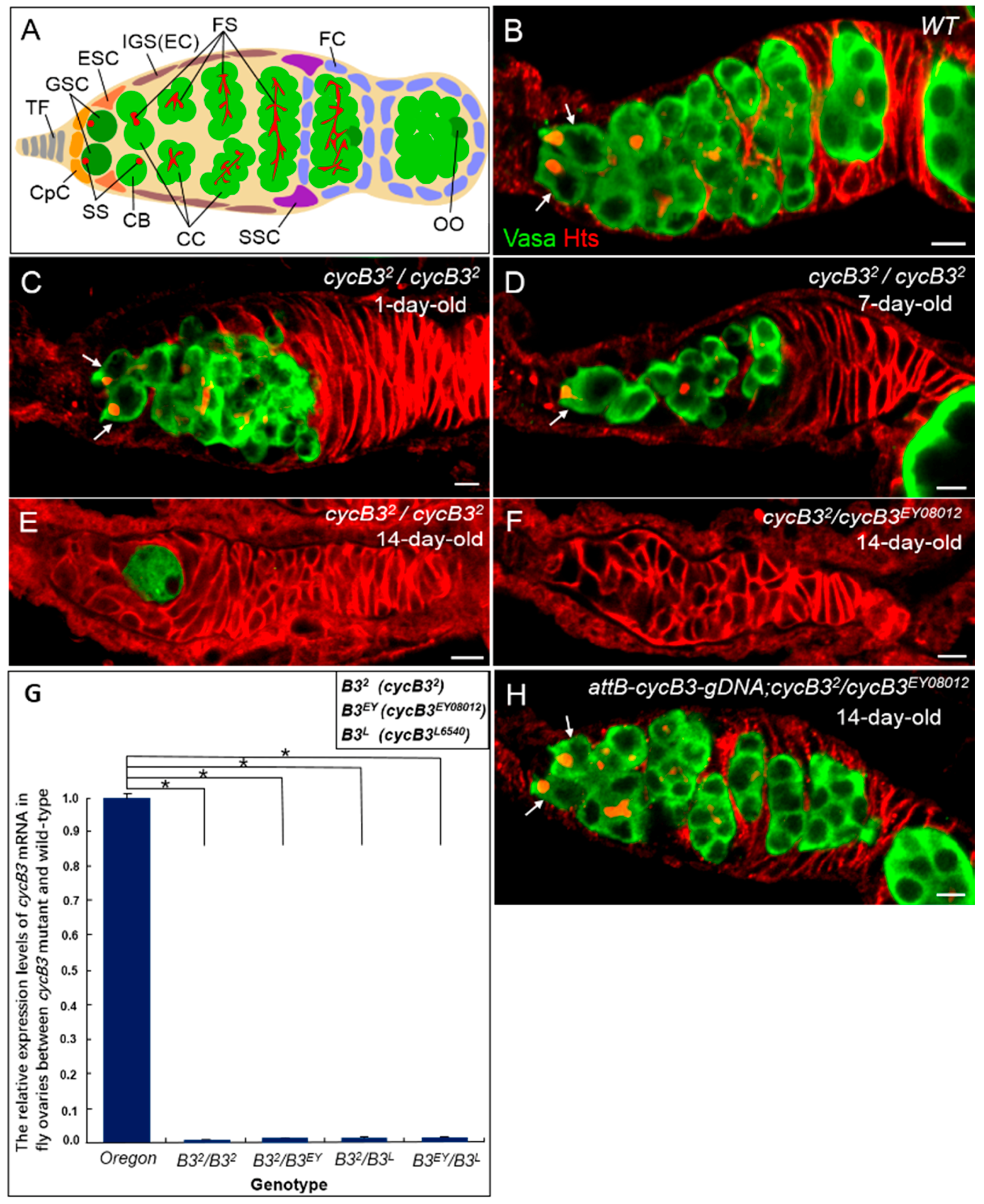
2.1. Deficiency of cycB3 Impairs GSCs Maintenance in Drosophila Ovary

To discover the genes that possibly influence the fate of Drosophila GSCs, we performed a genetic screen of female sterile lines. We isolated a null allele, cycB32, which carries a small deletion resulting from an imprecise excision of the P-element insertion in cyclin B3 [29]. Quite a few cycB32 homozygous mutant flies (>30%) displayed slim ovaries when dissected at day 7 after eclosion. This finding triggered us to thoroughly examine the behavior of GSCs in cycB3 mutant. We first obtained two additional P-element insertion mutation alleles of the gene cycB3 (i.e., P{EPgy2} cycB3EY08012 and P{lacW} cycB3L6540) from Bloomington Stock Center [35]. Next, we got three trans-heterozygous mutants (i.e., cycB32/cycB3EY08012, cycB32/cycB3L6540 and cycB3EY08012/cycB3L6540) other than the cycB32 homozygous mutant (cycB32/cycB32). We used anti-Hts and anti-Vasa antibodies to visualize the spectrosomes/fusomes and germ cells in adult ovaries, respectively (Figure 1B). Vasa displays a germ cell-specific expression pattern, while Hts exhibits an enrichment in both round fusomes (i.e., spectrosomes) and branched fusomes [36,37]. By using the methods described previously [15], we finally performed phenotypic analyses by measuring the ratios of four types of germaria (i.e., 2–3 GSCs-, 1 GSC-, 0 GSC-containing and empty germaria) in different cycB3 mutation backgrounds, at different ages.
As shown in Table 1, in wild-type ovaries, the number of normal germaria (containing 2–3 GSCs), examined at three stages (days 1, 7 and 14, post-eclosion) was largely sustained at high levels, measured as 98.6% (n = 146), 95.5% (n = 178) and 90.5% (n = 148), respectively (Figure 1B). In contrast, the number of normal germaria from cycB32 homozygotes at three stages (day 1, 7 and 14) was reduced dramatically with time, counted as 89.1% (n = 274), 39.4% (n = 203) and 22.6% (n = 420), respectively (Table 1 and Figure 1C). Contrarily, the ratios of abnormal phenotypes (1 GSC, 0 GSC and empty germaria) from cycB32 homozygote were increased from the initial 10.9% (30/274), at day 1, to the final 77.4% (325/420), at day 14 (Table 1 and Figure 1D,E). These data demonstrate that the number of abnormal germaria in cycB32 mutant ovaries was elevated rapidly as time elapsed, suggesting a notable loss of GSCs in cycB3 deficient ovaries. Similarly, the ratios of normal germaria from three cycB3 trans-heterozygotes (cycB32/cycB3EY08012, cycB32/cycB3L6540 and cycB3EY08012/cycB3L6540), at day 1, were 94.3% (n = 209), 86.6% (n = 194) and 80.8% (n = 219), respectively (Table 1). Two weeks later, the ratios of normal germaria had decreased severely, measured as 27.6% (n = 340), 28.2% (n = 444) and 18.4% (n = 207), respectively (Table 1). Accordingly, the numbers of abnormal germaria from three cycB3 trans-heterozygotes increased with time. In these abnormal phenotypes, the total proportions of 0 GSC and empty germaria, from three trans-heterozygotes, at day 14, were increased to 44.4% (151/340), 45.3% (201/444) and 48.7% (101/207), respectively (Table 1 and Figure 1F). In addition, similar results were observed in cycB32/Df (fly deficiency strain of cycB3 gene) mutant ovaries (Table 1). Taken together, these statistical data strongly suggest that cycB3 deficiency causes a progressive loss of GSCs with ageing.
To determine if the GSC loss phenotype in cycB3 mutant ovaries is due to a reduced cycB3 expression level, we performed quantitative real-time polymerase chain reaction (qPCR) assays, to compare mRNA levels between wild-type and mutant ovaries [38]. According to the method described previously [39], we extracted total RNA from Drosophila ovaries and performed reverse transcription-based qPCR experiments to measure the cycB3 mRNA level, using the rp49 gene as a reference. Compared with wild-types, the cycB3 mRNA expression level in cycB3 mutant ovaries (cycB32/cycB32, cycB32/cycB3EY08012, cycB32/cycB3L6540 and cycB3EY08012/cycB3L6540) was reduced dramatically (Figure 1G). The data strongly suggest that cycB3 is reduced in cycB3 mutant ovaries, indicating that cycB3 protein is responsible for the loss of GSC phenotypes in cycB3 mutant flies.
To confirm a specific role of cycB3 in GSC maintenance, we performed a cycB3 rescue assay, by generating a transgene of P{attB-cycB3-gDNA}, in which a 9.5 kb genomic DNA fragment, encompassing the ~2.8 kb cycB3 transcript, was introduced into attP-phiC31 fly hosts, by attB/attP-element-mediated germline transformation [40]. We found that the GSC loss phenotype in three cycB3 allelic mutants was fully rescued by this transgenic line (Figure 1H and Table S1). Taken together, these results substantiate the idea that cycB3 plays an essential role in GSC maintenance.

2.2. The Gene cycB3 Functions as an Intrinsic Factor in Controlling GSC Maintenance

It has been reported that the GSC maintenance is modulated by intrinsic and extrinsic signaling pathways in the ovary [1,5,10,13]. To determine the role of cycB3 in the GSC maintenance, we generated a transgenic reporter, P{cycB3P-cycB3-gfp}, in which the cycB3-gfp fused coding sequence (encoding CycB3-Green fluorescent protein (GFP) fusion protein) was placed under the control of a 6.5 kb cycB3 promoter. The gfp expression pattern faithfully reflects cycB3 gene expression in this reporter system [41]. Thus, GFP expression can be used to represent that of cycB3. GFP expression was checked in transgenic fly ovaries with wild-types as a control (Figure 2A’,A). We found that GFP showed a specific expression in germ cells (e.g., GSCs) of fly ovaries (n > 100 germaria), and GFP was localized to the cell nuclei of GSCs (Figure 2B’,B), which is consistent with previous studies [29,42]. Therefore, this result suggests that cycB3 functions as an intrinsic factor.
To further test its intrinsic role, we performed tissue-specific rescue assays, under a cycB3 mutant background, by using the Gal4-UAS system [43], in which cycB3 protein could be expressed specifically, either in germ cells (e.g., GSCs) or in somatic cells (e.g., niche cells). When doing rescue assays, all of the tested flies were raised at 29 °C after eclosion, to obtain a higher activity of Gal4, which can enhance phenotypic severity [44]. We first generated a transgene, P{UASp-cycB3}, in which the cycB3 coding sequence was under the control of the UASp promoter [15]. Then we forcibly expressed this transgene by either the extrinsic or intrinsic driver (i.e., c587-gal4 and nanosP-gal4:vp16). We found that, compared to cycB3 mutants, the GSC loss phenotype was fully rescued in cycB3 mutant ovaries carrying the transgenes of P{UASp-cycB3} and P{nanosP-gal4:vp16} (abbreviated as P{nosP-gvp} in Figure 2) (p < 0.001, χ2 test), in which the cycB3 protein was specifically/highly expressed by a germline-specific nosP-gvp driver [45,46] (Figure 2C–E and Table S2). This result demonstrates that the intrinsic supplement of cycB3 completely rescues the GSC loss phenotype in cycB3 mutant ovaries, indicating that cycB3 intrinsically plays a role in GSC maintenance. To confirm this result, we generated another transgene, P{nosP-cycB3}, in which the cycB3 coding sequence was placed under the control of the promoter of the nanos gene, which exhibits a high expression level in germline cells. We observed that the GSC loss phenotype was fully rescued under a cycB3 mutant background (Figure 2F and Table S2). To exclude the possibility that cycB3 also plays an extrinsic role in GSC maintenance, we extrinsically expressed cycB3 protein in somatic niche cells by c587-gal4-driven UASp-cycB3 expression [15]. We found that the GSC loss phenotype was not rescued in cycB3 mutant ovaries carrying the UASp-cycB3 and c587-gal4 transgenes (Figure 2G and Table S2). Taken together, the data demonstrate that cycB3 is required intrinsically for GSC maintenance.
To further determine whether the intrinsic deficiency of cycB3 in GSCs is sufficient to result in the GSC loss phenotype, we used the FLP (flipase) /FRT-mediated mitotic recombination technique to generate marked cycB3 mutant GSC clones [14,47]. We analyzed the loss rate of GFP-negatively marked GSC clones, according to the method described previously [14,15]. The cycB3 mutant GSCs were marked by the absence of GFP expression after five days of heat-shock treatment. Using this system, we examined the loss rates of the marked GSCs between FRT control (hs-flp/+; FRT82B/FRT82B) and cycB3 mutant flies (hs-flp/+; FRT82B, cycB3/FRT82B, cycB3), at days 2, 7 and 14 after heat-shock treatments (AHST). In the non-heat-shock FRT control, GFP was expressed ubiquitously in Drosophila ovary (Figure 3A). For the FRT control, the percentages of marked GSC clones reduced weakly, from the initial 42.3% (n = 130) to the final 37.3% (n = 142), during a period of 12 days (Figure 3B,C,G). The data showed that merely 11.8% of the marked GSCs were lost during the 12-day AHST period. In contrast, the rates of marked cycB3 mutant GSC clones (cycB32, cycB3EY08012 and cycB3L6540) decreased rapidly from the initial 42.4% (n = 158), 40.9% (n = 132) and 43.6% (n = 163), respectively, at day 2 AHST, to the final 17.2% (n = 209), 19.2% (n = 151) and 23.0% (n = 152), respectively, at day 14 AHST (Figure 3D–F,G). These results indicated that 59.0%, 53.1% and 47.5% of marked cycB32, cycB3EY08012 and cycB3L6540 mutant GSCs were lost during the measured 12-day AHST. Put together, these findings suggest that cycB3 plays an intrinsic role for GSC maintenance.
To substantiate the role of cycB3 as an intrinsic GSC maintenance regulator, we performed a rescue assay in cycB3 mutant GSC clones, by supplementing cycB3 function, using the transgene of P{nosP-cycB3} [15]. As shown in Figure 3G, the rates of marked cycB3 mutant GSC clones (cycB32, cycB3EY08012 and cycB3L6540) decreased very weakly, from the initial 43.7% (n = 151), 42.6% (n = 122) and 43.0% (n = 149), respectively, at day 2 AHST, to the final 43.6% (n = 172), 42.2% (n = 161) and 40.1% (n = 192), respectively at day 14 AHST. The data showed that only 0.2%, 0.9% and 6.7% of marked cycB32, cycB3EY08012 and cycB3L6540 mutant GSCs were lost during the testing days. There were no differences between the FRT control and each of the rescue alleles (p > 0.05, χ2 test). Taken together, these results strongly support that idea that cycB3 functions as an intrinsic modulator for GSC maintenance.

2.3. cycB3 Does Not Influence Bmp/dpp-Mediated Dad Expression

The Bmp/Dpp target gene, Daughters against Dpp (Dad), whose expression is induced by Dpp, negatively regulates Dpp signaling and forms a negative-feedback loop in Drosophila wing development [48,49]. To explore whether cycB3 affects Dpp signaling, we examined the Dad expression pattern in the cycB3 mutant background with the transgenic reporter, DadP-GFP, in which the GFP-coding sequence was positioned downstream of the promoter of the gene Dad, so that the GFP expression pattern represented that of Dad [50]. As shown in Figure 4A, the DadP-GFP (designed as wild-type control) expression within the germline is strong in GSCs, which is rapidly downregulated in CBs from the 5-day-old ovaries (n > 150 germaria) [5,51]. A similar Dad expression pattern was also found in the cycB3 mutants (DadP-GFP; cycB32/cycB32) ovaries (n > 250 germaria) (Figure 4B). These results suggest that cycB3 is not involved in Dad-mediated regulation of Bmp signaling.

2.4. cycB3 Is Not Required for Bam Transcriptional Silencing and Acts Probably in a Bam-Dependent Manner, in Ovarian GSCs of Drosophila

It is known that Dpp-dependent bam transcriptional silencing is an essential mechanism for GSC self-renewal [5,10,11]. To test if cycB3 is involved in bam silencing, we examined bam expression patterns in cycB3 mutant ovaries, by observing the GFP expression with the bam transcriptional reporter, P{bamP-GFP} (a GFP coding sequence driven by a bam promoter) [10,15]. We found that, by marking the germ cells derived from 5-day-old flies with two antibodies (anti-Hts and anti-GFP) and 4′,6-diamidino-2-phenylindole (DAPI) staining, all of the putative ovarian GSCs from wild-type and cycB32 (a loss of function allele) [29] homozygote exhibited a negative GFP pattern (wild-type, n > 150 germaria; cycB3 mutant, n > 200 germaria) (Figure 5A,B). These results suggest that cycB3 is dispensable for bam silencing. Thus, cycB3 function is not located upstream of bam action in ovarian GSCs of Drosophila.
To determine whether the cycB3 gene functions in a bam-independent manner, we generated cycB3 and bam double mutant flies (cycB32, bamΔ86/cycB32, bamBG). In bam-single mutant ovaries from 14-day-old flies, we found that all germaria contained non-differentiated germ cells with characteristics similar to GSCs (GSC-like cells carrying either round spectrosomes or associated round spectrosomes) (Figure 5C) [10]. As shown in Figure 5D, the cycB3; bam-double mutants phenocopied the bam single mutants, by producing morphological germarium tumors (n > 150 germaria). The data demonstrate that the deficiency of bam represses the loss of GSCs caused by cycB3 mutations in the Drosophila ovary, suggesting that bam+ activity is required for cycB3 mutant GSC differentiation. Thus, we propose that cycB3 functions probably in a bam-dependent manner.

2.5. Deficiency of cycB3 Fails to Cause Apoptosis in Ovarian GSCs, Nor Influences CB Differentiation into Oocytes in Drosophila Ovary

To explore whether the loss of GSCs in cycB3 mutants was caused by its apoptosis-mediated cell death, we examined the rate of apoptosis in cycB3 mutant GSCs with Terminal deoxynucleotidyl transferase-mediated dUTP Nick End Labeling (TUNEL) assays [15]. We found that the apoptosis-occurring rates, in wild-type and cycB32 null mutant ovaries, at day 4 after eclosion, were 1.4% (3/210) and 1.8% (4/223), respectively (Figure 6A,B and Table S3), suggesting that there are no enhanced apoptosis rates in cycB32 null mutant GSCs. Similar observations were also found in FLP-FRT-induced cycB32 mutant GSC clones (i.e., GFP negatively-marked GSCs) in Drosophila ovaries. The apoptosis-occurring rates at day 6 AHST in FRT control and marked cycB3 mutant GSCs were 1.5% (3/201) and 1.0% (2/197), respectively (Figure 6C,D and Table S3). Put together, these results showed that the cycB3 mutation failed to cause apoptosis in ovarian GSCs, suggesting that mutant GSCs may precociously differentiate into CBs.
CBs can develop into cyst cells, eventually differentiate into oocytes during Drosophila oogenesis. If this process is disturbed, oocyte formation will be blocked. To determine whether the oocytes form normally in cycB3 mutant ovaries, we examined the expression of orb in cycB3 mutant ovaries. Orb protein preferentially accumulates in the newly formed oocytes [52], therefore Orb can be used as a marker of oocytes, to show the status of CB differentiation [53]. We found that the oocytes from both wild-type (n = 150 germaria) and cycB32 null mutants (n = 165 germaria) showed normal Orb-positive expression patterns in germaria (Figure 6E,F). Similar results were also observed in cycB3 mutant germaria, induced by FLP/FRT-mediated mitotic recombination. All the oocytes located in the 16-cell cyst-clones from FRT controls (n = 80 cyst clones) and cycB32 mutants (n = 100) exhibited normal Orb-positive staining (Figure 6G,H). Taken together, these data strongly indicate that cycB3 has no effects on CB differentiation.

2.6. Over-Expression of cycB3 Fails to Increases the Number of GSCs, but Dramatically Enhances the Number of CBs

Since a loss of function of cycB3 resulted in a loss of ovarian GSCs, while no enhanced apoptosis rates were measured in cycB3 mutant female GSCs, we hypothesized that an excess of cycB3 could promote GSC proliferation or/and delay CB differentiation. To test this hypothesis, we first generated the transgenic fly lines that carried the transgenes of P{attB-cycB3-gDNA} and P{nosP-cycB3}, as described in the above paragraph. Then, these two transgenic lines were crossed with the allele, P{bamP-GFP}, a bam transcriptional reporter [10]. Finally, we obtained two cycB3-overexpression strains, bamP-GFP; attB-cycB3-gDNA and bamP-GFP; nosP-cycB3, in which GSCs and CBs were clearly recognized by immunostaining anti-GFP and anti-Hts antibodies [10,36]. Fusomes are morphologically spherical spectrosomes in GSC/CBs (or a connected short bar in the case of a dividing GSC/CB) and branched in differentiated cysts (Figure 1A,B). GSCs and CBs are known to have negative and positive Bam-staining patterns, respectively [10,14].
We measured the number of germ cells carrying spectrosomes in germaria from wild-type (i.e., bamP-GFP) and cycB3-overexpressed flies, at day 10 after eclosion. As shown in Table 2, in wild-types, the average numbers of spectrosome-containing GSCs and CBs (SGAC) were 2.1 and 1.1 per germarium (n = 87), respectively (Figure 7A). In contrast, the numbers of SGAC from cycB3-overexpressed flies carrying bamP-GFP; attB-cycB3-gDNA were 2.6 and 2.8 per germarium (n = 83), respectively (Figure 7B and Table 2). These results demonstrated that, compared to wild-types, there was no obvious increase in GSC number (p < 0.05), but a dramatically enhanced CB number was observed (p < 0.01). Similar results were also observed in cycB3-overexpression ovaries of bamP-GFP; nosP-cycB3, and the average numbers of SGAC were 3.0 and 3.1 per germarium (n = 113), respectively (Figure 7C and Table 2).
To confirm these results, we generated a new transgenic line, carrying P{hsP-cycB3}, in which the cycB3-coding sequence was placed downstream of the heat-shock promoter [54]. The cycB3 overexpression in ovaries was stimulated by heat-shock, at 37 °C, for 60 min each time, for a total of three times per day. After seven consecutive days of heat-shock treatment, we measured the average numbers of SGAC, with P{hsP-cycB3} flies raised at 25 °C as a control. We found that there was no difference in the spectrosome-containing GSC number between the control and heat-shock flies (p > 0.05) (Figure 7D and Table 2). However, the average number of CBs was notably increased to 3.2 per germarium (n = 98), compared with the control flies (1.1 per germarium, n = 89), cultured at room temperature (p < 0.01). Taken together, these data suggest that enhanced cycB3 activity efficiently suppresses CB differentiation, but is not sufficient to accelerate GSC proliferation.

3. Discussion

The cycB3 gene is evolutionarily-conserved among higher eukaryotic organisms examined, from insects to mammalians [27,29,42,55,56]. The cycB3 protein is present as Cyclin A and B (two other B-type Cyclins) in mitotically-proliferating cells, and is involved in the regulation of mitosis, where it cooperates with Cyclin A and B [29,42]. It is reported that Cyclin A and B are involved in the regulation of ovarian GSC maintenance in Drosophila [9,15,16,18]. Our earlier observation showed that cycB32 homozygous mutant females partially exhibit thinned ovaries. Given the above reports on Cyclin activity in stem cells, the thinned ovaries prompted us to further explore the potential involvement of cycB3 in the maintenance of germline stem cells, in the Drosophila ovary. The phenotypic assays indicate that a cycB3 deficiency leads to GSC loss with ageing. The rescue assays and genetic mosaic analyses convincingly suggest that cycB3 functions as an intrinsic factor for controlling the fate of GSC.
Previous studies have discovered that the dpp/bam pathway is the essential signaling pathway for maintaining GSCs in the Drosophila ovary [5,6,7,8,9,10,11]. The bam gene is a key switch in regulating the fate of GSC [11]. Here, combining our results, we proposed a model to explain how cycB3 is involved in regulation of GSC/CB fate determination (Figure 8). In GSCs, our data show that cycB3 is not involved in Dpp-mediated bam transcriptional silencing (Figure 8A). The cycB3 deficiency triggers GSC pre-differentiation and eventually causes its loss phenotype. In CBs, the bam gene exhibits a high expression level, due to loss of the inhibition by Dpp signaling (Figure 5A), and the Bam protein can promote CB differentiation [10,11]. As shown in Figure 8B, our genetic interaction analyses strongly show that cycB3 function is positioned upstream of Bam action in CBs. The excess cycB3 come from cycB3 overexpression, which specifically suppresses CB differentiation, probably through repressing the activity of Bam. However, what are the factors that functionally position upstream of cycB3 in CBs of Drosophila ovary? This still remains elusive.
It is reported that cycB3 promotes metaphase–anaphase transition in Drosophila embryos [57]. Our data show that overexpression of cycB3 fails to increase the number of GSCs, suggesting that the excess cycB3 may fail to influence transition into the GSC system, whereas the excess cycB3 is sufficient to delay CB differentiation. The underlying molecular mechanism might be due to the fact that the increased cycB3 activity is sufficient to enhance CB proliferation, by promoting metaphase-anaphase transition.

4. Materials and Methods

4.1. Constructs

The pattB-UASp, pattB-nosP and pattB-hsP vectors (abbreviated as UASp, nosP and hsP) were constructed according to a previous method [58]. To generate the attB-cycB3-gDNA construct, the genomic DNA (gDNA) was prepared from wild-type flies, as described previously [59], which was used as template in PCR reactions, to amplify the 9.5 kb length of the cycB3 gDNA fragment (P1/P2 as primers, Table S4). Then, this fragment was subcloned to nosP with the restriction enzymes, SbfI and KpnI. To make the UASp-cycB3, hsP-cycB3 and nosP-cycB3 constructs, total RNA was isolated from wild-type ovaries and reverse-transcription was performed, using the methods and reagents described previously [59]. Then the total ovarian cDNA was used as a template to amplify the cycB3-coding sequence (P3/P4 as primers, Table S4), which was subcloned to UASp, nosP and hsP, with AscI and kpnI. To make the cycB3P-cycB3-gfp construct, the attB-cycB3-gDNA construct was used as a template to amplify the 6.5 kb length of the cycB3 promoter (P1/P5 as primers, Table S4), which was inserted to nosP-cycB3 with the enzymes, SbfI and AscI. The 714 bp GFP-coding fragment was amplified with pEGFP-N1 (Clontech) as a template (P6/P7 as primers, Table S4). Then, these two fragments of GFP- and cycB3-coding sequence were subcloned to attB-cycB3P, with AscI and KpnI as the connectors; finally the attB-cycB3P-cycB3-gfp (abbreviated as cycB3P-cycB3-gfp) construct was obtained.

4.2. Drosophila Stocks

Oregon-R was used as a wild-type strain. All fly stocks were cultured at room temperature on a standard Drosophila medium, except those with special requirements. The following strains were from Bloomington Stock Center: cycB32 (#6635), cycB3EY08012 (#20013), cycB3L6540 (#10337), cycB3 deficiency (abbreviated as Df) allele (#7679) and nosP-gvp (#31777). The attP-containing strain (#25709 and #25710) was used as the host for phiC31-mediated transformation [40]. The following lines were also used for experimentation: DadP-GFP and bamP-GFP [50]; c587-gal4, neoFRT82B/TM3 and hs-FLP; FRT82B, Ubi-GFP/TM3 [15].

4.3. Immunohistochemistry and Microscopy for Drosophila Ovary

Ovaries were prepared for immunohistochemistry, as described previously [7]. The following primary antibodies were used: rabbit anti-GFP (1:500, Abcam, Cambridge, MA, USA); mouse anti-Hts (1:100, DSHB, Iowa, IA, USA); mouse anti-Orb (1:200, DSHB); rabbit anti-Vasa (1:500, Yeasen, Shanghai, China). The following secondary antibodies were used at a 1:1000 dilution: goat anti-rabbit Alexa 488; goat anti-mouse Alexa 555 (Molecular Probes, Abcam, Cambridge, MA, USA). DAPI dye (Yeasen, Shanghai, China) was used to visualize cellular nuclei. All samples were examined with a Leica fluorescent microscope. All micrographs were taken with an Olympus confocal FV1000 microscope (Tokyo, Japan), and Z-stacks were obtained for presentation.

4.4. Quantitative Real-Time PCR (qPCR)

Total RNA was independently extracted from Drosophila ovaries with different genotypes (wild-type and cycB3 mutants), using the Trizol Reagent (Sangon, Shanghai, China), and cDNA was synthesized, according to the manufacturer’s protocol (PrimeScript RT reagent Kit with gDNA Eraser, Takara, Dalian, China). Quantitative PCR was run on a CFX96 Touch (BioRad, Hercules, CA, USA) to measure total cycB3 mRNAs, with rp49 as a reference, according to the manufacturer’s protocol (SYBR Premix EX Taq™ II qPCR Kit, Takara, Dalian, China). The following primers were used in this assay (Table 3):

4.5. TUNEL Apoptotic Cell Detection

The apoptotic cell analyses were performed using the terminal deoxynucleotidyl transferase-mediated dUTP nick end-labeling (TUNEL) technique. The apoptotic GSCs from wild-type and cycB3 mutant ovaries were analyzed according to the manufacturer’s protocol (TUNEL Assay Kit, Beyotime, Hangzhou, China) and the previous description [58].

4.6. Generating and Analyzing Marked Germline Clones

The FLP/FRT-mediated mitotic recombination technique was used to generate mutant GSCs and cyst clones, as described previously [15]. For example, to generate cycB3 GSC clones, 2-day-old female flies, carrying the genotype hs-flp/+; FRT82B, ubi-gfp/FRT82B, cycB32 (hs-flp/+; FRT82B, ubi-gfp/FRT82B as the wild-type control) were heat-shocked for 90 min at 37 °C, three times per day. After 5 consecutive days of heat-shock treatment, the flies were transferred to fresh food at room temperature, and ovaries were analyzed at days 2, 7, 14 after the last heat-shock treatment. GSC clones were identified by a lack of GFP expression, as well as from their attachment position to cap cells or terminal filament cells. Cyst clones were recognized by GFP-negative staining, as well as due to being far away from niche cells.

4.7. Statistical Analysis

A Chi-square test, or Student’s t-tests were used to assess relationships between allelic variables. The level of statistical significance was set at p < 0.05.

5. Conclusions

This study reveals that Drosophila cyclin B3 (cycB3) plays a key role in the determination of the fate of GSCs fate. cycB3 is required intrinsically for GSC maintenance. Our results indicate that cycB3 is not involved in Dad-mediated regulating Bmp signaling, nor is it required for bam silencing, and it functions in a bam-dependent manner. The cycB3 deficit fails to cause apoptosis in GSCs, and does not affect CB differentiation into oocytes. In addition, the overexpression of cycB3 notably delays CB differentiation.

Supplementary Materials

Supplementary materials can be found at www.mdpi.com/1422-0067/19/1/298/s1.

Acknowledgments

We are grateful to Qingchun Tong for critical readings of the manuscript, Xiao-Yan Ma and Hao Yan for technical assistance. This work was supported by the Innovation Team Program from Scientific Research Platform in Anhui Universities (#20151105), and National Basic Research Programs from National Science Foundation of China (#31071266, #30871441).

Author Contributions

Conceived and designed the experiments: Dongsheng Chen Performed the experiments: Dongsheng Chen, Lijuan Zhou, Fuling Sun, Mingzhong Sun and Xiaoqian Tao. Analyzed the data: Dongsheng Chen, Lijuan Zhou. Wrote the paper: Dongsheng Chen Obtained the funding: Dongsheng Chen. All authors approved the final manuscript.

Conflicts of Interest

The authors declare no conflict of interest.

Abbreviations

cycB3cyclin B3
GSCgermline stem cell
GFPgfp fluorescent protein
CBCystoblast
DfDeficiency strain of cycB3 gene
qPCRquantitative real-time PCR
nosP-gvpnanosP-gal4:vp16
AHSTafter heat-shock treatment
UASppattB-UAS
hsPpattB-hsP
nosPpattB-nosP
DadDaughters agaist Dpp
TUNELTerminal deoxynucleotidyl transferase(TdT)-mediated dUTP Nick End Labeling
WTwild-type

References

  1. Lin, H. The stem-cell niche theory: Lessons from flies. Nat. Rev. Genet. 2002, 3, 931–940. [Google Scholar] [CrossRef] [PubMed]
  2. Spradling, A.; Drummond-Barbosa, D.; Kai, T. Stem cells find their niche. Nature 2001, 414, 98–104. [Google Scholar] [CrossRef] [PubMed]
  3. Decotto, E.; Spradling, A.C. The Drosophila ovarian and testis stem cell niches: Similar somatic stem cells and signals. Dev. Cell 2005, 9, 501–510. [Google Scholar] [CrossRef] [PubMed]
  4. Kirilly, D.; Xie, T. The Drosophila ovary: An active stem cell community. Cell Res. 2007, 17, 15–25. [Google Scholar] [CrossRef] [PubMed]
  5. Song, X.; Wong, M.D.; Kawase, E.; Xi, R.; Ding, B.C.; McCarthy, J.J.; Xie, T. Bmp signals from niche cells directly repress transcription of a differentiation-promoting gene, bag of marbles, in germline stem cells in the Drosophila ovary. Development 2004, 131, 1353–1364. [Google Scholar] [CrossRef] [PubMed]
  6. Xie, T.; Spradling, A.C. Decapentaplegic is essential for the maintenance and division of germline stem cells in the Drosophila ovary. Cell 1998, 94, 251–260. [Google Scholar] [CrossRef]
  7. Xia, L.; Jia, S.; Huang, S.; Wang, H.; Zhu, Y.; Mu, Y.; Kan, L.; Zheng, W.; Wu, D.; Li, X.; et al. The Fused/Smurf complex controls the fate of Drosophila germline stem cells by generating a gradient BMP response. Cell 2010, 143, 978–990. [Google Scholar] [CrossRef] [PubMed]
  8. Xia, L.; Zheng, X.; Zheng, W.; Zhang, G.; Wang, H.; Tao, Y.; Chen, D. The Niche-dependent feedback loop generates a BMP activity gradient to determine the germline stem cell fate. Curr. Biol. 2012, 22, 515–521. [Google Scholar] [CrossRef] [PubMed]
  9. Ji, S.; Li, C.; Hu, L.; Liu, K.; Mei, J.; Luo, Y.; Tao, Y.; Xia, Z.; Sun, Q.; Chen, D. Bam-dependent deubiquitinase complex can disrupt germ-line stem cell maintenance by targeting cyclin A. Proc. Natl. Acad. Sci. USA 2017, 114, 6316–6321. [Google Scholar] [CrossRef] [PubMed]
  10. Chen, D.; McKearin, D.M. A discrete transcriptional silencer in the bam gene determines asymmetric division of the Drosophila germline stem cell. Development 2003, 130, 1159–1170. [Google Scholar] [CrossRef] [PubMed]
  11. Chen, D.; McKearin, D. Gene circuitry controlling a stem cell niche. Curr. Biol. 2005, 15, 179–184. [Google Scholar] [CrossRef] [PubMed]
  12. Lin, H.; Spradling, A.C. A novel group of pumilio mutations affects the asymmetric division of germline stem cells in the Drosophila ovary. Development 1997, 124, 2463–2476. [Google Scholar] [PubMed]
  13. Forbes, A.; Lehmann, R. Nanos and Pumilio have critical roles in the development and function of Drosophila germline stem cells. Development 1998, 125, 679–690. [Google Scholar] [PubMed]
  14. Jiang, X.; Xia, L.; Chen, D.; Yang, Y.; Huang, H.; Yang, L.; Zhao, Q.; Shen, L.; Wang, J.; Chen, D. Otefin, a nuclear membrane protein, determines the fate of germline stem cells in Drosophila via interaction with Smad complexes. Dev. Cell 2008, 14, 494–506. [Google Scholar] [CrossRef] [PubMed]
  15. Chen, D.; Wang, Q.; Huang, H.; Xia, L.; Jiang, X.; Kan, L.; Sun, Q.; Chen, D. Effete-mediated degradation of Cyclin A is essential for the maintenance of germline stem cells in Drosophila. Development 2009, 136, 4133–4142. [Google Scholar] [CrossRef] [PubMed]
  16. Wang, Z.; Lin, H. The division of Drosophila germline stem cells and their precursors requires a specific cyclin. Curr. Biol. 2005, 15, 328–333. [Google Scholar] [CrossRef] [PubMed]
  17. Ables, E.T.; Drummond-Barbosa, D. Cyclin E controls Drosophila female germline stem cell maintenance independently of its role in proliferation by modulating responsiveness to niche signals. Development 2013, 140, 530–540. [Google Scholar] [CrossRef] [PubMed]
  18. Liu, T.; Wang, Q.; Li, W.; Mao, F.; Yue, S.; Liu, S.; Liu, X.; Xiao, S.; Xia, L. Gcn5 determines the fate of Drosophila germline stem cells through degradation of Cyclin A. FASEB J. 2017, 31, 2185–2194. [Google Scholar] [CrossRef] [PubMed]
  19. Swenson, K.I.; Farrell, K.M.; Ruderman, J.V. The clam embryo protein cyclin A induces entry into M phase and the resumption of meiosis in Xenopus oocytes. Cell 1986, 47, 861–870. [Google Scholar] [CrossRef]
  20. Evans, T.; Rosenthal, E.T.; Youngblom, J.; Distel, D.; Hunt, T. Cyclin: A protein specified by maternal mRNA in sea urchin eggs that is destroyed at each cleavage division. Cell 1983, 33, 389–396. [Google Scholar] [CrossRef]
  21. Hoffmann, I.; Clarke, P.R.; Marcote, M.J.; Karsenti, E.; Draetta, G. Phosphorylation and activation of human cdc25-C by cdc2-cyclin B and its involvement in the self-amplification of MPF at mitosis. EMBO J. 1993, 12, 53–63. [Google Scholar] [PubMed]
  22. Tassan, J.P.; Jaquenoud, M.; Leopold, P.; Schultz, S.J.; Nigg, E.A. Identification of human cyclin-dependent kinase 8, a putative protein kinase partner for cyclin C. Proc. Natl. Acad. Sci. USA 1995, 92, 8871–8875. [Google Scholar] [CrossRef] [PubMed]
  23. Kato, J.; Matsushime, H.; Hiebert, S.W.; Ewen, M.E.; Sherr, C.J. Direct binding of cyclin D to the retinoblastoma gene product (pRb) and pRb phosphorylation by the cyclin D-dependent kinase CDK4. Genes Dev. 1993, 7, 331–342. [Google Scholar] [PubMed]
  24. Koff, A.; Giordano, A.; Desai, D.; Yamashita, K.; Harper, J.W.; Elledge, S.; Nishimoto, T.; Morgan, D.O.; Franza, B.R.; Roberts, J.M. Formation and activation of a cyclin E-cdk2 complex during the G1 phase of the human cell cycle. Science 1992, 257, 1689–1694. [Google Scholar] [CrossRef] [PubMed]
  25. Edwards, M.C.; Wong, C.; Elledge, S.J. Human cyclin K, a novel RNA polymerase II-associated cyclin possessing both carboxy-terminal domain kinase and Cdk-activating kinase activity. Mol. Cell. Biol. 1998, 18, 4291–4300. [Google Scholar] [CrossRef] [PubMed]
  26. Echard, A.; O’Farrell, P.H. The degradation of two mitotic cyclins contributes to the timing of cytokinesis. Curr. Biol. 2003, 13, 373–383. [Google Scholar] [CrossRef]
  27. Sigrist, S.; Jacobs, H.; Stratmann, R.; Lehner, C.F. Exit from mitosis is regulated by Drosophila fizzy and the sequential destruction of cyclins A, B and B3. EMBO J. 1995, 14, 4827–4838. [Google Scholar] [PubMed]
  28. Knoblich, J.A.; Lehner, C.F. Synergistic action of Drosophila cyclins A and B during the G2-M transition. EMBO J. 1993, 12, 65–74. [Google Scholar] [PubMed]
  29. Jacobs, H.W.; Knoblich, J.A.; Lehner, C.F. Drosophila Cyclin B3 is required for female fertility and is dispensable for mitosis like Cyclin B. Genes Dev. 1998, 12, 3741–3751. [Google Scholar] [CrossRef] [PubMed]
  30. Leismann, O.; Lehner, C.F. Drosophila securin destruction involves a D-box and a KEN-box and promotes anaphase in parallel with Cyclin A degradation. J. Cell Sci. 2003, 116 Pt 12, 2453–2460. [Google Scholar] [CrossRef] [PubMed]
  31. De Luca, G.; Ferretti, R.; Bruschi, M.; Mezzaroma, E.; Caruso, M. Cyclin D3 critically regulates the balance between self-renewal and differentiation in skeletal muscle stem cells. Stem Cells 2013, 31, 2478–2491. [Google Scholar] [CrossRef] [PubMed]
  32. Wang, Z.A.; Kalderon, D. Cyclin E-dependent protein kinase activity regulates niche retention of Drosophila ovarian follicle stem cells. Proc. Natl. Acad. Sci. USA 2009, 106, 21701–21706. [Google Scholar] [CrossRef] [PubMed]
  33. Patel, S.A.; Simon, M.C. Functional analysis of the Cdk7.cyclin H.Mat1 complex in mouse embryonic stem cells and embryos. J. Biol. Chem. 2010, 285, 15587–15598. [Google Scholar] [CrossRef] [PubMed]
  34. Dai, Q.; Lei, T.; Zhao, C.; Zhong, J.; Tang, Y.Z.; Chen, B.; Yang, J.; Li, C.; Wang, S.; Song, X.; et al. Cyclin K-containing kinase complexes maintain self-renewal in murine embryonic stem cells. J. Biol. Chem. 2012, 287, 25344–25352. [Google Scholar] [CrossRef] [PubMed]
  35. Bellen, H.J.; Levis, R.W.; Liao, G.; He, Y.; Carlson, J.W.; Tsang, G.; Evans-Holm, M.; Hiesinger, P.R.; Schulze, K.L.; Rubin, G.M.; et al. The BDGP gene disruption project: Single transposon insertions associated with 40% of Drosophila genes. Genetics 2004, 167, 761–781. [Google Scholar] [CrossRef] [PubMed]
  36. Lin, H.; Yue, L.; Spradling, A.C. The Drosophila fusome, a germline-specific organelle, contains membrane skeletal proteins and functions in cyst formation. Development 1994, 120, 947–956. [Google Scholar] [PubMed]
  37. Lasko, P.F.; Ashburner, M. The product of the Drosophila gene vasa is very similar to eukaryotic initiation factor-4A. Nature 1988, 335, 611–617. [Google Scholar] [CrossRef] [PubMed]
  38. Bustin, S.A. Quantification of mRNA using real-time reverse transcription PCR (RT-PCR): Trends and problems. J. Mol. Endocrinol. 2002, 29, 23–39. [Google Scholar] [CrossRef] [PubMed]
  39. Lhocine, N.; Ribeiro, P.S.; Buchon, N.; Wepf, A.; Wilson, R.; Tenev, T.; Lemaitre, B.; Gstaiger, M.; Meier, P.; Leulier, F. PIMS modulates immune tolerance by negatively regulating Drosophila innate immune signaling. Cell Host Microbe 2008, 4, 147–158. [Google Scholar] [CrossRef] [PubMed]
  40. Bischof, J.; Maeda, R.K.; Hediger, M.; Karch, F.; Basler, K. An optimized transgenesis system for Drosophila using germ-line-specific phiC31 integrases. Proc. Natl. Acad. Sci. USA 2007, 104, 3312–3317. [Google Scholar] [CrossRef] [PubMed]
  41. Ferrandon, D.; Jung, A.C.; Criqui, M.; Lemaitre, B.; Uttenweiler-Joseph, S.; Michaut, L.; Reichhart, J.; Hoffmann, J.A. A drosomycin-GFP reporter transgene reveals a local immune response in Drosophila that is not dependent on the Toll pathway. EMBO J. 1998, 17, 1217–1227. [Google Scholar] [CrossRef] [PubMed]
  42. Gallant, P.; Nigg, E.A. Identification of a novel vertebrate cyclin: Cyclin B3 shares properties with both A- and B-type cyclins. EMBO J. 1994, 13, 595–605. [Google Scholar] [PubMed]
  43. Rorth, P. Gal4 in the Drosophila female germline. Mech. Dev. 1998, 78, 113–118. [Google Scholar] [CrossRef]
  44. Brand, A.H.; Manoukian, A.S.; Perrimon, N. Ectopic expression in Drosophila. Methods Cell Biol. 1994, 44, 635–654. [Google Scholar] [PubMed]
  45. Sadowski, I.; Ma, J.; Triezenberg, S.; Ptashne, M. GAL4-VP16 is an unusually potent transcriptional activator. Nature 1988, 335, 563–564. [Google Scholar] [CrossRef] [PubMed]
  46. Van Doren, M.; Williamson, A.L.; Lehmann, R. Regulation of zygotic gene expression in Drosophila primordial germ cells. Curr. Biol. 1998, 8, 243–246. [Google Scholar] [CrossRef]
  47. Xu, T.; Rubin, G.M. Analysis of genetic mosaics in developing and adult Drosophila tissues. Development 1993, 117, 1223–1237. [Google Scholar] [PubMed]
  48. Tsuneizumi, K.; Nakayama, T.; Kamoshida, Y.; Kornberg, T.B.; Christian, J.L.; Tabata, T. Daughters against dpp modulates dpp organizing activity in Drosophila wing development. Nature 1997, 389, 627–631. [Google Scholar] [PubMed]
  49. Inoue, H.; Imamura, T.; Ishidou, Y.; Takase, M.; Udagawa, Y.; Oka, Y.; Tsuneizumi, K.; Tabata, T.; Miyazono, K.; Kawabata, M. Interplay of signal mediators of decapentaplegic (Dpp): Molecular characterization of mothers against dpp, Medea, and daughters against dpp. Mol. Biol. Cell. 1998, 9, 2145–2156. [Google Scholar] [CrossRef] [PubMed]
  50. Li, C.; Kan, L.; Chen, Y.; Zheng, X.; Li, W.; Zhang, W.; Cao, L.; Lin, X.; Ji, S.; Huang, S.; et al. Ci antagonizes Hippo signaling in the somatic cells of the ovary to drive germline stem cell differentiation. Cell Res. 2015, 25, 1152–1170. [Google Scholar] [CrossRef] [PubMed]
  51. Kai, T.; Spradling, A. An empty Drosophila stem cell niche reactivates the proliferation of ectopic cells. Proc. Natl. Acad. Sci. USA 2003, 100, 4633–4638. [Google Scholar] [CrossRef] [PubMed]
  52. Lantz, V.; Chang, J.S.; Horabin, J.I.; Bopp, D.; Schedl, P. The Drosophila orb RNA-binding protein is required for the formation of the egg chamber and establishment of polarity. Genes Dev. 1994, 8, 598–613. [Google Scholar] [CrossRef] [PubMed]
  53. Xi, R.; Doan, C.; Liu, D.; Xie, T. Pelota controls self-renewal of germline stem cells by repressing a Bam-independent differentiation pathway. Development 2005, 132, 5365–5374. [Google Scholar] [CrossRef] [PubMed]
  54. Ohlstein, B.; McKearin, D. Ectopic expression of the Drosophila Bam protein eliminates oogenic germline stem cells. Development 1997, 124, 3651–3662. [Google Scholar] [PubMed]
  55. Kreutzer, M.A.; Richards, J.P.; De Silva-Udawatta, M.N.; Temenak, J.J.; Knoblich, J.A.; Lehner, C.F.; Bennett, K.L. Caenorhabditis elegans cyclin A- and B-type genes: A cyclin A multigene family, an ancestral cyclin B3 and differential germline expression. J. Cell Sci. 1995, 108 Pt 6, 2415–2424. [Google Scholar] [PubMed]
  56. Nguyen, T.B.; Manova, K.; Capodieci, P.; Lindon, C.; Bottega, S.; Wang, X.Y.; Refik-Rogers, J.; Pines, J.; Wolgemuth, D.J.; Koff, A. Characterization and expression of mammalian cyclin b3, a prepachytene meiotic cyclin. J. Biol. Chem. 2002, 277, 41960–41969. [Google Scholar] [CrossRef] [PubMed]
  57. Yuan, K.; O’Farrell, P.H. Cyclin B3 is a mitotic cyclin that promotes the metaphase-anaphase transition. Curr. Biol. 2015, 25, 811–816. [Google Scholar] [CrossRef] [PubMed]
  58. Chen, D.; Tao, X.; Zhou, L.; Sun, F.; Sun, M.; Fang, X. Spaghetti, a homolog of human RPAP3 (RNA polymerase II-associated protein 3), determines the fate of germline stem cells in Drosophila ovary. Cell Biol. Int. 2017. [Google Scholar] [CrossRef] [PubMed]
  59. Chen, D.; Zhu, X.; Zhou, L.; Wang, J.; Tao, X.; Wang, S.; Sun, F.; Kan, X.; Han, Z.; Gu, Y. Gilgamesh is required for the maintenance of germline stem cells in Drosophila testis. Sci. Rep. 2017, 7, 5737. [Google Scholar] [CrossRef] [PubMed]
Figure 1. cycB3 is required for maintaining GSCs in the Drosophila ovary. (A) A cross-sectional diagram of an adult Drosophila germarium; (BF,H) germaria labeled with anti-Vasa antibody (green, germ cells), and with anti-Hts antibody (red, fusomes and spectrosomes). GSCs are indicated by arrows. (B) Wild-type germarium with two GSCs; (CF) cycB3 mutant ovaries on different days after eclosion. Germaria, containing two GSCs (C); one GSC (D); no GSCs (E) and an empty germarium (F); (G) Quantitative real-time PCR analyses of cycB3 mRNA levels in ovaries between wild-type and cycB3 mutants; (H) The transgene P{attB-cycB3-gDNA} rescued the cycB32/cycB3EY08012 mutant ovary to normal. Scale bars: 5 μm. * p < 0.001. Abbreviations: Terminal filament (TF), Germline stem cell (GSC), Escort stem cell (ESC), Inner germarium sheath cell (IGS) or Escort cell (EC), Fusomes (FS), Follicle cell (FC), Cap cell (CpC), Spectrosomes (SS), Cystoblast (CB), Cyst cell (CC), Somatic stem cell (SSC), OO(oocyte).
Ijms 19 00298 g001
Figure 2. cycB3 is required intrinsically for controlling the fate of GSCs. (A,B) Ovaries were stained with anti-Hts antibody (red) to visualize fusomes, 4',6-diamidino-2-phenylindole (DAPI) dye to label nuclei (blue), and anti-GFP antibody (green) to show the cycB3 expression pattern; (A’,A) Ovary from an Oregon fly (wild-type). GSCs (indicated by dashed circles) were GFP negative (noted by arrowheads); (B’,B) Ovary carrying a transgene P{cycB3P-cycB3-gfp}. GSCs (indicated by dashed circles) were GFP positive (green, noted by arrowheads); (CG) Fourteen-day-old germaria stained with anti-Vasa antibody (green, germ cells), and with anti-Hts antibody (red, spectrosomes and fusomes). GSCs are indicated by arrows. (C) cycB3 mutant germarium with one GSC; (D) cycB3 mutant with empty germarium. (E,F) cycB3 mutant ovaries were rescued by the transgenes of nos-gal4:vp16; UASp-cycB3 and nosP-cycB3. Germarium with two GSCs. (G) cycB3 mutant flies, carrying the genotype, c587-gal4; UASp-cycB3. Ovary with an empty germarium. Scale bars: 5 μm.
Ijms 19 00298 g002
Figure 3. The intrinsic deficiency of cycB3 leads to GSC loss in the Drosophila ovary. (AF) Ovarioles from FRT control (AC) and FRT, cycB3 flies (DF) were collected at different days after heat-shock treatment and stained with anti-GFP (green) and anti-Hts (red) antibodies. GFP-negatively marked GSC clones (indicated by dashed circles) and cyst clones (noted by arrowheads); (G) Percentages of negatively GFP-marked GSC clones in FRT control and cycB3 mutant alleles, at days 2, 7 and 14. Compared with marked GSCs (GFP-) in FRT control, the percentages of marked cycB3 mutant GSCs (GFP-) decreased dramatically. Scale bars: 5 μm.
Ijms 19 00298 g003
Figure 4. cycB3 doesn’t affect Dad expression pattern. (A,B) Ovaries were stained with anti-GFP (green), anti-Hts (red) antibodies and DAPI (blue). GSCs are indicated by arrows, CBs are noted by arrowheads. Germaria from both Dad-GFP (A) and Dad-GFP; cycB32/cycB32 (B) exhibited a higher GFP expression level in GSCs, and a lower level in CBs. Five-day-old flies were analyzed. Scale bars: 5 μm.
Ijms 19 00298 g004
Figure 5. The genetic relationship between cycB3 and bam. (A,B) cycB3 is not required for bam transcriptional silencing. Ovaries were stained with anti-GFP (green), anti-Hts (red) antibodies and DAPI (blue). GSCs are indicated by dashed circles. CBs and cyst cells are noted by arrowheads. Germaria both from bamP-GFP (A) and bamP-GFP; cycB32/cycB32 (B) exhibited a bam-GFP negative expression in GSCs, and a bam-GFP positive expression in CBs and cyst cells; (C,D) The gene bam mutation suppresses the GSC loss caused by a cycB3 deficit. Ovaries were stained with anti-Vasa (green) and anti-Hts (red) antibodies. Germaria both from bam single mutants (C) and bam-cycB3 double mutants (D) possessed a lot of GSC-like cells, carrying either round spectrosomes or associated round spectrosomes (noted by solid circles). Scale bars: 5 μm.
Ijms 19 00298 g005
Figure 6. The mutation in cycB3 fails to affect oocyte formation. (AD) The deficiency of cycB3 fails to lead to apoptosis in GSCs. Germaria from wild-type (A) and cycB32 mutants (B) were labeled by TUNEL (red, indicated by arrowheads) and stained with anti-Vasa antibody (green) and DAPI dye (blue); Germaria from FRT controls (C) and FRT cycB32 mutants (D) were labeled by TUNEL (red, indicated by arrowheads), stained with anti-GFP antibody (green) and DAPI (blue); (EH) The cycB3 mutation in CB fails to impair its differentiation into oocytes; Germaria from wild-type (E) and cycB32 mutants (F) were stained with anti-Orb antibody (red, indicated by arrowheads), anti-Vasa antibody (green) and DAPI (blue). The outlines of germaria are drawn by dashed circles. Germaria from FRT controls (G) and FRT cycB32 mutants (H) were stained by anti-Orb antibody (red, indicated by arrowheads), anti-GFP antibody (green) and DAPI (blue). GSCs are noted by arrows (A,B,E,F); GSC clones are indicated by dashed circles and cyst clones are noted by solid circles (C,D,G,H). The outlines of germaria are drawn with dashed circles (AF). Scale bars: 5 μm.
Ijms 19 00298 g006
Figure 7. Over-expression of cycB3 notably increases the number of CBs. Ovaries from wild-type (A) and cycB3-overexpression flies (BD) were stained with anti-Hts antibody (red), anti-Vasa antibody (green), and DAPI dye (blue). The micrographs were stacked together in the Z-axis direction, to visualize spectrosome-containing GSCs (indicated by arrows) and CBs (noted by arrowheads). Scale bars: 5 μm.
Ijms 19 00298 g007
Figure 8. Model to explain how cycB3 regulates GSC self-renewal and CB differentiation. (A) cycB3 is required for GSC maintenance, because it represses GSC differentiation; (B) Bam is necessary for CB differentiation and cycB3 function is positioned upstream of Bam action in CBs.
Ijms 19 00298 g008
Table 1. Phenotypic assay for cycB3 mutation in Drosophila ovary.
GenotypeAge 1Germaria
Empty0 Germline Stem Cell (GSC) (Cysts Only)1 GSC2–3 GSCsTotal
OregonDay 1001.4%98.6%146
Day 700.6%3.9%95.5%178
Day 140.7%1.4%7.4%90.5%148
cycB32/cycB32Day 12.9%2.2%5.8%89.1%274
Day 726.6%4.4%29.6%39.4%203
Day 1442.1%10.5%24.8%22.6%420 *
cycB32/cycB3EY08012Day 11.9%0.5%3.3%94.3%209
Day 722.5%6.0%25.8%45.7%267
Day 1438.8%5.6%28.0%27.6%340 *
cycB32/cycB3L6540Day 13.1%3.6%6.7%86.6%194
Day 78.0%1.9%26.5%63.6%162
Day 1429.5%15.8%26.5%28.2%444 *
cycB3EY08012/cycB3L6540Day 16.9%0.9%11.4%80.8%219
Day 715.9%6.0%47.5%30.6%183
Day 1435.2%13.5%32.9%18.4%207 *
cycB32/DfDay 14.3%3.8%6.9%85.0%160
Day 75.6%11.7%24.8%57.9%214
Day 1432.9%20.8%25.9%20.4%216 *
1 The days after eclosion. Df, Deficiency strain for cycB3 gene. * p < 0.001 (χ2 test) when the total percentages of abnormal germaria (containing 1 GSC, 0 GSC and empty) from different cycB3 mutant ovaries were compared with wild-types. Fourteen-day-old flies were selectively analyzed.
Table 2. Overexpression of cycB3 dramatically increases the average number of Cystoblasts (CBs).
GenotypeThe Average Number of GSCs (Mean ± SD)p ValueThe Average Number of CBs (Mean ± SD)p-Value
bamP-GFP (WT)2.1 ± 0.4 (n = 87)-1.1 ± 0.4 (n = 87)-
bamP-GFP; attB-cycB3-gDNA2.6 ± 0.9 (n = 83) p < 0.052.8 ± 1.3 (n = 83)p < 0.01
bamP-GFP; nosP-cycB33.0 ± 1.4 (n = 113) p < 0.053.1 ± 1.7 (n = 113)p < 0.01
bamP-GFP; hsP-cycB3 (RT)2.1 ± 0.5 (n = 89)p > 0.051.1 ± 0.5 (n = 89)p < 0.01
bamP-GFP; hsP-cycB3 (HS)2.2 ± 0.8 (n = 98) 3.2 ± 1.4 (n = 98)
RT, room temperature; HS, heat-shock; SD, standard deviation; n, number of examined germaria; unpaired t-test, compared to WT.
Table 3. The primers in conducting qPCR experiment
Primer NamesSequences of Primers
cycB35’-CAGTGCCCGAGGAAGTAGAG-3′ (sense)
5’-GCATATAGTCGGCAATGGGG-3′ (antisense)
rp495’-CACTTCATCCGCCACCAGTC-3′ (sense)
5’-CGCTTGTTCGATCCGTAACC-3′ (antisense)

