Article

New Thiosemicarbazide Derivatives with Multidirectional Biological Action

1
Doctoral School, Medical University of Lublin, Chodzki 7, 20-093 Lublin, Poland
2
Department of Pharmaceutical Microbiology, Faculty of Pharmacy, Medical University, 20-093 Lublin, Poland
3
Department of Biology and Genetics, Faculty of Pharmacy, Medical University of Lublin, 4A Chodzki Street, 20-093 Lublin, Poland
4
Department of Biochemistry, Medical University of Warsaw, 02-097 Warszawa, Poland
5
Department of Vascular Surgery, Medical University of Lublin, Staszica 11 St., 20-081 Lublin, Poland
6
Department of Organic Chemistry, Faculty of Pharmacy, Medical University of Lublin, 4A Chodzki Street, 20-093 Lublin, Poland
*
Author to whom correspondence should be addressed.
Molecules 2024, 29(7), 1529; https://doi.org/10.3390/molecules29071529
Submission received: 5 March 2024 / Revised: 24 March 2024 / Accepted: 27 March 2024 / Published: 29 March 2024

Abstract

:
Over the years, several new medicinal substances have been introduced for the treatment of diseases caused by bacteria and parasites. Unfortunately, due to the production of numerous defense mechanisms by microorganisms and parasites, they still pose a serious threat to humanity around the world. Therefore, laboratories all over the world are still working on finding new, effective methods of pharmacotherapy. This research work aimed to synthesize new compounds derived from 3-trifluoromethylbenzoic acid hydrazide and to determine their biological activity. The first stage of the research was to obtain seven new compounds, including six linear compounds and one derivative of 1,2,4-triazole. The PASS software was used to estimate the potential probabilities of biological activity of the newly obtained derivatives. Next, studies were carried out to determine the nematocidal potential of the compounds with the use of nematodes of the genus Rhabditis sp. and antibacterial activity using the ACCT standard strains. To determine the lack of cytotoxicity, tests were performed on two cell lines. Additionally, an antioxidant activity test was performed due to the importance of scavenging free radicals in infections with pathogenic microorganisms. The conducted research proved the anthelmintic and antibacterial potential of the newly obtained compounds. The most effective were two compounds with a 3-chlorophenyl substituent, both linear and cyclic derivatives. They demonstrated higher efficacy than the drugs used in treatment.

1. Introduction

Parasitic diseases continue to affect millions of people worldwide; they still pose a great challenge to public health and are a threat to livestock resources. A particular group of parasites are helminths, which include nematodes (round worms), trematodes (flat worms), and cestodes (tapeworms). The World Health Organization (WHO) classifies diseases caused by helminths, such as schistosomiasis, lymphatic filariasis, dracunculiasis, and soil-transmitted helminth infections, among the neglected tropical diseases, which affect multiple populations and cause disability and suffering. Those diseases could be controlled; however, scarce funding is devoted to them. Developing countries in which these diseases are endemic are often unable to afford research on new potential anthelmintic agents [1]. According to the Centers for Disease Control and Prevention (CDC), a large part of the world’s population is infected with soil-transmitted helminths (approximately 807–1121 million with Ascaris lumbricoides, 604–795 million with Trichuris trichuria, and 576–740 million with Necator americanus or Ancylostoma duodenale) [2].
Furthermore, there are only a few classes of anthelmintic drugs, which have multiple disadvantages, such as side effects, and the majority of them were introduced many years ago. An additional problem is widespread resistance to the medicines that are currently used, especially for the agricultural sections of both industrialized and developing countries’ economies [3]. The development of resistance, which has been reported in all classes of veterinary anthelmintics, is a result of the intensive application of antiparasitic drugs in light of the general lack of antiparasitic vaccines [4,5]. The mechanisms underlying resistance include upregulation of cellular efflux processes, enhanced drug metabolism, reduced receptor expression, or changes in drug receptor sites that reduce drug binding or receptor functions; for instance, mutations in the beta-tubulin gene may cause nematodes to develop resistance to benzimidazoles [6,7]. Given the above, there is a great necessity not only to detect, monitor, and investigate resistance mechanisms but also to search for new compounds that demonstrate antiparasitic activity. A great proportion of drugs have been introduced as an effect of known compound modification. The purpose of this work was to analyze the nematocidal properties of newly obtained thiosemicarbazide derivatives containing a trifluoromethyl group. The in vitro efficacy of these compounds was evaluated against free-living nematodes—Rhabditis sp.—as model organisms, with albendazole as a reference drug. Moreover, the antibacterial in vitro activity of newly synthesized compounds was investigated towards both Gram-positive and Gram-negative bacteria.
In recent years, etiological agents of infective diseases and new resistance mechanisms developed by bacteria have greatly reduced the number of available therapeutic options and antimicrobial agents effective against bacteria. Frequently, microorganisms mainly isolated from hospitalized patients but also in community-acquired infections show resistance to more than one group of antimicrobials. These microorganisms, known as multi-drug-resistant (MDR) bacteria have become a major problem for global health, and for this reason, the search for new compounds with antimicrobial activity is now a priority for researchers. According to the WHO, Staphylococcus aureus is one of the priority opportunistic bacteria with known pathogenicity for research on new antimicrobials. This microorganism can induce toxicoinfections and diseases of the soft tissues and skin, as well as the respiratory, urinary, or digestive system. In turn, Staphylococcus epidermidis, which colonizes the human body as a microbiota element in parts of the mucous membranes and skin, can be the major etiological factor of nosocomial infections [8,9].
Drugs used in both bacterial infections and parasitic diseases are often toxic. When looking for new structures, you should pay attention to the safety of their use. That is why cytotoxicity tests are performed. Reactive oxygen species are produced in many chemical reactions related to cellular metabolism but also as the body’s response to bacterial infections. ROS can cause oxidation of proteins and lipids, DNA damage, destruction of extracellular matrix components such as collagen, hyaluronans, and proteoglycans, as well as the induction of metalloproteinases [10]. For this reason, antioxidant activity is desirable for compounds with antimicrobial activity.
Thiosemicarbazides are compounds with a broad spectrum of biological activity. Their antimicrobial properties deserve special attention [11,12,13,14,15,16,17,18,19,20,21,22,23,24,25]. Derivatives with antiviral, antimalarial, and anticancer activity are also known [26,27,28,29,30]. Thiosemicarbazides are often an initial compound for the synthesis of new active substances that include antibacterial and antifungal properties [31,32,33,34,35,36].
The introduction of fluorine or trifluoromethyl groups into organic molecules, particularly in pharmaceuticals, has been a well-documented strategy in medicinal chemistry [37]. These modifications can lead to significant improvements in the pharmacokinetic and physicochemical properties of the molecules. Some of the benefits include enhanced metabolic stability by resisting enzymatic degradation and improving the lipophilicity of the molecule, which can enhance its ability to cross cellular membranes. Additionally, fluorine substitution can influence the binding interactions of a molecule with its target protein, potentially leading to increased potency or selectivity.
Considering the above, in the presented work, we proposed the synthesis of new thiosemicarbazide derivatives with a trifluoromethylphenyl substituent as promising antiparasitic, antibacterial, and antioxidant agents.

2. Results and Discussion

2.1. Chemistry

Based on the tests carried out in our research group, it can be stated with certainty that thiosemicarbazide derivatives are characterized by higher antimicrobial activity than their cyclic analog—1,2,4-triazole derivatives. Initially, we assumed that the aim of the work would be the synthesis of thiosemicarbazide derivatives. Based on the obtained results of biological tests, we also decided to synthesize a cyclic analog of the most active thiosemicarbazide derivative to determine the impact of closing the linear system in a 1,2,4-triazole ring on the biological activity.
To synthesize new derivatives, ethyl 3-trifluoromethylbenzoate (1) was used as a substrate. In the reaction with 100% hydrazine hydrate in anhydrous ethanol, 3-trifluoromethylbenzoic acid hydrazide (2) was obtained. Then, by reacting with selected isothiocyanates, new thiosemicarbazide derivatives were formed. The reactions were carried out by a known procedure, heating the substrates for 30 min in an ethanol medium [38,39,40]. Substituents for synthesis were selected in such a way that based on the results of the biological studies, it was possible to determine the effect of the type of substituent on the anthelmintic and antimicrobial activity. The new 1-(3-trifluoromethylbenzoyl)thiosemicarbazides 3a3f were prepared according the Scheme 1.
Compound NumberRCompound NumberR
3aMolecules 29 01529 i0013dMolecules 29 01529 i002
3bMolecules 29 01529 i0033eMolecules 29 01529 i004
3cMolecules 29 01529 i0053fMolecules 29 01529 i006
The synthesis of the 1,2,4-triazole-3-thione derivative (4) consisted of dissolving 4-(3-chlorophenyl)-1-(3-trifluoromethylbenzoyl)thiosemicarbazide in 2% aqueous NaOH solution and heating under reflux for 2 h (Scheme 2).
As a result of the reactions conducted, we obtained six undescribed derivatives of 3-trifluoromethylbenzoic acid thiosemicarbazide (3a3f) and one cyclic compound, a 1,2,4-triazole-3-thione derivative (4).
The yields of the conducted reactions were in the range of 58–69%. The structures of all compounds were determined by IR and NMR spectra. In the 1H NMR spectra of compounds 3a3f, signals characteristic of aromatic ring protons in the range of 6.80–8.43 ppm were observed. Signals characteristic for protons located on nitrogen atoms in thiosemicarbazide derivatives were observed as two or three singlets in the range of 8.25–10.93 ppm. In compound 4, a signal for an NH group as a singlet at δ = 14.30 ppm was observed. The 13C NMR spectra confirm the structures of the obtained compounds. Measurement of 19F NMR spectra was performed for two compounds: one linear (3a) and one cyclic (4). The signals of three fluorine atoms were observed for compounds 3a and 4 at δ = −61.11 and −61.66 ppm, respectively.

2.2. Predictions of the Biological Activity

Predictions of the potential biological activity of the newly obtained thiosemicarbazide derivatives were carried out using the PASS (Prediction of Activity Spectra for Substances) software [41].
Based on the obtained results, it was found that among all the activities, those presented in Table 1 seem to be the most probable.
Based on the obtained results, a decision was made to test the newly synthesized compounds confirming their antiparasitic and antibacterial activity, while confirmation of the antituberculosis activity was left to be carried out in the future due to the limited availability of such studies.

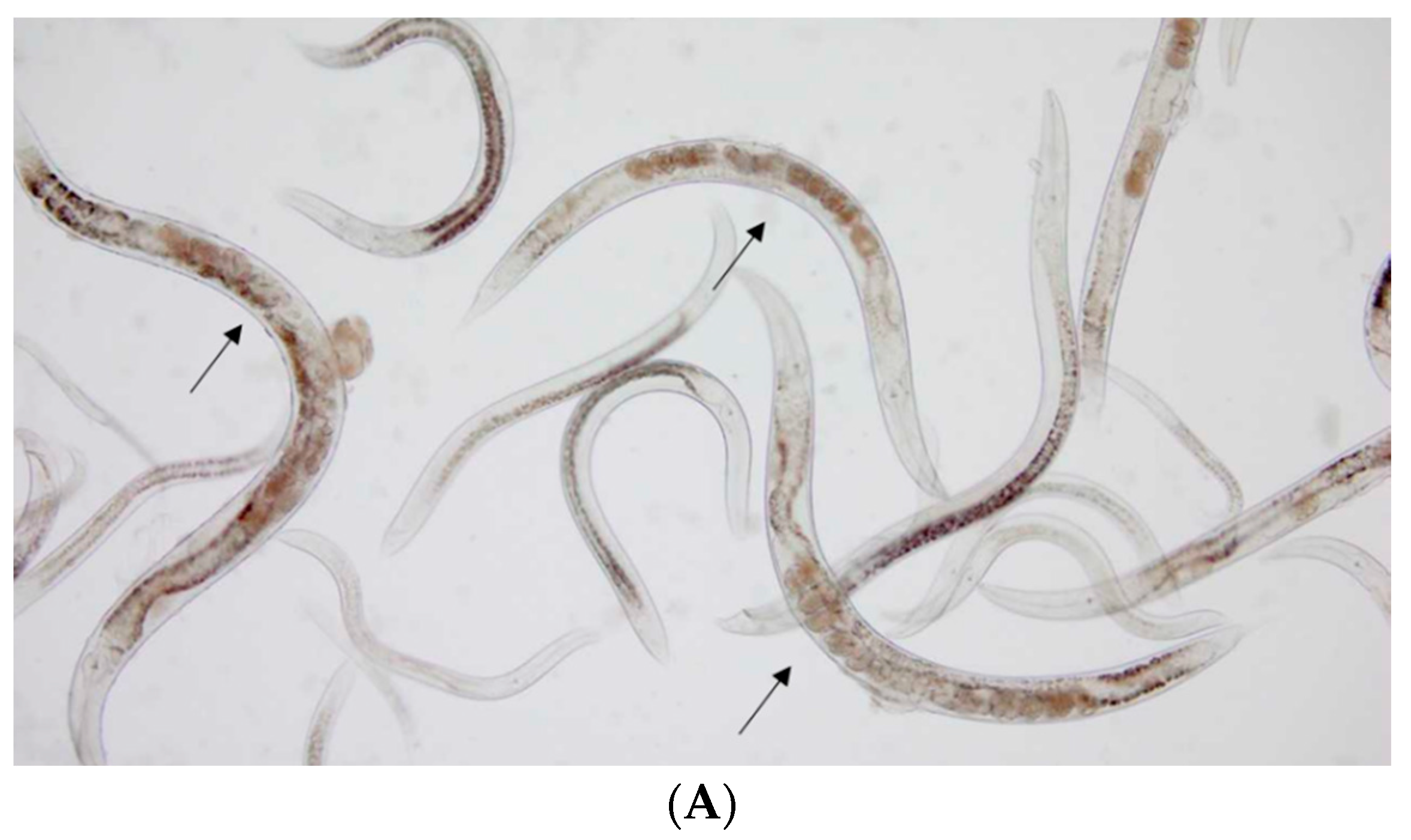
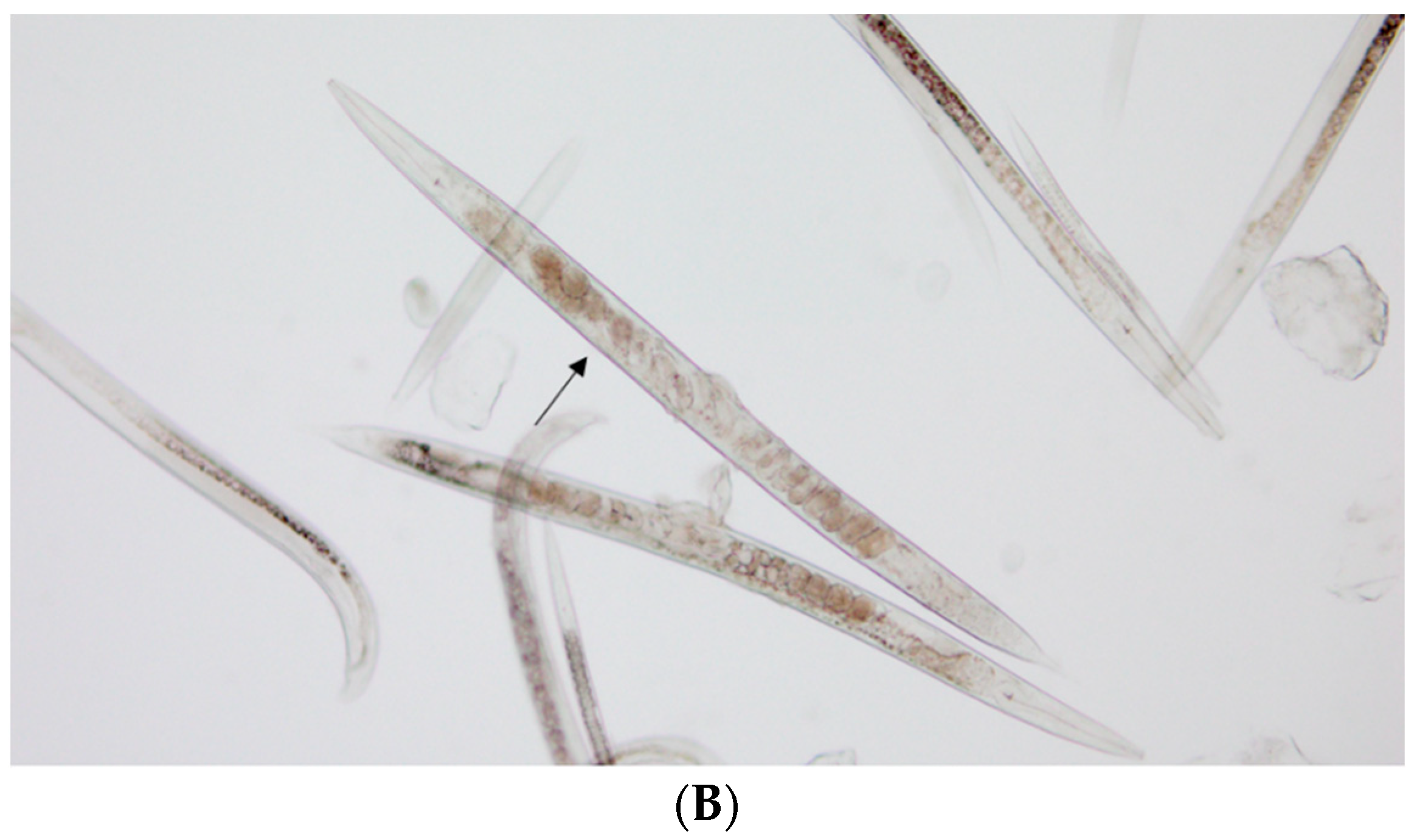
2.3. Anthelmintic Activity

Two of the tested compounds showed anthelmintic activity: 4-(3-chlorophenyl)-5-(3-trifluoromethylphenyl)-2,4-dihydro-3H-1,2,4-triazole-3-thione (4) and 4-(3-chlorophenyl)-1-(3-trifluoromethylbenzoyl)thiosemicarbazide (3a). Both had a 3-chlorophenyl substituent. The anthelmintic activity of the 1,2,4-triazole derivative (4) was better than the thiosemicarbazide derivative (3a). The LC50 value for compound 4 was 0.37 mg/mL, whereas for compound 3a it was 14.77 mg/mL. Figure 1 shows the nematode culture Rhabditis sp. alive, moving (control—A), and dead after the administration of compound 4 (B). The other compounds did not show anthelmintic activity in the concentrations tested. The two active compounds showed anthelmintic activity better than albendazole. The LC50 value for this drug was 19.24 mg/mL [42].
In our previous work [43], the most active compounds also had a chlorophenyl substituent, so it seems that it can be said to be a structural element promoting antiparasitic activity. Further, by comparing the antibacterial and antiparasitic activity of new compounds, it can be concluded that in the case of bacteria, the type of substituent has less influence than the structure of the basic skeleton. The linear structure is preferred. However, when analyzing the results of antiparasitic tests, it can be noticed that the type of substituent is of decisive importance, and in this case, the cyclic compounds are characterized by higher activity. Confirmation of these theses will be the subject of our further research.

2.4. Antimicrobial Activity

We described the results of experiments that aimed to evaluate the in vitro antibacterial activities of newly synthesized thiosemicarbazides and 1,2,4-triazole derivative. The broth microdilution method to examine the compounds’ ability to inhibit bacterial growth in vitro toward bacterial species from the ATCC (American Type Culture Collection) was used. We used five staphylococci strains including both Staphylococcus aureus (four strains) and Staphylococcus epidermidis (one strain) species. Staphylococci infections and increasing drug resistance have caused substantial public health problems. Represented by S. aureus, ATCC 43300 strain methicillin-resistant bacteria, known as MRSA, are of particular concern because of their ability to spread rapidly and extensively, along with their multi-drug resistance to various antimicrobials, especially β-lactam and aminoglycoside antibiotics [44,45]. MRSA-positive diseases are often connected with chronic or recurrent infections, including respiratory infections, and endocardial inflammation [46].
The minimum inhibitory concentration (MIC) values in µg/mL of the tested compounds against selected Gram-positive bacteria are reported in Table 2.
Newly synthesized compounds were examined for their antimicrobial activity with promising results against selected Gram-positive bacteria. According to our screening results, the highest antibacterial activity against all Gram-positive bacterial strains was demonstrated by thiosemicarbazide 3a, with an MIC value of 1.95 µg/mL against all Staphylococcus spp. strains except methicillin-resistant S. aureus (MRSA) ATCC 43300 strain with an MIC value of 3.9 µg/mL. Moreover, the MBC value was 1000 µg/mL towards all tested S. aureus reference strains. The lowest MBC value (15.63 µg/mL) was detected against S. epidermidis ATCC 12228, M. luteus ATCC 10240, and B. cereus ATCC 10876 (Table 2). The activity of compound 3a against staphylococci was bacteriostatic with an MBC/MIC ratio >4. This compound has a chlorine atom in position 3 in the phenyl ring.
The slightly less in vitro antibacterial activity against Gram-positive bacteria was demonstrated by thiosemicarbazide 3e with MIC values in the range of 7.8–31.25 µg/mL and MBC values > 1000 µg/mL. The MIC range for the methicillin-resistant S. aureus ATCC 43300 MRSA strain and all methicillin-sensitive (MSSA) S. aureus reference strains was 15.63–31.25 µg/mL. The thiosemicarbazide 3e was most active toward the B. cereus ATCC 10876 strain with an MIC value of 7.81 µg/mL. This compound has a fluorine atom in position 3 in the phenyl ring.
The weakest activity against Gram-positive bacteria was shown by thiosemicarbazide compounds 3b3d and 3f with MIC ranges from 500 to ≥1000 µg/mL toward seven strains of Gram-positive bacteria (Table 2). Low MICs for these compounds were observed in only one strain of staphylococci—S. aureus ATCC 25923. Their MIC values against this strain of S. aureus were sequentially as follows: 62.5 µg/mL, 250 µg/mL, 1000 µg/mL, and 62.5 µg/mL. The transformation of the linear system of thiosemicarbazide (3a) into a 1,2,4-triazole ring (4) in this research results in a drastic decrease in activity in relation to all tested strains.
The antibacterial effect examination showed that all tested compounds were active against Gram-positive bacteria and no activity was observed against Gram-negative strains. Our data indicate that all tested compounds were ineffective in tested concentrations against the reference strains of Gram-negative bacteria, Klebsiella pneumoniae ATCC 13883, Escherichia coli ATCC 25922, and Salmonella typhimurium ATCC 14028, with MIC values > 1000 µg/mL.
Based on the obtained results, it can be hypothesized that in the studied group of 3-trifluoromethylphenylthiosemicarbazide derivatives, the presence of a halogen in the aromatic ring is a prerequisite for obtaining antibacterial activity. However, comparing the activity of the thiosemicarbazides derived from fluorobenzoic acids obtained earlier, it can be concluded that they were more active than those with a trifluoromethylphenyl substituent. In the future, this should be confirmed by performing tests for compounds with bromine and iodine atoms.
Despite the fact that the obtained activities are at least four times lower than for the reference antibiotic, cefuroxime, they are very good due to the fact that bacteria produce mechanisms of resistance to known drugs and known chemical structures. Thiosemicarbazide derivatives have a structure not yet recognized by microorganisms, which makes them attractive structures for further research. In the future, we will try to determine the impact of the obtained compounds on multidrug-resistant strains.

2.5. Anticancer Activity

To establish the antiproliferative activity of newly obtained thiosemicarbazide derivatives, they were tested with an MTT assay using the human colon cancer cell line (SW 620) compared to a normal cell line (Chinese hamster lung cells V79). The cytotoxic activity of the studied compounds is presented in Table 3. IC50 values for most of the studied compounds were very high (>100 µM). For compound 3a, we observed a lower value (IC50 = 69.02 ± 12.63 µM for SW620). However, in contrast to the reference compound Doxorubicin (IC50 = 1.83 ± 0.10 µM for SW620 and IC50 = 0.46 ± 0.03 µM for V79), this result should be considered low as well. Thus, thiosemicarbazide derivatives (3a3f) and the tested triazole derivative (4) have not caused cytotoxic effects on studied cell lines.

2.6. Antioxidant Activity

Oxygen is an element necessary for the life of all aerobic organisms, including humans. Despite its key importance, especially in relation to energy processes and reactions accompanying inflammatory processes, it may pose a threat to cell metabolism. It is known that during the energy processes, including, among others, electron transport chains in mitochondria and microsomes, as well as the activities of enzymes such as superoxide dismutase, flavoprotein oxidases, or xanthine oxidase, oxygen radicals (ROS) may be generated. These include superoxide anions (the main representative of free radicals), hydrogen peroxide (a precursor of free radicals), and hydroxyl radicals. These compounds, which have a high oxidizing potential, are necessary for the proper conduct of many metabolic processes [47,48], but their excessive production may lead to damage to the structure of many macromolecules, such as proteins, nucleic acids, and unsaturated fatty acids [49,50,51].
Naturally occurring non-enzymatic and enzymatic antioxidant systems forming the antioxidant defence system (ADS) protect cells against the effects of ROS by reducing them to water or molecular oxygen and reducing their production [52,53].
Numerous studies have shown the relationship between overproduction of ROS and cancer, as well as cardiovascular, neurodegenerative, and inflammatory diseases [54,55,56,57].
Unsaturated fatty acids are the compounds most susceptible to the effects of ROS, which is particularly dangerous due to their common presence in all biological membranes. Their oxidation products, malondialdehyde (MDA) and trans-4-hydroxynonenal (4HNE), also affect cell metabolism by forming strong bonds with nucleophilic groups in proteins, nucleic acids, and membrane lipids. Both MDA and 4HNE are used as markers in the assessment of cellular oxidative stress [58,59].
The antioxidant potential of the newly synthesized derivatives was assessed based on the analysis of the malondialdehyde (MDA) concentration formed as a product of lipid oxidation stimulated by iron (II) ions [60]. The results are presented in Table 4.
Our research shows that all tested compounds exhibit antioxidant activities and reduced the level of MDA as compared to that in the control sample by 25.1% to 52.8%.
Inactivation of free oxygen radicals occurs as a result of the transfer of a hydrogen atom to this type of compound, the transfer of one electron, or in the process of electron transfer accompanied by the subsequent loss of a proton [61,62].
The presence of sulfur in the structure of thiourea in the tested compounds indicates the possibility of their antioxidant effect through the mechanism of chelation of Fe2+ ions, which are the most powerful pro-oxidants [63]. It is known that the introduction of an -OH group to the phenyl ring increases antioxidant activity due to the possibility of creating an aryloxy radical. Also, the introduction of a methoxy group to the phenolic ring significantly increases the antioxidant potential of phenolic acids, as well as stilbenes, flavonoids, and hydroxytyrosol, although it is much lower than in the case of the hydroxyl group [61,64]. The activity enhancement effect is related to the electron-donating properties of the methoxy moiety [65,66]. A similar effect is demonstrated by compound 3d containing a methoxy group on the phenyl ring compared to compound 3f containing only the phenyl ring (50.1% antioxidant activity vs. 46.3%). Rimal et al. showed that substituting chalcones with halogens increased their antioxidant activity both in the mechanism of iron ion chelation and free radical scavenging activity [67]. In the case of the tested compounds 3a and 3e containing a phenyl ring substituted with chlorine and fluorine, respectively, a slight increase in antioxidant activity was also found compared to compound 3f containing only the phenyl ring (48.3% and 52.8% of antioxidant activity vs. 46.3%). Cyclization of the thiosemicarbazide (compound 4) significantly reduces the antioxidant activity compared to compound 3a (25.1% vs. 48.3%).
Studies on the modification of phenolic compounds in order to obtain higher antioxidant capacity have shown that the introduction of a tert-butyl group into the phenyl structure increases their antioxidant potential. In turn, the introduction of linear substituents, especially long-chain alkanes, does not provide such effects [68]. The introduction of a butyl substituent into the phenyl ring (compound 3c) significantly reduced the antioxidant activity compared to compound 3f containing a phenyl ring (34.3% vs. 46.3%).

2.7. Lipinski’s Rule of Drug-Likeness

The compounds were filtered with Lipinski’s criteria to check for drug-like characteristics.
Table 5 shows the calculated parameters for thiosemicarbazide derivatives, including the molecular weight, the number of hydrogen bond donors and acceptors, log p (octanol-water partition coefficient), the number of rotational bonds, and the topological polar surface area of atoms in the molecule. Lipinski’s rule is observed by all tested compounds according to the results, indicating that these compounds have the potential to act as active drugs when administered orally.

3. Materials and Methods

3.1. Chemistry

All the substances used in this work were purchased from Sigma-Aldrich (Munich, Germany) and were used without further purification. On a Fisher-Johns melting point apparatus (Stuart SMP50, Schwerte, Germany), melting points were recorded. Using an elemental analyzer by Perkin-Elmer 2400 CHN (Waltham, MA, USA), elemental analyses of C, H, and N were performed. Using DMSO-d6 solvent, the 1H, 13C, and 19F NMR spectra were analyzed by using the Bruker Avance 600 (Bruker BioSpin GmbH, Rheinstetten, Germany). In the Supplementary Materials, 1H NMR spectra (Figures S1–S7), 13C NMR spectra (Figures S8–S14), and 19F NMR (Figures S15 and S16) are presented. The progress of the reactions was monitored by TLC (aluminum sheet 60 F254 plates (Merck Co., Kenilworth, NJ, USA). We used the solvent system CHCl3/EtOH (10:1, v/v).

3.1.1. The Procedure for the Synthesis of Hydrazide of 3-Trifluoromethylbenzoic Acid (2)

A total of 0.01 mole of ethyl 3-trifluoromethylbenzoate was dissolved in 5 mL anhydrous ethanol, and then 0.02 mole of 100% hydrazine hydrate was added. The solution was refluxed for 1 h. After cooling, the precipitate was filtered off and dried. The yield of the reaction was 99%. Using 80% hydrazine hydrate resulted in obtaining hydrazide with a yield of 80%.

3.1.2. The Procedure for the Synthesis of 1-(3-Trifluoromethylbenzoyl)-4-substituted Thiosemicarbazide (3a3f)

A total of 0.001 mol of 3-trifluoromethylbenzoic acid hydrazide (2) was dissolved hot in 5 mL of anhydrous ethanol. Then, 0.001 mol of isothiocyanate was added and the mixture was refluxed for 1/2 h. After this time, the contents of the flasks were cooled to room temperature, the precipitates were filtered, washed with water and diethyl ether, and allowed to air dry. The resulting thiosemicarbazide derivatives were purified by crystallization from 96% ethanol.

3.1.3. The Procedure for the Synthesis of 4-(3-Chlorophenyl)-5-(3-trifluoromethylphenyl)-2,4-dihydro-3H-1,2,4-triazole-3-thione (4)

A total of 0.1 g of 4-(3-chlorophenyl)-1-(3-trifluoromethylbenzoyl)thiosemicarbazide (3a) was dissolved in 5 mL of a 2% aqueous NaOH solution. The solution was refluxed for 2 h. After cooling to room temperature, the solution was treated 3N hydrochloric acid until slightly acidic to litmus paper. The resulting precipitate was filtered off and, after drying, crystallized from 96% ethanol.

4-(3-Chlorophenyl)-1-(3-trifluoromethylbenzoyl)thiosemicarbazide (3a)

Yield 65%, m.p. 185.4 °C, 1H NMR (DMSO-d6) δ (ppm): 7.22 (d, 1H, ArH, J = 7.9 Hz), 7.36 (t, 1H, ArH, J = 8.0 Hz), 7.44 (d, 1H, ArH, J = 8.3 Hz), 7.59 (bs, 1H, ArH), 7.78 (t, 1H, ArH, J = 8.0 Hz), 7.98 (d, 1H, ArH, J = 7.8 Hz), 8.23 (d, 1H, ArH, J = 7.9 Hz), 8.29 (s, 1H, ArH), 9.92–9.97 (m, 2H, 2NH), 10.86 (s, 1H, NH). 13C NMR (DMSO-d6) δ (ppm): 123.52, 123.79, 125.03, 125.33, 127.14, 128.93, 128.96, 129.59, 130.17, 132.45, 133.86, 141.15, 141.23, 165.15; 180.78. 19F NMR (DMSO-d6) δ (ppm): −61.11. Elemental analysis for C15H11ClF3N3OS. Calculated: C 48.20; H 2.97; N 11.24. Found: C 48.30; H 2.90; N 11.40.

1-(3-Trifluoromethylbenzoyl)-4-(3-methoxyphenyl)thiosemicarbazide (3b)

Yield 62%, m.p. 181.3 °C, 1H NMR (DMSO-d6) δ (ppm): 3.80 (s, 3H, CH3), 6.80–6.82 (m, 1H, ArH), 7.07–7.08 (m, 1H, ArH), 7.16 (bs, 1H, ArH), 7.30 (t, 1H, ArH J = 8.1 Hz) 7.84 (t, 1H, ArH J = 7.8 Hz), 8.04 (d, 1H, ArH J = 9.1 Hz), 8.29 (d, 1H, ArH J = 7.8 Hz), 8.35 (s, 1H, ArH), 9.85 (s, 2H, 2NH), 10.87 (s, 1H, NH). 13C NMR (DMSO-d6) δ (ppm): 55.58, 111.10, 112.24, 118.72, 121.73, 123.54, 125.03, 128.88, 129.34, 130.14, 132.46, 133.99, 140.74, 159.46, 165.17, 181.45. Elemental analysis for C16H14F3N3O2S. Calculated: C 52.03; H 3.82; N 11.38. Found: C 52.20; H 3.90; N 11.65.

4-Butyl-1-(3-trifluoromethylbenzoyl)thiosemicarbazide (3c)

Yield 67%, m.p. 161.6 °C, 1H NMR (DMSO-d6) δ (ppm): 0.87 (t, 3H, CH3, J = 7.3 Hz), 1.27–1.32 (m, 2H, CH2), 1.43–1.52 (m, 2H, CH2), 3.43 (d, 2H, CH2, J = 6.7 Hz), 7.73–7.78 (m, 1H, ArH), 7.94–7.97 (m, 1H, ArH), 9.15–8.20 (m, 2H, ArH), 8.25 (s, 1H, NH), 9.30 (s, 1H, NH), 10.57 (s, 1H, NH). 13C NMR (DMSO-d6) δ (ppm): 14.27, 19.92, 31.39, 48.89, 123.35, 124.94, 128.79, 129.48, 129.52, 130.10, 131.46, 132.36, 165.07, 177.78. Elemental analysis for C13H16F3N3OS. Calculated: C 48.89; H 5.05; N 13.16. Found: C 48.98; H 5.30; N 13.45.

1-(3-Trifluoromethylbenzoyl)-4-(3-methylphenyl)thiosemicarbazide (3d)

Yield 58%, m.p. 230.7 °C, 1H NMR (DMSO-d6) δ (ppm): 3.34 (s, 3H, CH3), 6.96–6.99 (m, 1H, ArH), 7.18–7.23 (m, 2H, ArH), 7.33–7.36 (m, 1H, ArH), 7.77 (t, 1H, ArH, J = 7.8 Hz) 7.97 (d, 1H, ArH, J = 7.8 Hz), 8.23 (d, 1H, ArH, J = 7.8 Hz), 8.29 (s, 1H, ArH), 9.75–9.80 (m, 2H, 2NH), 10.81 (s, 1H, NH). 13C NMR (DMSO-d6) δ (ppm): 21.39, 123.55, 125.04, 125.35, 126.34, 127.16, 128.33, 128.85, 129.32, 130.11, 132.46, 134.02, 137.77, 139.51, 165.17, 182.16. Elemental analysis for C16H14F3N3OS. Calculated: C 54.38; H 3.99; N 11.89. Found: C 54.60; H 4.20; N 11.90.

1-(3-Trifluoromethylbenzoyl)-4-(3-fluorophenyl)thiosemicarbazide (3e)

Yield 62%, m.p. 174.4 °C, 1H NMR (DMSO-d6) δ (ppm): 7.62 (t, 1H, ArH, J = 8.2 Hz) 7.77–7.82 (m, 1H, ArH) 7.98–8.01 (m, 3H, ArH) 8.25 (d, 1H, ArH, J = 7.9 Hz) 8.30 (s, 1H, ArH) 8.43 (s, 1H, ArH) 10.15 (s, 2H, 2NH) 10.93 (s, 1H, NH). 13C NMR (DMSO-d6) δ (ppm): 112.94, 121.85, 121.94, 123.52, 125.01, 125.33, 127.14, 128.95, 129.58, 130.18, 132.44, 133.89, 141.38, 141.45, 156.08, 165.16, 181.49. Elemental analysis for C15H11F4N3OS. Calculated: C 50.42; H 3.10; N 11.76. Found: C 50.50; H 3.40; N 11.95.

1-(3-Trifluoromethylbenzoyl)-4-(phenyl)thiosemicarbazide (3f)

Yield 69%, m.p. 176.4 °C, 1H NMR (DMSO-d6) δ (ppm): 7.17 (t, 1H, ArH, J = 7.2 Hz), 7.33 (t, 2H, ArH, J = 7.8 Hz), 7.41 (bs, 2H, ArH), 7.77 (t, 1H, ArH, J = 7.8 Hz), 7.97 (d, 1H, ArH, J = 7.8 Hz), 8.23 (d, 1H, ArH, J = 7.9 Hz), 9.78 (s, 1H, NH), 9.87 (s, 1H, NH), 10.83 (s, 1H, NH). 13C NMR (DMSO-d6) δ (ppm): 123.35, 125.04, 125.35, 126.67, 128.53, 128.86, 129.32, 130.12, 132.44, 134.01, 139.64, 165.17, 181.60. Elemental analysis for C15H12F3N3OS. Calculated: C 53.09; H 3.56; N 12.38. Found: C 53.30; H 3.90; N 12.45.

4-(3-Chlorophenyl)-5-(3-trifluoromethylphenyl)-2,4-dihydro-3H-1,2,4-triazole-3-thione (4)

Yield 65%, m.p. 241.2 °C, 1H NMR (DMSO-d6) δ (ppm): 7.40 (d, 1H, ArH, J = 7.7 Hz), 7.54 (t, 1H, ArH, J =8.0 Hz), 7.59–7.61 (m, 2H, ArH), 7.65–7.67 (m, 3H, ArH), 7.83 (d, 1H, ArH, J =7.0 Hz), 14.31 (s, 1H, NH). 13C NMR (DMSO-d6) δ (ppm): 123.12, 124.42, 125.44, 127.16, 127.52, 128.26, 129.49, 130.18, 130.43, 131.41, 132.84, 133.80, 136.01, 149.65, 169.20. 19F NMR (DMSO-d6) δ(ppm): −61.66. Elemental analysis for C15H9ClF3N3S. Calculated: C 50.64; H 2.55; N 11.81 Found: C 50.85; H 2.90; N 11.90.

3.2. Predictions of the Biological Activity

Predictions of the potential biological activity of the newly obtained thiosemicarbazide derivatives were carried out using the PASS (Prediction of Activity Spectra for Substances) software (http://www.ncss.com/software/pass/, accessed on 20 October 2022). [41]. The program enables the prediction of biological activity (including pharmacological effects, mechanisms of action, interactions, and toxicity). The principle of operation is based on the assessment of the similarity of the substance to the structures of biologically active compounds found in the program database (more than 46,000 medicines). The use of the above-mentioned software for the purpose of the above research work was determined by its high accuracy of predicting activity, being on average approx. 95.3%.

3.3. Anthelmintic Activity Assay

All compounds in the anthelmintic activity study were prepared in the form of a homogeneous suspension. The anthelmintic activity was tested according to the procedure developed by Bogucka-Kock and Kołodziej [69]. The tested compounds were added to the culture of Rhabditis sp. at experimentally determined concentrations. After a 24 h exposure, nematodes were observed in terms of development according to the earlier described procedure [42]. LC50 values (lethal concentration) were determined for two active substances. It was defined as the amount of the substance causing the death of 50% of nematodes [43].

3.4. Antimicrobial Activity Assay

The microdilution method was used to determine the MIC values of newly synthesized compounds. The antimicrobial activity of the thiosemicarbazides series was tested on ten reference bacterial strains purchased from the American Type Culture Collection (ATCC). Seven Gram-positive reference strains (S. aureus ATCC 29213, S. aureus ATCC 43300, S. aureus ATCC 6538, S. aureus ATCC 25923, S. epidermidis ATCC 12228, M. luteus ATCC 10240, and B. cereus ATCC 10876) and three Gram-negative reference bacteria (K. pneumoniae ATCC 13883, E. coli ATCC 25922, S. typhimurium ATCC 14028) were used. These bacterial strains are representatives of microorganisms that can cause the most common opportunistic infections among humans. The bacteria were grown on Mueller–Hinton agar (MHA) plates before examination. To perform the assay, all bacteria were placed in sterile saline (0.85% NaCl) to obtain a suspension of the optical density of the 0.5 McFarland standard (1.50 × 10⁸ colony-forming units (CFUs)/mL). A solution of the tested compounds was prepared in dimethyl sulfoxide (DMSO) at a concentration of 50 mg/mL as a stock solution.
A 96-well microtiter plate and microbroth two-fold dilution method was used to determine the minimum inhibitory concentration (MIC) according to the EUCAST (European Committee on Antimicrobial Susceptibility Testing) reference recommendation [70]. The solutions were then diluted with Mueller–Hinton broth (MHB, Biomaxima, Lublin, Poland) medium to obtain a solution with a basic concentration of 1000 µg/mL. Then, using the two-fold dilution method with MHB, the tested compounds were introduced into the wells of the microplate to provide a final concentration in the range of 0.49 to 1000 µg/mL. Then, 2 μL of bacterial inoculum with a density of 0.5 McFarland standard was added into each well. Each well was filled with a total volume of 100 μL. Then, microtiter plates were incubated for 18 ± 2 h at 35 °C. After incubation, the growth of bacteria was monitored by a Biotek ELx800 (Biokom, Janki, Poland) spectrophotometer at 600 nm (OD600). The MIC assays were performed in triplicate.
A negative control with MHB and without tested compounds was used. Clinically used anti-bacterial agents, cefuroxime and vancomycin, were included as positive controls for the purpose of comparison in a concentration range from 0.007 to 15.625 µg/mL.
The in vitro antimicrobial activity of the tested compounds was determined by the MIC, which was defined as the lowest concentration of the compound tested at which no visible growth of the test bacteria was observed. The MBC (the minimum bactericidal concentration) is defined as the lowest concentration of antimicrobial agent resulting in reduction in the viability of the initial bacterial inoculum by ≥99.9% on the plate. Generally, an MBC/MIC ratio less than 4 is considered bactericidal for an antimicrobial, and an MBC/MIC > 8 is indicative of bacteriostatic activity [71,72]. For the MBC test, the Mueller–Hinton agar (MHA, Biomaxima, Poland) plate assay was used. MBC was determined after the broth dilution period of an MIC test has been completed. After the MIC detection, 5 µL aliquots from each well were plated on an MHA plate and grown at 35 °C overnight. The MBC values were determined by observing the first clear area on the agar plate that has no observable bacterial growth.

3.5. Anticancer Activity

To conduct biological tests two cell lines were used: the human metastatic colon cancer cell lines SW 620 and V79 which are cells exhibiting fibroblast morphology isolated from the lung of a Chinese hamster. Both lines were obtained from American Type Culture Collection (ATCC, Rockville, MD, USA). The culture used to grow the SW 620 cells was prepared in MEM (ThermoSci, Waltham, MA, USA). The V79 cells were cultured in RPMI (Biowest SAS, Nuaillé, France) with the addition of 20 mM HEPES (Biowest, Nuaillé, France), 10% fetal bovine serum (FBS, Sigma-Aldrich, St. Louis, MO, USA), 100 µg/mL of streptomycin, and 100 U/mL of penicillin (Gibco, Grand Island, NY, USA). The eligible level of cells confluence was 80–90%, and both cell cultures were maintained in a humidified incubator set at 37 °C with 5% CO2 until they reached it.
The MTT assay was used to establish the cytotoxic activity of the studied compounds. The mechanism of this method relies on enzymatic conversion of 3-(4,5-dimethylthiazol-2-yl)-2,5-diphentyltetrazolium bromide salt (MTT) into insoluble formazan crystals by mitochondrial dehydrogenases within viable cells.
The process starts with the preparation of 96-well plates by placing cells at a density of 1 × 10⁴ per well and incubating them for 24 h to adhere. Afterwards, the cells were treated with different concentrations of thiosemicarbazide derivatives, from 20 to 100 µM, except the control group which remained not exposed. Next, cells were incubated for 72 h at 37 °C with a 5% CO2. Then, a solution of MTT (0.5 mg/mL) was added to each well and plates were incubated for 4 h more in the same conditions.
To dissolve precipitated formazan crystals, a mixture of DMSO and isopropanol (in a 1:1 ratio, v/v) was used. For the spectrophotometric survey, we used MultiscanGo from Thermo Scientific with the wavelength value set at 570 nm to measure absorbance.
Cell viability was determined as the percentage of MTT reduction in the treated cells compared to the control group (unexposed cells). The relative MTT level, represented as a percentage, was calculated using the formula [A]/[B] × 100, where [A] stands for the absorbance of the treated cell sample and [B] for the absorbance of unexposed to studied compounds sample (control group). Reduced cell viability reveals itself as decreased relative MTT levels. To calculate IC50 values (concentration at which 50% of cell viability is inhibited), we used GraphPad Prism 8.0.1 software.

3.6. Antioxidant Activity

The antioxidant activity of the tested compounds was measured in the brain homogenate of intact rats bred at the Central Laboratory of Experimental Animals of the Medical University of Warsaw. Brains were collected according to procedure no. 3R. A 10% (m/v) rat brain solution was obtained by homogenizing freshly prepared brain washed with cold 0.9% NaCl in cold 10 mM TRIS-HCl buffer, with pH = 7.5. The homogenate was centrifuged at 4 °C (2000 rpm) to obtain supernatant 1 [54,60]. Sample preparation was as follows: 100 μM stock solutions of test compounds in DMSO were added to 200 μM of supernatant 1 to a final concentration of 10 μM. The reference sample contained brain homogenate supernatant and DMSO.

3.6.1. Estimation of Lipid Peroxidation

Freshly prepared solutions of 0.1% FeCl2 (0.2 mL) and 1 mM ascorbic acid (0.4 mL) [60,73] were added to all samples to initiate the oxidation reaction. Samples were incubated for 1 h at 37 °C. Then, 8.1% sodium dodecyl sulfate (SDS) solution was added to inhibit the oxidation reaction. Acetate buffer (pH = 3.4) and 0.8% thiobarbituric acid (TBA) solution (pH = 6) were added to each sample to determine the concentration of malondialdehyde (MDA). The samples were incubated for 1 h at 100 °C, then cooled on ice and centrifuged (10 min, 4000 rpm). The absorbance was measured at a wavelength of 532 nm against a blank sample (acetate buffer with TBA solution) [60,74,75,76,77,78].
Calculation of malondialdehyde (MDA) concentration [60] was performed as follows:
M D A   c o n c e t r a t i o n =   A 532   n m × 1.56 × 10 6   [ n m o l   M D A h g   t i s s u e ]

3.6.2. Statistical Analyses

Statistical analyses were performed using GraphPad Prism 9 software (GraphPad Software, San Diego, CA, USA). The results are expressed as mean ± SD from at least three independent experiments. The statistical significance of differences between means was established by ANOVA with Dunnett’s multiple comparison pos hoc test. p values below 0.05 were considered statistically significant.

3.7. The Lipinski Rules

The Lipinski rule, also known as the “Rule of Five”, is a guideline used in drug discovery to assess the likelihood of a drug candidate’s success as an orally administered medication [79,80,81]. The rule states that a compound is more likely to be orally active if it has:
  • No more than five hydrogen bond donors (i.e., NH or OH groups);
  • No more than ten hydrogen bond acceptors (i.e., N or O atoms);
  • A molecular weight of less than 500 Daltons;
  • A partition coefficient (log P) of less than 5;
  • Molar refractivity ranging from 40 to 130.
The rule does not state that a medicine that is active when taken orally will be tiny and only mildly lipophilic. Rather, it suggests that a compound is more likely to be orally active if it has moderate lipophilicity, as indicated by a log p value between −0.4 and 5.0. Compounds that are too hydrophilic or too lipophilic may have poor oral absorption, distribution, metabolism, and excretion (ADME) properties, which can limit their effectiveness as oral medications. However, lipophilicity is just one of several factors that influence a drug’s oral bioavailability, and other physicochemical and pharmacological properties must also be considered in drug discovery and development. Molinspiration (http://www.molinspiration.com, accessed on 20 March 2023) is used to estimate the drug-likeness qualities of the compounds by verifying Lipinski’s rule of five.

4. Conclusions

The results of the research showed that the newly obtained compounds with thiosemicarbazide skeleton had anthelmintic and antibacterial potential. Moreover, the compounds did not show significant cytotoxic activity towards the tested cancer line and normal cell line. The most active was thiosemicarbazide with a 3-chlorophenyl substituent (3a). In fact, the efficacy of these compounds was found to be higher than that of the therapeutics currently used in the treatment of parasitic diseases. Additionally, all compounds showed antioxidant activity, but the most active was a molecule with a fluorine atom in the phenyl ring (3e). Analysis of the assumptions of the rule of five indicates that all obtained compounds should be characterized by good bioavailability. Our research shows that the newly synthesized compounds may constitute a promising group of compounds for further structural modifications.

Supplementary Materials

The following supporting information can be downloaded at: https://www.mdpi.com/article/10.3390/molecules29071529/s1, Figures S1–S7: 1H NMR spectra, Figures S8–S14 13C NMR spectra, Figures S15 and S16 19F NMR spectra. Figure S1. The 1H NMR of compound 3a. Figure S2. The 1H NMR of compound 3b. Figure S3. The 1H NMR of compound 3c. Figure S4. The 1H NMR of compound 3d. Figure S5. The 1H NMR of compound 3e. Figure S6. The 1H NMR of compound 3f. Figure S7. The 1H NMR of compound 4. Figure S8. The 13C NMR of compound 3a. Figure S9. The 13C NMR of compound 3b. Figure S10. The 13C NMR of compound 3c. Figure S11. The 13C NMR of compound 3d. Figure S12. The 13C NMR of compound 3e. Figure S13. The 13C NMR of compound 3f. Figure S14. The 13C NMR of compound 4. Figure S15. The 19F NMR of compound 3a. Figure S16. The 19F NMR of compound 4.

Author Contributions

Conceptualization, M.W.; methodology, M.W., U.K., A.B.-K., M.S., G.K.-T., P.K. and M.F.; investigation, P.L., M.W., U.K., P.K., G.K.-T. and T.S.; writing—original draft preparation, M.W., U.K., G.K.-T., T.S., N.K., P.K. and A.B.-K.; funding acquisition, M.W. and A.B.-K.; writing—review and editing, all authors. All authors have read and agreed to the published version of the manuscript.

Funding

This research was funded by Medical University of Lublin, Poland, grants number DS 15 and DS 43.

Institutional Review Board Statement

Not applicable.

Informed Consent Statement

Not applicable.

Data Availability Statement

The details of the data supporting the report results in this research were included in the paper and Supplementary Materials.

Conflicts of Interest

The authors declare no conflicts of interest.

References

  1. Neglected Tropical Diseases. Available online: https://www.who.int/health-topics/neglected-tropical-diseases#tab=tab_1 (accessed on 4 July 2022).
  2. CDC. Soil-Transmitted Helminths. Available online: https://www.cdc.gov/parasites/sth/index.html (accessed on 13 July 2022).
  3. Charlier, J.; Höglund, J.; von Samson-Himmelstjerna, G.; Dorny, P.; Vercruysse, J. Gastrointestinal Nematode Infections in Adult Dairy Cattle: Impact on Production, Diagnosis and Control. Vet. Parasitol. 2009, 164, 70–79. [Google Scholar] [CrossRef] [PubMed]
  4. Kaplan, R.M. Drug Resistance in Nematodes of Veterinary Importance: A Status Report. Trends Parasitol. 2004, 20, 477–481. [Google Scholar] [CrossRef] [PubMed]
  5. Baiak, B.H.B.; Lehnen, C.R.; da Rocha, R.A. Anthelmintic Resistance in Cattle: A Systematic Review and Meta-Analysis. Livestock Sci. 2018, 217, 127–135. [Google Scholar] [CrossRef]
  6. Redman, E.; Whitelaw, F.; Tait, A.; Burgess, C.; Bartley, Y.; Skuce, P.J.; Jackson, F.; Gilleard, J.S. The Emergence of Resistance to the Benzimidazole Anthelmintics in Parasitic Nematodes of Livestock Is Characterised by Multiple Independent Hard and Soft Selective Sweeps. PLoS Neglect. Trop. Dis. 2015, 9, e0003494. [Google Scholar] [CrossRef] [PubMed]
  7. Fissiha, W.; Kinde, M.Z. Anthelmintic Resistance and Its Mechanism: A Review. Infect. Drug Resist. 2021, 14, 5403. [Google Scholar] [CrossRef] [PubMed]
  8. Ghassemi, A.; Farhangi, H.; Badiee, Z.; Banihashem, A.; Mosaddegh, M.R. Evaluation of Nosocomial Infection in Patients at Hematology-Oncology Ward of Dr. Sheikh Children’s Hospital. Iran. J. Pediatr. Hematol. Oncol. 2015, 5, 179–185. [Google Scholar]
  9. Vuong, C.; Otto, M. Staphylococcus epidermidis infections. Microbes Infect. 2002, 4, 481–489. [Google Scholar] [CrossRef] [PubMed]
  10. Waddington, R.J.; Moseley, R.; Embery, G. Review: Periodontal disease mechanisms. Reactive oxygenspecies: A potential role in the pathogenesis of periodontal diseases. J. Oral Dis. 2000, 6, 138–151. [Google Scholar]
  11. Ameryckx, A.; Thabault, L.; Pochet, L.; Leimanis, S.; Poupaert, J.H.; Wouters, J.; Joris, B.; Van Bambeke, F.; Frédérick, R. 1-(2-Hydroxybenzoyl)-thiosemicarbazides are promising antimicrobial agents targeting D-alanine-D-alanine ligase in bacterio. Eur. J. Med. Chem. 2018, 159, 324–338. [Google Scholar] [CrossRef]
  12. Ameryckx, A.; Pochet, L.; Wang, G.; Yildiz, E.; Saadi, B.E.; Wouters, J.; Van Bambeke, F.; Frédérick, R. Pharmacomodulations of the benzoyl-thiosemicarbazide scaffold reveal antimicrobial agents targeting D-alanyl-D-alanine ligase in bacterio. Eur. J. Med. Chem. 2020, 200, 112444. [Google Scholar] [CrossRef]
  13. Al-Mutairi, A.A.; Al-Alshaikh, M.A.; Al-Omary, F.A.M.; Hassan, H.M.; El-Mahdy, A.M.; El-Emam, A.A. Synthesis, antimicrobial, and anti-proliferative activities of novel 4-(adamantan-1-yl)-1-arylidene-3-thiosemicarbazides, 4-arylmethyl N′-(Adamantan-1-yl)piperidine-1-carbothioimidates, and related derivatives. Molecules 2019, 24, 4308. [Google Scholar] [CrossRef]
  14. Chen, R.; Huo, L.; Jaiswal, Y.; Huang, J.; Zhong, Z.; Zhong, J.; Williams, L.; Xia, X.; Liang, Y.; Yan, Z. Design, synthesis, antimicrobial, and anticancer activities of acridine thiosemicarbazides derivatives. Molecules 2019, 24, 2065. [Google Scholar] [CrossRef]
  15. El-Sharief, M.A.M.S.; Abbas, S.Y.; El-Bayouki, K.A.M.; El-Gammal, E.W. Synthesis of thiosemicarbazones derived from N-(4-hippuric acid)thiosemicarbazide and different carbonyl compounds as antimicrobial agents. Eur. J. Med. Chem. 2013, 67, 263–268. [Google Scholar] [CrossRef]
  16. Wang, Y.; Dang, Q.; Liu, C.; Yu, D.; Pu, X.; Wang, Q.; Gao, H.; Zhang, B.; Cha, D. Selective adsorption toward Hg(II) and inhibitory effect on bacterial growth occurring on thiosemicarbazide-functionalized chitosan microsphere surface. ACS Appl. Mater. Interfaces 2018, 10, 40302–40316. [Google Scholar] [CrossRef]
  17. Chen, Y.-L.; Fang, K.-C.; Sheu, J.-Y.; Hsu, S.-L.; Tzeng, C.-C. Synthesis and antibacterial evaluation of certain quinolone derivatives. J. Med. Chem. 2001, 44, 2374–2377. [Google Scholar] [CrossRef]
  18. Devar, S.B.; Swamy, B.H.M.; Rao, B.N.; Shivkumar, H.; Shivkumar, B. Synthesis of new tetrazoloquinoline thiocarbohydrazides as potential antimicrobial agents. Indian J. Heterocycl. Chem. 2011, 21, 37–40. [Google Scholar]
  19. Umadevi, P.; Deepti, K.; Srinath, I.; Vijayalakshmi, G.; Tarakaramji, M. Synthesis and in-vitro antibacterial activity of some new urea, thiourea and thiosemicarbazide derivatives. Int. J. Pharm. Pharm. Sci. 2012, 4, 379–383. [Google Scholar]
  20. Pitucha, M.; Karczmarzyk, Z.; Swatko-Ossor, M.; Wysocki, W.; Woś, M.; Chudzik, K.; Ginalska, G.; Fruziński, A. Synthesis, in vitro screening and docking studies of new thiosemicarbazide derivatives as antitubercular agent. Molecules 2019, 24, 251. [Google Scholar] [CrossRef]
  21. Paneth, A.; Stączek, P.; Plech, T.; Strzelczyk, A.; Dzitko, K.; Wujec, M.; Kuśmierz, E.; Kosikowska, U.; Grzegorczyk, A.; Paneth, P. Biological evaluation and molecular modelling study of thiosemicarbazide derivatives as bacterial type IIA topoisomerases inhibitors. J. Enzym. Inhib. Med. Chem. 2016, 31, 14–22. [Google Scholar] [CrossRef]
  22. Gopalakrishnan, M.; Sureshkumar, P.; Thanusu, J.; Kanagarajan, V. Unusual formation of N-hydroxy-3,3-dimethyl-2,6-diarylpiperidin-4-one and its thiosemicarbazide derivative–synthesis and antimicrobial activity. Pharm. Chem. J. 2008, 42, 271–276. [Google Scholar] [CrossRef]
  23. Siddiqui, N.; Singh, O. Antibacterial activity of some 4-N-substituted thiosemicarbazides and thiosemicarbazones. Indian J. Pharm. Sci. 2003, 65, 423–425. [Google Scholar]
  24. Toan, V.N.; Tri, N.M.; Thanh, N.D. Substituted 4-formyl-2H-chromen-2-ones: Their reaction with N-(2,3,4,6-tetra-O-acetyl-β-D-galactopyranosyl)thiosemicarbazide, antibacterial and antifungal activity of their thiosemicarbazone products. Curr. Org. Chem. 2020, 24, 2272–2282. [Google Scholar] [CrossRef]
  25. Molnar, M.; Tomić, M.; Pavić, V. Coumarinyl thiosemicarbazides as antimicrobial agents. Pharm. Chem. J. 2018, 51, 1078–1081. [Google Scholar] [CrossRef]
  26. Han, M.İ.; İnce, U.; Gündüz, M.G.; Küçükgüzel, Ş.G. Synthesis, Antimicrobial Evaluation, and Molecular Modeling Studies of New Thiosemicarbazide-Triazole Hybrid Derivatives of (S)-Naproxen. Chem. Biodivers. 2022, 19, e202100900. [Google Scholar] [CrossRef]
  27. Sriram, D.; Yogeeswari, P.; Thirumurugan, R.; Pavana, R.K. Discovery of new antitubercular oxazolyl thiosemicarbazones. J. Med. Chem. 2006, 49, 3448–3450. [Google Scholar] [CrossRef]
  28. García, C.C.; Brousse, B.N.; Carlucci, M.J.; Moglioni, A.G.; Alho, M.M.; Moltrasio, G.Y.; Damonte, E.B. Inhibitory effect of thiosemicarbazone derivatives on Junin virus replication in vitro. Antivir. Chem. Chemother. 2003, 14, 99–105. [Google Scholar] [CrossRef]
  29. Coşkun, G.P.; Djikic, T.; Hayal, T.B.; Türkel, N.; Yelekçi, K.; Şahin, F.; Küçükgüzel, Ş.G. Synthesis, molecular docking and anticancer activity of diflunisal derivatives as cyclooxygenase enzyme inhibitors. Molecules 2018, 23, 1969. [Google Scholar] [CrossRef]
  30. Kaproń, B.; Czarnomysy, R.; Paneth, A.; Wujec, M.; Bielawski, K.; Bielawska, A.; Swiątek, Ł.; Rajtar, B.; Polz-Dacewicz, B.; Plech, T. Dual antibacterial and anticancer activity of 4-benzoyl-1-dichlorobenzoylthiosemicarbazide derivatives. Anti-Cancer Agents Med. Chem. 2018, 18, 529–540. [Google Scholar] [CrossRef]
  31. Prashanthi, M.; Babu, H.R.; Rani, J.U. Design, Synthesis and Molecular Docking Studies of Novel Indole–Isoxazole–Triazole Conjugates as Potent Antibacterial Agents. Russ. J. Bioorg. Chem. 2021, 47, 601–608. [Google Scholar] [CrossRef]
  32. Rimpiläinen, T.; Nunes, A.; Calado, R.; Fernandes, A.S.; Andrade, J.; Ntungwe, E.; Spengler, G.; Szemerédi, N.; Rodrigues, J.; Gomes, J.P.; et al. Increased Antibacterial Properties of Indoline-Derived Phenolic Mannich Bases. Eur. J. Med. Chem. 2021, 220, 113459. [Google Scholar] [CrossRef]
  33. Yang, G.; Shi, L.; Pan, Z.; Wu, L.; Fan, L.; Wang, C.; Xu, C.; Liang, J. The Synthesis of Coumarin Thiazoles Containing a Trifluoromethyl Group and Their Antifungal Activities. Arab. J. Chem. 2021, 14, 102880. [Google Scholar] [CrossRef]
  34. Antiqueira-Santos, P.; Teixeira, W.K.O.; Flores, A.F.C.; Piovesan, L.A.; Nery, L.E.M.; Votto, A.P. de S. Synthesis of Pyrazoline Fatty Chain Derivatives and Its Effects on Melanoma Cells. Bioorg. Med. Chem. Lett. 2021, 41, 127988. [Google Scholar] [CrossRef]
  35. Jung, Y.H.; Jain, S.; Gopinatth, V.; Phung, N.B.; Gao, Z.G.; Jacobson, K.A. Structure Activity Relationship of 3-Nitro-2-(Trifluoromethyl)-2H-Chromene Derivatives as P2Y6 Receptor Antagonists. Bioorg. Med. Chem. Lett. 2021, 41, 128008. [Google Scholar] [CrossRef]
  36. Krátký, M.; Svrčková, K.; Vu, Q.A.; Štěpánková, Š.; Vinšová, J. Hydrazones of 4-(Trifluoromethyl)Benzohydrazide as New Inhibitors of Acetyl- and Butyrylcholinesterase. Molecules 2021, 26, 989. [Google Scholar] [CrossRef]
  37. Shah, P.; Westwell, A.D. The role of fluorine in medicinal chemistry. J. Enzym. Inhib. Med. Chem. 2007, 22, 527–540. [Google Scholar] [CrossRef]
  38. Janowska, S.; Khylyuk, D.; Andrzejczuk, S.; Wujec, M. Design, Synthesis, Antibacterial Evaluations and In Silico Studies of Novel Thiosemicarbazides and 1,3,4-Thiadiazoles. Molecules 2022, 27, 3161. [Google Scholar] [CrossRef]
  39. Plech, T.; Wujec, M.; Siwek, A.; Kosikowska, U.; Malm, A. Synthesis and antimicrobial activity of thiosemicarbazides, s-triazoles and their Mannich bases bearing 3-chlorophenyl moiety. Eur. J. Med. Chem. 2011, 46, 241–248. [Google Scholar] [CrossRef]
  40. Janowska, S.; Stefańska, J.; Khylyuk, D.; Wujec, M. The Importance of Substituent Position for Antibacterial Activity in the Group of Thiosemicarbazide Derivatives. Molecules 2024, 29, 1333. [Google Scholar] [CrossRef]
  41. Power Analysis and Sample Size Software. NCSS, LLC.: Kaysville, UT, USA, 2015; Available online: https://www.ncss.com/software/pass/ (accessed on 20 October 2022).
  42. Tejchman, W.; Kołodziej, P.; Kalinowska-Tłuścik, J.; Nitek, W.; Żuchowski, G.; Bogucka-Kocka, A.; Żesławska, E. Discovery of Cinnamylidene Derivative of Rhodanine with High Anthelmintic Activity against Rhabditis sp. Molecules 2022, 27, 2155. [Google Scholar] [CrossRef]
  43. Dziduch, K.; Kołodziej, P.; Paneth, A.; Bogucka-Kocka, A.; Wujec, M. Synthesis and Anthelmintic Activity of New Thiosemicarbazide Derivatives—A Preliminary Study. Molecules 2020, 25, 2770. [Google Scholar] [CrossRef]
  44. Boucher, H.W.; Talbot, G.H.; Bradley, J.S.; Edwards, J.E.; Gilbert, D.; Rice, L.B.; Scheld, M.; Spellberg, B.; Bartlett, J. Bad Bugs, No Drugs: No ESKAPE! An Update from the Infectious Diseases Society of America. Clin. Infect. Dis. 2009, 48, 1–12. [Google Scholar] [CrossRef]
  45. Kolendi, C.L. Methicillin-Resistant Staphylococcus aureus (MRSA): Etiology, At-Risk Populations and Treatment; Nova Science Publishers Inc.: New York, NY, USA, 2010. [Google Scholar]
  46. Que, Y.A.; Haefliger, J.A.; Piroth, L.; François, P.; Widmer, E.; Entenza, J.M.; Sinha, B.; Herrmann, M.; Francioli, P.; Vaudaux, P.; et al. Fibrinogen and Fibronectin Binding Cooperate for Valve Infection and Invasion in Staphylococcus aureus Experimental Endocarditis. J. Exp. Med. 2005, 16, 201. [Google Scholar] [CrossRef]
  47. Valko, M.; Leibfritz, D.; Moncol, J.; Cronin, M.T.D.; Mazur, M.; Telser, J. Free Radicals and Antioxidants in Normal Physiological Functions and Human Disease. Int. J. Biochem. Cell Biol. 2007, 39, 44–84. [Google Scholar] [CrossRef]
  48. Zhao, W.; Diz, D.I.; Robbins, M.E. Oxidative Damage Pathways in Relation to Normal Tissue Injury. Br. J. Radiol. 2007, 80, 23–31. [Google Scholar] [CrossRef]
  49. Aruoma, O.I. Free Radicals, Oxidative Stress, and Antioxidants in Human Health and Disease. J. Am. Oil Chem. Soc. 1998, 75, 199–212. [Google Scholar] [CrossRef]
  50. Devasagayam, T.P.; Tilak, J.C.; Boloor, K.K.; Sane, K.S.; Ghaskadbi, S.S.; Lele, R.D. Free Radicals and Antioxidants in Human Health: Current Status and Future Prospects. J. Assoc. Physicians India 2004, 52, 794–804. [Google Scholar]
  51. Lobo, V.; Patil, A.; Phatak, A.; Chandra, N. Free Radicals, Antioxidants and Functional Foods: Impact on Human Health. Pharmacogn. Rev. 2010, 4, 118–126. [Google Scholar] [CrossRef]
  52. Marengo, B.; Nitti, M.; Furfao, A.L.; Colla, R.; de Cucis, C.; Marinari, U.M.; Pronzato, M.A.; Traverso, N.; Domenicotti, C. Redox Homeostasis and Cellular Antioxidant Systems: Crucial Players in Cancer Growth and Therapy. Oxid. Med. Cell Longev. 2016, 2016, 6235641. [Google Scholar] [CrossRef]
  53. Ali, S.S.; Ahsan, H.; Zia, M.K.; Siddiqui, T.; Khan, F.H. Understanding oxidants and antioxidants: Classical team with new players. J. Food Biochem. 2020, 44, e13145. [Google Scholar] [CrossRef]
  54. Garcia, Y.J.; Rodriguez-Malaver, A.J.; Penaloza, N. Lipid peroxidation measurement by thiobarbituric acid assay in rat cerebellar slices. J. Neurosci. Methods 2005, 144, 127–135. [Google Scholar] [CrossRef]
  55. Zhao, M.J.; Yuan, S.; Zi, H.; Gu, J.M.; Fang, C.; Zeng, X.T. Oxidative Stress Links Aging-Associated Cardiovascular Diseases and Prostatic Diseases. Oxid. Med. Cell Longev. 2021, 2021, 5896136. [Google Scholar] [CrossRef]
  56. Liguori, I.; Russo, G.; Curcio, F.; Bulli, G.; Aran, L.; Morte, D.D.; Gargiulo, G.; Testa, G.; Cacciatore, F.; Bonaduce, D.; et al. Oxidative stress, aging, and diseases. Clin. Interv. Aging 2018, 13, 757–772. [Google Scholar] [CrossRef]
  57. Serra, J.A.; Domínguez, R.O.; Marschoff, E.R.; Guareschi, E.M.; Famulari, A.L.; Boveris, A. Systemic Oxidative Stress Associated with the Neurological Diseases of Aging. Neurochem. Res. 2009, 34, 2122–2132. [Google Scholar] [CrossRef]
  58. Barrera, G.; Pizzimenti, S.; Daga, M.; Dianzani, C.; Arcaro, A.; Cetrangolo, G.P.; Giordano, G.; Cucci, M.A.; Graf, M.; Gentile, F. Lipid Peroxidation-Derived Aldehydes, 4-Hydroxynonenal and Malondialdehyde in Aging-Related Disorders. Antioxidants 2018, 7, 102. [Google Scholar] [CrossRef]
  59. Spickett, C.M. The Lipid Peroxidation Product 4-Hydroxy-2-Nonenal: Advances in Chemistry and Analysis. Redox Biol. 2013, 1, 145–152. [Google Scholar] [CrossRef]
  60. Ohkawa, H.; Ohishi, N.; Yagi, K. Assay for Lipid Peroxides in Animal Tissues by Thiobarbituric Acid Reaction. Anal. Biochem. 1979, 95, 351–358. [Google Scholar] [CrossRef]
  61. Chen, J.; Yang, J.; Ma, L.; Li, J.; Shahzad, N.; Kim, C.K. Structure-Antioxidant Activity Relationship of Methoxy, Phenolic Hydroxyl, and Carboxylic Acid Groups of Phenolic Acids. Sci. Rep. 2020, 10, 2611. [Google Scholar] [CrossRef]
  62. Mazzone, G.; Malaj, N.; Galano, A.; Russo, N.; Toscano, N. Antioxidant Properties of Several Coumarin–Chalcone Hybrids from Theoretical Insights. RSC Adv. 2015, 5, 565–575. [Google Scholar] [CrossRef]
  63. Gulcin, I. Antioxidant and Antiradical Activities of L-carnitine. Life Sci. 2006, 78, 803–811. [Google Scholar] [CrossRef]
  64. Spiegel, M.; Kapusta, K.; Kołodziejczyk, W.; Saloni, J.; Żbikowska, B.; Hill, G.A.; Sroka, Z. Antioxidant Activity of Selected Phenolic Acids-Ferric Reducing Antioxidant Power Assay and QSAR Analysis of the Structural Features. Molecules 2020, 25, 3088. [Google Scholar] [CrossRef]
  65. Castelluccio, C.; Paganga, G.; Melikian, N.; Bolwell, G.P.; Pridham, J.; Sampson, J.; Rice-Evans, C. Antioxidant Potential of Intermediates in Phenylpropanoid Metabolism in Higher Plants. FEBS Lett. 1995, 368, 188–192. [Google Scholar] [CrossRef]
  66. Razzaghi-Asl, N.; Garrido, J.; Khazraei, H.; Borges, F.; Firuzi, O. Antioxidant Properties of Hydroxycinnamic Acids: A Review of Structure-Activity Relationships. Curr. Med. Chem. 2013, 20, 4436–4450. [Google Scholar] [CrossRef]
  67. Rimal, I.; Reddy, P.S.; Chavali, M. Halogen Substituted Chalcone as Potential Antioxidants: An In Vitro Study. Adv. Sci. Eng. Med. 2012, 4, 499–504. [Google Scholar]
  68. Olajide, T.M.; Liu, T.; Liu, H.; Weng, X. Antioxidant Properties of Two Novel Lipophilic Derivatives of Hydroxytyrosol. Food Chem. 2020, 315, 126197. [Google Scholar] [CrossRef]
  69. PL 232918 B1; Method for the Culture of Nematodes of Rhabditis sp. Genus and Determination of the Nematocidal Substances Activity. Uniwersytet Medyczny w Lublinie PL: Warsaw, Poland, 2019.
  70. The European Committee on Antimicrobial Susceptibility Testing. Breakpoint Tables for Interpretation of MICs and Zone Diameters. Version 12.0, 2022. Ecast: EUCAST. Available online: https://www.eucast.org/ (accessed on 2 February 2023).
  71. O’Donnell, F.; Smyth, T.J.P.; Ramachandran, V.N.; Smyth, W.F. A Study of the Antimicrobial Activity of Selected Synthetic and Naturally Occurring Quinolines. Int. J. Antimicrob. Agents 2010, 35, 30–38. [Google Scholar] [CrossRef]
  72. Wiegand, I.; Hilpert, K.; Hancock, R.E.W. Agar and Broth Dilution Methods to Determine the Minimal Inhibitory Concentration (MIC) of Antimicrobial Substances. Nat. Protoc. 2008, 3, 163–175. [Google Scholar] [CrossRef]
  73. Kubiak-Tomaszewska, G. Effect of Hydroxyl Groups Esterification with Fatty Acids on the Cytotoxicity and Antioxidant Activity of Flavones. Molecules 2022, 27, 420. [Google Scholar] [CrossRef]
  74. Chawla, R.; Arora, R.; Kumar, R.; Sharma, A.; Prasad, J.; Singh, S.; Sagar, R.; Chaudhary, P.; Shukla, S.; Kaur, G.; et al. Antioxidant Activity of Fractionated Extracts of Rhizomes of High-altitude Podophyllum hexandrum: Role in Radiation Protection. Mol. Cell Biochem. 2005, 273, 193–208. [Google Scholar] [CrossRef]
  75. Ghani, M.A.; Barril, C.; Bedgood, D.R., Jr.; Prenzler, P.D. Measurement of Antioxidant Activity with the Thiobarbituric Acid Reactive Substances Assay. Food Chem. 2017, 230, 195–207. [Google Scholar] [CrossRef]
  76. Christodoulou, M.C.; Orellana Palacios, J.C.; Hesami, G.; Jafarzadeh, S.; Lorenzo, J.M.; Domínguez, R.; Moreno, A.; Hadidi, M. Spectrophotometric Methods for Measurement of Antioxidant Activity in Food and Pharmaceuticals. Antioxidants 2022, 11, 2213. [Google Scholar] [CrossRef]
  77. de Zwart, L.L.; Meerman, J.H.; Commandeur, J.N.; Vermeulen, N.P. Biomarkers of Free Radical Damage Applications in Experimental Animals and In Humans. Free Radic. Biol. Med. 1999, 26, 202–226. [Google Scholar] [CrossRef]
  78. Hou, Y.; Carne, A.; McConnell, M.; Bekhit, A.A.; Mros, S.; Amagase, K.; Bekhit, A.E.A. In Vitro Antioxidant and Antimicrobial Activities, and In Vivo Anti-inflammatory Activity of Crude and Fractionated PHNQs From Sea Urchin (Evechinus chloroticus). Food Chem. 2020, 316, 126339. [Google Scholar] [CrossRef]
  79. Lipinski, C.A.; Lombardo, F.; Dominy, B.W.; Feeney, P.J. Experimental and Computational Approaches to Estimate Solubility and Permeability in Drug Discovery and Development Settings. Adv. Drug Deliver. Rev. 2001, 46, 3–26. [Google Scholar] [CrossRef]
  80. Ertl, P.; Rohde, B.; Selzer, P. Fast Calculation of Molecular Polar Surface Area as a Sum of Fragment-based Contributions and its Application to the Prediction of Drug Transport Properties. J. Med. Chem. 2000, 43, 3714–3717. [Google Scholar] [CrossRef]
  81. Veber, D.F.; Johnson, S.R.; Cheng, H.-Y.; Smith, B.R.; Ward, K.W.; Kopple, K.D. Molecular Properties That Influence the Oral Bioavailability of Drug Candidates. J. Med. Chem. 2002, 45, 2615–2623. [Google Scholar] [CrossRef]
Scheme 1. Synthetic route for the compounds 13.
Molecules 29 01529 sch001
Scheme 2. Synthetic route for compound 4.
Molecules 29 01529 sch002
Figure 1. Example of a culture of nematodes of the genus Rhabditis sp. (A) control (arrow marked alive); (B) after the action of the compound 4 (arrow marked dead).
Molecules 29 01529 g001aMolecules 29 01529 g001b
Table 1. The probability of the predicted biological activity of the synthesized thiosemicarbazides.
CompoundAntituberculosisAntibacterialAntiparasitic
3a0.5510.3810.441
3b0.5130.3370.341
3c0.4790.4400.390
3d0.5550.3890.328
3e0.5410.3630.269
3f0.5940.4140.335
Table 2. The antimicrobial activity results of the newly synthesized compounds against reference strains of Gram-positive bacteria expressed as the values of the minimal inhibitory concentration (MIC; µg/mL) and the minimal bactericidal concentration (MBC; µg/mL).
CompoundsStaphylococcus aureus
ATCC 25923
Staphylococcus aureus
ATCC 29213
Staphylococcus aureus
ATCC 6538
Staphylococcus aureus
ATCC 43300
Staphylococcus epidermidis
ATCC 12228
Micrococcus luteus
ATCC 10240
Bacillus cereus
ATCC 10876
MICMBCMICMBCMICMBCMICMBCMICMBCMICMBCMICMBC
3a1.9510001.9510001.9510003.910001.9515.631.9515.631.9515.63
3b62.5nd *>1000nd *>1000nd *->1000nd *>1000nd *500nd *>1000nd *
3c250nd *>1000nd *->1000nd *->1000nd *1000nd *1000nd *>1000nd *
3d1000nd *>1000nd *->1000nd *->1000nd *1000nd *1000nd *>1000nd *
3e31.25>100015.63>100015.63>100031.25>100031.25>100015.63>10007.81>1000
3f62.5nd *>1000nd *>1000nd *>1000nd *1000nd *1000nd *>1000nd *
41000nd *>1000nd *>1000nd *>1000nd *500nd *1000nd *>1000nd *
1 Cef0.49 nt 0.98 nt 0.25 0.25 31.25
2 Van22 22
1 Cef—cefuroxime, 2 Van—vancomycin, nd *—not determined, nt—not tested.
Table 3. Cytotoxic activity of studied compounds estimated by MTT assay a.
Cancer CellsNormal Cells
SW 620 dV 79 e
CompoundIC50 bSI cIC50 b
3a69.02 ± 12.630.5940.49 ± 3.83
3b>1001>100
3c>1001>100
3d>1000.6969.09 ± 5.45
3e89.87 ± 14.611.06>100
3f>1001>100
4>1001>100
Ref f1.83 ± 0.100.160.46 ± 0.03
a The MTT assay is a colorimetric assay for measuring cell metabolic activity. It is based on the ability of nicotinamide adenine dinucleotide phosphate (NADPH)-dependent cellular oxidoreductase enzymes to reduce the tetrazolium dye MTT to its insoluble formazan, which has a purple color. Data are expressed as mean SD. b IC50 (µM)—the concentration of the compound that corresponds to a 50% growth inhibition of cell line (as compared to the control) after the cells were cultured for 72 h with the individual compound. c The SI (Selectivity Index) was calculated using the following formula: SI = IC50 for normal cell line/IC50 cancer cell line. d Human metastatic colon cancer (SW620). e Lung cells isolated from a Chinese hamster. f The selected reference compound commonly used in cancer treatment (Doxorubicin).
Table 4. Antioxidant potential of tested compounds estimated by malondialdehyde (MDA) concentration in rat brain homogenate.
CompoundMDA [mM × 10−3]Antioxidative Potential Value [%]
3a1.395 ± 0.232 *48.3
3b1.703 ± 0.177 **36.9
3c1.773 ± 0.077 ***34.3
3d1.320 ± 0.143 **51.1
3e1.274 ± 0.121 **52.8
3f1.450 ± 0.006 ***46.3
42.022 ± 0.121 **25.1
Control Sample2.699 ± 0.0880.0
Data represent the mean ± SD of 3 independent samples. Control sample: oxidation reaction was induced by 0.1% FeCl2 and 1 mM ascorbic acid. *** p ≤ 0.001, ** p ≤ 0.01, * p ≤ 0.05, difference as compared to the control sample. The antioxidant potential was determined based on the analysis of MDA concentrations between rat brain homogenates containing the tested compounds and the control sample.
Table 5. Lipinski’s parameters for compounds 3a3f.
CompoundLipinski’s Parameters
TPSAHydrogen Bond AcceptorHydrogen Bond Donorlog pMassRotatable Bonds
(No’s)
3a53.15433.67373.796
3b62.39533.05369.377
3c53.15433.39319.358
3d53.15433.44353.376
3e53.15433.15357.336
3f53.15433.01339.346
Disclaimer/Publisher’s Note: The statements, opinions and data contained in all publications are solely those of the individual author(s) and contributor(s) and not of MDPI and/or the editor(s). MDPI and/or the editor(s) disclaim responsibility for any injury to people or property resulting from any ideas, methods, instructions or products referred to in the content.

