Article

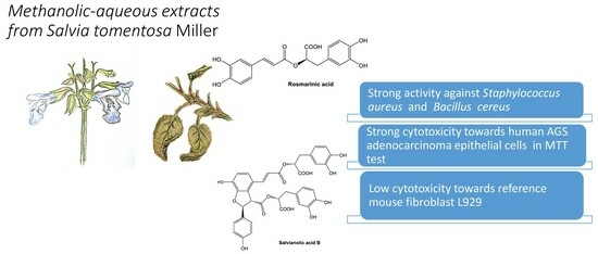
The Effect of Salvia tomentosa Miller Extracts, Rich in Rosmarinic, Salvianolic and Lithospermic Acids, on Bacteria Causing Opportunistic Infections

by
Ewelina Piątczak
1,*,
Joanna Kolniak-Ostek
2,
Weronika Gonciarz
3,
Paweł Lisiecki
4,
Urszula Kalinowska-Lis
5,
Magdalena Szemraj
4,
Magdalena Chmiela
3 and
Sylwia Zielińska
6
1
Department of Pharmaceutical Biotechnology, Faculty of Pharmacy, Medical University of Lodz, Muszyńskiego 1, 90-151 Lodz, Poland
2
Department of Fruit, Vegetable and Plant Nutraceutical Technology, Wrocław University of Environmental and Life Sciences, Chełmońskiego 37, 51-630 Wroclaw, Poland
3
Department of Immunology and Infectious Biology, Faculty of Biology and Environment Protections, University of Lodz, Banacha 12/16, 90-237 Lodz, Poland
4
Department of Pharmaceutical Microbiology and Microbiological Diagnostics, Faculty of Pharmacy, Medical University of Lodz, Muszyńskiego 1, 90-151 Lodz, Poland
5
Department of Cosmetic Raw Materials Chemistry, Faculty of Pharmacy, Medical University of Lodz, Muszyńskiego 1, 90-151 Lodz, Poland
6
Department of Pharmaceutical Biotechnology, Faculty of Pharmacy, Wroclaw Medical University, Borowska 211, 50-556 Wroclaw, Poland
*
Author to whom correspondence should be addressed.
Molecules 2024, 29(3), 590; https://doi.org/10.3390/molecules29030590
Submission received: 30 December 2023 / Revised: 16 January 2024 / Accepted: 22 January 2024 / Published: 25 January 2024
(This article belongs to the Special Issue Phytochemical Profile and Biological Evaluation of Plant Extracts)

Abstract

:
Methanolic-aqueous extracts of Salvia tomentosa Miller roots, aerial parts, and inflorescences were examined for their content of polyphenolic derivatives and the antimicrobial and cytotoxic effect. In the polyphenolic-rich profile, rosmarinic, salvianolic, and lithospermic acids along with various derivatives were predominant. A total of twenty phenolic compounds were identified using the UPLC/DAD/qTOF-MS technique. These were caffeic acid, rosmarinic acid derivatives, lithospermic acid derivatives, salvianolic acids B, F, and K derivatives, as well as sagerinic acid, although rosmarinic acid (426–525 mg/100 g of dry weight—D.W.) and salvianolic acid B (83–346.5 mg/100 g D.W.) were significantly predominant in the metabolic profile. Strong antibacterial activity of S. tomentosa extracts was observed against Staphylococcus epidermidis (MIC/MBC = 0.625 mg/mL) and Bacillus cereus (MIC = 0.312–1.25 mg/mL). The extracts showed low cytotoxicity towards the reference murine fibroblasts L929 and strong cytotoxicity to human AGS gastric adenocarcinoma epithelial cells in the MTT reduction assay. The observed cytotoxic effect in cancer cells was strongest for the roots of 2-year-old plant extracts.

Graphical Abstract

1. Introduction

Exposure to environmental pollution, some cleaning agents, and cosmetics has been associated with a significant decrease in immunity among the general population [1]. This can often result in opportunistic infections from generally non-pathogenic microorganisms which do not pose a threat to healthy people and may even play a beneficial role, such as Staphylococcus epidermidis or Bacillus cereus [2]. Such infections can be particularly severe in people struggling with chronic viral infections such as HIV or fighting cancer.
As such, there is a need to identify substances that can resist infection and maintain the proper condition of the body. Fortunately, many substances of natural origin, such as specialized plant metabolites, appear to be promising candidates. This group comprises an extensive and chemically diverse range of substances that have strong biological effects, both as single compounds and in mixtures with others [3]. These include alkaloids and terpenoids, which show strong biological activity, as well as phenylpropanoids, all of which have been found to possess antimicrobial and anticancer properties when present in plant essential oils and in non-volatile form [4].
The present study examines the phytochemical profile and the antimicrobial and cytotoxic effect of extracts obtained from the roots, aerial parts, and inflorescences of Salvia tomentosa Miller (Lamiaceae). The sage genus, Salvia, as a whole, is relatively well known, and many of its species are used as pharmaceutical raw materials [5]. S. tomentosa itself is native to South-East Europe and the Transcaucasus region (Albania, Bulgaria, Greece, Crimea, Lebanon-Syria, and Turkey) [6]. It is a perennial plant that flowers during its second year of vegetation. In Turkish folk medicine, the aerial parts of S. tomentosa are used as antibacterial and anti-inflammatory agents, as well as against rheumatic, stomach, and abdominal pain [7,8]. Its essential oils are dominated by β-pinene, α-pinene, and camphor [9], as well as 1,8-cineol, β-caryophylene, cyclophenchene [10]. In addition, the extracts from the aerial parts and leaves contain various phenolic acids (e.g., vanillic, galic, rosmarinic, and chlorogenic acids), flavonoids (e.g., rutin, miricetin, quercetin, luteolin, and flavone derivatives), and diterpenoids (7α-acetoxyroyleanone and 7α-hydroxyroyleanone, royleanone, and 6,7-dehydroxyroyleanone) [7,11,12]. The main compounds are known to possess antioxidant, anti-inflammatory, antibacterial, and antiviral properties and are promising anticancer drugs against various human cancer types [13,14,15].
Previous studies have only examined the essential oils of the plant and extracts of its aerial parts using antimicrobial and antioxidant assays [9,10], and no detailed study has been performed on the roots or inflorescences. However, several di- and triterpenoids have been noted in acetone extracts from roots [16]. To address this gap, the present study performs a detailed investigation of the profile of metabolites present in methanolic-aqueous extracts of S. tomentosa roots, aerial parts, and inflorescences using UPLC/DAD/qTOF-MS. The study also investigates the antimicrobial properties of the extracts and tests their cytotoxic properties.

2. Results and Discussion

2.1. Phytochemical Studies

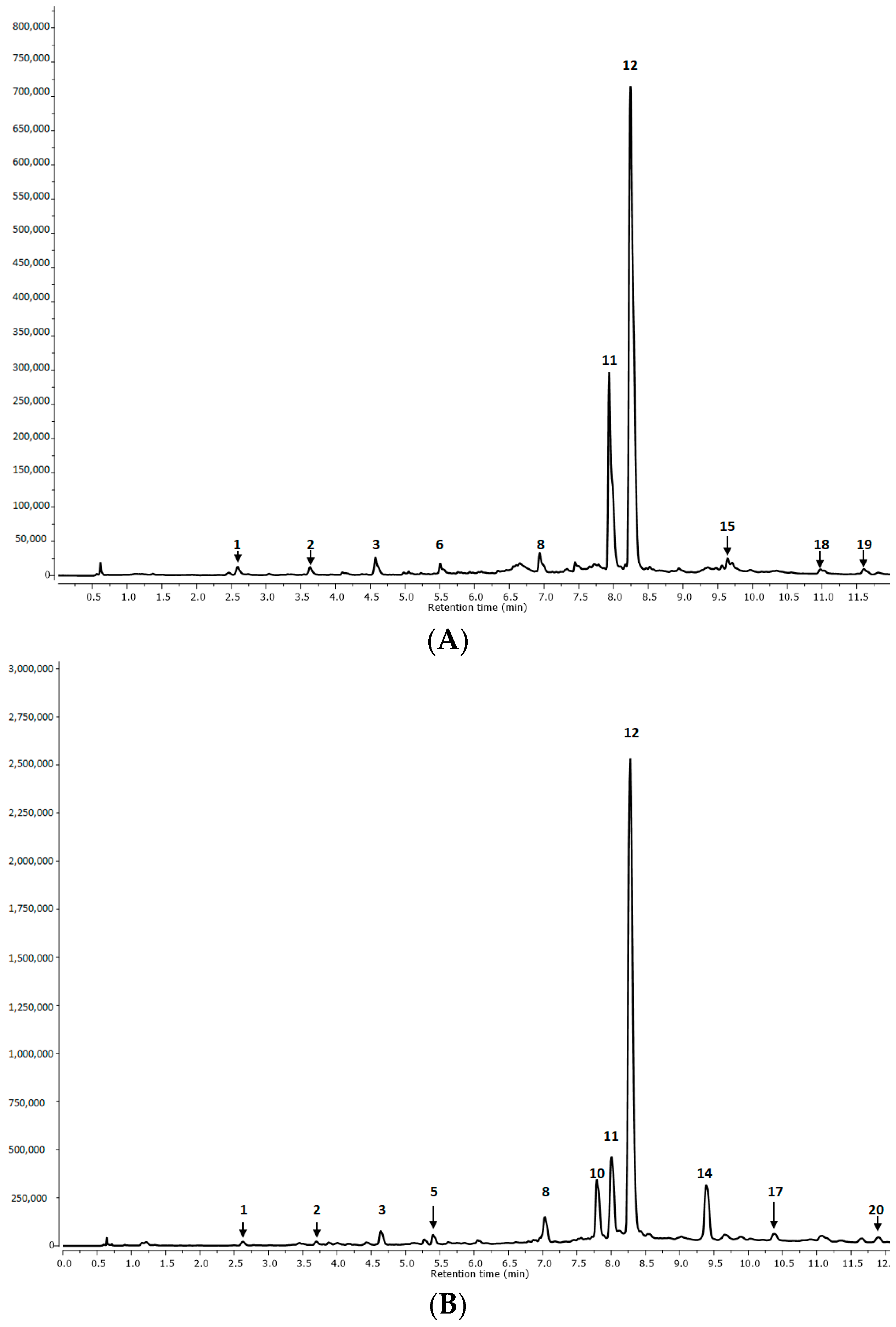
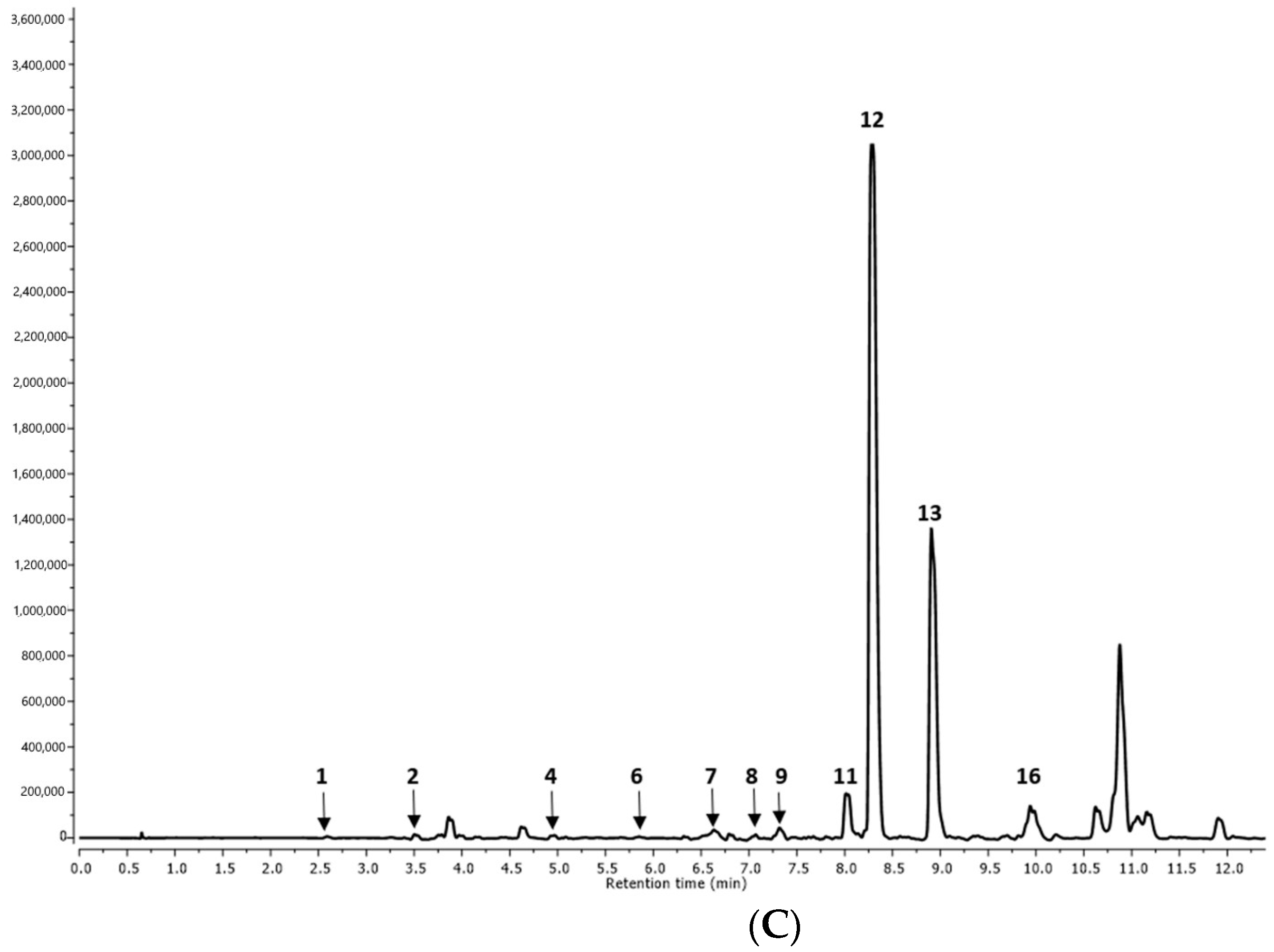
2.1.1. Qualitative Analysis

The roots and aerial parts of the one- and two-year-old plants (R1, R2, and AP1 and AP2, respectively) and the inflorescences of the two-year-old plants (I) were examined for specialized metabolites.
In total, 20 phenolic compounds were identified in the S. tomentosa extracts (Table 1 and Table 2). These included rosmarinic acid (RA), rosmarinic acid derivatives (RAd), salvianolic acid derivatives (SAd), and lithospermic acid derivatives (LAd), as well as cyclobutene lignan–sagerinic acid and free caffeic acid (CA).
The RA (peak no 12), i.e., the salvianic ester of caffeic acid, exhibited a deprotonated molecular ion at m/z 359, whose fragmentation led to characteristic ions at 197, 179, 161, and 135 (m/z). These peaks correspond to a deprotonated form of salvianic acid (danshensu or 3-(3,4-dihydroxyphenyl)lactic acid), a deprotonated CA, a dehydrated form of the mentioned acids, and a residue of CA after decarboxylation (Figure 1 and Figure 2; Table 1 and Table 2). These results were in agreement with the fragmentation described by Barros et al. [17], Katanić et al. [18], and Lecomte et al. [19]. Similarly, RA has also been found to be the predominant compound in numerous other Salvia species [16,17,18,19,20].
The RA derivatives (RAd) included three hexosides of RA with the same molecular ions [M-H] at m/z 521 (peaks no 2, 3, and 9). Their corresponding fragmentation ions at m/z 359 and (197, 179, 161, and 135) derived from rosmarinic acid (359) (the main component of its fragmentation), resulting from a loss of hexoside moiety (−162 u) from a molecular ion at m/z 521. Rosmarinic acid hexosides have been previously identified as the predominant ingredients in S. viridis [20], S. bulleyana [21], S. officinalis [22], S. blepharochlaena [23], S. viridis [24], and S. cadmica [25].
The second most abundant group of compounds present in the extracts was isomers and derivatives of salvianolic acids. These included salvianolic acid A (peak no 16), salvianolic acid B (SAB) (peaks no 11, 17, and 18), salvianolic acid F (peaks no 19 and 20) and salvianolic acid K (peaks no 4, 6, 7, and 15) (Table 1 and Table 2, Figure 1 and Figure 2).
Compound 16 (Table 1 and Table 2) found in the inflorescences was identified as salvianolic acid A (SAA) with a characteristic parent ion at m/z 493. Its fragmentation yielded a product ion typical for RA (359) and loss of fragments at m/z 197 and 179. SAA has also been found in the root extracts of S. miltiorrhiza [26,27].
Peak 11 (Rt = 11.08 min), identified as SAB, was present in all tested plant organs. It presented its main ion at m/z 717 and the following fragmentation ions at m/z 537, 519, 339, 321, 295, 197, and 179. The typical fragmentation ion for SAB after the cleavage of two danshensu units was the ion at m/z 321. The removal of salvianolic and caffeic acids from the SAB of mass 718 Da, [M-H-198-180], resulted in the formation of a fragmentation ion of m/z 339, the further decarboxylation of which (−44 u) may lead to ion formation at m/z 295.
Compound 17 (Rt = 10.37 min), present in the aerial parts, was identified as one of the two SAB isomers. The fragmentation ion at m/z 537 can be attributed to the loss of a caffeic acid (−180 u) unit from the parent ion at m/z 717. The remaining peaks at m/z 359 and 179 were analogous to those present in compound 11, which was identified as SAB.
Another isomer of SAB identified in the extracts in the present study was compound 18, which was only detected in the extracts of S. tomentosa roots. Its main fragmentation ion at m/z 519 can be attributed to the loss of a danshensu unit (−198 u) from the parent ion at m/z = 717. The compound showed m/z 359, with the characteristic loss of a fragment corresponding to the caffeic acid residue (−162 u). SAB isomers were one of the most abundant compounds in other Salvia species, for example Salvia miltiorrhiza [28]. SAB isomers have also previously been found in S. africana, S. officinalis ‘Icterina’, and S. mexicana aqueous extracts [29], and in S. apiana and S. farinacea var. Victoria Blue decoctions [30]; however, they were present in smaller amounts compared to the S. tomentosa methanolic-aqueous extracts described in our study.
In turn, compounds no 4 and 6 (Table 1 and Table 2, Figure 1), detected in the inflorescences and roots, showed parent ions at m/z 555 and identical fragmentation ions at 537, 359, 295, and 179. They were identified as hydrated salvianolic acids K (SAK) due to the fragmentation pattern with successive losses of danshensu (−197 u) and caffeic acid (−179 u or −161 u) moieties. Another SAK derivative (peak no 7) was found at Rt = 6.50 min, and its parent ion m/z 683 corresponded to salvianolic acid K methyl pentoside. The loss of the methyl-pentoside residue (−146 u) from the main molecular ion showed the presence of a fragmentation ion typical for SAK at m/z 537. All SAK derivatives were absent in the aerial parts extracts of S. tomentosa.
Peaks 19 and 20 (Rt = 11.79 and 11.89 min, respectively) represented by the same molecular ion ([M-H] at m/z 313) were assigned to salvianolic acid F (SAF) isomers. Compound 20, found in the aerial parts, has only one fragment ion at m/z 161, suggesting the decomposition of the main molecule to the dehydrated form of caffeic acid (CA). The second compound, 19, found only in inflorescences, possesses more fragmentation ions, i.e., at m/z 269, 203, 161, and 135: these may represent a decarboxylated form of SAF (269 u), an SAF residue after the loss of 3,4-dihydroxyphenyl (203 u), a dehydrated form of CA (161 u), and a decarboxylated form of CA (135 u). SAF isomers have been detected in the roots of S. viridis [20], S. cadmica [25], and S. bulleyana [27], and in the aerial parts of S. verticillata subsp. amasiaca [23], S. blepharochlaena [23], and S. cadmica [25].
Four compounds, represented by peaks 8 and 1315 were annotated as lithospermic acid derivatives (LAd) (Table 1 and Table 2, Figure 1). Peak no 8 was present in all analyzed extracts and was attributed to lithospermic acid (LA). Compound 15 (Rt = 9.72), detected in the roots, was annotated as an LAd with parent ion m/z 569. Peaks 13 and 14 (Rt = 8.91 and 9.38 min, respectively) that showed parent ion m/z 1074, were identified as dimers of lithospermic acid and were detected in aerial parts only. The MS/MS fragmentations yielded product ions at m/z 537 due to the loss of lithospermic acid A (537 Da) and fragments with m/z 359, 197, and 179. Some isomeric lithospermic acids were identified previously in S. miltiorrhiza, S. cadmica, S. cavaleriei, S. chinensis, and S. sonchifolia but were not present as dimers in any of these plants [22,25,31].
Moreover, free caffeic acid (CA, peak no 1) [m/z 179] was also found in all plant parts (Table 1, Figure 1). This compound could be the product of the hydrolysis of more complex structures like rosmarinic, lithospermic, or salvianolic acids. CA has been previously identified in the hydromethanolic extract of the roots and aerial parts of S. cadmica [25] and in South African Salvia species such as Salvia africana-lutea, S. lanceolata, and S. chamelaeagnea; these are used to treat fever and some inflammation [32].
Apart from phenylpropanoid derivatives, a cyclobutene lignan–sagerinic acid (peak no 10) (Rt = 7.80 min) with a parent ion at m/z 719 and a characteristic loss of fragments [m/z 359, 197, 179, 161, 135] was also identified in the aerial parts of S. tomentosa. These annotations were previously found in Rosmarinus officinalis, and three red-colored basil cultivars [33,34].

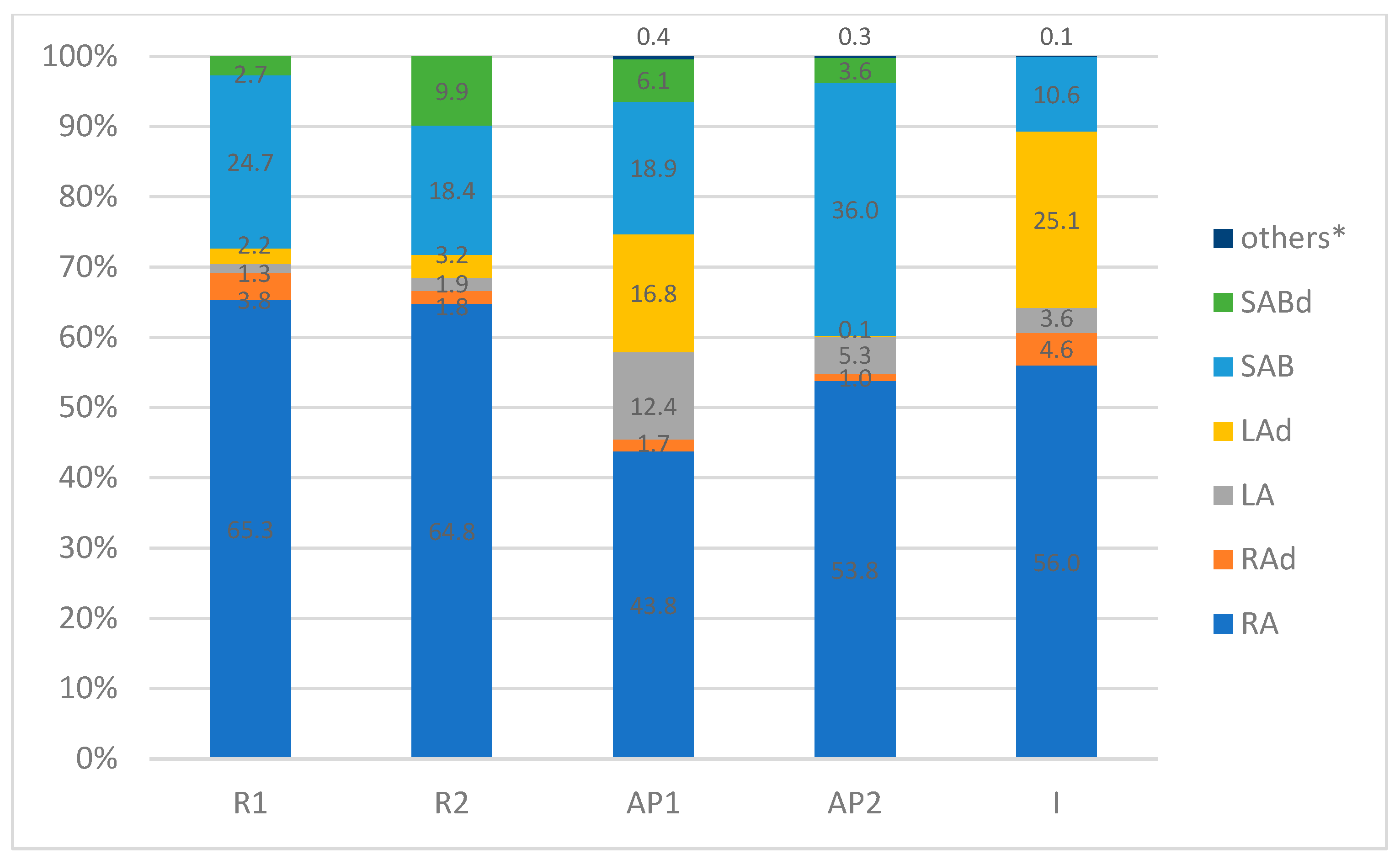
2.1.2. Quantitative Analysis

The methanolic-aqueous extracts of S. tomentosa roots, aerial parts, and inflorescences were found to be rich sources of phenylpropanoid compounds (Table 2). The phytochemical profile of all extracts was significantly predominated by RA (425.87–524.71 mg/100 g D.W.). A number of RA derivatives were also found, which, together with RA, accounted for up to 69% of the total share of all compounds (Figure 2). However, only 1.0–4.6% of RA was present in the glycoside and hydrated forms (Table 2, Figure 2).
Moreover, all extracts were relatively rich in SAB, its derivatives (SAd), and lithospermic acid (LA) dimers. SAB together with SAd accounted for 10.6% to 39.5% of the total composition, with the highest contents found in the aerial parts and roots (24.9–39.5% and 27.4–28.3%, respectively; Figure 2). In turn, LA and its derivatives were found in considerable amounts (28.7–29.2%) in the aerial parts, including inflorescences (Table 2, Figure 2). The presence of the remaining compounds was negligible, which was less than 0.4% (Table 2, Figure 2).

2.2. Antimicrobial Studies

The antimicrobial activity of the methanolic-aqueous extracts from S. tomentosa was tested against 14 Gram-positive bacterial strains, six Gram-negative bacterial strains, and two fungal strains. The extracts from both the aerial parts and roots demonstrated moderate inhibitory and biocidal effects against most of the tested strains compared to the routinely used antibiotics and fungicides (Table 3), demonstrating strong antibacterial effects against Staphylococcus epidermidis and Bacillus cereus (Table 3). Minimal inhibition concentration (MIC) and minimal bactericidal concentration (MBC) were obtained against Staphylococcus epidermidis when 0.625 mg/mL of the root, aerial part, and inflorescence extracts was used. In the case of Bacillus cereus, more potent effects were obtained from the root and inflorescence extracts (MIC/MBC = 0.312 mg/mL) than from the non-flowering aerial parts (MIC/MBC = 1.25 mg/mL). Previous studies on methanolic extracts of the aerial parts did not indicate any antimicrobial activity against Bacillus cereus [9]. Other studies have examined the antibacterial potential of essential oils from the non-aerial parts [9,10,35]; however, the results cannot be compared with ours due to methodological differences.
Both S. epidermidis and B. cereus are Gram-positive bacteria. Our present findings confirm those of other authors [25,36,37], indicating that extracts or essential oils from other plant species, including genus Salvia, are more active against Gram-positive than Gram-negative bacteria. This could be related to the cells lacking an external membrane, which may act as a barrier against antibacterial macromolecules [38]. Alternatively, Gram-positive strains may demonstrate greater ion permeability and leakage of intracellular components from bacterial cells or impaired bacterial enzyme systems [39].
B. cereus and S. epidermidis are opportunistic pathogens, indicating that they may induce severe infections in immunocompromised patients. B. cereus is a spore-forming bacterium, which allows it to survive longer in extremes of temperature. These bacteria are commonly found in the environment and can contaminate food such as beef, turkey, rice, beans, and vegetables. B. cereus can multiply rapidly at room temperature and produce toxins that can cause food poisoning, resulting in diarrhea and vomiting (emesis). The diarrheal illness is often related to meat, milk, vegetables, and fish; the emetic illness, in contrast, is typically associated with rice products but has also been related to other products such as potatoes, pasta, and cheese. Infections caused by B. cereus are usually self-limiting and do not require specific treatment, but in immunocompromised patients, antibiotic therapy is often necessary. B. cereus is also associated with infections of the eyes, respiratory tract, and wounds [40,41,42]. Most importantly, B. cereus is resistant to two major classes of antibiotics: trimethoprim/sulfamethoxazole and beta-lactam antibiotics [43]. Therefore, natural extracts exhibiting antibacterial activity may be useful in therapy.
Staphylococcus epidermidis forms the main part of the microbiota of the skin and mucous membranes. It is one of the most often isolated bacteria in hospitals and is commonly responsible for nosocomial infections of the bloodstream, cardiovascular system, eye, ear, nose, and throat. Its pathogenicity is mainly due to its ability to form biofilms [44,45]. A biofilm is a complex, three-dimensional structure consisting of a community of microorganisms surrounded by an exopolymer substance called a matrix. The exopolymer makes up 50–90% of the total organic mass of the biofilm and consists of extracellular DNA (eDNA), proteins, lipids, polysaccharides, divalent cations, and water channels. The matrix serves as a protective barrier against external factors, acts as a source of nutrients and enzymes, and enables the transport of intercellular substances [46]. As S. epidermidis is protected against attacks from the immune system and antibiotic treatments when in the biofilm, and such infections are difficult to eradicate. Approximately 80% of S. epidermidis strains from nosocomial infections are resistant to methicillin, and most are also resistant to other antibiotics [44,45]. Therefore, S. tomentosa extracts, demonstrating antibiotic potential, may be of value in countering such infections, especially in immunocompromised patients.

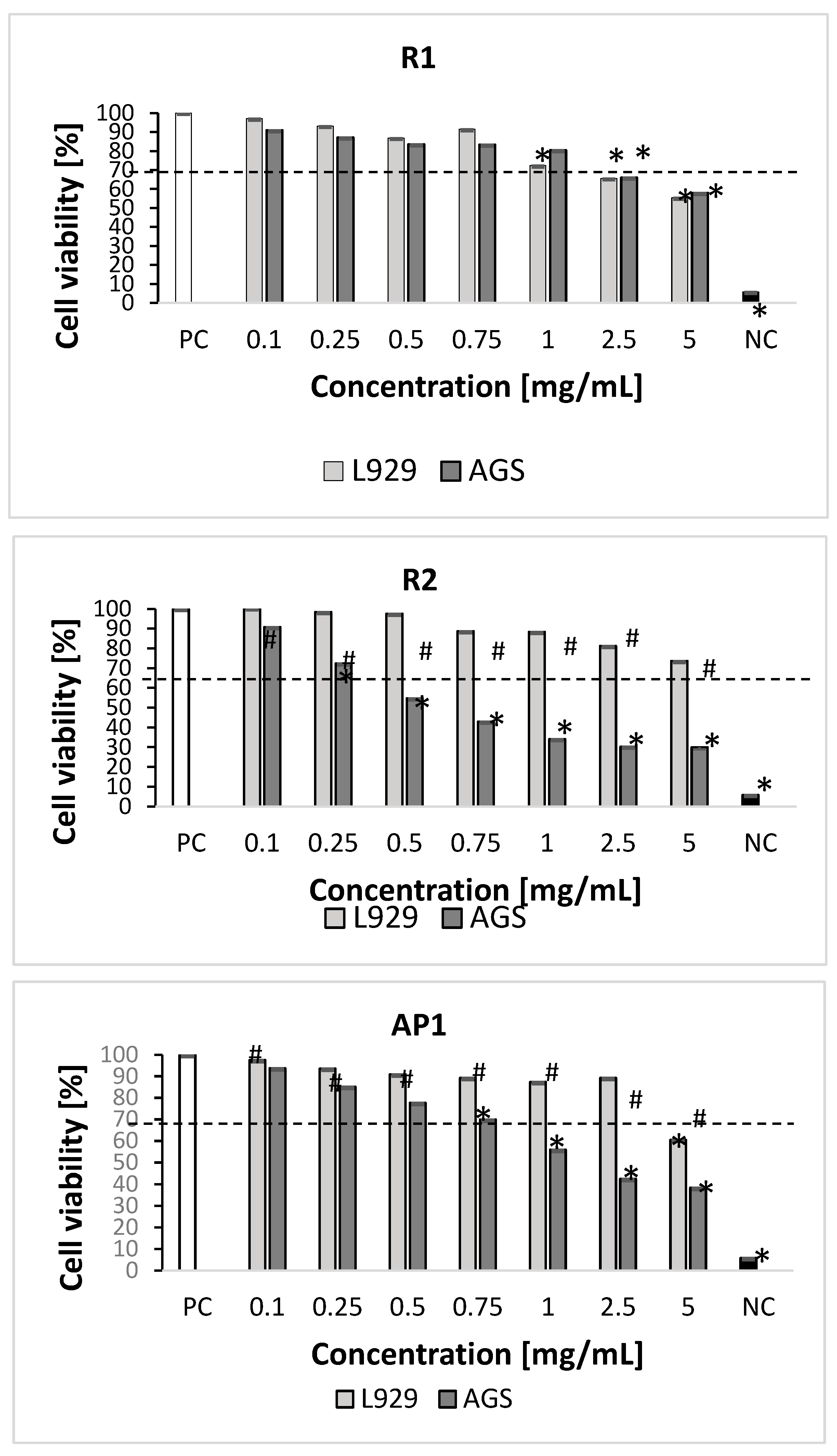
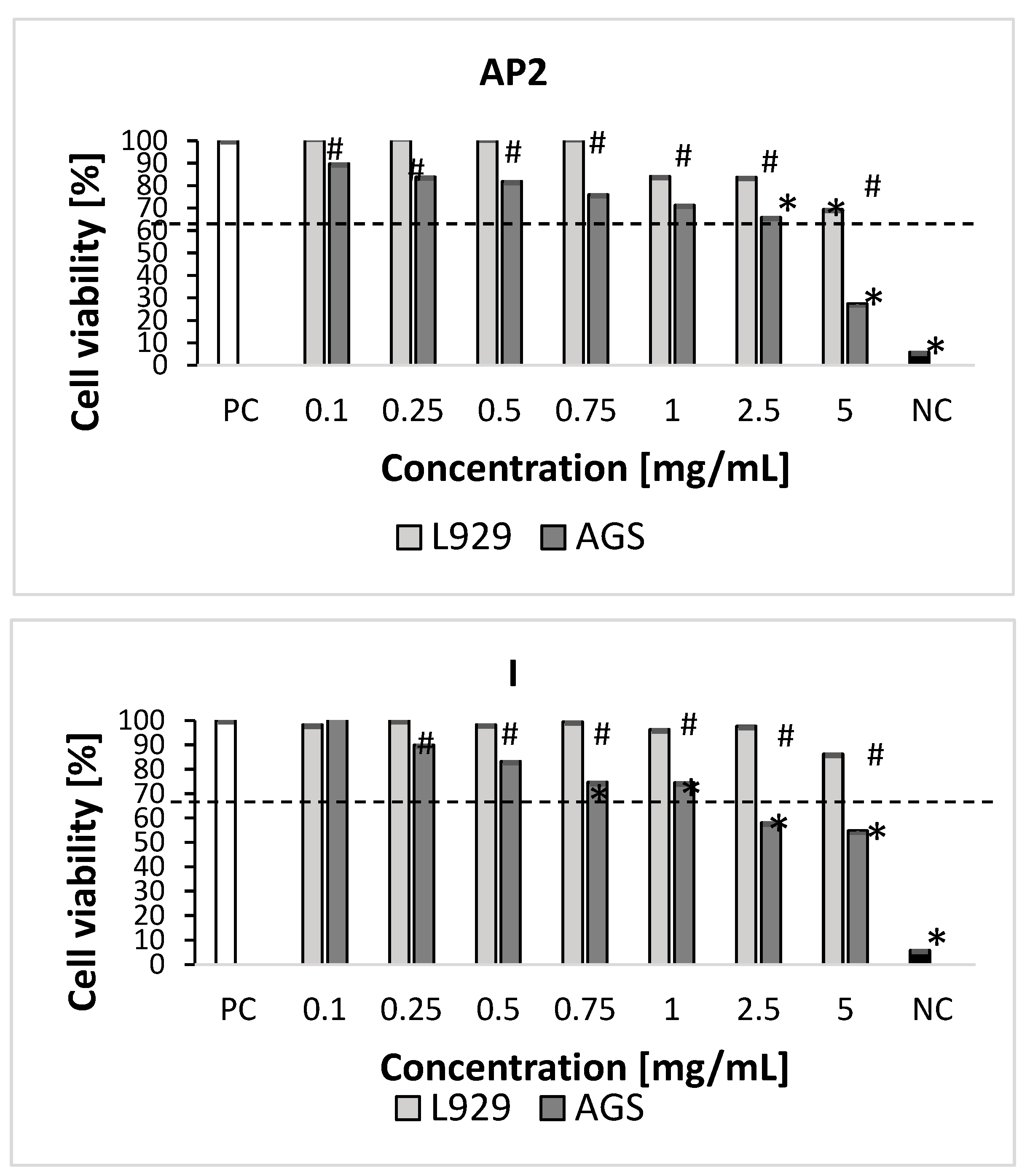
2.3. Cytotoxicity Studies

The methanolic-aqueous extracts from the aerial parts (one-year and two-year-old plants) in the highest concentration (5 mg/mL) significantly diminished the growth of L929 fibroblasts (30–39% of dead cells); however, the inflorescence extract did not show any cytotoxic activity at any of the tested concentrations (Figure 3). The root extract of one-year-old plants at the highest concentrations (5 mg/mL, 2.5 mg/mL, and 1 mg/mL) had a stronger inhibitory effect on L929 cell metabolic activity (28–45% of dead cells) than those of the two-year-old plants; the latter only reduced cell viability at the highest concentration (5 mg/mL) (27% of dead cells).
The root extract from the two-year-old plants also demonstrated the strongest inhibitory effect on the metabolic activity of AGS cells at the concentration range of 0.25–5 mg/mL (28–70% of dead cells). The root extract from the one-year-old plant significantly inhibited AGS cell growth at the two highest concentrations (2.5 and 5 mg/mL).
The extracts from inflorescences and aerial parts exhibited significantly greater cytotoxic activity against AGS cells in the range of 0.75–5 mg/mL (25–46% and 30–62% of dead cells, respectively) (Figure 3).
None of the inflorescence extracts demonstrated toxicity towards the reference mouse L929 fibroblasts at any tested concentrations. Similarly, no such cytotoxicity was noted for extracts from aerial parts and roots (two-year-old plants) at 2.5 mg/mL and below, or for root extracts (one-year-old) below 1 mg/mL.
Kirmizibekmez et al. [47] found various Salvia extracts, including S. tomentosa, to demonstrate low or absent cytotoxicity towards L6 mammalian cells. Similarly, hydromethanolic aerial and root extracts from S. cadmica at concentrations of 2.5 mg/mL and below did not demonstrate any cytotoxicity against the same cell line (L929) based on MTT assay [25]. Abu-Dahab et al. [48] also report that the extracts of the Salvia species S. fruticosa, S. horminum, and S. syriaca did not appear cytotoxic towards various normal cell lines in the concentration range 0.1 and 100 μg/mL.
In the present study, non-neoplastic cells like L-929 fibroblasts were found to be resistant to studied extracts. Only R1 and AP1 extracts diminished cell viability below 70%, at concentration ranges of 5–1 mg/mL and 5 mg/mL, respectively. Hence, the developed extracts appear to be very safe in contact with normal cells, which bodes well for their use in the development of formulations with antimicrobial or pro-regenerative effects. However, the root extracts (two-year cultivation; concentration: 0.25–5 mg/mL), inflorescence, and leaf extracts (concentration: 0.75–5 mg/mL) were found to be cytotoxic against the studied gastric cancer AGS cells. Therefore, S. tomentosa extracts may be of value in anticancer treatment. However, further research is needed in this area to explore their mechanism of action. Indeed, Berk et al. [49] report S. tomentosa extracts to have cytotoxic effects on hepatocellular carcinoma HepG2 cells (IC50 = 0.417 mg/mL).
Other Salvia extracts have also exhibited cytotoxic activity against different cancer cell lines. For example, Firuzi et al. [50] found that S. eremophila methanolic and hydromethanolic extracts significantly diminished the viability of breast cancer MCF-7 cell lines and increased the effectiveness of the reference drug cisplatin. Jiang et al. [51] report that ethanolic and acetone extracts from S. miltiorrhiza roots displayed high cytotoxic potential against HepG2 cells (IC50 = 17.3 and 83.2 µg/mL, respectively), as did extracts of S. officinalis roots and leaves (IC50 = 19.6–43.8 µg/mL and 64.4–90.0 µg/mL, respectively). Uysal et al. [52] showed that the methanolic and aqueous extracts of S. ceratophylla roots caused the death of HepG2 cells (30.9 and 34.5% of cell viability), while aqueous extracts from the aerial parts were more active against B16 4A5 cells (57.3% of cell viability).
Our present findings suggest that the cytotoxic activity of the tested plant extracts against AGS cells may be related to their phytochemical profile, with high amounts of polyphenols. Various studies based on animal models have found plant polyphenols to inhibit tumorigenesis and arrest cancer cell proliferation, potentially via the modulation of numerous signaling pathways dependent on nuclear factor B (NF-kB) or mitogen-activated protein kinase (MAPK). These pathways are related to cell autophagy, apoptosis, and inflammation [53,54,55]. Parsaee et al. [56] found S. chorassanica root extracts to have pro-apoptotic activity against human HeLa cells, which suggests that plant polyphenols can influence the course of an apoptotic process [57]; however, this thesis has yet to be confirmed.

3. Materials and Methods

3.1. Plant Material

S. tomentosa seeds were obtained from the Botanical Garden of Medicinal Plants, Wroclaw Medical University (Wroclaw, Poland) in 2018. They were sewn in the experimental plot at the Botanical Garden of Medicinal Plants, Medical University of Lodz (Lodz, Poland) (51°77′ N, 19°49′ E). The resulting plants were cultivated for two years and harvested during each vegetation season. A voucher specimen (no EP-ST-2019) was deposited at the Department of Pharmaceutical Biotechnology, Medical University of Lodz (Lodz, Poland). The plant material was collected and dried and then separated into aerial parts and roots. As the plant blooms only in the second year of vegetation, the collected inflorescences were analyzed separately.

3.2. Extract Preparation

Dried and powdered plant material (100 mg for phytochemical studies and 500 mg for antimicrobial and cytotoxic activities, separately) was pre-extracted with chloroform (20 or 100 mL) for 12 h at room temperature. Chloroform supernatants were filtered off. The defatted dried plant material was extracted three times with methanol:water solution at a ratio of 8:2 (v/v) according to the method described by Piątczak et al. [25]. The obtained dry extracts were stored at 4 °C until used.

3.3. Phytochemical Studies

Phytochemical analyses were performed using UPLC equipped with quadrupole Time of Flight—double mass spectrometry detector (UPLC/DAD/qTOF-MS/MS) (Waters Corporation, Milford, MA, USA) as described by Zielińska et al. [58]. Polyphenolic compounds were separated by a UPLC BEH C18 column (1.7 μm, 2.1 × 100 mm, Waters Corporation, Milford, MA, USA). The mobile phase consisted of solvent A (0.1% formic acid in LC–MS grade water, v/v, Merck, Darmstadt, Germany) and solvent B (0.1% formic acid in LC–MS grade acetonitrile, Merck Germany).
The retention times and spectra of the analyzed compounds were compared with those of authentic standards. Caffeic, rosmarinic, salvianolic acid B, salvianolic acid F, and its derivatives were quantified with their own standards. Salvianolic acid K and lithospermic acid derivatives were quantified as salvianolic acid A equivalents. The quantities of compounds were expressed as mg per 100 g of plant material dry weight (mg/100 g DW).

3.4. Antimicrobial Studies

The antimicrobial activity of the prepared extracts was investigated against fourteen strains of Gram-positive bacteria: Staphylococcus aureus ATCC 25923, S. aureus MM3 MRSA, S. aureus ZMF MLSB, Staphylococcus hominis DSM 20328, Staphylococcus epidermidis ATCC 12228, Staphylococcus haemolyticus PCM 2113, Staphylococcus simulans DSM 20723, Staphylococcus pseudintermedius PCM 2791, Enterococcus faecium PCM 1859, Enterococcus hirae ATCC 10541, Enterococcus faecalis ATCC 29212, Bacillus subtilis ATCC 6635, Bacillus cereus PCM 1948, and Listeria monocytogenes PCM 2191; six Gram-negative strains: Escherichia coli ATCC 25922, E. coli ZMF ESBL, Proteus vulgaris CCM 1799, Salmonella enteritidis ZMF 279, Shigella sonnei PCM 2466 FII, and Pseudomonas aeruginosa ATCC 27833; and two fungal strains: Candida albicans ATCC 10231 and Aspergillus brasiliensis ATCC 16404.
The S. haemolyticus PCM 2113, S. pseudintermedius PCM 2791, E. faecium PCM 1859, B. cereus PCM 1948, L. monocytogenes PCM 2191, and S. sonnei PCM 2466 FII strains were taken from the Polish Collection of Microorganisms (PCM). The P. vulgaris CCM 1799 strain was obtained from the Czech Collection of Microorganisms. The S. hominis DSMZ 20328 and S. simulans DSMZ 20723 were obtained from the German Collection of Microorganisms and Cell Cultures (DSMZ). Four bacterial strains, S. aureus MM3 MRSA, S. aureus MLSb, E. coli ZMF ESBL, and S. enteritidis ZMF 279, were derived from the collection of the Department of Pharmaceutical Microbiology and Diagnostic Microbiology of the Medical University of Lodz. The rest of the strains were received from the American Type Culture Collection (ATCC), Rockville, MD, USA.
All tested microorganisms were stored at −80 °C in 15% glycerol stocks. Before the experiment, the bacterial strains were transferred to the Mueller–Hinton agar medium (Oxoid, Thermo Fisher Scientific, Waltham, MA, USA) and cultured overnight at 37 °C. Fungal strains were transferred on agar RPMI-1640 medium and cultured for two days at 30 °C.
The antimicrobial activity of the methanolic-aqueous extracts from the aerial parts, roots, and inflorescences of S. tomentosa were determined based on MIC (minimum inhibitory concentration) and MBC/MFC (minimum bactericidal concentration/minimum fungicidal concentration) using the broth microdilution method. These assays were performed in the Mueller–Hinton liquid media for bacteria or RPMI-1640 for fungi. All tests were performed in 96-well microtiter plates (Kartell Labware, Noviglio, Italy) according to the European Committee on Antimicrobial Susceptibility recommendations (EUCAST) as described by Grzegorczyk-Karolak et al. [59]. The extracts were used to create a two-fold dilution series in a growth medium in the range of 5 mg–0.009 mg/mL. Gentamicin, fluconazole, and rosmarinic acid were used as positive controls.

3.5. Cytotoxicity Studies

3.5.1. In Vitro Cell Cultures

The in vitro cytotoxicity assays were performed using mouse fibroblast L929 cell line (LGC Standards, Middlesex, UK), according to ISO 10993-5 [60], and human gastric adenocarcinoma epithelial cells (AGS) (CRL-1739, ATCC, Rockville, MD, USA). The cells were maintained under standard conditions in complete culture medium (cRPMI 1640 medium) supplemented with fetal bovine serum (FBS) (10%), L-glutamine (2 mM, 1%), and penicillin (50 U mL−1)/streptomycin (50 μg mL−1) (1%) (Sigma-Aldrich, Darmstadt, Germany). The cultures were incubated in a humidified atmosphere at 37 °C and 5% CO2 and cultured according to Kamizela et al. [61]. Cell viability was assessed by exclusion of trypan blue, 93 to 95%.

3.5.2. Measurement of Cellular Metabolic Activity and Cell Growth Inhibition

The metabolic activity of both cell lines was tested in cell cultures in vitro after the application of S. tomentosa extracts. Cells suspended in complete culture medium (cRPMI-1640) were seeded (2 × 10⁵ cells/well) in 96-well plates for 24 h at 37 °C and 5% CO2. The tested extracts were diluted in cRPMI-1640 medium in concentrations of 5, 2.5, 1, 0.75, 0.5, 0.25, and 0.1 mg/mL, and added to the wells with the cells (100 μL/well). Cells were incubated under standard conditions for 24 h. Following incubation, cell morphology was determined via light microscopy, as recommended by ISO norm 10993-5 [60]. Cell metabolism was estimated according to [56] using MTT assay, as recommended by the Food and Drug Administration (FDA) and the International Organization for Standardization (IOS).

3.6. Statistical Analysis

The phytochemical analyses were performed in triplicate. All data are presented as mean values ± standard error (SE). Antibacterial and antifungal tests were repeated twice. Cell viability was determined using an MTT reduction assay (cytotoxicity studies) in four experiments. Statistical significance was considered at a p value of <0.05 using the Wilcoxon signed rank test (cytotoxicity assays) and Tukey’s HSD test (phytochemical analysis). Statistical analyses were performed using STATISTICA 13.3 PL software (TIBCO, Wroclaw, Poland).

4. Conclusions

Methanolic-aqueous extracts from different organs of field-grown S. tomentosa appear to be rich sources of rosmarinic acid, salvianolic acids, and lithospermic acid A derivatives. The metabolite profile was not influenced by the developmental stage of the plants harvested from two consecutive vegetation seasons. No significant differences in the metabolite profile of the roots and aerial extracts were noted between harvest periods. However, it is important to note that the inflorescences, which naturally appear in the second year of vegetation, contained considerable amounts of salvianolic acid B and K derivatives, as well as rosmarinic acid glycosides.
The studied extracts demonstrated strong antimicrobial activity against opportunistic bacterial species, such as Staphylococcus epidermidis and Bacillus cereus. This result is particularly important in the face of increasing antibiotic resistance and the need to identify alternative treatments for infections. Cytotoxic investigations on mouse fibroblasts indicated that all of the tested extracts were non-toxic against mammalian cells up to a concentration of 5 mg/mL, depending on the plant organ, but showed cytotoxicity against gastric cancer cells (AGS) across a broad concentration range. The extract of roots collected from the second year of cultivation showed the highest cytotoxic activity.
Our findings significantly improve the knowledge of the metabolite content of S. tomentosa and their acquisition for further use. As such, S. tomentosa merits further consideration as a potential herbal medicine suitable for the treatment of infections and for the support of anticancer therapy. Further detailed investigations are recommended into the isolation and study of these compounds in an intact and sustainable form, which is needed for their strong bioactivity.

Author Contributions

E.P. developed the idea and study concept. E.P., J.K.-O., W.G. and P.L. designed and conducted the experiments. E.P., U.K.-L. and S.Z. analyzed and interpreted the data. E.P., W.G., U.K.-L., P.L. and J.K.-O. prepared tables, figures, and a first draft of the manuscript. E.P., U.K.-L., M.S. and S.Z. prepared the final version of the manuscript. M.C., M.S. and S.Z. supervised the manuscript. All authors have read and agreed to the published version of the manuscript.

Funding

This research was financially supported by the Statutory Budget (503/3-012-02/503-31-001-19-00) of the Medical University of Lodz (Lodz, Poland).

Institutional Review Board Statement

Not applicable.

Informed Consent Statement

Not applicable.

Data Availability Statement

Data are contained within this article.

Acknowledgments

The authors would like to express their sincere thanks to Adam Matkowski (Medical University of Wrocław, Wroclaw, Poland) for all his valuable remarks and discussions which have improved the final version of the manuscript. Additionally, the authors would like to express the warmest gratitude to Hanna Zielińska for her drawings in the Graphical Abstract.

Conflicts of Interest

The authors declare no conflicts of interest.

References

  1. Suzuki, T.; Hidaka, T.; Kumagai, Y.; Yamamoto, M. Environmental Pollutants and the Immune Response. Nat. Immunol. 2020, 21, 1486–1495. [Google Scholar] [CrossRef] [PubMed]
  2. Messelhäußer, U.; Ehling-Schulz, M. Bacillus cereus—A Multifaceted Opportunistic Pathogen. Curr. Clin. Microbiol. Rep. 2018, 5, 120–125. [Google Scholar] [CrossRef]
  3. Weng, J.K.; Lynch, J.H.; Matos, J.O.; Dudareva, N. Adaptive Mechanisms of Plant Specialized Metabolism Connecting Chemistry to Function. Nat. Chem. Biol. 2021, 17, 1037–1045. [Google Scholar] [CrossRef] [PubMed]
  4. Rana, N.; Privitera, G.; Kondolf, H.C.; Bulek, K.; Lechuga, S.; De Salvo, C.; Corridoni, D.; Antanaviciute, A.; Maywald, R.L.; Hurtado, A.M.; et al. GSDMB Is Increased in IBD and Regulates Epithelial Restitution/Repair Independent of Pyroptosis. Cell 2022, 185, 283–298.e17. [Google Scholar] [CrossRef] [PubMed]
  5. Miraj, S.; Kiani, S. A Review Study of Therapeutic Effects of Salvia officinalis L. Der Pharm. Lett. 2016, 8, 299–303. [Google Scholar]
  6. Salvia tomentosa Mill.|Plants of the World Online|Kew Science. Available online: https://powo.science.kew.org/taxon/urn:isid:ipni.org:names:457406-1 (accessed on 23 May 2023).
  7. Ulubelen, A.; Miski, M.; Neuman, P.; Mabry, T.J. Flavonoidsof Salvia tomentosa (Labiatae). J. Nat. Prod. 1979, 42, 261–263. [Google Scholar] [CrossRef]
  8. Aşkun, T.; Tumen, G.; Satil, F.; Ates, M. Characterization of the Phenolic Composition and Antimicrobial Activities of Turkish Medicinal Plants. Pharm. Biol. 2009, 47, 563–571. [Google Scholar] [CrossRef]
  9. Tepe, B.; Daferera, D.; Sokmen, A.; Sokmen, M.; Polissiou, M. Antimicrobial and Antioxidant Activities of the Essential Oil and Various Extracts of Salvia tomentosa Miller (Lamiaceae). Food Chem. 2005, 90, 333–340. [Google Scholar] [CrossRef]
  10. Haznedaroglu, M.Z.; Karabay, N.U.; Zeybek, U. Antibacterial Activity of Salvia tomentosa Essential Oil. Fitoterapia 2001, 72, 829–831. [Google Scholar] [CrossRef]
  11. Dincer, C.; Tontul, I.; Çam, I.B.; Özdemir, K.S.; Topuz, A.; Şahin Nadeem, H.; Tugrul, A.Y.S.; Göktür, R.S. Phenolic Composition and Antioxidant Activity of Salvia tomentosa Miller: Effects of Cultivation, Harvesting Year, and Storage, Turk. J. Agric. For. 2013, 37, 561–567. [Google Scholar] [CrossRef]
  12. Nagy, G.; Gunther, G.; Mathe, I.; Blunden, G.; Yang, M.; Crabb, T.A. Diterpenoids from Salvia glutinosa, S. austriaca, S. tomentosa and S. verticillata Roots. Phytochemistry 1999, 52, 1105–1109. [Google Scholar] [CrossRef]
  13. Petersen, M.; Simmonds, M.S. Rosmarinic Acid. Phytochemistry 2003, 62, 121–125. [Google Scholar] [CrossRef] [PubMed]
  14. Tewtrakul, S.; Miyashiro, H.; Nakamura, N.; Hattori, M.; Kawahata, T.; Otake, T.; Rattanasuwon, P. HIV-1 integrase inhibitory substances from Coleus parvifolius. Phytother. Res. 2003, 17, 232–239. [Google Scholar] [CrossRef] [PubMed]
  15. Karak, P. Biological Activity of Flavonoids: An Overview. Int. J. Pharm. Sci. Res. 2019, 19, 1567–1574. [Google Scholar]
  16. Ulubelen, A.; Topçu, G. Terpenoids from the Roots of Salvia tomentosa. Nat. Prod. Lett. 1992, 1, 141–147. [Google Scholar] [CrossRef]
  17. Barros, L.; Dueñas, M.; Dias, M.I.; Sousa, M.J.; Santos-Buelga, C.; Ferreira, I.C.F.R. Phenolic Profiles of Cultivated, In Vitro Cultured and Commercial Samples of Melissa officinalis L. Infusions. Food Chem. 2013, 136, 1–8. [Google Scholar] [CrossRef] [PubMed]
  18. Katanić Stanković, J.S.; Srećković, N.; Mišić, D.; Gašić, U.; Imbimbo, P.; Monti, D.M.; Mihailović, V. Bioactivity, Biocompatibility and Phytochemical Assessment of Lilac Sage, Salvia verticillata L. (Lamiaceae)-A Plant Rich in Rosmarinic Acid. Ind. Crops Prod. 2020, 143, 111932. [Google Scholar] [CrossRef]
  19. Lecomte, J.; Lopez Giraldo, L.J.; Laguerre, M.; Baréa, B.; Villeneuve, P. Synthesis, Characterization and Free Radical Scavenging Properties of Rosmarinic Acid Fatty Esters. J. Am. Oil Chem. Soc. 2010, 87, 615–620. [Google Scholar] [CrossRef]
  20. Grzegorczyk-Karolak, I.; Kuźma, Ł.; Skała, E.; Kiss, A.K. Hairy Root Cultures of Salvia viridis L. for Production of Polyphenolic Compounds. Ind. Crops Prod. 2018, 117, 235–244. [Google Scholar] [CrossRef]
  21. Krzemińska, M.; Owczarek, A.; Gonciarz, W.; Chmiela, M.; Olszewska, M.A.; Grzegorczyk-Karolak, I. The Antioxidant, Cytotoxic and Antimicrobial Potential of Phenolic Acids-Enriched Extract of Elicited Hairy Roots of Salvia bulleyana. Molecules 2022, 27, 992. [Google Scholar] [CrossRef]
  22. Lu, Y.; Foo, L.Y. Polyphenolics of Salvia—A Review. Phytochemistry 2002, 59, 117–140. [Google Scholar] [CrossRef]
  23. Zengin, G.; Llorent-Martínez, E.J.; Córdova, M.L.F.; Bahadori, M.B.; Mocan, A.; Locatelli, M.; Aktumsek, A. Chemical Composition and Biological Activities of Extracts from Three Salvia species: S. blepharochlaena, S. euphratica var. leiocalycina, and S. verticillata subsp. amasiaca. Ind. Crops Prod. 2018, 111, 11–21. [Google Scholar] [CrossRef]
  24. Zengin, G.; Mahomoodally, F.; Picot-Allain, C.; Diuzheva, A.; Jekö, J.; Cziáky, Z.; Cvetanović, A.; Aktumsek, A.; Zeković, Z.; Rengasamy, K.R.R. Metabolomic Profile of Salvia viridis L. Root Extracts Using HPLC–MS/MS Technique and Their Pharmacological Properties: A Comparative Study. Ind. Crops Prod. 2019, 131, 266–280. [Google Scholar] [CrossRef]
  25. Piątczak, E.; Owczarek, A.; Lisiecki, P.; Gonciarz, W.; Kozłowska, W.; Szemraj, M.; Chmiela, M.; Kiss, A.K.; Olszewska, M.A.; Grzegorczyk-Karolak, I. Identification and Quantification of Phenolic Compounds in Salvia cadmica Boiss. and Their Biological Potential. Ind. Crops Prod. 2021, 160, 113113. [Google Scholar] [CrossRef]
  26. Fan, H.; Yang, L.; Fu, F.; Xu, H.; Meng, Q.; Zhu, H.; Teng, L.; Yang, M.; Zhang, L.; Zhang, Z.; et al. Cardioprotective Effects of Salvianolic Acid A on Myocardial Ischemia-Reperfusion Injury In Vivo and In Vitro. Evid.-Based Complement. Altern. Med. 2012, 2012, 508938. [Google Scholar] [CrossRef]
  27. Wojciechowska, M.; Owczarek, A.; Kiss, A.K.; Grąbkowska, R.; Olszewska, M.A.; Grzegorczyk-Karolak, I. Establishment of Hairy Root Cultures of Salvia bulleyana Diels for Production of Polyphenolic Compounds. J. Biotechnol. 2020, 318, 10–19. [Google Scholar] [CrossRef]
  28. Zhang, J.; Xu, W.; Wang, P.; Huang, J.; Bai, J.; Huang, Z.; Liu, X.; Qiu, X. Chemical Analysis and Multi-Component Determination in Chinese Medicine Preparation Bupi Yishen Formula Using Ultra-High Performance Liquid Chromatography With Linear Ion Trap-Orbitrap Mass Spectrometry and Triple-Quadrupole Tandem Mass Spectrometry. Front. Pharmacol. 2018, 9, 568. [Google Scholar] [CrossRef] [PubMed]
  29. Afonso, A.F.; Pereira, O.R.; Fernandes, Â.; Calhelha, R.C.; Silva, A.M.S.; Ferreira, I.C.F.R.; Cardoso, S.M. Phytochemical Composition and Bioactive Effects of Salvia africana, Salvia officinalis ‘Icterina’ and Salvia mexicana Aqueous Extracts. Molecules 2019, 24, 4327. [Google Scholar] [CrossRef] [PubMed]
  30. Afonso, A.F.; Pereira, O.R.; Fernandes, Â.S.F.; Calhelha, R.C.; Silva, A.M.S.; Ferreira, I.C.F.R.; Cardoso, S.M. The Health-Benefits and Phytochemical Profile of Salvia apiana and Salvia farinacea var. Victoria Blue Decoctions. Antioxidants 2019, 8, 241. [Google Scholar] [CrossRef] [PubMed]
  31. Zhu, Z.; Zhang, H.; Zhao, L.; Dong, X.; Li, X.; Chai, Y.; Zhang, G. Rapid Separation and Identification of Phenolic and Diterpenoid Constituents from Radix Salvia miltiorrhizae by High-Performance Liquid Chromatography Diode-Array Detection, Electrospray Ionization Time-of-Flight Mass Spectrometry and Electrospray Ionization. Rapid Commun. Mass. Spectrom. 2007, 21, 1855–1865. [Google Scholar] [CrossRef] [PubMed]
  32. Lim, M.J.; Tock, A.H.; Chen, W.; Combrinck, S.; Sandasi, M.; Kamatou, G.P.P.; Viljoen, A.M. Exploring the Phytochemical Variation of Non-Volatile Metabolites Within Three South African Salvia Species Using UPLC-MS Fingerprinting and Chemometric Analysis. Fitoterapia 2021, 52, 104940. [Google Scholar] [CrossRef] [PubMed]
  33. Sharma, Y.; Velamuri, R.; Fagan, J.; Schaefer, J. Full-Spectrum Analysis of Bioactive Compounds in Rosemary (Rosmarinus officinalis L.) as Influenced by Different Extraction Methods. Molecules 2020, 25, 4599. [Google Scholar] [CrossRef] [PubMed]
  34. Cruz, L.R.O.; Polyzos, N.; Fernandes, Â.; Petropoulos, S.A.; Gioia, F.D.; Dias, M.I.; Pinela, J.; Kostić, M.; Soković, M.; Ferreira, I.C.F.R.; et al. Effect of Saline Conditions on Chemical Profile and the Bioactive Properties of Three Red-Colored Basil Cultivars. Agronomy 2020, 10, 1824. [Google Scholar] [CrossRef]
  35. Ulukanli, Z.; Karabörklü, S.; Cenet, M.; Sagdic, O.; Ozturk, I.; Balcilar, M. Essential Oil Composition, Insecticidal and Antibacterial Activities of Salvia tomentosa Miller. Med. Chem. Res. 2013, 22, 832–840. [Google Scholar] [CrossRef]
  36. Eghbaliferiz, S.; Soheili, V.; Tayarani-Najaran, Z.; Asili, J. Antimicrobial and Cytotoxic Activity of Extracts from Salvia tebesana Bunge and Salvia sclareopsis Bornm Cultivated in Iran. Physiol. Mol. Biol. Plants 2019, 25, 1083–1089. [Google Scholar] [CrossRef] [PubMed]
  37. Duletić-Laušević, S.; Aradski, A.A.; Šavikin, K.; Knežević, A.; Milutinović, M.; Stević, T.; Vukojević, J.; Marković, S.; Marin, P. Composition and Biological Activities of Libyan Salvia fruticosa Mill. and S. lanigera Poir. Extracts. S Afr. J. Bot. 2018, 117, 101–109. [Google Scholar] [CrossRef]
  38. Gharenaghadeh, S.; Karimi, N.; Forghani, S.; Nourazarian, M.; Gharehnaghadeh, S.; Jabbari, V.; Khiabani, M.S.; Kafil, H.S. Application of Salvia multicaulis Essential Oil-Containing Nanoemulsion Against Food-borne Pathogens. Food Biosci. 2017, 19, 128–133. [Google Scholar] [CrossRef]
  39. Shakeri, A.; Khakdan, F.; Soheili, V.; Sahebkar, A.; Rassam, G.; Asili, J. Chemical Composition, Antibacterial Activity, and Cytotoxicity of Essential Oil from Nepeta ucrainica L. spp. kopetdaghensis. Ind. Crops Prod. 2014, 58, 315–321. [Google Scholar] [CrossRef]
  40. Kimura, K.; Yokoyama, S. Trends in the Application of Bacillus in Fermented Foods. Curr. Opin. Biotechnol. 2019, 56, 36–42. [Google Scholar] [CrossRef]
  41. Omer, M.K.; Álvarez-Ordoñez, A.; Prieto, M.; Skjerve, E.; Asehun, T.; Alvseike, O.A. A Systematic Review of Bacterial Foodborne Outbreaks Related to Red Meat and Meat Products. Foodborne Pathog. Dis. 2018, 15, 598–611. [Google Scholar] [CrossRef]
  42. Glasset, B.; Herbin, S.; Granier, S.A.; Cavalié, L.; Lafeuille, E.; Guérin, C.; Ruimy, R.; Casagrande-Magne, F.; Levast, M.; Chautemps, N.; et al. Bacillus cereus, a Serious Cause of Nosocomial Infections: Epidemiologic and Genetic Survey. PLoS ONE 2018, 13, e0194346. [Google Scholar] [CrossRef] [PubMed]
  43. Luna, V.A.; King, D.S.; Gulledge, J.; Cannons, A.C.; Amuso, P.T.; Cattani, J. Susceptibility of Bacillus anthracis, Bacillus cereus, Bacillus mycoides, Bacillus pseudomycoides and Bacillus thuringiensis to 24 Antimicrobials Using Sensititre Automated Microbroth Dilution and E-test Agar Gradient Diffusion Methods. J. Antimicrob. Chemother. 2007, 60, 555–567. [Google Scholar] [CrossRef]
  44. Nguyen, T.H.; Park, M.D.; Otto, M. Host Response to Staphylococcus epidermidis Colonization and Infections. Front. Cell. Infect. Microbiol. 2017, 7, 90. [Google Scholar] [CrossRef]
  45. Severn, M.M.; Horswill, A.R. Staphylococcus epidermidis and its Dual Lifestyle in Skin Health and Infection. Nat. Rev. Microbiol. 2023, 21, 97–111. [Google Scholar] [CrossRef]
  46. Srinivasan, R.; Santhakumari, S.; Poonguzhali, P.; Geetha, M.; Dyavaiah, M.; Xiangmin, L. Bacterial Biofilm Inhibition: A Focused Review on Recent Therapeutic Strategies for Combating the Biofilm Mediated Infections. Front. Microbiol. 2021, 12, 676458. [Google Scholar] [CrossRef]
  47. Kirmizibekmez, H.; Atay, I.; Kaiser, M.; Yesilada, E.; Tasdemir, D. In Vitro Antiprotozoal Activity of Extracts of Five Turkish Lamiaceae species. Nat. Prod. Commun. 2011, 6, 1697–1700. [Google Scholar] [CrossRef] [PubMed]
  48. Abu-Dahab, R.; Afifi, F.; Kasabri, V.; Majdalawi, L.; Naffa, R. Comparison of the Antiproliferative Activity of Crude Ethanol Extracts of Nine Salvia Species Grown in Jordan Against Breast Cancer Cell Line Models. Pharmacogn. Mag. 2012, 8, 319. [Google Scholar] [CrossRef] [PubMed]
  49. Berk, S.; Ergul, M.; Baltas, H.; Tutar, Y. Investigation of the cytotoxic and DNA damage protection potentials of O. syriacum and S. tomentosa. Turk. Jnl. Biochem. 2013, 38. Available online: http://ovidsp.ovid.com/ovidweb.cgi?T=JS&PAGE=reference&D=emed14&NEWS=N&AN=75004147 (accessed on 3 August 2022).
  50. Firuzi, O.; Miri, R.; Asadollahi, M.; Eslami, S.; Jassbi, A.R. Cytotoxic, Antioxidant and Antimicrobial Activities and Phenolic Contents of Eleven Salvia Species from Iran. Iran. J. Pharm. Res. 2013, 12, 801–810. [Google Scholar]
  51. Jiang, Y.; Zhang, L.; Rupasinghe, H.P.V. Antiproliferative Effects of Extracts from Salvia officinalis L. and Saliva miltiorrhiza Bunge on Hepatocellular Carcinoma Cells. Biomed. Pharmacother. 2017, 85, 57–67. [Google Scholar] [CrossRef]
  52. Uysal, S.; Zengin, G.; Sinan, K.I.; Ak, G.; Ceylan, R.; Mahomoodally, M.F.; Uysal, A.; Sadeer, N.B.; Jekő, J.; Cziáky, Z.; et al. Chemical Characterization, Cytotoxic, Antioxidant, Antimicrobial, and Enzyme Inhibitory Effects of Different Extracts from One Sage (Salvia ceratophylla L.) from Turkey: Open a New Window on Industrial Purposes. RSC Adv. 2021, 11, 5295–5310. [Google Scholar] [CrossRef]
  53. Nakazato, T.; Ito, K.; Miyakawa, Y.; Kinjo, K.; Yamada, T.; Hozumi, N.; Ikeda, Y.; Kizaki, M. Catechin, a Green Tea Component, Rapidly Induces Apoptosis of Myeloid Leukemic Cells via Modulation of Reactive Oxygen Species Production in vitro and Inhibits Tumor Growth in vivo. Haematologica 2005, 90, 317–325. [Google Scholar] [PubMed]
  54. Jeong, J.C.; Jang, S.W.; Kim, T.H.; Kwon, C.H.; Kim, Y.K. Mulberry Fruit (Moris fructus) Extracts Induce Human Glioma Cell Death in vitro Through ROS-dependent Mitochondrial Pathway and Inhibits Glioma Tumor Growth in vivo. Nutr. Cancer 2010, 62, 402–412. [Google Scholar] [CrossRef] [PubMed]
  55. Costa, A.; Bonner, M.Y.; Arbiser, J.L. Use of Polyphenolic Compounds in Dermatologic Oncology. Am. J. Clin. Dermatol. 2016, 17, 369–385. [Google Scholar] [CrossRef] [PubMed]
  56. Parsaee, H.; Asili, J.; Mousavi, S.H.; Soofi, H.; Emami, S.A.; Tayarani-Najaran, Z. Apoptosis Induction of Salvia chorassanica Root Extract on Human Cervical Cancer Cell Line. Iran. J. Pharm. Res. 2013, 12, 75–83. [Google Scholar]
  57. D’Archivio, M.; Santangelo, C.; Scazzocchio, B.; Varì, R.; Filesi, C.; Masella, R.; Giovannini, C. Modulatory Effects of Polyphenols on Apoptosis Induction: Relevance for Cancer Prevention. Int. J. Mol. Sci. 2008, 9, 213–228. [Google Scholar] [CrossRef] [PubMed]
  58. Zielińska, S.; Dryś, A.; Piątczak, E.; Kolniak-Ostek, J.; Podgórska, M.; Oszmiański, J.; Matkowski, A. Effect of LED Illumination and Amino Acid Supplementation on Phenolic Compounds Profile in Agastache rugosa in vitro Cultures. Phytochem. Lett. 2019, 31, 12–19. [Google Scholar] [CrossRef]
  59. Grzegorczyk-Karolak, I.; Kuźma, Ł.; Lisiecki, P.; Kiss, A. Accumulation of Phenolic Compounds in Different in vitro Cultures of Salvia viridis L. and Their Antioxidant and Antimicrobial Potential. Phytochem. Lett. 2019, 30, 324–332. [Google Scholar] [CrossRef]
  60. ISO 10993-5 norm; Biological Evaluation of Medical Devices—Part 5: Tests for In Vitro Cytotoxicity. International Organization for Standardization: Geneva, Switzerland, 2009.
  61. Kamizela, A.; Gawdzik, B.; Urbaniak, M.; Lechowicz, Ł.; Białonska, A.; Gonciarz, W.; Chmiela, M. Synthesis, characterization, cytotoxicity and antimicrobial properties of trans-γ-halo-δ-lactones. Chem. Open 2018, 7, 543–550. [Google Scholar] [CrossRef]
Figure 1. UPLC-DAD chromatogram at 280 nm of S. tomentosa methanolic-aqueous extracts from roots (A), aerial parts (B), and inflorescences (C). Peak number identities are displayed in Table 1.
Molecules 29 00590 g001aMolecules 29 00590 g001b
Figure 2. The content (%) of the main components in the various organs of S. tomentosa plants. * The sum of the percentages of ingredients whose individual content in the extract is less than 0.4%; R1—roots of 1-year-old plants; R2—roots of 2-year-old plants; I—inflorescences from 2-year-old plants; AP1—aerial parts from 1-year-old plants; AP2—aerial parts from 2-year-old plants. SABd—salvianolic acid B derivatives; SAB—salvianolic acid B; LAd—lithospermic acid derivatives; LA—lithospermic acid; RAd—rosmarinic acid derivatives; RA—rosmarinic acid.
Molecules 29 00590 g002
Figure 3. Cytotoxic effect of S. tomentosa plant extracts towards L929 and AGS cells. The cytotoxicity was assessed by MTT [(3-(4,5-dimethylthiazol-2-yl)-2,5-diphenyltetrazolium bromide)] reduction assay. Complete RMPI-1640 medium (cRPMI) was used as a positive control (PC) of cell viability (100% viable cells) and 0.03% H2O2 as a negative control (NC) of cell viability (100% dead cells). Statistical significance: p < 0.05; * untreated cells vs. cells treated with tested plant extracts; # L929 cell line vs. AGS cell line (Mann–Whitney U-test). R1—roots of 1-year-old plants; R2—roots of 2-year-old plants; AP1—aerial parts from 1-year-old plants; AP2—aerial parts from 2-year-old plants; I—inflorescences from 2-year-old plants.
Molecules 29 00590 g003aMolecules 29 00590 g003b
Table 1. UPLC-DAD ESI-MS detection and identification of phenolic compounds in methanolic-aqueous extracts from Salvia tomentosa different organs.
Peak No.RT
(min)
MS
[M-H]
MS/MS/Fragmentation Ions
[M-H]
Tentative IdentificationR1R2AP1AP2I
1.2.62179 Caffeic acidppppp
2.3.69521.0880359, 197, 179, 161, 135Rosmarinic acid hexoside Ippppn.d.
3.4.63521.1452359, 179, 135Rosmarinic acid hexoside IIppppn.d.
4.4.95555.1432537, 359, 295, 179Salvianolic acid K derivativen.d.n.d.n.d.n.d.p
5.5.41377.0422359, 197, 161Rosmarinic acid derivativen.d.n.d.ppn.d.
6.5.60555.1807537, 359, 295, 179Salvianolic acid K derivativeppn.d.n.d.p
7.6.50683.2615537, 357, 197, 179, 161, 135Salvianolic acid K methyl pentosiden.d.n.d.n.d.n.d.p
8.7.00735.2613537, 493, 359, 295Lithospermic acidppppp
9.7.32521.1120359, 197, 179, 161, 135Rosmarinic acid hexoside IIIn.d.n.d.n.d.n.d.p
10.7.80719.2865359, 197, 179, 161, 135Sagerinic acidn.d.n.d.ppn.d.
11.8.01717.2477537, 519, 339, 321, 295, 197, 179Salvianolic acid Bppppp
12.8.30359.0282197, 179, 161, 135Rosmarinic acidppppp
13.8.911074.6538537, 359, 197, 179Lithospermic acid dimer In.d.n.d.n.d.n.d.p
14.9.381074.6538537, 359, 197, 179Lithospermic acid dimer IIn.d.n.d.ppn.d.
15.9.72569.1367537, 359, 197, 179Lithospermic acid derivativeppn.d.n.d.n.d.
16.9.81493.0895359, 197, 179Salvianolic acid An.d.n.d.n.d.n.d.p
17.10.37717.2409537, 359, 179Salvianolic acid B isomer In.d.n.d.ppn.d.
18.11.08717.2438519, 359, 179Salvianolic acid B isomer IIppn.d.n.d.n.d.
19.11.79313.1357269, 203, 161, 135Salvianolic acid F isomer Ipn.d.n.d.n.d.n.d.
20.11.89313.0336161Salvianolic acid F isomer IIn.d.n.d.ppn.d.
R1—roots from 1-year-old plants; R2—roots from 2-year-old plants; AP1—aerial parts from 1-year-old plants; AP2—aerial parts from 2-year-old plants; I—inflorescences from 2-year-old plants. p—compound present, identified, and quantified in the organ; n.d.—compound not detected.
Table 2. Content of phenolic compounds (mg/100 g D.W.) in methanolic-aqueous extracts from different organs (roots, aerial parts, and inflorescences) of S. tomentosa plants.
Peak No.Tentative IdentificationR1R2AP1AP2I
1.Caffeic acid 0.27 ± 0.05 a0.17 ± 0.04 a0.62 ± 0.05 c0.41 ± 0.05 ac0.98 ± 0.08 b
2.Rosmarinic acid hexoside I0.87 ± 0.003 a0.27 ±0.09 b1.12 ± 0.02 a4.63 ± 0.11 c0.00 b
3.Rosmarinic acid hexoside II27.45 ± 0.12 a11.73 ± 1.11 b15.79 ± 1.11 d3.85 ± 0.3 e0.00 c
4.Salvianolic acid K derivative I 0.00 a0.00 a0.00 a0.00 a7.64 ± 0.26 b
5.Rosmarinic acid derivative 0.00 a0.00 a3.39 ± 0.22 a1.37 ± 0.05 b0.00 a
6.Salvianolic acid K derivative II 15.21 ± 0.1 a17.64 ± 0.13 b0.00 c0.00 c0.00 c
7.Salvianolic acid K methyl pentoside 0.00 b0.00 b0.00 b0.00 b15.63 ± 0.3 a
8.Lithospermic acid A9.34 ± 0.13 a12.51 ± 0.66 a148.20 ± 5.60 c50.71 ± 0.64 d28.01 ± 0.38 b
9.Rosmarinic acid hexoside 0.00 b0.00 b0.00 b0.00 b20.21 ± 0.77 a
10.Sagerinic acid0.00 a0.00 a4.04 ± 0.03 b2.03 ± 0.08 c0.00 a
11.Salvianolic acid B181.90 ± 0.58 a121.10 ± 0.94 b226.00 ± 0.72 d346.51 ± 4.91 e82.62 ± 1.42 c
12.Rosmarinic acid 481.78 ± 0.8 a425.87 ± 2.83 b524.71 ± 1.31 c518.25 ± 4.43 c436.86 ± 1.71 ab
13.Lithospermic acid dimer I 0.00 a0.00 a0.00 a0.00 a153.57 ± 2.35 b
14.Lithospermic acid dimer II 0.00 a0.00 a201.76 ± 1.42 b1.38 ± 0.07 c0.00 a
15.Lithospermic acid derivative 0.91 ± 0.04 a3.45 ± 0.12 b0.00 c0.00 c0.00 c
16.Salvianolic acid A 0.00 b0.00 b0.00 b0.00 b34.63 ± 0.49 a
17.Salvianolic acid B isomer I0.00 a0.00 a29.73 ± 2.5 b19.11 ± 2.92 c0.00 a
18.Salvianolic acid B isomer II12.72 ± 0.15 a64.83 ± 1.84 b0.00 c0.00 c0.00 c
19.Salvianolic acid F isomer I 7.25 ± 0.5 a0.00 b0.00 b0.00 b0.00 b
20.Salvianolic acid F isomer II 0.00 a0.00 a42.76 ± 1.95 b15.30 ± 0.82 c0.00 a
Sum [mg/100 g D.W.]737.70657.571198.12963.55780.15
R1—roots of 1-year-old plants; R2—roots of 2-year-old plants; AP1—aerial parts from 1-year-old plants; AP2—aerial parts from 2-year-old plants; I—inflorescences from 2-year-old plants. The results are means from 3–4 samples ± standard error (SE). Means with the same letter within each line are not statistically different in the Kruskal–Wallis test at p ≤ 0.05.
Table 3. Antimicrobial activity of methanolic-aqueous extracts from aerial parts, roots, and inflorescences of S. tomentosa.
StrainR1R2AP1AP2IRosmarinic
Acid
GentamicinFluconazole
Gram-positive bacteriaMIC/MBC (mg/mL)MIC/MBC (µg/mL)
Staphylococcus aureus
ATCC 25923
2.5/2.52.5/2.55/55/55/51/>10.5-
Staphylococcus aureus
MM3 MRSA
2.5/2.52.5/2.52.5/2.52.5/2.52.5/2.5>1/>12-
Staphylococcus aureus
ZMF MLSB
2.5/2.52.5/2.55/55/52.5/2.5>1/>10.12-
Staphylococcus epidermidis
ATCC 12228
0.625/0.6250.625/0.6250.625/0.6250.625/0.6250.625/0.6251/>116-
Staphylococcus haemolyticus
PCM 2113
2.5/2.52.5/2.5>5/>5>5/>52.5/2.51/>10.06-
Staphylococcus hominis
DSM 20328
5/55/5>5/>5>5/>52.5/2.51/>10.06-
Staphylococcus simulans
DSM 20723
>5/>5>5/>5>5/>5>5/>5>5/>51/>10.06-
Staphylococcus pseudintermedius
PCM 2791
2.5/2.52.5/2.55/55/55/51/>10.12-
Enterococcus feacalis
ATTC 29212
>5/>5>5/>5>5/>5>5/>5>5/>51/>116-
Enterococcus faecium
PCM 1859
>5/>5>5/>5>5/>5>5/>5>5/>51/>134-
Enterococcus hirae
ATCC 10541
>5/>5>5/>5>5/>5>5/>5>5/>51/>132
Bacillus cereus
PCM 1948
0.312/0.3120.312/0.3121.25/1.251.25/1.250.312/0.3121/10.25-
Bacillus subtilis
ATCC 6635
2.5/2.52.5/2.55/55/52.5/2.51/10.25-
Listeria monocytogenes
PCM 2191
>5/>5>5/>5>5/>5>5/>5>5/>5>1/>10.25-
Gram-negative bacteria
Escherichia coli
ATCC 25922
2.5/2.52.5/2.52.5/2.52.5/2.52.5/2.51/12-
Escherichia coli
ZMF ESBL
>5/>5>5/>5>5/>5>5/>5>5/>5>1/>1128-
Pseudomonas aeruginosa
ATCC 27853
2.5/2.52.5/2.52.5/2.52.5/2.52.5/25.1/>12-
Proteus vulgaris
CCM 1799
5/55/5>5/>5>5/>55/51/10.12-
Salmonella enteritidis
ZMF 279
>5/>5>5/>5>5/>5>5/>5>5/>5>1/>10.25-
Shigella sonnei
PCM 2466 FII
5/55/55/55/55/5>1/>15-
Fungi
Candida albicans
ATTC 10231
2.5/2.52.5/2.52.5/2.52.5/2.52.5/2.51/>1-5/>5
Aspergillus brasiliensis
ATCC 16404
>5/>5>5/>5>5/>5>5/>5>5/>51/>1-5/>5
R1—roots from 1-year-old plants; R2—roots from 2-year-old plants; AP1—aerial parts from 1-year-old plants; AP2—aerial parts from 2-year-old plants; I—inflorescences from 2-year-old plants; gentamicin—wide-spectrum antibacterial antibiotic: fluconazole—antifungal chemotherapeutic. MRSA—methicyllin-resistant Staphylococcus aureus; MLSB—resistance to macrolide; lincosamide and streptogramin B; ESBL—extended-spectrum beta-lactamases; MIC—minimal inhibitory concentration; MBC—minimal bactericidal concentration.
Disclaimer/Publisher’s Note: The statements, opinions and data contained in all publications are solely those of the individual author(s) and contributor(s) and not of MDPI and/or the editor(s). MDPI and/or the editor(s) disclaim responsibility for any injury to people or property resulting from any ideas, methods, instructions or products referred to in the content.

