
The High-Precision Liquid Chromatography with Electrochemical Detection (HPLC-ECD) for Monoamines Neurotransmitters and Their Metabolites: A Review

Abstract
:
1. Introduction
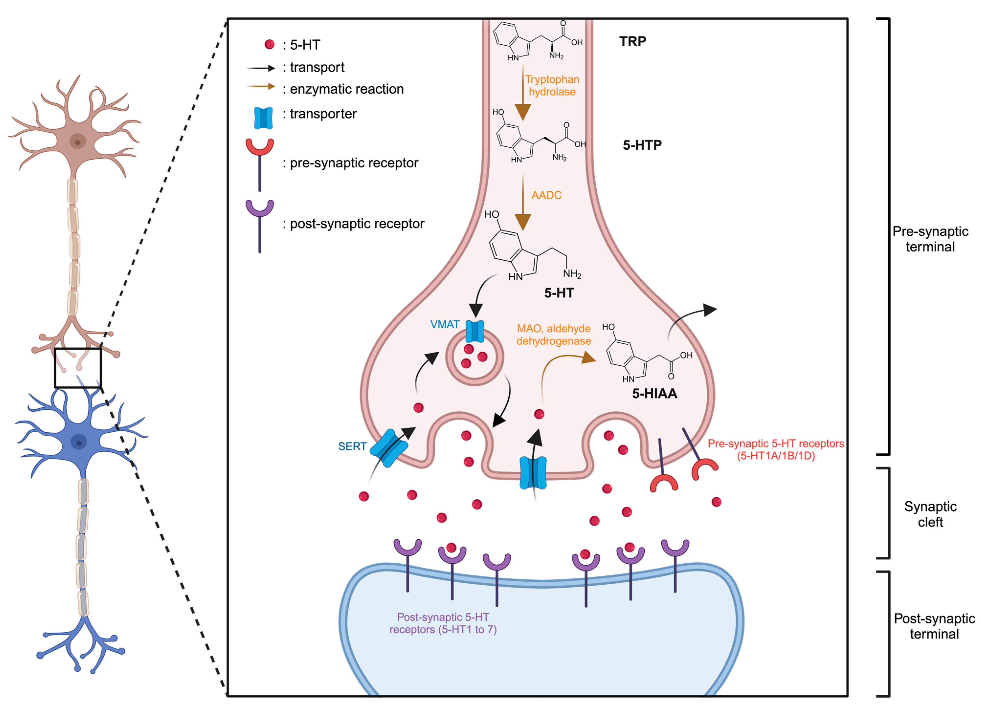
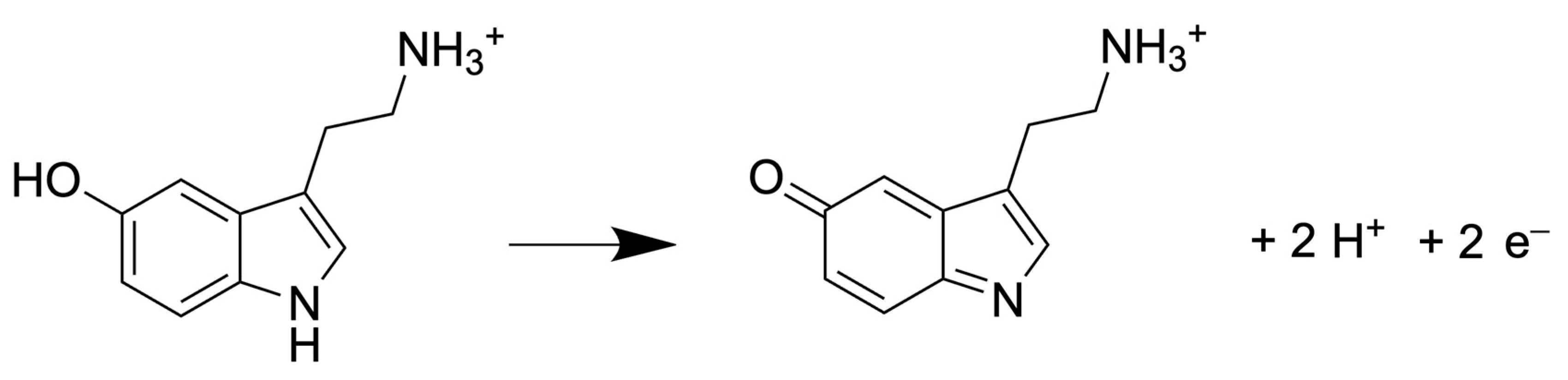
1.1. The Serotonergic System
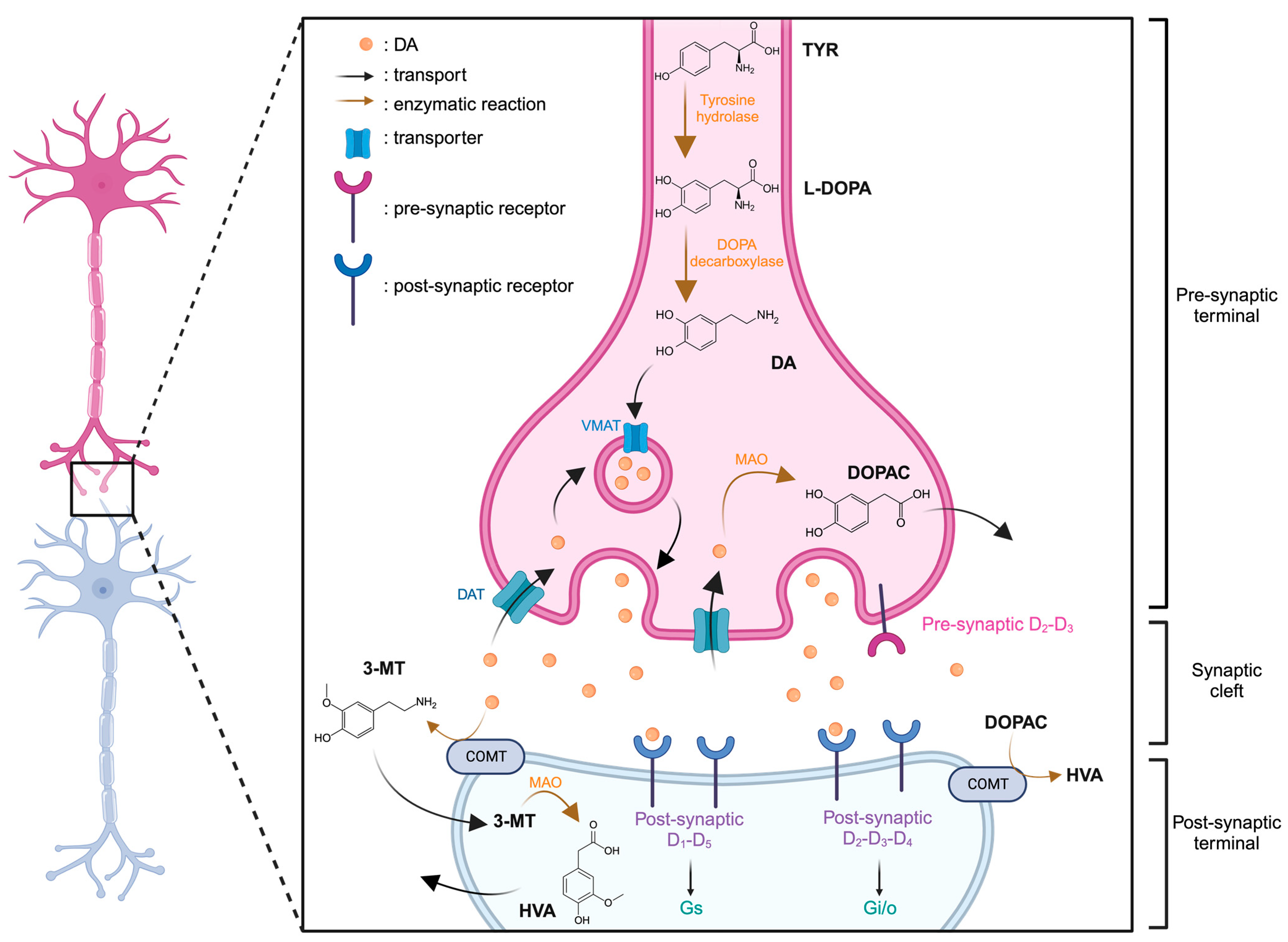
1.2. The Dopaminergic System
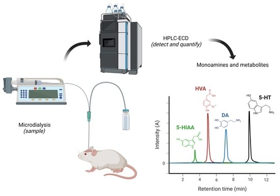
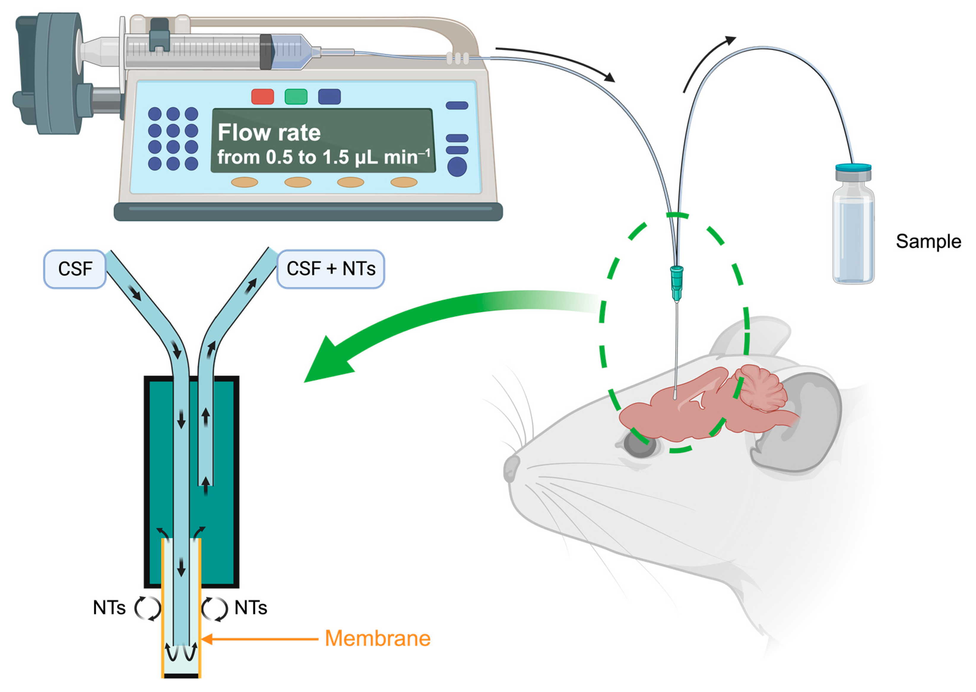
2. Microdialysis Experiment
3. Analytical Methods for Detection and Quantification of NTs and Metabolites
3.1. Gas Chromatography and Capillary Electrophoresis
3.2. HPLC
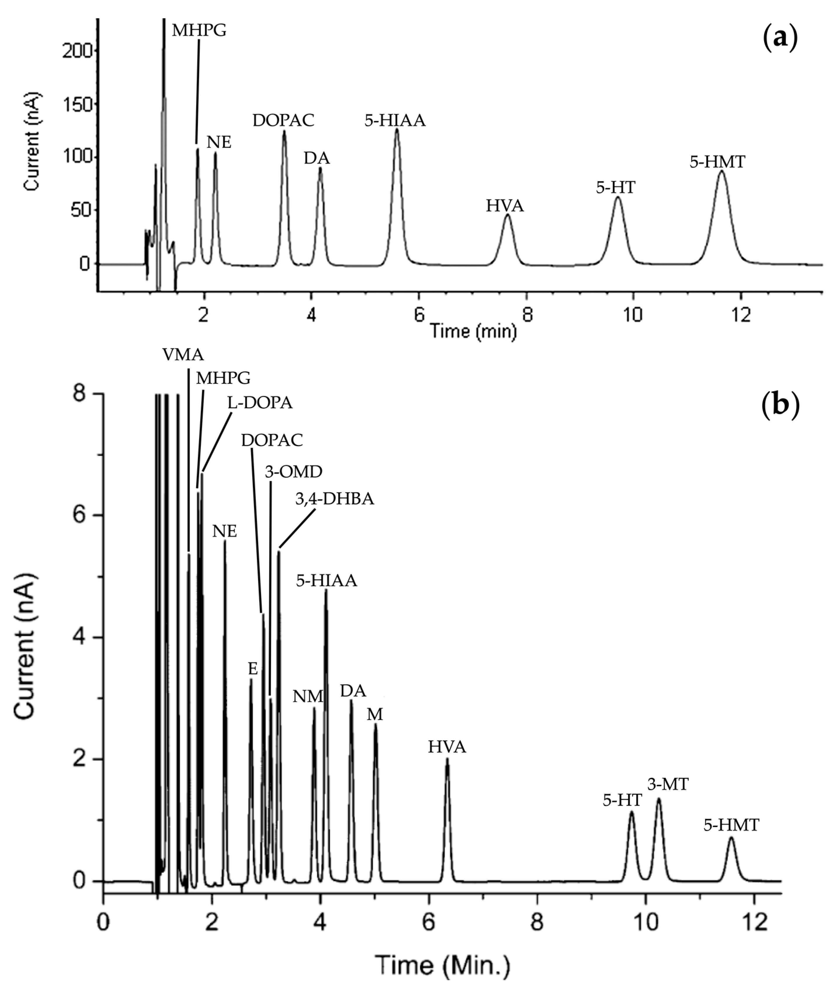
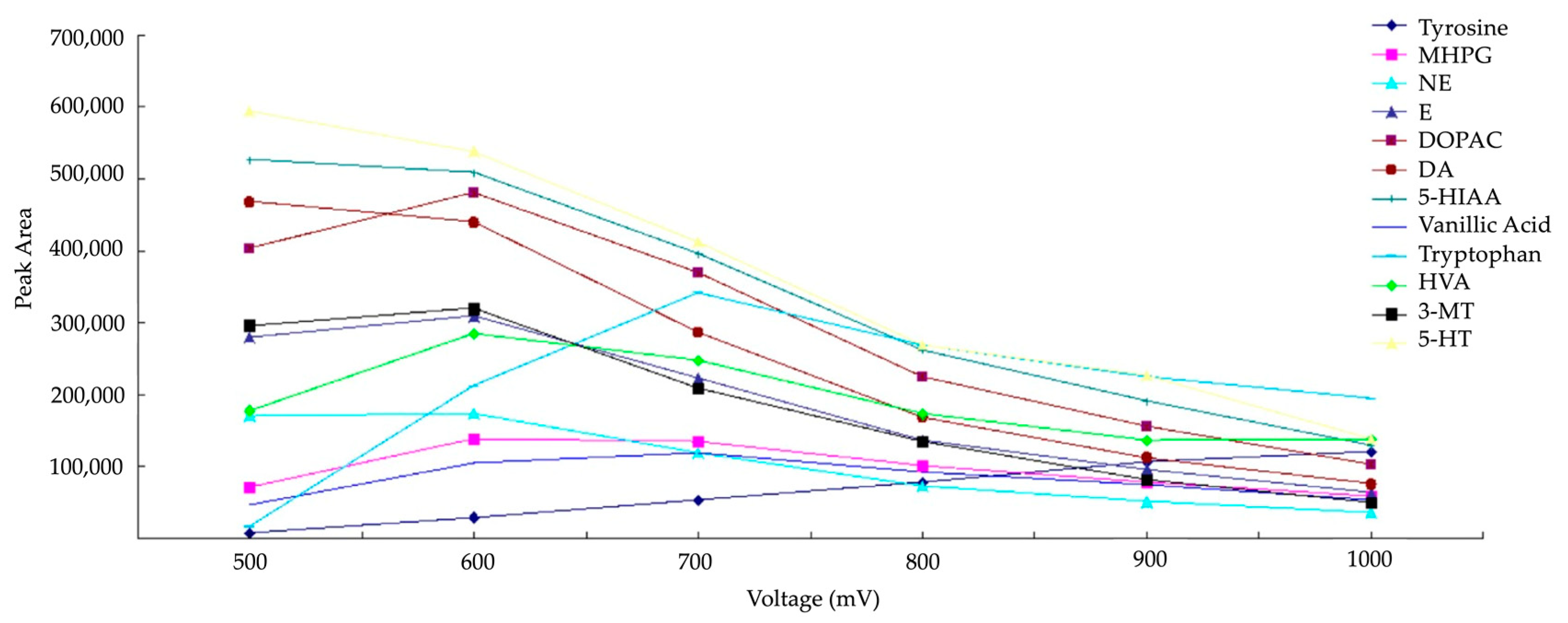
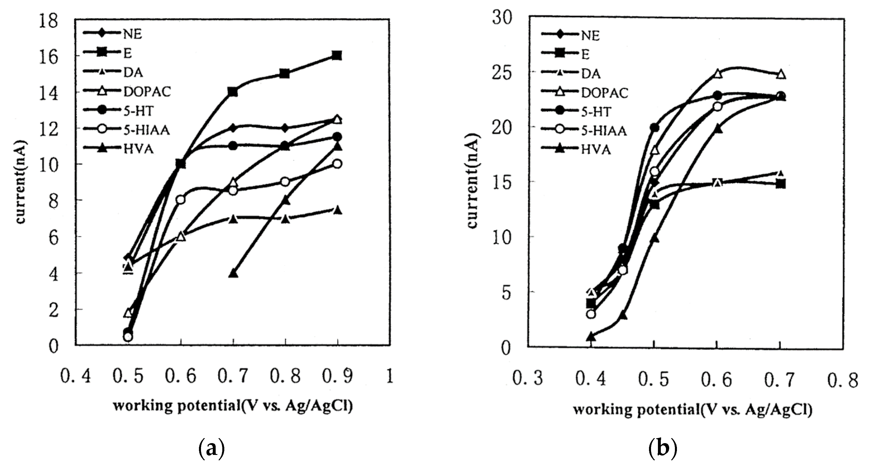
3.3. Detection of NTs and Their Metabolites
3.4. Limit of Detection and Quantification

| Reference | HPLC-ECD Conditions | Application | ||||||
|---|---|---|---|---|---|---|---|---|
| Reinhoud et al. (2013) [30] | Methods and Application | C18 column (1.0 × 100 mm, 1.7 µm particles size) Flow rate = 50 µL min−1 Column temperature = 37 °C Injection volume = 5 µL Mobile phase: 100 mM phosphoric acid, 100 mM citric acid, 8 mM KCl, 0.1 mM EDTA and 2.8 mM OSA. ACN/H2O (8/92; v/v) pH = 3 Detection: GC working electrode (+0.65 V vs. Ag/AgCl) | Development of analytical method for the determination of monoamines concentrations and application to a microdialysis in a prefrontal cortex. | |||||
| Detection (mol L−1) | 5-HT | 5-HIAA | DA | DOPAC | HVA | |||
| LOD | 8.3 × 10⁻¹¹ | 3.5 × 10⁻¹¹ | 4.2 × 10⁻¹¹ | 5.0 × 10⁻¹¹ | 4.7 × 10⁻¹¹ | |||
| Ferry et al. (2014) [63] | Methods and Application | C18 column (0.32 × 100 mm, 1.9 µm particles size) Flow rate = 8.5 µL min−1 Column temperature = 40 °C Injection volume = 1 µL Mobile phase: 140 mM potassium phosphate, 8 mM KCl, 0.1 mM EDTA, 6 mM OSA. MeOH/H2O (6/94; v/v) pH = 5 (adjusted with 10 mM NaOH) Detection: GC working electrode (+0.45 V vs. Ag/AgCl) | Development of analytical method for the determination of monoamines concentrations and application to microdialysis in the dorsal hippocampus | |||||
| Detection (mol L−1) | 5-HT | DA | 3-MT | |||||
| LOD | 1.5 × 10⁻⁹ | 7.5 × 10⁻¹⁰ | 1.5 × 10⁻⁹ | |||||
| LOQ | 5.0 × 10⁻⁹ | 2.5 × 10⁻⁹ | 5.0 × 10⁻⁹ | |||||
| Schou-Pedersen et al. (2016) [64] | Methods and Application | C18 column (4.6 × 100 mm, 2.6 µm particles size) Flow rate = 0.8 mL min−1 Column temperature = 30 °C Injection volume = 20 µLMobile phase: 70 mM potassium dihydrogen phosphate, 2 mM OSA, 0.1 mM EDTA. MeOH/H2O (10/90; v/v) pH = 3.12 (adjusted with 1 M citric acid) Detection: Porous graphite working electrode (+0.40 V vs. Pd) | Development of chromatographic method for the quantification of monoamine NTs from sub-regions of guinea pig brain (intracellular and extracellular). | |||||
| Detection (mol L−1) | 5-HT | 5-HIAA | DOPAC | DA | HVA | |||
| LOQ | 8.8 × 10⁻⁹ | 3.8 × 10⁻⁹ | 3.6 × 10⁻⁹ | 1.0 × 10⁻⁸ | 1.2 × 10⁻⁸ | |||
| L.R. | 8.8 × 10⁻⁹–1.0 × 10⁻⁶ | 3.8 × 10⁻⁹–1.0 × 10⁻⁶ | 3.6 × 10⁻⁹–1.0 × 10⁻⁶ | 1.0 × 10⁻⁸–7.5 × 10⁻⁷ | 1.2 × 10⁻⁸–5.0 × 10⁻⁷ | |||
| Van Dam et al. (2014) [65] | Methods and Application | C18 column (1.0 × 250 mm, 3.0 µm particles size) Flow rate = 40 µL min−1 Column temperature = from 30 °C to 36 °C Injection volume = 50 µL Mobile phase: 8 mM KCl, 50 mM phosphoric acid, 50 mM citric acid, 0.1 mM EDTA, from 1.8 to 2.2 mM OSA. MeOH/H2O (from 13/87 to 17/83; v/v) pH = from 3.0 to 3.6 (adjusted with NaOH) Detection: GC working electrode (from +0.63 V to +0.67 V vs. Ag/AgCl) | Development of analytical method for the quantification of biogenic amines and metabolites in human brain tissue. | |||||
| Detection (mol L−1) | 5-HT | 5-HIAA | DOPAC | DA | HVA | |||
| L.R. | 5.3 × 10⁻¹⁰–1.8 × 10⁻⁷ | 2.2 × 10⁻¹⁰–7.7 × 10⁻⁸ | 2.8 × 10⁻¹⁰–1.6 × 10⁻⁷ | 2.7 × 10⁻¹⁰–1.8 × 10⁻⁷ | 5.8 × 10⁻¹⁰–3.2 × 10⁻⁷ | |||
| Pantiya et al. (2024) [66] | Methods and Application | C18 column (3.0 × 500 mm, 2.6 µm particles size) Flow rate = 0.5 mL min−1 Column temperature = 25 °C Injection volume = 25 µLMobile phase: 130 mM sodium phosphate monobasic, 20 mM orthophosphoric acid, 2 mM sodium dodecyl sulfate, 50 µM EDTA. ACN, MeOH, H2O (5/10/95; v/v/v) pH = 3.2 (adjusted with phosphate buffer) Detection: GC working electrode (+0.50 V vs. Ag/AgCl) | Development of analytical method for the quantification of NTs and metabolites in brain mice microdialysates. | |||||
| Detection (mol L−1) | 5-HT | 5-HIAA | DA | HVA | ||||
| LOD | 3.1 × 10⁻¹⁰ | 5.4 × 10⁻¹⁰ | 3.7 × 10⁻¹⁰ | 4.1 × 10⁻¹⁰ | ||||
| LOQ | 3.9 × 10⁻¹⁰ | 5.8 × 10⁻¹⁰ | 4.2 × 10⁻¹⁰ | 4.3 × 10⁻¹⁰ | ||||
| Allen et al. (2017) [67] | Methods and Application | C18 column (3.2 × 150 mm, 3.0 µm particles size) Flow rate = 0.6 mL min−1 Column temperature = N/A Injection volume = 40 µLMobile phase: 100 mM sodium acetate, 20 mM citric acid, 0.38 mM sodium octyl sulfate, 0.15 mM EDTA. ACN/H2O (5/95; v/v) pH = 3.3 (adjusted with glacial acetic acid) Detection: Dual working electrode (−0.22 V and +0.375 V) | Development of analytical method for the quantification of monoamines and metabolites in brain tissue of mice. | |||||
| Detection (mol L−1) | 5-HT | 5-HIAA | DOPAC | DA | HVA | |||
| LOD | 1.7 × 10⁻⁹ | 6.5 × 10⁻¹⁰ | 7.4 × 10⁻¹⁰ | 8.2 × 10⁻¹⁰ | 4.1 × 10⁻¹⁰ | |||
| LOQ | 3.6 × 10⁻⁹ | 1.3 × 10⁻⁹ | 1.5 × 10⁻⁹ | 1.6 × 10⁻⁹ | 1.4 × 10⁻⁹ | |||
| L.R. | 3.6 × 10⁻⁹–1.7 × 10⁻⁴ | 1.3 × 10⁻⁹–7.9 × 10⁻⁵ | 1.5 × 10⁻⁹–8.9 × 10⁻⁵ | 1.6 × 10⁻⁹–2.0 × 10⁻⁴ | 1.4 × 10⁻⁹–8.2 × 10⁻⁵ | |||
| Yardimci et al. (2023) [68] | Methods and Application | C18 column (4.6 × 250 mm, 5.0 µm particles size) Flow rate = 1 mL min−1 Column temperature = 36 °C Injection volume = 20 µL Mobile phase: 35 mM citric acid, 19 mM sodium citrate, 0.16 mM EDTA, 1.1 mM heptasulfonic acid. Glacial acetic acid/tetrahydrofuran/MeOH/H2O (0.11/0.3/2.5/97.085; v/v/v/v) pH = 4.9 (adjusted with 10 M NaOH) Detection: GC working electrode (+0.50 V vs. Ag/AgCl) | Analysis in hypothalamic and subcortical nuclei | |||||
| Detection (mol L−1) | 5-HT | 5-HIAA | DA | DOPAC | ||||
| LOQ | 5.7 × 10⁻⁷ | 5.2 × 10⁻⁷ | 6.5 × 10⁻⁷ | 6.0 × 10⁻⁷ | ||||
| Du et al. (2018) [69] | Methods and Application | C18 column (4.6 × 250 mm, 5.0 µm particles size) Flow rate = 1 mL min−1 Column temperature = 25 °C Injection volume = 20 µL Mobile phase: 25 mM sodium citrate, 0.01 mM EDTA. ACN/H2O (5/95; v/v) pH = 4.5 (adjusted with 1 M acetic acid) Detection: BDD working electrode (+0.70 V vs. Ag/AgCl) | Analytical method development | |||||
| Detection (mol L−1) | 5-HT | 5-HIAA | ||||||
| LOD | 2.1 × 10⁻⁸ | 1.6 × 10⁻⁸ | ||||||
| LOQ | 2.8 × 10⁻⁸ | 4.2 × 10⁻⁸ | ||||||
| L.R. | 2.8 × 10⁻⁸–1.1 × 10⁻⁶ | 2.6 × 10⁻⁸–2.6 × 10⁻⁶ | ||||||
| Zhang et al. (2016) [53] | Methods and Application | C18 column (4.6 × 250 mm, 5.0 µm particles size) Flow rate = 1 mL min−1 Column temperature = 25 °C Injection volume = 20 µL Mobile phase: 25 mM sodium citrate, 0.01 mM EDTA. ACN/H2O (5/95; v/v) pH = 4.5 (adjusted with 1 M acetic acid) Detection: BDD working electrode (+0.70 V vs. Ag/AgCl) | Determination of the concentrations of monoamines NTs in rat cortex and hippocampus tissues. | |||||
| Detection (mol L−1) | 5-HT | 5-HIAA | DA | DOPAC | 3-MT | HVA | ||
| LOD | 2.3 × 10⁻⁸ | 1.1 × 10⁻⁸ | 2.6 × 10⁻⁸ | 1.2 × 10⁻⁸ | 3.6 × 10⁻⁸ | 2.7 × 10⁻⁸ | ||
| LOQ | 8.5 × 10⁻⁸ | 3.1 × 10⁻⁸ | 9.8 × 10⁻⁸ | 4.8 × 10⁻⁸ | 1.2 × 10⁻⁷ | 8.2 × 10⁻⁸ | ||
| L.R. | 8.5 × 10⁻⁸–1.4 × 10⁻⁶ | 3.1 × 10⁻⁸–7.9 × 10⁻⁷ | 9.8 × 10⁻⁸–2.3 × 10⁻⁶ | 6.0 × 10⁻⁸–3.0 × 10⁻⁶ | 1.2 × 10⁻⁷–1.8 × 10⁻⁶ | 8.2 × 10⁻⁸–1.4 × 10⁻⁶ | ||
| Jiang et al. (2015) [70] | Methods and Application | C18 column (4.6 × 250 mm, 5.0 µm particles size) Flow rate = 1 mL min−1 Column temperature = 30 °C Injection volume = 10 µL Mobile phase: 50 mM potassium dihydrogen phosphate, 0.1 mM octane sulfonic acid. MeOH/H2O (5/95; v/v) pH = N/A Detection: GC working electrode (+0.70 V vs. Ag/AgCl) | Development of the analytical method for monoamines NTs in human urine. | |||||
| Detection (mol L−1) | 5-HT | DA | ||||||
| LOD | 6.1 × 10⁻⁸ | 3.9 × 10⁻⁸ | ||||||
| Lokhande et al. (2022) [71] | Methods and Application | C18 column (4.6 × 250 mm, 5.0 µm particles size) Flow rate = 1.3 mL min−1 Column temperature = 35 °C Injection volume = 20 µL Mobile phase: 50 mM potassium dihydrogen phosphate, 0.99 mM SOS and 53 µM EDTA. MeOH/H2O (12/88; v/v) pH = 2.5 (adjusted with 85% phosphoric acid) Detection: BDD working electrode (+0.70 V vs. Ag/AgCl) | Development of the analytical method for metabolites quantification in human CSF. | |||||
| Detection (mol L−1) | 5-HIAA | HVA | ||||||
| LOQ | 6.5 × 10⁻⁸ | 6.9 × 10⁻⁸ | ||||||
| L.R. | 6.5 × 10⁻⁸–2.6 × 10⁻⁶ | 6.9 × 10⁻⁸–2.7 × 10⁻⁶ | ||||||
4. Conclusions
Author Contributions
Funding
Institutional Review Board Statement
Informed Consent Statement
Data Availability Statement
Conflicts of Interest
References
- Berger, M.; Gray, J.A.; Roth, B.L. The Expanded Biology of Serotonin. Annu. Rev. Med. 2009, 60, 355–366. [Google Scholar] [CrossRef]
- Factor, S.A.; McDonald, W.M.; Goldstein, F.C. The Role of Neurotransmitters in the Development of Parkinson’s Disease-related Psychosis. Eur. J. Neurol. 2017, 24, 1244–1254. [Google Scholar] [CrossRef] [PubMed]
- Tajeddinn, W.; Fereshtehnejad, S.; Ahmed, M.; Yoshitake, T.; Kehr, J.; Shahnaz, T.; Milovanovic, M.; Behbahani, H.; Hoglund, K.; Winblad, B.; et al. Association of Platelet Serotonin Levels in Alzheimer’s Disease with Clinical and Cerebrospinal Fluid Markers. J. Alzheimer Dis. 2016, 53, 621–630. [Google Scholar] [CrossRef]
- Xu, Y.; Yan, J.; Zhou, P.; Li, J.; Gao, H.; Xia, Y.; Wang, Q. Neurotransmitter Receptors and Cognitive Dysfunction in Alzheimer’s Disease and Parkinson’s Disease. Prog. Neurobiol. 2012, 97, 1–13. [Google Scholar] [CrossRef] [PubMed]
- Bahi, A.; Dreyer, J.-L. Anxiety and Ethanol Consumption in Socially Defeated Mice; Effect of Hippocampal Serotonin Transporter Knockdown. Behav. Brain Res. 2023, 451, 114508. [Google Scholar] [CrossRef]
- Kraus, C.; Castrén, E.; Kasper, S.; Lanzenberger, R. Serotonin and Neuroplasticity—Links between Molecular, Functional and Structural Pathophysiology in Depression. Neurosci. Biobehav. Rev. 2017, 77, 317–326. [Google Scholar] [CrossRef]
- Yagishita, S. Transient and Sustained Effects of Dopamine and Serotonin Signaling in Motivation-related Behavior. Psychiatry Clin. Neurosci. 2020, 74, 91–98. [Google Scholar] [CrossRef] [PubMed]
- Speranza, L.; Di Porzio, U.; Viggiano, D.; De Donato, A.; Volpicelli, F. Dopamine: The Neuromodulator of Long-Term Synaptic Plasticity, Reward and Movement Control. Cells 2021, 10, 735. [Google Scholar] [CrossRef]
- Weinstein, J.J.; Chohan, M.O.; Slifstein, M.; Kegeles, L.S.; Moore, H.; Abi-Dargham, A. Pathway-Specific Dopamine Abnormalities in Schizophrenia. Biol. Psychiatry 2017, 81, 31–42. [Google Scholar] [CrossRef]
- Di Forti, M.; Lappin, J.M.; Murray, R.M. Risk Factors for Schizophrenia—All Roads Lead to Dopamine. Eur. Neuropsychopharmacol. 2007, 17, S101–S107. [Google Scholar] [CrossRef]
- Latif, S.; Jahangeer, M.; Maknoon Razia, D.; Ashiq, M.; Ghaffar, A.; Akram, M.; El Allam, A.; Bouyahya, A.; Garipova, L.; Ali Shariati, M.; et al. Dopamine in Parkinson’s Disease. Clin. Chim. Acta 2021, 522, 114–126. [Google Scholar] [CrossRef] [PubMed]
- Wong, D.L. Epinephrine Biosynthesis: Hormonal and Neural Control During Stress. Cell. Mol. Neurobiol. 2006, 26, 889–898. [Google Scholar] [CrossRef]
- Harrison, N.A.; Morgan, R.; Critchley, H.D. From Facial Mimicry to Emotional Empathy: A Role for Norepinephrine? Soc. Neurosci. 2010, 5, 393–400. [Google Scholar] [CrossRef]
- Alves, E.; Lukoyanov, N.; Serrão, P.; Moura, D.; Moreira-Rodrigues, M. Epinephrine Increases Contextual Learning through Activation of Peripheral Β2-Adrenoceptors. Psychopharmacology 2016, 233, 2099–2108. [Google Scholar] [CrossRef] [PubMed]
- Ferry, B.; Roozendaal, B.; McGaugh, J.L. Role of Norepinephrine in Mediating Stress Hormone Regulation of Long-Term Memory Storage: A Critical Involvement of the Amygdala. Biol. Psychiatry 1999, 46, 1140–1152. [Google Scholar] [CrossRef] [PubMed]
- Tossman, U.; Ungerstedt, U. Microdialysis in the Study of Extracellular Levels of Amino Acids in the Rat Brain. Acta Physiol. Scand. 1986, 128, 9–14. [Google Scholar] [CrossRef]
- Ungerstedt, U.; Hallström, Å. In Vivo Microdialysis-a New Approach to the Analysis of Neurotransmitters in the Brain. Life Sci. 1987, 41, 861–864. [Google Scholar] [CrossRef] [PubMed]
- Zhou, S.Y.; Zuo, H.; Stobaugh, J.F.; Lunte, C.E.; Lunte, S.M. Continuous in Vivo Monitoring of Amino Acid Neurotransmitters by Microdialysis Sampling with Online Derivatization and Capillary Electrophoresis Separation. Anal. Chem. 1995, 67, 594–599. [Google Scholar] [CrossRef]
- Van Schoors, J.; Lens, C.; Maes, K.; Michotte, Y.; Smolders, I.; Van Eeckhaut, A. Reassessment of the Antioxidative Mixture for the Challenging Electrochemical Determination of Dopamine, Noradrenaline and Serotonin in Microdialysis Samples. J. Chromatogr. B 2015, 998–999, 63–71. [Google Scholar] [CrossRef]
- Guiard, B.; Lanfumey, L.; Gardier, A. Microdialysis Approach to Study Serotonin Outflow in Mice Following Selective Serotonin Reuptake Inhibitors and Substance P (Neurokinin 1) Receptor Antagonist Administration: A Review. Curr. Drug Targets 2006, 7, 187–201. [Google Scholar] [CrossRef]
- Zhang, P.; Wang, B.; Sun, Y.; Gao, J.; Lian, K. Analysis of 5-Hydroxytryptamine and Its Related Indoles in Cerebrospinal Fluid of Leukemic Children by Gas Chromatography-Mass Spectrometry. J. Lab. Med. 2020, 44, 41–45. [Google Scholar] [CrossRef]
- Shi, H.; Wang, B.; Niu, L.; Cao, M.; Kang, W.; Lian, K.; Zhang, P. Trace Level Determination of 5-Hydroxytryptamine and Its Related Indoles in Amniotic Fluid by Gas Chromatography–Mass Spectrometry. J. Pharm. Biomed. Anal. 2017, 143, 176–182. [Google Scholar] [CrossRef] [PubMed]
- Tsai, H.; Whang, C. Capillary Electrophoresis of Monoamines and Catechol with Indirect Chemiluminescence Detection. Electrophoresis 1999, 20, 2533–2538. [Google Scholar] [CrossRef]
- Benturquia, N.; Couderc, F.; Sauvinet, V.; Orset, C.; Parrot, S.; Bayle, C.; Renaud, B.; Denoroy, L. Analysis of Serotonin in Brain Microdialysates Using Capillary Electrophoresis and Native Laser-Induced Fluorescence Detection. Electrophoresis 2005, 26, 1071–1079. [Google Scholar] [CrossRef] [PubMed]
- Hattox, S.E.; Murphy, R.C. Mass Spectrometry and Gas Chromatography of Trimethylsilyl Derivatives of Catecholamine Related Molecules. Biol. Mass Spectrom. 1978, 5, 338–345. [Google Scholar] [CrossRef] [PubMed]
- Donzanti, B.A.; Yamamoto, B.K. An Improved and Rapid HPLC-EC Method for the Isocratic Separation of Amino Acid Neurotransmitters from Brain Tissue and Microdialysis Perfusates. Life Sci. 1988, 43, 913–922. [Google Scholar] [CrossRef]
- Peinado, J.M.; McManus, K.T.; Myers, R.D. Rapid Method for Micro-Analysis of Endogenous Amino Acid Neurotransmitters in Brain Perfusates in the Rat by Isocratic HPLC-EC. J. Neurosci. Methods 1986, 18, 269–276. [Google Scholar] [CrossRef]
- Rogers, K.L.; Philibert, R.A.; Allen, A.J.; Molitor, J.; Wilson, E.J.; Dutton, G.R. HPLC Analysis of Putative Amino Acid Neurotransmitters Released from Primary Cerebellar Cultures. J. Neurosci. Methods 1987, 22, 173–179. [Google Scholar] [CrossRef]
- Bidel, F.; Corvaisier, S.; Jozet-Alves, C.; Pottier, I.; Dauphin, F.; Naud, N.; Bellanger, C. An HPLC-ECD Method for Monoamines and Metabolites Quantification in Cuttlefish (Cephalopod) Brain Tissue: Biogenic Monoamines and Metabolites in Cuttlefish (Cephalopod) Brain. Biomed. Chromatogr. 2016, 30, 1175–1183. [Google Scholar] [CrossRef]
- Reinhoud, N.J.; Brouwer, H.-J.; Van Heerwaarden, L.M.; Korte-Bouws, G.A.H. Analysis of Glutamate, GABA, Noradrenaline, Dopamine, Serotonin, and Metabolites Using Microbore UHPLC with Electrochemical Detection. ACS Chem. Neurosci. 2013, 4, 888–894. [Google Scholar] [CrossRef]
- Tufi, S.; Lamoree, M.; De Boer, J.; Leonards, P. Simultaneous Analysis of Multiple Neurotransmitters by Hydrophilic Interaction Liquid Chromatography Coupled to Tandem Mass Spectrometry. J. Chromatogr. A 2015, 1395, 79–87. [Google Scholar] [CrossRef] [PubMed]
- Chirita, R.-I.; West, C.; Finaru, A.-L.; Elfakir, C. Approach to Hydrophilic Interaction Chromatography Column Selection: Application to Neurotransmitters Analysis. J. Chromatogr. A 2010, 1217, 3091–3104. [Google Scholar] [CrossRef]
- Zhou, G.-S.; Yuan, Y.-C.; Yin, Y.; Tang, Y.-P.; Xu, R.-J.; Liu, Y.; Chen, P.-D.; Yin, L.; Duan, J.-A. Hydrophilic Interaction Chromatography Combined with Ultrasound-Assisted Ionic Liquid Dispersive Liquid–Liquid Microextraction for Determination of Underivatized Neurotransmitters in Dementia Patients’ Urine Samples. Anal. Chim. Acta 2020, 1107, 74–84. [Google Scholar] [CrossRef]
- Boulghobra, A.; Bonose, M.; Billault, I.; Pallandre, A. A Rapid and Sensitive Method for the Quantification of Dopamine and Serotonin Metabolites in Cerebrospinal Fluid Based on UHPLC with Fluorescence Detection. J. Chromatogr. B 2022, 1200, 123264. [Google Scholar] [CrossRef] [PubMed]
- De Benedetto, G.E.; Fico, D.; Pennetta, A.; Malitesta, C.; Nicolardi, G.; Lofrumento, D.D.; De Nuccio, F.; La Pesa, V. A Rapid and Simple Method for the Determination of 3,4-Dihydroxyphenylacetic Acid, Norepinephrine, Dopamine, and Serotonin in Mouse Brain Homogenate by HPLC with Fluorimetric Detection. J. Pharm. Biomed. Anal. 2014, 98, 266–270. [Google Scholar] [CrossRef]
- Zhao, H.-X.; Mu, H.; Bai, Y.-H.; Yu, H.; Hu, Y.-M. A Rapid Method for the Determination of Dopamine in Porcine Muscle by Pre-Column Derivatization and HPLC with Fluorescence Detection. J. Pharm. Anal. 2011, 1, 208–212. [Google Scholar] [CrossRef] [PubMed]
- Carrera, V.; Sabater, E.; Vilanova, E.; Sogorb, M.A. A Simple and Rapid HPLC–MS Method for the Simultaneous Determination of Epinephrine, Norepinephrine, Dopamine and 5-Hydroxytryptamine: Application to the Secretion of Bovine Chromaffin Cell Cultures. J. Chromatogr. B 2007, 847, 88–94. [Google Scholar] [CrossRef]
- Xu, N.; Qiu, C.; Wang, W.; Wang, Y.; Chai, C.; Yan, Y.; Zhu, D. HPLC/MS/MS for Quantification of Two Types of Neurotransmitters in Rat Brain and Application: Myocardial Ischemia and Protection of Sheng-Mai-San. J. Pharm. Biomed. Anal. 2011, 55, 101–108. [Google Scholar] [CrossRef]
- Kovac, A.; Somikova, Z.; Zilka, N.; Novak, M. Liquid Chromatography–Tandem Mass Spectrometry Method for Determination of Panel of Neurotransmitters in Cerebrospinal Fluid from the Rat Model for Tauopathy. Talanta 2014, 119, 284–290. [Google Scholar] [CrossRef]
- Shao, Z.; Chang, Y.; Venton, B.J. Carbon Microelectrodes with Customized Shapes for Neurotransmitter Detection: A Review. Anal. Chim. Acta 2022, 1223, 340165. [Google Scholar] [CrossRef]
- Polo, E.; Kruss, S. Nanosensors for Neurotransmitters. Anal. Bioanal. Chem. 2016, 408, 2727–2741. [Google Scholar] [CrossRef] [PubMed]
- Banerjee, S.; McCracken, S.; Hossain, M.F.; Slaughter, G. Electrochemical Detection of Neurotransmitters. Biosensors 2020, 10, 101. [Google Scholar] [CrossRef]
- Dicgory, G.L.; Buckett, W.R. An Automated Method to Measure Monoamines and Metabolites Using Elevated Temperature Reversed Phase HPLC with Electrochemical Detection Application to Striatal Dopamine and Hippocampal Serotonin Turnover. J. Pharmacol. Methods 1984, 11, 207–217. [Google Scholar] [CrossRef] [PubMed]
- Cannazza, G.; Di Stefano, A.; Mosciatti, B.; Braghiroli, D.; Baraldi, M.; Pinnen, F.; Sozio, P.; Benatti, C.; Parenti, C. Detection of Levodopa, Dopamine and Its Metabolites in Rat Striatum Dialysates Following Peripheral Administration of l-DOPA Prodrugs by Mean of HPLC–EC. J. Pharm. Biomed. Anal. 2005, 36, 1079–1084. [Google Scholar] [CrossRef] [PubMed]
- Birch, P.J.; Fillenz, M. Measurement of Noradrenaline Synthesis in Rat Hippocampal Synaptosomes Using HPLC with ECD. J. Neurosci. Methods 1985, 13, 231–238. [Google Scholar] [CrossRef]
- Saito, H.; Murai, S.; Abe, E.; Masuda, Y.; Itoh, T. Rapid and Simultaneous Assay of Monoamine Neurotransmitters and Their Metabolites in Discrete Brain Areas of Mice by HPLC with Coulometric Detection. Pharmacol. Biochem. Behav. 1992, 42, 351–356. [Google Scholar] [CrossRef]
- Bard, A.J.; Faulkner, L.R. Electrochemical Methods, Fundamentals and Applications; John Wiley & Sons Inc.: New York, NY, USA, 2001; ISBN 0-471-04372-9. [Google Scholar]
- Sharma, S.; Singh, N.; Tomar, V.; Chandra, R. A Review on Electrochemical Detection of Serotonin Based on Surface Modified Electrodes. Biosens. Bioelectron. 2018, 107, 76–93. [Google Scholar] [CrossRef]
- Błaszczyk, J.W. Parkinson’s Disease and Neurodegeneration: GABA-Collapse Hypothesis. Front. Neurosci. 2016, 10, 239. [Google Scholar] [CrossRef]
- Nägga, K.; Bogdanovic, N.; Marcusson, J. GABA Transporters (GAT-1) in Alzheimer’s Disease. J. Neural Transm. 1999, 106, 1141–1149. [Google Scholar] [CrossRef]
- Panrod, K.; Tansirikongkol, A.; Panapisal, V. Comparison of Validated High-Performance Liquid Chromatography Methods Using Two Derivatizing Agents for Gamma-Aminobutyric Acid Quantification. Thai J. Pharm. Sci. 2016, 40, 203–208. [Google Scholar]
- Narmadha, M.; Noel, M.; Suryanarayanan, V. Relative Deactivation of Boron-Doped Diamond (BDD) and Glassy Carbon (GC) Electrodes in Different Electrolyte Media Containing Substituted Phenols—Voltammetric and Surface Morphologic Studies. J. Electroanal. Chem. 2011, 655, 103–110. [Google Scholar] [CrossRef]
- Zhang, L.; Yang, J.; Luo, Y.; Shang, J.; Jiang, X. Simultaneous Determination of Eleven Compounds Related to Metabolism of Bioamines in Rat Cortex and Hippocampus by HPLC-ECD with Boron-Doped Diamond Working Electrode. J. Pharm. Biomed. Anal. 2016, 118, 41–51. [Google Scholar] [CrossRef] [PubMed]
- Gotti, G.; Evrard, D.; Gros, P. Simultaneous Electrochemical Detection of Oxygen (O2) and Hydrogen Peroxide (H2O2) in Neutral Media. Int. J. Electrochem. Sci. 2023, 18, 100262. [Google Scholar] [CrossRef]
- Gotti, G.; Fajerwerg, K.; Evrard, D.; Gros, P. Kinetics of Dioxygen Reduction on Gold and Glassy Carbon Electrodes in Neutral Media. Int. J. Electrochem. Sci. 2013, 8, 12643–12657. [Google Scholar] [CrossRef]
- Xu, F.; Gao, M.; Shi, G.; Wang, L.; Zhang, W.; Xue, J.; Jin, L.; Jin, J. Simultaneous Detection of Monoamines in Rat Striatal Microdialysate at Poly(Para-Aminobenzoic Acid) Modified Electrode by High-Performance Liquid Chromatography. Anal. Chim. Acta 2001, 439, 239–246. [Google Scholar] [CrossRef]
- Guo, Y.-X.; Xia, C.-Y.; Yan, Y.; Han, Y.; Shi, R.; He, J.; Wang, Y.-M.; Wang, Z.-X.; Zhang, W.-K.; Xu, J.-K. Loganin Improves Chronic Unpredictable Mild Stress-Induced Depressive-like Behaviors and Neurochemical Dysfunction. J. Ethnopharmacol. 2023, 308, 116288. [Google Scholar] [CrossRef] [PubMed]
- Lei, C.; Chen, H.; Chen, K. Effects of Bone Marrow Mesenchymal Stem Cells on Myelin Repair and Emotional Changes of a Cuprizone-Induced Demyelination Model. J. Integr. Neurosci. 2023, 22, 40. [Google Scholar] [CrossRef]
- Wang, Z.-X.; Lian, W.-W.; He, J.; He, X.-L.; Wang, Y.-M.; Pan, C.-H.; Li, M.; Zhang, W.-K.; Liu, L.-Q.; Xu, J.-K. Cornuside Ameliorates Cognitive Impairments in Scopolamine Induced AD Mice: Involvement of Neurotransmitter and Oxidative Stress. J. Ethnopharmacol. 2022, 293, 115252. [Google Scholar] [CrossRef]
- Kalantary, R.R.; Moradi, M.; Pirsaheb, M.; Esrafili, A.; Jafari, A.J.; Gholami, M.; Vasseghian, Y.; Antolini, E.; Dragoi, E.-N. Enhanced Photocatalytic Inactivation of E. Coli by Natural Pyrite in Presence of Citrate and EDTA as Effective Chelating Agents: Experimental Evaluation and Kinetic and ANN Models. J. Environ. Chem. Eng. 2019, 7, 102906. [Google Scholar] [CrossRef]
- Broch, S.C.; Celia García Alvarez-Coque, M.; Broch, S.C.; Esteve-Romero, J.S. Liquid Chromatographic Determination of Some Thiazide Diuretics in Pharmaceuticals with a Sodium Dodecyl Sulfate Mobile Phase. Analyst 1998, 123, 301–306. [Google Scholar] [CrossRef]
- Umek, N.; Geršak, B.; Vintar, N.; Šoštarič, M.; Mavri, J. Dopamine Autoxidation Is Controlled by Acidic pH. Front. Mol. Neurosci. 2018, 11, 467. [Google Scholar] [CrossRef] [PubMed]
- Ferry, B.; Gifu, E.-P.; Sandu, I.; Denoroy, L.; Parrot, S. Analysis of Microdialysate Monoamines, Including Noradrenaline, Dopamine and Serotonin, Using Capillary Ultra-High Performance Liquid Chromatography and Electrochemical Detection. J. Chromatogr. B 2014, 951–952, 52–57. [Google Scholar] [CrossRef] [PubMed]
- Schou-Pedersen, A.M.V.; Hansen, S.N.; Tveden-Nyborg, P.; Lykkesfeldt, J. Simultaneous Quantification of Monoamine Neurotransmitters and Their Biogenic Metabolites Intracellularly and Extracellularly in Primary Neuronal Cell Cultures and in Sub-Regions of Guinea Pig Brain. J. Chromatogr. B 2016, 1028, 222–230. [Google Scholar] [CrossRef] [PubMed]
- Van Dam, D.; Vermeiren, Y.; Aerts, T.; De Deyn, P.P. Novel and Sensitive Reversed-Phase High-Pressure Liquid Chromatography Method with Electrochemical Detection for the Simultaneous and Fast Determination of Eight Biogenic Amines and Metabolites in Human Brain Tissue. J. Chromatogr. A 2014, 1353, 28–39. [Google Scholar] [CrossRef] [PubMed]
- Pantiya, P.; Guiard, B.P.; Gotti, G. Sensitive and Fast Detection of Monoamines and Their Metabolites by High-Performance Liquid Chromatography Coupled with an Electrochemical Detector (HPLC-ECD) under Isocratic Conditions. Application to Intracerebral Microdialysis in Mice Treated by Fluoxetine and Atomoxetine. Chromatographia 2024, in press. [Google Scholar]
- Allen, S.A.; Rednour, S.; Shepard, S.; Pond, B.B. A Simple and Sensitive High-performance Liquid Chromatography–Electrochemical Detection Assay for the Quantitative Determination of Monoamines and Respective Metabolites in Six Discrete Brain Regions of Mice. Biomed. Chromatogr. 2017, 31, e3998. [Google Scholar] [CrossRef]
- Yardimci, A.; Ertugrul, N.U.; Ozgen, A.; Ozbeg, G.; Ozdede, M.R.; Ercan, E.C.; Canpolat, S. Effects of Chronic Irisin Treatment on Brain Monoamine Levels in the Hypothalamic and Subcortical Nuclei of Adult Male and Female Rats: An HPLC-ECD Study. Neurosci. Lett. 2023, 806, 137245. [Google Scholar] [CrossRef]
- Du, T.; Cui, T.; Qiu, H.; Wang, N.; Huang, D.; Jiang, X. Simultaneous Determination of Tryptophan, Kynurenine, Kynurenic Acid and Two Monoamines in Rat Plasma by HPLC-ECD/DAD. J. Pharm. Biomed. Anal. 2018, 158, 8–14. [Google Scholar] [CrossRef]
- Jiang, L.; Chen, Y.; Chen, Y.; Ma, M.; Tan, Y.; Tang, H.; Chen, B. Determination of Monoamine Neurotransmitters in Human Urine by Carrier-Mediated Liquid-Phase Microextraction Based on Solidification of Stripping Phase. Talanta 2015, 144, 356–362. [Google Scholar] [CrossRef]
- Lokhande, R.V.; Bhagure, G.R.; Dherai, A.J.; Naik, P.R.; Udani, V.P.; Desai, N.A.; Ashavaid, T.F. Analytical Method Validation for Estimation of Neurotransmitters (Biogenic Monoamines) from Cerebrospinal Fluid Using High Performance Liquid Chromatography. Ind. J. Clin. Biochem. 2022, 37, 85–92. [Google Scholar] [CrossRef]


| Electrode Geometry | Limiting Current Equation |
|---|---|
| Tubular | |
| Planar, parallel flow in channel | |
| Planar, perpendicular flow | |
| Wall jet |
Disclaimer/Publisher’s Note: The statements, opinions and data contained in all publications are solely those of the individual author(s) and contributor(s) and not of MDPI and/or the editor(s). MDPI and/or the editor(s) disclaim responsibility for any injury to people or property resulting from any ideas, methods, instructions or products referred to in the content. |
© 2024 by the authors. Licensee MDPI, Basel, Switzerland. This article is an open access article distributed under the terms and conditions of the Creative Commons Attribution (CC BY) license (https://creativecommons.org/licenses/by/4.0/).
Share and Cite
Guiard, B.P.; Gotti, G. The High-Precision Liquid Chromatography with Electrochemical Detection (HPLC-ECD) for Monoamines Neurotransmitters and Their Metabolites: A Review. Molecules 2024, 29, 496. https://doi.org/10.3390/molecules29020496
Guiard BP, Gotti G. The High-Precision Liquid Chromatography with Electrochemical Detection (HPLC-ECD) for Monoamines Neurotransmitters and Their Metabolites: A Review. Molecules. 2024; 29(2):496. https://doi.org/10.3390/molecules29020496
Chicago/Turabian StyleGuiard, Bruno P., and Guillaume Gotti. 2024. "The High-Precision Liquid Chromatography with Electrochemical Detection (HPLC-ECD) for Monoamines Neurotransmitters and Their Metabolites: A Review" Molecules 29, no. 2: 496. https://doi.org/10.3390/molecules29020496