
Cd(II) and Pd(II) Mixed Ligand Complexes of Dithiocarbamate and Tertiary Phosphine Ligands—Spectroscopic, Anti-Microbial, and Computational Studies
 , , ,
, , , 
Abstract
:1. Introduction
2. Results
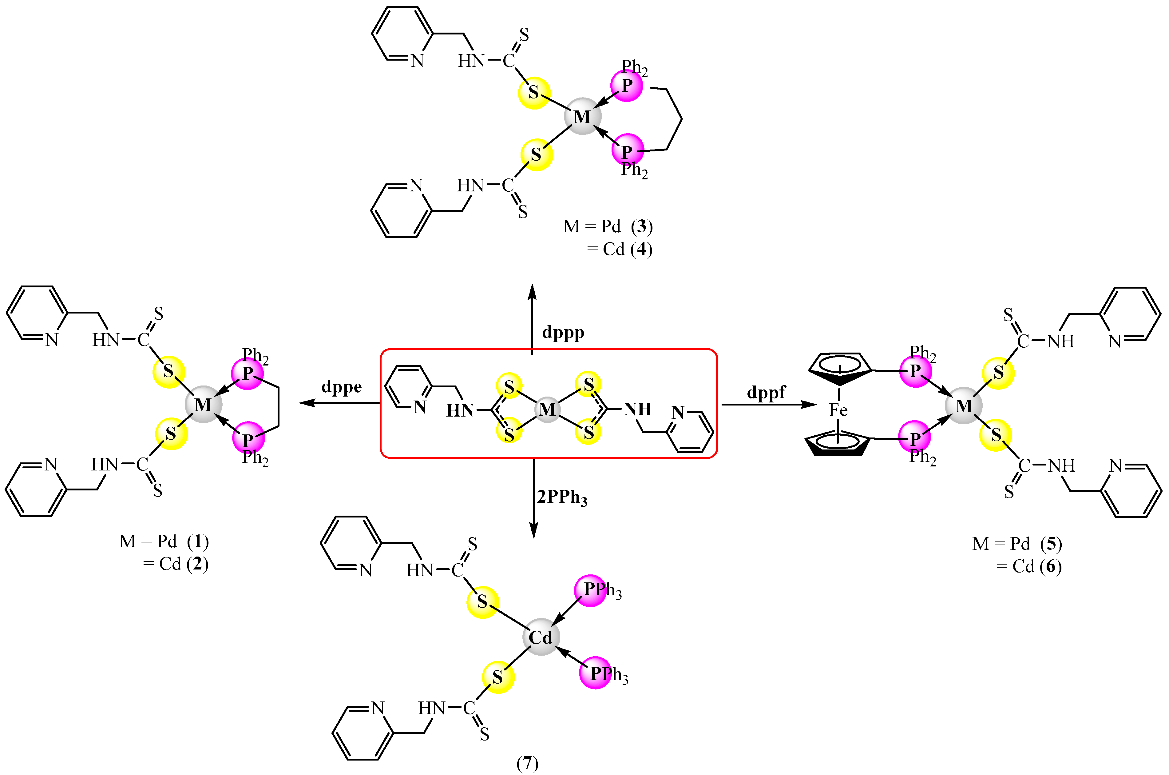
2.1. Synthesis
2.2. Characterization of Prepared Complexes
2.2.1. 31P-{1H} and 1H nmr Spectra
2.2.2. IR Data
2.3. Antimicrobial Studies
2.4. Computational Studies
2.4.1. Geometrical and Electronic Properties
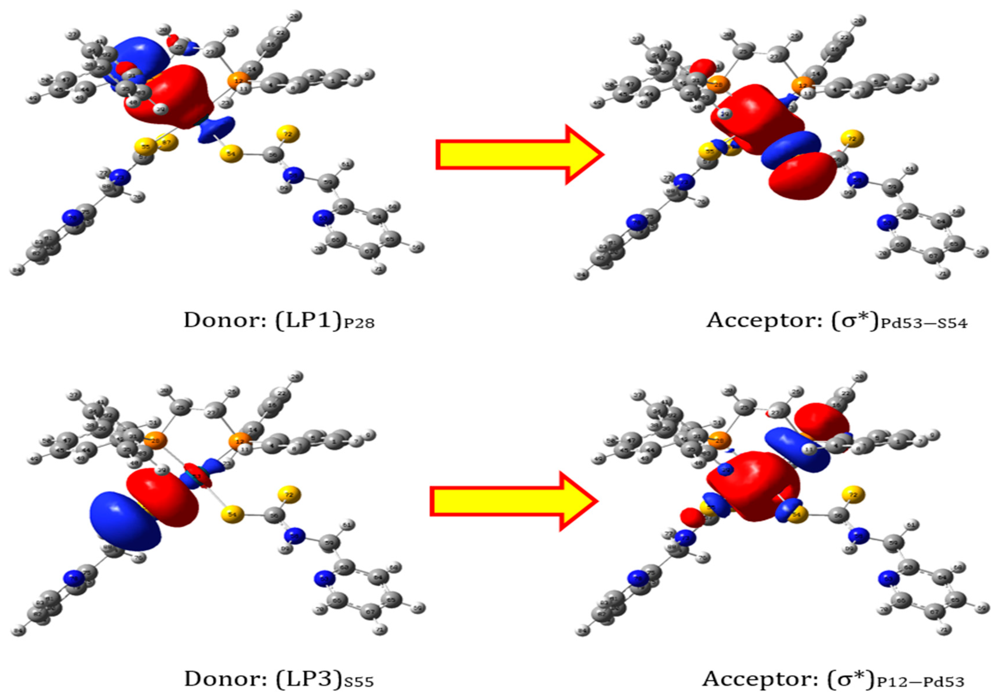
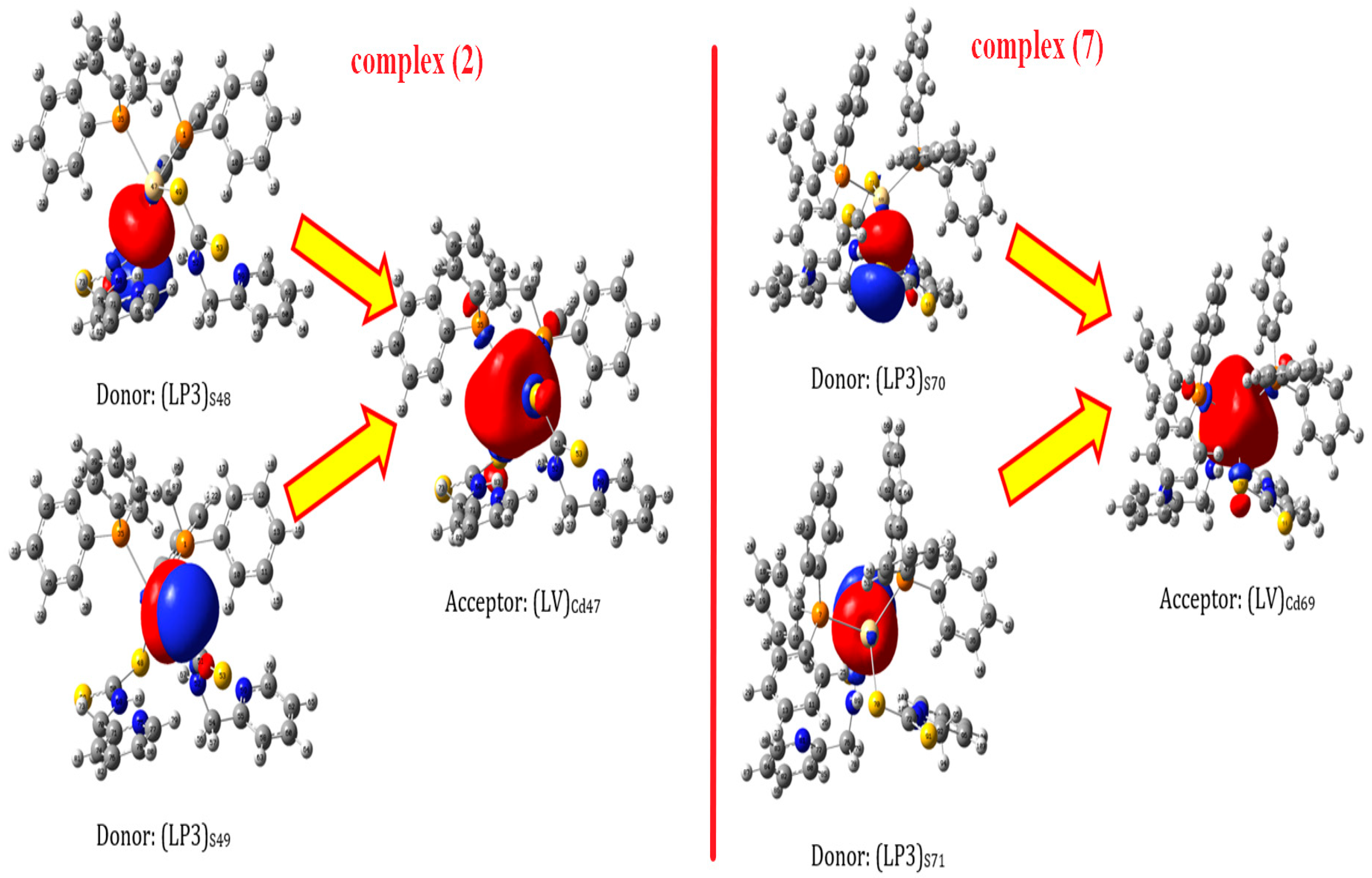
2.4.2. NBO Analysis
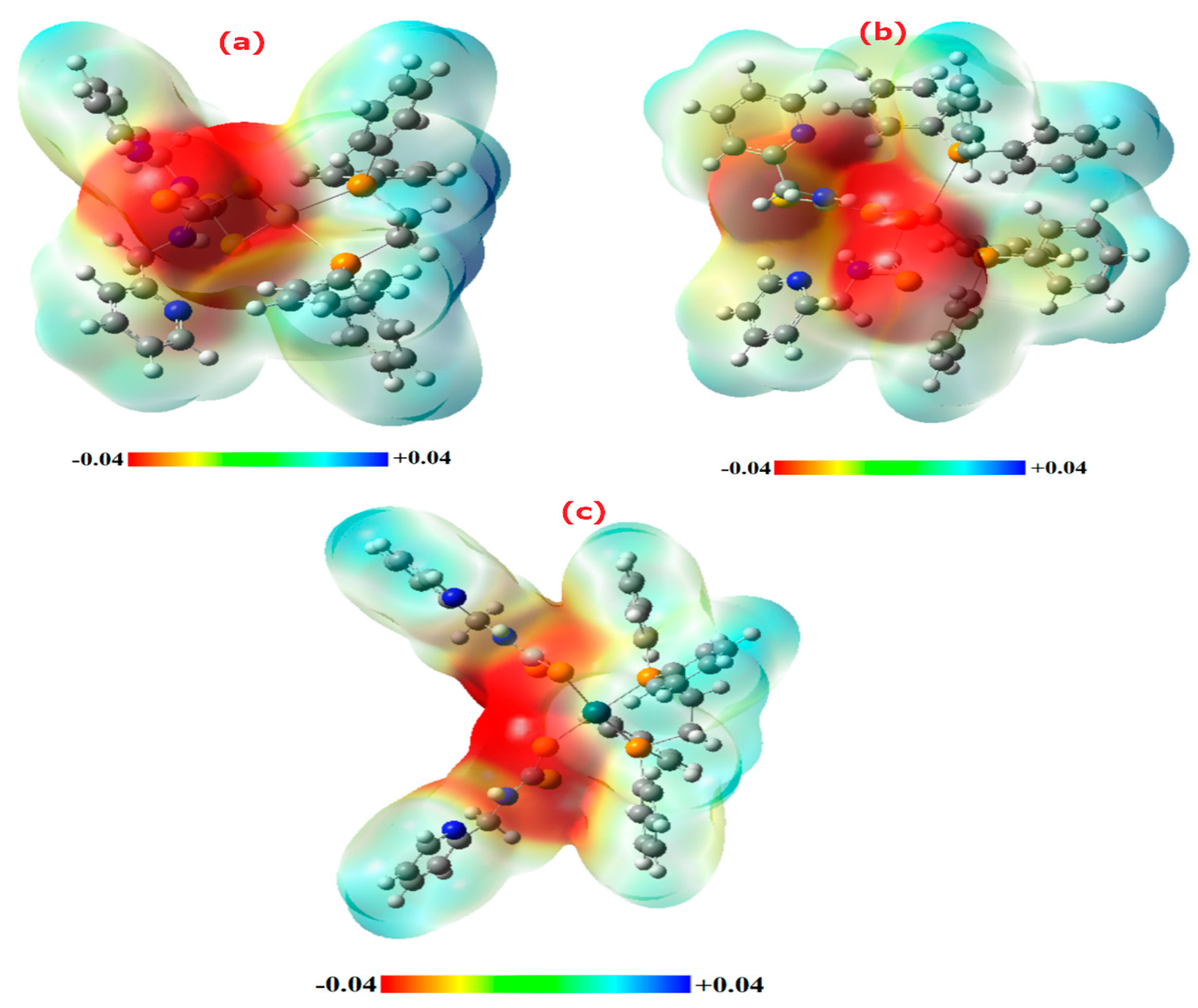
2.4.3. MEP Analysis
3. Experimental Part
3.1. Material and Physical Measurements
3.2. Synthesis of Complexes
3.2.1. Synthesis of [Pd(PCA-dtc)2(dppe)]Complex (1)
3.2.2. Synthesis of [Cd(PCA-dtc)2(PPh3)2]complex (7)
3.3. Antibacterial Studies
3.4. Computational Detail
4. Conclusions
Supplementary Materials
Author Contributions
Funding
Institutional Review Board Statement
Informed Consent Statement
Data Availability Statement
Acknowledgments
Conflicts of Interest
References
- Hogarth, G. Transition metal dithiocarbamates: 1978–2003. Prog. Inorg. Chem. 2005, 53, 71–561. [Google Scholar]
- Hogarth, G. Metal-dithiocarbamate complexes: Chemistry and biological activity. Mini-Rev. Med. Chem. 2012, 12, 1202–1215. [Google Scholar] [CrossRef] [PubMed]
- Faihan, A.S.; Hatshan, M.R.; Alqahtani, A.S.; Nasr, F.A.; Al-Jibori, S.A.; Al-Janabi, A.S. New divalent metal ion complexes with 1,8-diaminonapthalene-2-thione: Synthesis, Spectroscopic, anti-bacterial and anticancer activity studies. J. Mol. Struct. 2022, 1247, 131291. [Google Scholar] [CrossRef]
- Faihan, A.S.; Hatshan, M.R.; Kadhim, M.M.; Alqahtani, A.S.; Nasr, F.A.; Saleh, A.M.; Al-Jibori, S.A.; Al-Janabi, A.S. Promising bio-active complexes of platinum (II) and palladium (II) derived from heterocyclic thiourea: Synthesis, characterization, DFT, molecular docking, and anti-cancer studies. J. Mol. Struct. 2022, 1252, 132198. [Google Scholar] [CrossRef]
- Al-Mouqdady, O.D.; Al-Janabi, A.S.; Hatshan, M.R.; Al-Jibori, S.A.; Fiahan, A.S.; Wagner, C. Synthesis, characterization, anti-bacterial and anticancer activities of Palladium (II) mixed ligand complexes of 2-mercapto-5-methyl-1, 3, 4-thiadiazole (HmtzS) and phosphines. Crystal structure of [Pd (mtzS) 2 (dppf)]. H2O. EtOH. J. Mol. Struct. 2022, 1264, 133219. [Google Scholar] [CrossRef]
- Faihan, A.S.; Al-Jibori, S.A.; Al-Janabi, A.S. Novel base-free dianion complexes of Pt(II) and Pd(II) derived from heterocyclic thiourea and tertiary phosphine ligands. J. Mol. Struct. 2022, 1251, 131966. [Google Scholar] [CrossRef]
- Halimehjani, A.Z.; Marjani, K.; Ashouri, A.; Amani, V. Synthesis and characterization of transition metal dithiocarbamate derivatives of 1-aminoadamantane: Crystal structure of (N-adamantyldithiocarbamato)nickel(II). Inorganica Chim. Acta 2011, 373, 282–285. [Google Scholar] [CrossRef]
- Kitagawa, S.; Munakata, M.; Shimono, H.; Matsuyama, S.; Masuda, H. Synthesis and crystal structure of hexanuclear copper (I) complexes of µ3-pyridine-2-thionate. J. Chem. Soc. Dalton Trans. 1990, 7, 2105–2109. [Google Scholar] [CrossRef]
- Onwudiwe, D.C.; Arfin, T.; Strydom, C.A.; Kriek, R.J. Synthesis, spectroscopic characterization and behavior of AC impedance spectroscopy of Cd(II) bis(N-para-methylphenyl dithiocarbamate). Electrochimica Acta 2013, 104, 19–25. [Google Scholar] [CrossRef]
- Memon, A.A.; Afzaal, M.; Malik, M.A.; Nguyen, C.Q.; O’Brien, P.; Raftery, J. The N-alkyldithiocarbamato complexes [M (S2CNHR)2](M = Cd (ii) Zn (ii); R = C2H5, C4H9, C6H13, C12H25); their synthesis, thermal decomposition and use to prepare of nanoparticles and nanorods of CdS. Dalton Trans. 2006, 37, 4499–4505. [Google Scholar] [CrossRef]
- Kumar, V.; Singh, V.; Gupta, A.N.; Manar, K.K.; Drew, M.G.B.; Singh, N. Influence of ligand environments on the structures and luminescence properties of homoleptic cadmium(ii) pyridyl functionalized dithiocarbamates. Crystengcomm 2014, 16, 6765–6774. [Google Scholar] [CrossRef]
- Trindade, T.; O’Brien, P. Synthesis of CdS and CdSe nanoparticles by thermolysis of diethyldithio-or diethyldiseleno-carbamates of cadmium. J. Mater. Chem. 1996, 6, 343–347. [Google Scholar] [CrossRef] [Green Version]
- Tan, Y.S.; Sudlow, A.L.; Molloy, K.C.; Morishima, Y.; Fujisawa, K.; Jackson, W.J.; Henderson, W.; Halim, S.N.B.A.; Ng, S.W.; Tiekink, E.R. Supramolecular isomerism in a cadmium bis (N-hydroxyethyl, N-isopropyldithiocarbamate) compound: Physiochemical characterization of ball (n = 2) and chain (n = ∞) forms of {Cd [S2CN (iPr) CH2CH2OH]2·solvent}n. Cryst. Growth Des. 2013, 13, 3046–3056. [Google Scholar] [CrossRef]
- Thirumaran, S.; Ramalingam, K.; Bocelli, G.; Righi, L. XPS, single crystal X-ray diffraction and cyclic voltammetric studies on 1,10-phenanthroline and 2,2′-bipyridine adducts of bis (piperidinecarbodithioato-S,S′) cadmium (II) with CdS4N2 environment–A stereochemical and electronic distribution investigation. Polyhedron 2009, 28, 263–268. [Google Scholar] [CrossRef]
- Faraglia, G.; Fregona, D.; Sitran, S.; Giovagnini, L.; Marzano, C.; Baccichetti, F.; Casellato, U.; Graziani, R. Platinum (II) and palladium (II) complexes with dithiocarbamates and amines: Synthesis, characterization and cell assay. J. Inorg. Biochem. 2001, 83, 31–40. [Google Scholar] [CrossRef]
- Faihan, A.S.; Al-Jibori, S.A.; Hatshan, M.R.; Al-Janabi, A.S. Antibacterial, spectroscopic and X-ray crystallography of newly prepared heterocyclic thiourea dianion platinum (II) complexes with tertiary phosphine ligands. Polyhedron 2022, 212, 115602. [Google Scholar] [CrossRef]
- Fregona, D.; Giovagnini, L.; Ronconi, L.; Marzano, C.; Trevisan, A.; Sitran, S.; Biondi, B.; Bordin, F. Pt (II) and Pd (II) derivatives of ter-butylsarcosinedithiocarbamate: Synthesis, chemical and biological characterization and in vitro nephrotoxicity. J. Inorg. Biochem. 2003, 93, 181–189. [Google Scholar] [CrossRef]
- Marzano, C.; Fregona, D.; Baccichetti, F.; Trevisan, A.; Giovagnini, L.; Bordin, F. Cytotoxicity and DNA damage induced by a new platinum(II) complex with pyridine and dithiocarbamate. Chem. Interact. 2002, 140, 215–229. [Google Scholar] [CrossRef]
- Marzano, C.; Trevisan, A.; Giovagnini, L.; Fregona, D. Synthesis of a new platinum(II) complex: Anticancer activity and nephrotoxicity in vitro. Toxicol. Vitr. 2002, 16, 413–419. [Google Scholar] [CrossRef]
- Mansouri-Torshizi, H.; Saeidifar, M.; Divsalar, A.; Saboury, A.A. Interaction studies between a 1, 10-phenanthroline adduct of palladium (II) dithiocarbamate anti-tumor complex and calf thymus DNA. A synthesis spectral and in-vitro study. Spectrochim. Acta Part A Mol. Biomol. Spectrosc. 2010, 77, 312–318. [Google Scholar] [CrossRef]
- Khan, M.S.; Hayat, M.U.; Khanam, M.; Saeed, H.; Owais, M.; Khalid, M.; Shahid, M.; Ahmad, M. Role of biologically important imidazole moiety on the antimicrobial and anticancer activity of Fe(III) and Mn(II) complexes. J. Biomol. Struct. Dyn. 2021, 39, 4037–4050. [Google Scholar] [CrossRef] [PubMed]
- Chen, J.; Cheng, F.; Luo, D.; Huang, J.; Ouyang, J.; Nezamzadeh-Ejhieh, A.; Khan, M.S.; Liu, J.-Q.; Peng, Y. Recent advances in Ti-based MOFs in biomedical applications. Dalton Trans. 2022, 51, 14817–14832. [Google Scholar] [CrossRef] [PubMed]
- Wang, H.; Wei, J.; Jiang, H.; Zhang, Y.; Jiang, C.; Ma, X. Design, Synthesis and Pharmacological Evaluation of Three Novel Dehydroabietyl Piperazine Dithiocarbamate Ruthenium (II) Polypyridyl Complexes as Potential Antitumor Agents: DNA Damage, Cell Cycle Arrest and Apoptosis Induction. Molecules 2021, 26, 1453. [Google Scholar] [CrossRef] [PubMed]
- Duminy, W.; Pillay, M.N.; van Zyl, W.E. Silver(I) and Gold(I) Mono-thiocarbonate Complexes: Synthesis, Structure, Luminescence. Inorganics 2022, 10, 19. [Google Scholar] [CrossRef]
- Pozza, M.D.; Orvain, C.; Brustolin, L.; Pettenuzzo, N.; Nardon, C.; Gaiddon, C.; Fregona, D. Gold(III) to Ruthenium(III) Metal Exchange in Dithiocarbamato Complexes Tunes Their Biological Mode of Action for Cytotoxicity in Cancer Cells. Molecules 2021, 26, 4073. [Google Scholar] [CrossRef]
- Alhoshani, A.; Sulaiman, A.; Sobeai, H.; Qamar, W.; Alotaibi, M.; Alhazzani, K.; Monim-Ul-Mehboob, M.; Ahmad, S.; Isab, A. Anticancer Activity and Apoptosis Induction of Gold(III) Complexes Containing 2,2′-Bipyridine-3,3′-dicarboxylic Acid and Dithiocarbamates. Molecules 2021, 26, 3973. [Google Scholar] [CrossRef]
- Geary, W.J. The use of conductivity measurements in organic solvents for the characterization of coordination compounds. Coord. Chem. Rev. 1971, 7, 81–122. [Google Scholar] [CrossRef]
- Al-Janabi, A.S.; Jerjes, H.M.; Salah, M.H. Synthesis and characterization of new metal complexes of thione and phosphines Ligands. Tikrit J. Pure Sci. 2018, 22, 53–57. [Google Scholar]
- Al-Janabi, A.S.M.; Al-Nassiry, A.I. Synthesis, characterization and antibacterial studies of some of phenyl mercury (II) complexes of 1, 3-benzothiazole-2-thione and phosphine or amines. Res. J. Chem. Environ. 2020, 24, 90–97. [Google Scholar]
- Al-Janabi, A.S.; Al-Soumadaiy, G.A.; Khear-Allah, B.A. Preparation ligand 5-(3-chlorophenyl)-1,3,4-oxadiazole-2-thiol by new method and complexation with transition metals. Orient. J. Chem. 2011, 27, 1465. [Google Scholar]
- Brisdon, A.; Nakamoto, K. Infrared and Raman Spectra of Inorganic and Coordination Compounds, Part B, Applications in Coordination, Organometallic, and Bioinorganic Chemistry, 6th ed.; Wiley: New York, NY, USA, 2010. [Google Scholar]
- Kuchen, W.; Buchwald, H. Zur Kenntnis der Organophosphorverbindungen, II. Das Tetraphenyldiphosphin. Eur. J. Inorg. Chem. 1958, 91, 2871–2877. [Google Scholar] [CrossRef]
- Witschard, G.; Griffin, C. Infrared absorption characteristics of alkyl and aryl substituted phosphonium salts. Spectrochim. Acta 1963, 19, 1905–1910. [Google Scholar] [CrossRef]
- Al-Janabi, A.S.; Kadhim, M.M.; Al-Nassiry, A.I.; Yousef, T.A. Antimicrobial, computational, and molecular docking studies of Zn (II) and Pd (II) complexes derived from piperidine dithiocarbamate. Appl. Organomet. Chem. 2021, 35, e6108. [Google Scholar] [CrossRef]
- Salman, M.M.; Al-Dulaimi, A.A.; Al-Janabi, A.S.; Alheety, M.A. Novel dithiocabamate nano Zn (II), Cd (II) and Hg (II) complexes with pyrrolidinedithiocarbamate and N,N-diethyldithiocarbamate. Mater. Today Proc. 2021, 43, 863–868. [Google Scholar] [CrossRef]
- Al-Janabi, A.S.; Al-Samrai OA, A.; Yousef, T.A. New palladium (II) complexes with 1-phenyl-1H-tetrazole-5-thiol and diphosphine Synthesis, characterization, biological, theoretical calculations and molecular docking studies. Appl. Organomet. Chem. 2020, 34, e5967. [Google Scholar] [CrossRef]
- Faihan, A.S.; Aziz, N.M.; Ashfaq, M.; Hassan, W.M.; Al-Jibori, S.A.; Al-Janabi, A.S.; Tahir, M.N.; Al-Barwari, A.S. Synthesis, characterization, and x-ray crystallography of unexpected chloro-substitution on 1-(4-chlorophenyl)-3-phenylthiourea platinum(II) complex with tertiary phosphine ligand. J. Mol. Struct. 2022, 1270, 133985. [Google Scholar] [CrossRef]
- Van Beusichem, M.; Farrell, N. Activation of the trans geometry in platinum antitumor complexes. Synthesis, characterization, and biological activity of complexes with the planar ligands pyridine, N-methylimidazole, thiazole, and quinoline. Crystal and molecular structure of trans-dichlorobis (thiazole) platinum (II). Inorg. Chem. 1992, 31, 634–639. [Google Scholar]
- Bauer, A.W.; Perry, D.M.; Kirby, W.M.M. Single disc antibiotic sensitivity testing of Staphylococci. AMA Arch. Intern. Med. 1959, 104, 208–216. [Google Scholar] [CrossRef]
- Abdullah, T.B.; Jirjes, H.M.; Faihan, A.S.; Al-Janabi, A.S. Spectroscopic, computational, anti-bacterial studies of bivalent metal complexes of N-picolyl-amine dithiocarbamate. J. Mol. Struct. 2023, 1276, 134730. [Google Scholar] [CrossRef]
- Frisch, M.J.; Trucks, G.W.; Schlegel, H.B.; Scuseria, G.E.; Robb, M.A.; Cheeseman, J.R.; Scalmani, G.; Barone, V.; Mennucci, B.; Petersson, G.A.; et al. Fox Gaussian 09 Rev. E.01; Gaussian Inc.: Wallingford, CT, USA, 2009. [Google Scholar]
- Glendening, E.D.; Landis, C.R.; Weinhold, F. NBO 6.0: Natural bond orbital analysis program. J. Comput. Chem. 2013, 34, 1429–1437. [Google Scholar] [CrossRef]
- Roy, D.; Keith, T.A.; Millam, J.M. GaussView, Version 5.0; Semichem Inc.: Shawnee Mission, KS, USA, 2016. [Google Scholar]


| Seq. | Color | Yield (%) | Conductivity | m.p (°C) | Elemental Analysis Calc. (Found) % | ||
|---|---|---|---|---|---|---|---|
| C | H | N | |||||
| 1 | Greenish yellow | 91 | 12.1 | 190–192 | 55.14 (54.93) | 4.40 (4.61) | 6.43 (6.78) |
| 2 | White | 74 | 8.0 | 162–165 | 54.76 (54.90) | 4.37 (4.20) | 6.39 (6.45) |
| 3 | Greenish yellow | 89 | 3.8 | 152–154 | 55.62 (55.78) | 4.55 (4.72) | 6.33 (6.51) |
| 4 | Olive | 86 | 2.9 | 89–92 | 55.24 (55.37) | 4.52 (4.43) | 6.29 (6.48) |
| 5 | Dark brown | 92 | 10.1 | 168–174 | 56.12 (56.06) | 4.12 (3.98) | 5.45 (5.71) |
| 6 | Olive | 45 | 11.0 | 121–124 | 55.79 (55.81) | 4.10 (3.96) | 5.42 (5.70) |
| 7 | Green | 96 | 13.8 | 101–103 | 59.84 (59.74) | 4.42 (4.71) | 5.58 (5.62) |
| Complexes | δP (ppm) | δH (ppm) | |||
|---|---|---|---|---|---|
| NH | CH2 or H-Cp PAC-dtc | CH2 phosphine | Aryl Rings | ||
| 1 | 30.60 | 11.58 (s, 2H) | 4.93 (s, 4H) | 2.43 (s, 4H) | 7.49–8.84 (m, 28H) |
| 2 | 30.34 | 13.37 (bs, 2H) | 4.40 (s, 4H) | 2.72 (s, 4H) | 6.63–8.11 (m, 28H) |
| 3 | 30.23 | 10.28 (s, 2H) | 4.32 (s, 2H) | 3.01 (bs, 4H); 1.89 (bs, 2H); | 6.97–8.11 (m, 28H) |
| 4 | 30.28 | 11.04 (bs, 2H) | 4.46 (s, 2H) | 3.09 (s, 4H); 1.84 (s, 2H); | 6.81–8.39 (m, 28H) |
| 5 | 26.00 | 10.71 (s, 2H) | 4.35 (s, 4H) | 4.84 (bs, 8H) | 6.96–8.85 (m, 28H) |
| 6 | 39.83 | 10.84 (d, 2H) | 4.21 (d, 4H) | 4.90–5.28 (m, 8H) | 6.70–8.66 (m, 28H) |
| 7 | 25.83 | 10.45 (d, 2H) | 4.25 (dd, 4H) | -- | 6.64–8.68 (m, 38H) |
| Comps. | ν(N-H) | ν(C-H) | Phosphine Ligands | ν(C=N)pyridyl ν(C-N) | ν(CS2) | ν(M-P) ν(M-S) | |||
|---|---|---|---|---|---|---|---|---|---|
| Aliph. | Arom. | ν(P-Ph) | ν(P-C)st | δ(P-C) | |||||
| 1 | 3203b | 3057w | 2943w | 1435s | 1119s | 532s | 1618m 1533s | 1001m 694s | 493w 428w |
| 2 | 3331m | 3008w | 2918w | 1433s | 1093s | 530s | 1595m 1533s | 987m 694s | 479w 425w |
| 3 | 3308b | 3108w | 2943w | 1433s | 1120s | 511s | 1624m 1533s | 1001m 694s | 463w 424w |
| 4 | 3338m | 3053w | 2935w | 1437s | 1118s | 509s | 1618m 1541s | 995m 694s | 464w 427w |
| 5 | 3318m | 3103w | 2947w | 1433s | 1118s | 532s | 1622s 1531s | 1001m 698s | 491w 440w |
| 6 | 3291m | 3057w | 2953w | 1437s | 1120s | 497s | 1623m 1541s | 1036m 700m | 497w 424w |
| 7 | 3318m | 3053w | 2947w | 1437s | 1120s | 502s | 1595s 1548s | 997m 694m | 462w 412w |
| Complex | S | S | P | P | Cd/Pd |
|---|---|---|---|---|---|
| Cd(2) | −0.46 | −0.41 | +0.73 | +0.73 | +1.27 |
| Cd(7) | −0.47 | −0.42 | +0.78 | +0.78 | +1.26 |
| Pd(1) | −0.17 | −0.15 | +1.0 | +1.0 | +0.26 |
| Donor | Type | Acceptor | Type | E(2) |
|---|---|---|---|---|
| (LP1)P28 | 13% (s) + 87% (p) | P12-Pd53 | σ * | 71.5 |
| (LP1)P28 | 13% (s) + 87% (p) | C25-P28 | σ * | 23.6 |
| (LP1)P28 | 13% (s) + 87% (p) | P28-C31 | σ * | 22.0 |
| (LP1)P28 | 13% (s) + 87% (p) | P28-C42 | σ * | 23.8 |
| (LP1)P28 | 13% (s) + 87% (p) | Pd53-S54 | σ * | 236.5 |
| (LP2)S54 | 100% (p) | C56-S72 | π * | 47.0 |
| (LP1)N58 | 100% (p) | C56-S72 | π * | 81.5 |
| )LP2(S72 | 100% (p) | C56-N58 | σ * | 12.1 |
| P12-Pd53 | σ | Pd53-S54 | σ * | 31.5 |
| Pd53-S54 | σ | P12-Pd53 | σ * | 66.7 |
| Pd53-S54 | σ | Pd53-S54 | σ * | 21.5 |
| (LP2(S55 | 74% (s) + 26% (p) | P12-Pd53 | σ * | 12.3 |
| (LP3(S55 | 100% (p) | P12-Pd53 | σ * | 106.5 |
| (LP3(S55 | 100%(p) | Pd53-S54 | σ * | 19.4 |
| (LP2(S55 | 100%(p) | C57-S87 | σ * | 43.9 |
| (LP1(N73 | 100%(p) | C57-S87 | σ * | 85.2 |
| (LP2(S87 | 100%(p) | C57-N73 | σ * | 12.5 |
| Donor | Type | Acceptor | Type | E(2) |
|---|---|---|---|---|
| (LP1)P1 | 45% (s) + 55% (p) | (LV)Cd47 | 100% (s) | 64.0 |
| (LP1)S48 | 70% (s) + 30% (p) | (LV)Cd47 | 100% (s) | 7.4 |
| (LP2)S48 | 7% (s) + 93% (p) | (LV)Cd47 | 100% (s) | 7.9 |
| (LP3)S48 | 8% (s) + 92% (p) | (LV)Cd47 | 100% (s) | 131.4 |
| (LP3)S48 | 8% (s) + 92% (p) | C50-S69 | π * | 22.7 |
| (LP1)N68 | 100% (p) | C50-S69 | π * | 70.8 |
| (LP2)S48 | 7% (s) + 93% (p) | N52-H67 | σ * | 8.3 |
| (LP3)S49 | 12% (s) + 88% (p) | (LV)Cd47 | 100% (s) | 140.0 |
| (LP2)S49 | 100% (p) | C51-S53 | σ * | 13.0 |
| (LP1)N52 | 100% (p) | C51-S53 | σ * | 24.7 |
| (LP1)N52 | 100% (p) | C51-S53 | π * | 14.8 |
| (LP2)S53 | 100% (p) | S49-C51 | σ * | 10.5 |
| (LP2)S53 | 100% (p) | C51-N52 | σ * | 11.4 |
| Donor | Type | Acceptor | Type | E(2) |
|---|---|---|---|---|
| (LP1)P7 | 45% (s) + 55% (p) | (LV)Cd69 | 100% (s) | 58.8 |
| (LP1)P46 | 45% (s) + 55% (p) | (LV)Cd69 | 100% (s) | 70.0 |
| (LP3)S70 | 8% (s) + 92% (p) | (LV)Cd69 | 100% (s) | 124.3 |
| (LP3)S70 | 8% (s) + 92% (p) | C72-S91 | π * | 23.5 |
| (LP1)N90 | 100% (s) | C72-S91 | π * | 72.7 |
| (LP2)S91 | 1% (s) + 99% (p) | S70-C72 | σ * | 11.3 |
| (LP2)S91 | 1% (s) + 99% (p) | C72-N90 | σ * | 11.4 |
| (LP2)S70 | 8% (s) + 92% (p) | N74-H89 | σ * | 10.1 |
| (LP3)S71 | 12% (s) + 88% (p) | (LV)Cd69 | 100% (s) | 128.6 |
| (LP2)S71 | 100% (p) | C73-S75 | π * | 41.3 |
| (LP1)N74 | 100% (p) | C73-S75 | π * | 88.5 |
| (LP2)S75 | 100% (p) | S71-C73 | σ * | 10.4 |
| (LP2)S75 | 100% (p) | C73-N74 | σ * | 11.2 |
Disclaimer/Publisher’s Note: The statements, opinions and data contained in all publications are solely those of the individual author(s) and contributor(s) and not of MDPI and/or the editor(s). MDPI and/or the editor(s) disclaim responsibility for any injury to people or property resulting from any ideas, methods, instructions or products referred to in the content. |
© 2023 by the authors. Licensee MDPI, Basel, Switzerland. This article is an open access article distributed under the terms and conditions of the Creative Commons Attribution (CC BY) license (https://creativecommons.org/licenses/by/4.0/).
Share and Cite
Abdullah, T.B.; Behjatmanesh-Ardakani, R.; Faihan, A.S.; Jirjes, H.M.; Abou-Krisha, M.M.; Yousef, T.A.; Kenawy, S.H.; Al-Janabi, A.S.M. Cd(II) and Pd(II) Mixed Ligand Complexes of Dithiocarbamate and Tertiary Phosphine Ligands—Spectroscopic, Anti-Microbial, and Computational Studies. Molecules 2023, 28, 2305. https://doi.org/10.3390/molecules28052305
Abdullah TB, Behjatmanesh-Ardakani R, Faihan AS, Jirjes HM, Abou-Krisha MM, Yousef TA, Kenawy SH, Al-Janabi ASM. Cd(II) and Pd(II) Mixed Ligand Complexes of Dithiocarbamate and Tertiary Phosphine Ligands—Spectroscopic, Anti-Microbial, and Computational Studies. Molecules. 2023; 28(5):2305. https://doi.org/10.3390/molecules28052305
Chicago/Turabian StyleAbdullah, Tohama B., Reza Behjatmanesh-Ardakani, Ahmed S. Faihan, Hayfa M. Jirjes, Mortaga M. Abou-Krisha, Tarek A. Yousef, Sayed H. Kenawy, and Ahmed S. M. Al-Janabi. 2023. "Cd(II) and Pd(II) Mixed Ligand Complexes of Dithiocarbamate and Tertiary Phosphine Ligands—Spectroscopic, Anti-Microbial, and Computational Studies" Molecules 28, no. 5: 2305. https://doi.org/10.3390/molecules28052305