Article

Germacrane Sesquiterpene Dilactones from Mikania micrantha and Their Antibacterial and Cytotoxic Activity

1
State Key Laboratory for Conservation and Utilization of Subtropical Agro-bioresources/Guangdong Key Laboratory for Innovative Development and Utilization of Forest Plant Germplasm, College of Forestry and Landscape Architecture, South China Agricultural University, Guangzhou 510642, China
2
Guangdong Eco-Engineering Polytechnic, Guangzhou 510520, China
3
Guangdong Provincial Key Laboratory of Silviculture, Protection and Utilization, Guangdong Academy of Forestry, Guangzhou 510520, China
*
Author to whom correspondence should be addressed.
These authors contributed equally to this work.
Molecules 2023, 28(5), 2119; https://doi.org/10.3390/molecules28052119
Submission received: 14 February 2023 / Revised: 23 February 2023 / Accepted: 23 February 2023 / Published: 24 February 2023
(This article belongs to the Special Issue Plants Extractions in Health Care)

Abstract

:
Four new germacrane sesquiterpene dilactones, 2β-hydroxyl-11β,13-dihydrodeoxymikanolide (1), 3β-hydroxyl-11β,13-dihydrodeoxymikanolide (2), 1α,3β-dihydroxy-4,9-germacradiene-12,8:15,6-diolide (3), and (11β,13-dihydrodeoxymikanolide-13-yl)-adenine (4), together with five known ones (59) were isolated from the aerial parts of Mikania micrantha. Their structures were elucidated on the basis of extensive spectroscopic analysis. Compound 4 is featured with an adenine moiety in the molecule, which is the first nitrogen-containing sesquiterpenoid so far isolated from this plant species. These compounds were evaluated for their in vitro antibacterial activity against four Gram-(+) bacteria of Staphyloccocus aureus (SA), methicillin-resistant Staphylococcus aureus (MRSA), Bacillus cereus (BC) and Curtobacterium. flaccumfaciens (CF), and three Gram-(–) bacteria of Escherichia coli (EC), Salmonella. typhimurium (SA), and Pseudomonas Solanacearum (PS). Compounds 4 and 79 were found to show strong in vitro antibacterial activity toward all the tested bacteria with the MIC values ranging from 1.56 to 12.5 µg/mL. Notably, compounds 4 and 9 showed significant antibacterial activity against the drug-resistant bacterium of MRSA with MIC value 6.25 µg/mL, which was close to reference compound vancomycin (MIC 3.125 µg/mL). Compounds 4 and 79 were further revealed to show in vitro cytotoxic activity toward human tumor A549, HepG2, MCF-7, and HeLa cell lines, with IC50 values ranging from 8.97 to 27.39 μM. No antibacterial and cytotoxic activity were displayed for the other compounds. The present research provided new data to support that M. micrantha is rich in structurally diverse bioactive compounds worthy of further development for pharmaceutical applications and for crop protection in agricultural fields.

1. Introduction

Mikania micrantha H. B. K. (Asteraceae) is a perennial creeping vine indigenous to Central and South America. However, for the people in some other tropical and subtropical areas of the world, especially in Southeast and South Asia, Australia, and some Pacific islands, this plant is more often recognized by the local people as a harmful exotic invasive plant [1]. In those new Asia-Pacific habitats far away from its native America homeland, M. micrantha grows very fast and has been significantly hampering the normal growth of other local plants and thereby seriously threatening the local biodiversity, due to which M. micrantha has now been recorded as one of the 100 most invasive species in the world [2]. It was reported that the first appearance of M. micrantha in China was in the 1980s and now it has spread widely and rapidly in a large area of southern China; its long-lasting invasion has caused huge economic losses to the local agricultural and forestry production, and seriously damaged the original ecological balance [1,3].
Apart from the recognition of this plant as a very successful exotic invasive plant species, it is very interesting to know that, in Mexico, M. micrantha has long been used as a traditional folk remedy for the treatments of skin diseases, snake bites, and scorpion stings [4]. In addition, this plant has long been used in Jamaica as a traditional herbal medicine to treat skin itches and athlete’s foot [5]. In the past twenty years or so, some bioactive screening studies on exploring the potential values of this plant species have indicated that M. micrantha possesses a wide range of biological activities, including antibacterial, antitumor, analgesic, cytotoxic, and phytotoxic activities [5,6,7,8,9,10,11,12,13,14]. These literature reports indicated that potentially rich biologically active chemical constituents would certainly exist in this plant. To date, some phytochemical studies conducted by several research groups in different countries have discovered a series of structurally diverse chemicals from this plant species, including terpenoids, steroids, flavonoids, and phenolic compounds, but only a small part of those identified compounds were addressed for their biological activities [11,15,16,17,18,19,20,21,22,23,24].
Recently, in our pre-test, we noticed that the extracts of the aerial part material of this plant collected in Guangzhou, China, showed obviously detectable antibacterial and cytotoxic activity, and therefore we initiated a phytochemical study on this plant, by which a group of rare C-9 hydroxylated ent-kaurene diterpene glucosides and some phenolic compounds with antioxidant activities were revealed [25,26]. During our ongoing effort to clarify those potential antibacterial and cytotoxic natural products in this plant, a series of germacrane sesquiterpene dilactones, including four new (14) and five known ones (59) (Figure 1), were further isolated and identified from the aerial parts of M. micrantha. The new structures were established based on detailed analysis of their MS and NMR spectra (see Supplementary Materials). Among those isolates, compound 4 is novelly featured with an adenine moiety in the molecule which is the first nitrogen-containing sesquiterpenoid so far isolated from this plant species. The in vitro bioassays further revealed that part of these germacrane sesquiterpene dilactones showed significant antibacterial (including anti-MRSA) and cytotoxic activities. In this paper, we describe the isolation and structure elucidation of these compounds, as well as the evaluation for their in vitro antibacterial and cytotoxic activity.

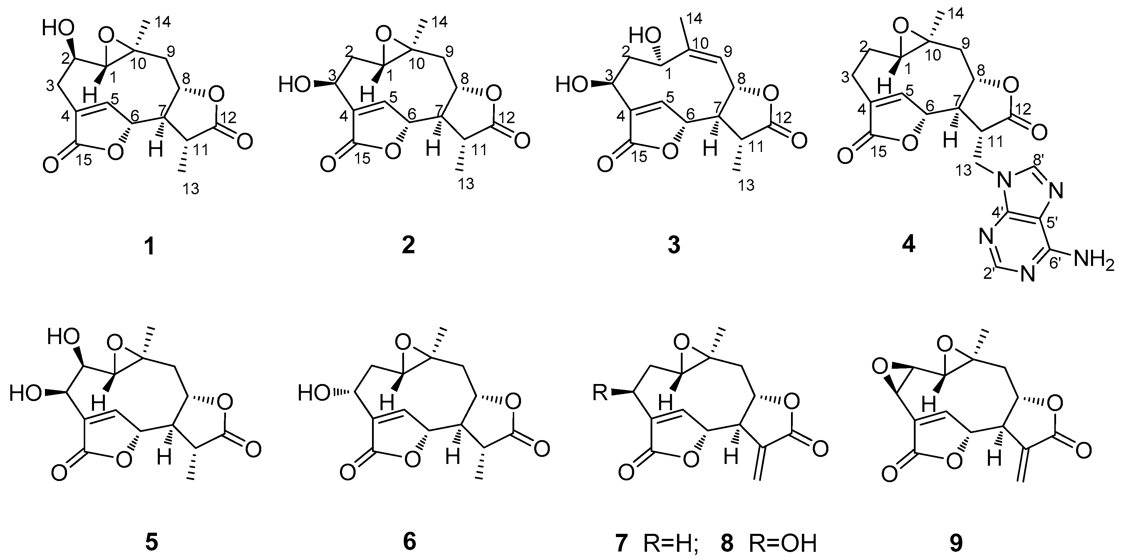
2. Results and Discussion

The air-dried and powdered leaf material of M. micrantha was extracted with 95% ethanol (in water). The resultant crude ethanol extract was then suspended with water and sequentially partitioned with petroleum ether (PE), ethyl acetate (EtOAc), and n-butanol (n-But), respectively, to generate the PE, EtOAc, and n-But extracts. The obtained EtOAc extract was then subjected to a series of column chromatographic steps over silica gel, ODS, and Sephadex LH-20 to afford the four new (14) and five known (59) germacrane sesquiterpene dilactones.
Compound 1, obtained as colorless needles, was determined to have a molecular formula C15H18O6 from pseudo-molecular ions at m/z 317 [M + Na]+, 611 [2M + Na]+ in positive ESI-MS and m/z 293 [M − H] and 329 [M + Cl] in negative ESI-MS as well as m/z 329.0805 [M + Cl] in HR-ESI-MS (calcd for C15H18O6Cl, 329.0797). In the 1H NMR spectrum (Table 1), signals for a tertiary methyl group at δ 1.40 (3H, d, Me-13), a quaternary methyl group at δ 1.25 (3H, s, Me-14), three oxygenated methine groups at δ 3.73 (1H, H-2), 5.27 (1H, H-6) and 4.64 (1H, H-8), an oxymethine proton belonging to an epoxy group at 2.85 (1H, H-1) and an olefinic proton at δ 7.79 (1H, H-5) were all observed. The 13C NMR spectrum (Table 2), coupled with HSQC analysis, showed resonances for fifteen carbons (4×C, 7×CH, 2×CH2, 2×CH3), in which two lactone carboxyl carbons, four oxymethines, one oxygenated quaternary carbon, one olefinic methine, and one olefinic quaternary carbon were deduced. The 1H–1H COSY spectrum showed correlations of H-1/H-2/H2-3, H-5/H-6, and H2-9/H-8/H-7/H-11/H3-13, indicating the presence of structural fragments of C-1–C-2–C-3, C-5–C-6 and C-9–C-8–C-7–C-11–C-13 (Figure 2). In the HMBC spectrum, the observation of 1H−13C long-range correlation signals from δH 7.79 (1H, H-5) to 32.2 (C-3) and 174.3 (C-15), and from δH 2.58 (1H, H-3β) to δC 174.3 (C-15) supported the connections of C-4 with C-3, C-5, and C-15. The HMBC correlations from δH 5.27 (1H, H-6) to 79.2 (C-8) and 41.4 (C-11), and from δH 2.62 (1H, H-7) to δC 151.4 (C-5), 79.2 (C-8) and 13.5 (C-13) supported the connections of C-7 with C-6, C-8, and C-11. The HMBC correlations from δH 1.25 (1H, H-14) to 66.9 (C-1), 44.4 (C-9) and 58.8 (C-10), and from δH 3.73 (1H, H-2) to δC 58.8 (C-10) supported the direct linkages of C-10 with C-1, C-9 and C-14. The HMBC correlations from δH 1.40 (1H, H-13) to 178.0 (C-12) supported C-11 linked with the carboxyl carbon C-12. The HMBC correlations from H-6 to C-15 and from δH 4.64 (1H, H-8) to C-12 supported the ester bond linkages of C-6 with C-15 and of C-8 with C-12, respectively. Those above spectral data and literature precedents supported 1 to be a germacrane sesquiterpene dilactone [19,24]. Careful comparison indicated that the 1H and 13C NMR spectral data of 1 closely resembled those of a previously reported known germacrane sesquiterpenoid 1,10-epoxy-4-germacrene-12,8;15,6-diolide [16], except that the resonances for the methylene group at C-2 in the known compound were absent in 1. Instead, additional signals for an oxygenated methine group [δH 3.73 (1H, H-2); δC 66.9 (C-2)] were exhibited in the spectra. This observation supported that the structure of 1, as shown in Figure 1, was close to 1,10-epoxy-4-germacrene-12,8;15,6-diolide with the only difference that one more hydroxy group attached at C-2 in 1. This deduction was in complete accord with the molecular formula of 1 and the β-orientation of the hydroxy group at C-2 was supported by the observed large coupling constant of H-1 with H-2 (JH-1β,H-2α = 11.7 Hz) [19]. Moreover, the β-orientation of the 2-hydroxy group was further well evidenced by the NOESY spectrum, in which significant NOE correlation signals of H-2 with Me-14, but not H-2 with H-1β, were observed (Figure 3). Therefore, the structure of compound 1 was identified as 2β-hydroxyl-11β,13-dihydrodeoxymikanolide, trivially named dihydrodeoxymikanolide M.
Compound 2 was deduced to have a molecular formula C15H18O6, the same as that of compound 1, as determined by HR-ESI-MS (m/z 329.0792 [M + Cl], calcd for C15H18O6Cl, 329.0797). The 1H NMR spectrum (Table 1) of 2 showed signals readily recognized for two methyl groups at δH 1.40 (3H, d, Me-13) and 1.19 (3H, s, Me-14), one olefinic methine at δH 7.81 (1H, H-5), and four oxymethines at δ H 3.07 (1H, dd, H-1), 4.88 (1H, dd, H-3), 5.33 (1H, H-6) and 4.57 (1H, ddd, H-8). The 13C NMR data (Table 1) supported the above analysis, which showed fifteen carbon signals including two methyls at δC 13.5 (C-13) and 20.6 (C-14), two methylenes, seven methines (including one olefinic methine at δC 147.8 (C-5) and four oxygenated methines at δC 60.2 (C-1), 65.8 (C-3), 81.8 (C-6), and 79.5 (C-8)), one oxygenated quaternary carbon at δC 58.5 (C-10), and two carboxyl carbon at δC 178.1 (C-12) and 172.6 (C-15). The closely resembled NMR spectral data (Table 1 and Table 2) of 2 with those of 1 supported 2 also being a germacrane sesquiterpene dilactone close to 1. Detailed comparison of the 1H- and 13C-NMR spectra revealed that the signals for the hydroxy-methine group at C-2 and the methylene group at C-3 in 1 were replaced by signals for a new methylene group (δH 1.75 (1H, ddd, H-2α), 2.17 (1H, ddd, H-2β); δC 32.8 (C-2)) and a new oxymethine group (δH 4.88 (1H, dd, H-3); δC 65.8 (C-3)), respectively. This observation indicated the difference between the two compounds that the hydroxy group located at C-2 in 1 was changed to attach at C-3 in 2. The rationality for assigning the hydroxy group at C-3 in 2 was supported by observation of significant HMBC correlations from δH 7.81 (1H, H-5) to δC 65.8 (C-3) (Figure 3), and accurately interpreted the significant chemical shift change of C-4 and C-5 from δC 131.1 (C-4) and 151.4 (C-5) in 1 to δC 138.0 (C-4) and 147.8 (C-5) in 2, respectively. Furthermore, the β-orientation of the hydroxy group at C-3 was supported by the observed small coupling constants of H-3 with H-2 (JH-3α,H-2α = 4.0 Hz and JH-3α,H-2β = 2.2 Hz) [19] and further well evidenced by the NOESY spectrum, in which significant NOE correlations of H-3 with H2-2, but no NOE correlation of H-3 with H-1, were displayed (Figure 3). Moreover, compound 2 showed closely similar chemical shift values and proton coupling constants from C-6 through C-14 (Table 1 and Table 2) to those of 1, which indicated that the other part structure and relative configurations (from C-6 through C-14) of 2 was the same as that of 1. Thus, compound 2 was identified as 3β-hydroxyl-11β,13-dihydrodeoxymikanolide, trivially named dihydrodeoxymikanolide N, and all the other spectroscopic data supported this deduction.
Compound 3 was obtained as a colorless oil with a molecular formula C15H18O6 the same as that of compound 2, as determined by HR-ESI-MS, m/z 329.0802 [M + Cl] (calcd. for C15H18O6Cl, 329.0797). The closely related 1H- and 13C-NMR (Table 1) spectral data of 3 with those of 2 supported 3 being an isomeric germacrane sesquiterpenoid of 2. Comparison of the NMR spectral data revealed the major difference between the two compounds, that the resonances in 2 for the oxygenated quaternary carbon at C-10 and for the methylene group at C-9 were absent in 3. Instead, additional signals for one olefinic quaternary carbon (δC 148.2 (C-10)) and one olefinic methine group (δC 121.8 (C-9) and δH 5.29 (1H, d, H-9)) were exhibited in the spectra. This observation led to primarily establish 3, as shown in Figure 1, to be a compound possessed a similar structural skeleton to compound 2, with the only difference of the epoxy group at C-1(C-10) and the methylene group at C-9 in 2 was replaced by a hydroxy-methine group at C-1 and a trisubstituted double bond between C-9 and C-10, respectively. This deduction was consistent with the molecular formula of 3 and further well supported by the following 1D and 2D-NMR spectral analysis. In the 1H–1H COSY spectrum, coupled with HSQC analysis, correlation signals for H-atom spin systems corresponding to structural fragments of C-1–C-2–C-3, C-5–C-6 and C-9–C-8–C-7–C-11–C-13 (Figure 2) were all displayed. The HMBC spectrum showed significant 1H−13C long-range correlations from δH 8.12 (1H, H-5) to δC 65.7 (C-3), 138.6 (C-4) and 171.8 (C-15), and from δH 5.47 (1H, H-6) to C-4 and C-15, which confirmed the connections of C-4 with C-3, C-5 and C-15, and evidenced the ester bond linkage between C-6 and C-15. The observation of HMBC correlations from δH 2.62 (1H, H-7) to δC 145.6 (C-5), 13.2 (C-13), from δH 5.47 (1H, H-6) to 38.7 (C-11), and from δH 1.38 (H-13) to δC 51.8 (C-7), 177.5 (C-12) and 38.7 (C-11), evidenced the connections of C-7 with C-6, and C-11 with C-7, C-12 and C-13. The HMBC correlations from δH 2.09 (Me-14) to δC 64.4 (C-1), 121.8 (C-9), 148.2 (C-10) supported C-10 linked with C-1, C-9, and C-14. The HMBC correlation from H-8 to C-12 supported the ester bond linkage of C-8 with C-12. Furthermore, both the α-orientation of the hydroxy group at C-1 and the β-orientation of the hydroxy group at C-3 were first supported by the large coupling constant of H-1 with H-2α (JH-1β,H-2α = 9.3 Hz) and the small coupling constant of H-3α with H-2α (JH-3α,H-2α = 3.3 Hz) [19], and further supported by NOESY experiment, in which H-1 showed significant NOE correlations with H-5 and H-8, while no NOE correlations of H-1 with H-2α and H-3α (Figure 3). Consequently, the structure of compound 3, as shown in Figure 1, was elucidated as 1α,3β-dihydroxy-4,9-germacradiene-12,8:15,6-diolide, trivially named dihydrogermacradienolide.
Compound 4 was obtained as a white powder. Its molecular formula C20H21N5O5 was determined by HR-ESI-MS (m/z 446.1248 [M + Cl], calcd for C20H21N5O5Cl, 446.1237). The presence of an adenine moiety in the molecule was suggested by the presence of characteristic proton signals at δH 8.14 (1H, s, H-2′) and 8.06 (1H, s, H-8′) in the 1H NMR spectrum, and the carbon signals in the 13C NMR spectrum at δC 152.6 (C-2′), 149.7 (C-4′), 118.6 (C-5′), 156.0 (C-6′) and 141.1 (C-8′) [27]. Apart from those signals due to the adenine moiety, the remaining signals in the 1H NMR spectrum for the aglycone of 2 were recognizable for a methyl group at δH 0.98 (3H, Me-14), an olefinic methine at δH 7.70 (1H, H-5), two oxymethines at δH 4.93 (1H, H-6) and 4.60 (1H, m, H-8), and one oxymethine proton belonging to an epoxy group at δ H 2.77 (1H, dd, H-1). The 13C NMR spectrum indicated, beside the signals for the adenine moiety, fifteen carbons including one methyl, four methylenes, six methines (including an olefinic methine at δC 149.4 (C-5) and three oxygenated methines at δC 60.9 (C-1), 79.9 (C-6) and 77.5 (C-8)), and four quaternary carbons (including an olefinic quaternary carbon at δC 131.1 (C-4) and two carboxyl carbons at δC 173.8 (C-12), 171.9 (C-15)). By comparison, it was found that the 1H- and 13C-NMR spectroscopic data (Table 1 and Table 2) of the aglycone of 4 were quite similar to those of the known compound 1,10-epoxy-4-germacrene-12,8;15,6-diolide [16], except that the resonances for the methyl group at C-13 in the known compound were replaced by the signals for a methylene group (δC 41.8 (C-13) and δH 4.64 (1H, m, H-13a), 4.50 (1H, dd, H-13b)) in 4. These findings supported the structure of 4, as shown in Figure 1, being a closely related derivative of 1,10-epoxy-4-germacrene-12,8;15,6-diolide with specially an adenine moiety linked at C-13. This deduction was in complete accord with the molecular formula of 4, and the linkage of the adenine moiety at C-13 was well supported by the observation of significant 1H–13C long-range correlations of H2-13 (δ H 4.64, 4.50) with δC 149.7 (C-4′) and 141.1 (C-8′) in the HMBC spectrum. Moreover, the 1H–1H COSY and HMBC spectra showed significant correlation signals permitting to establish the structural fragments of C-1–C-2–C-3, C-5–C-6 and C-9–C-8–C-7–C-11–C-13, and construct their connections as depicted in Figure 3. The same relative configurations of the aglycone part of 4 as those of 1,10-epoxy-4-germacrene-12,8;15,6-diolide were well confirmed by the NOESY spectrum which displayed significant NOE correlation signals as delineated in Figure 3. Therefore, the structure of compound 4 was unambiguously identified as (11β,13-dihydrodeoxymikanolide-13-yl)-adenine, trivially named dihydrodeoxymikadenine, as shown in Figure 1.
The five known compounds obtained in this study were identified as 2β,3β-dihydroxy-11β,13-dihydroxydeoxymikanolide (5) [19], 3α-hydroxy-11β,13-dihydroxydeoxymikanolide (6) [19], deoxymikanolide (7) [16], 3β-hydroxy-deoxymikanolide (8), and mikanolide (9) [28] by comparison of their spectroscopic data with literature values.
All nine germacrane sesquiterpenoids were evaluated for their in vitro antibacterial activity against four Gram-(+) bacteria of Staphyloccocus aureus (SA), methicillin-resistant Staphylococcus aureus (MRSA), Bacillus cereus (BC), and Curtobacterium flaccumfaciens (CF), and three Gram-(−) bacteria of Escherichia coli (EC), Salmonella typhimurium (ST), and Pseudomonas solanacearum (PS), by using a microdilution titer method as described in the experimental section [29]. As shown in Table 3, compounds 4 and 79 were found to display significant in vitro antibacterial activity toward all the tested bacteria with MIC values ranging from 6.25 to 12.5 µg/mL, which were close to that of the reference compounds kanamycin sulfate and vancomycin, while no detectable antibacterial activity was displayed for the other compounds. Among the seven test microorganisms, SA, BC, EC, and ST were human pathogenic bacteria, and CF and PS were two Agro-pathogenic bacteria capable of widely causing crop infection diseases. The exhibition of strong and broad-spectrum antibacterial activity of compounds 4 and 79 against all the test human pathogenic and agro-pathogenic bacteria in this bioassay indicated that these four compounds were not only valuable to be developed in medical usage as antibacterial agents for the treatment of human infectious diseases, but also valuable to be applied in agriculture for crop protection. It is worth noting that compounds 4 and 79 showed significant antibacterial activity against MRSA. MRSA infection is responsible for a rapidly increasing number of serious infectious diseases severely threatening global public health [30,31], and its control and clinic therapy are urgently lacking effective and safe anti-MRSA agents. The strong anti-MRSA activity of these four compounds suggested that they have the potential to be developed as an effective anti-MRSA agent.
Compounds 19 were further screened for their in vitro cytotoxic activity against human tumor A549, HepG2, MCF-7, and HeLa cell lines, using a microdilution titer technique as recently we described [32]. The resulting IC50 values are displayed in Table 4, compared to Adriamycin as positive control. Compounds 4 and 79 were found to obviously show cytotoxic activity against all the four tested cancer cell lines, with IC50 values ranging from 8.97 to 27.39 μM. No obvious cytotoxic activity was displayed for the other compounds. The bioassay data indicated that compounds 4 and 79 were bioactive compounds with certain anticancer potentials highly valuable to be further developed as therapeutic drugs or health care agents for cancer treatment or prevention.
Based on comparison of the chemical structures and their antibacterial and cytotoxic activities of compounds 16 (except 4) versus 79, we can find out that the existence of the α-methylene-γ-lactone moiety, i.e., the carboxyl group of O=C(12) conjugated with the exocyclic double bond –C(11)=C(13)H2, would be essential for this type of germacrane sesquiterpenoid to display strong antibacterial potential. It was reported that the mechanism for those sesquiterpenoids, characterized with a α-methylene-γ-lactone moiety in the molecule, to display strong antibacterial and cytotoxic activity, it would be due to the potential Michael-type addition reactions that easily happen in vivo by nucleophiles, such as thiol groups of cysteine residues in proteins, to react with the α-methylene-γ-lactone moiety [33]. However, at this point, it was exceptional for compound 4 which also showed strong antibacterial and cytotoxic activity toward all the seven bacterial strains, but evidently, this compound did not contain the aforementioned α-methylene-γ-lactone moiety in the structure. In fact, compound 4 was novelly characterized with a nitrogen-containing adenine motif attaching at C-13 in the molecule, which suggested that compound 4 might employ a different mechanism to show its antibacterial and cytotoxic activity, and the present data supported a presumption that the introduction of nitrogen atom or nitrogen-containing moiety into the structure might be another effective way to remarkably enhance the antibacterial and cytotoxic potentials of sesquiterpenoids, at least reasonable for this type of sesquiterpene dilactones [34].
As a very successful invasive plant, M. micrantha can annually produce a huge plant biomass at its invasion areas, but this biomass resource has yet not been well-developed and utilized so far. In recent years, some phytochemical studies on M. micrantha have revealed a variety of chemical constituents, with some of them addressed with biological activities, and those findings also indicated the existence of variability in the chemical constituent compositions among various populations of this plant species distributed in different parts of the world. In the present study, the obtained data provided new evidences to supported that the populations of M. micrantha growth in Guangzhou area in China is rich in bioactive natural products, at least rich in antibacterial and cytotoxic chemicals, potentially highly valuable to be developed in medical usage or in agriculture for crop protection.

3. Materials and Methods

3.1. General Experimental Procedures

Optical rotations were measured by a Perkin-Elmer 341 polarimeter (Perkin-Elmer, Inc., Waltham, MA, USA). UV spectra were acquired on a Perkin-Elmer Lambda 650 UV-Vis spectrometer (Perkin-Elmer, Inc.,Waltham, MA, USA). General electrospray ionization-mass spectrometry (ESI-MS) were recorded on a MDS SCIEX API 2000 liquid chromatography/tandem mass spectrometry (LC/MS/MS) instrument (Applied Biosystems, Inc., Forster, CA, USA). High-resolution ESI-MS (HR-ESI-MS) were acquired on a Bruker maXis spectrometer (Bruker Daltonics GmbH, Bremen, Germany). Nuclear magnetic resonance (NMR) spectra were carried out on a Bruker AVANCE HD 500 spectrometer (Bruker Biospin Gmbh, Rheistetten, Germany) with deuterated solvent residues as internal standard. Silica gel (80–100 and 200–300 mesh, Qingdao Haiyang Chemical Co., Qingdao, China), YMC ODS-A (50 μm, YMC Co., Ltd., Kyoto, Japan) and Sephadex LH-20 (Pharmacia Fine Chemcial Co. Ltd., Uppsala, Sweden) were used for open column chromatography (CC). ODS CC was carried out on a CXTH P3000 instrument (Beijing Chuang Xin Tong Heng Science and Technology Co., Ltd., Beijing, China) equipped with a UV 3000 UV–vis Detector and a C-18 column (50 μm, 50 × 500 mm). Preparative HPLC was performed with an HPLC system equipped with a Shimadzu LC-6AD pump and a Shimadzu RID-10A refractive index detector using a Shim-pack PRC-ODS C-18 column (5 μm, 20 × 250 mm). Fractions were monitored by precoated HSGF254 TLC (Yantai Jiangyou Silica Gel Development Co., Ltd., Yantai, China) and spot detection was performed by spraying 10% H2SO4 in ethanol, followed by heating. Kanamycin sulfate, resazurin, vancomycin, and adriamycin were purchased from Sigma-Aldrich (Shanghai, China) Trading Co. (Shanghai, China).

3.2. Plant Material

The aerial part materials of M. micrantha was collected at the South China Botanical Garden, Guangzhou, Guangdong Province, P.R. China, and identified by Dr. Xinsheng Qin at the College of Forestry and Landscape Architecture, South China Agricultural University. A voucher specimen (No.20190623) was deposited in the phytochemical laboratory at the College of Forestry and Landscape Architecture, South China Agricultural University.

3.3. Extraction and Isolation

The air-dried aerial part materials of M. micrantha (18.5 kg) were powdered and extracted three times with 95% EtOH at room temperature for 3 days each time. After evaporation to remove the solvent ethanol under reduced pressure, the produced viscous concentrate was suspended in water (3 L) and sequentially partitioned with petroleum ether (3 L × 3) and EtOAc (3 L × 3) to afford the corresponding petroleum ether-soluble extract (520 g) and the EtOAc-soluble fraction extract (260 g). The EtOAc-soluble extract was subjected to silica gel column chromatography (CC), eluted with an increasing polarity gradient solvent system of CHCl3/MeOH (from 98:2 to 70:30, v/v, each 15 L) to afford fractions Fr.1–Fr.8 after pooling according to their TLC profiles. The fraction Fr.2 (11 g), collected from the elution of 98:2 CHCl3/MeOH, was separated by an ODS CC eluted with a declining polarity solvent system of MeOH/H2O (40:60–100:0, v/v) to provide fractions Fr.2-1–Fr.2-20. Fraction Fr.2-10 was then separated by a silica-gel CC, with elution system of CHCl3/Acetone (from 100:0 to 70:30, v/v), and further purified by a Sephadex LH-20 CC eluted with CHCl3/MeOH (1:4, v/v) to afford compounds 2 (9 mg), 7 (16 mg) and 9 (5 mg). The fraction Fr.3 (20 g), obtained from the elution of 97:3 CHCl3/MeOH, was separated by an ODS CC using a gradient elution system of MeOH/H2O (from 40:60–100:0, v/v) to provide fractions Fr.3-1–Fr.3-20. Fraction Fr.3-1 was first separated by a silica-gel CC, eluted with a gradient solvent system of CHCl3/Acetone (from 95:5 to 70:30, v/v), and then further purified by a Sephadex LH-20 CC with the elution of CHCl3/MeOH (1:4, v/v) to afford 6 (12 mg) and 5 (13.6 mg). The fraction Fr.4 (18.8 g), obtained from the elution of 95:5 CHCl3/MeOH, was separated by an ODS CC using a declining polarity elution system of MeOH/H2O (30:70–100:0, v/v) to give fractions Fr.4-1–Fr.4-18. Fraction Fr.4-4, obtained from the elution with MeOH/H2O (40:60), was subjected to a Sephadex LH-20 CC eluted with CHCl3/MeOH (1:4, v/v) to provide pure compound 1 (5 mg). Fraction Fr.4-5, obtained from the elution of MeOH/H2O (45:55), was first subjected to a Sephadex LH-20 CC eluted with pure MeOH and further separated by preparative HPLC with a Shim-pack PRC-ODS C-18 column (5 μm, 20 × 250 mm) using 25% methanol in water (v/v) as a mobile phase at the flow rate of 10 mL/min to obtain 8 (4 mg, tR 105 min). Fraction Fr.4-8, obtained from the elution of MeOH/H2O (55:45), was first subjected to a Sephadex LH-20 CC eluted with CHCl3/MeOH (1:4, v/v) and further separated by preparative HPLC with a Shim-pack PRC-ODS C-18 column (5 μm, 20 × 250 mm) using 10% methanol in water (v/v) as a mobile phase at the flow rate of 8 mL/min to afford 3 (5 mg, tR 70 min). The fraction Fr.8 (11.3 g), obtained from the elution of 80:20 CHCl3/MeOH, was separated by an ODS CC using a gradient elution system of MeOH/H2O (from 10:90–100:0, v/v) to provide fractions Fr.8-1–Fr.8-17. Fraction Fr.8-4, obtained from the elution of MeOH/H2O (25:75), was separated by a Sephadex LH-20 CC eluted with CHCl3/MeOH (1:4, v/v) to afford 4 (5 mg).

3.4. Spectroscopic Data of Compounds 1–4

2β-Hydroxyl-11β,13-dihydrodeoxymikanolide (1). Colorless needles. [α] D 20 +68.0 (c 0.50, CH3OH); UV (CH3OH) λmax nm (log ε) 220 (3.13); CD (CH3OH) λmax nm (∆ε) 221 (-2.88), 242 (+1.32); ESI-MS (neg.) m/z 293 [M − H], 329 [M + Cl]; ESI-MS (pos.) m/z 317 [M + Na]+, m/z 611 [2M + Na]+; HR-ESI-MS (neg.), m/z 329.0805 [M + Cl] (calcd for C15H18O6Cl, 329.0797). The 1H (500 MHz) and 13C (125 MHz) NMR data in methanol-d4, see Table 1 and Table 2.
3β-Hydroxyl-11β,13-dihydrodeoxymikanolide (2). Colorless needles. [α ] D 20 +66.9 (c 0.63, CH3OH). UV (CH3OH) λmax nm (log ε) 220 (3.09). ESI-MS (neg.) m/z 293 [M − H], 329 [M + Cl]; ESI-MS (pos.) m/z 317 [M + Na]+, m/z 611 [2M + Na]+. HR-ESI-MS (neg.), m/z 329.0792 [M + Cl] (calcd for C15H18O6Cl, 329.0797). The 1H (500 MHz) and 13C (125 MHz) NMR data in methanol-d4, see Table 1 and Table 2.
1α,3β-Dihydroxy-4,9-germacradiene-12,8:15,6-diolide (3). Colorless oil. [α] D 20 +108.7 (c 0.46, C5H5N). UV (C5H5N) λmax nm (log ε) 251 (4.11), 257 (4.14). ESI-MS (neg.) m/z 329 [M + Cl], 623 [2M + Cl]; ESI-MS (pos.) m/z 317 [M + Na]+, m/z 611 [2M + Na]+. HR-ESI-MS (neg.), m/z 329.0802 [M + Cl] (calcd for C15H18O6Cl, 329.0797). The 1H (500 MHz, C5D5N) and 13C (125 MHz) NMR data in C5D5N, see Table 1 and Table 2.
(11β,13-Dihydrodeoxymikanolide-13-yl)-adenine (4). White amorphous powder. [α] D 20 +10.5 (c 0.60, C5H5N). UV (C5H5N) λmax nm (log ε) 260 (3.03). ESI-MS (neg.) m/z 410 [M − H], 446 [M + Cl]; ESI-MS (pos.) m/z 434 [M + Na]+, m/z 845 [2M + Na]+. HR-ESI-MS (neg.), m/z 446.1248 [M + Cl] (calcd for C20H21N5O5Cl, 446.1237). The 1H (500 MHz) and 13C (125 MHz) NMR data in DMSO-d6, see Table 1 and Table 2.

3.5. Antibacterial Assay

The antibacterial activity of the nine germacrane sesquiterpenoids 19 was evaluated by using a method as we described recently [29], with slight modifications. In brief, 100 μL indicator solution (resazurin in sterile water, 100 μg/mL) was first placed into each of the sterility control wells (11th column) on 96 well plates, and the indicator solution (about 7.5 mL, 100 μg/mL) was mixed with test bacteria (5 mL, 106 cfu/mL, OD600 = 0.07) followed by transferring (100 μL, each) to growth control wells (12th column) and all test wells (1–10th column). Then, each of 100 μL of test compounds (0.8 mg/mL) in beef extract peptone medium, along with the positive control solutions (prepared by kanamycin sulfate and vancomycin instead of the test compounds) and the negative control solution (3% DMSO in beef extract peptone medium), were applied to the wells in the 1st column of the plates. Once all samples (test compounds) and controls were properly applied to the 1st column of wells on the plate, half of the homogenized content (100 μL solution) was then parallel transferred from the 1st column wells to the 2nd column wells, and each subsequent well was treated similarly (doubling dilution) up to the 10th column, followed by discarding the last 100 μL aliquot. Finally, the plates were incubated at 37 °C for about 5–6 h until the color of growth control change to pink. The lowest concentration for each test compound at which color change occurred was recorded as the MIC value of the test compound. In the bioassay, a total of seven microorganisms including four Gram-(+) bacteria of Staphyloccocus aureus (SA), methicillin-resistant Staphylococcus aureus (MRSA), Bacillus cereus (BC), Curtobacterium flaccumfaciens (CF), and three Gram-(–) bacteria of Escherichia coli (EC), Salmonella typhimurium (SA), and Pseudomonas solanacearum (PS) were used in the bioassay.

3.6. Cytotoxic Assay

The cytotoxic activity of sesquiterpenoid compounds 19 against human tumor A549, HepG2, MCF-7, and HeLa cell lines were assayed by using 96 well plates according to a literature MTT method with slight modification [32]. Briefly, the cells for the test were cultured in RPMI-1640 medium supplemented with 10% fetal bovine serum in a humidified atmosphere with 5% CO2 at 37 °C. Each well of 96-well cell culture plates was seeded with 100 μL adherent cells (5 × 10⁴ cell/mL) and placed in an atmosphere with 5% CO2 at 37 °C for 24 h to form a monolayer on the flat bottoms. Subsequently, the supernatant in each well was removed and 100 μL fresh medium and 100 μL medium containing one of the test compounds was added. Then, the test plate was incubated in a humidified atmosphere with 5% CO2 at 37 °C for three days. Afterwards, 20 μL MTT at concentration 5 mg/mL in DMSO was added into each well and incubated for 4 h. Carefully, the supernatant in each well was removed and 150 μL DMSO was added. Then, the plate was vortex shaken for 15 min to dissolve blue formazan crystals. The OD (optical density) value of each well was tested on a Genois microplate reader (Tecan GENios, Männedorf, Switzerland) at 570 nm. All the tests were carried out by three individual experiments and adriamycin was applied as a positive control. In a test, for each of the tumor cell lines, each of the test compounds was set at concentrations 50, 25, 12.5, 6.25, 3.125, and 1.5625 µg/mL. The inhibitory rate of tumor cell growth was calculated by the formula: Inhibition rate (%) = (ODcontrol − ODtreated)/ODcontrol × 100%, and the IC50 values were calculated by SPSS 16.0 statistic software. The four tumor cell lines were obtained from the Kunming Institute of Zoology, CAS. The resulting IC50 values listed in Table 4 were based on three individual experiments and represented as means ± standard deviation (SD).

4. Conclusions

Four new germacrane sesquiterpene dilactones, trivially named dihydrodeoxymikanolide M (1), dihydrodeoxymikanolide N (2), dihydrogermacradienolide (3) and dihydrodeoxymikadenine (4), along with five known ones (59) were isolated from the aerial parts of Mikania micrantha. Their structures were determined by extensive spectroscopic analysis. Compound 4 features an adenine moiety in the molecule, which is the first nitrogen-containing sesquiterpenoid so far isolated from this plant species. Bioassays indicated that compounds 4 and 79 were active to show strong in vitro antibacterial activity toward all the seven tested bacteria with the MIC values ranging from 1.56 to 12.5 µg/mL. Notably, compounds 4 and 9 showed significant antibacterial activity against the drug-resistant bacterium of MRSA with MIC value close to reference compound vancomycin. Compounds 4 and 79 were further revealed to show in vitro cytotoxic activity toward human tumor A549, HepG2, MCF-7 and HeLa cell lines, with IC50 values ranging from 8.97 to 27.39 µM. No antibacterial and cytotoxic activity were displayed for the other compounds. The current research provided new data to support that M. micrantha is rich in structurally diverse bioactive compounds worthy of further development in medicinal or healthcare application and for crop-protection in agricultural fields.

Supplementary Materials

The following supporting information can be downloaded at: https://www.mdpi.com/article/10.3390/molecules28052119/s1, HR-ESI-MS, 1H-NMR, 13C-NMR, HSQC, and HMBC spectra of new compounds 14.

Author Contributions

L.-M.D. and Q.-L.X. performed the isolation, structural elucidation of the chemicals; S.-B.L. and S.-X.Z. contributed to the plant material collection and extraction; M.-F.L. and J.-L.D. contributed to the cytotoxic assay experiments; L.-M.D. and J.-K.O. contributed to the antibacterial assay experiments; J.-T.H. and F.-Y.F. partially contributed to the separation process; Q.-L.X. and J.-W.T. prepared the manuscript; J.-W.T. designed this study and supervised all the experiments. All authors have read and agreed to the published version of the manuscript.

Funding

This work was supported by the National Natural Science Foundation of China (Grant No. 32170401 and 31270406), the Guangdong Basic and Applied Basic Research Foundation, China (Grant No. 2023A1515012805 and 2019A1515011236), and the Forestry Science and Technology Innovation Project of Guangdong Province, China (Grant No. 2020KJCX013, 2021-KJXM-14 and 2021-KYXM-01).

Institutional Review Board Statement

Not applicable.

Informed Consent Statement

Not applicable.

Data Availability Statement

Not applicable.

Conflicts of Interest

The authors declare no conflict of interest.

Sample Availability

Samples of the compounds are not available from the authors.

References

  1. Zhang, L.Y.; Ye, W.H.; Cao, H.L.; Feng, H.L. Mikania micrantha H.B.K. in China—An overview. Weed Res. 2004, 44, 42–49. [Google Scholar] [CrossRef]
  2. Lowe, S.; Browne, M.; Boudjelas, S. 100 of the World’s Worst Invasive Alien Species, a Selection from the Global Invasive Species Database; IUCN/SSC Invasive Species Specialist Group (ISSG): Auckland, New Zealand, 2001. [Google Scholar]
  3. Feng, H.L.; Cao, H.L.; Liang, X.D.; Zhou, X.; Ye, W.H. The distribution and harmful effect of Mikania micrantha in Guangdong. J. Trop. Subtrop. Bot. 2002, 10, 263–270. [Google Scholar]
  4. Rufatto, L.C.; Gower, A.; Schwambach, J.; Moura, S. Genus Mikania: Chemical composition and phytotherapeutical activity. Braz. J. Pharmacogn. 2012, 22, 1384–1403. [Google Scholar] [CrossRef] [Green Version]
  5. Facey, P.C.; Pascoe, K.O.; Porter, R.B.; Jones, A. Investigation of plants used in Jamaican folk medicine for anti-bacterial activity. J. Pharm. Pharmacol. 1999, 51, 1455–1460. [Google Scholar] [CrossRef]
  6. Ahmed, M.; Rahman, M.T.; Alimuzzaman, M. Analgesic sesquiterpene dilactone from Mikania cordata. Fitoterapia 2001, 72, 919–921. [Google Scholar] [CrossRef] [PubMed]
  7. Dou, X.; Zhang, Y.; Sun, N.; Wu, Y.; Li, L. The anti-tumor activity of Mikania micrantha aqueous extract in vitro and in vivo. Cytotechnology 2014, 66, 107–117. [Google Scholar] [CrossRef] [Green Version]
  8. Ghosh, A.; Das, B.K.; Roy, A.; Mandal, B.; Chandra, G. Antibacterial activity of some medicinal plant extracts. J. Nat. Med. 2008, 62, 259–262. [Google Scholar] [CrossRef]
  9. Laurella, L.C.; Frank, F.M.; Sarquiz, A.; Alonso, M.R.; Giberti, G.; Cavallaro, L.; Catalan, C.A.; Cazorla, S.I.; Malchiodi, E.; Martino, V.S.; et al. In vitro evaluation of antiprotozoal and antiviral activities of extracts from Argentinean Mikania species. Sci. World J. 2012, 12, 121–133. [Google Scholar]
  10. Zhang, L.L.; Wen, D.Z.; Fu, S.L. Responses of photosynthetic parameters of Mikania micrantha and Chromolaena odorata to contrasting irradiance and soil moisture. Biol. Plant. 2009, 53, 517–522. [Google Scholar] [CrossRef]
  11. Ríos, E.; León, A.; Chávez, M.I.; Torres, Y.; Ramírez-Apan, M.T.; Toscano, R.A.; Bravo-Monzón, Á.E.; Espinosa-García, F.J.; Delgado, G. Sesquiterpene lactones from Mikania micrantha and Mikania cordifolia and their cytotoxic and anti-inflammatory evaluation. Fitoterapia 2014, 94, 155–163. [Google Scholar] [CrossRef] [PubMed]
  12. Zhuang, S.H.; Hao, C.Q.; Feng, J.T.; Zhang, X. Active antifungal components of Mikania micrantha H. B. K. J. Zhejiang Univ. 2010, 36, 293–298. [Google Scholar]
  13. Yan, Y.B.; Huang, Y.L.; Fang, X.T.; Lu, L.; Zhou, R.C.; Ge, X.J.; Shi, S.H. Development and characterization of EST-SSRs in an invasive weed Mikania micrantha. Am. J. Bot. 2011, 98, e1–e3. [Google Scholar] [CrossRef] [PubMed]
  14. Li, Y.; Shen, B.B.; Li, J.; Li, Y.; Wang, X.X.; Cao, A.C. Antimicrobial potential and chemical constituent of Mikania micrantha H. B. K. Afr. J. Microbiol. Res. 2013, 7, 2409–2415. [Google Scholar]
  15. Boeker, R.; Jakupovic, J.; Bohlmann, F.; Schmeda-Hirschmann, G. Germacra-1,10Z,4E-dien-12,8α-olides from Mikania micrantha. Planta Med. 1987, 53, 105–106. [Google Scholar] [CrossRef]
  16. But, P.P.H.; He, Z.D.; Ma, S.C.; Chan, Y.M.; Shaw, P.C.; Ye, W.C.; Jiang, R.W. Antiviral constituents against respiratory viruses from Mikania micrantha. J. Nat. Prod. 2009, 72, 925–928. [Google Scholar] [CrossRef] [PubMed]
  17. Cuenca, M.D.R.; Bardon, A.; Catalan, C.A.N. Sesquiterpene lactones from Mikania micrantha. J. Nat. Prod. 1988, 51, 625–626. [Google Scholar] [CrossRef]
  18. Herz, W.; Srinivasan, A.; Kalyanaraman, P.S. Mikanokryptin, a new guianolide from Mikania. Phytochemistry 1975, 14, 233–237. [Google Scholar] [CrossRef]
  19. Huang, H.J.; Ye, W.H.; Wu, P.; Lin, L.D.; Wei, X.Y. New sesquiterpene lactones from Mikania micrantha. J. Nat. Prod. 2004, 67, 734–736. [Google Scholar] [CrossRef]
  20. Nicollier, G.; Thompson, A.C. Allelopathic potential of sesquiterpene lactones and phenolic constituents from Mikania micrantha H. B. K. Phytochemistry 1981, 20, 2587–2588. [Google Scholar] [CrossRef]
  21. Wei, X.Y.; Huang, H.J.; Wu, P.; Cao, H.L.; Ye, W.H. Phenolic constituents from Mikania micrantha. Biochem. Syst. Ecol. 2004, 32, 1091–1096. [Google Scholar] [CrossRef]
  22. Xu, Q.L.; Xie, H.H.; Xiao, H.L.; Lin, L.D.; Wei, X.Y. Two new ent-kaurene diterpene glucosides from the roots of Mikania micrantha. Phytochem. Lett. 2013, 6, 425–428. [Google Scholar] [CrossRef]
  23. Xu, Q.L.; Xie, H.H.; Xiao, H.L.; Wei, X.Y. Phenolic constituents from the roots of Mikania micrantha and their allelopathic effects. J. Agric. Food Chem. 2013, 61, 7309–7314. [Google Scholar] [CrossRef] [PubMed]
  24. Ma, Q.; Li, J.Y.; Wu, X.M.; Wang, Z.G.; Yang, X.; Pan, W.D.; Su, S.; Li, Y. New Germacrane-sesquiterpenoids from the leaves of Mikania micrantha Kunth. Phytochem. Lett. 2020, 40, 49–52. [Google Scholar] [CrossRef]
  25. Dong, L.-M.; Jia, X.-C.; Luo, Q.-W.; Zhang, Q.; Luo, B.; Liu, W.-B.; Zhang, X.; Xu, Q.-L.; Tan, J.-W. Phenolics from Mikania micrantha and their antioxidant activity. Molecules 2017, 22, 1140. [Google Scholar] [CrossRef] [PubMed]
  26. Dong, L.-M.; Jia, X.-C.; Luo, Q.-W.; Peng, Y.-M.; Zhang, Q.; Luo, B.; Tan, J.-W. Four new ent-kaurene diterpene glucosides from Mikania micrantha. Phytochem. Lett. 2017, 20, 155–159. [Google Scholar] [CrossRef]
  27. Cai, W.; Zhang, J.; Liu, Y.; Lu, J. Two new sesquiterpene lactones from Ixeris sonchifolia. Chem. Nat. Compd. 2019, 55, 674–676. [Google Scholar] [CrossRef]
  28. Aguinaldo, A.M.; ABE, F.; Yamauchi, T.; Padolina, W.G. Germacranolides of Mikania cordata. Phytochemistry 1995, 38, 1441–1443. [Google Scholar] [CrossRef]
  29. Liu, S.-B.; Zeng, L.; Xu, Q.-L.; Chen, Y.-L.; Lou, T.; Zhang, S.-X.; Tan, J.-W. Polycyclic phenol derivatives from the leaves of Spermacoce latifolia and their antibacterial and α-glucosidase inhibitory activity. Molecules 2022, 27, 3334. [Google Scholar] [CrossRef]
  30. Brown, E.D.; Wright, G.D. Antibacterial drug discovery in the resistance era. Nature 2016, 529, 336–343. [Google Scholar] [CrossRef]
  31. Piasecki, B.; Biernasiuk, A.; Skiba, A.; Skalicka-Wo’zniak, K.; Ludwiczuk, A. Composition, Anti-MRSA Activity and Toxicity of Essential Oils from Cymbopogon Species. Molecules 2021, 26, 7542. [Google Scholar] [CrossRef]
  32. Zhang, M.; Ouyang, J.-K.; Xu, Q.-L.; Liu, S.-B.; Qian, T.; Dong, L.-M.; Tan, J.-W. Thymol derivatives with antibacterial and cytotoxic activity from the aerial parts of Ageratina adenophora. RSC Adv. 2021, 11, 5755–5761. [Google Scholar] [CrossRef] [PubMed]
  33. Scotti, M.T.; Fernandes, M.B.; Ferreira, M.J.P.; Emerenciano, V.P. Quantitative structure–activity relationship of sesquiterpene lactones with cytotoxic activity. Bioorgan. Med. Chem. 2007, 15, 2927–2934. [Google Scholar] [CrossRef] [PubMed]
  34. Dong, L.-M.; Huang, L.-L.; Dai, H.; Xu, Q.-L.; Ouyang, J.-K.; Jia, X.-C.; Gu, W.-X.; Tan, J.-W. Anti-MRSA sesquiterpenes from the semi-mangrove Plant Myoporum Bontioides A. Gray. Marine Drugs 2018, 16, 438. [Google Scholar] [CrossRef] [PubMed] [Green Version]
Figure 1. Chemical structures of compounds 1–9.
Molecules 28 02119 g001
Figure 2. Key 1H-1H COSY (Molecules 28 02119 i001
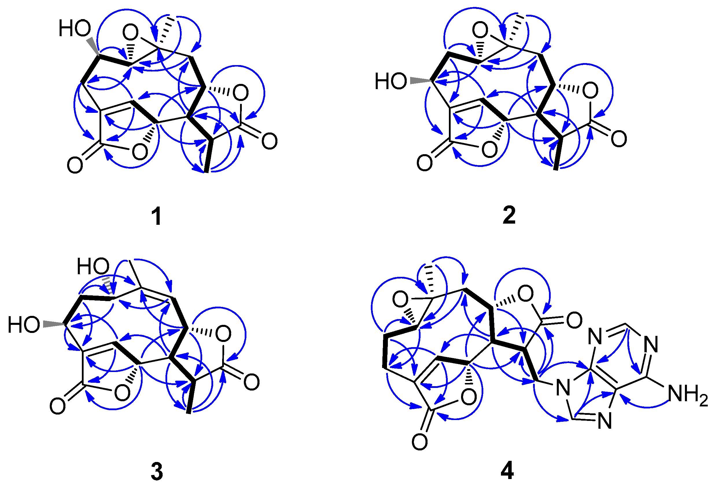
) and selected HMBC (Molecules 28 02119 i002) correlations of 14.
Molecules 28 02119 g002
Figure 3. Key NOESY (Molecules 28 02119 i003) correlations of compounds 14.
Molecules 28 02119 g003
Table 1. 1H NMR data of compounds 14, δ in ppm and J in Hz.
No1 a2 a3 b4 c
12.85, d (11.7)3.07, dd (11.5, 2.0)5.43, d (9.3)2.77, br.d (11.1)
23.73, ddd (11.7, 9.7, 4.2)α 1.75, ddd (14.2, 11.5, 4.0)α 3.13, ddd (14.5, 9.3, 3.3)α 1.22, m
β 2.17, ddd (14.2, 2.2, 2.0)β 2.57, dd (14.5, 3.1)β 1.96, m
3α 2.96, dd (12.6, 4.2)4.88, dd (4.0, 2.2)5.26, dd (3.3, 3.1)α 2.42, dd (12.9, 5.3)
β 2.58, dd (12.6, 9.7) β 2.52, ddd (12.9, 10.2, 4.1)
57.79, br s7.81, t (1.6)8.12, d (1.2)7.70, br.s
65.27, br s5.33, br s5.47, d (1.2)4.93, br s
72.62, (13.9, 11.1)2.61, dd (13.9, 10.0)2.62, overlapped2.81, m
84.64, ddd (11.1, 10.8, 4.6)4.57, ddd (10.7, 10.0, 4.6)5.51, dd (10.6, 10.2)4.60, overlapped
9α 2.06, dd (14.2, 10.8)α 2.05, dd (14.3, 10.7)5.29, d (10.6)α 1.93, overlapped
β 2.12, dd (14.2, 4.6)β 2.10, dd (14.3, 4.6) β 1.81, dd (13.8, 4.3)
112.90, dq (13.9, 7.0)2.93, dq (13.9, 7.0)2.97, dq (13.2, 6.9)3.68, dt (12.1, 6.0)
131.40, d (7.0)1.40, d (7.0)1.38, d (6.9)4.64, overlapped
4.50, dd (14.5, 6.0)
141.25, s1.19, s2.09, s0.98, s
2′ 8.14, s
8′ 8.06, s
-NH2 7.23, s
a Measured in methanol-d4; b Measured in pyridine-d5; c Measured in DMSO-d6.
Table 2. 13C NMR data of compounds 14, δ in ppm.
No1 a2 a3 b4 c
166.9, CH60.2, CH64.4, CH60.9, CH
267.1, CH32.8, CH241.2, CH222.3, CH2
332.2, CH265.8, CH65.7, CH21.2, CH
4131.1, C138.0, C138.6, C131.1, C
5151.4, CH147.8, CH145.6, CH149.4, CH
681.5, CH81.8, CH79.3, CH79.9, CH
755.2, CH55.3, CH51.8, CH49.1, CH
879.2, CH79.5, CH75.6, CH77.5, CH
944.4, CH243.8, CH2121.8, CH42.7, CH2
1058.8, C58.5, C148.2, C56.6, C
1141.4, CH41.2, CH38.7, CH45.0, CH
12178.0, C178.1, C177.5, C173.8, C
1313.5, CH313.5, CH313.2, CH341.8, CH2
1421.5, CH320.6, CH318.1, CH319.7, CH3
15174.3, C172.6, C171.8, C171.9, C
2′ 152.6, CH
4′ 149.7, C
5′ 118.6, C
6′ 156.0, C
8′ 141.1, CH
a Measured in methanol-d4; b Measured in pyridine-d5; c Measured in DMSO-d6.
Table 3. Antibacterial activity of compounds 19 (MIC, µg/mL).
CompoundsSAMRSABCCFECSTPS
1>100>100>100>100>100>100>100
2>100>100>100>100>100>100>100
3>100>100>100>100>100>100>100
46.256.253.1251.566.256.256.25
5>100>100>100>100>100>100>100
6>100>100>100>100>100>100>100
76.2512.56.253.1256.256.253.125
86.2512.56.253.1256.256.253.125
96.256.253.1253.1256.253.1253.125
K1.56500.781.561.560.781.56
V1.563.1251.563.1251.560.781.56
K = Kanamycin sulfate; V = Vancomycin.
Table 4. Cytotoxic activity of compounds 19, (IC50, µM) a.
NoA549HepG2MCF-7HeLa
1>100>100>100>100
2>100>100>100>100
3>100>100>100>100
417.97 ± 0.8915.62 ± 1.0225.81 ± 1.2227.39 ± 2.46
5>100>100>100>100
6>100>100>100>100
7>100>100>100>100
814.28 ± 0.7116.36 ± 1.669.85 ± 0.6413.92 ± 0.97
916.48 ± 1.3518.06 ± 1.1310.24 ± 0.9511.79 ± 1.05
1012.26 ± 1.3215.84 ± 1.168.97 ± 0.9310.75 ± 0.81
Adriamycin0.68 ± 0.071.12 ± 0.160.86 ± 0.090.92 ± 0.11
a Values represent mean ± SD (n = 3) based on three individual experiments.
Disclaimer/Publisher’s Note: The statements, opinions and data contained in all publications are solely those of the individual author(s) and contributor(s) and not of MDPI and/or the editor(s). MDPI and/or the editor(s) disclaim responsibility for any injury to people or property resulting from any ideas, methods, instructions or products referred to in the content.

