
Characterization of Bioactive Compounds and Novel Proteins Derived from Promising Source Citrullus colocynthis along with In-Vitro and In-Vivo Activities

 , , , and
Abstract
:1. Introduction
2. Results
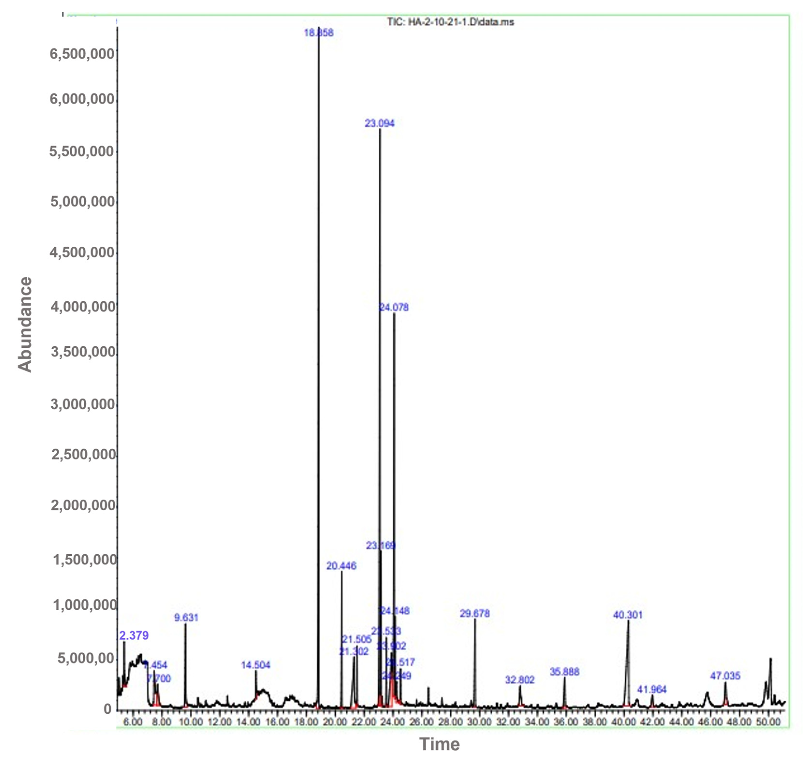
2.1. GC-MS Analysis
2.2. HPLC Chromatogram of Methanolic Extract
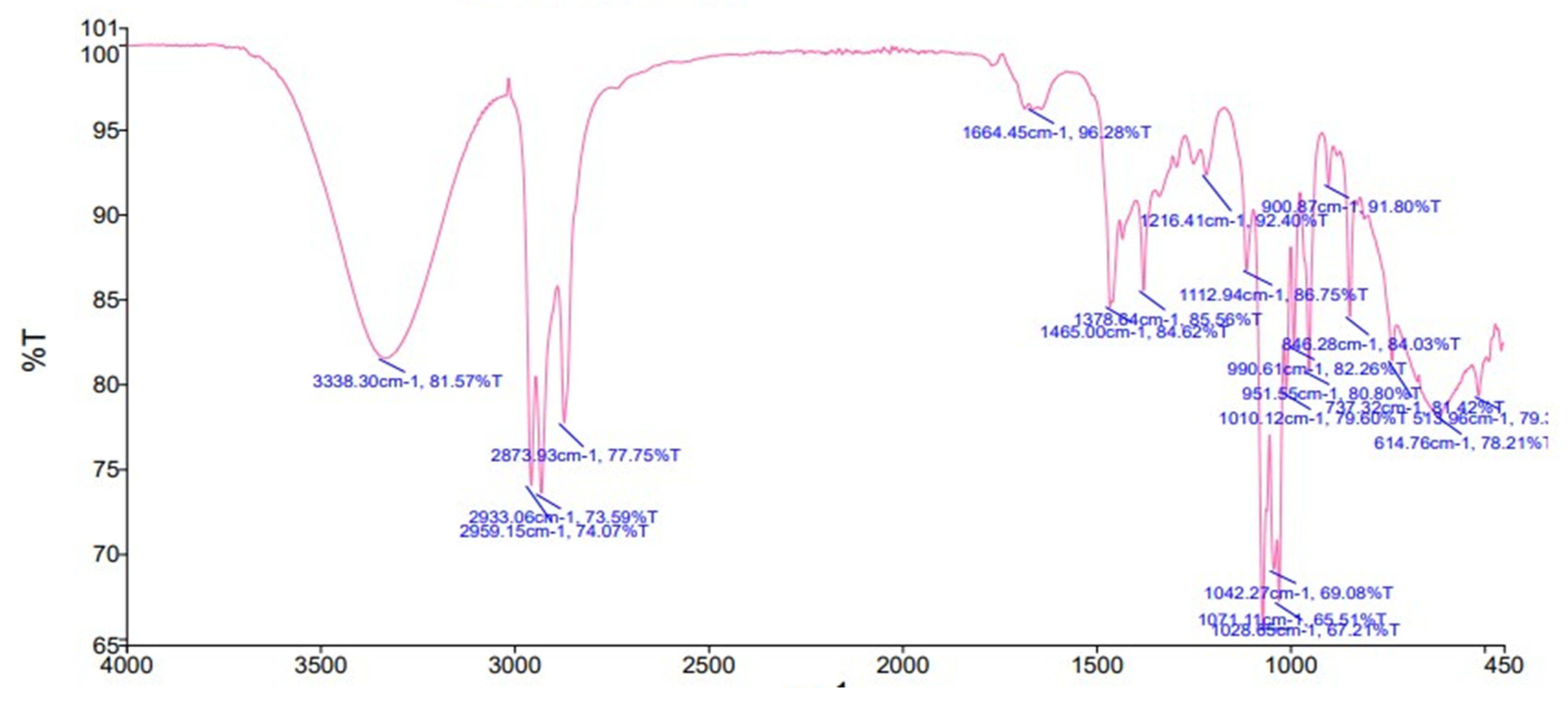
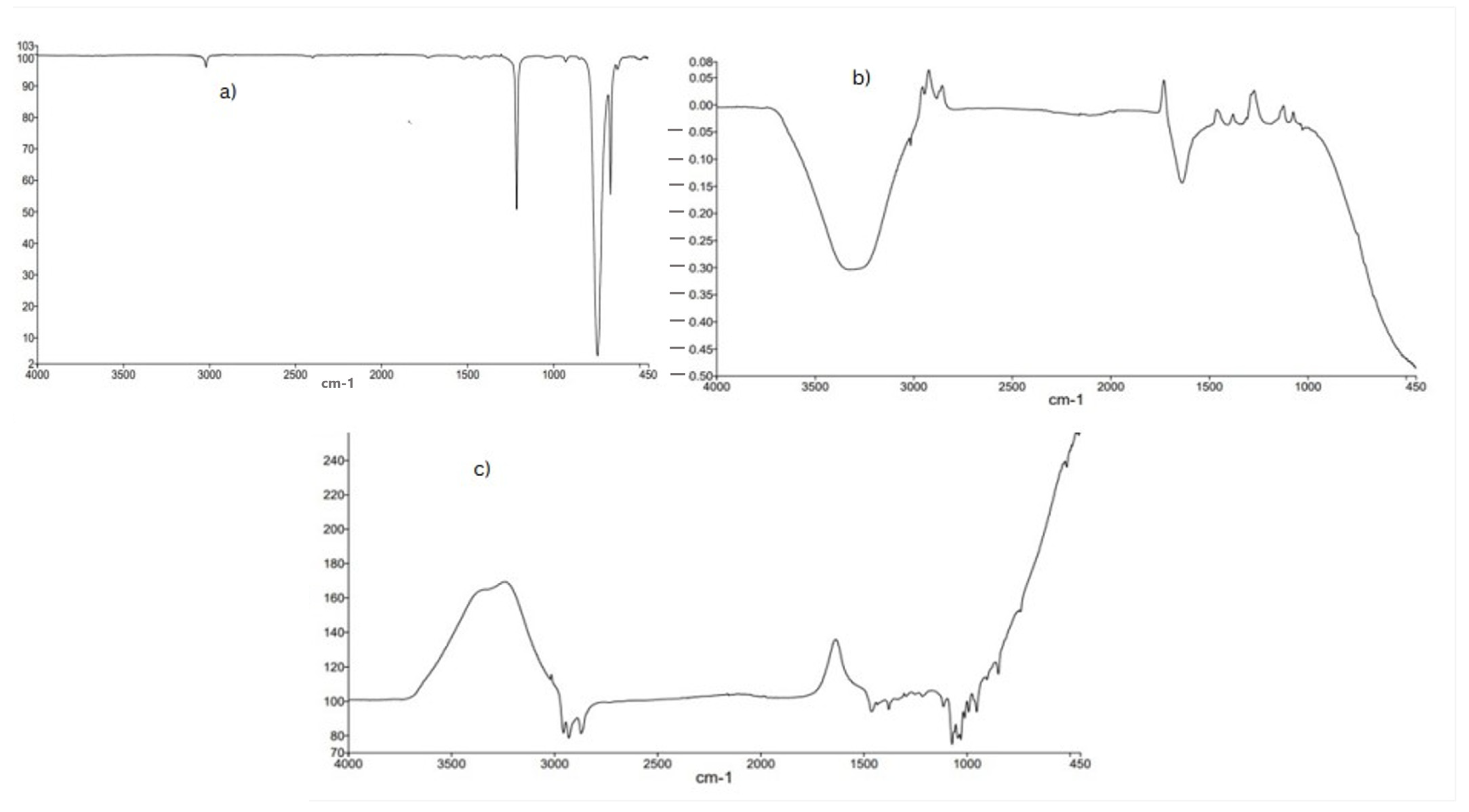
2.3. FTR-ATR Analysis
2.4. Antioxidant Activity of Methanolic Extracts
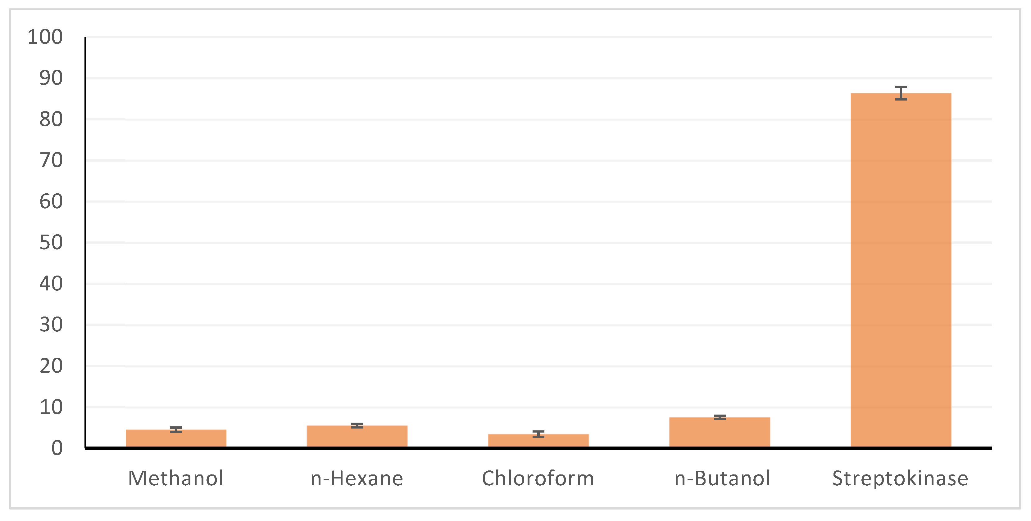
2.5. Thrombolytic Activity
2.6. Hemolytic Activity
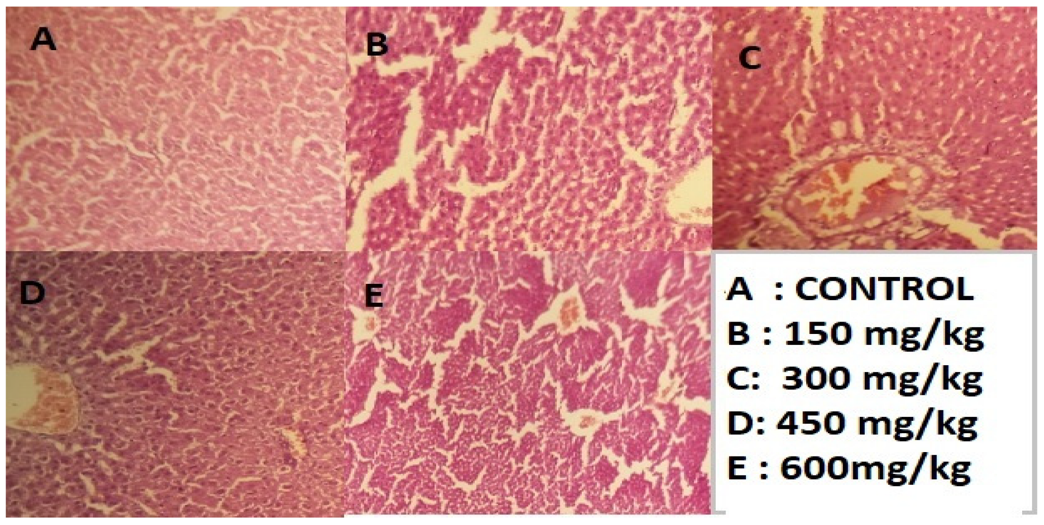
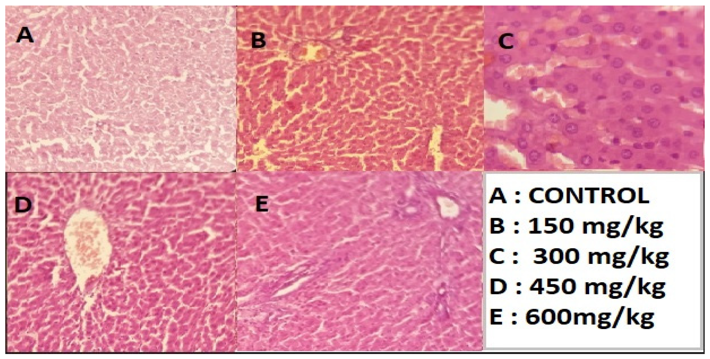
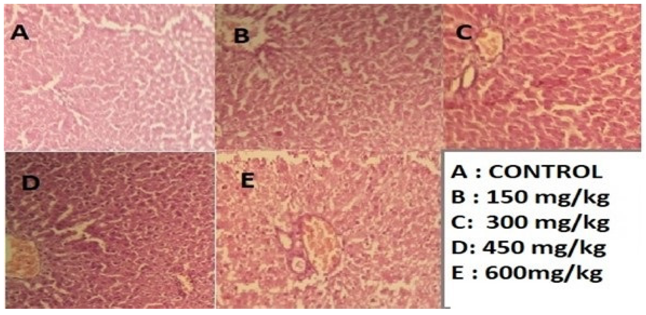
2.7. Histopathological Examination
2.8. Anti-Bacterial Activity
2.9. Molecular Docking
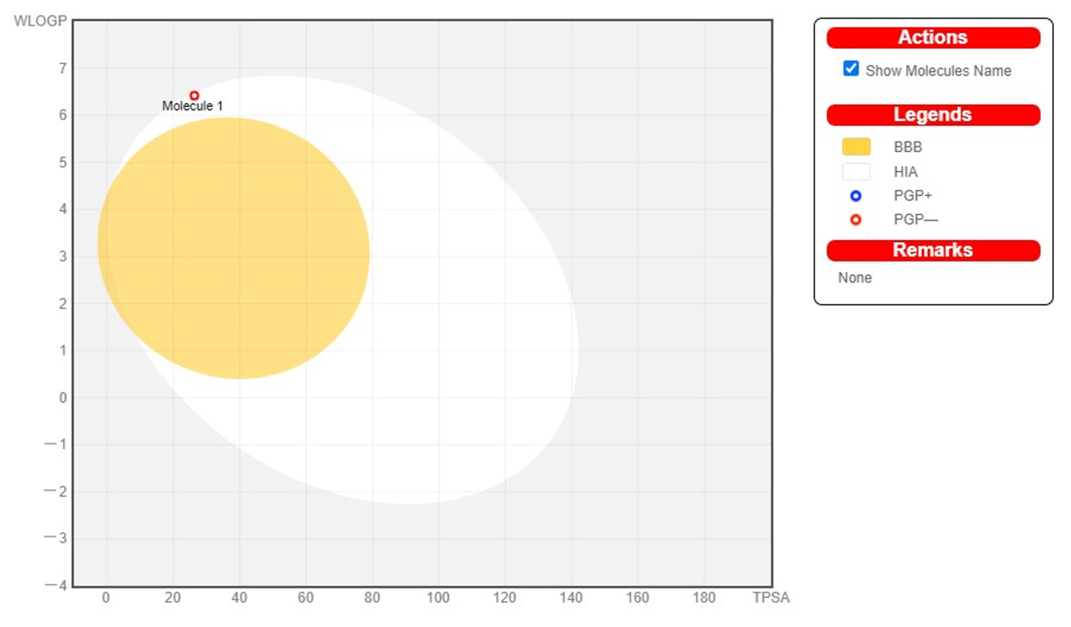
2.10. Assessment of Pharmacokinetics Properties
3. Discussion
4. Materials and Methods
4.1. Plant Collection
4.2. Preparation of Plant Extract: C. colocynthis
4.3. HPLC and GC-MS
4.4. GC-MS Analysis
4.5. FTIR-ATR Analysis
4.6. Biological Activities
4.6.1. Antioxidant Activity
4.6.2. Hemolytic Activity
4.6.3. Thrombolytic Activity
4.6.4. Anti-Diabetic Activity
4.6.5. Anti-Bacterial Activity
Disc Diffusion Method
4.6.6. In-Vivo Hepatotoxicity of Fractions of Methanolic Extracts
Preparation of Sample for Hepatotoxicity
Experimental Protocol
Histopathological Studies
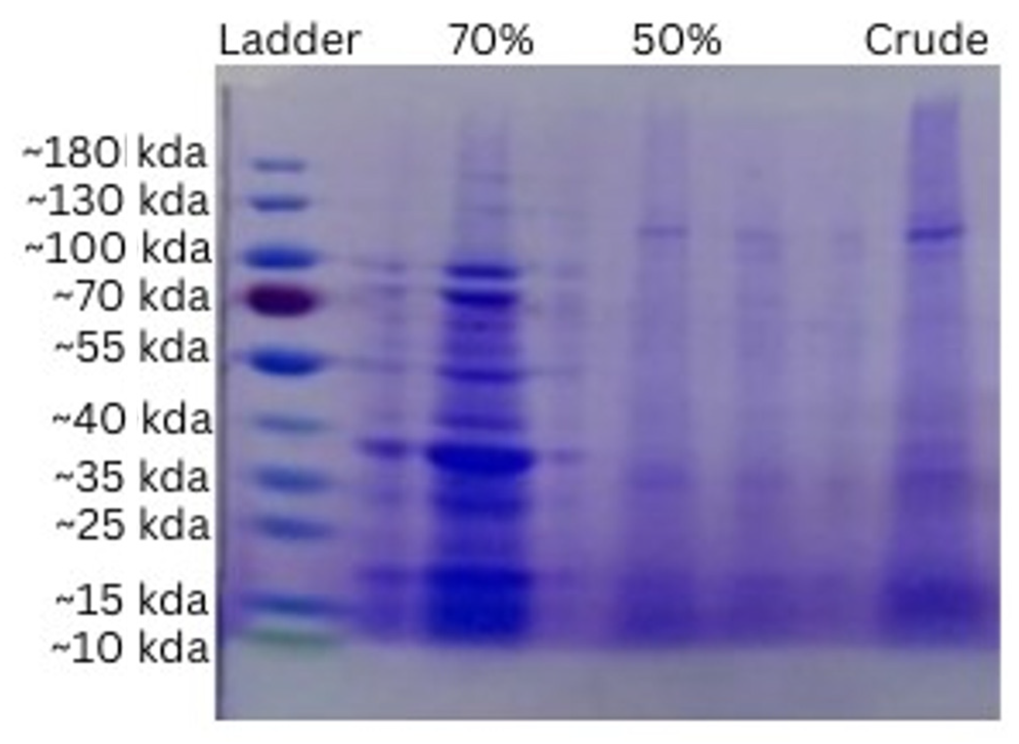
4.6.7. Extraction of Peptide
Buffer Extract Preparation
Salting In and Salting Out
Bradford Assay
4.6.8. In Silico Studies of Antioxidant
Ligand Selection
Selection of Target Protein
Docking Studies
Pharmacokinetic Properties
Statistical Analysis
5. Conclusions
Author Contributions
Funding
Institutional Review Board Statement
Informed Consent Statement
Data Availability Statement
Acknowledgments
Conflicts of Interest
References
- Yonbawi, A.R.; Abdallah, H.M.; Alkhilaiwi, F.A.; Koshak, A.E.; Heard, C.M. Anti-proliferative, cytotoxic and antioxidant properties of the methanolic extracts of five Saudi Arabian flora with folkloric medicinal use: Aizoon canariense, Citrullus colocynthis, Maerua crassifolia, Rhazya stricta and Tribulus macropterus. Plants 2021, 10, 2073. [Google Scholar] [CrossRef] [PubMed]
- Rahimi, R.; Amin, G.; Ardekani, M.R. A review on Citrullus colocynthis Schrad.: From traditional Iranian medicine to modern phytotherapy. J. Altern. Complement Med. 2012, 18, 551–554. [Google Scholar] [CrossRef] [PubMed]
- Abudayeh, Z.; Lamazian, G.; Sereda, P.; Chekman, I.; Alkhalifa, D.I.; Al Azzam, K.; Hassouneh, D.L. Comparative Study of Amino Acid Composition in the Seeds, Pulp and Rind from Citurllus colocynthis Fruits. J. Pharmacogn. Phytochem. 2016, 8, 433–437. [Google Scholar]
- Shafaei, H.; Esmaeili, A.; Rad, J.S.; Delazar, A.; Behjati, M. Citrullus colocynthis as a medicinal or poisonous plant: A revised fact. J. Med. Plant Res. 2012, 6, 4922–4927. [Google Scholar] [CrossRef]
- Khan, M.; Khan, M.; Al-hamoud, K.; Adil, S.F.; Shaik, M.R.; Alkhathlan, H.Z. Diversity of Citrullus colocynthis (L.) Schrad Seeds Extracts: Detailed Chemical Profiling and Evaluation of Their Medicinal Properties. Plants 2023, 12, 567. [Google Scholar] [CrossRef]
- Al-Nablsi, S.; El-Keblawy, A.; Ali, M.A.; Mosa, K.A.; Hamoda, A.M.; Shanableh, A.; Almehdi, A.M.; Soliman, S.S. Phenolic Contents and Antioxidant Activity of Citrullus colocynthis Fruits, Growing in the Hot Arid Desert of the UAE, Influenced by the Fruit Parts, Accessions, and Seasons of Fruit Collection. Antioxidants 2022, 11, 656. [Google Scholar] [CrossRef]
- Rizvi, T.S.; Mabood, F.; Ali, L.; Al-Broumi, M.; Al Rabani, H.K.; Hussain, J.; Jabeen, F.; Manzoor, S.; Al-Harrasi, A. Application of NIR spectroscopy coupled with PLS regression for quantification of total polyphenol contents from the fruit and aerial parts of Citrullus colocynthis. Phytochem Anal. 2018, 29, 16–22. [Google Scholar] [CrossRef]
- Birgani, G.A.; Ahangarpour, A.; Khorsandi, L.; Moghaddam, H.F. Anti-diabetic effect of betulinic acid on streptozotocin-nicotinamide induced diabetic male mouse model. Braz. J. Pharm. Sci. 2018, 54, 1590. [Google Scholar] [CrossRef]
- Patel, D.; Prasad, S.K.; Kumar, R.; Hemalatha, S. An overview on antidiabetic medicinal plants having insulin mimetic property. Asian Pac. J. Trop. Biomed. 2012, 2, 320–330. [Google Scholar] [CrossRef]
- Lemos, M.F.; Lemos, M.F.; Pacheco, H.P.; Guimarães, A.C.; Fronza, M.; Endringer, D.C.; Scherer, R. Seasonal variation affects the composition and antibacterial and antioxidant activities of Thymus vulgaris. Ind. Crops Prod. 2017, 95, 543–548. [Google Scholar] [CrossRef]
- Chauhan, A.; Semwal, D.K.; Mishra, S.P.; Semwal, R.B. Ayurvedic research and methodology: Present status and future strategies. AYU Int. Q. J. Res. Ayurveda 2015, 36, 364–369. [Google Scholar] [CrossRef]
- Shaikh, S.I.; Nagarekha, D.; Hegade, G.; Marutheesh, M. Postoperative nausea and vomiting: A simple yet complex problem. Anesth. Essays Res. 2016, 10, 388–396. [Google Scholar]
- Javadzadeh, H.R.; Davoudi, A.; Davoudi, F.; Valizadegan, G.; Goodarzi, H.; Mahmoodi, S.; Ghane, M.R.; Faraji, M. Citrullus colocynthis as the Cause of Acute Rectorrhagia. J. Emerg. Med. Case Rep. 2013, 2013, 652192. [Google Scholar] [CrossRef]
- Koche, D.; Shirsat, R.; Kawale, M. An overerview of major classes of phytochemicals: Their types and role in disease prevention. Hislopia J. 2016, 9, 0976–2124. [Google Scholar]
- Ak, M. A Brief Review of Traditional plants as Sources of Pharmacological interests. J. Plant Sci. 2019, 39, 001–008. [Google Scholar] [CrossRef]
- Falade, O.S.; Otemuyiwa, I.O.; Adekunle, A.S.; Adewusi, S.A.; Oluwasefunmi, O. Nutrient composition of watermelon (Citrullis lanatus (Thunb.) Matsum. &Nakai) and egusi melon (Citrullus colocynthis (L.) Schrad.) seeds. Agric. Conspec. Sci. 2020, 85, 43–49. [Google Scholar]
- Daoudi, A.; Bousta, D.; Aarab, L.; Abdel-Sattar, E. Evaluation and characterization of the immunomodulatory activity of the protein extract from Citrullus colocynthis (L). Food Agric. Immunol. 2013, 24, 47–57. [Google Scholar] [CrossRef]
- Rasool Hassan, B. Medicinal plants (importance and uses). Pharm. Anal. Acta 2012, 3, 2153–2435. [Google Scholar] [CrossRef]
- Gurudeeban, S.; Ramanathan, T. Antidiabetic effect of Citrullus colocynthis in alloxon-induced diabetic rats. J. Ethnopharmacol. 2010, 1, 1112–1115. [Google Scholar]
- Mohanta, Y.K.; Panda, S.K.; Jayabalan, R.; Sharma, N.; Bastia, A.K.; Mohanta, T.K. Antimicrobial, antioxidant and cytotoxic activity of silver nanoparticles synthesized by leaf extract of Erythrina suberosa (Roxb.). Front. Mol. Biosci. 2017, 4, 14. [Google Scholar] [CrossRef]
- Vakiloddin, S.; Fuloria, N.; Fuloria, S.; Dhanaraj, S.A.; Balaji, K.; Karupiah, S. Evidences of hepatoprotective and antioxidant effect of Citrullus colocynthis fruits in paracetamol induced hepatotoxicity. Pak. J. Pharm. Sci. 2015, 28, 951–957. [Google Scholar] [PubMed]
- Arshed Iqbal, D.; Ramesh Chandra, S.; Suresh Kumar, B. Hepatoprotection: A hallmark of Citrullus colocynthis L. against paracetamol induced hepatotoxicity in Swiss albino rats. Am. J. Plant Sci. 2012, 3, 1022–1027. [Google Scholar]
- Tabani, K.; Birem, Z.; Halzoune, H.; Saiah, W.; Lahfa, F.; Koceir, E.A.; Omari, N. Therapeutic effect of alkaloids and glycosides of colocynth seeds on liver injury, associated with metabolic syndrome in wistar rats, subject to nutritional stress. Pak. J. Pharm. Sci. 2018, 31, 277–290. [Google Scholar] [PubMed]
- Fattah, M.A.; El Baz, M.; Sherif, A.; Adel, A. Complement components (C3, C4) as inflammatory markers in asthma. Indian J. Pediatr. 2010, 77, 771–773. [Google Scholar] [CrossRef]
- Reddy, C.V.K.; Sreeramulu, D.; Raghunath, M. Antioxidant activity of fresh and dry fruits commonly consumed in India. Int. Food Res. J. 2010, 43, 285–288. [Google Scholar] [CrossRef]
- Salla, H.R.; Al Jahwari, M.R.H.; Al Tobi, Z.M.R. A Study on FTIR, Antimicrobial, Antioxidant and Hypogycaemic Effect of Diospyros Kaki and Citrullus colocynthis. Int. J. Phytomedicine. 2019, 11, 23–31. [Google Scholar]
- Kumar, S.; Kumar, D.; Manjusha; Saroha, K.; Singh, N.; Vashishta, B. Antioxidant and free radical scavenging potential of Citrullus colocynthis (L.) Schrad. methanolic fruit extract. Acta Pharm. 2008, 58, 215–220. [Google Scholar] [CrossRef]
- Alabdallat, N.G.; Bin Dukhyil, A.A.A. In vitro anticoagulant activity of methanolic extracts of Artemisia herba-alba, Achillea fragrantissima and Citrullus colocynthis grown in Saudi Arabia. Indian J. Tradit. Knowl. 2021, 20, 344–350. [Google Scholar]
- Khyade, M.S.; Varpe, S.N.; Padwal, A.D. Evaluation of chemical profile and antioxidant potential of Trichodesma indicum (L.) R. Br. Int. J. Phytomedicine. 2017, 9, 416–425. [Google Scholar] [CrossRef]
- Martínez-Maqueda, D.; Hernández-Ledesma, B.; Amigo, L.; Miralles, B.; Gómez-Ruiz, J.Á. Extraction/fractionation techniques for proteins and peptides and protein digestion. In Proteomics in Foods; Springer: Berlin/Heidelberg, Germany, 2013; Volume 2, pp. 21–50. [Google Scholar]
- Perveen, S.; Ashfaq, H.; Ambreen, S.; Ashfaq, I.; Kanwal, Z.; Tayyeb, A. Methanolic extract of Citrullus colocynthis suppresses growth and proliferation of breast cancer cells through regulation of cell cycle. Saudi J. Biol. Sci. 2021, 28, 879–886. [Google Scholar] [CrossRef]
- Zhang, H.; Wang, Y.; Zhang, X.; Liu, M.; Hu, Z. Analysis of vicine in bitter melon with high performance liquid chromatography. Anal. Lett. 2003, 36, 1597–1605. [Google Scholar] [CrossRef]
- Konappa, N.; Udayashankar, A.C.; Krishnamurthy, S.; Pradeep, C.K.; Chowdappa, S.; Jogaiah, S. GC–MS analysis of phytoconstituents from Amomum nilgiricum and molecular docking interactions of bioactive serverogenin acetate with target proteins. Sci. Rep. 2020, 10, 16438. [Google Scholar] [CrossRef]
- Shameer, P.M.; Nishath, P.M. Exploration and enhancement on fuel stability of biodiesel: A step forward in the track of global commercialization. In Advanced Biofuels, 1st ed.; Elsevier: Amsterdam, The Netherlands, 2019; pp. 181–213. [Google Scholar]
- Bourhia, M.; Bouothmany, K.; Bakrim, H.; Hadrach, S.; Salamatullah, A.M.; Alzahrani, A.; Khalil Alyahya, H.; Albadr, N.A.; Gmouh, S.; Laglaoui, A. Chemical profiling, antioxidant, antiproliferative, and antibacterial potentials of chemically characterized extract of citrullus colocynthis L. seeds. Separations 2021, 8, 114. [Google Scholar] [CrossRef]
- Kauser, A.; Shah, S.M.A.; Iqbal, N.; Murtaza, M.A.; Hussain, I.; Irshad, A.; Nasir, S.; Akram, M.; Munir, N.; Riaz, M. In vitro antioxidant and cytotoxic potential of methanolic extracts of selected indigenous medicinal plants. Prog. Nutr. 2018, 20, 706–712. [Google Scholar]
- Powell, W.; Catranis, C.; Maynard, C. Design of self-processing antimicrobial peptides for plant protection. Lett. Appl. Microbiol. 2000, 31, 163–168. [Google Scholar] [CrossRef]
- Abed, H.H.; Alwasiti, E.; Tawfeeq, A.T. Titanium nanoparticles conjugated with streptokinase as a modified thrombolytic agent. Int. J. Curr. Pharm. Res. 2020, 112, 18–25. [Google Scholar] [CrossRef]
- Khair, A.; Ibrahim, M.; Ahsan, Q.; Homa, Z.; Kuddus, M.R.; Rashid, R.B.; Rashid, M.A. Pharmacological activities of Blumea lacera (Burm. f) DC: A medicinal plant of Bangladesh. Br. J. Pharm. Res. 2014, 4, 1677. [Google Scholar] [CrossRef]
- Mostafa, A.A.; Al-Askar, A.A.; Almaary, K.S.; Dawoud, T.M.; Sholkamy, E.N.; Bakri, M.M. Antimicrobial activity of some plant extracts against bacterial strains causing food poisoning diseases. Saudi J. Biol. Sci. 2018, 25, 361–366. [Google Scholar] [CrossRef]
- Mohamed, E.A.A.; Muddathir, A.M.; Osman, M.A. Antimicrobial activity, phytochemical screening of crude extracts, and essential oils constituents of two Pulicaria spp. growing in Sudan. Sci. Rep. 2020, 10, 17148. [Google Scholar] [CrossRef]
- Dehghani, F. and Panjehshahin, M.R. The toxic effect of alcoholic extract of Citrullus colocynthis on rat liver. Iran. J. Pharmacol. Ther. 2006, 5, 117–119. [Google Scholar]
- Gräslund, S.; Nordlund, P.; Weigelt, J.; Hallberg, B.M.; Bray, J.; Gileadi, O.; Knapp, S.; Oppermann, U.; Arrowsmith, C.; Hui, R. Protein production and purification. Nat. Methods 2008, 5, 135–146. [Google Scholar]
- Kielkopf, C.L.; Bauer, W.; Urbatsch, I.L. Bradford assay for determining protein concentration. Cold Spring Harb. Protoc. 2020, 2020, pdb-prot102269. [Google Scholar] [CrossRef] [PubMed]
- Saravanan, R.; Raja, K.; Shanthi, D. GC–MS Analysis, Molecular Docking and Pharmacokinetic Properties of Phytocompounds from Solanum torvum Unripe Fruits and Its Effect on Breast Cancer Target Protein. Appl. Biochem. Biotechnol. 2022, 194, 529–555. [Google Scholar] [CrossRef] [PubMed]
- Mishra, S.; Dahima, R. In vitro ADME studies of TUG-891, a GPR-120 inhibitor using SWISS ADME predictor. J. Drug Deliv. Ther. 2019, 9, 366–369. [Google Scholar]
- Steel, R.G.D.; Torrie, J.W.; Dickey, M. Principles and Procedures of Statistics: A Biometrical Approach, 1st ed.; McGraw-Hill Book Company: New York, NY, USA, 1997. [Google Scholar]
- Zar, J.H. Biostatistical Analysis, 3rd ed.; Prentice Hall Inc.: Upper Saddle River, NJ, USA, 1996. [Google Scholar]


| Sr. No. | Compounds | Retention Time | Area (%) |
|---|---|---|---|
| 1 | Ether, 3-butenyl pentyl | 5.047 | 9.36 |
| 2 | oxime-, methoxy phenyl | 5.620 | 3.45 |
| 3 | 4-Dimethylbenzamide | 6.186 | 0.94 |
| 4 | Betaine | 6.277 | 0.91 |
| 5 | Ethyl 4-chlorobutanimidoate | 7.417 | 1.82 |
| 6 | Benzofuran, 2,3-dihydro | 7.823 | 0.58 |
| 7 | Cyclotrisiloxane, hexamethyl | 8.604 | 1.26 |
| 8 | Cyclohexasiloxane, dodecamethy | 8.909 | 2.89 |
| 9 | 2-Methoxy-4-vinylphenol | 9.745 | 5.27 |
| 10 | p-Cymen-7-ol | 10.059 | 0.51 |
| 11 | 3-Isopropoxy-1,1,1,7,7,7-hexamet. | 12.616 | 2.13 |
| 12 | 2,4-Di-tert-butylphenol | 13.471 | 0.88 |
| 13 | Cyclooctasiloxane, hexadecamethyl | 15.413 | 1.1 |
| 14 | Triisopropyl phosphite | 16.354 | 1.68 |
| 15 | Cyclopropanecarboxylic acid, 2,2. | 16.654 | 2.59 |
| 16 | 1-Octadecenoic acid, methyl est | 16.863 | 6.44 |
| 17 | cis-13-Octadecenoic acid, methyl | 23.211 | 3.16 |
| 18 | Methyl stearate | 23.511 | 9.00 |
| Sr. No. | Compound | Retention Time | Area (%) | Conc (ppm) |
|---|---|---|---|---|
| 1 | Quercetin | 3.040 | 0.6 | 12.2127 |
| 2 | Gallic Acid | 4.807 | 5.0 | 74.8542 |
| 3 | Vanillic Acid | 13.287 | 4.7 | 122.6160 |
| 4 | Chlorogenic Acid | 15.513 | 3.4 | 110.7420 |
| 5 | Syringic acid | 16.967 | 2.4 | 25.2823 |
| 6 | Feralic Acid | 22.647 | 3.4 | 101.0458 |
| 7 | Cinnamic acid | 24.753 | 2.4 | 35.4156 |
| 8 | Sinapic Acid | 26.333 | 1.4 | 1.5497 |
| Sr. No. | Frequency Range (cm−1) | Functional Group |
|---|---|---|
| 1 | 3400–3200 | Hydroxyl Compound |
| 2 | 2855–2975 | Cyclo alkane |
| 3 | 1458–1591 | Phenol ring |
| 4 | 1150–911 | C-O-C |
| 5 | 858–733 | C-H |
| 6 | 600–700 | C-S |
| 7 | 550–690 | Halogen compound (bromo-compound) C-Br |
| Methanol Extract | n-Hexane Fraction | Chloroform Fraction | n-Butanol Fraction | Ascorbic Acid | |||||||
|---|---|---|---|---|---|---|---|---|---|---|---|
| Sr. No. | Conc. (mg/mL) | DPPH % Scavenging | IC50 | DPPH % Scavenging | IC50 | DPPH % Scavenging | IC50 | DPPH % Scavenging | IC50 | DPPH % Scavenging | IC50 |
| 1 | 0.15 | 28.59 ± 0.45 | 1.54 | 25.66 ± 0.45 | 0.84 | 26.67 ± 0.45 | 2.87 | 17.48 ± 0.49 | 3 | 40.28 ± 0.28 | 0.29 |
| 2 | 0.3 | 29.38 ± 0.43 | 32.39 ± 0.49 | 28.57 ± 0.40 | 19.59 ± 0.51 | 50.58 ± 0.50 | |||||
| 3 | 0.45 | 29.36 ± 0.39 | 38.34 ± 0.31 | 29.61 ± 0.49 | 21.33 ± 0.56 | 60.57 ± 0.28 | |||||
| 4 | 0.6 | 30.57 ± 0.49 | 40.51 ± 0.49 | 29.39 ± 0.52 | 22.61 ± 0.44 | 70.59 ± 0.35 | |||||
| 5 | 0.75 | 38.41 ± 0.45 | 45.59 ± 0.51 | 30.44 ± 0.39 | 23.75 ± 0.27 | 80.63 ± 0.51 | |||||
| 6 | 0.9 | 40.57 ± 0.30 | 52.54 ± 0.49 | 34.59 ± 0.42 | 26.45 ± 0.49 | 93.50 ± 0.35 | |||||
| Conc. (mg/mL) | Methanol Extract | n-Hexane Fraction | Chloroform Fraction | n-Butanol Fraction | Metformin (Standard) | |||||
|---|---|---|---|---|---|---|---|---|---|---|
| Antidiabetic Activity | IC50 | Antidiabetic Activity | IC50 | Antidiabetic Activity | IC50 | Antidiabetic Activity | IC50 | Antidiabetic Activity | IC50 | |
| 0.15 | 20.99 ± 0.24 | 0.56 | 15.99 ± 0.38 | 0.6 | 10.99 ± 0.99 | 0.51 | 25.99 ± 0.45 | 05.99 ± 0.10 | 2.3 | |
| 0.3 | 25.60 ± 0.42 | 20.60 ± 0.34 | 15.60 ± 0.50 | 30.60 ± 0.24 | 10.66 ± 0.49 | |||||
| 0.45 | 35.19 ± 0.40 | 30.19 ± 0.37 | 25.19 ± 0.16 | 40.19 ± 0.20 | 20.19 ± 0.37 | |||||
| 0.6 | 40.71 ± 0.33 | 35.71 ± 0.47 | 30.71 ± 0.18 | 45.71 ± 0.45 | 25.71 ± 0.30 | |||||
| 0.75 | 75.23 ± 0.42 | 70.61 ± 0.23 | 65.23 ± 0.47 | 80.23 ± 0.41 | 60.23 ± 0.06 | |||||
| 0.9 | 83.34 ± 0.47 | 82.64 ± 0.36 | 81.54 ± 0.45 | 83.64 ± 0.27 | 78.67 ± 0.44 | |||||
| Methanol Extract | n-Hexane Fraction | Chloroform Fraction | n-Butanol Fraction | Triton X-100 Standard | |||
|---|---|---|---|---|---|---|---|
| Sr. No. | Conc. (mg/mL) | Activity (%) | Activity (%) | Activity (%) | Activity (%) | Conc. | Activity (%) |
| 1 | 0.15 | 2.50 ± 0.43 | 2.45 ± 0.40 | 1.49 ± 0.40 | 1.53 ± 0.30 | 0.1% | 99.90 ± 0.15 |
| 2 | 0.3 | 2.30 ± 0.33 | 5.42 ± 0.45 | 2.67 ± 0.33 | 2.50 ± 0.40 | ||
| 3 | 0.45 | 4.48 ± 0.33 | 8.43 ± 0.27 | 4.32 ± 0.06 | 3.32 ± 0.37 | ||
| 4 | 0.6 | 5.45 ± 0.44 | 9.63 ± 0.23 | 5.19 ± 0.16 | 5.45 ± 0.49 | ||
| 5 | 0.75 | 6.51 ± 0.47 | 11.50 ± 0.41 | 7.68 ± 0.22 | 8.37 ± 0.38 | ||
| 6 | 0.9 | 8.43 ± 0.38 | 14.66 ± 0.22 | 9.53 ± 0.40 | 10.26 ± 0.40 | ||
| Sr. No. | Sample Fractions | Zone of Inhibition (mm) | |||
|---|---|---|---|---|---|
| Staphylococcus aureu | Enterococcus faecalis | Klebsiella pneumoniae | Pseudomonas aeruginosa | ||
| 1 | Crude | 3.0 ± 0.001 | 2.76 ± 0.009 | 7.1 ± 0.178 | - |
| 2 | 50% | 8.83 ± 0.022 | 10.12 ± 0.017 | 8.27 ± 0.001 | 6.4 ± 0.279 |
| 3 | 70% | 6.96 ± 0.047 | 8.58 ± 0.012 | 5.86 ± 0.022 | 4.94 ± 0.473 |
| 4 | Imipenem | 19.4 ± 0.049 | 15.2 ± 0.057 | 18.1 ± 0.017 | 9.05 ± 0.042 |
| 5 | Vancomycin | 6.61 ± 0.062 | - | - | - |
| 6 | Aztreonam | - | - | 18.7 ± 0.206 | - |
| Compound | PUB CHEM I.D. | Standard Melatonin | Myeloperoxidase | NADPH-Oxidase |
|---|---|---|---|---|
| Methyl stearate | 8201 | −6.4 | −6.1 | −5.1 |
| Methyltrans-9-(2-butylcyclopentyl) nonanoate | 14389759 | −6.1 | −6.2 | −4.6 |
| trans-11-Octadecenoic acid methyl ester | 5364432 | −6.1 | −6.0 | −5.3 |
| 9,12-Octadecadienoic acid | 3931 | −6.0 | −6.1 | −5.1 |
| 4,7-Octadecadiynoic acid methyl ester | 569159 | −6.0 | −6.7 | −5.2 |
| Benzofuran, 2,3-dihydro | 47756 | −6.0 | −6.2 | −4.9 |
| cis-13-Octadecenoic acid | 5312441 | −5.6 | −5.7 | −5.1 |
| 2-Butanone, 4-phenyl | 91752799 | −5.2 | −5.4 | −5.8 |
| 10.Benzamide, N-ethyl-N-(3-methylphenyl)-4-ethyl | 533234 | −5.3 | −5.5 | −4.2 |
| Ether, 3-butenyl pentyl | 537745 | −5.0 | −4.6 | −4.2 |
| TRIISOPROPYL PHOSPHITE | 8304 | −5.0 | −4.9 | −4.5 |
| Cyclopropanecarboxylic acid | 6451381 | −4.8 | −5.1 | −4.5 |
| 2,4-Di-tert-butylphenol | 7311 | −4.6 | −4.9 | −4.1 |
| [1,1′-Biphenyl]-2,5-diol | 82722 | −4.5 | −4.6 | −4.1 |
| N,N-Dimethyl-p-(1-pyrrolyl)aniline | 272429 | −4.4 | −4.4 | −4.1 |
| 3,4-Dimethylbenzamide | 21755 | −4.2 | −4.3 | −3.9 |
| ethyl 4-chlorobutanimidoate | 13163358 | −4.2 | −4.3 | −3.7 |
| 6-Chloropiperonyl alcohol | 7015319 | −4.2 | −4.6 | −3.9 |
| 2-Methoxy-4-vinylphenol | 332 | −4.2 | −4.2 | −3.9 |
| Oxime-, methoxy-phenyl | 9602988 | −4.0730 | −4.4 | −4.1 |
| Betaine | 247 | −3.7 | −3.9 | −3.6 |
| Doses | Group I (Control, Normal Saline mL/kg) n = 4 | Group II (n-butanol Fraction mg/kg) n = 4 | Group III (Chloroform Fraction vs. mg/kg n = 4 | Group IV (n-hexane Fraction mg/kg) n = 4 |
|---|---|---|---|---|
| 1st | 1 | 150 | 150 | 150 |
| 2nd | 1 | 300 | 300 | 300 |
| 3rd | 1 | 450 | 450 | 450 |
| 4th | 1 | 600 | 600 | 600 |
Disclaimer/Publisher’s Note: The statements, opinions and data contained in all publications are solely those of the individual author(s) and contributor(s) and not of MDPI and/or the editor(s). MDPI and/or the editor(s) disclaim responsibility for any injury to people or property resulting from any ideas, methods, instructions or products referred to in the content. |
© 2023 by the authors. Licensee MDPI, Basel, Switzerland. This article is an open access article distributed under the terms and conditions of the Creative Commons Attribution (CC BY) license (https://creativecommons.org/licenses/by/4.0/).
Share and Cite
Afzal, M.; Khan, A.S.; Zeshan, B.; Riaz, M.; Ejaz, U.; Saleem, A.; Zaineb, R.; Sindhu, H.A.; Yean, C.Y.; Ahmed, N. Characterization of Bioactive Compounds and Novel Proteins Derived from Promising Source Citrullus colocynthis along with In-Vitro and In-Vivo Activities. Molecules 2023, 28, 1743. https://doi.org/10.3390/molecules28041743
Afzal M, Khan AS, Zeshan B, Riaz M, Ejaz U, Saleem A, Zaineb R, Sindhu HA, Yean CY, Ahmed N. Characterization of Bioactive Compounds and Novel Proteins Derived from Promising Source Citrullus colocynthis along with In-Vitro and In-Vivo Activities. Molecules. 2023; 28(4):1743. https://doi.org/10.3390/molecules28041743
Chicago/Turabian StyleAfzal, Muhammad, Anis Shahzad Khan, Basit Zeshan, Muhammad Riaz, Umer Ejaz, Ayesha Saleem, Rida Zaineb, Haseeb Akram Sindhu, Chan Yean Yean, and Naveed Ahmed. 2023. "Characterization of Bioactive Compounds and Novel Proteins Derived from Promising Source Citrullus colocynthis along with In-Vitro and In-Vivo Activities" Molecules 28, no. 4: 1743. https://doi.org/10.3390/molecules28041743